A Comprehensive Guide to Validating CRISPR Edits: From Sanger to NGS Sequencing Methods
This article provides researchers, scientists, and drug development professionals with a definitive guide to validating CRISPR-Cas9 gene edits.
A Comprehensive Guide to Validating CRISPR Edits: From Sanger to NGS Sequencing Methods
Abstract
This article provides researchers, scientists, and drug development professionals with a definitive guide to validating CRISPR-Cas9 gene edits. It covers the critical transition from basic DNA analysis to advanced RNA-seq, detailing robust sequencing methodologies for confirming on-target efficiency, detecting unintended transcriptomic changes, and troubleshooting low knockout efficiency. The content synthesizes current best practices and emerging trends, including the role of AI and clinical validation frameworks, to ensure reliable and comprehensive analysis of gene editing outcomes for both research and therapeutic applications.
Why DNA Sequencing Alone is Not Enough: The Critical Need for Comprehensive CRISPR Validation
While PCR amplification followed by Sanger sequencing remains the gold standard for confirming targeted CRISPR edits due to its accuracy and low cost, this method faces significant limitations in scalability, sensitivity, and ability to detect complex modifications. This comparison guide objectively evaluates the performance of PCR+Sanger against emerging sequencing technologies for CRISPR validation. We present quantitative data demonstrating how next-generation sequencing (NGS) and specialized computational tools are overcoming these limitations, enabling researchers to comprehensively assess editing efficiency, detect off-target effects, and characterize complex editing outcomes with unprecedented resolution.
The CRISPR-Cas9 system has revolutionized genetic engineering, providing an easily programmable platform for precise genome editing. However, accurately validating these edits remains a critical bottleneck in the research pipeline. For years, PCR amplification coupled with Sanger sequencing has served as the primary validation method, offering a seemingly straightforward approach to confirm intended edits. This method involves amplifying the target region via PCR and subsequently determining its nucleotide sequence through Sanger's chain-termination method.
Despite being considered the gold standard for detecting single nucleotide variants and small insertions/deletions, this approach faces inherent technological constraints. As research progresses toward more complex editing strategies—including multiplex editing, large knock-ins, and therapeutic applications—the limitations of PCR+Sanger become increasingly problematic. This guide examines these limitations through experimental data and presents viable alternatives for comprehensive CRISPR validation.
Experimental Comparison of CRISPR Validation Methods
Methodologies and Protocols
PCR + Sanger Sequencing Protocol The standard protocol for validating CRISPR edits begins with PCR amplification of the target region from purified genomic DNA, followed by Sanger sequencing. Critical steps include:
- DNA Extraction: Purify genomic DNA from edited cells or tissues.
- Target Amplification: Design locus-specific primers flanking the edit site (typically generating 500-1000 bp amplicons).
- PCR Purification: Clean amplified products to remove primers and enzymes.
- Sanger Sequencing: Prepare sequencing reaction with fluorescent dye-terminators.
- Capillary Electrophoresis: Separate termination products by size.
- Data Analysis: Interpret chromatograms for sequence variants [1] [2].
NGS-Based Validation Protocol Next-generation sequencing provides a comprehensive alternative:
- Library Preparation: Fragment DNA and ligate with platform-specific adapters.
- Target Enrichment: Use hybridization baits or PCR to enrich for target regions.
- Cluster Amplification: Bridge amplify fragments on flow cell (Illumina).
- Sequencing by Synthesis: Parallel sequencing with fluorescent nucleotides.
- Data Analysis: Map reads to reference genome, call variants [1] [3].
High-Throughput Genotyping Protocol Specialized NGS approaches like genoTYPER-NEXT offer optimized workflows:
- Direct Submission: Submit CRISPR-edited cells in multiwell plates.
- Cell Lysis & Barcoding: Lyse cells and amplify with barcoded primers.
- Pooled Sequencing: Multiplex thousands of samples on Illumina platforms.
- Interactive Analysis: Visualize results via dedicated browser [3].
Performance Comparison Across Key Metrics
Table 1: Quantitative comparison of CRISPR validation methods across critical performance metrics
| Metric | PCR + Sanger | T7 Endonuclease Assay | NGS (Amplicon) | High-Throughput Genotyping |
|---|---|---|---|---|
| Detection Sensitivity | ~15-20% allele frequency [4] | ~5% [4] | <1% allele frequency [3] | <1% allele frequency [3] |
| Multiplexing Capacity | 1 target per reaction [1] | 1 target per reaction | 1 to >10,000 targets [1] | Up to 10,000 samples per run [3] |
| Edit Characterization | Limited to predominant variants [4] | Indel frequency only [4] | Full sequence resolution | Full INDEL resolution, frameshift analysis [3] |
| Quantitative Capability | Not quantitative [1] | Semi-quantitative | Quantitative [1] | Quantitative with allele frequency [3] |
| Turnaround Time | ~8 hours sequencing + analysis [1] | ~3-4 hours | Days (library prep + sequencing) [1] | Varies with scale |
| Cost Per Sample | $ (Low) [1] | $ (Low) | $$ to $$$$ (Medium-High) [1] | Varies with scale |
Table 2: Accuracy assessment of computational tools for analyzing Sanger sequencing data of CRISPR edits (based on artificial templates with predetermined indels) [4]
| Tool | Simple Indels (1-3 bp) | Complex Indels | Knock-in Sequences | Indel Sequence Deconvolution |
|---|---|---|---|---|
| TIDE | Good accuracy | Variable estimation | Limited capability | Limited |
| ICE | Good accuracy | Variable estimation | Limited capability | Moderate |
| DECODR | Best overall accuracy | Most accurate for complex indels | Limited capability | Best among tools |
| SeqScreener | Good accuracy | Variable estimation | Limited capability | Moderate |
| TIDER | Not specialized for knock-ins | Not specialized for knock-ins | Best for knock-in efficiency | Specialized for HDR events |
Limitations of PCR and Sanger Sequencing in CRISPR Validation
Limited Sensitivity and Inability to Detect Mosaicism
PCR+Sanger sequencing requires a homogeneous template for clear interpretation, limiting its ability to detect mosaic populations where editing efficiency is below 15-20% [2] [4]. The method struggles to resolve complex mixtures of editing outcomes, as evidenced by chromatograms becoming indecipherable with overlapping traces when analyzing amplicons from heterogeneous samples [1]. This poses significant challenges in detecting off-target effects, which typically occur at low frequencies across the genome.
Lack of Quantitative Capability
Unlike qPCR or NGS, Sanger sequencing provides no quantitative information about editing efficiency or allele frequency [1]. While computational tools like TIDE and ICE can estimate indel frequencies from trace files, their accuracy varies considerably, particularly with complex indels or extreme (low or high) editing efficiencies [4]. This limitation prevents accurate assessment of editing efficiency in polyclonal populations.
Low Scalability and Throughput
Sanger sequencing is fundamentally low-throughput, limited to analyzing one target per reaction without multiplexing capability [1]. This creates a significant bottleneck in large-scale projects requiring analysis of multiple targets or samples. As CRISPR applications expand to genome-wide screens and therapeutic development, this scalability limitation becomes increasingly prohibitive.
Inability to Resolve Complex Editing Outcomes
While Sanger excels at confirming specific intended edits, it provides limited resolution of diverse editing outcomes within a population. In studies comparing computational tools, the ability to deconvolute complex indel sequences varied significantly, with most tools struggling to accurately characterize more complicated indel patterns [4]. This is particularly problematic for CRISPR applications where heterogeneous editing outcomes are common.
Restricted Sequence Context
Sanger sequencing read lengths typically max out at approximately 500-1000 base pairs per reaction [1] [2], limiting the genomic context that can be assessed in a single assay. This constraint hinders comprehensive analysis of large knock-ins, deletions, or rearrangements that may result from CRISPR editing. Furthermore, the technology cannot reliably detect structural variations or complex rearrangements that may occur at off-target sites.
Advanced Solutions for Comprehensive CRISPR Validation
Next-Generation Sequencing Approaches
Targeted NGS methods address nearly all limitations of Sanger sequencing by providing:
- Ultra-sensitive detection of low-frequency edits (<1% allele frequency) [3]
- Multiplexed analysis of thousands of targets or samples simultaneously [1] [3]
- Complete sequence resolution of all editing outcomes in a population [3]
- Quantitative measurement of allele frequencies [1]
These approaches enable researchers to simultaneously assess on-target efficiency, characterize editing profiles, and detect off-target effects in a single assay. While NGS has higher per-sample costs and longer turnaround times, its comprehensive data output often makes it more cost-effective for large-scale studies [1].
Computational Tools for Enhanced Sanger Data
Specialized algorithms can extend the utility of Sanger data for CRISPR validation:
- TIDE (Tracking of Indels by Decomposition): Estimates editing efficiency and indel distribution by decomposing Sanger trace data [5] [4]
- TIDER (Tracking of Insertions, Deletions, and Recombination events): Specialized for analyzing homology-directed repair events and small edits using an additional donor sequencing trace [5]
- DECODR (Deconvolution of Complex DNA Repair): Shows superior performance for complex indel patterns according to comparative studies [4]
- ICE (Inference of CRISPR Edits): Provides reliable estimation of editing efficiency for simple indels [4]
These tools enable more quantitative analysis from Sanger data but still face limitations with highly complex editing outcomes.
Integrated Workflows for Comprehensive Validation
Leading laboratories are adopting tiered validation strategies that combine multiple methods:
- Rapid screening using T7E1 or similar mismatch detection assays
- Initial confirmation of editing with PCR+Sanger and computational analysis
- Comprehensive characterization using targeted NGS for off-target assessment and precise quantification
This integrated approach balances speed, cost, and comprehensiveness while addressing the limitations of any single method.
Table 3: Key research reagent solutions for CRISPR validation
| Reagent/Resource | Function | Examples/Providers |
|---|---|---|
| Sanger Sequencing Reagents | Chain-termination sequencing with fluorescent detection | BigDye Terminator kits (Thermo Fisher) [2] |
| NGS Library Prep Kits | Prepare sequencing libraries for high-throughput platforms | Illumina Nextera, Swift Biosciences Accel-NGS [1] |
| Computational Analysis Tools | Deconvolute complex editing outcomes from sequencing data | TIDE, ICE, DECODR, CRISPResso [5] [4] |
| High-Throughput Genotyping Services | Large-scale validation of edited cell lines | genoTYPER-NEXT [3] |
| Digital PCR Systems | Absolute quantification of editing efficiency | Bio-Rad QX200, Thermo Fisher QuantStudio [1] |
| CRISPR Validation Panels | Targeted sequencing for on- and off-target assessment | Custom hybridization panels (Illumina, Agilent) |
The limitations of PCR and Sanger sequencing in characterizing CRISPR edits beyond the immediate target site are becoming increasingly apparent as applications advance toward therapeutic development. While Sanger remains valuable for confirming specific intended edits in small-scale studies, its inability to quantitatively assess complex editing outcomes and off-target effects necessitates complementary approaches.
Next-generation sequencing technologies provide the comprehensive profiling capability required for rigorous therapeutic development, enabling sensitive detection of off-target effects and complete characterization of editing outcomes. As the field progresses, integrated validation workflows that combine the cost-effectiveness of Sanger for initial screening with the comprehensiveness of NGS for final characterization will become standard practice.
Future advancements in long-read sequencing, single-cell technologies, and computational analysis will further enhance our ability to fully characterize CRISPR editing outcomes, ultimately supporting the safe and effective translation of CRISPR-based therapies into clinical applications.
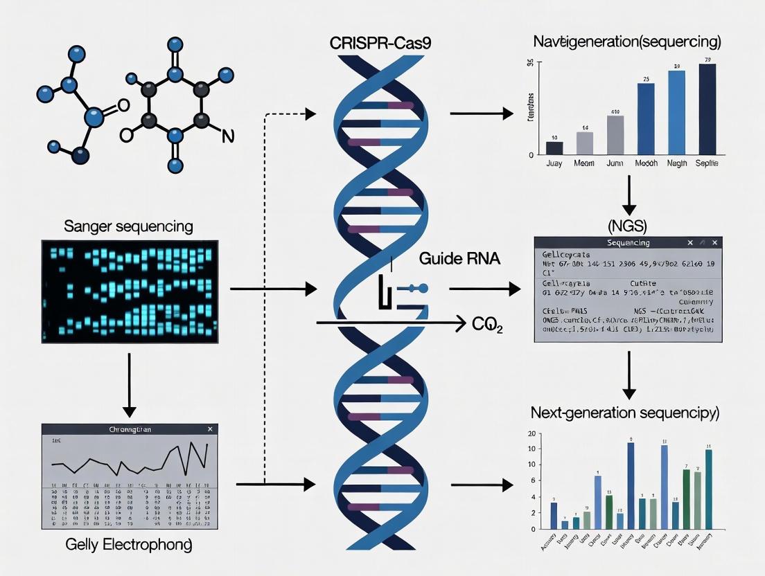
Methodological Diagrams
CRISPR-based genome editing technologies have revolutionized biological research and therapeutic development by enabling precise, programmable modification of the genome [6]. However, traditional validation methods focusing solely on DNA-level analysis provide an incomplete picture of editing outcomes. Emerging research demonstrates that RNA sequencing (RNA-seq) reveals a hidden landscape of transcriptional changes that remain undetectable through conventional PCR amplification and Sanger sequencing of target DNA sites [7] [8]. This comparison guide objectively evaluates RNA-seq against established CRISPR analysis methods, providing researchers and drug development professionals with experimental data to inform their validation strategies.
The Limitations of Traditional CRISPR Validation Methods
Standard approaches for validating CRISPR edits typically examine only the immediate target site, potentially missing substantial unintended consequences. DNA-based methods can confirm intended mutations but fail to detect transcriptome-wide alterations that significantly impact gene function and cellular phenotype [7].
Table 1: Comparison of CRISPR Validation Methods
| Method | Detection Capability | Unanticipated Change Detection | Throughput | Cost |
|---|---|---|---|---|
| Sanger Sequencing | Target site mutations | Limited | Low | Low |
| T7E1 Assay | Editing efficiency (indels) | None | Medium | Low |
| TIDE Analysis | Indel spectrum | Limited | Medium | Low-medium |
| ICE Analysis | Indel spectrum and efficiency | Moderate (large indels) | Medium | Low-medium |
| RNA-seq | Transcriptome-wide changes | Comprehensive | High | Medium-high |
Traditional DNA-focused methods like T7E1, TIDE, and ICE provide valuable data on editing efficiency and small indels at the target site [9]. However, these approaches cannot detect the full spectrum of transcriptional alterations occurring beyond the immediate target locus, creating significant blind spots in validation protocols [7].
RNA-Seq Reveals the Hidden Transcriptional Impact of CRISPR
RNA-seq provides a comprehensive view of CRISPR-induced changes by capturing the entire transcriptional landscape rather than just target DNA sequences. This approach has uncovered numerous unexpected consequences of genome editing that would otherwise remain undetected [7] [8].
Documented Unanticipated Changes Detected by RNA-Seq
Analysis of RNA-seq data from multiple CRISPR knockout experiments has revealed several categories of unintended transcriptional alterations:
- Inter-chromosomal fusion events: Chimeric transcripts connecting genes from different chromosomes
- Exon skipping: Complete exclusion of exons from mature transcripts
- Chromosomal truncation: Large-scale deletion of chromosomal segments
- Neighboring gene alterations: Unintentional transcriptional modification and amplification of genes adjacent to the target site [7]
These findings highlight a critical limitation of DNA-focused validation methods. As one study concluded, "The inadvertent modifications identified by the evaluation of 4 CRISPR experiments highlight the value of using RNA-seq to identify transcriptional changes to cells altered by CRISPR, many of which cannot be recognized by evaluating DNA alone" [8].
Experimental Evidence: Case Studies in CRISPR Validation
NF1 and SUZ12 Knockout Experiments
In CRISPR knockout experiments targeting Neurofibromin 1 (NF1) and SUZ12 in immortalized human Schwann cells, researchers employed Trinity software for de novo transcript assembly from RNA-seq data [7]. This approach identified numerous changes at the transcript level that escaped detection by standard DNA amplification methods, including:
- Small indels that avoided nonsense-mediated decay
- Alternative splicing events
- Potentially functional N-terminal truncated proteins resulting from alternative start codon usage [7]
SRGAP2 Multi-Copy Gene Knockout
When targeting multiple copies of SLIT-ROBO Rho GTPase Activating Protein 2 (SRGAP2) in human osteosarcoma cells, researchers discovered that RNA-seq analysis provided crucial information about the knockout completeness across all gene copies [7]. Quantitative RT-PCR and Western blotting complemented RNA-seq findings, demonstrating how multi-modal validation strengthens experimental conclusions.
Comparative Experimental Protocols
RNA-Seq Workflow for CRISPR Validation
Diagram 1: RNA-seq CRISPR validation workflow (6 words)
Trinity-Based Analysis for Novel Transcript Discovery
For detecting unanticipated transcriptional changes, the Trinity platform enables de novo transcript assembly without a reference genome [7]. This method proves particularly valuable for identifying:
- Novel fusion transcripts
- Alternative splicing variants
- Previously unannotated transcripts arising from CRISPR editing
- Chimeric RNA species resulting from inter-chromosomal rearrangements [7]
The protocol involves:
- Isolating RNA from CRISPR-treated and control cells
- Preparing sequencing libraries with sufficient depth for transcript characterization
- Running Trinity assembly on processed reads
- Comparing assembled transcripts between experimental conditions
- Validating unexpected findings through orthogonal methods
Advanced CRISPR Validation Technologies
CRISPRgenee: Enhanced Loss-of-Function Screening
A recent innovation called CRISPRgenee addresses limitations in conventional CRISPR knockout and interference systems by combining simultaneous gene knockout and epigenetic repression [10]. This dual-action system demonstrates:
- Improved depletion efficiency over individual CRISPRi or CRISPRko
- Reduced sgRNA performance variance
- Accelerated gene depletion kinetics
- Enhanced consistency in phenotypic effects [10]
Single-Cell CRISPRclean (scCLEAN)
The scCLEAN method utilizes CRISPR/Cas9 to selectively remove highly abundant transcripts from single-cell RNA-seq libraries, redistributing sequencing reads toward less abundant transcripts [11]. This approach:
- Targets the top 1% of the transcriptome while redistributing approximately half of reads
- Enhances detection of low-abundance biologically relevant transcripts
- Improves signal-to-noise ratio in single-cell experiments
- Uncovered inflammatory signatures in coronary artery cells relevant to disease pathogenesis [11]
Performance Comparison: Quantitative Data Analysis
Table 2: Detection Capabilities of CRISPR Validation Methods for Various Alterations
| Type of Change | DNA Methods | RNA-seq | Experimental Evidence |
|---|---|---|---|
| Small indels | Yes | Yes | Confirmed by both methods [7] |
| Large deletions | Limited | Yes | RNA-seq detected chromosomal truncation [7] |
| Exon skipping | No | Yes | Identified in multiple experiments [7] |
| Fusion transcripts | No | Yes | Inter-chromosomal fusions detected [7] |
| Neighboring gene effects | No | Yes | Unintentional modification of adjacent genes [7] |
| Alternative splicing | No | Yes | Multiple splicing alterations identified [7] |
| Expression changes | No | Yes | Genome-wide differential expression [7] |
The Scientist's Toolkit: Essential Research Reagents
Table 3: Key Reagent Solutions for CRISPR Validation Studies
| Reagent/Resource | Function | Application Notes |
|---|---|---|
| Trinity software | De novo transcript assembly | Identifies novel transcripts and fusion events [7] |
| Synthego ICE | Indel characterization from Sanger data | Provides NGS-comparable results without high cost [9] |
| scCLEAN reagents | Abundant transcript removal | Enhances detection of low-abundance transcripts in scRNA-seq [11] |
| CRISPRgenee system | Dual knockout and repression | Improves loss-of-function efficacy and reproducibility [10] |
| OptiType v1.3.5 | Cell line authentication | Confirms sample identity through HLA typing [7] |
| 10X Genomics platform | Single-cell RNA sequencing | Enables cellular heterogeneity analysis post-editing [11] |
| N1-Methoxymethyl picrinine | N1-Methoxymethyl picrinine, MF:C22H26N2O4, MW:382.5 g/mol | Chemical Reagent |
| 2-Phenoxy-1-phenylethanol-d2 | 2-Phenoxy-1-phenylethanol-d2, MF:C14H14O2, MW:216.27 g/mol | Chemical Reagent |
The evidence demonstrates that RNA-seq provides an essential dimension in CRISPR validation by revealing transcriptomic changes inaccessible to DNA-focused methods. While traditional techniques retain value for assessing target site editing efficiency, comprehensive validation requires transcriptome-wide analysis to detect unintended consequences. The integration of RNA-seq into standard CRISPR validation pipelines represents a critical advancement for basic research and therapeutic development, ensuring a complete understanding of editing outcomes and their functional implications. As CRISPR technologies continue evolving toward clinical applications, robust validation methodologies incorporating transcriptomic analysis will be essential for establishing safety and efficacy.
While CRISPR-based genome editing has revolutionized biological research and therapeutic development, the full spectrum of unintended structural consequences at the target site often goes undetected by conventional genotyping methods. Standard validation approaches relying on PCR amplification of the immediate target region followed by Sanger sequencing provide limited information, failing to reveal complex rearrangements and transcript-level alterations that can compromise experimental results and therapeutic safety [7] [8]. Advanced sequencing methodologies, particularly RNA-sequencing and specialized DNA-sequencing approaches, have uncovered a troubling prevalence of unintended on-target effects that escape conventional detection.
The most significant unintended effects include large deletions, exon skipping, and fusion transcripts – structural alterations that can disrupt gene function, create aberrant proteins, or eliminate therapeutic efficacy. These artifacts demonstrate that successful CRISPR editing requires moving beyond simple indel characterization to comprehensive structural analysis. This guide compares the detection capabilities of various sequencing methods for identifying these critical unintended effects, providing researchers with experimental data and protocols to enhance their CRISPR validation strategies.
Comparative Detection Capabilities of Sequencing Methods
Table 1: Detection Capabilities of Sequencing Methods for CRISPR Artifacts
| Sequencing Method | Large Deletions | Exon Skipping | Fusion Transcripts | Key Limitations |
|---|---|---|---|---|
| Sanger Sequencing | Limited to small indels near cut site | Undetectable | Undetectable | Limited by PCR primer placement; misses structural variants |
| Short-Read RNA-seq | Inferred from transcript absence | Detectable | Detectable if breakpoint within sequenced fragment | Cannot span complex rearrangements; alignment challenges in repetitive regions |
| Long-Read RNA-seq | Direct detection of large structural variants | Direct detection with full transcript context | Direct detection with phasing information | Higher cost; lower throughput; specialized expertise required |
| CRAFTseq (Multi-omic) | Targeted DNA sequencing with transcriptome correlation | Detectable via transcriptome | Detectable via transcriptome | Plate-based, lower throughput; requires customized design |
Table 2: Quantitative Comparison of Unintended Effect Frequencies in CRISPR Experiments
| Study System | Target Gene | Large Deletions Detected | Exon Skipping Frequency | Fusion Transcripts | Validation Method |
|---|---|---|---|---|---|
| HSC1λ Schwann Cells [7] | NF1 | Chromosomal truncation identified | Confirmed in multiple clones | Inter-chromosomal fusion event | RNA-seq with Trinity assembly |
| 143B Osteosarcoma [7] | SRGAP2 | Large deletions confirmed | Not reported | Unintentional transcriptional modification of neighboring gene | RNA-seq, ddPCR, Sanger sequencing |
| SKOV3 Ovarian [7] | STAT3 | Not specifically reported | Identified in CRISPR clones | Not reported | RNA-seq analysis |
| Primary Human Cells [12] | Multiple loci | Not quantified | Not quantified | Not quantified | CRAFTseq (targeted DNA + transcriptome) |
Detailed Characterization of Key Unintended Effects
Large Deletions
Experimental Evidence: Analysis of CRISPR knockout experiments in HSC1λ human Schwann cells targeting the NF1 gene revealed a chromosomal truncation that was not detectable through standard PCR amplification of the DNA around the CRISPR target site [7]. This finding demonstrates that the cellular repair processes following CRISPR-induced double-strand breaks can generate substantially larger genomic rearrangements than typically assayed. Similarly, in the 143B osteosarcoma cell line targeting SRGAP2, large deletions were confirmed through RNA-sequencing analysis, highlighting that DNA-level assessments alone provide an incomplete picture of editing outcomes [7].
Detection Methodology: The most effective approach for identifying large deletions involves long-range PCR followed by sequencing, which can capture deletions spanning thousands of bases. However, RNA-sequencing provides complementary evidence through the identification of transcriptional consequences, such as the complete absence of exons or altered expression of neighboring genes. For the SRGAP2 experiment, researchers employed droplet digital PCR (ddPCR) to precisely quantify copy number variations resulting from large deletions, providing absolute quantification of deletion frequencies [7].
Exon Skipping
Experimental Evidence: CRISPR-mediated editing can disrupt splicing patterns, leading to the exclusion of entire exons from mature transcripts. In the NF1 knockout experiment, RNA-seq analysis identified exon skipping events that would not be apparent from DNA-based genotyping [7]. This phenomenon has been particularly documented when CRISPR cuts occur near exon-intron boundaries, potentially disrupting splicing regulatory elements or creating new cryptic splice sites.
Detection Methodology: Full-length transcriptome assembly from RNA-seq data using tools like Trinity enables comprehensive characterization of splicing variants [7]. This approach reconstructs transcript isoforms without reference genome bias, allowing identification of novel splicing patterns induced by CRISPR editing. For the NF1 model, this analysis confirmed the success of CRISPR modifications while simultaneously identifying unexpected transcriptional consequences that would affect functional interpretation.
Fusion Transcripts
Experimental Evidence: One of the most striking findings from RNA-seq validation of CRISPR edits is the formation of inter-chromosomal fusion events. In the NF1 knockout experiment, researchers identified an inter-chromosomal fusion that joined sequences from different chromosomes, creating a novel chimeric transcript [7]. Additionally, in the SRGAP2 model, CRISPR editing led to unintentional transcriptional modification and amplification of a neighboring gene, demonstrating how on-target editing can have cis-regulatory consequences extending beyond the immediate target locus [7].
Detection Methodology: De novo transcriptome assembly from RNA-seq data is particularly powerful for identifying fusion transcripts, as it does not rely on existing transcript models and can reconstruct novel chimeric sequences. In the analyzed experiments, this approach successfully identified fusion events that connected the targeted gene with unexpected genomic regions, highlighting the potential for CRISPR to induce complex structural variations with potentially oncogenic consequences [7].
Advanced Experimental Protocols for Detection
RNA-sequencing with De Novo Transcript Assembly
Protocol Summary: This method enables comprehensive detection of transcript-level unintended effects without prior knowledge of potential outcomes [7].
- RNA Extraction: Isolate high-quality total RNA from CRISPR-edited cells and appropriate controls using column-based purification methods.
- Library Preparation: Prepare stranded RNA-seq libraries with ribosomal RNA depletion to ensure comprehensive transcriptome coverage.
- Sequencing: Perform paired-end sequencing (2×150 bp) on Illumina platforms to a minimum depth of 30-50 million reads per sample.
- Data Analysis:
- Perform quality control using FastQC and trim adapters with Trimmomatic.
- Conduct de novo transcript assembly using Trinity with default parameters.
- Align assembled transcripts to the reference genome using Minimap2.
- Identify aberrant transcripts, including exon skipping, novel exons, and fusion events through comparative analysis with control samples.
- Validate findings through PCR and Sanger sequencing of candidate aberrant transcripts.
Key Advantage: This approach identified an inter-chromosomal fusion event in the NF1 knockout experiment that was completely undetectable by DNA-focused methods [7].
CRAFTseq: Multi-omic Single-Cell Analysis
Protocol Summary: CRAFTseq (CRISPR by ADT, flow cytometry and transcriptome sequencing) enables simultaneous detection of editing outcomes and functional effects in single cells [12].
- Cell Preparation: Edit primary cells or cell lines using CRISPR RNPs or base editors via electroporation.
- Cell Hashing: Label different conditions with unique barcoded antibodies for multiplexing.
- Single-Cell Sorting: Sort single cells into 384-well plates containing lysis buffer.
- Library Construction:
- Perform nested PCR to amplify targeted genomic regions from single-cell lysates.
- Conduct full-length RNA-seq using a modified FLASH-seq protocol.
- Include antibody-derived tags (ADTs) for surface protein quantification.
- Sequencing: Sequence libraries on Illumina platforms with appropriate index reads.
- Data Integration:
- Call genotypes from targeted DNA amplicons with high confidence using specialized pipelines.
- Cluster cells based on transcriptome and protein expression.
- Correlate specific editing outcomes with transcriptional and proteomic changes.
Key Advantage: CRAFTseq achieves approximately 58% alignment of RNA reads to the transcriptome and recovers a mean of 5,089 genes and 57,540 UMIs per cell, enabling high-resolution correlation of genotypes with molecular phenotypes [12].
Droplet Digital PCR (ddPCR) for Copy Number Validation
Protocol Summary: ddPCR provides absolute quantification of copy number variations resulting from large deletions [7] [13].
- Assay Design: Design TaqMan probes targeting the region of interest and a reference gene.
- Partitioning: Partition each sample into approximately 20,000 nanoliter-sized droplets.
- PCR Amplification: Perform endpoint PCR on the droplet emulsion.
- Quantification: Count positive and negative droplets for target and reference assays.
- Copy Number Calculation: Calculate absolute copy number using Poisson statistics.
Application Example: In rice genome editing experiments, ddPCR successfully validated Cas3 nuclease-mediated reduction in OsMTD1 gene copy number, providing precise quantification of CNV modifications [13].
Visualizing Experimental Workflows
Figure 1: Comprehensive Workflow for Detecting CRISPR Unintended Effects
The Scientist's Toolkit: Essential Research Reagents
Table 3: Essential Reagents and Tools for CRISPR Validation Studies
| Reagent/Tool | Function | Example Application |
|---|---|---|
| Trinity | De novo transcriptome assembly | Identified fusion transcripts and exon skipping in NF1 KO [7] |
| Droplet Digital PCR | Absolute nucleic acid quantification | Verified copy number variations in SRGAP2 and rice CNV studies [7] [13] |
| FLASH-seq Reagents | Single-cell full-length RNA-seq | Enabled CRAFTseq transcriptome analysis with 5,089 genes/cell [12] |
| Cell Hashing Antibodies | Multiplexed single-cell experiments | Allowed pooling of multiple conditions in CRAFTseq [12] |
| Long-Range PCR Kits | Amplification of large genomic regions | Detection of large deletions spanning multiple exons |
| Barcoded Oligo-dT Primers | Single-cell RNA-seq | Captured transcriptomes in CRAFTseq platform [12] |
| Cas3 Nuclease | Large-scale deletion generation | Created CNV variants in rice OsMTD1 gene [13] |
| PROTAC BRD4 Degrader-17 | PROTAC BRD4 Degrader-17, MF:C49H47N7O9, MW:877.9 g/mol | Chemical Reagent |
| Chisocheton compound F | 20,21,22,23-Tetrahydro-23-oxoazadirone | Research-grade 20,21,22,23-Tetrahydro-23-oxoazadirone, a limonoid from Meliaceae. For Research Use Only. Not for human or veterinary diagnostic or therapeutic use. |
The evidence from multiple CRISPR editing experiments demonstrates that conventional DNA-centric validation approaches are insufficient for capturing the full spectrum of unintended on-target effects. Based on comparative analysis of detection methods:
RNA-sequencing with de novo assembly should be implemented as a standard validation step, as it uniquely identifies fusion transcripts and exon skipping events that escape DNA-based detection [7].
Multi-omic single-cell approaches like CRAFTseq provide the highest resolution view of editing outcomes, enabling direct correlation of specific genotypes with transcriptomic and proteomic consequences [12].
Absolute quantification methods including ddPCR offer crucial validation for structural variants identified through sequencing, providing orthogonal confirmation of findings [7] [13].
As CRISPR technologies advance toward clinical applications, comprehensive characterization of unintended effects becomes increasingly critical for ensuring both experimental validity and therapeutic safety. The methods and data presented here provide researchers with a framework for moving beyond simple indel analysis to fully characterize the structural consequences of genome editing.
The revolutionary power of CRISPR genome editing is undeniable, but its true value in research and therapy is wholly dependent on the rigorous validation of editing outcomes. A comprehensive validation pipeline is crucial to confirm intended on-target modifications, detect unwanted off-target effects, and ultimately, define the success of an experiment. While numerous detection methods exist, their performance varies significantly in accuracy, sensitivity, and cost. This guide provides an objective, data-driven comparison of CRISPR analysis techniques, framing them within a strategic validation pipeline to help researchers select the optimal methods for their specific applications.
Chapter 1: The Critical Need for Validation in CRISPR Experimentation
CRISPR-Cas9 functions by creating double-strand breaks in DNA, which are subsequently repaired by the cell's innate repair mechanisms. The primary pathway, non-homologous end joining (NHEJ), is error-prone and often results in insertions or deletions (indels). However, the editing outcomes are not always predictable or clean. Beyond intended indels, CRISPR can introduce complex outcomes like large deletions, chromosomal rearrangements, and structural variations [14].
Furthermore, a significant safety concern in therapeutic applications is off-target activity, where the nuclease cuts at unintended sites in the genome, potentially leading to adverse effects, including oncogenic mutations [14]. Traditional validation methods that focus solely on DNA sequence at the target site can miss these critical events. RNA-sequencing has revealed unanticipated transcriptional changes post-editing, such as exon skipping, inter-chromosomal fusion events, and the unintentional modification of neighboring genes [7]. Relying on a single, limited method can thus provide a false sense of security, underscoring the need for a multi-faceted validation pipeline that interrogates the genome, transcriptome, and phenome.
Chapter 2: A Comparative Analysis of CRISPR Detection Methods
Numerous molecular techniques have been adapted to detect and quantify CRISPR edits. The choice of method depends on the required resolution, throughput, and available resources. The table below summarizes the core characteristics of the most common approaches.
Table 1: Comparison of Primary Methods for Detecting CRISPR-Cas9 Edits
| Method | Detection Principle | Key Metric | Throughput | Advantages | Disadvantages |
|---|---|---|---|---|---|
| T7 Endonuclease I (T7E1) / SURVEYOR [15] [16] | Enzymatic cleavage of mismatched heteroduplex DNA | Indirect quantification of indel frequency via gel electrophoresis | Medium | Low cost; simple workflow; quick results [17] | Low accuracy and sensitivity; under-represents efficiency; no sequence information [15] [17] [16] |
| Sanger Sequencing + Deconvolution Software (ICE, TIDE) [15] [17] | Capillary electrophoresis of PCR amplicons deconvoluted via algorithms | Indel frequency and sequence context | Low to Medium | Cost-effective; provides sequence data; user-friendly software (e.g., ICE) [17] | Lower sensitivity for low-frequency edits (<5-10%); limited to small indels; results depend on base-calling software [15] |
| Quantitative PCR (qPCR) [18] | Amplification of target DNA sequence using specific primers | Cycle threshold (Ct) value indicating relative abundance | High | High throughput; low cost per sample | Fundamentally mismatched for KO validation; detects mRNA, not genomic DNA; poor detection of small indels [18] |
| Droplet Digital PCR (ddPCR) [15] | Partitioned PCR enabling absolute quantification of target sequences | Copies per microliter | High | High sensitivity and accuracy; absolute quantification without standards [15] | Requires specific probe/assay design; limited information on edit identity |
| Targeted Amplicon Sequencing (AmpSeq) [15] [16] | Next-generation sequencing of PCR-amplicons covering the target site | Indel frequency and precise sequence of each read | Medium to High | Gold standard for sensitivity and accuracy; provides complete mutational spectrum [15] | Higher cost and longer turnaround time than other methods [15] |
| Single-Cell DNA Sequencing (scDNA-seq) [19] [14] | Targeted DNA sequencing of thousands of individual cells | Editing co-occurrence, zygosity, and clonality at single-cell resolution | Medium | Reveals unique editing patterns in every cell; measures zygosity and complex heterogeneity [14] | Specialized equipment and expertise required; higher cost than bulk methods |
Quantitative Performance Benchmarking
A 2025 systematic benchmarking study directly compared the accuracy of several quantification methods against targeted amplicon sequencing (AmpSeq) as the gold standard. The results provide critical insights for method selection.
Table 2: Benchmarking Accuracy of CRISPR Quantification Methods vs. AmpSeq [15]
| Method | Performance Characteristics | Key Findings |
|---|---|---|
| PCR-Capillary Electrophoresis (PCR-CE/IDAA) | High Accuracy | Quantified edit frequencies showed strong correlation with AmpSeq data. |
| Droplet Digital PCR (ddPCR) | High Accuracy | Demonstrated high sensitivity and accurate quantification compared to AmpSeq. |
| Sanger Sequencing (Deconvolution Tools) | Variable Accuracy | Accuracy was highly dependent on the base-calling algorithm and software used. |
| T7 Endonuclease I (T7E1) | Low Accuracy | Consistently under-represented the true editing efficiency in a non-linear fashion. |
This data strongly suggests that for applications requiring precise quantification, PCR-CE/IDAA and ddPCR are reliable alternatives to AmpSeq, whereas T7E1 assays are not recommended for quantitative conclusions [15] [17].
Chapter 3: Building Your Validation Pipeline: A Strategic Workflow
A robust validation pipeline is multi-stage, employing different techniques at each step to balance rigor with practicality. The following workflow diagram and subsequent explanation outline a comprehensive strategy.
Stages of the Validation Pipeline:
Initial Rapid Triage: Immediately after generating a pool of edited cells, use a fast and cost-effective method like Sanger sequencing with ICE analysis or a T7E1 assay to confirm that editing has occurred [17]. This step is critical for deciding whether to proceed to single-cell cloning or re-optimize the transfection.
Comprehensive Bulk Characterization: For a detailed view of the editing landscape, use targeted amplicon sequencing (AmpSeq). This provides the precise spectrum of indels at the target site and can be adapted to screen nominated off-target sites [15] [16]. This is the recommended method for thorough, publication-quality validation.
Advanced Single-Cell Resolution: For therapeutic development or when assessing complex, heterogeneous populations, single-cell DNA sequencing (e.g., Tapestri) is invaluable. It can determine the co-occurrence of edits, their zygosity (homozygous/heterozygous), and clonality, revealing heterogeneity that bulk methods miss [14].
Functional Quality Control: DNA editing does not guarantee functional knockout. Validation should include:
Table 3: Key Research Reagent Solutions for CRISPR Validation
| Item | Function / Application | Examples / Notes |
|---|---|---|
| rhAmpSeq CRISPR Analysis System [16] | Targeted amplicon sequencing system for highly accurate, multiplexed on- and off-target quantification. | Includes optimized PCR technology and a cloud-based analysis pipeline. |
| Tapestri Platform [14] | Single-cell DNA sequencing platform for resolving co-editing, zygosity, and clonality. | Custom amplicon panels can be designed for on- and off-target sites. |
| Inference of CRISPR Edits (ICE) [17] | Software for deconvoluting Sanger sequencing traces to determine indel frequency. | Free, web-based tool; good balance of cost and accuracy for knockout validation. |
| Alt-R Genome Editing Detection Kit [16] | Kit for performing the T7E1 mismatch cleavage assay. | Provides a simple, gel-based method for quick confirmation of edits. |
| Validated sgRNA Libraries [20] | Pre-designed libraries of sgRNAs with high on-target efficiency, minimizing screening burden. | Libraries like "Vienna" (based on VBC scores) show superior performance in loss-of-function screens. |
| Droplet Digital PCR (ddPCR) Systems [15] | Platform for absolute quantification of editing efficiency with high sensitivity. | Accurate alternative to AmpSeq for quantifying specific edits. |
Establishing a rigorous validation pipeline is non-negotiable for credible CRISPR research. The data clearly shows that while simple methods like T7E1 have a role in initial triage, they lack the accuracy required for definitive conclusions. For most applications, Sanger sequencing with deconvolution software like ICE provides a strong balance of cost and information for routine knockouts, while targeted amplicon sequencing (AmpSeq) remains the gold standard for comprehensive, sensitive characterization of editing outcomes. As the field advances toward clinical applications, single-cell DNA sequencing is emerging as a powerful technology to ensure the highest safety standards by revealing the complex heterogeneity within edited cell populations. By strategically layering these methods, researchers can build a validation pipeline that truly defines—and ensures—the success of their CRISPR experiments.
A Practical Guide to Sequencing Validation Methods: From T7E1 to NGS
In the field of CRISPR-based genome editing, accurately measuring on-target editing efficiency is a critical step for both fundamental research and clinical application development [21]. Enzymatic mismatch detection assays provide a rapid and accessible method for initial screening of editing success. Among these, the T7 Endonuclease I (T7E1) assay has been a long-standing standard, while newer reagents like Authenticase have emerged with claims of enhanced performance [22]. This guide provides an objective comparison of these two enzymatic methods, detailing their protocols, performance characteristics, and appropriate applications within a comprehensive CRISPR validation workflow.
Mechanism of Action and Detection Capabilities
Both T7 Endonuclease I and Authenticase function by recognizing and cleaving mismatched regions in double-stranded DNA (dsDNA), which arise when edited and wildtype DNA strands hybridize after PCR amplification.
T7 Endonuclease I (T7E1)
T7E1 recognizes and cleaves at distorted DNA structures, including mismatches and small insertions or deletions (indels) [23]. It cleaves upstream of the mismatch site, generating discrete DNA fragments that can be visualized via gel electrophoresis [23]. A key limitation is that it may overlook single-nucleotide changes and its sensitivity is highly dependent on reaction conditions [16] [23].
Authenticase
Authenticase is described as a proprietary mixture of structure-specific nucleases that cleaves outside the mismatch and indel regions on dsDNA [24] [25]. It is reported to recognize a broader spectrum of mismatches, including single base mismatches (e.g., C/C, T/C, A/C, T/G, G/G, T/T, A/A) and indels ranging from 1 to 10 base pairs [24]. The formulation is also noted for having limited non-specific activity on perfectly matched homoduplex DNA [24].
Performance Comparison and Experimental Data
The following table summarizes the core characteristics of each assay based on available product information and comparative studies.
Table 1: Direct Comparison of T7 Endonuclease I and Authenticase Assays
| Feature | T7 Endonuclease I (T7E1) | Authenticase |
|---|---|---|
| Enzyme Composition | Single enzyme [23] | Proprietary mixture of nucleases [24] |
| Recognition Site | Cleaves upstream of the mismatch [23] | Cleaves outside the mismatch/indel region [24] |
| Detection Range | Small indels; less sensitive to single nucleotides [16] [23] | Indels (1-10 bp) and specific single-base mismatches [24] |
| Primary Applications | Mismatch detection for genome editing validation [23] | Error-correction in gene synthesis; mismatch detection assay [24] |
| Advantages | Simple, inexpensive, and provides rapid results [23] | Broader mismatch recognition; reduced non-specific cleavage [24] |
| Limitations | Semi-quantitative; requires optimization; may miss single-nucleotide edits [21] [23] | Research use only; not for human or animal diagnostics [24] |
A recent comparative analysis of methods for assessing on-target gene editing noted that while the T7E1 digestion is quick, it is only semi-quantitative, "even when using densitometric analysis of DNA band intensities" [21]. The study highlighted that T7E1 assays lack the sensitivity of more advanced quantitative techniques like sequencing-based methods [21]. In its product documentation, New England Biolabs states that Authenticase "can replace T7 Endonuclease I in the mismatch detection assay" and that it "outperforms T7 Endo I in detecting CRISPR-induced on-target mutations across a broad range of mutations/wild-types" [24] [22].
Detailed Experimental Protocols
T7 Endonuclease I (T7E1) Assay Protocol
The T7E1 assay is a well-established method for detecting CRISPR-induced indels. The following protocol is synthesized from published methodologies [23] [26].
- PCR Amplification: Amplify the target genomic region from both edited and control (wildtype) samples using a high-fidelity PCR master mix. The amplicon should be 400-800 base pairs in length, with the target site located such that the smallest expected cleavage product is >100 bp [23].
- Heteroduplex Formation: Purify the PCR products. To form heteroduplexes, mix the PCR products from edited and control samples, denature at 95°C for 5 minutes, and then reanneal by ramping down the temperature to 25°C at a rate of 0.1°C per second [26].
- T7E1 Digestion:
- Analysis by Gel Electrophoresis:
- Resolve the digestion products on a 1% to 1.5% agarose gel containing a DNA stain [21] [26].
- Visualize the gel and quantify the band intensities. The editing efficiency (indel frequency) can be estimated using the formula:
- Indel Frequency (%) = [1 - (1 / (a + b))] × 100
- Where
aandbare the integrated intensities of the cleavage bands, andcis the intensity of the undigested, parent band [23].
Authenticase Assay Protocol
The protocol for Authenticase shares a similar workflow with T7E1 but uses different reaction conditions optimized for the enzyme mixture [24].
- PCR Amplification and Heteroduplex Formation: This initial step is identical to the T7E1 protocol: amplify the target region and form heteroduplexes by denaturing and reannealing the PCR products.
- Authenticase Digestion:
- Set up the digestion reaction using the purified, reannealed PCR product.
- Use the supplied 1X Authenticase Reaction Buffer (10 mM Tris-HCl, 10 mM MgCl₂, 100 µg/ml Recombinant Albumin, pH 8.0 @ 25°C) [24].
- Incubate the reaction at 42°C for the recommended time (refer to the product manual for specific duration) [24].
- Analysis by Gel Electrophoresis: Analyze the cleavage products via agarose gel electrophoresis, similar to the T7E1 assay. Quantify band intensities to estimate editing efficiency.
Workflow Visualization
The diagram below illustrates the shared workflow for both enzymatic mismatch detection assays.
The Scientist's Toolkit: Essential Research Reagents
Successful execution of these assays requires a set of key reagents. The following table lists essential materials and their functions.
Table 2: Key Reagent Solutions for Enzymatic Mismatch Assays
| Reagent / Kit | Function / Application | Example Product & Source |
|---|---|---|
| High-Fidelity PCR Master Mix | Amplifies the target genomic region with low error rates to prevent false positives. | Q5 Hot Start High-Fidelity 2X Master Mix (NEB M0494) [21] |
| Mismatch-Specific Endonuclease | Cleaves heteroduplex DNA at mismatch sites to reveal editing events. | T7 Endonuclease I (NEB M0302) [21] or Authenticase (NEB M0689) [24] |
| Specialized Detection Kit | Provides optimized, complete reagent sets for streamlined workflow. | Alt-R Genome Editing Detection Kit (IDT) [16] or EnGen Mutation Detection Kit (NEB E3321) [22] |
| Gel Visualization Stain | Stains DNA for visualization and quantification after electrophoresis. | Ethidium Bromide or GelRed [21] |
| DNA Clean-Up Kit | Purifies PCR products prior to heteroduplex formation and digestion. | Gel and PCR Clean-Up Kit [21] |
| [3,5 Diiodo-Tyr7] Peptide T | [3,5 Diiodo-Tyr7] Peptide T, MF:C35H53I2N9O16, MW:1109.7 g/mol | Chemical Reagent |
| Antitubercular agent-12 | Antitubercular agent-12, MF:C13H7BrN4O5, MW:379.12 g/mol | Chemical Reagent |
Discussion: Position in CRISPR Validation Workflow
While enzymatic assays like T7E1 and Authenticase are valuable for initial, rapid screening due to their low cost and speed, their role in a comprehensive validation strategy must be considered alongside their limitations [21] [23].
The most significant limitation of both methods is that they are inference-based; they indicate that a sequence change has occurred but do not reveal the exact nucleotide composition of the edit [16]. They are semi-quantitative and may not detect all types of edits with equal sensitivity. Therefore, they are ideally used as a primary screening tool to identify candidate gRNAs or editing conditions with high activity.
For a complete understanding of editing outcomes, including precise sequence changes and off-target effects, these enzymatic methods should be followed by Next-Generation Sequencing (NGS) [16]. NGS provides the high resolution necessary to definitively characterize the spectrum of indels and base substitutions, making it the gold standard for confirmatory analysis [22] [16]. As one source concludes, "NGS is the recommended method for full investigation of CRISPR edits" [16].
Both T7 Endonuclease I and Authenticase offer efficient pathways for the initial detection of CRISPR-induced mutations. The classic T7E1 assay remains a widely used, cost-effective option. In contrast, Authenticase presents a potentially enhanced alternative with a broader recognition profile for mismatches and indels. The choice between them depends on the required sensitivity, the specific types of edits being screened for, and available resources. Crucially, neither method replaces the need for sequencing-based validation to obtain a complete and quantitative picture of genome editing outcomes, underscoring the importance of a multi-tiered analytical approach in rigorous scientific research.
Validating CRISPR edits is a critical step in genome engineering workflows, and the choice of analysis method directly impacts the accuracy and reliability of research outcomes. While next-generation sequencing (NGS) offers comprehensive detail, its cost and complexity often render it impractical for routine validation. This has established Sanger sequencing coupled with sophisticated analysis tools as a fundamental approach for indel characterization. Among available computational tools, the Inference of CRISPR Edits (ICE) method has emerged as a particularly robust solution, offering NGS-comparable quality with the accessibility and low cost of Sanger sequencing [27] [9]. This guide provides an objective comparison of leading Sanger-based analysis methods, detailing their performance, experimental protocols, and appropriate applications to inform researchers in selecting the optimal strategy for their CRISPR validation needs.
Methodological Comparison: ICE, TIDE, and T7E1
Various methods have been developed to assess CRISPR-Cas9 editing efficiency, each with distinct strengths and limitations. The table below summarizes the core characteristics of three common techniques.
Table 1: Key Features of Common CRISPR Analysis Methods
| Method | Principle | Quantitative Output | Indel Sequence Data | Key Advantage | Primary Limitation |
|---|---|---|---|---|---|
| ICE (Inference of CRISPR Edits) | Computational decomposition of Sanger sequencing traces [27] | Yes (Indel %, KO Score, R²) [27] | Yes, including complex edits [27] | NGS-level analysis from Sanger data; user-friendly [9] | Accuracy may vary with highly complex indel mixtures [4] |
| TIDE (Tracking of Indels by Decomposition) | Computational decomposition of Sanger sequencing traces [28] | Yes (Indel frequency, R²) [9] | Limited, best for simple indels [4] [9] | Established, widely-used method | Struggles with complex edits and large insertions [4] [9] |
| T7E1 Assay | Enzyme-based cleavage of heteroduplex DNA [28] [29] | Semi-quantitative [28] | No [9] | Fast, low-cost, and simple [9] | Lacks sequence-level detail; can underestimate efficiency [4] |
Performance and Experimental Data
A systematic 2024 comparison of computational tools using artificial sequencing templates with predetermined indels revealed important performance nuances [4]. While all tools estimated indel frequency with reasonable accuracy for simple indels, the estimated values became more variable among tools with more complex indels. DECODR provided the most accurate estimations for most samples, though TIDE-based TIDER was superior for analyzing knock-in efficiency of short epitope tags [4].
Independent research confirms that ICE analysis results are highly comparable to NGS, with a reported correlation of R² = 0.96 [9]. ICE also demonstrates capability to analyze edits from multiple gRNAs and non-SpCas9 nucleases like Cas12a and MAD7, a limitation of many traditional tools [27] [30].
Table 2: Quantitative Comparison of ICE and TIDE from Experimental Data
| Parameter | ICE | TIDE | Notes |
|---|---|---|---|
| Correlation with NGS (R²) | 0.96 [9] | Not specified | Demonstrates ICE's high accuracy |
| Analysis of Complex Edits | Supported [27] | Limited [4] | Complex edits include those from multiple gRNAs |
| Knock-in Analysis | Supported (Knock-in Score) [27] [30] | Limited, requires TIDER extension [4] | ICE provides a dedicated Knock-in Score metric |
| Typical Output Metrics | Indel %, KO Score, KI Score, R² [27] | Indel frequency, R² [9] | KO Score estimates functional knockout likelihood |
Experimental Protocol for Sanger Sequencing and ICE Analysis
Proper experimental execution from sample preparation to sequencing is fundamental for obtaining reliable ICE results. The following protocol outlines the critical steps.
Sample Preparation and DNA Extraction
- Genomic DNA Extraction: After CRISPR editing, harvest cells and extract genomic DNA. Use appropriate methods (e.g., phenol-chloroform for tissues, column-based kits for cells) to obtain high-purity DNA with OD260/OD280 ratios of 1.8-2.0 [31].
- PCR Amplification: Design primers flanking the target site to generate amplicons of optimal length (typically 300-500 bp). Use high-fidelity DNA polymerase to minimize PCR errors. Purify PCR products to remove primers and enzymes, ensuring a clean template for sequencing [31].
Sanger Sequencing
- Primer Design: Design a sequencing primer that anneals 50-150 bp upstream of the CRISPR cut site. Follow standard principles: 18-25 bases length, avoidance of secondary structures, and Tm ~50-60°C [31].
- Sequencing Reaction: Submit purified PCR products and the sequencing primer to a sequencing facility. Ensure adequate template concentration (10-50 ng/μL for PCR products) [31].
- Quality Control: Upon receipt, inspect sequencing chromatograms (.ab1 files) for quality. High-quality traces have low background noise, even peak spacing, and no signal degradation after the cut site [32].
ICE Analysis Workflow
- Data Upload: Access the ICE web tool (provided by Synthego or EditCo) [27] [30]. Upload the control (un-edited) and edited sample .ab1 files.
- Parameter Input: Enter the gRNA target sequence (excluding the PAM sequence) and select the nuclease used (e.g., SpCas9, Cas12a) from the dropdown menu [27].
- Analysis Execution: Initiate the analysis. For knock-in experiments, also provide the donor template sequence (up to 300 bp) [27] [30].
- Result Interpretation: Review the output dashboard:
- Indel Percentage: The overall editing efficiency [27] [30].
- Knockout Score: The proportion of edits likely to cause a functional gene knockout (frameshift or large ≥21 bp indel) [27] [30].
- R² Value: The goodness-of-fit; values >0.9 indicate high-confidence decomposition [27] [30].
- Indel Spectrum: Detailed breakdown of specific insertion and deletion sequences and their relative abundances [27].
The following workflow diagram illustrates the complete experimental process from sample preparation to final analysis:
Essential Research Reagent Solutions
Successful indel characterization depends on specific, high-quality reagents. The table below lists essential materials and their functions.
Table 3: Essential Reagents for Sanger Sequencing and ICE Analysis
| Reagent/Material | Function | Key Considerations |
|---|---|---|
| High-Fidelity DNA Polymerase | Amplifies the target genomic region for sequencing | Reduces PCR errors that can be misinterpreted as indels [31] |
| PCR Purification Kit | Removes primers, dNTPs, and enzymes post-amplification | Ensures clean template for sequencing reactions [31] |
| Sanger Sequencing Service | Generates sequencing chromatograms (.ab1 files) | Provider should return high-quality, low-noise traces [31] |
| ICE Software Tool | Computational analysis of Sanger data for indel characterization | Web-based platform; requires gRNA sequence and nuclease type [27] |
| Guide RNA (gRNA) | Targets the Cas nuclease to the genomic locus | Sequence is critical input for ICE analysis [27] |
Sanger sequencing remains an indispensable tool for validating CRISPR edits, particularly when paired with advanced analysis tools like ICE. While TIDE offers a valid approach for basic editing assessments and T7E1 provides a rapid, low-cost alternative, ICE delivers superior detail, accuracy, and versatility for characterizing complex indel profiles. Its performance, which closely mirrors NGS at a fraction of the cost, establishes the Sanger-ICE pipeline as a powerful and efficient gold standard for most indel characterization workflows. Researchers should select their method based on the required level of detail, experimental complexity, and available resources, but can rely on the robust, data-rich outputs of ICE for the majority of their CRISPR validation needs.
The advent of Clustered Regularly Interspaced Short Palindromic Repeats (CRISPR) technology has revolutionized genetic engineering, enabling precise modifications in genomic DNA across diverse organisms. However, verifying the accuracy and specificity of these edits remains a critical challenge. Next-generation sequencing (NGS) provides an powerful suite of tools for this validation, with amplicon sequencing and whole-transcriptome sequencing representing two complementary approaches. Amplicon sequencing focuses on deep sequencing of specific target regions to quantify editing efficiency and detect off-target effects at predicted sites, while whole-transcriptome sequencing (RNA-seq) captures the broader transcriptional consequences of genetic modifications, including unexpected perturbations in gene expression and splicing alterations. This guide objectively compares these methodologies, providing experimental data and protocols to inform researchers' validation strategies.
The critical importance of rigorous CRISPR validation stems from the potential for unintended effects. Off-target mutations with frequencies below 0.5% often remain undetected by conventional methods but can be identified with advanced NGS techniques [33]. Furthermore, CRISPR can cause unanticipated transcriptional changes—including inter-chromosomal fusion events, exon skipping, chromosomal truncation, and unintended modification of neighboring genes—that are not detectable by DNA-focused analysis alone [8] [7]. As CRISPR-based therapies advance toward clinical applications, comprehensive validation using these NGS methods becomes increasingly essential for ensuring efficacy and safety.
Technical Comparison of Amplicon and Whole-Transcriptome Sequencing
Amplicon sequencing (targeted amplicon sequencing) employs PCR to amplify specific genomic regions of interest, including CRISPR target sites and predicted off-target locations, followed by high-coverage sequencing [34]. This targeted approach enables ultra-deep sequencing—reaching coverage depths of thousands to millions of reads—allowing for the detection of very low-frequency mutations. In contrast, whole-transcriptome sequencing (RNA-seq) provides a global view of the transcriptome by sequencing all expressed genes, typically with lower per-transcript coverage but much broader scope [35]. While amplicon sequencing directly assesses DNA-level modifications, RNA-seq reveals the functional consequences of these edits at the transcriptional level, including changes in gene expression, alternative splicing, and the emergence of novel fusion transcripts.
The following table summarizes the core characteristics, strengths, and limitations of each method:
Table 1: Core Characteristics of Amplicon and Whole-Transcriptome Sequencing
| Feature | Amplicon Sequencing | Whole-Transcriptome Sequencing |
|---|---|---|
| Analytical Target | Specific genomic loci (DNA) | Entire transcriptome (RNA) |
| Primary Application in CRISPR Validation | On-target efficiency, indel quantification, off-target validation | Transcriptional profiling, aberrant splicing, fusion transcripts, unexpected expression changes |
| Key Strength | High sensitivity for low-frequency mutations (<0.1%-0.00001%) [33] [36] | Hypothesis-free, genome-wide detection of functional impacts |
| Key Limitation | Limited to pre-determined targets; misses novel off-target sites | Does not directly detect DNA mutations; higher RNA input requirement |
| Typical Read Depth | Very high (>10,000x) | Moderate (20-50 million reads/sample) |
| Best Suited For | Validating predicted edits, quantifying editing efficiency, sensitive off-target screening | Comprehensive safety assessment, functional annotation of edits, discovery of unexpected effects |
Performance Data and Experimental Evidence
Sensitivity and Detection Limits
Sensitivity is a critical parameter for CRISPR validation, particularly for detecting rare off-target events. Amplicon sequencing, especially when coupled with specialized enrichment techniques, demonstrates exceptional sensitivity. One study described a CRISPR amplification method that enriched mutant DNA over wild-type DNA, enabling the detection of indel mutations with a frequency as low as 0.00001%—significantly below the detection limit of conventional targeted amplicon sequencing [33]. This method detected off-target mutations at a 1.6 to 984-fold higher rate than standard methods. For standard targeted amplicon sequencing, the typical lower limit of detection is approximately 0.1% of alleles in a cell population [36]. Single-cell DNA sequencing approaches targeting predetermined loci can achieve a similar sensitivity of around 0.1%, with the potential for further improvement by increasing the number of cells analyzed [36].
Whole-transcriptome sequencing also offers advantages in detecting minority transcripts and quantitative expression changes. While typically not used for detecting very low-frequency DNA mutations, its ability to identify chimeric transcripts and allele-specific expression provides a different dimension of sensitivity to functional consequences that may affect only a subset of cells [7].
Comprehensiveness and Unbiased Discovery
While amplicon sequencing excels at targeted sensitivity, whole-transcriptome sequencing provides a comprehensive, unbiased view of CRISPR effects. A key advantage of RNA-seq is its ability to identify unexpected transcriptional changes that DNA-based methods miss entirely. Researchers analyzing RNA-seq data from four CRISPR knockout experiments identified numerous unanticipated events, including an inter-chromosomal fusion, exon skipping, chromosomal truncation, and the unintentional transcriptional modification and amplification of a neighboring gene [7]. These findings highlight that DNA confirmation alone is insufficient for a complete understanding of CRISPR outcomes.
Whole-genome sequencing (WGS) represents the most comprehensive DNA-based approach for unbiased validation. In one study, WGS of CRISPR/Cas9-engineered NF-κB reporter mice successfully validated the intended genetic modifications while also characterizing off-target effects across the entire genome [37]. This approach detected all CRISPR-induced variants without prior assumptions about their locations, though it comes with higher costs and computational demands than targeted approaches.
Table 2: Comparison of Mutation Types Detected by Different Validation Methods
| Mutation Type | Amplicon Sequencing | Whole-Transcriptome Sequencing | Whole-Genome Sequencing |
|---|---|---|---|
| Small indels (on-target) | Yes | Indirectly via transcript analysis | Yes |
| Small indels (off-target) | Only at predicted sites | No | Yes |
| Large structural variations | No | Yes (via aberrant transcripts) | Yes |
| Chromosomal translocations | No | Yes (via fusion transcripts) | Yes |
| Exon skipping/alternative splicing | No | Yes | No |
| Gene expression changes | No | Yes | No |
| Unexpected integration events | Limited | Yes | Yes |
Experimental Protocols and Workflows
Targeted Amplicon Sequencing for CRISPR Validation
The following workflow outlines a robust protocol for targeted amplicon sequencing to validate CRISPR edits, based on established methods [33] [34]:
Amplicon Sequencing Workflow for CRISPR Validation
Step 1: DNA Extraction and Amplification Extract high-quality genomic DNA from CRISPR-edited cells. Design primers to flank the CRISPR target site(s) and all in silico predicted off-target sites. Include partial Illumina sequencing adapters in these primers. Amplify target regions using a high-fidelity PCR polymerase [34].
Step 2: Optional CRISPR Enrichment For enhanced sensitivity to rare mutations, implement a CRISPR-based enrichment step. Incubate the initial PCR amplicons with the same CRISPR effector (Cas9 or Cas12a) and guide RNA used in the original editing experiment. The CRISPR complex will cleave wild-type sequences, thereby enriching for mutant alleles that resist cleavage. Perform additional PCR amplification after cleavage [33].
Step 3: Library Preparation and Sequencing In a second PCR reaction, add full Illumina sequencing adapters and unique dual indices to enable sample multiplexing. Quantify the final libraries using fluorometry, pool equimolar amounts, and sequence on an Illumina platform (e.g., MiSeq) with sufficient read depth to achieve the desired sensitivity [34].
Step 4: Data Analysis Process raw sequencing data through a standardized bioinformatics pipeline: (1) Demultiplex reads by sample-specific barcodes; (2) Trim adapter sequences; (3) Align reads to the reference genome; (4) Use specialized tools like CRISPResso2 [38] to quantify indel frequencies, characterize mutation spectra, and determine frameshift proportions.
Whole-Transcriptome Sequencing for Functional Assessment
The protocol below details whole-transcriptome sequencing to evaluate transcriptional consequences of CRISPR editing:
RNA Sequencing Workflow for CRISPR Functional Assessment
Step 1: RNA Extraction and Quality Control Extract total RNA from CRISPR-edited cells and appropriate control cells using a method that preserves RNA integrity. Assess RNA quality using an instrument such as an Agilent Bioanalyzer; an RNA Integrity Number (RIN) greater than 8.0 is generally recommended for reliable sequencing results [7] [35].
Step 2: Library Preparation For standard RNA-seq: Deplete ribosomal RNA or select polyadenylated RNA to enrich for mRNA. Fragment the RNA or resulting cDNA, then add sequencing adapters. For targeted transcriptome approaches like AmpliSeq: Convert RNA to cDNA, then amplify targeted genes using a multiplexed primer pool [35].
Step 3: Sequencing Sequence libraries on an appropriate NGS platform. Illumina HiSeq or NovaSeq systems are commonly used for standard RNA-seq, while Ion Torrent Proton is compatible with targeted approaches like AmpliSeq. Aim for 20-50 million reads per sample for standard differential expression analysis, though deeper sequencing may be required for detecting rare transcripts or complex splicing events [35].
Step 4: Bioinformatics Analysis A comprehensive RNA-seq analysis pipeline should include: (1) Read alignment to the reference genome using splice-aware aligners like STAR; (2) Differential expression analysis with tools such as DESeq2; (3) Alternative splicing analysis using tools like rMATS; (4) Fusion transcript detection with tools like FusionCatcher; (5) de novo transcript assembly using platforms like Trinity to identify novel transcripts that may result from CRISPR editing [7].
The Scientist's Toolkit: Essential Research Reagents and Solutions
Successful implementation of NGS-based CRISPR validation requires specific reagents and computational tools. The following table details essential components for establishing these workflows:
Table 3: Essential Research Reagents and Solutions for NGS-based CRISPR Validation
| Category | Specific Item | Function/Purpose | Examples/Notes |
|---|---|---|---|
| Sample Preparation | High-fidelity DNA polymerase | Amplification of target loci with minimal errors | GoTaq Flexi DNA Polymerase [39] |
| RNA extraction kit with DNase treatment | Isolation of high-quality, DNA-free RNA | High Pure RNA Isolation Kit [7] | |
| Reverse transcriptase kit | cDNA synthesis from RNA templates | SuperScript VILO cDNA Synthesis Kit [35] | |
| Library Construction | Targeted amplicon panel | Multiplexed amplification of specific gene targets | Ion AmpliSeq Transcriptome Human Gene Expression Kit [35] |
| Sequencing adapters and barcodes | Sample multiplexing and platform compatibility | Illumina sequencing adapters, Native Barcoding Kit [34] [38] | |
| Sequencing & Analysis | NGS sequencing platform | High-throughput DNA/RNA sequencing | Illumina MiSeq/HiSeq, Ion Torrent Proton [34] [35] |
| CRISPR analysis software | Quantification of editing efficiency and indel characterization | CRISPResso2, nCRISPResso2 (nanopore-compatible) [38] | |
| Transcriptome analysis suite | Differential expression, splicing, and fusion detection | Trinity for de novo assembly [7] | |
| Validation & QC | Bioanalyzer system | Quality control of nucleic acids and libraries | Agilent Bioanalyzer for RNA IQC [35] |
| 1-Bromo-3-ethylbenzene-d5 | 1-Bromo-3-ethylbenzene-d5, MF:C8H9Br, MW:190.09 g/mol | Chemical Reagent | Bench Chemicals |
| 1-Bromo-4-chlorobutane-d8 | 1-Bromo-4-chlorobutane-d8, MF:C4H8BrCl, MW:179.51 g/mol | Chemical Reagent | Bench Chemicals |
Amplicon sequencing and whole-transcriptome sequencing offer complementary strengths for CRISPR validation. Amplicon sequencing provides exceptional sensitivity for quantifying on-target efficiency and validating predicted off-target sites, with detection limits reaching 0.00001% using advanced enrichment methods [33]. Whole-transcriptome sequencing delivers comprehensive functional assessment, detecting unexpected transcriptional consequences that DNA-based methods miss, including fusion transcripts, aberrant splicing, and unintended effects on neighboring genes [7].
For researchers designing CRISPR validation strategies, the following evidence-based approach is recommended:
- For routine validation of editing efficiency: Implement targeted amplicon sequencing with tools like CRISPResso2 for cost-effective, high-sensitivity quantification of indels at specified loci.
- For comprehensive safety profiling: Combine amplicon sequencing with whole-transcriptome sequencing to assess both specific mutations and genome-wide functional impacts.
- When working with novel cell lines or therapeutic applications: Include whole-transcriptome analysis to identify cell-type-specific effects and unexpected transcriptional changes that could impact experimental results or clinical safety.
- For the most thorough unbiased assessment: Consider whole-genome sequencing when resources allow, as it provides the most complete picture of on-target and off-target modifications throughout the genome [37].
The integration of these NGS methodologies provides a robust framework for validating CRISPR edits, each contributing unique and essential information to fully characterize genetic modifications and their functional consequences.
The advent of CRISPR genome editing has revolutionized functional genomics, enabling precise manipulation of genes to study their function in disease development [40]. However, CRISPR interventions can cause many unanticipated transcriptional changes that are not detectable through DNA sequencing alone [8]. RNA sequencing (RNA-Seq) provides a powerful approach to fully characterize these transcriptional consequences, particularly when studying non-model organisms or systems where a reference genome is unavailable. In these contexts, de novo transcriptome assembly using tools like Trinity enables comprehensive transcript characterization without requiring a reference genome, making it an essential methodology for validating the full impact of CRISPR experiments [41] [42].
Trinity De Novo Assembly: Core Methodology and Workflow
Trinity is a novel method for efficient de novo reconstruction of transcriptomes from RNA-Seq data, combining three independent software modules that process large volumes of RNA-seq reads sequentially [42] [43]. Unlike genome-guided approaches that depend on reference genomes, Trinity's de novo assembly capability makes it particularly valuable for studying non-model organisms, cancer samples with altered genomes, or any system where a high-quality reference genome is unavailable [41].
The Trinity platform operates through three consecutive stages, each with distinct functions in the transcript reconstruction process [41] [42]:
Stage 1: Inchworm
Inchworm assembles RNA-Seq reads into linear transcript contigs using a greedy k-mer based approach. It begins by constructing a k-mer dictionary from all sequence reads (typically k=25), removes likely error-containing k-mers, then selects the most frequent k-mer to seed contig assembly. The algorithm extends seeds in both directions by finding the highest occurring k-mer with k-1 overlap, reporting linear contigs once extension is complete [42]. This stage efficiently generates unique transcript sequences but captures only a single representative for sets of alternative variants that share k-mers [42].
Stage 2: Chrysalis
Chrysalis clusters related Inchworm contigs into components representing full transcriptional complexity for given genes or gene families. It groups contigs that share perfect k-1 base overlaps with minimal read support spanning their junctions, then builds complete de Bruijn graphs for each component using k-1 word sizes for nodes and k for edges [42]. Finally, it assigns reads to components based on shared k-mers and partitions the data for parallel processing [41] [42].
Stage 3: Butterfly
Butterfly processes individual graphs in parallel, tracing paths taken by reads and read pairs to reconstruct full-length transcripts. It performs graph simplification by merging consecutive nodes in linear paths and pruning edges supported by few reads (likely sequencing errors). Through plausible path scoring, it identifies biologically relevant paths supported by actual reads and read pairs, ultimately reporting full-length isoforms and teasing apart transcripts from paralogous genes [42].
The following diagram illustrates Trinity's three-stage assembly workflow and its application in CRISPR validation:
Diagram Title: Trinity Workflow for CRISPR Validation
Performance Comparison: Trinity vs. Alternative Approaches
When selecting a de novo transcriptome assembly method, researchers must consider multiple performance dimensions. The table below summarizes key comparative data between Trinity and other approaches, based on independent evaluations:
Table 1: Performance Comparison of De Novo Transcriptome Assembly Tools
| Assembly Tool | Full-Length Transcript Recovery | Alternative Isoform Resolution | Paralog Handling | Computational Efficiency | Ease of Use |
|---|---|---|---|---|---|
| Trinity | High (most reference transcripts) | Excellent (reports multiple isoforms) | Good (teases apart paralogs) | Moderate (improved with normalization) | High (minimal parameter tuning) |
| Trans-ABySS | Moderate | Moderate | Moderate | Moderate | Moderate |
| Oases | Moderate | Moderate | Limited | Moderate | Low (requires parameter optimization) |
| SOAPdenovo-Trans | Moderate | Limited | Limited | High | Moderate |
Independent evaluations demonstrate that Trinity "recovers most of the reference expressed transcripts as full-length sequences, and resolves alternative isoforms and duplicated genes, performing better than other available transcriptome de novo assembly tools" [42]. Trinity's ability to resolve alternatively spliced isoforms and transcripts from recently duplicated genes makes it particularly valuable for detecting subtle transcriptional changes following CRISPR interventions [42].
Experimental Protocol: De Novo Assembly for CRISPR Validation
Implementing Trinity for CRISPR validation requires careful experimental design and execution. The following protocol outlines key steps:
Input Data Requirements and Preparation
- Sequence Data: Provide short-read data in FASTQ or FASTA format, either paired-end (preferable) or single-end [41].
- Read Specifications: Trinity supports common Illumina read lengths (76bp or 101bp). For paired reads, ensure names include /1 or /2 suffixes to indicate left/right ends [41].
- Data Integration: Concatenate multiple sequencing runs or biological replicates into single files (single-end) or paired files (left.fq and right.fq for paired-end) [41].
- Strand-Specificity: Leverage strand-specific RNA-Seq protocols when possible to improve assembly accuracy [41].
Trinity Assembly Execution
- In Silico Normalization: Run Trinity with in silico normalization to reduce memory requirements and improve efficiency for large datasets [41].
- Basic Command Structure: Execute the three-stage process with minimal parameter tuning required [41].
- Computational Resources: Allocate sufficient computing power - Trinity benefits from parallel computing infrastructure but can run on standard servers [41].
CRISPR Validation Analysis
- Transcriptome Characterization: Process RNA-Seq data from CRISPR-treated samples through Trinity to assemble full-length transcripts [8].
- Differential Expression: Use Trinity's supported utilities (RSEM for abundance estimation, edgeR/DESeq for differential expression) to identify expression changes [41].
- Alternative Splicing Detection: Leverage Trinity's ability to resolve isoforms to detect unintended splicing changes resulting from CRISPR edits [8].
- Structural Variant Identification: Analyze assembled transcripts for fusion events, exon skipping, or chromosomal truncations that may occur following CRISPR intervention [8].
The Scientist's Toolkit: Essential Research Reagents and Solutions
Successful implementation of RNA-Seq and de novo assembly for CRISPR validation requires specific research solutions. The table below outlines essential components:
Table 2: Essential Research Reagent Solutions for RNA-Seq and CRISPR Validation
| Reagent/Solution | Function | Application Notes |
|---|---|---|
| Strand-Specific RNA Library Prep Kits | Maintains transcriptional directionality during cDNA synthesis | Improves assembly accuracy and isoform resolution |
| CRISPR-Cas9 Editing Components | Enables targeted gene modifications | Use purified ribonucleoproteins (RNPs) to reduce off-target effects |
| mRNA Enrichment Reagents | Isolates polyadenylated transcripts | Reduces ribosomal RNA contamination, improving assembly efficiency |
| Fragmentation Buffers | Controls RNA fragment size | Optimizes library insert size for sequencing platforms |
| Library Quantification Kits | Accurately measures library concentration | Ensures optimal sequencing cluster density |
| Trinity-Compatible Alignment Tools | Maps reads to assembled transcripts | Enables transcript abundance estimation (e.g., RSEM) |
| Mal-PEG2-Val-Cit-PABA | Mal-PEG2-Val-Cit-PABA, MF:C27H38N6O9, MW:590.6 g/mol | Chemical Reagent |
| MART-1 (26-35) (human) | MART-1 (26-35) (human), MF:C42H74N10O14, MW:943.1 g/mol | Chemical Reagent |
Applications in CRISPR Validation
RNA-Seq analysis via de novo assembly provides critical advantages for comprehensive CRISPR validation. Research demonstrates that "various RNA-sequencing techniques can be used to identify these changes and effectively gauge the full impact of the CRISPR knockout," including detection of "inter-chromosomal fusion events, exon skipping, chromosomal truncation, and the unintentional transcriptional modification and amplification of a neighboring gene" [8]. These unintended transcriptional changes frequently escape detection by standard PCR amplification and Sanger sequencing of the CRISPR target site but are readily identified through transcriptome assembly and analysis [8].
Trinity provides a robust, efficient platform for de novo transcriptome assembly that enables comprehensive characterization of transcriptional changes following CRISPR interventions. Its ability to reconstruct full-length transcripts without a reference genome, resolve alternative isoforms, and tease apart paralogous genes makes it uniquely valuable for detecting both intended and unintended consequences of genome editing. When integrated into CRISPR validation pipelines, Trinity-powered RNA-Seq analysis offers researchers a more complete picture of transcriptional outcomes, ultimately leading to more reliable experimental results and safer therapeutic applications. As CRISPR technologies continue advancing toward clinical applications [44] [45], thorough transcriptional validation through de novo assembly will become increasingly critical for establishing intervention safety and efficacy.
In biomedical research, the integrity of cell line models is paramount. Cell line misidentification and cross-contamination are persistent, widespread problems that compromise scientific reproducibility, waste financial resources, and misdirect therapeutic development [46]. Studies indicate that 5-25% of cell lines used in research are misidentified, with some repositories reporting misidentification rates as high as 85.5% for locally established lines [46]. The financial impact is staggering, with an estimated $990 million spent on publications using just two known contaminated cell lines [46].
Within the specific context of CRISPR editing research, proper authentication becomes even more critical. Genome editing introduces additional complexities, including the need to distinguish between multiple clonal lines derived from the same donor and to confirm that observed phenotypes result from intended edits rather than unidentified cellular backgrounds [47] [8]. This guide provides a comprehensive comparison of authentication methodologies, focusing on the established gold standard of Short Tandem Repeat (STR) profiling and emerging approaches that integrate analysis of engineered mutations including nonsense variants.
STR Profiling: The Established Gold Standard
Short Tandem Repeat profiling analyzes polymorphic loci containing repetitive DNA sequences 1-6 base pairs in length that are scattered throughout the human genome. The method leverages the high variability in the number of these repeats between individuals, providing a unique genetic fingerprint for each cell line [48]. The technique is optimized for detecting interspecies and intraspecies contamination and is supported by international standards (ANSI/ATCC ASN-0002) and massive reference databases like Cellosaurus, which contains STR profiles for over 8,000 distinct human cell lines [46].
The standard methodology involves multiplex PCR amplification of core STR loci using commercial kits such as the AmpFLSTR Identifiler Plus, which simultaneously amplifies 15 tetranucleotide repeat loci plus the Amelogenin gender marker [49]. Capillary electrophoresis then separates the amplified fragments by size, generating a profile of allele sizes that serves as the cell line's unique identifier [48] [49]. Authentication occurs by comparing this profile to reference databases using similarity algorithms, with a match threshold of ≥80% generally indicating identity [48] [49].
Nonsense Mutation Analysis in CRISPR Validation
While not a standalone authentication method, nonsense mutation analysis plays a crucial role in validating CRISPR-edited cell lines. Unlike STR profiling, which confirms cellular identity, mutation analysis verifies the intended genetic modification has been achieved without unexpected alterations. RNA-sequencing has emerged as a powerful tool for this purpose, capable of identifying diverse unintended transcriptional consequences of CRISPR editing that DNA-based methods miss [8].
Critical applications include detecting inter-chromosomal fusions, exon skipping, chromosomal truncations, and unintentional modification of neighboring genes [8]. The integration of mutation verification with identity confirmation represents the new paradigm for comprehensive cell line validation in genome editing research. Advanced methods like the STRaM (Short Tandem Repeats and Mutations) pipeline now combine these approaches, using targeted amplicon sequencing to simultaneously assess STR profiles and specific engineered mutations in a single workflow [50].
Comparative Performance Analysis
Table 1: Key Performance Metrics of Authentication and Validation Methods
| Parameter | STR Profiling | OGM-ID | STRaM Method | RNA-seq Validation |
|---|---|---|---|---|
| Primary Function | Cell line identity confirmation | Karyotype + identity | Identity + mutation tracking | CRISPR edit verification |
| Discriminatory Power | Very High (1 in billion chance of identical profiles) [48] | High (Uses genome-wide insertions/deletions >500bp) [47] | Very High (Combines STRs + sequence variants) [50] | Functional impact assessment |
| Multiplexing Capability | 8-23 loci typically analyzed [48] [49] | Genome-wide variant analysis | 22 STR loci + engineered mutations [50] | Transcriptome-wide |
| Contamination Detection | Excellent for inter-species and intra-species [48] | Excellent for both inter-species and intra-species [47] | Excellent with purity index calculation [50] | Detects functional consequences |
| Handling Engineered Cells | Limited, can confuse edited clones | Excellent for distinguishing edited clones from same donor [47] | Excellent, specifically designed for edited cells [50] | Direct assessment of editing outcomes |
| Quantitative Metrics | Similarity scores (Tanabe/Masters algorithms) [48] | Jaccard similarity index [47] | Similarity Index, Purity Index, Editing/Mutation Index [50] | Indel frequency, transcript alteration |
| Standards Compliance | ANSI/ATCC standard (ASN-0002) [46] | Emerging | Compatible with existing STR databases [50] | Laboratory-developed |
Table 2: Experimental Data Comparison from Published Studies
| Study Focus | Method Used | Key Experimental Results | Limitations Identified |
|---|---|---|---|
| 34-year storage stability [48] | 23 forensic STR markers | 100% recovery of complete STR profiles from cryopreserved cells; Genetic stability maintained in majority of lines | Some lines showed loss of heterozygosity or additional alleles after long-term passaging |
| CRISPR-edited iPSC authentication [47] | Optical Genome Mapping (OGM-ID) | Correctly identified donor of wild-type and edited iPSCs after multiple clonal selections; Distinguished clones with large (>500bp) edits | Requires control of software version and similar coverage depths between samples |
| Sensitivity of off-target detection [33] | CRISPR amplification + NGS | Detected off-target mutations at rates 1.6-984x higher than conventional amplicon sequencing; Achieved sensitivity to 0.00001% indel frequency | Requires prior in silico prediction of off-target sites; Complex workflow |
| Engineered cell line tracking [50] | STRaM pipeline | 100% accuracy in STR identification vs. 83.3% for STR-FM; Successfully tracked homologous edited cells via combined STR+mutations | Requires custom panel development |
Experimental Protocols for Core Methodologies
Standard STR Profiling Workflow
Sample Preparation: Extract genomic DNA from approximately 5×10ⶠcells using commercial kits (e.g., QIAamp DNA Blood Mini Kit). Quantify DNA using fluorometric methods and dilute to 10 ng/μL in low TE buffer [48] [49]. High-quality DNA with 260/280 ratios of ~1.8 is essential for optimal amplification.
PCR Amplification: Perform multiplex PCR using commercially available kits such as the AmpFLSTR Identifiler Plus, which amplifies 15 autosomal STR loci and the Amelogenin gender marker in a single reaction [49]. Follow manufacturer-recommended thermal cycling conditions with approximately 28-30 cycles to avoid stutter peaks while maintaining strong signal.
Capillary Electrophoresis and Analysis: Separate amplified fragments by capillary electrophoresis on genetic analyzers. Use internal size standards for precise allele calling. Analyze data with specialized software that automatically calls alleles while flagging potential artifacts. Compare resulting profiles to reference databases using the Tanabe or Masters algorithms [48]:
- Tanabe Algorithm:
Similarity = (2 × number of shared alleles) / (total alleles in query + total alleles in reference) × 100% - Masters Algorithm:
Percent Match = (number of shared alleles / total alleles in query profile) × 100%
Interpretation: Match thresholds are ≥90% for Tanabe and ≥80% for Masters to declare a match. Scores below these ranges indicate unrelated cell lines [48].
Integrated STR and Mutation Analysis (STRaM Protocol)
The STRaM method employs a bioinformatic pipeline with three analysis modules that can be implemented on galaxy servers [50]:
Wet-Lab Procedure:
- Design a custom primer panel targeting 22 selected STR loci with simple tetranucleotide repeats and all engineered mutation sites
- Perform targeted amplicon sequencing using Illumina platforms with 150-300bp insert sizes
- Include control samples with known genotypes in each sequencing run
Bioinformatic Analysis:
- STR Analysis Module: Uses STR-FM package to identify continuous STRs, calculating repetitive motifs, lengths, and chromosomal coordinates
- STR Flanking Analysis Module: Employs Nucmer aligner (MUMmer4 package) to determine flanking sequences and compute lengths
- Error-Sensing Comparison: Cross-references outputs from both modules to correct or discard mismatched reads
- EMS Analysis Module: Identifies specific engineered mutations, edits, or transgene sequences
Output Generation: The pipeline generates three key indices:
- Similarity Index (SI): Reports identity match with reference cells
- Purity Index (PI): Quantifies potential contamination
- Editing/Mutation Index (EMI): Confirms intended genetic modifications
Authentication Workflows and Relationships
Table 3: Essential Research Reagents and Resources for Cell Authentication
| Reagent/Resource | Function | Example Products/Platforms |
|---|---|---|
| STR Profiling Kits | Multiplex amplification of core STR loci | AmpFLSTR Identifiler Plus, SiFaSTR 23-plex |
| DNA Extraction Kits | High-quality DNA isolation from cells | QIAamp DNA Blood Mini Kit |
| Genetic Analyzers | Fragment separation and sizing | Applied Biosystems Series, SUPER YEARS Classic 116 |
| Reference Databases | STR profile comparison | Cellosaurus (CLASTR), ATCC, DSMZ, JCRB |
| Targeted Sequencing Panels | Custom STR and mutation analysis | Illumina TruSeq, IDT xGen |
| Analysis Software | STR genotyping and similarity calculation | GeneMarker, GeneMapper, STRaM pipeline |
| Authentication Standards | Quality control guidelines | ANSI/ATCC ASN-0002-2021 |
Cell line authentication remains a cornerstone of reproducible biomedical research, with STR profiling maintaining its position as the validated gold standard for identity confirmation. However, the advent of CRISPR-based genome editing necessitates complementary approaches that verify intended genetic modifications while confirming cellular identity. Methods like OGM-ID and STRaM represent the next generation of authentication tools, offering integrated solutions that address both fundamental questions: "Are these the right cells?" and "Do they contain the expected genetic modifications?" [47] [50].
For researchers working with CRISPR-edited cell lines, a two-tiered approach is recommended: initial authentication through standard STR profiling followed by comprehensive mutation verification using RNA-seq or targeted sequencing. As the field evolves toward increasingly complex cellular models, the integration of identity confirmation with functional validation will become standard practice, ensuring both the authenticity and the experimental integrity of cell-based research.
Troubleshooting CRISPR Validation: Solving Low Efficiency and Complex Outcomes
Diagnosing and Overcoming Low Knockout Efficiency
In CRISPR-Cas9-based functional genomics, knockout efficiency—the percentage of cells in a population where the target gene has been successfully disrupted—is a fundamental determinant of experimental success and reliability [51]. Low knockout efficiency presents a significant translational research challenge, often resulting in variable phenotypic outcomes and obstructing subsequent steps in the drug discovery pipeline. Achieving high efficiency is particularly crucial for dependable functional studies, as it ensures observed phenotypes are a direct consequence of the intended gene loss rather than inconsistent editing [51]. Within the broader context of validating CRISPR edits with sequencing methods, accurately diagnosing the root causes of low efficiency enables researchers to select appropriate corrective strategies and validation protocols, ultimately accelerating precision medicine and therapeutic development.
The simplicity of CRISPR's programmable RNA-guided design belies the complex cellular and molecular interplay that determines its efficacy. This guide provides a systematic, evidence-based approach to diagnosing the predominant causes of low knockout efficiency and offers structured, comparative data on solutions to overcome them, with a particular focus on validation through advanced sequencing techniques.
Diagnosing the Causes of Low Knockout Efficiency
Successful diagnosis begins with a structured investigation of the most common failure points in a CRISPR knockout workflow. The following diagram outlines a logical diagnostic pathway.
Figure 1: A diagnostic pathway for investigating the root causes of low CRISPR knockout efficiency. The process involves assessing five key experimental components, each with specific contributing factors.
Suboptimal sgRNA Design
The single-guide RNA (sgRNA) is the cornerstone of CRISPR specificity and efficacy. Poorly designed sgRNA can result in inefficient binding to the target DNA, leading to dramatically reduced cleavage rates [51]. Performance is governed by several factors, including GC content (typically optimal between 40-60%), the potential for secondary structure formation that may occlude the targeting region, and the distance to the transcription start site [51]. Furthermore, the specificity of the sgRNA sequence is critical to minimize off-target binding, which can divert the Cas9 enzyme from its intended target [52].
Inefficient Delivery and Transfection
The successful delivery of sgRNA and Cas9 ribonucleoprotein complexes or encoding plasmids into cells is a primary determinant of knockout rates. Low transfection efficiency means only a subset of cells receive the editing components, inevitably leading to reduced overall efficiency [51]. Non-viral transfection methods, while convenient, often suffer from significant efficiency challenges compared to viral delivery systems. The method of delivery must be matched to the cell type, with challenging cells potentially requiring more robust techniques like electroporation.
Cell Line-Specific Variability and Biological Factors
Cellular context profoundly influences editing outcomes. Different cell lines exhibit varying levels of DNA repair enzyme activity [51]. Certain lines, such as HeLa cells, possess robust DNA repair mechanisms that can efficiently rectify Cas9-induced double-strand breaks, thereby diminishing knockout success [51]. The choice between using a stable Cas9 cell line versus transient transfection also impacts reproducibility; stable lines provide consistent Cas9 expression, avoiding the variability inherent in transient delivery methods [51].
Comparative Analysis of CRISPR Validation Methods
Selecting the appropriate analytical method is critical for accurately quantifying knockout efficiency and characterizing the spectrum of induced mutations. The choice hinges on the required level of detail, available budget, and throughput needs. The following table provides a structured comparison of the most common validation technologies, summarizing key performance data from experimental studies.
Table 1: Performance comparison of major CRISPR analysis methods
| Method | Principle | Throughput | Cost | Key Metric | Limitations |
|---|---|---|---|---|---|
| Next-Generation Sequencing (NGS) [53] [9] | Deep, targeted sequencing of the edited locus | High | High | Precise indel percentage and spectrum; Gold standard for sensitivity [9] | Time, labor, and cost-intensive; Requires bioinformatics support [9] |
| Inference of CRISPR Edits (ICE) [9] [27] | Computational deconvolution of Sanger sequencing traces | Medium | Low | ICE Score (indel %), KO Score (frameshift %), R² (model fit) [27] | Analysis of very complex edits may be less accurate than NGS |
| Tracking of Indels by Decomposition (TIDE) [9] | Decomposition of Sanger sequencing chromatograms | Medium | Low | Estimated indel frequency and p-value [9] | Limited ability to detect complex edits like large insertions [9] |
| T7 Endonuclease I (T7E1) Assay [9] | Cleavage of heteroduplex DNA at mismatch sites | Low | Very Low | Presence or absence of editing (non-quantitative) [9] | Not quantitative; No sequence-level information [9] |
Experimental Protocol: NGS-Based Validation
The NGS workflow represents the gold standard for validation, providing unparalleled resolution [53] [9].
- DNA Extraction & PCR Amplification: Genomic DNA is extracted from edited and control cell populations. The target locus is amplified via PCR.
- Library Preparation & Sequencing: PCR amplicons are processed into sequencing libraries, which are then sequenced on a high-throughput platform [53].
- Bioinformatic Analysis: Sequencing reads are aligned to a reference genome. Specialized algorithms (e.g., CRISPResso2) are used to identify and quantify insertion/deletion mutations (indels) relative to the cut site, providing a precise measure of editing efficiency and the distribution of specific edits [53].
Experimental Protocol: ICE Analysis
For most labs, ICE offers an optimal balance of cost, convenience, and information depth, producing NGS-quality analysis from Sanger sequencing data [9] [27].
- Sample Preparation: Genomic DNA is extracted from edited and control cells. The target region is PCR-amplified and submitted for Sanger sequencing.
- Data Upload: The Sanger sequencing chromatogram files (.ab1) and the sgRNA target sequence are uploaded to the web-based ICE tool (Synthego).
- Automated Analysis: The ICE algorithm compares the edited sample trace to the control trace, decomposing the complex signal into its constituent sequences.
- Result Interpretation: The tool outputs an ICE Score (total indel percentage), a Knockout Score (proportion of frameshift indels), and an R² value indicating confidence in the fit. It also provides a detailed breakdown of the types and abundances of all detected indels [27].
Research Reagent Solutions for Enhanced Knockout Efficiency
A successful CRISPR knockout experiment relies on a toolkit of high-quality reagents and tools. The following table details essential materials and their functions for optimizing and validating gene edits.
Table 2: Essential research reagents and tools for CRISPR knockout experiments
| Reagent / Tool | Function | Example Use-Case |
|---|---|---|
| Bioinformatics sgRNA Design Tools (e.g., CRISPR Design Tool, Benchling) [51] | Predict optimal sgRNA candidates by analyzing GC content, specificity, and potential off-target sites. | Selecting 3-5 high-likelihood sgRNAs per gene to screen for the most effective guide. |
| Stable Cas9-Expressing Cell Lines [51] | Provide consistent, endogenous expression of Cas9 nuclease, eliminating variability from transient transfection. | Generating a clonal cell line with reliable, high-efficiency editing across multiple targets. |
| High-Efficiency Transfection Reagents (e.g., lipid nanoparticles, DharmaFECT) [51] | Form complexes with CRISPR components to facilitate their entry into cells via endocytosis. | Delivering sgRNA into hard-to-transfect primary cells or sensitive cell lines. |
| Validation Assays (e.g., Western Blot, Flow Cytometry) [51] | Confirm functional knockout by detecting the absence of the target protein (phenotypic validation). | Verifying that a high ICE Score corresponds to a true loss of protein expression. |
Overcoming the challenge of low knockout efficiency requires a systematic, two-pronged approach: proactive optimization of experimental parameters and rigorous, quantitative validation. As demonstrated, key optimization strategies include the use of bioinformatic tools for sgRNA design, selecting the most efficient delivery method for the target cell line, and considering the use of stable Cas9 cell lines for reproducibility [51]. Critically, the choice of validation method must align with the project's goals. While rapid, low-cost methods like T7E1 have their place, the integration of quantitative sequencing-based analysis—either through the deep resolution of NGS or the accessibility of ICE—is indispensable for accurately measuring success and making informed decisions in translational research [9] [27]. By adopting this structured framework, researchers can significantly enhance the reliability and impact of their CRISPR knockout studies, thereby strengthening the pipeline from functional genomics to therapeutic discovery.
Addressing Unintended Protein Expression and Truncated Isoforms
Validating CRISPR-Cas9 gene editing extends beyond confirming DNA-level indels. A significant challenge in the field is the potential for unintended protein expression, particularly the formation of truncated protein isoforms that can arise from alternative translation start sites or in-frame mutations. These truncated isoforms may lack large portions of the annotated protein, including critical functional domains, and can exhibit condition-specific regulation, distinct subcellular localization, and functions different from their full-length counterparts [54]. This guide objectively compares sequencing-based methods and their capabilities for detecting these unexpected outcomes, providing a critical toolkit for ensuring the accuracy of genetic modifications.
Comparison of CRISPR Analysis Methods for Detecting Truncated Isoforms
While several methods exist to validate CRISPR edits, their sensitivity in identifying complex outcomes, such as truncated isoforms, varies significantly. The table below summarizes the capabilities of key validation techniques.
Table 1: Comparison of CRISPR Validation Methods for Detecting Truncated Isoforms
| Method | Principle | Detection of Truncated Isoforms | Key Advantages | Key Limitations |
|---|---|---|---|---|
| Next-Generation Sequencing (NGS) [3] [55] [9] | Massively parallel sequencing of PCR-amplified target sites. | High. Can detect specific indels and sequence variations that may lead to alternative start codons, though inference is primarily at the DNA level [3] [7]. | High sensitivity; detects low-frequency mutations; provides comprehensive sequence data for on- and off-target analysis [3] [55]. | Higher cost and complexity; requires bioinformatics expertise; does not directly confirm protein expression [55] [9]. |
| Sanger Sequencing + ICE/TIDE [56] [9] | Sanger sequencing of edited DNA analyzed by software (ICE or TIDE) to deconvolute indel mixtures. | Medium. Can predict frameshifts and potential premature stop codons, but cannot directly identify the use of downstream in-frame start sites or confirm the expression of resulting proteins [9]. | Cost-effective; user-friendly (ICE); provides indel spectrum and efficiency; good for mixed populations [56] [9]. | Limited ability to detect large deletions or complex rearrangements (TIDE); inference based on DNA sequence only [7] [9]. |
| T7 Endonuclease I (T7E1) Assay [55] [56] | Enzyme cleavage of heteroduplex DNA formed by wild-type and mutant strands. | Low. Can indicate the presence of a mutation but provides no sequence information to predict the potential for truncated isoform generation [55] [9]. | Inexpensive; fast; uses standard lab equipment [55] [56]. | Not quantitative; cannot identify specific edits; prone to false positives from natural polymorphisms [55] [56]. |
| RNA Sequencing (RNA-seq) [7] | High-throughput sequencing of cDNA to analyze the entire transcriptome. | Very High. Can empirically identify translated regions, detect exon skipping, novel fusion transcripts, and the expression of N-terminally truncated proteins through de novo transcript assembly [7]. | Directly profiles transcriptional changes; can identify unexpected outcomes like inter-chromosomal fusions, large deletions, and altered splicing not detectable at DNA level [7]. | Highest cost; complex data analysis required; results can be confounded by nonsense-mediated decay (NMD) of mRNAs with premature stop codons [7]. |
Experimental Protocols for Comprehensive CRISPR Validation
To reliably detect unintended protein expression and truncated isoforms, a multi-tiered validation strategy is recommended. The following protocols outline key experiments.
Protocol 1: DNA-Level Validation Using Next-Generation Sequencing
This protocol is ideal for high-throughput, sensitive detection of CRISPR-induced indels that could potentially lead to truncated proteins [3] [55].
- Genomic DNA Extraction: Isolate high-quality genomic DNA from CRISPR-edited and control cell populations.
- PCR Amplification: Design primers to amplify the genomic region spanning the CRISPR target site. Use a high-fidelity DNA polymerase to avoid introducing amplification errors [55].
- Library Preparation & Sequencing: Attach barcodes to PCR amplicons from different samples. Pool the barcoded libraries and sequence on an Illumina or similar NGS platform [3].
- Bioinformatic Analysis: Process sequencing data to identify and quantify insertion/deletion mutations (indels) and their frequencies. Analyze reads for in-frame mutations that create premature stop codons or alter the annotated start codon, which are potential precursors for truncated isoforms [3].
Protocol 2: Transcriptome-Level Analysis Using RNA-seq
This protocol is critical for identifying changes that are not evident from DNA sequencing alone, including the expression of N-terminally truncated proteins [7].
- RNA Harvesting: Extract total RNA from CRISPR-edited and control cells. Ensure RNA integrity is high (e.g., RIN > 8).
- Library Preparation and Sequencing: Convert RNA to cDNA and prepare sequencing libraries. Use ribosomal RNA depletion to enrich for mRNA. Sequence at sufficient depth (recommended >50 million reads per sample) to enable robust de novo transcript assembly [7].
- Bioinformatic Analysis:
- Differential Expression: Identify genes that are significantly up- or down-regulated.
- De Novo Transcript Assembly: Use tools like Trinity to reconstruct transcripts without a reference genome, which helps identify novel splice variants, fusion genes, and other unexpected transcripts [7].
- Identification of Truncated Isoforms: Look for evidence of translation initiation at downstream in-frame start codons, which can manifest as unique reads at these sites if specialized profiling data is available [54].
Protocol 3: Protein-Level Confirmation via Western Blotting
This method directly confirms the loss of full-length protein and can detect the presence of truncated isoforms [56].
- Protein Extraction: Lyse cells and quantify total protein concentration.
- Gel Electrophoresis and Transfer: Separate proteins by SDS-PAGE and transfer to a membrane.
- Antibody Probing: Probe the membrane with a well-validated antibody. Critical: When possible, use an antibody that recognizes an epitope in the C-terminal region of the protein. This ensures detection of both the full-length protein and any potential N-terminally truncated isoforms that retain the C-terminus [56].
- Analysis: Confirm the loss of the full-length protein band. Look for the appearance of lower molecular weight bands, which may indicate the expression of truncated protein isoforms.
Visualizing the CRISPR Validation Workflow for Truncated Isoforms
The following diagram illustrates the logical relationship between CRISPR-induced DNA damage, potential molecular outcomes, and the recommended methods for their detection.
The Scientist's Toolkit: Key Research Reagent Solutions
Table 2: Essential Reagents for Validating CRISPR Outcomes
| Item | Function in Validation | Key Consideration |
|---|---|---|
| High-Fidelity DNA Polymerase [55] | Accurately amplifies the target genomic region for NGS, T7E1, or TIDE/ICE analysis without introducing errors. | Prevents false positives in mismatch detection assays. |
| NGS Library Prep Kit | Prepares PCR amplicons or RNA for high-throughput sequencing. | Select kits designed for targeted sequencing to improve cost-effectiveness. |
| Ribo-depletion RNA-seq Kit [7] | Enriches for mRNA by removing ribosomal RNA, crucial for comprehensive transcriptome analysis. | Essential for identifying low-abundance or novel transcripts. |
| C-terminal Validated Antibody [56] | Detects both full-length and N-terminally truncated protein isoforms by Western blot. | An N-terminal specific antibody will fail to detect truncated isoforms. |
| ICE or TIDE Software [9] | Analyzes Sanger sequencing data from mixed cell populations to quantify editing efficiency and indel spectra. | ICE is generally more capable of detecting complex edits compared to TIDE. |
| Trinity Software [7] | Performs de novo transcript assembly from RNA-seq data without a reference genome. | Critical for identifying unexpected transcripts, fusion events, and exon skipping. |
| Mitapivat hemisulfate | Mitapivat hemisulfate, CAS:2329710-91-8, MF:C48H54N8O10S3, MW:999.2 g/mol | Chemical Reagent |
A critical challenge in CRISPR-based research is the efficient delivery of editing components into target cells. The choice of delivery method can profoundly impact editing efficiency, cell viability, and the reliability of subsequent sequencing validation. This guide provides a comparative analysis of three prominent non-viral delivery systems—electroporation, lipid nanoparticles (LNPs), and magnetofection—to inform experimental design and optimization.
At a Glance: Performance Comparison of CRISPR Delivery Methods
The table below summarizes key performance metrics for electroporation, LNPs, and magnetofection, based on recent comparative studies.
| Delivery Method | Editing Efficiency | Cell Viability | Key Advantages | Major Limitations |
|---|---|---|---|---|
| Electroporation | Up to 95% in susceptible lines (e.g., SaB-1); highly variable (e.g., 30% in DLB-1) [57] [58] | Variable; can be low (~50%) at high-efficiency settings [57] | High efficiency under optimized conditions; direct RNP delivery minimizes off-targets [59] | High cell toxicity; sensitivity to cell type and parameters [57] |
| Lipid Nanoparticles (LNPs) | Moderate (~25% in DLB-1); minimal in some cell lines (SaB-1) [57] [58] | Generally higher than electroporation [59] | Biocompatible; suitable for in vivo use; FDA-approved delivery platform [44] [59] | Lower and variable efficiency; endosomal entrapment; cell line-dependent uptake [57] [59] |
| Magnetofection (SPIONs) | Efficient cellular uptake but no detectable editing in some studies [57] [58] | High (efficient uptake at low toxicity) [57] | Magnetically guided delivery; high uptake with low cytotoxicity [57] | Post-entry barriers (e.g., endosomal escape) can prevent functional editing [57] |
Detailed Experimental Data and Protocols
Understanding the experimental context from which performance data are derived is crucial for interpreting these results and adapting protocols to your research needs.
Electroporation
The high efficiency of electroporation is well-documented but comes with significant trade-offs that require careful optimization.
Key Experimental Findings: A 2025 study on marine teleost cell lines delivered CRISPR/Cas9 as a ribonucleoprotein (RNP) complex via electroporation. The outcomes were highly cell line-dependent. In SaB-1 cells, editing efficiency of the ifi27l2a gene reached up to 95%, whereas in DLB-1 cells, efficiency was only about 30%. Furthermore, the DLB-1 cells exhibited potential structural genomic rearrangements at the target locus, a critical consideration for sequencing validation [57] [58]. Optimizing parameters is a balance; one study on human myoblasts identified a specific setting that maximized delivery while preserving cell viability, which was crucial for subsequent single-cell cloning [60].
Detailed Protocol: Electroporation of CRISPR/Cas9 RNP [57] [61]
- Complex Formation: Pre-complex the purified Cas9 protein with synthetic sgRNA (e.g., from Synthego) at a molar ratio of 1:1.2 in a suitable buffer. Incubate for 10-20 minutes at room temperature to form the RNP.
- Cell Preparation: Harvest and count the target cells. Resuspend the cells in an electroporation buffer (e.g., MaxCyte Electroporation Buffer) at a concentration of 1.2 × 10^6 cells per 20 µL of buffer [61].
- Electroporation: Mix the cell suspension with the pre-formed RNP complex (e.g., 3 µM final concentration). Transfer the mixture to an electroporation cuvette. Apply the electrical pulse. Optimal parameters must be empirically determined. For example:
- Post-Transfection Recovery: Immediately after electroporation, transfer the cells to pre-warmed culture medium. Allow the cells to recover in a standard culture incubator for 24-48 hours before assessing editing efficiency or proceeding with single-cell cloning.
Lipid Nanoparticles (LNPs)
LNPs offer a gentler delivery alternative but can be hindered by intracellular barriers, leading to variable outcomes.
Key Experimental Findings: In the same marine teleost study, Diversa LNPs were used to deliver sgRNA, followed by subsequent Cas9 protein internalization. This approach resulted in moderate editing efficiency of approximately 25% in DLB-1 cells but only minimal editing in SaB-1 cells. The study concluded that while LNPs facilitate cellular uptake, the lack of correlation between uptake and editing efficiency suggests significant post-entry barriers, such as endosomal retention and inefficient nuclear import [57] [58]. Recent advances focus on improving LNP design. For instance, novel CRISPR lipid nanoparticle-spherical nucleic acids (LNP-SNAs) have demonstrated a 2-3 fold increase in cellular uptake and superior gene-editing performance compared to standard LNPs [62].
Detailed Protocol: LNP-mediated Delivery of CRISPR Components [59]
- LNP Formulation: Formulate LNPs using a mixture of ionizable lipids, phospholipids, cholesterol, and PEG-lipids. The CRISPR cargo (mRNA, sgRNA, or RNP) is encapsulated within the aqueous core of the particle during the formulation process. Techniques like microfluidics are commonly used for this step to ensure uniform particle size.
- Cell Transfection: Incubate the target cells with the prepared LNP formulation. The ratio of LNP volume to cell number and media should be optimized. For example, a typical protocol might involve adding LNPs directly to cells in a serum-free medium, followed by incubation for several hours before replacing the medium.
- Analysis: Analyze editing efficiency 48-72 hours post-transfection.
Magnetofection
Magnetofection efficiently transports cargo into cells but its success is contingent on overcoming downstream biological hurdles.
Key Experimental Findings: Magnetofection was evaluated using gelatin-coated superparamagnetic iron oxide nanoparticles (SPIONs@Gelatin) conjugated to Cas9–sgRNA RNPs. The study reported efficient cellular uptake of the nanoparticles in both DLB-1 and SaB-1 cell lines. However, despite this successful entry, no detectable gene editing was observed. This starkly highlights that intracellular barriers, such as the inability of the RNP to escape from the endosomal compartment or degradation before reaching the nucleus, can completely abrogate functionality, even with high uptake [57] [58].
Detailed Protocol: SPION-based Magnetofection of RNP Complexes [57]
- Nanoparticle Complexation: Conjugate the Cas9–sgRNA RNP complexes to fluorescently labeled, gelatin-coated SPIONs according to the manufacturer's instructions. This often involves incubating the RNPs with the nanoparticles for a set period to allow for binding.
- Application and Magnetofection: Add the SPION-RNP complex to the cell culture medium. Place a permanent magnet underneath the culture plate to create a magnetic field. This field draws the nanoparticles down onto the cells and enhances uptake. Incubate for a specified duration (e.g., 15-30 minutes).
- Post-Transfection Processing: After incubation, remove the magnetic field and wash the cells to remove any non-internalized complexes. Continue culture in fresh medium for 48-72 hours before analysis.
The Scientist's Toolkit: Essential Reagents and Materials
The table below lists key reagents and materials central to the experiments cited in this guide.
| Item Name | Function/Description | Example Use Case |
|---|---|---|
| Synthego sgRNA | Chemically synthesized, high-purity sgRNA with modified bases for enhanced stability and reduced immunogenicity. | Compared to in vitro transcribed (IVT) sgRNA, yielded higher editing efficiency (~95%) in SaB-1 cells via electroporation [57] [58]. |
| MaxCyte ExPERT GTx | Clinical-grade electroporator system. Used in the first FDA-approved CRISPR therapy (Casgevy). | Achieved high viability (89.9%) and 100% on-target editing in primary mouse hepatocytes ex vivo [61]. |
| Diversa LNPs | A commercial lipid nanoparticle formulation designed for nucleic acid delivery. | Used for sgRNA delivery, achieving ~25% editing in DLB-1 cells, though efficiency was minimal in SaB-1 cells [57]. |
| SPIONs@Gelatin | Superparamagnetic Iron Oxide Nanoparticles coated with a gelatin shell. Used for magnetically guided transfection. | Successfully delivered fluorescently labeled Cas9 into DLB-1 and SaB-1 cells, but failed to produce detectable gene edits [57] [58]. |
| V3 SpCas9 Nuclease | An engineered, high-fidelity version of the Cas9 protein with reduced off-target effects. | Used as part of RNP complexes for electroporation in primary hepatocytes to model hereditary tyrosinemia type 1 (HT1) [61]. |
Experimental Workflow and Decision Pathway
The following diagram illustrates a general workflow for testing and optimizing a CRISPR delivery method, integrating the key findings from the discussed studies.
CRISPR Delivery Optimization Workflow
Key Takeaways for Research and Validation
- Method Selection is Cell-Type Dependent: There is no universally superior method. Electroporation excels in efficiency for amenable cells (like SaB-1) but can be toxic. LNPs are gentler but may fail in certain lines (like SaB-1), and magnetofection, despite high uptake, may not yield edits due to post-entry barriers [57] [58].
- Intracellular Trafficking is Crucial: Successful delivery is not just about cellular uptake. Nuclear localization and endosomal escape are critical determinants of functional editing efficiency, particularly for nanoparticle-based systems like LNPs and magnetofection [57].
- Editing Can Induce Complex Genomic Lesions: The observation of potential structural rearrangements in DLB-1 cells post-electroporation underscores a critical point for the validation thesis [57]. It highlights that Sanger sequencing or T7E1 assays alone may be insufficient for comprehensive validation. Next-Generation Sequencing (NGS) is often necessary to detect these more complex, on-target outcomes, which could have significant functional consequences.
The Impact of Genomic Stability and Intracellular Trafficking on Editing Success
The successful application of CRISPR-Cas9 technology in both basic research and clinical therapeutics depends on overcoming two fundamental biological challenges: efficient intracellular delivery of editing components and the preservation of genomic integrity at target sites. While much attention has focused on guide RNA design and nuclease activity, the critical roles of intracellular trafficking and genomic stability have often been undervalued in determining editing outcomes. Recent research demonstrates that these factors not only influence editing efficiency but also affect the safety profile of CRISPR-based interventions. The journey of CRISPR components from extracellular delivery to nuclear target sites involves numerous intracellular barriers, while the inherent stability of the target genome can predispose to both intended edits and unintended structural variations. This review systematically compares how different delivery strategies navigate cellular uptake mechanisms and how cellular context influences genomic outcomes, providing researchers with a framework for optimizing editing success through enhanced delivery and rigorous validation.
Intracellular Trafficking Pathways and Delivery Efficiency
Comparative Analysis of Delivery Methods
The intracellular journey of CRISPR-Cas9 components begins with cellular entry and culminates with nuclear localization and target engagement. Different delivery strategies employ distinct mechanisms to navigate this pathway, with varying efficiencies across cell types. A recent comparative study of delivery methods in marine teleost cell lines revealed striking differences in how electroporation, lipid nanoparticles (LNPs), and magnetofection facilitate component delivery and subsequent editing outcomes [63].
Table 1: Comparison of CRISPR-Cas9 Delivery Methods and Outcomes
| Delivery Method | Mechanism of Action | Editing Efficiency | Key Advantages | Major Limitations |
|---|---|---|---|---|
| Electroporation | Electrical pulses create transient pores in cell membrane | Up to 95% in permissive cell lines (SaB-1); ~30% in resistant lines (DLB-1) [63] | Direct delivery bypassing endocytic trafficking; High efficiency in susceptible cells | Cell-type dependent outcomes; Can induce cellular stress; Requires optimization for different cell types |
| Lipid Nanoparticles (LNPs) | Endocytosis-mediated uptake; endosomal escape required | ~25% in DLB-1; Minimal editing in SaB-1 [63] | Biocompatibility; Clinical validation; Potential for in vivo applications | Post-entry barriers including endosomal entrapment; Variable performance across cell types |
| Magnetofection (SPIONs) | Magnetic field-driven cellular uptake; endosomal escape | Efficient uptake but no detectable editing [63] | Rapid and efficient cellular uptake; Directional control via magnetic fields | Post-entry barriers preventing functional editing despite successful uptake |
| Viral Vectors (AAV) | Natural infection mechanisms; receptor-mediated entry | Varies by serotype and target cell | High transduction efficiency; Persistent expression | Limited packaging capacity; Immunogenicity concerns; Potential for insertional mutagenesis |
The study demonstrated that even when delivery methods successfully transport CRISPR components into cells, intracellular trafficking barriers can prevent functional editing. Confocal imaging and fluorescence correlation spectroscopy revealed that nuclear localization patterns and Cas9 aggregation states significantly influence editing success, highlighting the importance of post-entry trafficking [63].
Intracellular Barriers to Functional Delivery
The journey of CRISPR components from the cell membrane to the nucleus presents multiple barriers that differ by delivery method:
Endosomal Entrapment: LNP-based delivery requires efficient endosomal escape to prevent degradation in lysosomal compartments. The study found that despite moderate editing in DLB-1 cells, LNPs yielded minimal editing in SaB-1 cells, suggesting cell-type specific differences in endosomal escape efficiency [63].
Nuclear Import: The nuclear membrane represents a significant barrier for CRISPR ribonucleoproteins (RNPs) and plasmid DNA. Electroporation, which directly delivers components to the cytoplasm, still requires efficient nuclear import, potentially explaining differential efficiency between cell types [63].
Intracellular Localization and Aggregation: Confocal imaging revealed that Cas9 localization patterns and aggregation states correlate with editing efficiency. Methods that promote nuclear localization and prevent protein aggregation demonstrate superior editing outcomes [63].
Genomic Stability and Editing Outcomes
Spectrum of CRISPR-Induced Genetic Alterations
While CRISPR-Cas9 technology aims to create precise genetic modifications, the outcome of editing is significantly influenced by the genomic stability of the target locus and the cellular repair environment. Beyond the well-characterized small insertions and deletions (indels), CRISPR editing can induce a spectrum of structural variations that challenge both efficacy and safety.
Table 2: Types of CRISPR-Induced Genomic Alterations and Detection Methods
| Alteration Type | Size Range | Detection Methods | Biological Impact |
|---|---|---|---|
| Small indels | 1-50 bp | T7E1, Sanger sequencing with decomposition tools (TIDE, ICE), NGS [64] [4] | Gene disruption through frameshifts; Potential protein truncation |
| Large deletions | Kilobases to megabases | Long-range PCR, karyotyping, FISH, CAST-Seq, LAM-HTGTS [65] [66] | Loss of regulatory elements; Haploinsufficiency; Potential tumor suppressor loss |
| Chromosomal rearrangements | Megabase scale | Karyotyping, FISH, whole-genome sequencing [65] | Chromosomal instability; Oncogenic fusion genes; Cellular senescence |
| Chromosomal translocations | Interchromosomal | FISH, CAST-Seq [66] | Oncogenic activation; Genomic instability |
Recent evidence indicates that large structural variations (SVs) represent a more pressing challenge for clinical translation than previously recognized off-target effects. These include chromosomal translocations and megabase-scale deletions, particularly in cells treated with DNA-PKcs inhibitors to enhance homology-directed repair [66]. One study demonstrated that standard CRISPR mutagenesis protocols can induce large-scale rearrangements at target loci that escape detection by conventional screening methods [65].
Cellular Context Influences Genomic Stability
The impact of CRISPR editing on genomic stability varies significantly based on cellular context:
Cancer vs. Normal Cells: Cancer cell lines with pre-existing genomic instability are particularly susceptible to CRISPR-induced chromosomal abnormalities. One study found that correctly mutated clones identified by standard Sanger sequencing nonetheless carried widespread genomic instability and large-scale disruptions of the targeted locus [65].
DNA Repair Pathway Modulation: Strategies to enhance HDR efficiency by inhibiting non-homologous end joining (NHEJ) pathways, particularly using DNA-PKcs inhibitors, can dramatically increase the frequency of kilobase- and megabase-scale deletions as well as chromosomal arm losses [66]. One study reported that DNA-PKcs inhibition led to a thousand-fold increase in the frequency of chromosomal translocations [66].
p53 Status: Cells with functional p53 pathways may undergo apoptosis or cell cycle arrest following CRISPR-induced DNA damage, potentially selecting for p53-deficient clones with increased genomic instability [66].
Figure 1: CRISPR-Induced DNA Repair Pathways and Genomic Outcomes. DNA-PKcs inhibitors used to enhance HDR efficiency can exacerbate large deletions and chromosomal translocations through NHEJ pathway modulation.
Validation Methods for Accurate Editing Assessment
Comparative Performance of Editing Assessment Methods
Rigorous validation of CRISPR editing outcomes is essential for accurate interpretation of experimental results, particularly given the complex landscape of potential genetic alterations. Multiple methods exist for quantifying editing efficiency, each with distinct strengths and limitations.
Table 3: Comparison of CRISPR-Cas9 Editing Validation Methods
| Method | Principle | Detection Range | Advantages | Limitations |
|---|---|---|---|---|
| T7E1 Assay | Mismatch cleavage in heteroduplex DNA | Small indels | Cost-effective; Technically simple [64] | Underestimates efficiency >30%; Low dynamic range; Sequence-dependent efficiency [64] |
| TIDE/ICE | Decomposition of Sanger sequencing traces | Small indels | Simple workflow; No special equipment; Quantitative for small indels [4] | Miscalls alleles in edited clones; Limited for complex indels [64] [4] |
| IDAA | Capillary electrophoresis of labeled amplicons | Small indels | Medium-throughput; Size resolution | Does not provide sequence information; Limited for complex indels [64] |
| Targeted NGS | High-throughput sequencing of amplicons | All variant types | Gold standard; Detects all variant types; Quantitative [64] | Higher cost; Computational requirements; PCR bias [64] |
| Karyotyping/FISH | Chromosomal visualization | Large SVs, translocations | Detects large structural variations; No amplification bias [65] | Low resolution; Labor-intensive |
A systematic comparison of computational tools for analyzing Sanger sequencing data revealed that while TIDE, ICE, DECODR, and SeqScreener can estimate indel frequency with reasonable accuracy for simple indels, their performance varies significantly with complex editing patterns. DECODR provided the most accurate estimations for most samples, particularly for identifying indel sequences [4].
Limitations of Common Validation Approaches
Traditional validation methods have significant limitations in detecting the full spectrum of CRISPR-induced genetic alterations:
Amplification Bias: PCR-based methods including T7E1, TIDE, and targeted NGS rely on amplification of the target region. Large deletions that eliminate primer binding sites render these alterations undetectable, leading to overestimation of desired editing outcomes [66].
Misinterpretation of Editing Efficiency: The T7E1 assay frequently misrepresents true editing efficiency. One study found that sgRNAs with apparently similar activity by T7E1 (both ~28%) actually had dramatically different efficiencies by NGS (40% vs. 92%) [64].
Failure to Detect Structural Variations: Conventional screening methods including PCR and Sanger sequencing fail to identify large-scale rearrangements. One study reported that correctly mutated clones identified by standard screening nonetheless carried large-scale deletions and disruptions detectable only by cytogenetic methods [65].
Experimental Protocols for Comprehensive Assessment
Protocol 1: Delivery Efficiency Assessment
To systematically evaluate CRISPR delivery efficiency across different methods:
Cell Preparation: Culture target cell lines (e.g., DLB-1 and SaB-1 for marine teleosts or appropriate mammalian lines) to 70-80% confluence [63].
Delivery Methods:
- Electroporation: Complex CRISPR RNPs with Cas9 protein and sgRNA in a 3:1 molar ratio. Use optimized electrical parameters (e.g., 1300V, 20ms, 2 pulses for SaB-1; 1100V, 30ms, 1 pulse for DLB-1) [63].
- LNP Formulation: Encapsulate CRISPR mRNA or RNPs in Diversa LNPs at N:P ratio of 6:1. Incubate with cells for 48 hours [63].
- Magnetofection: Complex CRISPR components with gelatin-coated SPIONs (2:1 w/w ratio). Apply magnetic field (1T) for 15 minutes, then continue incubation [63].
Efficiency Assessment:
- Analyze transfection efficiency at 24h using fluorescently tagged Cas9 or sgRNA.
- Evaluate intracellular localization via confocal microscopy with Cas9 immunostaining.
- Quantify editing efficiency at 72h by targeted NGS of the ifi27l2a gene or other target loci [63].
Protocol 2: Comprehensive Genomic Stability Assessment
To evaluate both intended editing and structural variations:
Editing Validation:
- Extract genomic DNA 72h post-transfection using silica membrane columns.
- Amplify target locus with primers flanking 300-500bp around cut site.
- Perform targeted NGS with 2×250bp paired-end sequencing on Illumina platform to quantify indel frequency [64].
Structural Variation Detection:
- Prepare metaphase spreads 96h post-transfection using colcemid arrest, hypotonic treatment, and Carnoy's fixative [65].
- Perform locus-specific FISH using BAC probes spanning the target region and flanking sequences [65].
- Conduct karyotyping with DAPI staining, analyzing minimum of 25 metaphases per sample [65].
- For sensitive detection of translocations, utilize CAST-Seq or LAM-HTGTS methods [66].
Data Analysis:
- Compare editing efficiency estimates from T7E1, TIDE, and NGS methods.
- Correlate editing efficiency with observed structural variations.
- Calculate frequency of large deletions (>1kb) and chromosomal rearrangements.
Figure 2: Comprehensive Workflow for CRISPR Editing Validation. Integrated approach combining multiple methods to detect both small indels and large structural variations.
Research Reagent Solutions
Table 4: Essential Research Reagents for CRISPR Delivery and Validation Studies
| Reagent Category | Specific Products | Application | Key Considerations |
|---|---|---|---|
| Cas9 Expression Systems | px330 (Addgene), lentiCas9-Blast | Stable Cas9 expression | Select based on delivery method (plasmid, lentiviral, mRNA) |
| Lipid Nanoparticles | Diversa LNPs [63] | In vitro and in vivo delivery | Optimize N:P ratio for different cargo types (DNA, RNA, RNP) |
| Electroporation Systems | Amaxa 4D-Nucleofector [65] | Hard-to-transfect cells | Requires extensive optimization of cell-specific programs |
| Magnetofection Reagents | Gelatin-coated SPIONs [63] | Directional delivery | Efficient uptake but may face post-entry barriers |
| Genomic DNA Extraction | Silica membrane columns, proteinase K lysis [65] | All validation methods | Ensure high molecular weight DNA for structural variation detection |
| PCR Amplification | KOD One Master Mix [4] | Amplicon generation for validation | Use high-fidelity polymerases to minimize amplification errors |
| Sequencing Tools | Illumina MiSeq for targeted NGS [64] | Comprehensive variant detection | Requires bioinformatics analysis capability |
| Cytogenetic Reagents | Colcemid, Carnoy's fixative, BAC probes [65] | Structural variation detection | Specialized expertise required for interpretation |
The successful application of CRISPR-Cas9 technology requires careful consideration of both intracellular trafficking barriers and the genomic context of target cells. The delivery method significantly influences editing outcomes by determining how efficiently CRISPR components navigate cellular compartments to reach their nuclear targets. Simultaneously, the genomic stability of target cells and the specific locus being edited predispose to different classes of genetic alterations, from small indels to large structural variations. Comprehensive validation using integrated methods that detect both intended edits and unintended structural variations is essential for accurate interpretation of editing outcomes. As CRISPR-based therapies advance clinically, understanding and mitigating the impact of intracellular trafficking and genomic stability will be crucial for developing safe and effective genetic interventions.
CRISPR-Cas9 genome editing has revolutionized biological research and therapeutic development, yet validating edits in complex genomic regions remains a substantial technical challenge. Genes with multiple copies or those embedded in repetitive sequences present unique obstacles for accurate genotyping and outcome assessment. Standard validation techniques often fail to distinguish between identical gene copies or resolve structural rearrangements that occur during repair processes. Recent studies have revealed that CRISPR editing in these problematic regions can induce unexpected large insertions (LgIns) of retrotransposable elements and regulatory sequences, with one study reporting LgIns frequencies of 0.43-1.61% depending on donor template type [67]. This guide objectively compares current validation methodologies, their performance limitations, and optimized experimental protocols for addressing these complexities, providing researchers with data-driven solutions for confident edit verification in challenging genomic contexts.
Analysis of Editing Complexities in Multi-Copy and Repetitive Regions
Multi-Copy Gene Challenges
The ploidy of an organism and copy number variations (CNVs) significantly impact CRISPR editing efficiency and validation feasibility. In human cell lines, which are frequently hypotriploid or near-diploid rather than perfectly diploid, the presence of multiple gene copies complicates complete editing [68]. Research indicates that approximately 12% of the human genome contains CNVs, with each individual typically harboring about 12 CNVs [68]. When attempting knockout experiments, failure to edit all gene copies typically results in persistent wildtype expression that confounds functional analyses. Similarly, for knock-in approaches, researchers must introduce the desired mutation into every copy to ensure complete phenotypic penetration, a technically demanding proposition [68].
Repetitive Element Complications
Recent investigations using long-read sequencing technologies have revealed that CRISPR-Cas9 editing consistently induces unintended large insertions (LgIns), with retrotransposable elements (REs) being particularly prevalent. One comprehensive study found that 46.15% of LgIns originated from repetitive genomic regions, with retrotransposable elements—including long terminal repeats (LTRs), long interspersed elements (LINEs), and short interspersed elements (SINEs)—accounting for 86.21% of these repeat insertions [67]. Statistical analysis suggests these insertions occur randomly, with DNA repair mechanisms acquiring genomic fragments in a seemingly stochastic manner [67]. These unintended integrations can alter gene expression and function in ways that standard validation methods may miss if they focus exclusively on the targeted edit.
DNA Accessibility Limitations
Beyond sequence multiplicity, chromatin organization presents another significant barrier. Genes located within heterochromatin—tightly packed DNA regions—demonstrate reduced editing efficiency due to limited Cas9 enzyme accessibility [68]. Additionally, GC-rich regions and repetitive nucleotide stretches pose challenges for PCR amplification and sequencing, potentially yielding unreliable genotyping results that fail to accurately represent the true editing outcomes [68].
Comparative Performance of Validation Methodologies
Table 1: Comparison of Validation Methods for Complex Gene Edits
| Validation Method | Key Strengths | Limitations for Complex Regions | Quantitative Performance Data |
|---|---|---|---|
| Sanger Sequencing with TIDE/TIDER | Cost-effective; provides indel quantification; suitable for bulk population analysis [5] | Cannot resolve complex rearrangements or distinguish between nearly identical gene copies [5] | Editing efficiency quantification in bulk populations; requires ~200bp flanking sequence for PCR [5] |
| Long-Range Amplicon Sequencing (IDMseq) | Detects large structural variants (>30bp); identifies insertion origins; haplotype-resolved analysis [67] | Higher cost; computationally intensive; requires specialized analysis pipelines | Identified LgIns (32-629bp) at 0.43-1.61% frequency; detected 25% of insertions from ±2kb of cut site [67] |
| RNA-seq Analysis | Reveals transcriptional consequences; detects aberrant splicing, fusion transcripts, and exon skipping [7] | Does not directly assess DNA-level changes; may miss edits in non-expressed genes | Identified inter-chromosomal fusions, exon skipping, and unintended transcriptional modifications in CRISPR knockouts [7] |
| CRISPR-Cas9 Targeted Enrichment | Improves NGS performance; enriches target regions; can isolate native large fragments [69] | Protocol complexity; potential for off-target enrichment | Enables detection of structural variants, short tandem repeats, and fusion genes [69] |
Table 2: Specialized Techniques for Specific Editing Challenges
| Technique | Application | Experimental Workflow | Performance Metrics |
|---|---|---|---|
| CRISPR-based Repeat Depletion (CRISPRclean) | Reduces repetitive element sequencing; concentrates data on coding/regulatory regions [70] | Custom gRNA design targeting repeats; Cas9 cleavage of unwanted library fragments; sequencing of intact fragments | 40% reduction in repeat-mapping reads; 2.6-fold increase in single-copy region reads; ~10x more genotyped bases [70] |
| Pathway Inhibition + Long-Read Sequencing | Reduces imprecise integration patterns; improves perfect HDR efficiency [71] | NHEJ inhibition (Alt-R HDR Enhancer V2); MMEJ suppression (ART558); SSA inhibition (D-I03); PacBio amplicon sequencing | NHEJi increased perfect HDR from 5.2% to 16.8% (Cpf1) and 6.9% to 22.1% (Cas9); SSA suppression reduced asymmetric HDR [71] |
| Iterative Multi-copy Integration (IMIGE) | Simultaneous multi-copy integration in yeast; exploits δ and rDNA repetitive sequences [72] | Combines Cas9-sgRNA with split-marker strategy; growth-based phenotypic screening; iterative cycles | 407.39% yield improvement for ergothioneine; 222.13% for cordycepin in 2 cycles (5.5-6 days) [72] |
Experimental Protocols for Enhanced Validation
Protocol 1: Comprehensive Edit Characterization Using IDMseq and Long-Read Sequencing
The IDMseq (Indel Detection by Multiplexed Sequencing) methodology enables sensitive, quantitative, and haplotype-resolved analysis of Cas9-mediated on-target mutagenesis, particularly valuable for detecting complex edits in repetitive regions [67].
- UMI-tagged Amplicon Generation: Amplify the target region using primers containing Unique Molecular Identifiers (UMIs) to label original DNA molecules, enabling accurate consensus sequencing and variant frequency quantification [67].
- Long-read Sequencing: Perform sequencing using Oxford Nanopore Technologies (ONT) or PacBio platforms to generate full-length haplotypes, capable of resolving structural variations and complex rearrangements [67] [71].
- Variant Calling Pipeline: Process sequencing data through specialized computational frameworks (e.g., VAULT or knock-knock) to detect single nucleotide variants (SNVs), large insertions, deletions, and imprecise integration events [67] [71].
- Origin Analysis: Align inserted sequences to reference genomes (including T2T-CHM13/hs1 for repetitive elements) using RepeatMasker annotation to identify sources of unintended DNA integrations [67].
Protocol 2: RNA-seq Based Transcriptional Validation
RNA sequencing provides critical functional validation by revealing how DNA edits manifest at the transcriptional level, especially important for multi-copy genes where partial editing may occur [7].
- Deep Sequencing Library Preparation: Isolate high-quality RNA from edited cells and prepare sequencing libraries with sufficient depth (recommended >50 million reads per sample) to characterize transcript alterations beyond differential expression analysis [7].
- De Novo Transcript Assembly: Use Trinity software to reconstruct transcripts without reference bias, enabling identification of novel splicing patterns, fusion events, and unexpected transcriptional consequences of editing [7].
- Variant Analysis: Compare edited and control transcriptomes to detect aberrant splicing, exon skipping, interchromosomal fusion events, and the presence of neo-transcripts arising from unintended editing outcomes [7].
- Functional Confirmation: Correlate transcriptional findings with protein-level analyses (Western blotting) or functional assays to validate biological impact [7].
Protocol 3: Multi-Pathway Inhibition for Enhanced Precision
Simultaneous targeting of multiple DNA repair pathways can significantly improve precise editing outcomes in complex genomic contexts [71].
- CRISPR Delivery: Form ribonucleoprotein (RNP) complexes with recombinant Cas nuclease (Cas9 or Cpf1) and guide RNAs, then introduce via electroporation along with donor DNA template [71].
- Pathway Inhibition: Immediately post-electroporation, treat cells with specific inhibitors:
- NHEJ inhibition: Alt-R HDR Enhancer V2
- MMEJ suppression: ART558 (POLQ inhibitor)
- SSA inhibition: D-I03 (Rad52 inhibitor) Maintain inhibitor treatment for 24 hours to cover peak DNA repair activity [71].
- Outcome Analysis: After 4 days, analyze editing outcomes via flow cytometry for efficiency assessment and long-read amplicon sequencing for precise molecular characterization of repair patterns [71].
- Pattern Classification: Categorize sequencing results using computational frameworks (e.g., knock-knock) to distinguish perfect HDR from imprecise integration events including asymmetric HDR and partial donor integration [71].
Visualizing Experimental Strategies and Outcomes
Experimental Strategy for Complex Gene Validation
The Scientist's Toolkit: Essential Research Reagents
Table 3: Key Reagents for Validating Edits in Complex Genomic Regions
| Reagent/Resource | Primary Function | Application Notes | Validation Context |
|---|---|---|---|
| Alt-R HDR Enhancer V2 | NHEJ pathway inhibition | Increases perfect HDR frequency; 24-hour treatment post-electroporation [71] | Improved HDR from 5.2% to 16.8% (Cpf1) and 6.9% to 22.1% (Cas9) [71] |
| ART558 | POLQ inhibition suppresses MMEJ pathway | Reduces large deletions (≥50nt) and complex indels [71] | Increases perfect HDR frequency when combined with NHEJ inhibition [71] |
| D-I03 | Rad52 inhibition suppresses SSA pathway | Reduces asymmetric HDR and imprecise donor integration [71] | Most effective for reducing specific imprecise integration patterns [71] |
| CRISPRclean gRNAs | Repeat depletion in sequencing libraries | Custom design excluding functional genomic elements; targets repetitive regions [70] | 566,766 gRNAs targeting 2.9 Gbp of repeats in lentil genome; 40% reduction in repeat reads [70] |
| IDMseq with UMIs | Unique Molecular Identifiers | Enables accurate consensus sequencing and quantitative variant frequency analysis [67] | Detected LgIns at frequencies as low as 0.43%; identified insertion origins [67] |
| Phosphorylated dsDNA Donors | Reduced unintended integration | Modified donor design to minimize concatemeric integration [67] | Nearly two-fold reduction in large insertions and deletions without HDR efficiency compromise [67] |
Validating CRISPR edits in multi-copy and repetitive genomic regions demands specialized approaches that combine molecular biology innovations with advanced sequencing technologies. The methodologies compared in this guide demonstrate that no single technique provides comprehensive validation; rather, researchers must select complementary approaches based on their specific genomic context and editing goals. Long-read sequencing reveals structural complexities missed by short-read technologies, while RNA-seq captures transcriptional consequences invisible to DNA-centric methods. Pathway inhibition strategies significantly improve precise editing outcomes but require validation approaches capable of detecting subtle repair pattern differences. As CRISPR applications advance toward clinical translation, particularly for conditions involving repetitive genomic regions or complex gene families, these enhanced validation frameworks will become increasingly essential for establishing safety and efficacy. Future developments in single-cell multi-omics and computational prediction of editing outcomes will further refine our ability to confidently characterize edits in the most challenging genomic environments.
Building a Robust Validation Framework: Integrating DNA and RNA Analysis
The advent of CRISPR-based genome editing has revolutionized biological research and therapeutic development. However, the precision of these tools does not negate a fundamental reality: confirming successful and intended editing requires a multi-level analytical approach. Relying on a single assay often provides an incomplete picture, potentially missing critical nuances such as partial editing, transcriptional inefficiency, or unexpected protein expression. This guide synthesizes current methodologies to establish a robust framework for validating CRISPR edits, integrating genomic, transcriptomic, and proteomic data to deliver a comprehensive confirmation of editing outcomes. This integrated "multi-omics" strategy is crucial for advancing the field from basic research to reliable clinical applications [73].
Genomic-Level Confirmation: Verifying the Blueprint
Genomic confirmation is the first and most direct step, aimed at analyzing the DNA sequence at the target locus to identify the introduced modifications.
Key Methodologies and Workflows
- PCR and Agarose Gel Electrophoresis: For large deletions or insertions, a simple PCR followed by gel electrophoresis can provide initial confirmation. Primers are designed to flank the target site, and a successful edit is indicated by a observable shift in the amplicon size on the gel [74] [5].
- Sanger Sequencing with Decomposition Analysis: For a more detailed view, the PCR product is sequenced via Sanger sequencing. In a heterogeneous cell population, this produces mixed chromatograms. Tools like Tracking of Indels by Decomposition (TIDE) analyze these traces to quantify the spectrum and frequency of insertions and deletions (indels) in the bulk population [5]. For knock-in edits, the related method TIDER can be used to quantify precise template-directed repairs [5].
- Next-Generation Sequencing (NGS): NGS offers the most comprehensive and quantitative genomic analysis. By sequencing the target region from a pooled cell population, it provides a precise measurement of editing efficiency and the exact distribution of all indel types. Furthermore, with the appropriate design, NGS can be used to screen for potential off-target effects across the genome [75] [5].
Table 1: Comparison of Genomic Confirmation Methods
| Method | Typical Application | Key Advantage | Key Limitation | Throughput |
|---|---|---|---|---|
| PCR & Gel Electrophoresis | Large deletions/insertions | Rapid, low-cost, and simple | Low resolution; no sequence detail | Low |
| TIDE/TIDER Analysis | Indel and knock-in frequency in bulk populations | Quantitative from standard Sanger sequencing | Less effective for highly complex mixtures | Medium |
| Next-Generation Sequencing | Comprehensive indel analysis & off-target screening | Highly quantitative; detects all variants | Higher cost and complex data analysis | High |
Transcriptomic-Level Confirmation: Analyzing the Message
After confirming the genomic change, the next step is to verify that the edit has produced the intended effect on gene expression using transcriptomic assays.
Key Methodologies and Workflows
The primary tool for this is quantitative Reverse Transcription PCR (qRT-PCR). This technique involves extracting RNA from edited and control cells, converting it to complementary DNA (cDNA), and then using quantitative PCR to measure the abundance of the target transcript. For a successful knockout, researchers expect to see a significant reduction in the mRNA level of the targeted gene [74].
The field of transcriptomics is rapidly advancing with the development of sequencing-based spatial transcriptomics (sST). These methods allow for comprehensive spatial profiling of gene expression patterns within the context of a tissue section. A recent systematic benchmark of 11 sST methods revealed significant variability in performance. Key findings are summarized in the table below, which can guide platform selection based on the needs of a validation project [76].
Table 2: Selected Spatial Transcriptomic Methods and Performance Characteristics
| sST Method | Spatial Indexing Strategy | Key Finding from Benchmark |
|---|---|---|
| Visium (probe-based) | Microarray | Demonstrated high sensitivity and high summed total counts in mouse eye and hippocampus regions [76]. |
| Slide-seq V2 | Bead-based | Showed higher sensitivity than other platforms in the mouse eye when sequencing depth was controlled [76]. |
| Stereo-seq | Polony/Nanoball-based | Exhibited the highest molecule-capture capability and sequencing scale, though sensitivity was highly dependent on sequencing depth [76]. |
| DBiT-seq | Microfluidics | Capture area is dependent on microfluidic channel width, offering a different approach to spatial patterning [76]. |
Protein-Level Confirmation: Assessing the Functional Output
The ultimate confirmation of a gene knockout's success often lies in the absence or reduction of the corresponding protein, which is assessed through proteomic assays.
Key Methodologies and Workflows
- Western Blotting: This is the most common method for protein-level validation. It involves extracting proteins from edited and control cells, separating them by size via gel electrophoresis, transferring them to a membrane, and probing with an antibody specific to the target protein. A successful knockout should show a loss or reduction of the protein band. It is critical to use an antibody that recognizes an epitope in the C-terminal region of the protein if the edit is expected to cause truncation [74].
- Multiplex Bead Array Assays (MBAA): Technologies like Luminex xMAP allow for the simultaneous quantification of multiple proteins from a single small-volume sample. While studies show good correlation with ELISA, the quantitative values may not always concur perfectly, indicating that these assays should be validated against established standards for each specific application [77].
An Integrated Workflow for Multi-Level CRISPR Validation
The following diagram illustrates the logical progression through these three levels of confirmation, from DNA to functional protein output, ensuring a thorough validation of CRISPR editing.
Successful multi-level validation depends on access to specific, high-quality reagents. The table below lists key solutions required for the experiments described.
Table 3: Research Reagent Solutions for CRISPR Validation
| Reagent / Solution | Function / Application | Example Use-Case |
|---|---|---|
| Target-Specific PCR Primers | Amplifying the genomic region flanking the edit for initial screening and sequencing. | Used in GCD assays, TIDE, and preparing amplicons for Sanger or NGS [75]. |
| TrueGuide Synthetic gRNA | Provides validated, high-efficiency guide RNAs for positive and negative control experiments. | Serves as a transfection control to benchmark editing efficiency against user-designed gRNAs [75]. |
| GeneArt Genomic Cleavage Detection Kit | A standardized kit for rapidly evaluating indel formation efficiency in a pooled cell population [75]. | Quick assessment of whether a significant number of cells have been edited before moving to clonal isolation [75]. |
| Validated Antibodies | Detecting the presence, absence, or size change of the target protein. | Critical for Western Blot; must be targeted to an appropriate epitope (e.g., C-terminal for knockouts) [74]. |
| TIDE & TIDER Web Tool | Algorithmic decomposition of Sanger sequencing traces to quantify editing efficiency and specificity. | Provides a quantitative estimate of indel or HDR frequency in a bulk cell population without needing NGS [5]. |
The journey from introducing a CRISPR edit to confidently confirming its success is not complete with a single positive result. A multi-level confirmation strategy that integrates genomic, transcriptomic, and proteomic data is no longer a luxury but a necessity for rigorous science, particularly as CRISPR technologies move into the clinical arena. By systematically employing the suite of tools and methodologies outlined in this guide, researchers can paint a complete and reliable picture of their editing outcomes, ensuring that their conclusions and therapeutic applications are built upon a solid experimental foundation.
The therapeutic application of CRISPR-based genome editing is fundamentally constrained by the challenge of delivery. The efficiency, precision, and safety of genetic modifications are directly influenced by the method used to transport CRISPR machinery into target cells. While the core CRISPR technologies—such as nucleases, base editors, and prime editors—continue to advance, their clinical translation requires delivery vehicles that can navigate biological barriers, minimize toxicity, and maximize editing outcomes in therapeutically relevant cells. This guide provides a comparative analysis of the primary delivery methods, synthesizing recent data on their performance to aid researchers in selecting the optimal strategy for specific experimental or therapeutic contexts. Understanding these trade-offs is essential for validating CRISPR edits and advancing the field of genetic medicine.
The success of CRISPR editing is contingent on the type of molecular cargo and the physical vehicle used for its delivery. The cargo can consist of plasmid DNA (pDNA) encoding Cas9 and guide RNA, messenger RNA (mRNA) for Cas9 translation along with a separate guide RNA, or pre-assembled ribonucleoprotein (RNP) complexes of the Cas9 protein and guide RNA [78]. RNP delivery is increasingly favored for its rapid activity and reduced off-target effects, as it minimizes the duration of nuclease exposure to the genome [78].
The cargo is transported using three primary vehicle strategies:
- Physical Methods, such as electroporation and microinjection, which create transient pores in the cell membrane.
- Viral Vectors, including adenoviruses (AV), adeno-associated viruses (AAV), and lentiviruses (LV), which leverage natural viral infectivity.
- Non-Viral Nanoparticles, such as lipid nanoparticles (LNPs) and extracellular vesicles (EVs), which encapsulate the cargo for cellular delivery [79] [78].
The editing outcome is a product of the complex interplay between the chosen cargo and vehicle.
Comparative Performance of Delivery Methods
The table below summarizes the key characteristics, performance metrics, and ideal use cases for the major delivery methods, based on current experimental data.
Table 1: Comprehensive Comparison of CRISPR Delivery Methods
| Delivery Method | Typical Cargo | Reported Editing Efficiency (Range) | Key Advantages | Key Limitations & Toxicity Concerns | Ideal Application Context |
|---|---|---|---|---|---|
| Electroporation | RNP, mRNA, pDNA | Up to 90% indel formation in HSPCs [78] | High efficiency for ex vivo editing; direct delivery of RNP complexes. | High cell mortality if optimized incorrectly; not suitable for in vivo therapy. | Ex vivo editing of immune cells (CAR-T), hematopoietic stem cells (HSCs). |
| Virus-Like Particles (VLPs) | RNP | Up to 97% transduction efficiency in human neurons [80] | Efficient delivery to hard-to-transfect cells (e.g., neurons); transient, protein-level delivery avoids genomic integration. | Efficiency depends on pseudotype and nuclear localization signal [80]. | Editing of primary and post-mitotic cells (neurons, cardiomyocytes) ex vivo. |
| Lipid Nanoparticles (LNPs) | mRNA, RNP | Tripled gene-editing efficiency vs. standard LNPs [81] | Improved safety profile; reduced toxicity; can be targeted to specific tissues. | Can become trapped in endosomes, limiting cargo release [81]. | In vivo therapeutic delivery; ongoing clinical trials. |
| Lipid Nanoparticle Spherical Nucleic Acids (LNP-SNAs) | Full CRISPR machinery (Cas9, gRNA, repair template) | 3x more effective cell entry; >60% improvement in precise DNA repair [81] | DNA coating enhances cell uptake and dictates organ/tissue targeting; dramatically reduces toxicity. | Emerging technology, requires further in vivo validation. | Potential for safer, more reliable in vivo genetic medicines. |
| Adeno-Associated Virus (AAV) | pDNA | High in certain contexts (e.g., retinal editing) | High transduction efficiency for in vivo delivery. | Limited cargo capacity; can trigger immune responses; risk of off-target integration [79]. | In vivo delivery to tissues like retina and liver, where cargo size is not limiting. |
| Lentivirus (LV) | pDNA | Varies | Stable, long-term expression due to genomic integration. | Insertional mutagenesis risk; persistent expression may increase off-target potential. | Ex vivo engineering of cells for long-term transgene expression. |
Experimental Protocols for Key Studies
Delivery to Non-Dividing Cells Using Virus-Like Particles (VLPs)
Objective: To study and manipulate DNA repair outcomes in post-mitotic human neurons, which are resistant to standard transfection methods [80].
Detailed Methodology:
- Cell Differentiation: Human induced pluripotent stem cells (iPSCs) are differentiated into cortical-like excitatory neurons using a established protocol. Purity is confirmed via immunocytochemistry (e.g., >99% Ki67-negative, ~95% NeuN-positive).
- VLP Production: VLPs are produced by co-transfecting packaging cells with:
- Plasmids encoding viral structural proteins (Gag-Pol).
- A plasmid encoding a fusion protein of Cas9 and the viral accessory protein Vpr.
- An mRNA encoding the sgRNA.
- Plasmids for envelope glycoproteins, such as VSVG or the co-pseudotype VSVG/BaEVRless (BRL), to enhance neuronal transduction.
- Transduction: The harvested VLPs are applied to the iPSC-derived neuronal cultures. The media is replaced after 24 hours to remove residual particles.
- Outcome Analysis: Editing efficiency and kinetics are assessed by tracking the accumulation of indels over time using next-generation sequencing (NGS) of the target locus. DSB formation is confirmed by immunostaining for markers like γH2AX and 53BP1.
Key Finding: Neurons repair Cas9-induced double-strand breaks (DSBs) over a much longer timeframe (up to two weeks) compared to dividing cells, and they favor non-homologous end joining (NHEJ)-like repair outcomes over microhomology-mediated end joining (MMEJ) [80].
Enhancing Delivery with Nanostructures (LNP-SNAs)
Objective: To supercharge CRISPR's ability to safely and efficiently enter cells, overcoming the limitations of standard lipid nanoparticles [81].
Detailed Methodology:
- Synthesis: A standard LNP core is loaded with the full CRISPR machinery (Cas9 enzyme, guide RNA, and a DNA repair template).
- Surface Functionalization: The LNP surface is coated with a dense shell of short, oriented DNA strands, creating a spherical nucleic acid (SNA) architecture.
- In Vitro Validation: The synthesized LNP-SNAs are added to various human cell cultures, including skin cells, white blood cells, bone marrow stem cells, and kidney cells.
- Efficiency Assessment: Multiple endpoints are measured:
- Cellular Uptake: Quantified using flow cytometry or similar techniques.
- Cytotoxicity: Assessed with cell viability assays.
- Editing Efficiency: Analyzed by sequencing the target DNA to quantify the frequency of desired edits and precise DNA repairs.
Key Finding: The LNP-SNAs entered cells up to three times more effectively, tripled gene-editing efficiency, and caused far less toxicity than standard LNPs [81].
Visualization of Workflows and Relationships
The following diagrams, generated with DOT language, illustrate the core workflows and decision-making processes for CRISPR delivery and validation.
CRISPR Delivery and Validation Workflow
Delivery Method Selection Logic
The Scientist's Toolkit: Key Research Reagents
The table below details essential materials and their functions for implementing the delivery methods discussed in this guide.
Table 2: Essential Research Reagents for CRISPR Delivery Experiments
| Research Reagent / Tool | Function in Delivery Experiments | Example Context |
|---|---|---|
| Prime Editing Guide RNA (pegRNA) | A specialized guide RNA that directs the editor to the target site and contains a template for the desired edit. | Essential for all prime editing experiments; requires careful design of PBS and RTT sequences [82] [83]. |
| Cas9 Nickase (H840A) | A mutated form of Cas9 that cuts only one DNA strand, essential for prime editing and reducing off-target effects [82]. | Used in prime editor (PE) fusions with reverse transcriptase (e.g., PE2, PE3 systems) [82]. |
| Virus-Like Particles (VLPs) | Engineered particles that deliver Cas9 protein (as RNP) transiently without a viral genome, minimizing immune concerns [80]. | Delivery of CRISPR components to post-mitotic cells like neurons and cardiomyocytes [80]. |
| Lipid Nanoparticles (LNPs) | Synthetic nanoparticles that encapsulate and protect mRNA, pDNA, or RNP cargo for efficient cellular delivery. | In vivo delivery of CRISPR-mRNA for therapeutic gene editing (e.g., inherited glaucoma model) [84]. |
| Spherical Nucleic Acids (SNAs) | Nanostructures with a dense, oriented shell of DNA, which enhance cellular uptake and targeting of nanoparticle cargo [81]. | Coating for LNPs (LNP-SNAs) to boost editing efficiency and reduce toxicity [81]. |
| Mismatch Repair Inhibitors (e.g., MLH1dn) | Suppresses the cellular mismatch repair pathway to prevent the reversal of prime edits, thereby increasing editing efficiency [82]. | Co-expressed with prime editors in systems like PE4 and PE5 to enhance editing outcomes [82]. |
The CRISPR-Cas9 system has revolutionized biological research and therapeutic development by enabling precise, programmable genome editing. However, a significant challenge complicating its clinical translation is the potential for off-target effects—unintended modifications at genomic sites with sequence similarity to the target. These effects can lead to detrimental consequences, including the disruption of essential genes or oncogenic mutations. Consequently, a robust framework for assessing off-target activity is a critical component of the gene-editing workflow. This guide provides a comparative analysis of the computational and experimental methods used for off-target assessment, framed within the broader thesis of validating CRISPR edits. It is designed to equip researchers and drug development professionals with the data needed to select appropriate strategies for ensuring the safety and efficacy of their genome-editing applications.
Computational Prediction of Off-Target Effects
Computational tools are indispensable for the in silico prediction of off-target sites during the initial guide RNA (gRNA) design phase. They allow for the preliminary screening and selection of gRNAs with higher predicted specificity before committing to costly experimental work.
Comparison of Prediction Tools
The following table summarizes the operational characteristics of several state-of-the-art computational tools.
Table 1: Comparison of Computational Off-Target Prediction Tools
| Tool Name | Core Methodology | Key Features | Inputs Required | Primary Output |
|---|---|---|---|---|
| CCLMoff [85] | Deep Learning (RNA language model) | Incorporates a pre-trained RNA model (RNA-FM); strong generalization across datasets [85]. | sgRNA sequence, target DNA sequence [85]. | Likelihood score of off-target cleavage. |
| DNABERT-Epi [86] | Deep Learning (DNA language model) | Integrates DNABERT model with epigenetic features (H3K4me3, H3K27ac, ATAC-seq) [86]. | sgRNA sequence, target DNA sequence, epigenetic data [86]. | Enhanced off-target prediction score with biological context. |
| iGWOS [87] | Ensemble Learning | Integrates multiple OTS prediction algorithms using an AdaBoost framework [87]. | sgRNA sequence, reference genome. | A ranked list of predicted off-target sites. |
| Cas-OFFinder [85] | Alignment-based | Searches for genomic sites with a user-defined number of mismatches and bulges [85]. | sgRNA sequence, reference genome, mismatch/bulge tolerance. | List of potential off-target genomic loci. |
| CRISPR-Net [85] | Deep Learning | Automatically extracts sequence patterns from training data [85]. | sgRNA sequence, target DNA sequence. | Prediction of off-target activity. |
Performance Benchmarking
A comprehensive benchmark study evaluating 17 different prediction tools highlighted the superior performance of deep learning models, particularly those leveraging pre-trained foundational models and multi-modal data [87]. The integration of epigenetic features, such as histone modifications (H3K4me3, H3K27ac) and chromatin accessibility (ATAC-seq), provides the model with crucial information about the local chromatin environment, which significantly influences Cas9 binding and cleavage efficiency [86]. Ablation studies for DNABERT-Epi quantitatively confirmed that both genomic pre-training and epigenetic data are critical factors that substantially enhance predictive accuracy [86]. Similarly, CCLMoff demonstrated strong cross-dataset generalization, a key advantage over models trained on limited, technique-specific data [85].
The following diagram illustrates the typical workflow for computational off-target prediction, highlighting the integration of sequence and epigenetic information in modern deep learning models like DNABERT-Epi.
Experimental Detection of Off-Target Effects
While computational predictions are a vital first step, experimental validation is essential to empirically identify where off-target editing has actually occurred. Various high-throughput, genome-wide methods have been developed for this purpose.
Categorization and Comparison of Detection Methods
These techniques can be broadly categorized based on what aspect of the CRISPR-Cas9 activity they detect.
Table 2: Comparison of Genome-Wide Experimental Off-Target Detection Methods
| Method Category | Method Name | Detection Principle | Key Advantage | Key Limitation |
|---|---|---|---|---|
| Detects Cas9 Binding | SITE-Seq [85] | In vitro capture of Cas9-bound DNA fragments. | High sensitivity; works without cellular context. | Does not confirm actual cleavage, only binding. |
| Detects Double-Strand Breaks (DSBs) | CIRCLE-Seq [87] [85] | In vitro sequencing of circularized DNA to detect DSBs. | Extremely high sensitivity; controlled in vitro conditions. | Purely in vitro, may not reflect cellular repair. |
| DISCOVER-Seq [87] [85] | In vivo identification of DSBs via recruitment of repair protein MRE11. | Direct in vivo application; captures cellular context. | Lower sensitivity compared to in vitro methods. | |
| Digenome-Seq [87] [85] | In vitro sequencing of Cas9-digested genomic DNA. | No size selection bias; uses purified genomic DNA. | Requires high sequencing depth; in vitro method. | |
| Detects Repair Products | GUIDE-Seq [87] [85] | Captures DSB repair products via integration of a double-stranded oligodeoxynucleotide tag. | Effective in vivo mapping; widely adopted. | Requires delivery of a foreign DNA tag. |
| HTGTS [85] | Identifies translocation partners of a programmed DSB. | Can detect large structural variations. | Complex data analysis. |
Experimental Protocols
Protocol for GUIDE-Seq [85]:
- Transfection: Co-deliver the CRISPR-Cas9 components (e.g., as plasmid, ribonucleoprotein) along with the proprietary GUIDE-Seq dsODN (double-stranded oligodeoxynucleotide) into the target cells using an appropriate method like electroporation.
- Genomic DNA Extraction: Allow editing to occur for 24-72 hours, then harvest the cells and extract high-molecular-weight genomic DNA.
- Library Preparation and Sequencing: Shear the DNA and perform PCR-free next-generation sequencing (NGS) library preparation. A PCR-based enrichment step is typically used to specifically amplify sequences flanking the integrated dsODN tags.
- Data Analysis: Process the NGS data using specialized bioinformatics pipelines (e.g., the original GUIDE-Seq software) to map the locations of the integrated tags, which correspond to DSB sites, thereby generating a genome-wide profile of on- and off-target edits.
Protocol for CIRCLE-Seq [85]:
- Genomic DNA Isolation and Circularization: Extract genomic DNA from the organism or cells of interest. The DNA is then fragmented and circularized using DNA ligase.
- In Vitro Cleavage: Incubate the circularized DNA library with the pre-formed CRISPR-Cas9 ribonucleoprotein (RNP) complex in vitro.
- Linearization and Enrichment: Cas9 cleavage linearizes the circular DNA molecules. These linearized fragments are then selectively enriched, often through an exonuclease treatment that degrades unused circular DNA.
- Sequencing and Analysis: Prepare an NGS library from the enriched, linearized DNA and sequence it. The resulting reads are mapped to the reference genome to identify the precise sites of Cas9 cleavage with high sensitivity.
The workflow below outlines the general process for experimentally detecting off-target effects, from the initial cellular experiment to final sequencing-based analysis.
The Scientist's Toolkit: Essential Reagents for Validation
A successful off-target assessment strategy, whether computational or experimental, relies on a suite of reliable reagents and tools. The following table details key solutions used in this field.
Table 3: Research Reagent Solutions for CRISPR Validation
| Item | Function/Application | Example Product/Note |
|---|---|---|
| T7 Endonuclease I | An enzyme used in cleavage detection assays to identify and quantify indels by recognizing and cleaving heteroduplex DNA formed from edited and unedited sequences [88]. | EnGen Mutation Detection Kit (NEB #E3321) [89]. |
| Advanced Nuclease | A proprietary enzyme mixture for enhanced detection of CRISPR-induced mutations, often offering superior performance over T7 Endonuclease I [89]. | Authenticase (NEB #M0689) [89]. |
| Cas9 Nuclease | Can be used directly in a digestion assay to detect indels, as it cleaves unedited, perfectly matched sequences but not most edited ones [89]. | Cas9 Nuclease, S.pyogenes (NEB #M0386) [89]. |
| NGS Library Prep Kit | Prepares DNA fragments for next-generation sequencing, which is the gold standard for confirming edits and investigating off-target effects [89] [90]. | NEBNext Ultra II DNA Library Prep Kit for Illumina (NEB #E7645) [89]. PCR-free kits are recommended to avoid bias [89]. |
| Anti-Cas9 Antibody | Used in immunocytochemistry to verify the successful delivery and expression of the Cas9 protein in cells on a per-cell basis [88]. | Available from various suppliers; used with a fluorescently-labeled secondary antibody [88]. |
| Fluorescent Reporters | Plasmid vectors or viral particles encoding fluorescent proteins (e.g., GFP, OFP) to visually monitor and quantify transfection/transduction efficiency [88]. | Invitrogen GeneArt CRISPR Nuclease Vector with OFP Reporter [88]. |
The safe application of CRISPR technology mandates a multi-faceted approach to off-target assessment. The current landscape is defined by a powerful synergy between sophisticated computational predictions and rigorous experimental validations. Computational tools, especially modern deep learning models like DNABERT-Epi and CCLMoff that integrate sequence and epigenetic data, provide the first and most efficient screen for gRNA specificity [86] [85]. However, these must be followed by experimental methods like GUIDE-Seq and CIRCLE-Seq, which offer empirical, genome-wide evidence of actual cleavage events [87] [85]. The choice of experimental method involves a key trade-off: in vitro techniques like CIRCLE-Seq offer unparalleled sensitivity, while in vivo methods like DISCOVER-Seq provide critical biological context. For the highest-risk applications, such as clinical therapies, a combination of both computational and multiple orthogonal experimental techniques is becoming the gold standard. This integrated strategy, supported by a robust toolkit of validation reagents, provides the most comprehensive safety profile, paving the way for the development of safer and more effective CRISPR-based genetic therapies.
The transition of CRISPR-based therapies from research tools to approved medicines represents a landmark achievement in modern biotechnology. The 2024 approval of Casgevy, a therapy for sickle cell disease and transfusion-dependent beta thalassemia, marked a pivotal moment, demonstrating that CRISPR cures are a clinical reality [44]. This milestone was built upon a foundation of rigorous pre-clinical validation, underscoring that accurate, sensitive methods for confirming genome edits are not merely academic exercises but are critical for patient safety and therapeutic efficacy. The clinical success of these therapies provides a clear directive: the journey from benchtop experiments to bedside treatments is paved with sequencing data. As the field advances toward more complex in vivo treatments, including the first personalized CRISPR therapy for an infant with CPS1 deficiency, the role of robust analytical methods has never been more important [44]. This guide objectively compares the performance of CRISPR validation methods, drawing on data from clinical development and current research to inform scientists and drug development professionals.
The Critical Role of Validation in the CRISPR Workflow
Before delving into specific methods, it is crucial to understand where validation fits within the overall CRISPR research and development pipeline. The following workflow outlines the key stages from initial design to final validation, highlighting the iterative nature of the process.
Figure 1: CRISPR Validation Workflow. The process is iterative, with validation results often necessitating optimization of guide RNAs or experimental conditions before proceeding to therapeutic development.
Comparative Analysis of CRISPR Validation Methods
Multiple methods exist for validating CRISPR editing efficiency, each with distinct strengths, limitations, and appropriate use cases. The table below provides a structured comparison of the most common techniques, drawing on performance data from controlled studies.
Table 1: Comparison of Primary CRISPR Validation Methods
| Method | Principle | Sensitivity | Quantitative Accuracy | Information Depth | Cost & Time | Best Use Cases |
|---|---|---|---|---|---|---|
| T7 Endonuclease I (T7E1) | Cleaves heteroduplex DNA formed by mismatched indel and WT sequences [9] | Low (cannot detect indels <5% frequency) [64] | Low (underestimates efficiency, especially >30%) [64] | Low (confirms editing but provides no sequence detail) [9] | Low cost, rapid (hours) [9] | Initial gRNA screening when resources are limited; qualitative assessment only [9] |
| Tracking Indels by Decomposition (TIDE) | Decomposes Sanger sequencing chromatograms to estimate indel spectra [9] | Medium | Medium (can miscall alleles in clones; deviation >10% in 50% of clones) [64] | Medium (provides limited indel spectrum but struggles with complex mixtures) [9] | Medium cost, rapid (days) [9] | Rapid analysis of pooled cell populations where NGS is not feasible [9] |
| Inference of CRISPR Edits (ICE) | Analyzes Sanger sequencing data to determine relative abundance and types of indels [9] | High (comparable to NGS, R²=0.96) [9] | High (accurately quantifies indel frequency and distribution) [9] | High (identifies all indels and relative contributions, including large insertions/deletions) [9] | Medium cost, rapid (days) [9] | Standard for most preclinical validation; high accuracy without NGS cost [9] |
| Next-Generation Sequencing (NGS) | Deep, targeted sequencing of amplified target loci [9] [3] | Very High (detects <1% allele frequency) [3] | Very High (gold standard for quantitative accuracy) [9] [64] | Very High (comprehensive indel spectrum, including precise nucleotide changes) [9] | High cost, slow (weeks) [9] | Clinical trial support; essential gene therapy safety studies; definitive characterization [44] [91] |
The data clearly demonstrates a trade-off between accessibility and analytical power. While T7E1 offers a rapid, low-cost option, its limitations in sensitivity and accuracy make it unsuitable for critical applications. As one study comparing T7E1 to NGS concluded, "estimates of nuclease activity determined by T7E1 most often do not accurately reflect the activity observed in edited cells" [64]. For therapeutic development, the high accuracy and comprehensive data provided by ICE and NGS are indispensable.
Validation in Clinical Development: Case Studies
Case Study 1: Hereditary Transthyretin Amyloidosis (hATTR)
The clinical development of Intellia Therapeutics' treatment for hATTR exemplifies the rigorous application of validation in translational research. In their Phase I trial, researchers used lipid nanoparticles (LNPs) to deliver CRISPR-Cas9 components systemically to target the TTR gene in the liver [44]. To validate editing efficacy, they did not rely on surrogate markers but directly quantified the reduction in serum TTR protein levels, demonstrating an average of ~90% reduction that was sustained over two years [44]. This correlation between molecular validation and clinical outcome was crucial for establishing biological proof-of-concept and advancing to Phase III trials.
Case Study 2: Personalized CRISPR for CPS1 Deficiency
A landmark 2025 case demonstrated the extreme precision required for bespoke therapies. Researchers developed a personalized in vivo CRISPR treatment for an infant with CPS1 deficiency, progressing from diagnosis to treatment in just six months [44]. The validation approach was equally innovative: using LNP delivery enabled multiple doses without the immune concerns associated with viral vectors. After each administration, precise molecular analyses confirmed increased editing percentages with corresponding clinical improvement [44]. This case establishes a new regulatory and validation paradigm for ultra-personalized CRISPR treatments.
Experimental Protocols for Robust Validation
Protocol 1: Validation Using ICE with Sanger Sequencing
This protocol provides a cost-effective alternative to NGS while maintaining high accuracy [9].
- DNA Extraction: Harvest cells 48-72 hours post-editing and extract genomic DNA using standard methods.
- PCR Amplification: Design primers flanking the target site to generate amplicons of 300-500 bp. Amplify using high-fidelity polymerase.
- Sanger Sequencing: Purify PCR products and submit for Sanger sequencing with the forward and reverse PCR primers.
- ICE Analysis:
- Upload the sequencing chromatogram files (.ab1) from both edited and unedited control samples to the ICE online tool (ice.synthego.com).
- Input the target sequence and guide RNA sequence when prompted.
- The algorithm aligns sequences and decomposes the mixed chromatograms to quantify editing efficiency (ICE score) and identify specific indels.
- Interpretation: The ICE report provides the knockout score (proportion of frameshift mutations), specific indel distribution, and overall editing efficiency.
Protocol 2: Validation Using Targeted Next-Generation Sequencing
This gold-standard method provides the most comprehensive data for preclinical and clinical applications [3] [91].
- Sample Preparation: Extract genomic DNA from edited cells or tissues. Include appropriate controls (unmodified and positive controls if available).
- Library Preparation:
- Amplify the target locus using primers with overhangs containing Illumina adapter sequences.
- Alternatively, for high-throughput applications like the genoTYPER-NEXT platform, lyse cells directly in 96-well plates and amplify with barcoded primers for multiplexing [3].
- Clean up amplicons and index samples with dual indices to enable pooling.
- Sequencing: Pool libraries and sequence on an Illumina platform (MiSeq or NextSeq) with 2×250 bp paired-end reads to cover the entire target region.
- Bioinformatic Analysis:
- Demultiplex reads and perform quality control (FastQC).
- Align reads to the reference genome (BWA, Bowtie2).
- Use specialized tools (CRISPResso2, MAGeCK) to quantify indel frequencies and characterize spectra [92].
- Quality Metrics: Ensure >1000x read depth per sample and include no-template controls to detect contamination.
Computational Analysis of CRISPR Data
For NGS data, selecting appropriate bioinformatics tools is essential for accurate interpretation. The computational workflow extends beyond simple indel detection to comprehensive characterization of editing outcomes.
Table 2: Bioinformatics Tools for CRISPR Analysis
| Tool | Method | Key Features | Applications |
|---|---|---|---|
| MAGeCK | Robust Rank Aggregation (RRA) on sgRNA counts from negative binomial distribution [92] | Identifies positively and negatively selected genes; calculates FDR; pathway analysis [92] | Genome-wide CRISPR knockout screens; essential gene identification [92] |
| CRISPResso2 | Alignment-based quantification of editing efficiency from amplicon sequencing [92] | Quantifies HDR and NHEJ outcomes; characterizes precise indel sequences; detects base editing [92] | Targeted validation experiments; precise quantification of editing efficiency [92] |
| TIDE | Decomposition of Sanger sequencing chromatograms [9] | Web-based tool; rapid analysis; no specialized bioinformatics needed [9] | Quick assessment of editing efficiency in small-scale experiments [9] |
Figure 2: Computational Analysis Workflow for CRISPR NGS Data. Specialized tools like CRISPResso2 are essential for accurate quantification of editing outcomes and safety assessment.
The Scientist's Toolkit: Essential Research Reagents
Successful validation requires not only appropriate methods but also high-quality reagents. The table below summarizes key solutions used in CRISPR validation workflows.
Table 3: Essential Research Reagents for CRISPR Validation
| Reagent/Tool | Function | Examples & Notes |
|---|---|---|
| High-Fidelity DNA Polymerase | Amplifies target locus with minimal errors for downstream sequencing | Q5 High-Fidelity, KAPA HiFi HotStart ReadyMix; critical for reducing PCR artifacts [93] |
| T7 Endonuclease I | Detects heteroduplex DNA in mismatch cleavage assays | Available in EnGen Mutation Detection Kits; cost-effective but limited accuracy [93] |
| NGS Library Prep Kits | Prepares amplicons for sequencing on Illumina platforms | NEBNext Ultra II DNA Library Prep; genoTYPER-NEXT for high-throughput applications [3] [93] |
| Cell Lysis Reagents | Releases genomic DNA while maintaining sample integrity | Direct lysis buffers enable high-throughput processing in 96-well plates [3] |
| Bioinformatic Tools | Analyzes sequencing data to quantify editing outcomes | CRISPResso2, MAGeCK; ICE for Sanger data; web-based and command-line options available [9] [92] |
The journey from initial discovery to approved CRISPR therapeutics demands increasingly stringent validation approaches. While research-grade tools like T7E1 may suffice for early-stage gRNA screening, the progression toward clinical application necessitates the precision of sequencing-based methods. The recent clinical successes demonstrate that targeted NGS emerges as the non-negotiable gold standard for IND-enabling studies and clinical trial support, providing the comprehensive dataset required by regulatory agencies.
Future directions point toward even more sophisticated validation paradigms. The integration of single-cell CRISPR screening with multi-omics readouts is already providing unprecedented resolution of gene function and therapeutic mechanisms [94]. Furthermore, the emerging capability for redosing LNP-delivered therapies, as demonstrated in both the hATTR and CPS1 deficiency trials, introduces new validation challenges and opportunities for monitoring cumulative editing effects over time [44]. As CRISPR medicine continues its rapid evolution, so too must the analytical methods that ensure its safety and efficacy, maintaining a steadfast commitment to validation rigor from benchtop discovery to patient bedside.
The integration of Artificial Intelligence (AI) into CRISPR workflow represents a paradigm shift, moving gRNA design from a trial-and-error process to a precise, predictive science. For researchers focused on validating CRISPR edits with sequencing methods, AI tools are becoming indispensable for initial design, dramatically increasing the odds of first-attempt success and reducing the burden of downstream validation. This guide objectively compares the performance of emerging AI-driven platforms against traditional methods, providing a clear framework for selecting tools that enhance the efficiency and reliability of gene editing experiments, with a constant view towards final sequencing-based confirmation.
Understanding the AI Toolbox for gRNA Design
Artificial intelligence, particularly machine learning (ML) and deep learning (DL), addresses the core challenges of CRISPR design by learning complex patterns from vast experimental datasets. The primary goal is to predict two key outcomes before an experiment begins: on-target efficiency (how well the gRNA will edit the intended site) and off-target effects (the potential for editing unintended sites) [95] [96].
AI models excel by integrating multiple data types that influence editing outcomes:
- Sequence Composition: The specific nucleotides in the gRNA and their target DNA context.
- Epigenetic Features: Chromatin accessibility and histone modifications that determine if the target site is physically accessible to the CRISPR machinery [6] [96].
- Cellular Context: Features specific to the cell type being edited.
- Enzyme-Specific Rules: The unique characteristics of the Cas nuclease variant being used, such as SpCas9, base editors, or prime editors [95].
This computational pre-screening allows researchers to prioritize gRNAs with the highest predicted activity and lowest predicted off-target risk, making the subsequent validation phase via sequencing far more efficient.
Comparative Analysis of AI Design Tools
The following section compares leading AI-driven gRNA design tools based on their published performance, architectures, and suitability for different experimental needs. This comparison is crucial for aligning tool selection with project goals, whether prioritizing raw efficiency, specificity, or novelty.
Table 1: Performance Comparison of Key AI Models for gRNA Design
| Model / Tool | Primary Function | Key AI Innovation | Reported Performance Advantage | Best For |
|---|---|---|---|---|
| CRISPRon [97] [96] | On-target efficiency prediction | Deep learning trained on multiple datasets with dataset-of-origin labeling | Significantly outperformed DeepABE/CBE, BE-HIVE, and BEDICT2.0 in independent tests [97] | Base editor design; projects with heterogeneous data sources |
| DeepCRISPR [95] | On-target & off-target prediction | Unsupervised pre-training on billions of gRNA sequences | Superior performance in identifying efficiency-influencing sequence positions [95] | General-purpose knockout screens; learning feature importance |
| CRISPR-GPT [95] | End-to-end experimental design | Large Language Model (LLM) trained on 11 years of literature and data | Enabled first-attempt success in gene activation for novice users [95] | Experimental planning; troubleshooting; interdisciplinary teams |
| CRISPR-M [95] | Off-target prediction | Multi-view deep learning for sites with indels and mismatches | Demonstrated superior prediction of off-target effects, especially complex variants [95] | Therapeutic development requiring high specificity |
| DeepHF [95] | On-target for HiFi Cas9 variants | RNNs combined with biological features for engineered Cas9 | Outperformed other tools for high-fidelity variants like eSpCas9(1.1) [95] | Projects using high-fidelity Cas enzymes to minimize off-targets |
Key Experimental Protocols
The performance data cited in Table 1 is derived from rigorous, high-throughput experimental validations. A typical protocol for generating training and validation data involves:
- Library Design: Designing oligonucleotide libraries encompassing tens of thousands of gRNA sequences targeting diverse genomic loci [97] [95].
- Delivery & Editing: Delivering these gRNA libraries, along with the Cas nuclease (e.g., SpCas9, ABE7.10, BE4), into human cell lines like HEK293T via lentiviral transduction [97].
- Sequencing & Analysis: Harvesting cells, extracting genomic DNA, and preparing next-generation sequencing (NGS) libraries of the target regions. Editing efficiency is quantified by analyzing the frequency of insertions and deletions (indels) or base conversions from the NGS data [97] [3].
- Model Training & Testing: Using this large dataset of gRNA sequences and their corresponding efficiency measurements to train the AI model. The model's performance is then tested on a held-out portion of the data not seen during training, or on independent, published datasets [97] [95].
Validating AI-Designed gRNAs: From Prediction to Sequencing Confirmation
While AI tools provide powerful predictions, sequencing remains the gold standard for definitive validation of CRISPR edits. The workflow below illustrates how AI design and sequencing validation are complementary phases in a robust gene-editing pipeline.
Essential Reagent Solutions for Validation
The following table details key reagents and technologies essential for the validation phase of the workflow, particularly following the use of AI design tools.
Table 2: Key Research Reagent Solutions for CRISPR Validation
| Reagent / Solution | Function in Workflow | Application in Validation |
|---|---|---|
| High-Throughput Genotyping (e.g., genoTYPER-NEXT) [3] | NGS-based multiplexed assay for genotyping edited cell pools. | Ultra-sensitive detection of editing events (<1% allele frequency) and full INDEL resolution in 96- or 384-well formats, ideal for screening large numbers of clones [3]. |
| T7 Endonuclease I (T7EI) Assay [98] | Enzyme-based mismatch detection for initial editing screening. | A quick, cost-effective method to confirm the presence of induced mutations at the target site before proceeding to sequencing. |
| Sanger Sequencing [98] | Capillary electrophoresis-based DNA sequencing. | The traditional method for validating edits in a small number of samples. Requires cloning of PCR products for clonal analysis, which can be time-consuming. |
| Next-Generation Sequencing (NGS) [3] | Massively parallel sequencing of amplified target sites. | The gold standard for comprehensive validation. Provides deep, quantitative data on on-target efficiency, exact edit sequences, and can be used for genome-wide off-target analyses. |
| PCR Reagents & Barcoded Primers [3] | Amplification of specific on- and off-target loci from genomic DNA. | Essential for preparing sequencing libraries from edited samples. Barcoding allows multiplexing of hundreds of samples in a single NGS run. |
The Future of AI in gRNA Design and Workflow Integration
The field is rapidly evolving beyond simple efficiency prediction. Emerging trends include:
- Explainable AI (XAI): New methods are making AI predictions more interpretable, showing researchers which nucleotide positions or features most influenced the gRNA's score, building trust and providing biological insights [99] [96].
- Generative AI for Novel Editors: AI is now being used to design entirely new CRISPR systems and editors that do not exist in nature, such as the OpenCRISPR-1 system, which promises reduced off-target effects [95].
- Virtual Cell Models: The development of AI-powered "virtual cells" will allow scientists to simulate the outcome of genome edits on cellular behavior before any wet-lab experiment, further future-proofing the research workflow [6] [100].
The integration of AI into gRNA design is no longer a speculative advantage but a concrete step for future-proofing the CRISPR workflow. As the data shows, tools like CRISPRon and CRISPR-GPT can significantly elevate the success rate of editing experiments, directly reducing the time and resource cost of the essential, downstream sequencing validation. By objectively selecting AI tools based on project-specific needs and coupling them with robust, sequencing-based confirmation protocols, researchers can achieve a new standard of precision and efficiency in genome engineering.
Conclusion
Effective validation of CRISPR edits is a multi-layered process that extends far beyond initial DNA confirmation. A robust framework integrating DNA-level indel detection with RNA-seq transcriptome analysis is crucial for identifying the full spectrum of editing outcomes, including unintended consequences like large deletions, exon skipping, and inter-chromosomal fusions. As CRISPR technology advances toward clinical applications, comprehensive validation becomes non-negotiable for ensuring both experimental reliability and therapeutic safety. Future directions will be shaped by the integration of artificial intelligence for predicting editing outcomes, the development of more sophisticated single-cell multi-omics validation techniques, and the establishment of standardized regulatory-grade validation protocols for clinical development. By adopting the comprehensive sequencing strategies outlined, researchers can confidently advance their CRISPR-based discoveries from fundamental research to transformative medicines.
References
- 1. NGS, qPCR, or Sanger Sequencing: An Assay Selection ... [https://blog.genewiz.com/ngs-qpcr-or-sanger-sequencing-an-assay-selection-guide]
- 2. Sanger sequencing — Knowledge Hub [https://www.genomicseducation.hee.nhs.uk/genotes/knowledge-hub/sanger-sequencing/]
- 3. High-Throughput Genotyping | CRISPR Validation [https://www.genewiz.com/public/services/next-generation-sequencing/high-throughput-genotyping]
- 4. Systematic Comparison of Computational Tools for Sanger ... [https://pmc.ncbi.nlm.nih.gov/articles/PMC10854981/]
- 5. CRISPR 101: Validating Your Genome Edit [https://blog.addgene.org/crispr-101-validating-your-genome-edit]
- 6. Harnessing artificial intelligence to advance CRISPR ... [https://www.nature.com/articles/s41576-025-00907-1]
- 7. Techniques for Validating CRISPR Changes Using RNA- ... [https://pmc.ncbi.nlm.nih.gov/articles/PMC12027141/]
- 8. Techniques for Validating CRISPR Changes Using RNA ... [https://pubmed.ncbi.nlm.nih.gov/40282329/]
- 9. How to Pick the Best CRISPR Data Analysis Method for ... [https://www.synthego.com/guide/how-to-use-crispr/analysis-methods/]
- 10. CRISPR GENome and epigenome engineering improves loss ... [https://pmc.ncbi.nlm.nih.gov/articles/PMC12272254/]
- 11. A CRISPR/Cas9-based enhancement of high-throughput ... [https://pmc.ncbi.nlm.nih.gov/articles/PMC12089397/]
- 12. Precisely defining disease variant effects in CRISPR-edited ... [https://www.nature.com/articles/s41586-025-09313-3]
- 13. CRISPR/Cas9- and Cas3-mediated modification of copy ... [https://www.frontiersin.org/journals/genome-editing/articles/10.3389/fgeed.2025.1652950/full]
- 14. Precise measurement of CRISPR genome editing ... [https://pmc.ncbi.nlm.nih.gov/articles/PMC11987616/]
- 15. Comprehensive benchmarking of genome editing ... [https://pmc.ncbi.nlm.nih.gov/articles/PMC12144422/]
- 16. How to confirm CRISPR gene editing success | IDT [https://www.idtdna.com/pages/technology/crispr/crispr-genome-editing/crispr-detection]
- 17. Insights on Analyzing CRISPR Edits [https://www.synthego.com/blog/crispr-analysis-benchmark/]
- 18. Limitations of qPCR detection in evaluating CRISPR/Cas9 ... [https://www.genuinbiotech.com/post/limitations-of-qpcr-detection-in-evaluating-crispr-cas9-gene-knockout-efficiency]
- 19. Precise measurement of CRISPR genome editing ... [https://www.sciencedirect.com/science/article/pii/S2329050125000440]
- 20. A benchmark comparison of CRISPRn guide-RNA design ... [https://bmcgenomics.biomedcentral.com/articles/10.1186/s12864-025-11386-3]
- 21. Analysis of Methods for Assessing On-Target Gene... Comparative [https://pmc.ncbi.nlm.nih.gov/articles/PMC11932317/]
- 22. Validation of CRISPR-based Gene Editing [https://www.neb.com/en/products/crispr-cas-gene-editing/crispr-validation?srsltid=AfmBOop0Egpr_dOSOQFvFzmSvknKlyBGTO_d5-3HEhLyJFJuo-7AaJXv]
- 23. Genome Editing Validation using T7 Endonuclease I [https://www.goldbio.com/blogs/articles/genome-editing-validation-using-t7-endonuclease?srsltid=AfmBOor7FQirNgJDVRZkO2WAFKQ6Nhk_zZpPEFRO3sbEIXVv2zECs_9B]
- 24. ® NEB M0689 New England Biolabs Authenticase [https://www.bioke.com/webshop/neb/m0689.html]
- 25. Authenticase [https://www.neb.com/en/products/m0689-authenticase?srsltid=AfmBOooLSJcUkqKzqtACQ7UoGx21lhu662I_RkG8hwEA8HPUPeyDXizO]
- 26. T7 Endonuclease I Assay (T7E1) [https://bio-protocol.org/exchange/minidetail?id=3804635&type=30]
- 27. How To Use ICE: A Detailed Guide for Analyzing CRISPR ... [https://www.synthego.com/guide/how-to-use-crispr/ice-analysis-guide/]
- 28. Comparative Analysis of Methods for Assessing On-Target ... [https://www.mdpi.com/2409-9279/8/2/23]
- 29. A simple validation and screening method for CRISPR/Cas9 ... [https://journals.plos.org/plosone/article?id=10.1371/journal.pone.0312722]
- 30. Step-by-Step Guide for Analyzing CRISPR Editing Results ... [https://www.editco.bio/step-by-step-ice-guide]
- 31. Sanger Sequencing: Experimental Design and Sample ... [https://www.cd-genomics.com/blog/sanger-sequencing-sample-prepation-experimental-design/]
- 32. Mastering DNA chromatogram analysis in Sanger ... [https://www.sciencedirect.com/science/article/pii/S1687157X23010077]
- 33. Prediction-based highly sensitive CRISPR off-target ... [https://www.nature.com/articles/s41467-020-17418-8]
- 34. Next Generation Sequencing Validating Your CRISPR ... [https://www.cd-genomics.com/next-generation-sequencing-validating-your-crispr-cas9-edit.html]
- 35. Comprehensive evaluation of AmpliSeq transcriptome, a ... [https://bmcgenomics.biomedcentral.com/articles/10.1186/s12864-015-2270-1]
- 36. Improving resolution with single-cell detection of CRISPR ... [https://pmc.ncbi.nlm.nih.gov/articles/PMC12179590/]
- 37. Whole genome sequencing of CRISPR/Cas9-engineered ... [https://www.nature.com/articles/s41597-024-04064-8]
- 38. Case study: streamlining routine CRISPR validation with ... [https://nanoporetech.com/resource-centre/case-study-streamlining-routine-crispr-validation-with-oxford-nanopore-sequencing]
- 39. A Comparison of Sanger Sequencing and Amplicon-Based ... [https://pmc.ncbi.nlm.nih.gov/articles/PMC11437444/]
- 40. Past, present, and future of CRISPR genome editing ... [https://www.sciencedirect.com/science/article/pii/S0092867424001119]
- 41. De novo transcript sequence reconstruction from RNA-Seq [https://pmc.ncbi.nlm.nih.gov/articles/PMC3875132/]
- 42. Trinity: reconstructing a full-length transcriptome without ... [https://pmc.ncbi.nlm.nih.gov/articles/PMC3571712/]
- 43. RNA-Seq De novo Assembly Using Trinity [https://github.com/trinityrnaseq/trinityrnaseq/wiki]
- 44. CRISPR Clinical Trials: A 2025 Update [https://innovativegenomics.org/news/crispr-clinical-trials-2025/]
- 45. Design of highly functional genome editors by modelling ... [https://www.nature.com/articles/s41586-025-09298-z]
- 46. Cell line authentication: a necessity for reproducible ... [https://pmc.ncbi.nlm.nih.gov/articles/PMC9289526/]
- 47. Cell line authentication using optical genome mapping [https://bmcgenomics.biomedcentral.com/articles/10.1186/s12864-025-11799-0]
- 48. Innovative use of forensic STRs for authentication ... [https://www.oaepublish.com/articles/jtgg.2025.08]
- 49. Human Cell Line Authentication Through STR Profiling [https://www.cgm.northwestern.edu/cores/nuseq/services/cell-line-authentication.html]
- 50. STRaM: A genetic framework for improved cell product ... [https://www.nature.com/articles/s42003-025-08547-1]
- 51. Troubleshooting Low Knockout Efficiency in CRISPR ... [https://www.biosynsis.com/blog/troubleshooting-low-knockout-efficiency-in-crispr-experiments/]
- 52. CRISPR/Cas System and Factors Affecting Its Precision ... [https://pmc.ncbi.nlm.nih.gov/articles/PMC8652214/]
- 53. Genome Editing & NGS | Off-target analysis and CRISPR ... knockout [https://www.illumina.com/techniques/popular-applications/genome-editing.html]
- 54. Truncated protein isoforms generate diversity of ... [https://pmc.ncbi.nlm.nih.gov/articles/PMC11075746/]
- 55. Validating CRISPR/Cas9-mediated Gene Editing [https://www.sigmaaldrich.com/US/en/technical-documents/technical-article/genomics/advanced-gene-editing/validating-crispr-cas9-mediated-gene-editing?srsltid=AfmBOoo9Ukp1sMfi9Ra87hudZuBcJvve2eTKUvLY5CkDUWzzF5cydpGN]
- 56. Validating CRISPR: How to Confirm Successful Editing [https://bitesizebio.com/47275/validate-crispr-experiment/]
- 57. Analysis of Comparative /Cas9 Delivery Methods in Marine... CRISPR [https://www.mdpi.com/1422-0067/26/21/10703]
- 58. Comparative Analysis of CRISPR/Cas9 Delivery Methods in ... [https://pmc.ncbi.nlm.nih.gov/articles/PMC12610623/]
- 59. This Way: Choosing Between Delivery System | VectorBuilder CRISPR [https://en.vectorbuilder.com/resources/vector-academy/lessons/crispr-delivery-systems.html]
- 60. Optimized CRISPR/Cas9 Electroporation and Single Cell Cloning ... [https://sciety-labs.elifesciences.org/articles/by?article_doi=10.1101/2025.09.08.674725]
- 61. Validation of Clinical-Grade Electroporation Systems for ... [https://pmc.ncbi.nlm.nih.gov/articles/PMC12109753/]
- 62. CMN Weekly (5 September 2025) [https://crisprmedicinenews.com/news/cmn-weekly-5-september-2025-your-weekly-crispr-medicine-news/]
- 63. Comparative Analysis of CRISPR/Cas9 Delivery Methods ... [https://pubmed.ncbi.nlm.nih.gov/41226739/]
- 64. A Survey of Validation Strategies for CRISPR-Cas9 Editing [https://www.nature.com/articles/s41598-018-19441-8]
- 65. CRISPR-Cas9 Causes Chromosomal Instability and ... [https://pmc.ncbi.nlm.nih.gov/articles/PMC6919265/]
- 66. The hidden risks of CRISPR/Cas: structural variations and ... [https://www.nature.com/articles/s41467-025-62606-z]
- 67. Prevalent integration of genomic repetitive and regulatory ... [https://www.nature.com/articles/s42003-025-07539-5]
- 68. Why Are Some Genes Difficult to CRISPR Edit? [https://www.synthego.com/blog/genes-difficult-crispr-edit/]
- 69. CRISPR-Cas9 Targeted Enrichment and Next-Generation ... [https://www.sciencedirect.com/science/article/pii/S1525157823000296]
- 70. CRISPR-Cas9-based repeat depletion for high-throughput ... [https://pmc.ncbi.nlm.nih.gov/articles/PMC10317117/]
- 71. Comparative analysis of multiple DNA double-strand break ... [https://pmc.ncbi.nlm.nih.gov/articles/PMC12075812/]
- 72. CRISPR/Cas9-based iterative multi-copy integration for ... [https://www.sciencedirect.com/science/article/pii/S2405805X2500033X]
- 73. Genome, transcriptome and proteome: the rise of omics data ... [https://pmc.ncbi.nlm.nih.gov/articles/PMC6018996/]
- 74. Confirm CRISPR Success: 5 Simple Methods [https://bitesizebio.com/44103/how-to-confirm-your-crispr-cas9-genome-editing-was-successful/]
- 75. Verification of CRISPR Gene Editing Efficiency [https://www.thermofisher.com/us/en/home/life-science/genome-editing/genome-editing-learning-center/genome-editing-resource-library/crispr-validated-protocols/verification-crispr-gene-editing-efficiency.html]
- 76. Systematic comparison of sequencing-based spatial ... [https://www.nature.com/articles/s41592-024-02325-3]
- 77. Multiplex bead array assays: Performance evaluation and ... [https://www.sciencedirect.com/science/article/abs/pii/S1046202306000107]
- 78. CRISPR/Cas9 Delivery Systems to Enhance Gene Editing ... [https://www.mdpi.com/1422-0067/26/9/4420]
- 79. Overcoming the Delivery Challenges in CRISPR/Cas9 Gene ... [https://pmc.ncbi.nlm.nih.gov/articles/PMC12434829/]
- 80. Characterizing and controlling CRISPR repair outcomes in ... [https://www.nature.com/articles/s41467-025-66058-3]
- 81. Scientists just made CRISPR three times more effective [https://www.sciencedaily.com/releases/2025/09/250907024543.htm]
- 82. Emerging trends in prime editing for precision genome ... [https://pmc.ncbi.nlm.nih.gov/articles/PMC12322275/]
- 83. Prime Editing as a Precision Gene Editing Tool [https://www.synthego.com/guide/crispr-methods/prime-editing/]
- 84. CMN Weekly (17 October 2025) [https://crisprmedicinenews.com/news/cmn-weekly-17-october-2025-your-weekly-crispr-medicine-news/]
- 85. A versatile CRISPR/Cas9 system off-target prediction tool ... [https://www.nature.com/articles/s42003-025-08275-6]
- 86. Improved CRISPR/Cas9 off-target prediction with ... [https://pubmed.ncbi.nlm.nih.gov/41223195/?utm_source=FeedFetcher&utm_medium=rss&utm_campaign=None&utm_content=1-SjSZJSoHBGsnEXcLDjZZ8F-vz5YagNLIUYugSQFZXvZle9Rx&fc=None&ff=20251113024544&v=2.18.0.post22+67771e2]
- 87. Benchmarking and integrating genome-wide CRISPR off ... [https://pubmed.ncbi.nlm.nih.gov/33137817/]
- 88. TALEN and CRISPR Validation Tools [https://www.thermofisher.com/us/en/home/life-science/genome-editing/crispr-validation.html]
- 89. Validation of CRISPR-based Gene Editing [https://www.neb.com/en-us/products/crispr-cas-gene-editing/crispr-validation?srsltid=AfmBOooKP3AAQXay6_Z-ot7x6Ot5ERu9U5ilNcIXblPYGJpTUSM1vq_h]
- 90. CRISPR Sequencing: Introduction, Workflow, and ... [https://www.cd-genomics.com/resource-crispr-sequencing-introduction-workflow-and-applications.html]
- 91. CRISPR/Cas9 and next generation sequencing in the ... [https://pmc.ncbi.nlm.nih.gov/articles/PMC8944095/]
- 92. Bioinformatics approaches to analyzing CRISPR screen data [https://pmc.ncbi.nlm.nih.gov/articles/PMC10019185/]
- 93. Validation of CRISPR-based Gene Editing [https://www.neb.com/en-us/products/crispr-cas-gene-editing/crispr-validation?srsltid=AfmBOoryiSJm7lws89Pyk3EiJYK-RCgrzbInuyfs4LJTcbIpsitprrXD]
- 94. Unraveling the future of genomics: CRISPR, single-cell ... [https://www.frontiersin.org/journals/genome-editing/articles/10.3389/fgeed.2025.1565387/full]
- 95. AI & CRISPR 2025: How AI Is Revolutionizing Genome Editing [https://aichronicle.co/how-artificial-intelligence-is-revolutionizing-precision-genome-editing-in-2025/]
- 96. Artificial Intelligence for CRISPR Guide RNA Design [https://arxiv.org/html/2508.20130v1]
- 97. Breaking: Deep learning multi-dataset training improves ... [https://crisprmedicinenews.com/news/breaking-deep-learning-multi-dataset-training-improves-crispr-base-editing-predictions/]
- 98. A simple validation and screening method for CRISPR ... [https://pmc.ncbi.nlm.nih.gov/articles/PMC11949363/]
- 99. How can we enhance biological research with AI? [https://sangerinstitute.blog/2024/12/18/how-can-we-enhance-biological-research-with-ai/]
- 100. AI in life sciences helps us reimagine the future of health [https://www.weforum.org/stories/2025/10/life-sciences-generative-ai-future-human-health/]