CRISPR-Cas9 Mechanism of Action: A Foundational Guide for Research and Therapeutic Development
This article provides a comprehensive exploration of the CRISPR-Cas9 mechanism of action, tailored for researchers, scientists, and drug development professionals.
CRISPR-Cas9 Mechanism of Action: A Foundational Guide for Research and Therapeutic Development
Abstract
This article provides a comprehensive exploration of the CRISPR-Cas9 mechanism of action, tailored for researchers, scientists, and drug development professionals. It details the foundational biology, from its origins as a bacterial immune system to its function as a programmable genome-editing tool. The scope encompasses the core mechanism involving sgRNA guidance, PAM recognition, and DNA cleavage, followed by cellular repair pathways. It further covers advanced applications in drug target screening and disease modeling, critical troubleshooting for off-target effects and delivery challenges, and a comparative analysis with other nuclease platforms. Finally, it synthesizes the current clinical landscape, including approved therapies and emerging technologies poised to shape future biomedical research.
The Foundational Mechanics of CRISPR-Cas9: From Bacterial Immunity to Genetic Scissors
The Discovery of an Adaptive Immune System in Prokaryotes
The story of CRISPR-Cas9 begins not in human genetics laboratories, but in the intricate defense mechanisms of single-celled organisms. Clustered Regularly Interspaced Short Palindromic Repeats (CRISPR) was first identified in 1987 by Japanese scientist Yoshizumi Ishino and his team, who accidentally discovered unusual repetitive DNA sequences in the Escherichia coli genome while analyzing the gene for alkaline phosphatase [1]. These sequences consisted of direct repeats interspersed with unique spacer sequences, but their biological function remained a mystery at the time.
Throughout the 1990s, similar sequences were reported in various bacteria and archaea. Francisco Mojica at the University of Alicante played a pivotal role in recognizing that these disparate sequences shared common features, and in 2000, he coined the term CRISPR through correspondence with Ruud Jansen, who first used the term in print in 2002 [2]. The critical breakthrough came in 2005 when Mojica and independent researchers recognized that the spacer sequences between CRISPR repeats often matched snippets of bacteriophage genomes, leading to the hypothesis that CRISPR serves as an adaptive immune system in prokaryotes [1] [2].
Concurrent research by Alexander Bolotin at the French National Institute for Agricultural Research revealed another crucial component. While studying Streptococcus thermophilus, he noted an unusual CRISPR locus containing novel Cas genes, including one encoding a large protein with predicted nuclease activity - now known as Cas9 [2]. Bolotin also observed that viral-derived spacers shared a common sequence at one end, now recognized as the protospacer adjacent motif (PAM), which is essential for target recognition [2].
The adaptive immunity hypothesis was experimentally validated in 2007 by Philippe Horvath and his team at Danisco France SAS, who demonstrated that S. thermophilus integrates new phage DNA into its CRISPR array, enabling resistance to subsequent phage attacks [1] [2]. This landmark study confirmed CRISPR as an adaptive immune system and showed that Cas9 is likely the only protein required for interference - the process of inactivating invading phage [2].
Table 1: Key Historical Discoveries of the CRISPR-Cas System
| Year | Researcher(s) | Discovery | Significance |
|---|---|---|---|
| 1987 | Ishino et al. | Accidental discovery of unusual repeats in E. coli | First identification of what would later be called CRISPR |
| 1993-2005 | Francisco Mojica | Characterized CRISPR loci across microbes; recognized phage sequence matches | Coined the term CRISPR; hypothesized adaptive immune function |
| 2005 | Alexander Bolotin | Identified Cas9 and PAM sequence in S. thermophilus | Revealed key components for targeted DNA cleavage |
| 2007 | Philippe Horvath et al. | Experimental demonstration of adaptive immunity in bacteria | Proved CRISPR provides resistance against viruses |
| 2008 | van der Oost et al. | Showed spacers are transcribed into crRNAs | Revealed RNA-guided nature of the system |
| 2011 | Emmanuelle Charpentier | Discovery of tracrRNA in S. pyogenes | Identified second essential RNA component for Cas9 system |
Molecular Mechanism of the Bacterial CRISPR-Cas9 System
The native CRISPR-Cas9 system in bacteria functions as a sophisticated immune defense through three distinct stages: adaptation, expression, and interference.
Stage 1: Adaptation - Capturing Foreign Genetic Memories
When a bacterium first survives infection by a virus or bacteriophage, it captures short fragments (typically 20-40 base pairs) of the invader's DNA and integrates them as spacers into its own CRISPR array [1] [3]. This array consists of identical direct repeats separated by these unique spacers, creating a genetic memory of past infections [4]. The Cas1 and Cas2 proteins are primarily responsible for this adaptation process, facilitating the acquisition and integration of new spacers [3]. These spacers serve as a heritable record of infections, enabling the bacterium to recognize and mount a defense against future attacks by the same phage [1].
Stage 2: Expression - Manufacturing the Defense Machinery
During subsequent infections, the CRISPR array is transcribed as a long precursor CRISPR RNA (pre-crRNA) [2]. In the Type II CRISPR system (which includes Cas9), a trans-activating CRISPR RNA (tracrRNA) forms a duplex with the repeat regions of the pre-crRNA [1] [4]. This duplex is processed by RNase III into mature crRNAs, each containing a single spacer sequence that serves as a guide to identify complementary foreign DNA [2] [4]. The mature crRNA then complexes with both the tracrRNA and the Cas9 protein to form the functional surveillance complex [1].
Stage 3: Interference - Neutralizing Invaders
The Cas9-crRNA-tracrRNA complex scans the cellular environment for foreign DNA sequences complementary to the crRNA spacer [4]. Recognition and binding require two key conditions: (1) complementarity between the crRNA and the target DNA (protospacer), and (2) the presence of a short protospacer adjacent motif (PAM) immediately downstream of the target sequence [1] [3]. For the commonly used Streptococcus pyogenes Cas9 (SpCas9), the PAM sequence is 5'-NGG-3' (where N is any nucleotide) [1] [4].
Once a matching sequence with the correct PAM is identified, Cas9 undergoes a conformational change that activates its nuclease domains [1]. The HNH domain cleaves the DNA strand complementary to the crRNA, while the RuvC domain cleaves the opposite strand, creating a precise double-stranded break (DSB) 3 base pairs upstream of the PAM sequence [1] [2]. This cleavage effectively neutralizes the invading pathogen by destroying its genetic material [4].
Table 2: Core Components of the Native Bacterial CRISPR-Cas9 System
| Component | Type | Function in Bacterial Immunity |
|---|---|---|
| CRISPR Array | Genomic locus | Stores genetic memory of past infections as spacer sequences between direct repeats |
| Spacers | DNA sequences | 20-40 bp sequences derived from previous invaders; serve as templates for recognition |
| Cas9 | Protein enzyme | Multidomain nuclease that cleaves target DNA; contains HNH and RuvC nuclease domains |
| crRNA | RNA molecule | Contains spacer sequence that guides Cas9 to complementary target DNA |
| tracrRNA | RNA molecule | Scaffold RNA that facilitates crRNA maturation and Cas9 binding |
| PAM | DNA sequence (e.g., NGG) | Short motif adjacent to target site; essential for self/non-self discrimination |
The Revolutionary Repurposing for Genome Engineering
The transformation of CRISPR-Cas9 from a bacterial immune mechanism to a versatile genome-editing tool required key insights and modifications by several research groups.
Foundational Research for Repurposing
Critical discoveries between 2010-2012 enabled the reprogramming of CRISPR-Cas9:
In 2011, Virginijus Siksnys and colleagues demonstrated that the CRISPR-Cas system could function heterologously by transferring the entire locus from S. thermophilus to E. coli, where it conferred plasmid resistance [2]. This established CRISPR-Cas as a self-contained, portable system that could function across species boundaries.
Simultaneously, Emmanuelle Charpentier's team discovered tracrRNA in S. pyogenes, revealing the dual-RNA structure that guides Cas9 to its targets [2]. This completed our understanding of the natural Cas9 complex components.
The seminal breakthrough came in 2012 when both Siksnys's group and the collaboration between Charpentier and Jennifer Doudna independently reconstituted the CRISPR-Cas9 system in vitro [2]. They demonstrated that Cas9 could be programmed with RNA guides to cleave specific DNA sequences of their choosing. Most importantly, Charpentier and Doudna simplified the system by fusing crRNA and tracrRNA into a single guide RNA (sgRNA) [1] [2] [4]. This engineering innovation dramatically simplified the system, requiring only two components: Cas9 protein and a customizable sgRNA.
Adaptation for Eukaryotic Genome Editing
In January 2013, Feng Zhang's lab at the Broad Institute and George Church's lab at Harvard University independently reported the successful adaptation of CRISPR-Cas9 for genome editing in eukaryotic cells [2]. Zhang's team engineered two different Cas9 orthologs and demonstrated targeted genome cleavage in human and mouse cells, showing the system could drive homology-directed repair and target multiple genomic loci simultaneously [2].
This established the modern CRISPR-Cas9 gene-editing platform, where researchers need only to express the Cas9 protein and design an sgRNA complementary to their DNA target of interest. When delivered into cells, this complex will create precise double-strand breaks at designated locations, enabling gene knockout, insertion, or modification through the cell's endogenous repair mechanisms.
Experimental Protocols for Key CRISPR-Cas9 Studies
Protocol 1:In VitroCleavage Assay (Jinek et al., 2012)
This foundational protocol demonstrated the programmable DNA cleavage capability of CRISPR-Cas9 and established the single-guide RNA concept.
Materials and Reagents:
- Purified Cas9 protein from S. pyogenes
- Custom crRNA and tracrRNA transcripts (or fused sgRNA)
- Linearized plasmid DNA containing target sequences with PAM sites
- Reaction buffer: 20 mM HEPES (pH 7.5), 150 mM KCl, 10 mM MgCl₂, 5% glycerol
- DNase-free water and standard molecular biology reagents
Methodology:
- RNP Complex Formation: Pre-incubate Cas9 protein (100 nM) with equimolar amounts of crRNA and tracrRNA (or sgRNA) in reaction buffer for 10 minutes at 37°C to form ribonucleoprotein (RNP) complexes.
- Cleavage Reaction: Add target DNA (10 nM) to the RNP complex and incubate at 37°C for 60 minutes.
- Reaction Termination: Stop the reaction with EDTA (final concentration 25 mM) and Proteinase K treatment.
- Analysis: Separate cleavage products by agarose gel electrophoresis and visualize with ethidium bromide staining.
Key Controls:
- Omit Cas9 protein to verify cleavage is enzyme-dependent
- Use scrambled RNA guides to demonstrate sequence specificity
- Include DNA with mutated PAM sequences to confirm PAM requirement
Protocol 2: Eukaryotic Genome Editing in Human Cells (Cong et al., 2013)
This protocol established CRISPR-Cas9 as a practical tool for mammalian genome engineering.
Materials and Reagents:
- Human embryonic kidney (HEK) 293FT cells
- Plasmid vectors expressing:
- Codon-optimized Cas9 for human cells
- sgRNA expression cassette with U6 promoter
- Transfection reagent (e.g., Lipofectamine 2000)
- Antibiotics for selection (if using stable expression)
- PCR reagents and sequencing primers for validation
- Surveyor or T7E1 mismatch cleavage assay reagents
Methodology:
- Vector Construction: Clone sgRNA target sequences (20-nt guide + NGG PAM) into the sgRNA expression vector.
- Cell Transfection: Co-transfect HEK293FT cells with Cas9 and sgRNA expression plasmids using lipid-based transfection.
- Incubation: Culture transfected cells for 48-72 hours to allow expression and editing.
- Genomic DNA Extraction: Harvest cells and isolate genomic DNA using standard protocols.
- Editing Efficiency Analysis:
- Amplify target region by PCR
- Use Surveyor nuclease or T7 endonuclease I to detect mismatches from indels
- Alternatively, clone PCR products and sequence individual colonies
- For HDR, include donor template and screen for precise edits
Critical Parameters:
- sgRNA design: Avoid off-target sites with high similarity
- Control with empty vector and non-targeting sgRNA
- Optimize transfection efficiency for each cell type
- Include multiple sgRNAs per target to improve efficiency
Table 3: Research Reagent Solutions for CRISPR-Cas9 Experiments
| Reagent/Category | Specific Examples | Function/Application |
|---|---|---|
| Cas9 Expression Systems | S. pyogenes Cas9 (SpCas9), S. thermophilus Cas9 | Source of nuclease activity; different variants offer varying PAM specificities |
| Guide RNA Design Tools | CHOPCHOP, CRISPRscan, CRISPick | Bioinformatics platforms for designing specific sgRNAs with minimal off-target effects |
| Delivery Vehicles | Lentiviral vectors, AAV vectors, lipid nanoparticles (LNPs) | Methods for introducing CRISPR components into target cells |
| Validation Assays | T7E1 mismatch assay, Surveyor assay, next-generation sequencing | Methods to confirm editing efficiency and specificity |
| Repair Templates | Single-stranded oligodeoxynucleotides (ssODNs), double-stranded donor plasmids | Provide homologous sequences for precise edits via HDR pathway |
| Cell Culture Models | HEK293T, induced pluripotent stem cells (iPSCs), primary cell lines | Cellular systems for testing and applying CRISPR editing |
The repurposing of the bacterial CRISPR-Cas9 system represents one of the most significant transformations in modern biotechnology. What began as fundamental research into how bacteria defend themselves against viruses has become a precise programmable genome-editing technology with far-reaching applications across basic research, therapeutic development, and biotechnology. The innate biological mechanism - with its simple RNA-guided targeting and precise DNA cleavage - required minimal engineering to become a versatile tool that has democratized genetic manipulation across biological systems. This remarkable journey from bacterial adaptive immunity to revolutionary genetic tool highlights the importance of basic scientific research in driving technological revolutions that reshape our approach to treating disease and understanding fundamental biological processes.
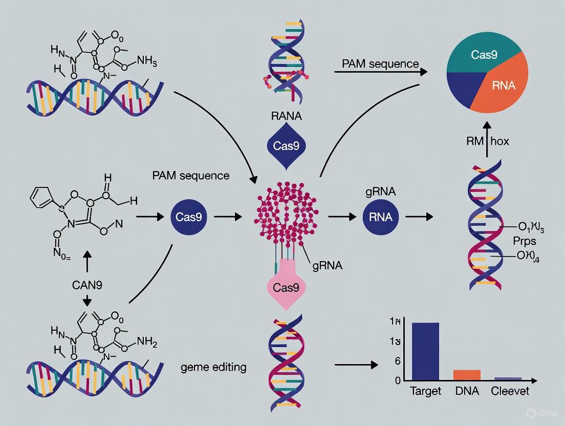
The CRISPR-Cas9 system represents a transformative technology in genome engineering, adapted from a natural adaptive immune system in bacteria. This system provides unprecedented capability for making precise, targeted changes to the genome of living cells [5]. Unlike previous genome-editing technologies such as zinc-finger nucleases (ZFNs) and transcription activator-like effector nucleases (TALENs), which required complex protein re-engineering for each new target, CRISPR-Cas9 simplifies targeted DNA modification through a programmable RNA-guided mechanism [6] [5]. At the heart of this system are two core components: the Cas9 nuclease, which acts as a "molecular scissor" to cut DNA, and a guide RNA (gRNA), which directs Cas9 to a specific genomic location [7] [8]. The simplicity, cost-effectiveness, and high specificity of CRISPR-Cas9 have revolutionized biomedical research, enabling applications ranging from gene function studies to the development of novel gene therapies for genetic disorders [6] [5].
Molecular Architecture of Cas9
Structural Domains and Functional Motifs
The Cas9 nuclease from Streptococcus pyogenes (SpCas9) features a bilobed architecture composed of a recognition lobe (REC) and a nuclease lobe (NUC) [7]. These lobes contain specialized domains that work in concert to enable precise DNA targeting and cleavage:
REC Lobe: Facilitates binding between the guide RNA and target DNA through several key domains. The bridge helix connects the two lobes and aids in gRNA recognition, while REC1, REC2, and REC3 domains stabilize the gRNA-Cas9 complex and interact with the RNA-DNA hybrid [7] [9]. Recent studies show that REC3 specifically docks onto the PAM-distal region of the RNA-DNA duplex once the R-loop extends beyond 14 base pairs, playing a critical role in Cas9 activation [9].
NUC Lobe: Contains the nuclease domains responsible for DNA cleavage. The HNH domain cleaves the DNA strand complementary to the guide RNA, while the RuvC domain cleaves the non-complementary strand [7] [5]. This lobe also houses the PAM-interacting domain, which recognizes the protospacer adjacent motif (PAM)—a short DNA sequence adjacent to the target site that serves as a binding checkpoint [7].
For CRISPR-Cas9 to function in eukaryotic cells, the enzyme must localize to the nucleus. This is achieved by fusing nuclear localization signals (NLS) to Cas9, enabling active transport through nuclear pores [7].
Cas9 Variants for Precision Engineering
Wild-type Cas9 has limitations in specificity and targeting range, prompting the development of engineered variants with improved properties:
Table 1: Engineered Cas9 Variants and Their Applications
| Variant Type | Key Mutations | Mechanism of Action | Primary Applications |
|---|---|---|---|
| High-Fidelity Cas9 (e.g., SpCas9-HF1, eSpCas9(1.1), HypaCas9) | Mutations in REC or NUC lobes (e.g., K848A, K1003A) | Reduces tolerance for mismatches between sgRNA and target DNA | Minimizes off-target editing while maintaining on-target efficiency [7] |
| Cas9 Nickase (nCas9) | D10A (inactivates RuvC) or H840A (inactivates HNH) | Cuts only one DNA strand, creating a single-strand break | Paired nickase system (two nCas9 complexes targeting opposite strands) creates staggered DSB with enhanced specificity [7] [5] |
| Catalytically Dead Cas9 (dCas9) | D10A and H840A (inactivates both nuclease domains) | Binds DNA without cleavage, blocking transcription or serving as a targeting platform | CRISPR interference (CRISPRi) for gene repression; epigenetic modulation when fused to effector domains [7] [5] |
Guide RNA (sgRNA): Design and Function
Composition and Structure
The guide RNA (gRNA) serves as the targeting component of the CRISPR-Cas9 system, determining its specificity. In nature, two separate RNA molecules—the CRISPR RNA (crRNA) and trans-activating crRNA (tracrRNA)—guide Cas9 to its target [10] [5]. For research and therapeutic applications, these are typically combined into a single-guide RNA (sgRNA) molecule, which includes both the target-specific crRNA region and the tracrRNA scaffold connected by a linker loop [10]. The sgRNA molecule can be divided into distinct functional regions:
- crRNA-derived region: A 17-20 nucleotide sequence complementary to the target DNA that provides targeting specificity [10] [11]
- tracrRNA scaffold: Maintains the structural integrity necessary for Cas9 binding and activation [10]
- Linker loop: Connects the crRNA and tracrRNA components into a single molecule [10]
The total length of a functional sgRNA is typically about 100 nucleotides, with 19-20 bases comprising the target-specific spacer and approximately 80 bases forming the universal sgRNA scaffold [11].
sgRNA Design Parameters
Effective sgRNA design is critical for successful genome editing, impacting both on-target efficiency and off-target effects. Key design considerations include:
PAM Sequence Selection: The Cas9 nuclease from Streptococcus pyogenes requires a 5'-NGG-3' protospacer adjacent motif (PAM) sequence immediately downstream of the target site, where "N" can be any nucleotide [10] [7]. The PAM is essential for Cas9 activation but is not part of the sgRNA sequence itself [10].
GC Content: Optimal sgRNA sequences typically have GC content between 40-60%, which balances stability and specificity. Higher GC content can increase sgRNA stability but may also promote off-target binding [7] [11].
Specificity Considerations: The sgRNA sequence should be unique to the target site to minimize off-target effects. Mismatches between the sgRNA and target DNA, particularly in the PAM-proximal region (seed region), can significantly reduce cleavage efficiency [7] [5].
Length Optimization: Standard sgRNAs utilize 20-nucleotide targeting sequences, but truncated sgRNAs with 17-18 nucleotides can reduce off-target effects while maintaining on-target activity [9] [5]. Recent research demonstrates that strategically designed truncated sgRNAs with terminal mismatches (e.g., 15-nt sgRNA with two additional mismatched nucleotides at positions +16 and +17) can promote multi-turnover Cas9 activity while maintaining efficiency [9].
Table 2: sgRNA Design Criteria and Optimization Strategies
| Parameter | Optimal Range | Impact on Editing | Design Tips |
|---|---|---|---|
| Spacer Length | 17-23 nucleotides | Longer guides increase specificity but may reduce efficiency; shorter guides (14-17 nt) can promote multi-turnover kinetics [10] [9] | For standard applications, use 20-nt guides; consider truncated guides (17-18 nt) to reduce off-target effects [9] |
| GC Content | 40-60% | Guides with GC content >80% may form stable secondary structures; <20% GC may be unstable [7] [11] | Avoid extreme GC content; aim for balanced distribution along the sequence |
| PAM Proximity | Immediately 5' of PAM | The 10-12 bases adjacent to PAM (seed region) are most critical for specificity [7] [5] | Ensure perfect complementarity in seed region; mismatches here dramatically reduce cleavage |
| Off-Target Potential | Minimal similarity to other genomic sites | Sequences with high similarity to off-target sites increase risk of unintended editing [11] [5] | Use tools like BLAST or specialized CRISPR design software to check genome-wide specificity |
The CRISPR-Cas9 Mechanism of Action
DNA Target Recognition and Cleavage
The CRISPR-Cas9 mechanism begins with the formation of a ribonucleoprotein (RNP) complex between Cas9 and the sgRNA [7]. This complex then scans the genome for complementary DNA sequences adjacent to a PAM sequence [7]. The process proceeds through several well-defined steps:
PAM Recognition: The Cas9-sgRNA complex first identifies the PAM sequence (5'-NGG-3' for SpCas9) through the PAM-interacting domain. This serves as an initial checkpoint, as Cas9 will not engage DNA sequences lacking the correct PAM [7] [5].
DNA Melting and R-loop Formation: Upon PAM recognition, Cas9 unwinds the DNA duplex, allowing the sgRNA to base-pair with the target strand. This forms an R-loop structure where the target strand hybridizes with the sgRNA while the non-target strand is displaced [9]. R-loop propagation proceeds directionally from the PAM-distal to PAM-proximal end, with full R-loop formation (typically beyond 14 base pairs) triggering conformational changes that activate Cas9's nuclease domains [9].
DNA Cleavage: Successful R-loop formation activates the two nuclease domains of Cas9. The HNH domain cleaves the target DNA strand complementary to the sgRNA, while the RuvC domain cleaves the non-target strand [7] [5]. This coordinated action creates a double-strand break (DSB) 3-4 nucleotides upstream of the PAM sequence [10].
Following DNA cleavage, Cas9 remains stably associated with the DNA product, exhibiting single-turnover kinetics that can block access to repair machinery [9]. Recent studies show that strategic sgRNA design (e.g., truncated guides with terminal mismatches) can promote multi-turnover behavior, enhancing editing efficiency [9].
DNA Repair Pathways and Editing Outcomes
The cellular response to CRISPR-induced double-strand breaks determines the final editing outcome. Cells primarily utilize two distinct repair pathways:
Non-Homologous End Joining (NHEJ): This pathway directly ligates broken DNA ends without a template, often resulting in small insertions or deletions (indels) that can disrupt gene function. NHEJ is efficient but error-prone, making it suitable for gene knockout applications [7] [5].
Homology-Directed Repair (HDR): This high-fidelity pathway uses a homologous DNA template to repair the break, enabling precise gene modifications including specific point mutations, gene corrections, or insertions. HDR is less efficient than NHEJ and requires co-delivery of a donor DNA template [7] [5].
Recent research has identified additional repair mechanisms, including CRISPR-homology-mediated end joining (HMEJ), which may operate through a single-strand annealing process and shows promise for gene therapy applications [6].
Advanced Experimental Applications
Enhanced Specificity and Control Systems
Recent advances address the critical challenge of off-target effects in CRISPR applications. Several innovative approaches have been developed:
Anti-CRISPR Protein Systems: Researchers have engineered cell-permeable anti-CRISPR protein systems (e.g., LFN-Acr/PA) that can rapidly enter human cells and inhibit Cas9 activity after genome editing is complete. This technology reduces off-target activity by preventing prolonged Cas9 exposure to genomic DNA, boosting genome-editing specificity by up to 40% [12].
High-Fidelity Cas9 Variants: Engineered Cas9 variants with enhanced specificity contain mutations that reduce non-specific interactions with DNA. These high-fidelity variants demonstrate significantly reduced off-target effects while maintaining robust on-target activity [7].
Computational Design Tools: AI-driven approaches for sgRNA design now enable more accurate prediction of on-target efficiency and off-target effects, optimizing guide selection for specific experimental contexts [6].
Multi-Turnover Cas9 Engineering
Traditional Cas9 exhibits single-turnover kinetics, remaining tightly bound to DNA after cleavage and limiting catalytic efficiency. Recent structural insights using cryo-EM have revealed strategies for engineering multi-turnover Cas9 systems [9]:
sgRNA Truncation: Shortening the sgRNA spacer to 15-17 nucleotides promotes faster product release and multi-turnover behavior, though with reduced cleavage rates [9].
Terminal Mismatch Strategy: Adding strategically positioned mismatches at the PAM-distal end of the sgRNA (e.g., 15-nt sgRNA with two terminal mismatches) enhances turnover while maintaining efficient cleavage across multiple targets [9].
Structural Insights: Cryo-EM studies of multi-turnover Cas9 complexes reveal that product inhibition is primarily due to retention of the PAM-containing DNA product, which occludes binding of new targets. These structural findings guide rational engineering of improved Cas9 variants [9].
Research Reagent Solutions
Successful implementation of CRISPR-Cas9 technology requires carefully selected reagents and delivery methods. The table below outlines essential materials and their applications in CRISPR experiments:
Table 3: Essential Research Reagents for CRISPR-Cas9 Experiments
| Reagent Type | Key Examples | Function & Mechanism | Applications & Advantages |
|---|---|---|---|
| Cas9 Expression Systems | Wild-type Cas9, Hi-Fi Cas9, Cas9D10A nickase, dCas9 | Provides the nuclease component; different variants offer tailored cleavage properties (full cleavage, nicking, or DNA binding without cleavage) | Gene knockout (WT-Cas9), precise editing with reduced off-targets (nickase), gene regulation (dCas9) [7] [5] |
| sgRNA Format Options | Synthetic sgRNA, IVT sgRNA, plasmid-expressed sgRNA | Delivers targeting component; format affects efficiency, toxicity, and delivery options | Synthetic sgRNA offers high purity and reduced immune activation; plasmid-based allows stable expression [10] |
| Delivery Methods | Electroporation, lipofection (LNP), microinjection, viral vectors | Introduces CRISPR components into cells; method affects efficiency, cell type compatibility, and toxicity | RNP electroporation for primary cells; LNP for in vivo delivery; viral vectors for stable expression [7] |
| Validation Tools | T7 Endonuclease I assay, NGS validation, PCR genotyping | Detects editing efficiency and identifies potential off-target effects | T7E1 for quick efficiency assessment; NGS for comprehensive off-target profiling [5] |
| Specificity Enhancers | Anti-CRISPR proteins (e.g., LFN-Acr/PA), truncated sgRNAs, computational design tools | Reduces off-target effects through various mechanisms including Cas9 inhibition and improved guide design | Anti-CRISPR proteins for temporal control; computational tools for guide optimization [9] [12] |
The core components of the CRISPR-Cas9 system—the Cas9 nuclease and guide RNA—represent a powerful platform for precision genome engineering. Understanding the molecular architecture of Cas9, the design principles of sgRNAs, and the detailed mechanism of DNA targeting and cleavage is essential for harnessing this technology effectively. Recent advances in Cas9 engineering, including high-fidelity variants, anti-CRISPR control systems, and strategies to enhance catalytic turnover, continue to expand the capabilities and safety of CRISPR-based applications. As these components evolve through ongoing research, particularly with integration of artificial intelligence and structural insights, CRISPR-Cas9 promises to drive further innovations in basic research and therapeutic development. The continued refinement of these core components will undoubtedly overcome current limitations and open new frontiers in genome engineering.
The CRISPR-Cas9 (Clustered Regularly Interspaced Short Palindromic Repeats and associated protein 9) system has revolutionized molecular biology by providing an unprecedented ability to make precise, targeted changes to the genome of living cells [13]. Originally discovered as an adaptive immune system in bacteria and archaea that defends against viral infections [1] [6], researchers have harnessed this natural mechanism to develop a powerful genome-editing tool. The system functions through a simplified, two-component mechanism comprising a Cas9 nuclease that cuts DNA and a guide RNA (gRNA) that directs the nuclease to a specific genomic location [6] [13]. This review focuses on the core mechanistic principles of the CRISPR-Cas9 system, breaking down the process into three fundamental steps: recognition, cleavage, and repair. Understanding this three-step mechanism is crucial for researchers and drug development professionals aiming to apply CRISPR technology to model diseases, develop therapies, and advance biomedical research.
Molecular Components of the CRISPR-Cas9 System
The CRISPR-Cas9 system's functionality depends on two essential molecular components that work in concert to achieve targeted DNA modification.
The Cas9 Nuclease: The Cas9 protein, often derived from Streptococcus pyogenes (SpCas9), is a large multi-domain DNA endonuclease (1,368 amino acids) that acts as the executive module of the system [1]. Structurally, Cas9 consists of two primary lobes: the recognition (REC) lobe, which binds the guide RNA, and the nuclease (NUC) lobe [1]. The NUC lobe contains two distinct nuclease domains responsible for DNA strand cleavage: the HNH domain, which cleaves the DNA strand complementary to the guide RNA, and the RuvC domain, which cleaves the non-complementary strand [1] [13]. A third critical region, the PAM-interacting domain, is responsible for initiating the binding to target DNA by recognizing a short, conserved sequence adjacent to the target site [1].
The Guide RNA (gRNA): The guiding module is a synthetic single-guide RNA (sgRNA), which combines the functions of two natural RNA components—the CRISPR RNA (crRNA) and the trans-activating crRNA (tracrRNA)—into a single molecule [1] [13]. The 5' end of the sgRNA contains a ~20-nucleotide spacer sequence that is complementary to the target DNA sequence and dictates the system's specificity through Watson-Crick base pairing [14]. The 3' end forms a hairpin structure that serves as a binding scaffold for the Cas9 protein [1].
Table 1: Core Components of the CRISPR-Cas9 System
| Component | Type | Function | Key Features |
|---|---|---|---|
| Cas9 Nuclease | Protein (Endonuclease) | Executes DNA cleavage | Contains HNH and RuvC nuclease domains; requires PAM sequence for activation [1] [13] |
| Guide RNA (gRNA) | RNA (Synthetic single-guide RNA) | Specifies target location | 5' end provides target complementarity; 3' end binds Cas9 protein [1] [14] |
| HNH Domain | Protein Domain (within Cas9) | Cleaves complementary DNA strand | Target specificity depends on RNA-DNA hybridization [1] [13] |
| RuvC Domain | Protein Domain (within Cas9) | Cleaves non-complementary DNA strand | Works with HNH to create a double-stranded break [1] [13] |
The Core Three-Step Mechanism
The process of CRISPR-Cas9-mediated genome editing can be systematically divided into three sequential steps: recognition, cleavage, and repair.
Step 1: Recognition
The recognition step initiates the genome-editing process, where the CRISPR-Cas9 complex locates and binds to its specific target site within the vast genome.
Target Site Identification: The Cas9 protein, pre-complexed with the sgRNA, scans the DNA for a short, conserved nucleotide sequence known as the Protospacer Adjacent Motif (PAM) [1] [13]. For the commonly used SpCas9, the PAM sequence is 5'-NGG-3', where "N" can be any nucleotide [1]. The presence of a compatible PAM is an absolute requirement for the initiation of Cas9 activity; even DNA sequences that are fully complementary to the sgRNA will be ignored if not adjacent to a PAM [13].
DNA Melting and Hybridization: Once Cas9 binds to a valid PAM sequence, it triggers local unwinding, or "melting," of the double-stranded DNA [1] [15]. This allows the ~20-nucleotide spacer sequence at the 5' end of the sgRNA to form a heteroduplex with the target DNA strand via complementary base pairing [1]. This sequence-specific hybridization is the primary determinant of the system's targeting precision.
Step 2: Cleavage
Following successful recognition and hybridization, the Cas9 nuclease is activated for DNA cleavage.
- Double-Strand Break Formation: The binding of the sgRNA to its complementary DNA target induces a conformational change in the Cas9 protein, activating its catalytic domains [1]. This results in the creation of a double-strand break (DSB) precisely 3 base pairs upstream of the PAM sequence [1] [15]. The cleavage is achieved through the coordinated action of the two nuclease domains: the HNH domain cleaves the strand complementary to the sgRNA, while the RuvC domain cleaves the opposite strand [1] [13]. This typically results in a blunt-ended DSB [15].
Step 3: Repair
The cellular DNA damage response machinery detects the induced DSB and initiates repair. The outcome of genome editing is determined by which of the two major endogenous repair pathways is employed.
Non-Homologous End Joining (NHEJ): This is the dominant and most active repair pathway throughout the cell cycle [1] [15]. NHEJ directly ligates the broken DNA ends without requiring a homologous template. As this process is error-prone, it often results in small random insertions or deletions (indels) at the cleavage site [1]. These indels can disrupt the coding sequence of a gene, leading to frameshift mutations and premature stop codons, effectively knocking out the target gene [1] [13].
Homology-Directed Repair (HDR): This pathway is highly precise but less frequent and primarily active in the late S and G2 phases of the cell cycle [1] [15]. HDR requires the presence of an exogenous donor DNA template containing homologous sequences flanking the DSB. This template is used to accurately copy genetic information, enabling precise gene insertion, correction, or replacement [1] [13]. The efficiency of HDR is generally lower than that of NHEJ.
The following diagram illustrates the logical sequence and key events of this three-step mechanism.
Table 2: Double-Strand Break Repair Pathways
| Feature | Non-Homologous End Joining (NHEJ) | Homology-Directed Repair (HDR) |
|---|---|---|
| Template Required | No | Yes (donor DNA template) |
| Primary Mechanism | Direct ligation of broken ends | Uses homologous sequence for precise repair |
| Fidelity | Error-prone | High-fidelity |
| Key Outcome | Small insertions/deletions (indels); gene knockout [1] | Precise nucleotide changes; gene correction [1] |
| Cell Cycle Activity | All phases | Late S and G2 phases [1] |
| Relative Efficiency | High (predominant pathway) [1] | Low (less frequent) [1] |
Technical Considerations and Experimental Protocols
For researchers aiming to utilize CRISPR-Cas9, understanding the practical considerations for designing and validating experiments is paramount.
Designing and Validating sgRNA
The design of the sgRNA is the most critical factor determining the success and specificity of a CRISPR experiment.
- Protocol: sgRNA Design and In Silico Analysis
- Target Selection: Identify a 20-nucleotide target sequence immediately adjacent to a 5'-NGG PAM sequence on the genomic DNA strand of interest [1] [13].
- Specificity Check: Use bioinformatics tools (e.g., CHOPCHOP, CRISPR Design Tool) to perform a genome-wide alignment to minimize off-target effects [14] [13]. Select sgRNAs with minimal homology to other genomic sites, especially in the "seed" region proximal to the PAM.
- Efficiency Prediction: Employ algorithms that predict sgRNA on-target activity based on sequence features, such as GC content and nucleotide composition [14].
- Cloning and Delivery: Clone the selected sgRNA sequence into an appropriate expression plasmid containing a RNA polymerase III promoter (e.g., U6) and deliver it alongside a plasmid expressing the Cas9 nuclease into the target cells [13].
Analyzing Editing Outcomes
Detecting the genetic modifications introduced by CRISPR-Cas9 is essential for evaluating editing efficiency.
Protocol: T7 Endonuclease I Mutation Detection Assay
- PCR Amplification: After allowing time for editing to occur, isolate genomic DNA from transfected cells. Amplify the target genomic region by PCR [13].
- Heteroduplex Formation: Denature and reanneal the PCR products. In samples containing a mix of wild-type and edited alleles, the reannealing process will create heteroduplex DNA where the strands are mismatched at the site of indels [13].
- Digestion and Analysis: Treat the reannealed DNA with T7 Endonuclease I, which cleaves at heteroduplex sites. Analyze the digestion products by gel electrophoresis. The presence of cleavage bands indicates successful genome editing, and band intensity can be used to estimate the mutation frequency [13].
Advanced Analysis: For a comprehensive assessment of editing outcomes, including the precise spectrum of indels, Sanger sequencing of the PCR amplicons followed by analysis with tools like CRISPResso is recommended [14]. It is crucial to note that traditional short-read sequencing may miss large, unintended structural variations, such as kilobase-scale deletions or chromosomal rearrangements [16]. Techniques like CAST-Seq or LAM-HTGTS are increasingly used to profile these genotoxic side effects in safety-critical applications [16].
The Scientist's Toolkit: Essential Research Reagents
Table 3: Key Reagents for CRISPR-Cas9 Research
| Reagent / Tool | Category | Function in Experiment |
|---|---|---|
| SpCas9 Expression Plasmid | Molecular Biology Reagent | Provides the genetic code for the Cas9 nuclease, often under a strong promoter like CMV [13] [17]. |
| sgRNA Expression Vector | Molecular Biology Reagent | Plasmid with a U6 promoter for in-cell transcription of the designed sgRNA [13] [17]. |
| Donor DNA Template | Molecular Biology Reagent | Single-stranded or double-stranded DNA containing homologous arms and the desired sequence for precise HDR editing [1]. |
| Lipid Nanoparticles (LNPs) | Delivery Vehicle | Synthetic particles used to encapsulate and deliver CRISPR ribonucleoproteins (RNPs) or plasmids in vivo, with a natural affinity for the liver [18] [17]. |
| Adeno-Associated Virus (AAV) | Delivery Vehicle | Viral vector with low immunogenicity used for in vivo delivery; limited by a small packaging capacity (~4.7 kb) [17]. |
| Electroporation System | Physical Delivery Equipment | Applies electrical pulses to temporarily permeabilize cell membranes, enabling efficient delivery of CRISPR components into hard-to-transfect cells (e.g., stem cells) ex vivo [17]. |
| T7 Endonuclease I | Detection Assay Reagent | Enzyme used to detect heteroduplex DNA formed from indels, enabling quantification of editing efficiency [13]. |
| HiFi Cas9 | Protein Engineering | Engineered Cas9 variant with enhanced specificity, reducing off-target effects at the cost of potentially lower on-target activity [16]. |
| Anti-CRISPR Proteins (Acrs) | Control/Enhancement Reagent | Naturally occurring proteins that inhibit Cas9 activity. Novel cell-permeable systems (e.g., LFN-Acr/PA) can be used to rapidly deactivate Cas9 after editing to minimize off-target effects [12]. |
Challenges and Future Perspectives
Despite its transformative potential, the practical application of the CRISPR-Cas9 three-step mechanism faces several challenges that are active areas of research.
Off-Target Effects: The Cas9 nuclease can tolerate mismatches between the sgRNA and DNA, leading to unintended cleavage at off-target sites with sequence similarity [16] [13]. Strategies to mitigate this include using engineered high-fidelity Cas9 variants (e.g., HiFi Cas9) [16], truncated sgRNAs [13], and the recently developed cell-permeable anti-CRISPR proteins that rapidly inactivate Cas9 after the intended editing is complete [12].
On-Target Genomic Aberrations: Beyond small indels, CRISPR-Cas9 can induce large, on-target structural variations (SVs), including kilobase- to megabase-scale deletions and chromosomal translocations [16]. These SVs pose substantial safety concerns for clinical applications and are often undetected by standard short-read sequencing methods [16].
Delivery Efficiency: The efficient delivery of all CRISPR components into the nucleus of target cells remains a major bottleneck, particularly for in vivo therapeutic applications [17]. The large size of the Cas9 protein challenges the packaging capacity of popular viral vectors like AAV, spurring the development of novel delivery platforms such as lipid nanoparticles (LNPs) and the discovery of smaller Cas orthologs [18] [17].
Future advancements are focused on improving the precision and safety of the technology. The integration of artificial intelligence (AI) and machine learning is refining sgRNA design and predicting off-target effects with greater accuracy [14] [6]. Furthermore, new editing systems like base editing and prime editing offer alternatives to DSBs, enabling precise nucleotide changes with potentially reduced risks of genotoxic off-target effects and large structural variations [6]. As these technologies mature, they will broaden the clinical potential of CRISPR, solidifying its role as a cornerstone of modern genetic engineering.
The Protospacer Adjacent Motif (PAM) serves as an essential molecular signature that enables CRISPR-Cas systems to distinguish between self and non-self DNA, representing a critical checkpoint in the target selection process. This in-depth technical guide examines the fundamental mechanisms of PAM recognition, its structural basis across different Cas proteins, and recent advances in PAM characterization and engineering. Within the broader context of CRISPR-Cas9 mechanisms of action, we explore how PAM requirements influence genome editing efficiency, specificity, and therapeutic applicability. For researchers, scientists, and drug development professionals, this review provides both foundational knowledge and cutting-edge methodologies to navigate PAM constraints in experimental design and therapeutic development.
The Protospacer Adjacent Motif (PAM) is a short, specific DNA sequence (typically 2-6 base pairs) adjacent to the target DNA region (protospacer) that is essential for cleavage by CRISPR-associated (Cas) nucleases [19]. This motif serves as the fundamental "gatekeeper" in CRISPR-mediated immunity and genome engineering applications, enabling the system to distinguish between invasive genetic elements and the host's own CRISPR arrays [19] [20]. In natural bacterial immunity, the PAM prevents autoimmunity by ensuring that Cas nucleases do not target the bacterial genome itself, as the CRISPR arrays within the bacterial genome lack PAM sequences [19]. The PAM is generally found 3-4 nucleotides downstream from the Cas9 cut site for type II systems, with the exact position and sequence varying depending on the specific Cas nuclease and CRISPR type [19] [1].
The dual functionality of PAM sequences in both spacer acquisition (adaptation) and target interference has led to proposals for more precise terminology: Spacer Acquisition Motif (SAM) for the conserved sequence recognized during spacer acquisition and Target Interference Motif (TIM) for the sequence recognized during interference [21] [20]. This distinction reflects the different molecular mechanisms and potential stringency requirements between these two processes in the CRISPR immune response [21]. From a therapeutic perspective, PAM recognition represents both a necessity for CRISPR activity and a significant constraint on targetable genomic loci, driving extensive research into understanding and engineering PAM specificity [22] [23] [24].
Molecular Mechanisms of PAM Recognition
Structural Basis of PAM Interaction
PAM recognition occurs through specific protein domains within Cas nucleases that interact directly with the DNA motif. For the well-characterized Streptococcus pyogenes Cas9 (SpCas9), the PAM-interacting domain recognizes a 5'-NGG-3' sequence through specific amino acid residues, particularly an arginine dyad (R1333 and R1335) that forms critical contacts with the guanine bases [22]. Structural studies reveal that when Cas9 identifies the correct PAM, it triggers local DNA melting, followed by the formation of an RNA-DNA hybrid (R-loop) that enables target recognition and cleavage [1] [25].
The PAM recognition mechanism follows an ordered process: Cas nuclease surveillance complexes first scan DNA for PAM sequences; specific Cas proteins then recognize and bind the PAM sequence, unwinding the adjacent dsDNA helix; the opened DNA becomes available for hybridization with the crRNA, forming a triple-stranded R-loop structure; seed sequences near the PAM are interrogated for complementarity with the crRNA spacer [20]. This sequential mechanism ensures that only bona fide targets with correct PAM sequences undergo cleavage, providing the specificity required for both bacterial immunity and precise genome editing.
Molecular Dynamics of PAM Recognition
Recent research has elucidated the sophisticated molecular dynamics underlying PAM recognition. In wild-type SpCas9, stringent guanine selection is enforced through the rigidity of its interacting arginine dyad, particularly R1335 [22]. Advanced molecular simulations and metadynamics studies of engineered variants like xCas9 reveal that increased flexibility in R1335 enables selective recognition of alternative PAM sequences while maintaining discrimination against non-target sequences [22]. This flexibility confers a pronounced entropic preference that surprisingly also improves recognition of the canonical NGG PAM [22].
The diagram below illustrates the core mechanism of PAM-dependent DNA target recognition:
PAM-dependent DNA Target Recognition - This workflow illustrates the sequential process from initial PAM scanning to final DNA cleavage.
The kinetic basis of PAM recognition explains several observed off-targeting rules, with binding being more promiscuous than cleavage due to kinetically stalled hybridization [26]. This understanding has enabled engineering of systems with increased specificity without losing on-target efficiency [26].
PAM Requirements Across CRISPR-Cas Systems
Diversity of PAM Sequences
Different Cas nucleases recognize distinct PAM sequences, reflecting their evolutionary origins in various bacterial species. The table below summarizes the PAM specificities of commonly used and engineered Cas nucleases in CRISPR experiments:
Table 1: PAM Sequences of Selected CRISPR Nucleases
| CRISPR Nuclease | Organism Isolated From | PAM Sequence (5' to 3') | Key Characteristics |
|---|---|---|---|
| SpCas9 | Streptococcus pyogenes | NGG [19] | Most commonly used nuclease; well-characterized |
| xCas9 | Engineered from SpCas9 | NG, GAA, GAT [22] [25] | Expanded PAM recognition; increased fidelity |
| SpCas9-NG | Engineered from SpCas9 | NG [25] | Expanded PAM recognition from NGG to NG |
| SpRY | Engineered from SpCas9 | NRN/NYN [25] | Near-PAMless variant; extremely broad targeting |
| SaCas9 | Staphylococcus aureus | NNGRRT or NNGRRN [19] | Compact size beneficial for viral delivery |
| NmeCas9 | Neisseria meningitidis | NNNNGATT [19] | Longer PAM; potentially higher specificity |
| CjCas9 | Campylobacter jejuni | NNNNRYAC [19] | Compact size for therapeutic applications |
| LbCas12a (Cpf1) | Lachnospiraceae bacterium | TTTV [19] | Creates staggered cuts; requires different gRNA structure |
| AacCas12b | Alicyclobacillus acidiphilus | TTN [19] | Thermostable variant |
| FnCas9 | Francisella novicida | NGG [24] | High intrinsic specificity; enhanced variants available |
| hfCas12Max | Engineered from Cas12i | TN and/or TNN [19] | High-fidelity variant with relaxed PAM |
This diversity enables researchers to select appropriate nucleases based on PAM availability in their target genomic regions. While SpCas9 remains the most widely used nuclease, its NGG PAM requirement occurs approximately every 8-12 base pairs in the human genome, potentially limiting targeting of specific loci of interest [19] [25]. The development of engineered Cas variants with altered PAM specificities has significantly expanded the targeting range of CRISPR technologies [23] [25] [24].
PAM Recognition in Type I, II, and III Systems
CRISPR-Cas systems are classified into two classes (1 and 2) and six types (I-VI) based on their effector module composition and sequences [20]. Each type employs distinct PAM recognition strategies:
- Type I systems: Utilize multi-subunit effector complexes (e.g., Cascade) for PAM recognition and target binding. The PAM is typically located at the 3' end of the protospacer and recognition involves multiple protein subunits working in concert [21] [20].
- Type II systems: Employ single-protein effectors (e.g., Cas9) that integrate both PAM recognition and nuclease activities in one molecule. The PAM is located at the 3' end of the protospacer [1] [20].
- Type III systems: Target RNA rather than DNA and generally do not require a classical PAM sequence for target recognition [20].
- Type V systems: Include Cas12 family proteins that recognize T-rich PAM sequences typically located at the 5' end of the protospacer [19] [20].
This diversity in PAM recognition mechanisms reflects the evolutionary arms race between bacteria and viruses, where varying PAM requirements help circumvent viral anti-CRISPR measures that alter PAM sequences or accessibility [20].
Experimental Methods for PAM Characterization
Established PAM Identification Techniques
Several experimental approaches have been developed to characterize PAM requirements for novel or engineered Cas nucleases:
In silico analysis: Computational identification of conserved sequences adjacent to protospacers matching CRISPR spacers in bacterial genomes [21] [20]. Tools like CRISPRTarget facilitate this analysis but require available phage genome sequences [20].
Plasmid depletion assays: Library-based approaches where randomized DNA sequences are inserted adjacent to target sites within plasmids transformed into hosts with active CRISPR-Cas systems [20]. Functional PAMs are identified through sequencing of retained plasmids after selection [20].
PAM-SCANR (PAM screen achieved by NOT-gate repression): High-throughput in vivo method utilizing catalytically dead Cas9 (dCas9) coupled with fluorescence-activated cell sorting (FACS) to identify functional PAM motifs through GFP repression [20].
In vitro cleavage assays: Utilization of purified Cas effector complexes to cleave target DNA libraries with randomized PAM sequences, followed by sequencing of cleavage products [20] [27].
HT-PAMDA (High-Throughput PAM Determination Assay): Scalable method combining human cell expression with in vitro cleavage reactions to characterize PAM preferences [27].
Each method presents distinct advantages and limitations regarding library coverage, physiological relevance, and technical requirements, necess careful selection based on research objectives.
GenomePAM: A Novel Mammalian Cell-Based Approach
A recent innovative method named GenomePAM leverages genomic repetitive sequences as naturally occurring target sites for direct PAM characterization in mammalian cells [27]. This approach utilizes highly repetitive sequences in the mammalian genome flanked by diverse sequences, where the constant sequence serves as the protospacer. The method identifies a 20-nt sequence (5′-GTGAGCCACTGTGCCTGGCC-3′, termed Rep-1) that occurs approximately 8,471 times (~16,942 occurrences in human diploid cells) distributed across the human genome with nearly random flanking sequences [27].
The experimental workflow involves:
- Cloning the Rep-1 sequence into a gRNA expression cassette
- Co-transfection with candidate Cas nuclease plasmid into human cells (e.g., HEK293T)
- Capturing cleaved genomic sites using GUIDE-seq methodology
- Sequencing and bioinformatic analysis to identify functional PAMs based on cleavage patterns [27]
GenomePAM offers significant advantages over previous methods by eliminating the need for protein purification or synthetic oligo libraries, while providing PAM characterization in the physiologically relevant context of mammalian chromatin [27]. The method has been successfully validated for characterizing PAM requirements of type II and type V nucleases, including the minimal PAM requirement of the near-PAMless SpRY and extended PAM for CjCas9 [27].
The following diagram illustrates the GenomePAM workflow:
GenomePAM Experimental Workflow - This diagram outlines the key steps in the GenomePAM method for PAM characterization using genomic repeats.
Engineering PAM Specificity and Novel Variants
Rational Engineering Approaches
Protein engineering strategies have successfully created Cas variants with altered PAM specificities to expand the targeting range of CRISPR technologies. Rational engineering approaches include:
Structure-guided mutagenesis: Using crystal structures of Cas nucleases to identify residues involved in PAM recognition for targeted mutagenesis. For example, enhanced FnCas9 (enFnCas9) variants were developed by modifying the WED-PI domain and phosphate-lock loop (PLL) to create additional interactions with the DNA backbone [24].
Directed evolution: Employing iterative rounds of selection to identify variants with desired PAM specificities. The xCas9 variant was developed through directed evolution, introducing seven amino acid substitutions that expanded PAM compatibility to include guanine- and adenine-containing PAMs while improving specificity [22].
Machine learning-guided engineering: Combining high-throughput protein engineering with neural networks to predict PAM specificity from amino acid sequences. The PAM machine learning algorithm (PAMmla) enables in silico-directed evolution for user-directed Cas9 enzyme design [23].
These engineering efforts have produced variants with significantly expanded PAM recognition, including SpCas9 variants recognizing NG PAMs (SpCas9-NG), NGN PAMs (SpG), and even near-PAMless variants (SpRY) that recognize NRN and NYN sequences [25].
Enhanced Specificity Variants
Engineering efforts have also addressed the challenge of off-target effects by developing high-fidelity variants with reduced non-specific activity:
- eSpCas9(1.1): Weakened interactions between the HNH/RuvC groove and the non-target DNA strand [25]
- SpCas9-HF1: Disrupted Cas9 interactions with the DNA phosphate backbone [25]
- HypaCas9: Enhanced proofreading and discrimination capabilities [25]
- evoCas9: Decreased off-target effects through laboratory evolution [25]
- Sniper-Cas9: Reduced off-target activity while maintaining on-target efficiency [25]
- SuperFi-Cas9: Dramatically increased fidelity with moderately reduced nuclease activity [25]
These variants maintain efficient on-target activity while significantly reducing off-target effects, addressing a major concern for therapeutic applications [25] [24].
Research Reagents and Experimental Tools
Table 2: Essential Research Reagents for PAM Studies
| Reagent/Tool Category | Specific Examples | Function/Application | Key Features |
|---|---|---|---|
| Cas Nuclease Variants | SpCas9, SaCas9, FnCas9, LbCas12a [19] | Core editing machinery with different PAM requirements | Diverse PAM specificities; varying sizes for delivery |
| Engineered Cas Variants | xCas9, SpCas9-NG, SpRY, enFnCas9 [22] [25] [24] | Expanded targeting range beyond wild-type PAM constraints | Broader PAM recognition; maintained or improved specificity |
| High-Fidelity Variants | eSpCas9(1.1), SpCas9-HF1, HypaCas9, evoCas9 [25] | Reduce off-target effects in therapeutic applications | Enhanced specificity while maintaining efficiency |
| PAM Characterization Systems | PAM-SCANR, HT-PAMDA, GenomePAM [20] [27] | Determine PAM preferences of novel nucleases | Various throughput levels; different experimental contexts |
| gRNA Design Tools | CRISPRTarget, various web tools [20] [25] | Select optimal target sites and predict off-targets | Algorithm-based specificity prediction |
| Specialized gRNA Formats | x-gRNAs, sx-gRNAs [24] | Enhance activity with certain Cas variants | Extended length spacers for improved kinetics |
Applications and Therapeutic Implications
Overcoming PAM Limitations in Genome Editing
PAM requirements traditionally constrained the targeting scope of CRISPR technologies, particularly for therapeutic applications requiring precise editing at specific genomic loci. Several strategies have emerged to overcome these limitations:
- Nuclease selection: Choosing appropriate Cas nucleases or variants based on PAM availability at the target locus [19] [25]
- PAM-flexible variants: Utilizing engineered Cas proteins with relaxed PAM requirements, such as SpRY or xCas9, to access previously inaccessible sites [25] [24]
- Extended gRNAs: Employing longer guide RNAs (x-gRNAs or sx-gRNAs) that enhance editing efficiency with certain Cas variants, particularly for base editing applications [24]
- Multiplex approaches: Combining multiple gRNAs to target several sites simultaneously, increasing the probability of successful editing when PAM availability is limited [25]
These approaches have significantly expanded the therapeutic applicability of CRISPR technologies, enabling targeting of previously inaccessible disease-relevant loci.
Therapeutic Applications and Clinical Relevance
The engineering of PAM specificity has direct implications for therapeutic development:
- Allele-selective targeting: PAMmla-designed Cas9 enzymes enable allele-selective targeting of specific mutations, such as the RHOP23H allele in human cells and mice, demonstrating potential for dominant disorder treatment [23]
- Gene correction: enFnCas9-based adenine base editors have been used to correct disease-associated point mutations, such as in the RPE65 gene causing Leber congenital amaurosis type 2 (LCA2) in patient-specific iPSC-derived retinal pigmented epithelium [24]
- Diagnostic applications: PAM-flexible enFnCas9 proteins expand the target range of CRISPR diagnostics (CRISPRDx) for detecting pathogenic DNA signatures [24]
- Enhanced safety profiles: High-fidelity variants with reduced off-target effects address critical safety concerns for clinical applications [25] [24]
These advances highlight how understanding and engineering PAM specificity contributes to the development of safer, more effective CRISPR-based therapeutics.
PAM recognition remains a fundamental aspect of CRISPR biology with significant implications for basic research and therapeutic development. Future directions in this field include:
- Continued expansion of PAM compatibility: Ongoing engineering efforts aim to develop truly PAMless Cas variants without compromising efficiency or specificity [23] [25]
- Computational prediction and design: Advanced machine learning approaches will enable more accurate prediction of PAM specificity and guide rational design of novel variants [23]
- Therapeutic optimization: Tailoring PAM specificity for particular clinical applications, such as allele-specific editing for dominant disorders [23] [24]
- Novel nuclease discovery: Exploration of natural CRISPR diversity continues to uncover novel Cas proteins with unique PAM specificities and biochemical properties [19] [20]
In conclusion, PAM recognition serves as the essential gatekeeper for DNA target selection in CRISPR-Cas systems, balancing the competing demands of specificity and flexibility. Through continued mechanistic studies and protein engineering, researchers have made significant progress in overcoming natural PAM limitations, expanding the targeting scope of CRISPR technologies while enhancing their precision. As these efforts continue, PAM engineering will remain central to realizing the full potential of CRISPR-based genome editing in both basic research and therapeutic applications.
The CRISPR-Cas9 system has revolutionized genome engineering by providing a programmable platform for precise DNA cleavage. At the core of this technology lies the Cas9 endonuclease, which creates double-strand breaks (DSBs) in target DNA through the coordinated activities of its two distinct nuclease domains: HNH and RuvC. This whitepaper provides an in-depth technical analysis of the structure-function relationships, cleavage mechanisms, and catalytic coordination of these domains. Within the broader context of CRISPR-Cas9 mechanism of action research, we examine how these domains achieve precise DNA cleavage, the experimental methodologies for studying their functions, and recent advances in enhancing their specificity and efficiency for therapeutic applications. The structural determinants and kinetic parameters governing HNH and RuvC activities represent critical considerations for researchers developing novel gene-editing therapeutics and experimental approaches.
CRISPR-Cas systems function as RNA-guided nucleases that provide adaptive immune protection in bacteria and archaea against invading genetic materials [28]. The type II-A CRISPR effector protein Cas9 from Streptococcus pyogenes (SpyCas9) has been widely adopted for gene editing applications due to its simplicity requiring only a single guide RNA (sgRNA) to direct DNA cleavage [28]. The catalytic cycle of SpyCas9 initiates with the formation of a binary complex upon sgRNA binding, which induces large conformational changes in the protein to accommodate duplex DNA [28]. The guide region of sgRNA adopts a pseudo A-form conformation that searches for complementarity in the target DNA, a process stabilized by the arginine-rich bridge helix (BH) of SpyCas9 [28].
The Cas9-sgRNA complex scans and locates the protospacer adjacent motif (PAM) in target DNA, followed by R-loop formation via complementary base pairing of the sgRNA with the target DNA [28]. This R-loop formation triggers substantial conformational changes in the REC lobe and HNH domain, which are essential for sequence-specific DNA cleavage [28]. The REC lobe senses nucleic acids and plays a critical role in the conformational transition of the HNH domain and its subsequent docking at the cleavage site [28]. The activated HNH and RuvC domains mediate target strand (TS) and non-target strand (NTS) cleavages, respectively, to generate a DSB in the target DNA [28] [29].
Structural Organization of Cas9 Nuclease Domains
Cas9 is a large multidomain protein structurally organized into two primary lobes: the nuclease (NUC) lobe and the recognition (REC) lobe, connected by an arginine-rich bridge helix (BH) [28]. The NUC lobe contains the two endonuclease domains (HNH and RuvC) along with a PAM-interacting domain, while the REC lobe comprises multiple α-helical recognition domains (REC1-REC3) that facilitate binding to RNA and DNA [28]. This bilobed architecture creates a central channel where the RNA-DNA heteroduplex resides, with the HNH and RuvC domains positioned to cleave opposite DNA strands [30].
HNH Domain Structure and Dynamics
The HNH domain is responsible for cleaving the target strand (complementary to the sgRNA) and exhibits remarkable conformational flexibility during catalytic activation [30]. Structural studies have revealed at least three distinct conformational states of the HNH domain:
- HNH-state 1: The HNH active site is positioned more than 32 Å from the cleavage site
- HNH-state 2: An intermediate state where the active site is approximately 19 Å from the cleavage site
- HNH-state 3: The active conformation where the HNH domain is closest to the DNA cleavage site, with the active site shifted about 25 Å and 13 Å compared to states 1 and 2, respectively [30]
This mobility enables the HNH domain to rotate approximately 170° around an axis to achieve proper positioning for catalysis [30]. The transition between these states involves a helix-to-loop conformational change in the L2 linker region (residues 906-923), similar to observations in Staphylococcus aureus Cas9 (SaCas9) [30].
RuvC Domain Organization
The RuvC domain cleaves the non-target DNA strand and shares structural homology with retroviral integrase superfamily members [29]. This domain contains a RuvC-like nuclease fold located at the amino terminus of Cas9 and is responsible for generating a single-stranded break in the non-complementary DNA strand [29]. Unlike the highly mobile HNH domain, the RuvC domain maintains a relatively stable position but undergoes allosteric activation upon HNH docking [28].
Table 1: Structural Characteristics of Cas9 Nuclease Domains
| Domain | Structural Features | Catalytic Residues | DNA Strand Targeted | Conformational Flexibility |
|---|---|---|---|---|
| HNH | ββα-metal fold, resembles HNH homing endonucleases | H840 (SpyCas9) | Target strand (complementary) | High - rotates ~170°, moves >25 Å |
| RuvC | RuvC-like fold, RNase H superfamily | D10, E762, H983 (SpyCas9) | Non-target strand | Moderate - allosteric activation |
| Bridge Helix | Arginine-rich α-helix | L64, K65 | N/A (regulatory) | Moderate - influences coordination |
DNA Cleavage Mechanism and Coordination
Sequential Activation and Cleavage Pathways
The HNH and RuvC domains operate through coordinated actions to linearize DNA, with evidence supporting the existence of parallel sequential routes for DNA cleavage [28]. Kinetic analysis using supercoiled plasmid substrates has revealed two primary pathways:
- TS Pathway: Nicking by HNH followed by RuvC cleavage
- NTS Pathway: Nicking by RuvC followed by HNH cleavage [28]
The relative usage of these pathways is modulated by the integrity of the bridge helix and the position of mismatches in the substrate, with each condition producing distinct conformational energy landscapes [28]. This coordinated cleavage between HNH and RuvC is facilitated by BH interactions with RNA/DNA, enabling target DNA discrimination through differential use of these parallel sequential pathways [28].
Allosteric Regulation and Communication
Extensive allosteric communication exists between the HNH and RuvC domains to ensure coordinated DSB formation. The REC lobe senses nucleic acids and plays an important role in the conformational transition of the HNH domain [28]. Specifically:
- REC3 allosterically activates HNH upon binding to RNA-DNA
- REC2 moves outward to prevent steric occlusion with HNH
- REC1 assists by locking HNH in an active state through ionic interactions [28]
Activation of the HNH domain concomitantly induces conformational changes in the hinge regions at the HNH-RuvC junctions and allosterically controls the RuvC domain [28]. Solution NMR and atomistic MD simulation studies have revealed the presence of an allosteric path through HNH that connects the REC2 and RuvC domains [28]. This intricate network ensures that both nuclease domains become activated only when proper target recognition has occurred, enhancing the fidelity of DNA cleavage.
Catalytic Mechanism and Cleavage Products
Both HNH and RuvC domains cleave DNA with similar catalytic rate constants (kchem) once properly positioned [28]. The HNH domain cleaves the complementary DNA strand, while the RuvC domain cleaves the non-complementary strand, generating DSBs located approximately 3-4 nucleotides upstream of the PAM sequence [25]. The resulting DSB is then repaired by cellular repair pathways:
- Non-Homologous End Joining (NHEJ): An efficient but error-prone pathway that frequently introduces small insertions or deletions (indels)
- Homology-Directed Repair (HDR): A less efficient but high-fidelity pathway that requires a repair template [25]
Table 2: Cleavage Parameters and Kinetic Data for HNH and RuvC Domains
| Parameter | HNH Domain | RuvC Domain | Experimental Conditions |
|---|---|---|---|
| Catalytic Rate Constant (kchem) | Similar for both domains [28] | Similar for both domains [28] | Measured in SpyCas9WT |
| Cleavage Position | ~3-4 nt upstream of PAM [25] | ~3-4 nt upstream of PAM [25] | Relative to NGG PAM |
| Strand Specificity | Target strand (complementary) [29] | Non-target strand [29] | Based on RNA-DNA hybridization |
| Metal Cofactor Requirement | Mg²⁺ dependent | Mg²⁺ dependent | Standard buffer conditions |
| Pathway Preference | Initiates TS pathway [28] | Initiates NTS pathway [28] | Depends on BH integrity and mismatches |
Experimental Analysis of DNA Cleavage
Kinetic Analysis Methodology
The cleavage kinetics of HNH and RuvC domains can be analyzed using supercoiled plasmid substrates, which allow independent measurements of nicked intermediate and linearized DNA products [28]. The experimental protocol involves:
- Plasmid Substrate Preparation: Supercoiled plasmid DNA containing the target protospacer sequence
- Cas9-sgRNA Complex Formation: Incubate SpyCas9 (WT or mutant) with sgRNA to form ribonucleoprotein complexes
- Time-Course Cleavage Assays: Initiate reactions by adding plasmid substrates and quench at various time points
- Product Separation and Quantification: Analyze reaction products using agarose gel electrophoresis to resolve supercoiled, nicked, and linear DNA forms
- Data Modeling: Fit time-dependent changes in DNA forms to parallel sequential cleavage models to derive individual rate constants [28]
This approach enables researchers to determine how BH substitutions and DNA mismatches alter individual rate constants and affect the relative use of TS versus NTS pathways [28].
Structural Determination Techniques
Several structural biology techniques have been employed to characterize HNH and RuvC conformations during DNA cleavage:
- Cryo-Electron Microscopy: Has revealed DNA cleavage-activating states of Cas9, showing HNH domain movements of >25 Å between conformational states [30]
- X-ray Crystallography: Provided initial structural insights into Cas9 domain organization [30]
- Single-molecule FRET: Has shown that HNH fluctuates between multiple inactive and active conformations before reaching cleavage-competent states [28]
- Molecular Dynamics Simulations: Have revealed allosteric pathways connecting REC2, HNH, and RuvC domains [28]
For cryo-EM structure determination, researchers typically form ternary complexes using nuclease activity-dead Cas9 (D10A/H840A) with sgRNA and target DNA, followed by rapid freezing, image acquisition, 2D classification, and 3D reconstruction [30].
Visualization of DNA Cleavage Mechanism
Diagram 1: Cas9 DNA Cleavage Pathway Coordination. This diagram illustrates the sequential activation of HNH and RuvC nuclease domains, highlighting the two parallel pathways (TS and NTS) that lead to double-strand break formation. The process initiates with PAM recognition and proceeds through R-loop formation, domain activation, and coordinated DNA cleavage.
Diagram 2: HNH Domain Conformational Transitions. This diagram depicts the three identified conformational states of the HNH domain during activation, showing the progressive movement toward the DNA cleavage site involving substantial rotational movement and structural rearrangements in the L2 linker region.
Research Reagents and Experimental Tools
Table 3: Essential Research Reagents for Studying HNH and RuvC Function
| Reagent / Tool | Function / Application | Key Features / Examples |
|---|---|---|
| Wild-type SpyCas9 | Full nuclease activity for DSB formation | Contains functional HNH (H840) and RuvC (D10) catalytic residues [29] |
| Cas9D10A | RuvC-inactivated nickase | Cleaves only target strand via HNH; useful for HDR studies [29] |
| Cas9H840A | HNH-inactivated nickase | Cleaves only non-target strand via RuvC [29] |
| dCas9 (D10A/H840A) | Catalytically dead Cas9 | DNA binding without cleavage; base for fusion proteins [25] [29] |
| Bridge Helix Mutants | Study allosteric regulation | SpyCas9-L64P-K65P (SpyCas92Pro) alters cleavage selectivity [28] |
| Supercoiled Plasmid Substrates | Kinetic analysis of cleavage pathways | Enables measurement of nicked intermediate and linear products [28] |
| Cryo-EM Sample Prep Systems | Structural studies of ternary complexes | Requires Cas9-sgRNA-DNA complexes with full-length targets [30] |
| Single-molecule FRET Systems | Monitoring HNH conformational dynamics | Reveals fluctuations between inactive/active states [28] |
Recent Advances and Therapeutic Implications
Engineering Enhanced Specificity
Recent efforts have focused on engineering Cas9 variants with reduced off-target cleavage by modulating conformational changes associated with RNA/DNA binding [28]. Proline substitutions in the arginine-rich bridge helix (SpyCas9-L64P-K65P, SpyCas92Pro) have been shown to improve target DNA cleavage selectivity and alter mismatch sensitivity [28]. Additionally, high-fidelity Cas9 variants (hfCas9) have been developed through various approaches:
- eSpCas9(1.1): Weakens interactions between the HNH/RuvC groove and non-target DNA strand
- SpCas9-HF1: Disrupts Cas9's interactions with DNA phosphate backbone
- HypaCas9: Increases proofreading and discrimination capabilities [25]
Emerging Applications and Delivery Systems
Novel delivery approaches are enhancing the therapeutic potential of CRISPR-Cas9 systems. Recent developments include:
- Lipid Nanoparticle Spherical Nucleic Acids (LNP-SNAs): A new nanostructure that improves CRISPR delivery efficiency threefold while reducing toxicity [31]
- Viral Vector Systems: Efficient but potentially immunogenic delivery methods
- Ex Vivo Approaches: Cells edited outside the body then reintroduced [31]
The structural insights into HNH and RuvC function have direct implications for therapeutic development, particularly in optimizing specificity and efficiency for clinical applications such as the treatment of sickle cell disease and beta thalassemia using Casgevy, the first approved CRISPR-based medicine [18].
The HNH and RuvC nuclease domains of Cas9 represent elegantly coordinated biological machinery that enables precise DNA cleavage through complex allosteric regulation and conformational dynamics. Understanding their structural determinants, kinetic parameters, and coordination mechanisms provides researchers with fundamental knowledge to develop improved genome-editing tools with enhanced specificity and efficacy. Continued research into the bridge helix modulation, allosteric communication networks, and cleavage pathway preferences will further advance both basic science understanding and therapeutic applications of CRISPR-Cas9 technology. The experimental methodologies and reagents outlined in this technical guide provide a foundation for researchers to investigate and manipulate these crucial nuclease domains for diverse genome engineering applications.
Within the framework of CRISPR-Cas9 research, the mechanism of action extends far beyond the initial cut made by the Cas9 nuclease. The ultimate genetic outcome is determined by the cell's endogenous DNA repair machinery, which processes the double-strand break (DSB) [32]. The two primary competing pathways for repairing DSBs are Non-Homologous End Joining (NHEJ) and Homology-Directed Repair (HDR) [33] [32]. The choice between these pathways is a critical determinant in CRISPR-based genome editing experiments, influencing whether a researcher achieves a targeted gene knockout via disruptive mutations or a precise gene knock-in or correction [34]. This guide provides an in-depth technical comparison of NHEJ and HDR, detailing their mechanisms, regulatory controls, and experimental methodologies tailored for researchers, scientists, and drug development professionals.
Core Mechanisms of NHEJ and HDR
The Non-Homologous End Joining (NHEJ) Pathway
NHEJ is an error-prone pathway that directly ligates the broken ends of a DSB without requiring a homologous DNA template [35]. It is active throughout the cell cycle and is the predominant DSB repair pathway in mammalian cells [35]. The process can be broken down into key steps involving a core set of repair proteins, detailed in Table 1.
Table 1: Key Protein Complexes in the NHEJ Pathway
| Protein/Complex | Primary Function in NHEJ |
|---|---|
| Ku70/Ku80 Heterodimer | Initial recognition and binding to DSB ends; serves as a scaffold for other NHEJ factors [36]. |
| DNA-PKcs | Serine/threonine kinase recruited by Ku; stabilizes broken ends and phosphorylates other repair proteins [36]. |
| Artemis | Nuclease that processes incompatible ends, including hairpins generated during V(D)J recombination [35]. |
| Pol λ and Pol μ | X-family DNA polymerases that fill in short gaps during end processing [35]. |
| XRCC4 | Scaffold protein that stabilizes DNA Ligase IV [36]. |
| DNA Ligase IV | Catalyzes the final ligation step to reseal the DNA backbone [35]. |
| XLF (Cernunnos) | Interacts with XRCC4/Ligase IV to stimulate ligation activity [35]. |
The mechanism initiates when the Ku70/Ku80 heterodimer recognizes and binds to the DSB ends with high affinity [36]. Ku then recruits the DNA-dependent protein kinase catalytic subunit (DNA-PKcs), forming the active DNA-PK holoenzyme. This complex aligns and bridges the broken DNA ends. If the ends are incompatible or damaged, they are processed by nucleases like Artemis and polymerases from the Pol X family. Finally, the XRCC4-DNA Ligase IV complex, stimulated by XLF, catalyzes the ligation of the DNA strands [35] [36]. The error-prone nature of this end processing often results in small insertions or deletions (indels) at the repair site [32].
The Homology-Directed Repair (HDR) Pathway
In contrast, HDR is a precise repair mechanism that utilizes a homologous DNA sequence as a template to accurately repair the DSB [33]. This pathway is predominantly active in the S and G2 phases of the cell cycle, when a sister chromatid is available [33] [37]. The process can occur through several sub-pathways, with Synthesis-Dependent Strand Annealing (SDSA) being a primary mechanism for CRISPR-mediated precise editing [38].
Table 2: Key Protein Complexes in the HDR Pathway
| Protein/Complex | Primary Function in HDR |
|---|---|
| MRE11-RAD50-NBS1 (MRN) | Initial DSB recognition and end resection [33]. |
| CtIP | Promotes initial short-range end resection [33]. |
| Exonuclease 1 (Exo1), DNA2 | Perform extensive long-range end resection to generate 3' ssDNA overhangs [33]. |
| Replication Protein A (RPA) | Coats and protects the ssDNA overhangs [33]. |
| BRCA2 | Facilitates the replacement of RPA with Rad51 [33]. |
| RAD51 | Forms a nucleoprotein filament on ssDNA; catalyzes strand invasion into the homologous donor template [33]. |
| DNA Polymerase | Extends the invading 3' end using the homologous template [33]. |
The HDR mechanism begins with the MRN complex recognizing the DSB. The 5' strands on either side of the break are then resected by nucleases to generate long 3' single-stranded DNA (ssDNA) overhangs [38]. This ssDNA is rapidly coated by RPA, which is subsequently replaced by RAD51 with the help of BRCA2. The RAD51-coated nucleoprotein filament then invades a homologous DNA sequence (the donor template). The invading 3' end serves as a primer for DNA synthesis, using the donor template to copy the genetic information. After synthesis, the newly formed strand is displaced and anneals to the complementary resected end on the other side of the break. The remaining gaps and nicks are then filled in and ligated, resulting in precise repair [38].
Direct Comparative Analysis: NHEJ vs. HDR
The choice between NHEJ and HDR is tightly regulated by the cell and has profound implications for genome editing outcomes. Table 3 summarizes the critical differences between these two pathways.
Table 3: Comparative Analysis of NHEJ and HDR Characteristics
| Feature | Non-Homologous End Joining (NHEJ) | Homology-Directed Repair (HDR) |
|---|---|---|
| Template Requirement | Not required; direct end-joining [35]. | Mandatory homologous template (sister chromatid, dsDNA, ssODN) [33] [32]. |
| Fidelity | Error-prone; often results in small insertions or deletions (indels) [32] [34]. | High-fidelity; enables precise, error-free repair [32] [34]. |
| Primary Editing Outcome | Gene knockouts due to disruptive indels [34]. | Precise gene knock-ins, point mutations, or corrections [34]. |
| Cell Cycle Activity | Active throughout all phases (G1, S, G2) [35]. | Primarily restricted to S and G2 phases [33] [37]. |
| Relative Efficiency | Highly efficient; dominant pathway in most mammalian cells [35] [34]. | Less efficient; competes with NHEJ [34] [37]. |
| Key Initiating Step | Binding of Ku70/Ku80 to DNA ends [36]. | 5' to 3' end resection to create ssDNA overhangs [38]. |
| Critical Regulating Kinase | DNA-PKcs [36]. | CDK1/Cdc28 (promotes resection by phosphorylating Sae2) [35]. |
| Core Effector Proteins | Ku70/80, DNA-PKcs, XRCC4, DNA Ligase IV [35]. | MRN Complex, BRCA2, RAD51 [33]. |
The regulatory switch between NHEJ and HDR is governed by the initial step of 5' end resection. Resection inhibits NHEJ by removing the DNA ends that Ku binds to and commits the break to repair by HDR [35]. This resection is, in turn, regulated by cyclin-dependent kinases (CDKs), which are active in S and G2 phases, thus linking HDR to the presence of a homologous sister chromatid [35].
Pathway Regulation and Cross-Talk in CRISPR-Cas9 Editing
In the context of CRISPR-Cas9, the Cas9 nuclease induces a DSB at a targeted genomic location [32]. The cell then perceives this break as DNA damage and activates both the NHEJ and HDR repair pathways. The competitive dynamics between these pathways ultimately determine the editing outcome. The following diagram illustrates the critical decision points and regulatory mechanisms that follow a CRISPR-induced DSB.
NHEJ is often the default pathway due to its speed and activity in all cell cycle phases. For researchers aiming to achieve precise HDR-mediated edits, this natural competition presents a significant challenge. Consequently, several experimental strategies have been developed to shift the balance toward HDR, including [34] [38]:
- Inhibition of NHEJ: Using small molecule inhibitors or siRNA to transiently knock down key NHEJ proteins (e.g., Ku, DNA-PKcs).
- Cell Cycle Synchronization: Synchronizing cells in S/G2 phase, when HDR is most active, to enhance HDR efficiency.
- Using Single-Stranded Oligodeoxynucleotides (ssODNs): Designing high-quality ssODN donor templates, which can improve HDR rates compared to double-stranded DNA donors.
Advanced and Emerging Repair Pathways
Beyond classical NHEJ and HDR, several alternative pathways play significant roles in DNA repair and genome editing. Microhomology-Mediated End Joining (MMEJ) is an error-prone pathway that uses short microhomology sequences (5-25 bp) flanking the break for repair, resulting in deletions [35]. Recent advances also include CRISPR-mediated HMEJ, which combines aspects of HDR and single-strand annealing (SSA) and can offer higher knock-in efficiency for large insertions [6]. Furthermore, newer gene-editing technologies like prime editing and base editing have been developed to achieve precise alterations without inducing DSBs, thereby bypassing the endogenous repair pathways altogether and reducing unwanted indel formation [6].
Experimental Design and Methodology
Designing the CRISPR-Cas9 System
A foundational experiment in CRISPR research involves comparing the outcomes of NHEJ and HDR. The core components required are consistent, but the strategy diverges based on the desired outcome.
Table 4: Research Reagent Solutions for CRISPR Editing
| Reagent Category | Specific Examples & Details | Function in Experiment |
|---|---|---|
| Nuclease System | Cas9 protein (or mRNA), guide RNA (gRNA) [32] [34]. | Creates a targeted double-strand break in the genome. |
| HDR Donor Template | Single-stranded oligodeoxynucleotide (ssODN) for small edits (<50 bp); double-stranded DNA (plasmid, PCR fragment) for large insertions [38]. | Provides the homologous template for precise repair. |
| NHEJ-Favoring Reagents | - | Relies on endogenous cellular machinery; no exogenous donor needed. |
| HDR-Enhancing Reagents | Small molecule NHEJ inhibitors (e.g., Scr7, Ku-0060648); cell cycle synchronizing agents (e.g., nocodazole, mimosine) [34]. | Shifts repair balance from NHEJ toward HDR. |
| Validation Primers | PCR primers flanking the target site; deep sequencing primers [34]. | Amplifies and sequences the edited genomic locus to assess outcomes. |
| Delivery Method | Electroporation, lipofection, viral transduction (lentivirus, AAV) [37]. | Introduces CRISPR components into the target cells. |
A Standard Protocol for HDR vs. NHEJ Workflow
The following diagram and protocol outline a standard experimental workflow for conducting a CRISPR-Cas9 gene editing experiment and analyzing the resulting repair outcomes.
Detailed Experimental Steps:
Design and Synthesis:
- Target Selection: Design a gRNA with high on-target efficiency and minimal predicted off-target effects.
- HDR Donor Design: For HDR experiments, design a donor template. For point mutations or small insertions, use an ssODN with 30-50 bp homology arms. Ensure the donor disrupts the gRNA binding site or PAM sequence to prevent re-cleavage [38]. For larger insertions (e.g., fluorescent proteins), use a dsDNA donor with 500-1000 bp homology arms [38].
- Controls: Include a negative control (cells without Cas9/gRNA) and an NHEJ-only control (cells with Cas9/gRNA but no donor template).
Delivery: Co-deliver the Cas9 nuclease (as protein, mRNA, or encoded plasmid), the gRNA, and, for HDR experiments, the donor template into your target cells using an appropriate method (e.g., electroporation for primary cells, lipofection for cell lines) [34].
Culture and Expansion: Allow the cells to recover and repair the DSB. Culture them for several days to allow expression of the edited genome. If using selection markers in a donor plasmid, apply appropriate selection pressure.
Outcome Analysis:
- Genomic DNA Extraction: Harvest cells and isolate genomic DNA.
- PCR Amplification: Amplify the targeted genomic region using primers flanking the edit site.
- Analysis Method: Use a combination of the following:
- Next-Generation Sequencing (NGS): The gold standard. Provides quantitative data on the spectrum and frequency of all HDR and NHEJ events (indels) [34].
- T7 Endonuclease I or Surveyor Assay: Detects the presence of indels (NHEJ products) but does not quantify HDR accurately.
- Restriction Fragment Length Polymorphism (RFLP): If the HDR edit introduces or disrupts a restriction site, this can be a quick validation method.
- Sanger Sequencing: Useful for validating clonal edits but is low-throughput for analyzing a mixed population.
Recent Advances and Future Perspectives
The field of genome editing repair is rapidly evolving. Recent research has focused on improving the safety and precision of CRISPR systems. A significant advance is the development of fast-acting, cell-permeable anti-CRISPR proteins (e.g., LFN-Acr/PA) that can deactivate Cas9 after the initial cut, thereby reducing off-target effects caused by prolonged Cas9 activity [12]. The integration of artificial intelligence (AI) is also refining gRNA design and predicting off-target effects with greater accuracy [6]. Furthermore, DSB-free editing systems like prime editing and base editing are gaining traction for therapeutic applications where minimizing NHEJ-induced indels is critical [6]. These technologies represent the next frontier in precise genome manipulation, moving beyond the manipulation of endogenous repair pathways.
CRISPR-Cas9 in Action: Methodologies and Applications in Biomedical Research and Drug Discovery
Isogenic cell lines, which are genetically identical except for a specific modification, are powerful tools in functional genomics for elucidating the direct phenotypic consequences of genetic variations. By controlling for the confounding effects of genetic background variability, these models enable researchers to establish clear causal links between genotypes and phenotypes. The combination of human induced pluripotent stem cells (hiPSCs) with CRISPR-Cas9 genome editing represents a particularly advanced approach for generating such models, allowing for the precise correction of disease-causing mutations or the introduction of specific genetic variants in an otherwise identical genetic background [39]. This methodology is revolutionizing the study of complex diseases by providing isogenic cellular material that can highlight phenotypic differences and identify pathological mechanisms [39]. For drug discovery, these precision models offer human-relevant systems that can more accurately predict clinical outcomes, thereby bridging the critical gap between laboratory research and therapeutic success [40].
CRISPR-Cas9 Mechanism of Action in Genome Editing
The CRISPR-Cas9 system functions as a programmable bacterial adaptive immune system that has been repurposed for precise genetic manipulation in eukaryotic cells. Its operation can be divided into three fundamental stages: recognition, cleavage, and repair [17].
Molecular Components
The system primarily requires two molecular components: the Cas9 endonuclease protein, which acts as molecular scissors to cut DNA, and a guide RNA (gRNA) [17]. The gRNA is a synthetic single RNA molecule that combines the functions of the natural crRNA and tracrRNA. This gRNA is designed with a ~20 nucleotide sequence that is complementary to the specific DNA target site, directing the Cas9 protein to that precise location in the genome [17] [41].
The Cleavage Mechanism
Upon delivery into the cell nucleus, the Cas9-gRNA complex scans the DNA for a sequence that matches the gRNA's guide sequence and is immediately adjacent to a short, conserved DNA motif known as the Protospacer Adjacent Motif (PAM) [17]. For the commonly used Streptococcus pyogenes Cas9, the PAM sequence is 5'-NGG-3'. Once a matching target is identified, the Cas9 protein unwinds the DNA double helix and creates a double-strand break (DSB) precisely 3 base pairs upstream of the PAM sequence [17].
Cellular Repair Pathways
The cellular repair machinery then addresses these induced DSBs primarily through two distinct pathways [17]:
- Non-Homologous End Joining (NHEJ): This is an error-prone repair pathway that often results in small insertions or deletions (indels) at the break site. When targeted to a gene's coding region, these indels can disrupt the open reading frame, leading to gene knockout.
- Homology-Directed Repair (HDR): This is a precise repair mechanism that uses a DNA template to faithfully repair the break. By co-delivering an exogenous donor DNA template with homology arms flanking the desired edit, researchers can harness HDR to introduce specific nucleotide changes, insert genes, or correct mutations, enabling precise genome editing.
The following diagram illustrates this core mechanism:
Experimental Workflow for Generating Isogenic iPSC Lines
The generation of genetically corrected isogenic iPSC lines involves a multi-stage, optimized protocol that ensures high efficiency and fidelity [42] [39]. The following workflow outlines the key steps from guide RNA design to functional validation of the resulting clonal lines.
Detailed Methodologies
Guide RNA Design and Cloning
- sgRNA Design: Design a 20-nucleotide guide RNA sequence with high on-target efficiency and minimal off-target potential. The target site should be as close as possible to the mutation site (within 10 bp or less is ideal) to maximize HDR efficiency. The sequence must be immediately followed by a 5'-NGG PAM sequence [17].
- Cloning into CRISPR Vector: Clone the synthesized sgRNA sequence into the pX330 or a similar CRISPR plasmid containing the Cas9 coding sequence under a CMV promoter. The sgRNA is typically expressed under a U6 promoter. Verify the construct by Sanger sequencing before proceeding [42].
Delivery into Human iPSCs
- Electroporation: For human iPSCs, electroporation is a highly effective physical delivery method. Use a specialized stem cell electroporation system with optimized parameters. Typically, 2-5 million iPSCs are resuspended in an electroporation buffer with 2-5 µg of the CRISPR plasmid (or Cas9 protein/gRNA ribonucleoprotein complex) and 10-50 pmol of single-stranded oligodeoxynucleotide (ssODN) HDR donor template. Apply a single electrical pulse (e.g., 1200-1400 V for 20 ms) to temporarily permeabilize the cell membrane [17] [39].
- Post-Electroporation Recovery: Immediately after electroporation, plate the cells onto Matrigel-coated plates in recovery medium supplemented with a Rho-associated kinase (ROCK) inhibitor (e.g., Y-27632) to enhance cell survival. Change to fresh, complete stem cell medium without the ROCK inhibitor after 24 hours [39].
Two-Step Clonal Isolation and Expansion
- Step 1 - Mechanical Picking: Between days 5-7 post-editing, when distinct colonies become visible, manually pick well-defined, undifferentiated colonies under a microscope using a sterile pipette tip or a specialized picking tool. Transfer each colony to a separate well of a 96-well plate pre-coated with Matrigel. This step minimizes the selection of mixed or mosaic colonies [39].
- Step 2 - Enzymatic Dissociation and Expansion: Once the picked colonies reach approximately 70-80% confluence, dissociate them using a gentle enzymatic reagent like Accutase or Dispase. Replate the dissociated cells into larger culture vessels (e.g., 24-well plate, then 6-well plate) for expansion. Maintain meticulous records of each clonal line throughout the process [39].
Genotypic Screening and Validation
- Initial Screening by PCR: Extract genomic DNA from a portion of each expanded clonal culture. Perform PCR amplification of the targeted genomic region. For point mutation corrections, the introduction or removal of a specific restriction enzyme site (Restriction Fragment Length Polymorphism, RFLP) by the edit can be used for rapid initial screening.
- Sequencing for Confirmation: Sanger sequence the PCR products of candidate clones identified by initial screening to confirm the precise incorporation of the desired edit and to check for the presence of random indels at the target site. Next-generation sequencing of the on-target region is recommended to definitively rule out low-frequency off-target edits or mosaicism within the clone [39].
- Off-Target Assessment: To assess potential off-target effects, use computational tools to identify the top potential off-target sites in the genome based on sequence similarity to the sgRNA. Amplify and sequence these loci from the final validated clones to ensure no unintended mutations have occurred [17] [41].
Quantitative Data and Delivery Method Comparison
The success of generating isogenic cell lines is highly dependent on the efficiency of the CRISPR-Cas9 delivery method. Different methods offer varying advantages and limitations in terms of editing efficiency, applicability, and safety profile. The tables below summarize key quantitative data and comparative analyses of common delivery strategies.
Table 1: Efficiency Metrics in Isogenic iPSC Line Generation
| Parameter | Typical Efficiency Range | Notes and Optimization Strategies |
|---|---|---|
| HDR (Point Correction) | ~2% of sequenced colonies [39] | Efficiency can be enhanced by using single-stranded DNA (ssODN) donors, synchronizing cells in S-phase, and using small molecule inhibitors of the NHEJ pathway (e.g., KU-0060648). |
| NHEJ (Gene Knockout) | ~15% of sequenced colonies [39] | Higher than HDR as it is the dominant repair pathway in most cells. |
| Clonal Survival Post-Picking | Variable; highly dependent on iPSC line health and technical skill | Use of ROCK inhibitor (Y-27632) is critical for improving survival of single cells and small clones. |
| Overall Workflow Success Rate | Dependent on cumulative efficiencies of each step | Optimized protocols reporting >2% final efficiency for precise correction are considered highly efficient [39]. |
Table 2: Comparison of CRISPR-Cas9 Delivery Methods for iPSCs
| Delivery Method | Application Context | Key Advantages | Key Limitations |
|---|---|---|---|
| Electroporation [17] | Ex vivo, human and non-human | High efficiency for hard-to-transfect cells like iPSCs; direct delivery of RNP complexes possible, reducing off-target time. | Can cause significant cell death; requires optimization of electrical parameters. |
| Lipid Nanoparticles [17] | Ex vivo and in vivo, human | Good efficiency and low cytotoxicity; suitable for RNA and RNP delivery. | Can have variable efficiency depending on cell type; potential for immune activation in vivo. |
| Lentiviral Vectors (LV) [17] | Ex vivo and in vivo, human and animal | High transduction efficiency; stable long-term expression. | Limited packaging capacity (~8kb); integrates into genome, raising safety concerns for clinical use. |
| Adeno-Associated Virus (AAV) [17] | In vivo, human | Excellent in vivo delivery; low immunogenicity; tissue-specific serotypes. | Very limited packaging capacity (~4.7kb), often requiring split Cas9 systems; potential pre-existing immunity in humans. |
| Microinjection [17] | Ex vivo, animal embryos/zygotes | Precise control over delivered dose; no size limitations on cargo. | Low throughput; technically demanding; not suitable for large cell populations. |
The Scientist's Toolkit: Essential Research Reagent Solutions
Table 3: Key Reagents for Generating Isogenic iPSC Lines
| Reagent / Material | Function / Application | Technical Notes |
|---|---|---|
| CRISPR Plasmid (e.g., pX330) [42] | Provides the Cas9 and gRNA expression cassettes for genome editing. | Allows for transient expression. Using a plasmid with a fluorescence marker (e.g., GFP) can help enrich for transfected cells by FACS. |
| Cas9 Nuclease (Wild-Type) | The effector protein that creates a double-strand break at the target DNA site. | Can be delivered as plasmid DNA, in vitro transcribed mRNA, or pre-complexed as a Ribonucleoprotein (RNP) with the gRNA. RNP delivery is faster and can reduce off-target effects. |
| Synthetic sgRNA | Guides the Cas9 protein to the specific genomic target locus. | Chemically modified sgRNAs can enhance stability and editing efficiency. |
| HDR Donor Template (ssODN) | Serves as the repair template for introducing precise edits via the HDR pathway. | For single nucleotide changes, a single-stranded oligodeoxynucleotide (ssODN) of ~100-200 nucleotides with homologous arms is typically used. Phosphorothioate modifications on the ends can improve stability. |
| Stem Cell Culture Medium | Supports the growth and maintenance of pluripotent iPSCs. | Must be supplemented with essential growth factors (e.g., bFGF) to maintain pluripotency. Use of defined, xeno-free media is recommended for clinical applications. |
| Extracellular Matrix (e.g., Matrigel, Vitronectin) | Provides a substrate for iPSC attachment and growth, mimicking the natural stem cell niche. | Essential for feeder-free culture of iPSCs. |
| Rho-Kinase (ROCK) Inhibitor (Y-27632) | Improves survival of iPSCs after dissociation (e.g., after electroporation or during subcloning). | Critical for increasing cloning efficiency during single-cell isolation. Typically used for 24-48 hours post-passaging. |
| Genomic DNA Extraction Kit | Isolates high-quality genomic DNA from clonal iPSC lines for genotyping analysis. | Non-destructive methods (e.g., using a small fraction of cells) allow the valuable clone to be preserved while screening. |
| PCR Reagents & Sanger Sequencing | For amplification and sequence verification of the targeted genomic locus in candidate clones. | Initial high-throughput screening can be done using T7 Endonuclease I or TIDE assays, but Sanger sequencing is the gold standard for confirmation. |
Applications in Disease Modeling and Drug Development
The application of isogenic iPSC lines extends across fundamental research and translational medicine, providing highly controlled systems for understanding disease mechanisms and evaluating therapeutic candidates.
Modeling Inherited Blood Disorders
Isogenic iPSCs have shown remarkable success in modeling hematological diseases. For instance, isogenic lines have been generated for sickle cell disease and β-thalassemia by correcting the underlying point mutations in the β-globin gene (HBB) [41]. These corrected lines can be differentiated into hematopoietic stem and progenitor cells, offering a potential autologous cell therapy source and a perfect in vitro system to study disease pathophysiology and test new genetic therapies without the confounding effects of genetic background [41]. Furthermore, CRISPR-Cas9 is a powerful tool for engineering chimeric antigen receptor (CAR)-T cells for cancer immunotherapy, allowing for the precise insertion of CAR genes into specific genomic loci to enhance anti-tumor efficacy and persistence [41].
Enhancing Drug Discovery Pipelines
The integration of genetically precise isogenic models into 3D cellular systems is transforming the drug discovery pipeline. These advanced models bridge the critical gap between traditional 2D cell culture and clinical outcomes by providing human-relevant models that more accurately predict patient responses [40]. For example, in cystic fibrosis and cancer research, 3D organoid models derived from isogenic iPSCs with defined mutations have shown strong correlations with clinical trial results, enabling more reliable assessment of drug efficacy and toxicity during preclinical stages [40]. This approach facilitates the discovery of next-generation biomarkers and improves safety assessments by capturing species-specific toxicity effects that are often missed in animal models [40].
Challenges and Future Perspectives
Despite its transformative potential, the generation and application of isogenic cell lines using CRISPR-Cas9 face several technical and biological hurdles that must be addressed for broader implementation.
Technical and Safety Challenges
A primary concern is the occurrence of off-target effects, where the CRISPR-Cas9 system cleaves at unintended genomic sites with sequence similarity to the guide RNA [17] [41]. Strategies to mitigate this include using computational tools to design highly specific gRNAs, employing modified high-fidelity Cas9 variants (e.g., eSpCas9, SpCas9-HF1), and delivering the system as a pre-formed ribonucleoprotein (RNP) complex to shorten its activity window [17]. Another significant challenge is the inherently low efficiency of the HDR pathway, especially in non-dividing cells, which is often outpaced by the error-prone NHEJ pathway [17]. The delivery of the relatively large CRISPR components also remains a bottleneck, particularly for in vivo applications, with ongoing research focused on optimizing viral and non-viral (e.g., nanoparticle) vectors [17] [41].
Emerging Technologies and Future Directions
Emerging genome-editing technologies like base editing and prime editing offer promising alternatives by enabling precise nucleotide changes without requiring DSBs or donor DNA templates, thereby potentially reducing off-target effects and improving the efficiency of precise editing [41]. The integration of artificial intelligence and machine learning is also poised to revolutionize the field by enhancing gRNA design algorithms, predicting off-target sites with greater accuracy, and optimizing experimental design [17]. Furthermore, interdisciplinary approaches that combine isogenic models with advanced 3D culture systems like organoids and organ-on-a-chip technologies are creating unprecedentedly faithful human disease models for both basic research and drug screening [40]. As these technologies mature, they will solidify the role of isogenic cell lines as an indispensable cornerstone in the evolution of functional genomics and precision medicine.
The mechanism of Clustered Regularly Interspaced Short Palindromic Repeats (CRISPR) and its associated protein Cas9 represents the most effective, efficient, and accurate genome editing tool in living cells [1]. Originally discovered as an adaptive immune system in prokaryotes that defends against viruses or bacteriophages, CRISPR-Cas9 has been repurposed as a formidable tool for precise genome interrogation and manipulation [1] [43]. For researchers in drug discovery, this technology has revolutionized therapeutic target identification by enabling high-throughput functional genetic screens that systematically investigate gene-drug interactions across the entire genome [44] [45].
High-throughput CRISPR screening leverages the efficiency and versatility of CRISPR-Cas genome editing, allowing researchers to perform loss-of-function and gain-of-function studies on a genomic scale [43]. These screens have become powerful tools for identifying genes essential for cell survival, drug resistance mechanisms, and synthetic lethal interactions—findings that directly inform therapeutic development [44] [45]. The development of extensive single-guide RNA (sgRNA) libraries has enabled systematic investigation of gene functions across diverse biological contexts, from traditional 2D cell cultures to more physiologically relevant 3D organoid models and in vivo systems [43] [46].
Core Mechanism of the CRISPR-Cas9 System
Molecular Components
The CRISPR-Cas9 system requires two fundamental components [1] [4]:
- Cas9 Nuclease: A large (1368 amino acids) multi-domain DNA endonuclease, often derived from Streptococcus pyogenes (SpCas9), that cleaves target DNA to create double-stranded breaks (DSBs). It consists of a recognition (REC) lobe for guide RNA binding and a nuclease (NUC) lobe containing RuvC and HNH domains for DNA cleavage [1].
- Guide RNA: A synthetic single guide RNA (sgRNA) that combines the functions of CRISPR RNA (crRNA) and trans-activating CRISPR RNA (tracrRNA). The 5' end of the sgRNA contains a ~20 nucleotide sequence that specifies the target DNA through complementary base pairing [1] [4].
Mechanism of Action
The CRISPR-Cas9 genome editing mechanism occurs in three sequential steps: recognition, cleavage, and repair [1]:
- Recognition: The sgRNA directs Cas9 to the target genomic locus through complementary base pairing. Cas9 scans DNA for a short Protospacer Adjacent Motif (PAM) sequence—5'-NGG-3' for SpCas9—which is essential for initiating binding [1] [4].
- Cleavage: Once Cas9 identifies a target site with the appropriate PAM, it triggers local DNA melting and RNA-DNA hybrid formation. The Cas9 protein then activates for DNA cleavage, with the HNH domain cleaving the complementary strand and the RuvC domain cleaving the non-complementary strand [1].
- Repair: The cellular machinery repairs the double-stranded break through one of two primary pathways [1] [4]:
- Non-Homologous End Joining (NHEJ): An error-prone mechanism that often results in small insertions or deletions (indels), typically leading to gene knockout.
- Homology-Directed Repair (HDR): A precise repair mechanism that requires a donor DNA template for accurate gene correction or insertion.
Figure 1: CRISPR-Cas9 Mechanism of Action. The diagram illustrates the core components and sequential steps in CRISPR-Cas9 mediated genome editing, from complex formation to DNA repair.
High-Throughput Screening Modalities and Experimental Designs
Screening Approaches and Formats
High-throughput CRISPR screens generally follow two primary formats, each with distinct advantages and applications [43]:
- Pooled Screens: A library of CRISPR guide RNAs (gRNAs) is introduced into cells in bulk, with each cell receiving a distinct gRNA driving specific genetic perturbations. After applying selective pressure (e.g., drug treatment), gRNAs in surviving cells are counted via high-throughput sequencing to identify genes affecting viability [43].
- Arrayed Screens: Each genetic perturbation is performed in physically separate compartments (e.g., individual wells of 96-well plates). While more labor-intensive and limited in scale, arrayed screens enable integration with diverse readouts including imaging, proteomics, and metabolomics [43].
Advanced CRISPR Perturbation Systems
Beyond standard gene knockout approaches, the CRISPR toolbox has expanded to include diverse perturbation modalities that enable more sophisticated screening applications [43] [46]:
- CRISPR Interference (CRISPRi): Utilizes catalytically dead Cas9 (dCas9) fused to transcriptional repressors (e.g., KRAB domain) to silence gene expression without altering DNA sequences [46].
- CRISPR Activation (CRISPRa): Employs dCas9 fused to transcriptional activators (e.g., VPR) to enhance gene expression, enabling gain-of-function screens [46].
- Base Editing: Cas proteins fused to deaminases enable precise nucleotide conversions without creating double-stranded breaks [43].
- Single-Cell CRISPR Screens: Combine CRISPR perturbations with single-cell RNA sequencing to simultaneously capture sgRNA identities and transcriptomic profiles in individual cells [46].
Table 1: CRISPR Screening Modalities and Applications
| Screening Modality | Mechanism | Primary Applications | Key Advantages |
|---|---|---|---|
| CRISPR Knockout | Cas9-induced double-stranded breaks followed by NHEJ repair [1] | Identification of essential genes, synthetic lethal interactions [44] | Permanent gene disruption; strong phenotypic effects |
| CRISPRi | dCas9-KRAB mediated transcriptional repression [46] | Tunable gene suppression; essential gene analysis [46] | Reversible; avoids DNA damage toxicity; reduced off-target effects |
| CRISPRa | dCas9-activator mediated transcriptional enhancement [46] | Gain-of-function screens; gene dosage studies [46] | Enables overexpression phenotypes; tunable activation |
| Single-Cell CRISPR | Combines perturbations with scRNA-seq [46] | Mapping gene regulatory networks; heterogeneous responses [46] | Resolves cellular heterogeneity; rich molecular phenotypes |
Applications in Therapeutic Target Identification
Oncology Target Discovery
High-throughput CRISPR screens have demonstrated remarkable utility in identifying novel therapeutic targets for cancer treatment. In acute myeloid leukemia (AML), genome-wide CRISPR screens have uncovered essential genes central to epigenetic regulation, signaling transduction, transcriptional control, and energy metabolism [44]. Notable discoveries include:
- Epigenetic Regulators: Screens identified histone modifiers including MOF (KAT8), SETDB1, and BRD4 as essential for AML survival, revealing new targets for epigenetic therapy [44].
- Kinase Pathways: Genetic dependencies in kinase signaling pathways such as GSK3, ROCK1, and LKB1 have been uncovered through systematic screening approaches [44].
- Therapeutic Response Modulators: Screens have identified genes that influence response to chemotherapeutics (e.g., WEE1, DCK for cytarabine response) and targeted agents (e.g., SPRY3 for FLT3 inhibitor response) [44].
Complex Physiological Models: Organoid and In Vivo Screening
Recent advances have extended CRISPR screening to more physiologically relevant models that better recapitulate human biology and disease states:
- 3D Organoid Models: Large-scale CRISPR screens in primary human 3D gastric organoids have enabled comprehensive dissection of gene-drug interactions in tissue-relevant contexts [46]. These systems preserve tissue architecture, stem cell activity, and genomic alterations of primary tissues, providing more clinically predictive results [46].
- In Vivo Screening: Genome-wide CRISPR screening in mouse models allows for genetic dissection within the native physiological context of the organism [47]. While technically challenging due to delivery barriers and coverage requirements, innovative approaches including improved viral vectors and transposon systems are expanding in vivo screening capabilities [47].
Quantitative Data Analysis in High-Throughput Screening
Screening Data Analysis and Hit Identification
Robust statistical analysis is crucial for distinguishing true genetic hits from background noise in high-throughput CRISPR screens. The standard analytical workflow involves [44]:
- Sequencing Read Processing: Raw sequencing reads are processed to count sgRNA representations across experimental conditions.
- Normalization: sgRNA counts are normalized to account for variations in sequencing depth and library size.
- Enrichment/Depletion Scoring: Statistical algorithms (e.g., MAGeCK) calculate phenotype scores for each sgRNA and gene based on differential abundance between conditions [44].
- Hit Prioritization: Genes are ranked by statistical significance and effect size, with candidate selection based on predetermined thresholds.
In typical genome-wide knockout screens, effective gene perturbation requires multiple sgRNAs per gene (traditionally ≥4) with sufficient cellular coverage (traditionally ≥250 cells per sgRNA) to ensure statistical power and minimize false discoveries [47] [48].
Experimental Protocols: Representative Screening Workflow
A standardized protocol for pooled CRISPR knockout screens in 3D organoid models includes these critical steps [46]:
Cell Line Preparation:
- Generate stable Cas9-expressing organoid lines using lentiviral transduction.
- Validate Cas9 activity through GFP reporter assays (≥95% knockout efficiency recommended).
Library Transduction:
- Transduce pooled lentiviral sgRNA library (e.g., 12,461 sgRNAs targeting 1,093 genes) at low MOI to ensure single integration.
- Maintain cellular coverage >1000 cells per sgRNA throughout screening.
- Apply puromycin selection (2-5 days) to eliminate untransduced cells.
Phenotypic Selection:
- Harvest reference sample (T0) 2 days post-selection for baseline sgRNA representation.
- Culture remaining organoids under experimental conditions (e.g., drug treatment, growth competition) for predetermined duration (e.g., 28 days).
- Maintain consistent cellular coverage throughout screening period.
Sequencing and Analysis:
- Extract genomic DNA from T0 and endpoint samples.
- Amplify integrated sgRNA sequences with barcoded primers for multiplexing.
- Perform high-throughput sequencing to quantify sgRNA abundance.
- Calculate gene-level phenotype scores using specialized algorithms.
Figure 2: High-Throughput CRISPR Screening Workflow. The diagram outlines key experimental phases in pooled CRISPR screening, from cell preparation to hit identification.
Research Reagent Solutions for CRISPR Screening
Table 2: Essential Research Reagents for High-Throughput CRISPR Screening
| Reagent Category | Specific Examples | Function & Application |
|---|---|---|
| CRISPR Effectors | SpCas9, Cas12a, dCas9-KRAB (CRISPRi), dCas9-VPR (CRISPRa) [43] [46] | DNA cleavage or transcriptional modulation; choice depends on screening modality |
| sgRNA Libraries | Genome-scale CRISPR Knockout (GeCKO), RNAi Consortium (TRC) [44] | Pre-designed pooled libraries targeting entire genome or specific pathways |
| Delivery Systems | Lentiviral vectors (VSVG-pseudotyped), AAV vectors, lipid nanoparticles [47] | Efficient sgRNA/Cas9 delivery; choice depends on cell type and application |
| Cell Models | Immortalized cell lines, primary cells, 3D organoids, in vivo models [46] | Biological context for screening; increasingly complex models enhance translational relevance |
| Selection Markers | Puromycin, blasticidin, GFP/mCherry reporters [46] | Enrichment for successfully transduced cells; tracking transduction efficiency |
| Sequencing Reagents | sgRNA amplification primers, barcoded sequencing adapters [44] | sgRNA quantification and deconvolution from pooled screens |
Technical Challenges and Mitigation Strategies
Despite its transformative potential, high-throughput CRISPR screening faces several technical challenges that require careful consideration:
Off-Target Effects: CRISPR-Cas9 may cleave unintended genomic sites with sequence similarity to the sgRNA. Mitigation strategies include [1] [4]:
- Using high-fidelity Cas9 variants (SpCas9-HF1, evoCas9, HiFi Cas9)
- Employing dual nickase systems (Cas9n) that require two adjacent sgRNAs for cleavage
- Careful sgRNA design to minimize off-target potential
Delivery Efficiency: Achieving sufficient sgRNA delivery, particularly in complex models like organoids and in vivo systems, remains challenging [47]. Innovative approaches include:
- Engineered viral vectors with enhanced tropism
- Hybrid AAV-transposon systems for stable sgRNA integration
- Nanoparticle-based non-viral delivery methods
Data Complexity and Analysis: The scale and multidimensionality of screening data present analytical challenges. Advanced computational methods, including machine learning algorithms and specialized software (e.g., MAGeCK), are essential for robust hit identification [44].
Physiological Relevance: While 2D cell cultures offer technical advantages, 3D organoid and in vivo models provide superior pathophysiological relevance. The field is increasingly moving toward these complex systems despite their technical challenges [47] [46].
Future Directions and Concluding Perspectives
The integration of high-throughput CRISPR screening with emerging technologies is poised to further accelerate therapeutic target identification:
- Multi-Omic Integration: Combining CRISPR screening with single-cell transcriptomics, proteomics, and epigenomics provides multidimensional insights into gene function and drug mechanisms [46].
- Artificial Intelligence: Machine learning approaches, including deep learning frameworks like DeepCE, are being developed to predict chemical-induced gene expression profiles and optimize screening strategies [49].
- Humanized Models: Advances in organoid technology and humanized mouse models are creating more physiologically relevant screening platforms that better predict clinical efficacy [46].
- High-Content Readouts: Integration of high-resolution imaging, metabolic profiling, and other rich phenotypic readouts with genetic perturbations is expanding the depth of biological insights [43].
In conclusion, high-throughput genetic screens using CRISPR-Cas9 technology have fundamentally transformed therapeutic target identification by enabling systematic, genome-wide functional analysis. As screening methodologies continue to evolve toward more physiologically relevant models and integrate with advanced computational approaches, their impact on drug discovery will continue to grow, ultimately accelerating the development of novel therapeutics for human diseases.
CRISPR-Cas9 technology has revolutionized biomedical research and therapeutic development by providing a precise and programmable method for modifying the human genome. This in-depth technical guide explores the application of in vivo CRISPR-Cas9 systems for treating monogenic disorders and oncology, framed within the broader context of CRISPR-Cas9 mechanism of action research. For researchers and drug development professionals, understanding the transition from ex vivo manipulation to direct in vivo gene editing represents a critical frontier in therapeutic development [50] [51]. While ex vivo approaches involve editing cells outside the body before reinfusion, in vivo delivery administers CRISPR components directly into the patient, targeting affected tissues systemically or locally [51]. This paradigm shift introduces unique challenges in delivery efficiency, tissue specificity, and safety profiles that must be addressed through sophisticated vector engineering and delivery chemistry [3] [52].
The therapeutic landscape is rapidly evolving, with the first FDA-approved CRISPR-based therapy (Casgevy for sickle cell disease) utilizing ex vivo editing of hematopoietic stem cells [53] [54]. However, recent advances now enable direct in vivo editing, potentially unlocking treatments for a broader range of genetic disorders and cancers that cannot be addressed through cell extraction and reintroduction [18]. This guide examines current delivery platforms, mechanistic considerations, and experimental approaches that form the foundation of this emerging therapeutic modality, with particular emphasis on technical protocols and reagent solutions essential for research and development.
CRISPR-Cas9 Mechanism: From Bacterial Immunity to Therapeutic Genome Editing
The CRISPR-Cas9 system functions as a bacterial adaptive immune system that has been repurposed for precise genome editing in eukaryotic cells [1] [3]. The system comprises two fundamental components: the Cas9 nuclease, which creates double-strand breaks (DSBs) in DNA, and a guide RNA (gRNA), which directs Cas9 to specific genomic loci through complementary base pairing [1] [53]. The minimal requirement for target recognition is a short protospacer adjacent motif (PAM) sequence adjacent to the target site, which for the most commonly used Streptococcus pyogenes Cas9 is 5'-NGG-3' [1] [3].
Following DSB formation, cellular repair mechanisms are activated through two primary pathways [1] [53]. Non-homologous end joining (NHEJ) directly ligates broken DNA ends without a template, often resulting in small insertions or deletions (indels) that disrupt gene function. Alternatively, homology-directed repair (HDR) uses a donor DNA template to enable precise gene correction or insertion when coincident with DSB formation. The balance between these pathways has significant implications for therapeutic outcomes, with NHEJ favoring gene disruption and HDR enabling precise correction [53].
Figure 1: CRISPR-Cas9 System Mechanism Overview. The diagram illustrates the core components, molecular mechanism of action, and resulting DNA repair pathways that define CRISPR-Cas9 gene editing functionality.
Recent advances have expanded the CRISPR toolkit beyond standard nuclease approaches. Base editing systems fuse catalytically impaired Cas9 variants with nucleobase deaminases to enable direct chemical conversion of DNA bases without creating DSBs [53] [55]. Cytosine base editors (CBEs) convert C•G to T•A base pairs, while adenine base editors (ABEs) convert A•T to G•C base pairs. Prime editing represents a further refinement, using a Cas9 nickase fused to a reverse transcriptase that can directly write new genetic information into a target site using a prime editing guide RNA (pegRNA) [53]. These advanced editors are particularly valuable for therapeutic applications where minimizing DNA damage is critical.
Delivery Systems for In Vivo CRISPR Applications
Efficient delivery remains the primary challenge for in vivo CRISPR-Cas9 applications. The ideal delivery system must protect CRISPR components from degradation, facilitate cellular uptake, enable endosomal escape, and achieve efficient editing with minimal off-target effects [51] [52]. Delivery strategies are broadly categorized into viral vectors, non-viral nanoparticles, and physical methods, each with distinct advantages and limitations for specific therapeutic contexts.
Viral Vector Systems
Viral vectors leverage evolved mechanisms for efficient gene transfer and represent the most mature delivery platform for in vivo applications [51] [56].
Table 1: Viral Delivery Systems for In Vivo CRISPR-Cas9 Applications
| Vector Type | Packaging Capacity | Advantages | Limitations | Therapeutic Examples |
|---|---|---|---|---|
| Adeno-Associated Virus (AAV) | ~4.7 kb | Low immunogenicity; Tissue-specific serotypes; Long-term expression | Limited cargo size; Potential pre-existing immunity | Duchenne Muscular Dystrophy (Dmd) correction in mdx mice [51] |
| Adenovirus (AV) | ~8-36 kb | High transduction efficiency; Broad tropism; Episomal maintenance | Significant immune response; Inflammatory potential | Pcsk9 editing in mouse liver (50% indel, 35-40% cholesterol reduction) [51] |
| Lentivirus | ~8 kb | Stable integration; Infects dividing and non-dividing cells | Insertional mutagenesis risk; Complex production | CAR-T cell engineering for cancer immunotherapy [56] |
A significant constraint for viral delivery is the limited packaging capacity of AAV vectors (~4.7 kb), which is insufficient for the standard SpCas9 (4.3 kb) when combined with necessary regulatory elements and gRNA sequences [51]. Solutions include the use of smaller Cas orthologs (e.g., SaCas9 from Staphylococcus aureus), split-Cas9 systems delivered via dual AAVs, and compact editors like CasMINI [51] [56].
Non-Viral Delivery Systems
Non-viral approaches offer advantages including reduced immunogenicity, larger payload capacity, and potential for redosing [51] [52].
Table 2: Non-Viral Delivery Systems for In Vivo CRISPR-Cas9
| Delivery Method | CRISPR Format | Advantages | Limitations | Applications |
|---|---|---|---|---|
| Lipid Nanoparticles (LNPs) | mRNA/gRNA or RNP | Clinical validation; Liver tropism; Redosing possible | Limited tissue targeting; Variable efficiency | hATTR therapy (90% TTR reduction); CPS1 deficiency [18] |
| Polymer Nanoparticles | DNA, mRNA or RNP | Tunable properties; Biodegradable options | Lower efficiency than viral vectors; Complexity | Experimental cancer models [3] |
| Electroporation | RNP or mRNA | High efficiency ex vivo | Tissue damage; Limited to accessible tissues | CAR-T cell engineering [56] |
| Hydrodynamic Injection | Plasmid DNA | Simple; Cost-effective | Mostly restricted to liver; Stress to organism | Fah mutation correction in mouse hepatocytes [51] |
Lipid nanoparticles (LNPs) have emerged as a leading non-viral platform, particularly for liver-directed therapies. Recent clinical advances demonstrate that LNP delivery enables redosing capability, as evidenced by multiple administrations in patients with CPS1 deficiency and hATTR without significant adverse effects [18]. This represents a significant advantage over viral vectors, which typically elicit immune responses that preclude repeated administration.
In Vivo Applications for Monogenic Disorders
In vivo CRISPR editing for monogenic disorders focuses primarily on direct correction of pathogenic mutations or disruption of disease-driving genes. Success in this area requires efficient delivery to affected tissues, high specificity, and durable editing effects.
Liver-Directed Therapies
The liver is a prime target for in vivo editing due to its vascular accessibility, role in producing circulating proteins, and the tropism of LNPs for hepatic tissue [18].
Hereditary Transthyretin Amyloidosis (hATTR)
- Target: TTR gene
- Mechanism: NHEJ-mediated gene disruption to reduce mutant transthyretin production
- Delivery: LNP-formulated Cas9 mRNA and sgRNA
- Clinical Results: ~90% reduction in serum TTR protein sustained over 2 years in all participants; phase III trials ongoing [18]
Hereditary Angioedema (HAE)
- Target: Kallikrein B1 (KLKB1) gene
- Mechanism: Gene disruption to reduce kallikrein protein
- Delivery: LNP-based in vivo editing
- Clinical Results: 86% reduction in kallikrein; 8 of 11 high-dose participants attack-free during 16-week observation [18]
Cardiovascular Metabolic Disorders
- CTX310: Targets ANGPTL3 for homozygous familial hypercholesterolemia and severe hypertriglyceridemia; phase I data shows 82% triglyceride reduction and 86% LDL reduction [54]
- CTX320: Targets LPA gene for elevated lipoprotein(a); phase I trial ongoing with update expected 2026 [54]
Musculoskeletal Disorders
Duchenne Muscular Dystrophy (DMD)
- Therapeutic Approach: Exon skipping via deletion to restore reading frame
- Delivery System: Dual AAV vectors encoding SaCas9 and sgRNA
- Animal Model Results: Frame restoration in myofibers, cardiomyocytes, and muscle stem cells with ~3% indel rate; partial dystrophin expression recovery [51]
- Key Challenge: Efficient delivery to muscle tissue requires improved vectors with enhanced muscle tropism
Advanced Editing Approaches for Monogenic Disorders
Base Editing Applications Base editors offer particular advantages for monogenic disorders caused by point mutations, enabling precise nucleotide conversion without DSB formation [53] [55]. The theoretical correction potential covers approximately 95% of pathogenic transition mutations in the ClinVar database. Current applications focus on disorders where single-nucleotide changes can restore function, including certain metabolic disorders and hemoglobinopathies.
Personalized In Vivo Therapy A landmark case demonstrated the potential for rapid development of personalized CRISPR therapies [18]. An infant with CPS1 deficiency received a bespoke LNP-delivered CRISPR therapy developed within six months, with three doses administered safely and resulting in clinical improvement. This establishes a proof-of-concept for "on-demand" gene editing for ultra-rare disorders.
Figure 2: In Vivo Therapeutic Applications for Monogenic Disorders. The diagram illustrates major disease categories and molecular targets for CRISPR-based interventions, highlighting liver-directed therapies, muscle disorders, neurological conditions, and emerging personalized approaches.
In Vivo Applications in Oncology
CRISPR-based approaches in oncology focus on multiple strategic fronts: engineering immune cells for enhanced anti-tumor activity, directly targeting oncogenes and tumor suppressor genes, and developing novel diagnostics. The versatility of CRISPR technology enables both ex vivo cell engineering and direct in vivo tumor editing.
CAR-T Cell Engineering
Chimeric antigen receptor (CAR) T-cell therapy has been revolutionized by CRISPR-based engineering approaches [50] [56].
Next-Generation CAR-T Development
- CTX112: Targets CD19 for B-cell malignancies and autoimmune diseases; incorporates novel potency edits enhancing expansion and cytotoxicity; granted RMAT designation for follicular lymphoma and marginal zone lymphoma [54]
- CTX131: Targets CD70 for solid tumors and hematologic malignancies; designed for improved persistence and activity [54]
- Engineering Strategy: Simultaneous knockout of endogenous T-cell receptor and PD-1 to enhance efficacy and reduce alloreactivity while inserting CAR construct at specific genomic safe harbor sites
Clinical Trial Progress
- NCT02793856: First CRISPR clinical trial in oncology (2016) using ex vivo edited T-cells with PD-1 knockout for non-small cell lung cancer; demonstrated safety and feasibility [56]
- Current Focus: Multiplexed editing to disrupt multiple immune checkpoints while introducing enhanced recognition and persistence modules
Direct In Vivo Tumor Editing
Direct in vivo approaches aim to edit tumor cells or microenvironment elements without cell extraction.
Oncogene Disruption
- Targets: Critical drivers like BRAF, EGFR, KRAS
- Delivery Challenge: Achieving sufficient editing in tumor tissue while sparing healthy cells
- Approaches: Tissue-specific promoters, tumor-homing vectors, and conditionally active gRNAs
Tumor Suppressor Reactivation
- Strategy: HDR-mediated correction of mutated tumor suppressor genes (e.g., TP53, PTEN)
- Technical Hurdle: Low efficiency of HDR in non-dividing cells requires advanced editors like base editors or prime editors
Microenvironment Modulation
- Targets: Immunosuppressive factors (TGF-β, PD-L1), angiogenic drivers (VEGFA)
- Mechanism: Disruption of genes creating favorable tumor microenvironment
CRISPR-Enhanced Oncolytic Virotherapy and Phage Therapy
Emerging approaches combine CRISPR with biological agents for targeted anti-cancer effects [18].
CRISPR-Armed Oncolytic Viruses
- Mechanism: Engineered viruses with CRISPR components that selectively replicate in and edit cancer cells
- Dual Action: Direct oncolysis plus genetic modification of survival pathways
Phage-Based Antimicrobial Therapy
- Application: Treatment of cancer-associated infections and microbiome modulation
- Mechanism: CRISPR-enhanced bacteriophages targeting antibiotic-resistant bacteria in immunocompromised cancer patients
- Clinical Status: Positive results in early trials for dangerous/chronic infections [18]
Experimental Protocols and Methodologies
This section provides detailed methodologies for key experiments cited in this review, enabling researchers to implement and adapt these approaches.
LNP-Mediated In Vivo Delivery (Based on hATTR Clinical Trial)
Materials:
- Cas9 mRNA (modified nucleotides for stability)
- Synthetic sgRNA (targeting TTR gene)
- Ionizable lipid (proprietary formulations)
- Cholesterol, DSPC, DMG-PEG
- Microfluidic mixer
Protocol:
- LNP Formulation: Combine lipid components in ethanol phase at molar ratio 50:10:38.5:1.5 (ionizable lipid:cholesterol:DSPC:DMG-PEG)
- Aqueous Phase Preparation: Dilute Cas9 mRNA and sgRNA in citrate buffer (pH 4.0)
- Nanoparticle Assembly: Mix ethanol and aqueous phases at 3:1 ratio using microfluidic device with total flow rate 12 mL/min
- Buffer Exchange: Dialyze against PBS (pH 7.4) for 24h at 4°C
- Characterization: Determine particle size (Z-average ~80 nm), PDI (<0.2), encapsulation efficiency (>90%)
- In Vivo Administration: Intravenous injection via tail vein (mice) or peripheral IV (human) at dose 0.75 mg/kg mRNA
Validation Methods:
- Next-generation sequencing of target locus for indel analysis
- ELISA for TTR protein quantification in serum
- Liver enzyme monitoring (ALT, AST) for toxicity assessment
- Immunohistochemistry for off-target tissue editing
AAV-Mediated Muscle Editing (Based on DMD Preclinical Studies)
Materials:
- AAV9 vectors (separate for SaCas9 and sgRNA)
- PBS for dilution
- mdx mice (DMD model)
Protocol:
- Vector Preparation: Purify AAV vectors by iodixanol gradient ultracentrifugation; titrate by ddPCR
- Dual AAV Administration: Mix AAV-SaCas9 and AAV-sgRNA (1:1 ratio, total dose 5×10^13 vg/kg) in PBS
- Delivery Route: Intraperitoneal injection in postnatal day 14 mice
- Tissue Collection: Harvest quadriceps, diaphragm, and heart at 4-8 weeks post-injection
Analysis Methods:
- DNA extraction and PCR amplification of dystrophin target region
- T7E1 assay or tracking of indels by decomposition (TIDE) for editing efficiency
- Western blot for dystrophin expression
- Immunofluorescence staining of muscle sections
- Treadmill exhaustion test for functional assessment
Tumor Editing via Intratumoral Injection
Materials:
- Cas9 ribonucleoprotein (RNP) complexes
- DNA nanoclews (partially complementary to sgRNA)
- Polyethylenimine (PEI) coating
- U2OS-GFP tumor mouse model
Protocol:
- RNP Complex Formation: Incubate Cas9 protein with sgRNA (1:2 molar ratio) for 15min at 25°C
- Nanoclew Loading: Mix DNA nanoclews with RNP complexes (weight ratio 5:1) in nuclease-free water
- PEI Coating: Add branched PEI (10kDa) at N:P ratio 10 for endosomal escape enhancement
- Intratumoral Injection: Administer 50μL containing 20μg RNP into established tumors
- Analysis Timeline: Assess editing at 3, 7, and 14 days post-injection
Validation:
- Flow cytometry for GFP signal loss
- Immunohistochemistry for cleavage markers (γH2AX)
- NGS for on-target and potential off-target editing
Table 3: Research Reagent Solutions for CRISPR In Vivo Applications
| Reagent Category | Specific Examples | Function | Application Notes |
|---|---|---|---|
| Cas9 Variants | SpCas9, SaCas9, Cas12f | DNA cleavage engine | SaCas9 preferred for AAV delivery due to smaller size |
| Guide RNA | Synthetic sgRNA, crRNA+tracrRNA | Target recognition | Chemical modifications enhance stability in vivo |
| Delivery Materials | AAV serotypes, LNPs, PEI, RNAiMAX | Vector and transfection | LNP preferred for liver; AAV for muscle/CNS |
| Editing Detection | T7E1 assay, NGS, TIDE, GUIDE-seq | Outcome validation | NGS gold standard for quantitative indel analysis |
| Control Elements | On-target gRNAs, inactive Cas9 | Experimental controls | Essential for specificity assessment |
| Cell Markers | GFP, surface antigens | Tracking and selection | Fluorescent reporters enable enrichment |
The therapeutic application of in vivo CRISPR-Cas9 systems represents a paradigm shift in how we approach genetic disorders and oncology. As detailed in this technical guide, the field has progressed from conceptual framework to clinical validation, with approved therapies for hematologic disorders and promising results across multiple disease areas. The convergence of advanced editors (base and prime editors), sophisticated delivery systems (LNPs, engineered AAVs), and tissue-specific targeting approaches creates a powerful toolkit for addressing previously untreatable conditions.
For researchers and drug development professionals, several key considerations emerge. First, the choice of editing approach must align with the therapeutic goal—NHEJ-mediated disruption for gene silencing versus HDR or base editing for precise correction. Second, delivery limitations continue to constrain applications, though recent advances in LNP technology and viral vector engineering are rapidly expanding the possible target tissues. Third, safety considerations including off-target editing, immunogenicity, and long-term consequences require careful evaluation through sensitive detection methods and extended follow-up.
The clinical trajectory suggests an accelerating pace of development, with personalized in vivo editing becoming feasible within compressed timelines. As the field addresses remaining challenges in delivery efficiency, tissue specificity, and safety profiling, CRISPR-based in vivo therapies are poised to expand from rare monogenic disorders to common conditions including cardiovascular disease, neurodegenerative disorders, and cancer. The integration of CRISPR technology with other therapeutic modalities will likely yield combinatorial approaches that maximize efficacy while minimizing limitations, ultimately fulfilling the promise of precision genetic medicine.
Sickle cell disease (SCD) and beta-thalassemia represent the vanguard of a new therapeutic era, being the first human genetic disorders treated with CRISPR-Cas9-based gene therapies approved by regulatory agencies. These inherited hemoglobinopathies, which affect hundreds of thousands worldwide, have historically been managed primarily through symptomatic treatment and chronic transfusions. The recent approval of exagamglogene autotemcel (exa-cel, marketed as CASGEVY) and other advanced therapies marks a paradigm shift from disease management toward potential curative approaches [57] [58]. This milestone demonstrates the successful translation of CRISPR-Cas9 mechanisms from basic research to clinical application, validating the potential of precision genetic engineering to address monogenic disorders. The convergence of molecular biology, hematology, and gene editing technologies has created a new therapeutic landscape for these conditions, offering sustained clinical benefits where previous treatments offered only temporary relief [41].
CRISPR-Cas9 Mechanism of Action: From Bacterial Immunity to Therapeutic Application
The CRISPR-Cas9 system functions as a precise genome-editing tool through a multi-step process of identification, cleavage, and repair. Understanding this mechanism is fundamental to appreciating its therapeutic application.
Molecular Components and Mechanism
The CRISPR-Cas9 system consists of two core components: the Cas9 nuclease enzyme and a guide RNA (gRNA) sequence. The gRNA is a synthetic fusion of CRISPR RNA (crRNA) and trans-activating crRNA (tracrRNA), which directs Cas9 to a specific DNA sequence complementary to the 20-nucleotide spacer region of the gRNA [41]. This targeted recognition requires the presence of a protospacer adjacent motif (PAM) sequence (5'-NGG-3' for Streptococcus pyogenes Cas9) immediately downstream of the target site [41].
Upon binding to the target DNA, Cas9 undergoes a conformational change that positions its nuclease domains (RuvC and HNH) to create a double-strand break (DSB) precisely 3 base pairs upstream of the PAM site [41]. This break then activates the cell's endogenous DNA repair machinery:
- Non-Homologous End Joining (NHEJ): An error-prone repair pathway that often results in small insertions or deletions (indels) at the break site, typically leading to gene disruption or knockout.
- Homology-Directed Repair (HDR): A precise repair mechanism that uses a donor DNA template to introduce specific genetic modifications, including gene corrections or insertions [41].
Therapeutic Application for Hemoglobinopathies
For SCD and beta-thalassemia, the exa-cel therapy utilizes an ex vivo approach where patient-derived hematopoietic stem and progenitor cells (HSPCs) are edited outside the body. The therapeutic strategy does not directly correct the disease-causing mutations in the HBB gene but employs a functional genomics approach that targets the BCL11A gene enhancer region [59]. BCL11A encodes a transcriptional repressor that silences fetal hemoglobin (HbF) expression after birth. By disrupting this repressor through CRISPR-Cas9-mediated knockout, the therapy reactulates HbF production, which does not sickle and can effectively compensate for the defective adult hemoglobin in both SCD and beta-thalassemia [59].
Figure 1: CRISPR-Cas9 Mechanism: From Bacterial Immunity to Therapeutic Application. This diagram illustrates the transition of the CRISPR-Cas9 system from its natural function in bacterial adaptive immunity to its application as a therapeutic genome-editing tool. The process spans from initial spacer acquisition in bacteria to the creation of targeted double-strand breaks and subsequent DNA repair pathways in therapeutic contexts.
Clinical Trial Milestones and Efficacy Data
Approved CRISPR-Based Therapies
CASGEVY (exagamglogene autotemcel) received landmark approvals from both the UK Medicines and Healthcare products Regulatory Agency (MHRA) in November 2023 and the U.S. Food and Drug Administration (FDA) in December 2023 for patients aged 12 years and older with severe SCD or transfusion-dependent beta-thalassemia (TDT) [57] [59]. This therapy involves ex vivo editing of autologous CD34+ hematopoietic stem and progenitor cells at the BCL11A erythroid-specific enhancer, leading to sustained induction of fetal hemoglobin production [59].
Clinical trials demonstrated remarkable efficacy with durable responses. In the CLIMB-SCD-121 trial for SCD, 93% of evaluable patients (95.6% in extended follow-up) were free from vaso-occlusive crises (VOCs) for at least 12 consecutive months, with sustained benefits observed for over 5.5 years [59] [60]. For TDT patients in the CLIMB-THAL-111 trial, 98.2% achieved transfusion independence, defined as maintaining weighted average hemoglobin levels ≥9 g/dL without any red blood cell transfusions for at least 12 consecutive months, with benefits sustained over 6 years [59] [60].
Lyfgenia (lovotibeglogene autotemcel), a lentiviral vector-based gene therapy, also received FDA approval in December 2023 for SCD patients aged 12 years and older [57]. This therapy works through a different mechanism, using a lentiviral vector to introduce a functional hemoglobin gene into hematopoietic stem cells. Clinical results showed that 94% of evaluable patients achieved freedom from severe VOCs between 6 and 18 months post-infusion, with 88% experiencing complete resolution of all VOCs during this period [57].
Table 1: Clinical Efficacy Outcomes of Approved Gene Therapies for Sickle Cell Disease and Beta-Thalassemia
| Therapy | Mechanism of Action | SCD Efficacy (Clinical Trials) | TDT Efficacy (Clinical Trials) | Duration of Follow-up |
|---|---|---|---|---|
| CASGEVY (exa-cel) | CRISPR-Cas9 editing of BCL11A enhancer in autologous CD34+ cells | 93-95.6% free from VOCs for ≥12 months [59] [60] | 98.2% achieved transfusion independence [59] [60] | 5.5+ years (SCD), 6+ years (TDT) [60] |
| Lyfgenia | Lentiviral vector-mediated addition of functional hemoglobin gene | 94% free from severe VOCs (6-18 months post-infusion) [57] | N/A (Approved for SCD only) | Ongoing trials with 18-month data [57] |
Quality of Life Outcomes
Beyond clinical efficacy metrics, patient-reported outcomes demonstrate substantial improvements in quality of life following exa-cel therapy. Studies published in Blood Advances reported clinically meaningful improvements across physical, social, emotional, and functional well-being domains starting as early as six months post-infusion and sustained for the duration of follow-up (median 33.6 months for SCD, 38.4 months for TDT) [58].
For SCD patients, the most significant improvements were observed in social impact (+16.5), emotional impact (+8.5), and sleep impact (+5.7) on the ASCQ-Me quality of life scale. Adolescent SCD patients demonstrated remarkable improvements in school functioning (+45), social functioning (+18.3), and emotional functioning (+17.7) [58]. These findings represent the first comprehensive evidence of quality of life improvements following CRISPR-based gene editing therapy, highlighting the transformative potential of these treatments for patients' daily lives.
Emerging Therapies and Late-Stage Clinical Developments
Several promising therapies are advancing through clinical development with notable milestones anticipated in 2025:
Reni-cel (EDIT-301) from Editas Medicine utilizes CRISPR-Cas12a ribonucleoprotein to edit the gamma globin gene promoters, upregulating fetal hemoglobin production. Recent data from the Phase 1/2/3 RUBY trial showed that 27 of 28 patients were free of vaso-occlusive events post-infusion, with robust increases in both total hemoglobin and fetal hemoglobin levels [57].
BEAM-101 from Beam Therapeutics represents a technological evolution, employing base editing technology to make precise single-nucleotide changes in the HBG1/2 promoter regions without creating double-strand breaks. This approach inhibits the transcriptional repressor BCL11A from binding without disrupting its expression. Phase 1/2 trial data demonstrated that all 17 treated patients achieved robust and durable increases in fetal hemoglobin (>60%) with reductions in sickle hemoglobin (<40%), with no vaso-occlusive crises reported post-treatment and durable responses up to 15 months [57] [60].
Mitapivat (PYRUKYND) from Agios Pharmaceuticals represents a small-molecule approach as an oral pyruvate kinase activator. The Phase 3 RISE UP study evaluating mitapivat in SCD has completed enrollment, with topline results expected in late 2025 and potential U.S. commercial launch in 2026 [61].
Table 2: Selected Investigational Therapies in Late-Stage Development for Sickle Cell Disease
| Therapy | Developer | Mechanism | Latest Phase | Key Efficacy Results | 2025 Anticipated Milestones |
|---|---|---|---|---|---|
| Reni-cel (EDIT-301) | Editas Medicine | CRISPR-Cas12a editing of HBG1/2 promoters | Phase 3 | 96.4% (27/28) VOC-free post-infusion [57] | Continued trial enrollment and data updates |
| BEAM-101 | Beam Therapeutics | Adenine base editing of HBG promoters | Phase 2 | >60% HbF, no VOCs post-treatment (17/17 patients) [57] [60] | Additional patient follow-up and data readouts |
| Mitapivat | Agios Pharmaceuticals | Oral pyruvate kinase activator | Phase 3 | Trial completed enrollment [61] | Topline results in late 2025 [61] |
Experimental Protocols and Methodologies
Ex Vivo Gene Editing Workflow for CASGEVY
The manufacturing process for CASGEVY represents a sophisticated integration of cell biology and genetic engineering, executed through a meticulously controlled workflow:
Hematopoietic Stem Cell Collection: CD34+ hematopoietic stem and progenitor cells are collected from the patient via apheresis following mobilization with granulocyte colony-stimulating factor (G-CSF) and plerixafor [59].
Cell Processing and Culture: Collected cells undergo processing and quality control checks before being placed in culture medium optimized for stem cell maintenance and proliferation.
CRISPR-Cas9 Electroporation: Cells are transfected with the CRISPR-Cas9 components—including Cas9 nuclease and guide RNA targeting the BCL11A enhancer—via electroporation. This delivery method creates temporary pores in cell membranes through electrical pulses, allowing direct intracellular entry of editing components [59].
Quality Assessment and Expansion: Successfully edited cells are analyzed for editing efficiency and expanded ex vivo to achieve the target cell dose required for therapeutic efficacy.
Patient Conditioning and Reinfusion: Patients undergo myeloablative conditioning with busulfan to create marrow space for the engineered cells, which are then infused back into the patient via a standard intravenous infusion process [59].
Engraftment Monitoring and Follow-up: Patients are closely monitored for neutrophil and platelet engraftment, with ongoing assessment of hemoglobin F levels, vaso-occlusive events (for SCD), transfusion requirements (for TDT), and potential adverse events.
Figure 2: Ex Vivo Gene Therapy Manufacturing and Treatment Workflow. This diagram outlines the comprehensive process for autologous ex vivo gene therapy, from initial patient stem cell collection through manufacturing to patient re-infusion and long-term monitoring. The manufacturing process typically requires 8-10 weeks to complete.
In Vivo Gene Editing Approaches
While current approved therapies utilize ex vivo approaches, significant research is advancing in vivo delivery systems that could potentially transform treatment paradigms:
Lipid Nanoparticle (LNP) Delivery: CRISPR-Cas9 components are encapsulated in lipid nanoparticles that facilitate cellular uptake through endocytosis. Following internalization, the LNP payload is released into the cytoplasm, and the editing components traffic to the nucleus [18]. Recent advances have demonstrated the potential for redosing with LNP-delivered CRISPR therapies, as demonstrated in trials for hereditary transthyretin amyloidosis (hATTR) where patients safely received multiple doses [18].
Novel Delivery Platforms: Research continues into improving delivery specificity and efficiency through engineered viral vectors (AAV) and non-viral delivery systems. Editas Medicine has reported promising preclinical data using targeted lipid nanoparticles for in vivo delivery of their AsCas12a-based editing system, achieving 58% on-target editing of HBG1/2 promoters in non-human primates—exceeding the 25% threshold predicted for therapeutic benefit [60].
The Scientist's Toolkit: Essential Research Reagents and Technologies
Table 3: Essential Research Reagents and Technologies for CRISPR-Based Therapy Development
| Reagent/Technology | Function | Application in Hemoglobinopathy Research |
|---|---|---|
| CRISPR-Cas9 Nucleases | RNA-guided DNA endonuclease that creates targeted double-strand breaks | Multiple variants (SpCas9, SaCas9) with different PAM requirements for target site flexibility [41] |
| Guide RNA Libraries | Synthetic RNA molecules that direct Cas9 to specific genomic loci | Designed to target therapeutic loci (BCL11A enhancer, HBG promoters) with minimal off-target potential [41] |
| Electroporation Systems | Physical delivery method that uses electrical pulses to introduce macromolecules into cells | Efficient delivery of CRISPR ribonucleoproteins (RNPs) to hematopoietic stem cells [59] |
| Lipid Nanoparticles (LNPs) | Non-viral delivery vehicles that encapsulate nucleic acids or proteins | Emerging technology for in vivo delivery of CRISPR components; liver-tropic for hematological targets [18] |
| Hematopoietic Stem Cell Media | Specialized culture media formulations that maintain stemness during ex vivo manipulation | Supports viability and pluripotency of CD34+ cells during editing process [59] |
| Next-Generation Sequencing | High-throughput DNA sequencing for assessing on-target and off-target editing | Comprehensive genomic analysis including GUIDE-seq, CIRCLE-seq for safety profiling [41] |
Technical Challenges and Safety Considerations
Off-Target Effects and Specificity Enhancement
A primary safety concern in CRISPR-based therapies is the potential for off-target effects—unintended edits at genomic sites with sequence similarity to the target. Several strategies have been developed to mitigate this risk:
Computational gRNA Design: Advanced algorithms predict potential off-target sites based on sequence similarity, chromatin accessibility, and genomic context, enabling selection of guide RNAs with maximal on-target and minimal off-target activity [41].
High-Fidelity Cas9 Variants: Engineered Cas9 mutants (e.g., eSpCas9, SpCas9-HF1) with reduced off-target activity while maintaining robust on-target editing through weakened non-specific DNA contacts [41].
Anti-CRISPR Proteins: Recently developed cell-permeable anti-CRISPR protein systems (LFN-Acr/PA) can rapidly shut down Cas9 activity after successful editing, reducing the time window for off-target effects. This technology boosts genome-editing specificity up to 40% by preventing prolonged Cas9 activity in cells [12].
Advanced Detection Methods: Sensitive assays like GUIDE-seq and CIRCLE-seq comprehensively profile off-target sites in an unbiased manner, providing critical safety data for regulatory submissions [41].
Delivery Optimization and Manufacturing
The efficient delivery of editing components to target cells remains a significant challenge. For ex vivo therapies, optimization of electroporation parameters is critical to balance high editing efficiency with maintenance of stem cell viability and engraftment potential. For emerging in vivo approaches, lipid nanoparticle formulation and dosing regimens require careful optimization to maximize therapeutic index.
Manufacturing complexities include maintaining stringent quality control throughout the multi-week process, ensuring consistent editing efficiencies across batches, and managing the logistical challenges of patient-specific autologous therapies [59].
The approval of CRISPR-based therapies for sickle cell disease and beta-thalassemia represents a transformative milestone in genetic medicine, demonstrating the successful translation of fundamental CRISPR-Cas9 mechanisms into clinically meaningful treatments. These therapies have moved beyond proof-of-concept to demonstrate durable efficacy and substantial quality-of-life improvements in clinical trials.
The field continues to evolve rapidly, with next-generation approaches including base editing, prime editing, and in vivo delivery systems offering potential improvements in safety, efficacy, and accessibility. As research addresses ongoing challenges related to off-target effects, delivery optimization, and manufacturing scalability, CRISPR-based therapies are poised to expand their impact beyond hemoglobinopathies to address a broad spectrum of genetic disorders.
The convergence of continued basic research on CRISPR mechanisms with clinical development experience creates a virtuous cycle that accelerates therapeutic innovation. The milestones achieved in sickle cell disease and beta-thalassemia treatment thus represent not only a conclusion of decades of research but also a foundation for the next generation of genetic medicines.
The advent of CRISPR-Cas9-based genome editing offered a convenient alternative to existing complex genome engineering techniques. Using CRISPR-Cas9, researchers can edit DNA sequences in the genome with utmost precision, enabling gene insertions, deletions, and base pair changes [62]. However, CRISPR technology has the power to go beyond permanent gene editing. A slight tweak in the CRISPR-Cas9 system turns it into a gene-specific regulator, adding the functionality of a light dimmer to an already formidable on-off switch [62]. The sub-applications of CRISPR that render this fine-tuned brightening and dimming are called CRISPR activation (CRISPRa) and CRISPR interference (CRISPRi), respectively.
These technologies employ a catalytically dead Cas9 (dCas9) that contains point mutations (D10A and H840A) deactivating its nuclease domains [63]. Although dCas9 cannot cut DNA, it retains its ability to be precisely recruited to target DNA sequences by a guide RNA (gRNA) [62] [63]. By fusing or co-recruiting transcriptional effector domains to dCas9, scientists have created powerful tools for reversible and tunable transcriptional control without altering the underlying DNA sequence [62] [64] [65]. This technical guide explores the mechanisms, experimental considerations, and applications of CRISPRa and CRISPRi, framing them within the broader context of CRISPR-Cas9 mechanism of action research.
Core Mechanisms: How CRISPRi and CRISPRa Work
CRISPR Interference (CRISPRi) for Gene Repression
CRISPR interference (CRISPRi) is a technology in which a dCas9 is fused with a transcriptional repressor to modulate target gene expression [62]. When gRNA navigates to the target genome locus, the dCas9-repressor complex binds DNA and represses downstream gene expression instead of cutting it [62].
In prokaryotes, targeting dCas9 alone to a promoter region can inhibit downstream gene expression through steric hindrance of RNA polymerase [62]. In mammalian cells, dCas9 alone achieves only modest repression (60-80%), therefore the optimized CRISPRi system includes dCas9 fused with a potent repressor domain such as the Krüppel-associated box (KRAB), which helps silence genes in an inducible, reversible, and non-toxic manner [62] [64]. The KRAB domain recruits additional proteins that promote heterochromatin formation, leading to more effective transcriptional silencing [64].
CRISPR Activation (CRISPRa) for Gene Upregulation
CRISPR activation (CRISPRa) is a variant of CRISPR in which dCas9 is fused with a transcriptional activator to modulate target gene expression [62]. Once the guide RNA directs the complex to the target locus, the transcriptional effector activates downstream gene expression [62].
Early implementations of CRISPRa used simple fusions of dCas9 to a single activator domain like VP64 (a tetrameric derivative of VP16), but achieved only modest activation [64]. To enhance gene activation in mammalian cells, more sophisticated dCas9 fusions that recruit multiple activator domains have been developed, primarily through three strategies [62] [64]:
- Direct fusions to dCas9: Exemplified by the VPR approach, which uses a tripartite fusion of VP64 with the activation domains of the p65 subunit of NF-κB and Epstein-Barr virus R transactivator (Rta) [64].
- Protein scaffolds: Exemplified by the SunTag system, where multiple copies of an epitope tag recruit multiple copies of an activator domain (e.g., VP64) fused to a superfolder GFP and an antibody single-chain variable fragment [62] [64].
- RNA scaffolds: Exemplified by the Synergistic Activation Mediator (SAM) system, in which engineered gRNA containing RNA aptamers recruits activator domains (p65 and HSF1) fused to the MS2 coat protein [64].
CRISPRa Complex Mechanism: Diagram illustrating how dCas9, activator domains, and gRNA assemble into a complex that binds target gene promoters to enhance transcription.
Comparative Advantages Over Alternative Technologies
CRISPRi offers several advantages over RNA interference (RNAi):
- Fewer off-target effects: CRISPRi demonstrates higher specificity with minimal sequence-specific off-target effects compared to RNAi [62] [63].
- Broader target range: CRISPRi can modulate both coding and non-coding genes, while RNAi primarily works on cytoplasmic mRNAs and is less effective for nuclear RNAs like non-coding RNAs [62] [63].
- More robust phenotypes: CRISPRi generates more consistent and stronger phenotypes in large-scale screens [63].
CRISPRi versus CRISPR knockout (CRISPRn):
- Reversibility: CRISPRi effects are typically reversible, while knockout mutations are permanent [63].
- Essential gene studies: CRISPRi enables study of essential genes where complete knockout would be lethal [62] [63].
- Reduced cytotoxicity: CRISPRi avoids DNA double-strand breaks that can cause genomic instability and cytotoxicity in some cell lines [63].
- Non-coding gene targeting: CRISPRi can effectively target non-coding regions that would require large deletions for functional knockout [63].
CRISPRa versus ORF overexpression:
- Physiological expression: CRISPRa upregulates genes from their endogenous promoters, resulting in more physiologically relevant expression levels and splice variants compared to the often supraphysiological expression from strong viral promoters in ORF overexpression [62] [63].
- Native context preservation: CRISPRa enables gene overexpression in its native genomic context, including regulatory elements [62].
- Large transcript handling: CRISPRa can overexpress large transcripts for which ORF-based methods are impractical [62].
- Simpler library construction: Genome-scale CRISPRa libraries are generally easier to synthesize than equivalent ORF libraries [63].
Experimental Design and Optimization
Guide RNA (gRNA) Design Rules
A fundamental requirement of CRISPR-based gene regulation is for the sgRNA-dCas9 system to bind the appropriate target region. The design of gRNAs differs significantly between CRISPRa/CRISPRi and CRISPR knockout approaches [63].
Optimal gRNA positioning has been determined through systematic tiling screens of sgRNAs around transcription start sites (TSS) of multiple genes [65] [63]:
Table: Optimal gRNA Positioning for CRISPRi and CRISPRa
| Technology | Optimal Target Region Relative to TSS | Peak Activity Region | Key Considerations |
|---|---|---|---|
| CRISPRi (dCas9-KRAB) | -50 to +300 bp | +50 to +100 bp downstream of TSS | Combines steric hindrance with KRAB-mediated repression [65] [63] |
| CRISPRa (all effectors) | -400 to -50 bp | Varies by specific CRISPRa system | Targets upstream regulatory regions for activator recruitment [63] |
Additional factors affecting gRNA efficacy include [65] [63]:
- Protospacer length: 18-21 base pairs show significantly higher activity than longer protospacers
- Sequence composition: Avoid nucleotide homopolymers (e.g., AAAA or GGGG)
- Chromatin accessibility: The local chromatin environment can limit dCas9 access to target sites
For genome-scale screens, libraries typically include 3-10 gRNAs per gene to ensure adequate coverage and account for variable gRNA efficacy [63]. Compact, optimized libraries have been developed to reduce the number of cells needed for screening while maintaining comprehensive coverage [63].
Quantitative Modulation of Gene Expression
A key advantage of CRISPRi and CRISPRa is their ability to tune gene expression over a wide dynamic range. When used together, these technologies can control transcript levels of endogenous genes over several orders of magnitude (up to and exceeding 1,000-fold) [64] [63]. This tunability enables researchers to define precise relationships between phenotypes and gene expression levels in an isogenic background [64].
This precise control is particularly valuable for:
- Modeling genetic diseases: Mimicking haploinsufficiency or gene dosage effects [66]
- Studying essential genes: Titrating expression to minimally sufficient levels [62]
- Chemical biology applications: Measuring compound sensitivity as a function of target expression levels [64]
- Pathway analysis: Testing robustness and sensitivity of biological systems to expression changes [62]
Key Experimental Protocols and Workflows
Implementation of Pooled Genetic Screens
Pooled CRISPRi and CRISPRa screens have become powerful tools for functional genomics. The general workflow involves [64]:
Pooled CRISPR Screen Workflow: Key steps in implementing genome-wide CRISPRi or CRISPRa screens.
Library design and construction: Genome-scale sgRNA libraries (containing thousands to hundreds of thousands of sgRNAs) are designed according to the optimal positioning rules and cloned into lentiviral vectors [64] [65].
Generation of stable "helper" cell lines: Cells are engineered to stably express the dCas9-effector fusion (dCas9-KRAB for CRISPRi or dCas9-activator for CRISPRa) [64] [63]. Both monoclonal and polyclonal cell lines can be used, with monoclonal lines potentially offering more uniform expression [66].
Library delivery and selection: The sgRNA library is delivered via lentiviral transduction at a low multiplicity of infection (MOI ~0.3) to ensure most cells receive only one sgRNA [64]. Cells are then selected (typically with antibiotics) to generate a representative pool of mutants.
Phenotypic screening and sequencing: The sgRNA-expressing cell population is subjected to selective pressure (e.g., drug treatment, growth competition, FACS sorting) [64]. Genomic DNA is harvested at baseline and after selection, followed by PCR amplification of sgRNA sequences and next-generation sequencing to determine sgRNA enrichment or depletion [64].
Bioinformatic analysis: Sequencing reads are counted and analyzed to identify sgRNAs (and their target genes) that are significantly enriched or depleted under the selective condition [64].
Advanced Screening Modalities
Single-cell CRISPR screening combines pooled CRISPR perturbations with single-cell RNA sequencing (scRNA-seq) [66]. In this approach, random combinations of multiple gRNAs are introduced into cells, followed by single-cell transcriptome profiling [66]. Cells are computationally partitioned into test and control groups based on their gRNA content, enabling detection of gene expression changes resulting from CRISPR perturbations [66]. This method is particularly powerful for identifying cell-type-specific effects of gene perturbations in heterogeneous cell populations [66].
Combinatorial CRISPR screening enables targeting of large numbers of gene pairs to construct genetic interaction maps [64]. These screens can reveal functional relationships between genes, synthetic lethal interactions, and pathway relationships [64].
Research Reagent Solutions and Essential Materials
Table: Essential Research Reagents for CRISPRi/a Experiments
| Reagent Category | Specific Examples | Function & Importance |
|---|---|---|
| dCas9-Effector Fusions | dCas9-KRAB (CRISPRi), dCas9-VP64, SunTag, SAM system, VPR (CRISPRa) | Core transcriptional modulators; determine system efficacy and dynamic range [62] [64] |
| Guide RNA Libraries | Genome-wide libraries (human/mouse), focused sub-libraries, custom designs | Target specificity; library quality directly impacts screen success [64] [65] |
| Delivery Systems | Lentiviral vectors, piggyBac transposon systems | Enable stable genomic integration and persistent expression [66] [67] |
| Cell Line Engineering Tools | Selection markers (puromycin, GFP), reporter constructs (GFP, tdTomato) | Facilitate generation and enrichment of CRISPRi/a-competent cells [66] [67] |
| Analysis Tools | Next-generation sequencing platforms, bioinformatic pipelines (e.g., MAGeCK) | Essential for quantifying sgRNA abundance and identifying hit genes [64] |
Applications in Biological Research and Drug Discovery
Functional Genomics and Target Identification
CRISPRi and CRISPRa are particularly powerful for functional genomics screens to identify genes involved in specific biological processes or disease states [62] [64]. By using genome-wide libraries of guide RNAs, researchers can modulate gene expression up or down in an unbiased manner and screen for genes that affect various phenotypes [62].
Key screening applications include:
- Growth and proliferation screens: Identification of essential genes, tumor suppressors, and regulators of differentiation [64] [65]
- Drug sensitivity/resistance screens: Uncovering mechanisms of drug action and resistance pathways [62] [64]
- Pathway-specific screens: Using fluorescent reporters or specific stimuli to interrogate signaling pathways [64]
In drug target identification, combined CRISPRi and CRISPRa screening offers particular power. The example of rigosertib, a cancer therapeutic candidate, illustrates this approach: genome-wide CRISPRi and CRISPRa screens revealed that sensitivity to rigosertib was specifically modulated by tubulin expression, pointing to microtubules as its direct target [68]. This "two-tiered" approach provides a filter for removing genes that indirectly affect drug sensitivity, enabling more confident target identification [68].
Characterization of Non-Coding Genomic Elements
CRISPRa has emerged as a powerful tool for characterizing the function of non-coding regulatory elements and their target genes [66]. While CRISPRi screens can determine whether a regulatory element is necessary for gene expression, CRISPRa can test whether it is sufficient to drive expression [66].
Recent advances in single-cell CRISPRa screening have enabled systematic identification of cell-type-specific enhancers [66]. For example, screening the same set of candidate cis-regulatory elements (cCREs) in both K562 cells and iPSC-derived excitatory neurons revealed that enhancer responsiveness to CRISPRa is frequently restricted to specific cell types, dependent on the native chromatin landscape and trans-acting factors [66].
Therapeutic Applications and Disease Modeling
CRISPRi and CRISPRa show promise for therapeutic development and disease modeling:
- Haploinsufficiency disorders: CRISPRa can potentially compensate for deficient gene expression in haploinsufficient disorders [66]. Successful upregulation of several autism spectrum disorder and neurodevelopmental disorder risk genes in neurons demonstrates this potential [66].
- Cancer biology: CRISPRi/a screens have identified growth-driving genes in leukemia cells and mediators of resistance to targeted therapies like BRAF inhibitors in melanoma [62] [64].
- Stem cell and disease modeling: CRISPRi has been used in human induced pluripotent stem cells (iPSCs) and their derivatives to uncover cell-type-specific essential genes [62] [64].
- Infectious disease: CRISPRi has been adapted to probe gene function in challenging pathogens like the malaria parasite Plasmodium, potentially identifying new therapeutic targets [62].
Technical Challenges and Optimization Strategies
Despite their power, CRISPRi and CRISPRa technologies present specific technical challenges that require optimization:
Chromatin accessibility limitations: The local chromatin environment can restrict dCas9 access to target sites [63]. This can be partially addressed by testing multiple gRNAs targeting the same gene and considering the native chromatin state when interpreting results.
Optimal effector selection: Different CRISPRa effectors (VP64, SAM, SunTag, VPR) show varying efficiencies across cell types and target genes [66] [67]. Empirical testing is often necessary to identify the most effective system for a specific application.
Delivery and stable expression: Efficient delivery and maintained expression of the often-large dCas9-effector constructs can be challenging. The use of self-selecting systems like the CRISPRa-sel (CRISPRa selection) platform, which links effector expression to a selectable marker, can help generate uniform, high-efficiency cell populations [67].
gRNA scaffold optimization: Subtle changes in gRNA scaffold sequence and structure can significantly affect functionality [67]. Ongoing engineering efforts continue to improve gRNA design for enhanced activity and specificity.
CRISPRa and CRISPRi represent powerful additions to the molecular biology toolkit, extending CRISPR technology beyond permanent genome editing to reversible, tunable transcriptional control. By leveraging a catalytically dead Cas9 fused to transcriptional effector domains, these technologies enable precise modulation of gene expression without altering DNA sequences.
The applications of CRISPRi and CRISPRa span functional genomics, drug discovery, disease modeling, and basic biological research. When combined in parallel screens, they can provide complementary insights into gene function and pathway relationships. As optimization of effector domains, delivery systems, and gRNA design continues, these technologies are poised to become increasingly specific, efficient, and broadly accessible.
For researchers and drug development professionals, mastering CRISPRi and CRISPRa approaches provides a powerful means to address fundamental questions in gene regulation and identify novel therapeutic targets, ultimately advancing both basic science and translational medicine.
The liver is a critical organ for metabolic functions and a primary target for gene therapies aimed at treating inherited genetic disorders such as hemophilia, ornithine transcarbamylase deficiency (OTCD), and hereditary transthyretin-mediated amyloidosis [69]. While viral vectors, particularly adeno-associated viruses (AAVs), have been the preferred delivery vehicles in gene therapy, they present significant limitations including pre-existing immunity in patients, debilitating immune responses, limited packaging capacity, and concerns regarding insertional mutagenesis [69] [70] [71]. Furthermore, the exorbitant costs of viral vector-based therapies—exceeding $2 million per treatment—restrict patient access [69].
Lipid nanoparticles (LNPs) have emerged as a promising non-viral alternative, overcoming many challenges associated with viral vectors. The clinical success of LNP-based COVID-19 vaccines and the FDA-approved siRNA drug Onpattro demonstrated their potential for nucleic acid delivery [69] [72]. LNPs offer a versatile platform for delivering multiple nucleic acid forms—including plasmid DNA (pDNA), messenger RNA (mRNA), small interfering RNA (siRNA), and ribonucleoproteins (RNPs)—enabling diverse therapeutic strategies from gene silencing and protein replacement to precise gene editing [69]. This technical guide explores the composition, mechanisms, and experimental applications of LNPs for efficient, liver-directed in vivo delivery of CRISPR-Cas9 therapeutics.
LNP Composition and Design Rationale
LNPs are sophisticated delivery systems whose performance hinges on the careful selection and ratio of their lipid components. The ionizable lipid is the core functional component, enabling nucleic acid complexation and facilitating endosomal escape. Unlike permanently cationic lipids, which cause toxicity and rapid clearance, ionizable lipids are neutral at physiological pH but become positively charged in acidic endosomal environments, promoting membrane destabilization and payload release [72]. Key design strategies include incorporating ester bonds into hydrophobic tails to enhance biodegradability and reduce long-term accumulation toxicity, and modulating tail length and branching to optimize delivery efficiency and organ targeting [73] [74].
Table 1: Core Components of Lipid Nanoparticles for Liver-Directed Delivery
| Component | Function | Examples | Molar Ratio Range |
|---|---|---|---|
| Ionizable Lipid | Nucleic acid complexation; pH-dependent endosomal escape | ALC-0315, SM-102, DLin-MC3-DMA, Novel Lipid 7 | 35-50% |
| Phospholipid | Structural integrity of bilayer; influences fusion and stability | DSPC, DOPE, Sphingomyelin | 10-15% |
| Cholesterol | Enhances membrane stability and fluidity; promotes cellular uptake | Cholesterol | 38.5-40% |
| PEG-Lipid | Reduces aggregation, opsonization, and clearance; modulates circulation time | PEG2000-DMG | 1.5-2% |
The helper phospholipid significantly influences LNP function. Replacing the conventional DSPC with more fusogenic lipids like DOPE (dioleoyl-phosphatidylethanolamine) can promote the formation of inverted hexagonal phases that destabilize endosomal membranes, thereby enhancing payload release into the cytoplasm [71]. Incorporating sphingomyelin can further improve performance. Within acidic endosomes, acid sphingomyelinase cleaves sphingomyelin to generate ceramide, a cone-shaped lipid that drives membrane curvature and facilitates endosomal leakage [71]. Cholesterol is a critical structural component that modulates membrane fluidity and permeability, while the PEG-lipid controls particle size and stability during formulation and in circulation [71] [72].
Mechanisms of LNP-Mediated Delivery and Intracellular Trafficking
The journey of an LNP from systemic administration to intracellular payload release involves a coordinated sequence of events. The following diagram illustrates the primary mechanisms of LNP-mediated delivery to hepatocytes.
Following systemic administration, LNPs accumulate in the liver through passive targeting, facilitated by the fenestrated endothelium of liver sinusoids [74]. Once in the liver, apolipoprotein E (ApoE) adsorbs onto the LNP surface, acting as an opsonin that facilitates recognition and uptake by hepatocytes primarily via low-density lipoprotein (LDL) receptor-mediated endocytosis [71] [72]. The critical step of endosomal escape is triggered by endosome acidification. The drop in pH protonates the ionizable lipid, increasing its positive charge and promoting interactions with the anionic endosomal membrane. This, combined with the fusogenic activity of helper lipids like DOPE, leads to membrane destabilization and the release of the nucleic acid payload (e.g., Cas9 mRNA and sgRNA) into the cytosol [71] [72]. The delivered mRNA is then translated into functional Cas9 protein, which complexes with the sgRNA to form a ribonucleoprotein (RNP). This RNP enters the nucleus to perform precise genome editing at the target site [70].
Quantitative Comparison of LNP Performance in Liver Editing
Recent preclinical studies have demonstrated the significant potential of novel LNP formulations to achieve high editing efficiencies in the liver. The following table summarizes key performance metrics from cutting-edge research.
Table 2: Performance Metrics of Advanced LNP Formulations in Preclinical Liver Editing
| LNP Formulation / Technology | Payload Delivered | Target Gene / Model | Editing Efficiency | Key Advantage |
|---|---|---|---|---|
| Biomembrane-Inspired LNPs [71] | Cas9 mRNA + sgRNA + AAV8 donor | F8 (Hemophilia A mice) | >50% F8 activity restoration | 2.3-fold higher efficiency vs. ALC-0315; low off-target effects |
| iGeoCas9 RNP-LNPs [75] | iGeoCas9 RNP (evolved) | PCSK9 (Wild-type mice) | 31% editing | Efficient RNP delivery; high thermostability |
| iGeoCas9 RNP-LNPs [75] | iGeoCas9 RNP (evolved) | Ai9 reporter mice | 37% of entire liver tissue | High-efficiency editing in a single dose |
| Lipid 7 LNPs [73] | HPV mRNA (for cancer immunotherapy) | HPV tumor model | Comparable tumor suppression to SM-102 | 3x higher mRNA expression at site; reduced hepatotoxicity |
The data reveal that optimizing LNP composition can yield substantial gains. The biomembrane-inspired LNP, incorporating sphingomyelin and DOPE, achieved a 2.3-fold increase in editing efficiency compared to the benchmark ALC-0315 LNP [71]. This highlights the critical role of helper lipid selection. Furthermore, the direct delivery of stable RNP complexes, such as those based on the engineered iGeoCas9, can achieve high editing rates (31-37%) while minimizing the risks of prolonged nuclease expression and immune activation [75]. A pivotal consideration in LNP design is the balance between efficacy and safety. Novel lipids like Lipid 7 are engineered to reduce liver accumulation, thereby mitigating hepatotoxicity—a common concern with first-generation LNPs—while maintaining robust therapeutic activity [73].
Experimental Protocols for LNP Formulation and In Vivo Assessment
LNP Formulation via Microfluidic Mixing
This protocol describes a standardized method for encapsulating CRISPR payloads into LNPs [73] [71].
- Prepare Lipid Stock Solution: Dissolve the ionizable lipid, phospholipid (e.g., DSPC, DOPE, or sphingomyelin), cholesterol, and PEG-lipid in absolute ethanol at a specific molar ratio (e.g., 50:10:38.5:1.5 or an optimized ratio of 45:15:38.5:1.5). The total lipid concentration is typically 10-20 mM.
- Prepare Aqueous Payload Solution: Dissolve the CRISPR payload (e.g., Cas9 mRNA, sgRNA, or precomplexed RNP) in an acidic aqueous buffer, such as 25 mM sodium acetate buffer (pH 5.0). The payload concentration should be tailored to the desired N/P ratio (molar ratio of ionizable lipid nitrogen to nucleic acid phosphate), often optimized between 6 and 12.
- Microfluidic Mixing: Use a microfluidic device (e.g., NanoAssemblr, Precision NanoSystems). Load the lipid and aqueous solutions into separate syringes. Pump the two solutions at a controlled flow rate (e.g., 1:3 volumetric ratio of lipid to aqueous phase) into the mixing chamber. The rapid mixing facilitates instantaneous self-assembly of LNPs.
- Buffer Exchange and Purification: Post-assembly, dialyze the LNP formulation against a neutral buffer (e.g., Tris-HCl, pH 7.4) or phosphate-buffered saline (PBS) for several hours at 4°C to remove ethanol and adjust the pH. Alternatively, use tangential flow filtration (TFF) for large-scale preparations.
- Characterization: Determine the particle size, polydispersity index (PDI), and zeta potential using dynamic light scattering (DLS). Measure encapsulation efficiency using a dye-based assay (e.g., Quant-iT RiboGreen RNA Assay), where total mRNA is measured with and without detergent disruption of the LNPs [73].
In Vivo Assessment of Genome Editing Efficiency
To evaluate the functional delivery of CRISPR-LNPs in animal models, the following workflow is employed [73] [71] [75].
- Animal Dosing: Administer the formulated LNPs intravenously to mice (e.g., C57BL/6, BALB/c) via tail vein injection. A standard dose for mRNA or RNP ranges from 0.5 to 2 mg/kg of body weight.
- Tissue Collection and Analysis:
- At endpoint (e.g., 3-7 days post-injection), euthanize the animals and harvest the liver and other organs for analysis.
- Genomic DNA Extraction: Isolate genomic DNA from homogenized liver tissue using a commercial kit.
- Editing Efficiency Quantification: Use targeted next-generation sequencing (NGS) of the PCR-amplified genomic region surrounding the target site to precisely quantify the percentage of insertions and deletions (indels). Alternatively, for reporter models, flow cytometry can be used to measure the percentage of cells expressing the corrected gene (e.g., tdTomato in Ai9 mice) [75].
- Functional Assays: For disease models like hemophilia A, measure the restoration of physiological function. Collect plasma at regular intervals and assay for Factor VIII activity using a chromogenic or coagulation-based assay. Therapeutic efficacy is achieved when activity is restored to >50% of wild-type levels [71].
- Safety and Biodistribution:
- Toxicity Profile: Collect blood for plasma chemistry analysis to assess liver damage (e.g., ALT, AST levels) and measure serum levels of key inflammatory cytokines (e.g., TNF-α, IL-6) to evaluate immunogenicity.
- Biodistribution: Using LNPs loaded with reporter mRNA (e.g., Firefly luciferase, Fluc) or RNP, track organ distribution via in vivo imaging systems (IVIS). This confirms hepatotropic delivery and identifies potential off-target accumulation [73].
The Scientist's Toolkit: Essential Research Reagents and Materials
Table 3: Key Reagents and Materials for LNP-CRISPR Research
| Reagent / Material | Function / Application | Example Sources / Notes |
|---|---|---|
| Ionizable Lipids | Core component for nucleic acid complexation and endosomal escape | ALC-0315, SM-102, DLin-MC3-DMA (commercially available); Novel lipids (e.g., Lipid 7) require synthetic chemistry [73] [74]. |
| Helper Phospholipids | Modulate LNP structure, stability, and fusogenicity | DSPC, DOPE, Sphingomyelin (available from Avanti Polar Lipids) [71]. |
| Microfluidic Mixer | Enables reproducible, scalable LNP formulation | NanoAssemblr (Precision NanoSystems), or other commercially available chips. |
| Cas9 mRNA & sgRNA | CRISPR genome editing machinery | Produced via in vitro transcription (IVT) from a linearized pDNA template; modified nucleotides (e.g., pseudouridine) can enhance stability and reduce immunogenicity [73]. |
| Stable Cas9 RNP | Direct delivery of preassembled editing complex; reduces off-target effects | iGeoCas9, a thermostable variant, shows high efficiency in RNP-LNP formats [75]. |
| Quant-iT RiboGreen Assay | Precisely measures mRNA encapsulation efficiency in LNPs | Thermo Fisher Scientific; requires measurement of total vs. unencapsulated mRNA [73]. |
| Ai9 tdTomato Reporter Mice | A robust in vivo model for visually quantifying editing efficiency | Jackson Laboratory; editing of a stop cassette activates red fluorescence, measurable by flow cytometry [75]. |
Lipid nanoparticles represent a transformative platform for liver-directed in vivo delivery of CRISPR-Cas9 therapeutics. Rational design of ionizable lipids and helper lipids, coupled with advanced payload engineering such as stable RNPs, has led to systems capable of achieving high editing efficiencies with improved safety profiles. Future developments will focus on further enhancing cell-type specificity within the liver, such as targeting hepatic stellate cells for treating fibrosis, and refining LNP properties to enable re-dosing strategies—a significant advantage over viral vectors [71] [74]. As LNP technology continues to mature, it holds the promise of providing accessible, safe, and curative treatments for a wide spectrum of liver disorders.
Troubleshooting CRISPR-Cas9: Overcoming Off-Target Effects, Delivery Hurdles, and Efficiency Challenges
The CRISPR-Cas9 system has emerged as a revolutionary technology for precision genome editing, enabling targeted modifications with unprecedented ease and efficiency. Derived from a bacterial adaptive immune system, this RNA-guided nuclease platform allows researchers to make precise double-strand breaks (DSBs) at specific genomic locations [1] [4]. The system comprises two fundamental components: the Cas9 endonuclease, which cleaves DNA, and a single guide RNA (sgRNA) that directs Cas9 to a complementary target sequence adjacent to a protospacer adjacent motif (PAM) [4] [53]. Despite its transformative potential, the therapeutic application of CRISPR-Cas9 faces a significant challenge: off-target effects. These unintended genomic alterations occur when the CRISPR machinery cleaves DNA at sites other than the intended target, potentially leading to confounding experimental results and serious safety concerns in clinical applications [76] [77] [16]. For drug development professionals and researchers, understanding the mechanisms behind these effects and implementing robust quantification strategies is paramount for developing safe, effective CRISPR-based therapies.
Molecular Mechanisms of Off-Target Effects
Fundamental Mechanisms of Off-Target Activity
The propensity of CRISPR-Cas9 to generate off-target effects stems primarily from the molecular flexibility of the Cas9-sgRNA complex during target recognition. The system's inherent simplicity and predictability are counterbalanced by a surprising tolerance for imperfect matches between the sgRNA and genomic DNA [76]. The wild-type Cas9 from Streptococcus pyogenes (SpCas9) can tolerate between three and five base pair mismatches, depending on their position and distribution, enabling cleavage at genomic sites bearing only partial homology to the intended target sequence [77]. This promiscuity is influenced by several key factors, including the nucleotide context, guide RNA secondary structure, enzyme concentration, and the energetics of the RNA-DNA hybrid formation [76].
The process begins when the Cas9-sgRNA complex scans the genome for PAM sequences (5'-NGG-3' for SpCas9). Upon identifying a PAM, the complex initiates local DNA melting, allowing the sgRNA to form a heteroduplex with the target DNA strand [1] [4]. Even when mismatches occur in this heteroduplex, the complex can remain stable enough to trigger catalytic activity of the Cas9 HNH and RuvC nuclease domains, resulting in a DSB at an incorrect genomic location [1]. The position of mismatches significantly impacts their tolerance; mismatches distal to the PAM sequence, particularly in the PAM-distal region of the sgRNA, are generally more tolerated than those closer to the PAM [76] [77].
Structural and Energetic Determinants
The structural biology of Cas9 provides insights into the mechanisms underlying off-target activity. The Cas9 protein consists of two primary lobes: a recognition lobe (REC) responsible for sgRNA binding, and a nuclease lobe (NUC) containing the DNA cleavage domains [76]. Allosteric regulation between these domains plays a crucial role in determining specificity. Recent structural studies have revealed that conformational changes in the REC lobe upon target binding can propagate to the NUC lobe, activating the nuclease function even with imperfectly matched targets [76].
The energetics of RNA-DNA hybridization also contribute significantly to off-target potential. Guides with lower thermodynamic stability in the seed region (PAM-proximal 10-12 nucleotides) may exhibit increased off-target activity due to more promiscuous binding [76] [77]. Furthermore, certain sgRNA sequences can form secondary structures that reduce on-target efficiency while potentially increasing interactions with off-target sites [76]. Electrostatic interactions between the positively charged Cas9 surface and the negatively charged DNA backbone further contribute to non-specific binding, enabling the complex to interact transiently with non-target DNA sequences during genome scanning [4].
Quantification and Detection Methodologies
Experimental Approaches for Off-Target Detection
Accurately quantifying off-target effects requires sophisticated methodological approaches with varying levels of comprehensiveness and sensitivity. The selection of an appropriate detection strategy depends on the specific application, with clinical development demanding the most rigorous assessment.
Table 1: Methodologies for Detecting CRISPR Off-Target Effects
| Method | Principle | Sensitivity | Advantages | Limitations |
|---|---|---|---|---|
| Candidate Site Sequencing [77] | Sequencing of in silico predicted off-target sites | Moderate | Simple, cost-effective; good for validation | Limited to predicted sites; may miss novel off-targets |
| GUIDE-Seq [77] | Captures DSB locations via integration of oligonucleotide tags | High | Genome-wide; captures actual cleavage events | Requires efficient dsODN delivery; may miss off-targets in certain genomic contexts |
| CIRCLE-Seq [77] [78] | In vitro circularization and amplification of cleaved genomic DNA | Very High | Ultra-sensitive; controlled conditions | In vitro approach may not reflect cellular context |
| DISCOVER-Seq [77] | Utilites MRE11 recruitment to DSB sites | High | In vivo applicability; captures cellular repair response | Requires specific antibodies; complex protocol |
| CAST-Seq [77] [16] | Detection of chromosomal rearrangements and structural variations | High for SVs | Specifically designed to identify translocations and large deletions | Focused on structural variations rather than single edits |
| Whole Genome Sequencing [77] [16] | Comprehensive sequencing of entire genome | Ultimate comprehensiveness | Most complete picture; detects all mutation types | Expensive; bioinformatically challenging; may lack sensitivity for rare events |
Experimental Protocol for GUIDE-Seq
For researchers requiring robust genome-wide off-target detection, GUIDE-seq represents one of the most widely adopted methods. The following detailed protocol ensures comprehensive assessment:
Cell Transfection: Co-transfect cells with CRISPR-Cas9 components (e.g., Cas9 mRNA/sgRNA or ribonucleoprotein complexes) along with the GUIDE-seq oligonucleotide (dsODN) using an appropriate method (electroporation for hard-to-transfect cells, chemical transfection for others). Use approximately 100,000-500,000 cells and 100-200 pmol of dsODN [77].
Genomic DNA Extraction: Harvest cells 72 hours post-transfection. Extract genomic DNA using a method that yields high-molecular-weight DNA (e.g., phenol-chloroform extraction or commercial kits). Quantify DNA quality and concentration via spectrophotometry and confirm high molecular weight by gel electrophoresis [77].
Library Preparation:
- Fragment DNA to approximately 400-600 bp using acoustic shearing.
- End-repair, A-tail, and ligate sequencing adapters following standard Illumina library preparation protocols.
- Perform two successive rounds of PCR enrichment to specifically amplify fragments containing integrated dsODN tags:
- First PCR: Use one primer binding to the Illumina adapter and another binding to the dsODN.
- Second PCR: Add full Illumina adapter sequences and sample barcodes.
- Purify amplified libraries using size-selection magnetic beads [77].
Sequencing and Data Analysis:
- Sequence libraries on an Illumina platform (minimum recommended depth: 10-20 million reads per sample).
- Process raw sequencing data through the GUIDE-seq analysis pipeline:
- Align reads to the reference genome using BWA or Bowtie2.
- Identify genomic locations with dsODN integration sites.
- Cluster integration sites and rank potential off-target sites based on read counts.
- Filter results to distinguish true off-target sites from background [77].
Validation: Confirm high-ranking off-target sites using amplicon sequencing of the identified loci in independent samples.
Consequences and Clinical Implications
Spectrum of Genomic Consequences
Off-target editing can generate a diverse spectrum of unintended genetic alterations with varying functional consequences. These range from small, localized mutations to large-scale chromosomal rearrangements:
- Small Insertions/Deletions (Indels): The most common off-target effects, typically resulting from error-prone non-homologous end joining (NHEJ) repair. These indels usually span less than 50 bp and can disrupt coding sequences when occurring in exons, potentially leading to frameshift mutations and premature stop codons [1] [79].
- Large Structural Variations (SVs): Recent studies have revealed that CRISPR editing can induce kilobase- to megabase-scale deletions, chromosomal translocations, and even chromothripsis (massive chromosomal rearrangement in a single event) [16]. These SVs represent a particularly concerning class of off-target effects due to their potential to simultaneously disrupt multiple genes and regulatory elements.
- Chromosomal Translocations: Simultaneous off-target cleavage at two genomic locations can lead to chromosomal translocations, especially problematic when involving oncogenes or tumor suppressor genes [16]. Studies using CAST-Seq have identified such translocations in CRISPR-edited cells, with frequencies potentially increased by certain editing conditions [16].
The functional impact of these alterations depends critically on their genomic context. Off-target effects in intergenic regions or introns may have minimal functional consequences, while those affecting protein-coding exons, promoter elements, or non-coding regulatory sequences can significantly alter gene expression and cellular function [77]. Particularly concerning are edits that activate oncogenes or inactivate tumor suppressor genes, creating a potential pathway to malignant transformation [77] [16].
Quantitative Assessment of Off-Target Frequencies
Understanding the frequency and distribution of off-target effects is essential for risk assessment in therapeutic applications. The table below summarizes reported off-target frequencies across various experimental systems:
Table 2: Reported Off-Target Frequencies in CRISPR-Cas9 Systems
| System/Cell Type | On-Target Efficiency | Off-Target Frequency | Detection Method | Key Observations |
|---|---|---|---|---|
| HEK293 Cells (Common sgRNA sites) [79] | Variable (up to 90%) | 0.1-50% of on-target rate | Targeted sequencing | Highly dependent on sgRNA design; some guides show minimal off-target activity |
| Therapeutic Editing (exa-cel) [16] | Therapeutic level | Low but detectable | WGS, CAST-Seq | FDA focused on off-target risk assessment during review; specific analysis for rare genetic variants |
| Primary Human T Cells [77] | 40-80% | Typically <0.1-5% | GUIDE-seq | Varies with delivery method; RNP complexes generally show lower off-target rates |
| Stem Cells (HSCs) [16] | 20-60% | Kilobase-scale deletions detected | Long-read WGS | Large structural variations observed at on-target and off-target sites |
| High-Fidelity Cas9 Variants [77] | Slightly reduced | 10-100x reduction | CIRCLE-seq | Improved specificity but not complete elimination of off-target effects |
Clinical Implications and Regulatory Considerations
The clinical translation of CRISPR-based therapies demands rigorous assessment and mitigation of off-target risks. Regulatory agencies including the FDA and EMA now require comprehensive off-target characterization as part of investigational new drug applications [77] [16]. For the first approved CRISPR therapy, Casgevy (exa-cel) for sickle cell disease and beta-thalassemia, the FDA review process included extensive analysis of potential off-target effects, particularly focusing on patients with rare genetic variants that might increase susceptibility to off-target editing [77].
Beyond off-target effects at sites with sequence similarity to the intended target, recent evidence highlights additional safety concerns. The formation of large on-target deletions, chromosomal rearrangements, and p53-mediated stress responses in edited cells presents complex safety challenges [16]. Furthermore, strategies to enhance homology-directed repair (HDR) efficiency, such as DNA-PKcs inhibitors, have been shown to exacerbate genomic aberrations, including megabase-scale deletions and increased chromosomal translocations [16]. These findings underscore the delicate balance between editing efficiency and safety in therapeutic genome editing.
Mitigation Strategies and Research Reagents
Computational and Experimental Approaches
Multiple strategies have been developed to minimize off-target effects of CRISPR-Cas9, focusing on both the initial design phase and subsequent experimental optimization:
- Enhanced sgRNA Design: Careful selection of guide sequences represents the most straightforward approach to reduce off-target potential. Optimal sgRNAs typically feature high specificity scores in prediction algorithms, moderate to high GC content (40-60%), and minimal similarity to other genomic sequences, particularly in the seed region [77]. Tools such as CRISPOR provide off-target specificity scores that strongly correlate with experimental outcomes [77].
- Chemical Modifications: Incorporating specific chemical modifications into synthetic sgRNAs can significantly reduce off-target effects while maintaining on-target activity. Common modifications include 2'-O-methyl analogs (2'-O-Me) and 3' phosphorothioate bonds (PS), which enhance nuclease resistance and improve specificity [77].
- High-Fidelity Cas Variants: Several engineered Cas9 variants with enhanced specificity have been developed, including SpCas9-HF1, eSpCas9(1.1), and HiFi Cas9 [77] [16]. These variants contain mutations that reduce non-specific interactions with the DNA backbone, thereby increasing specificity while sometimes trading off some on-target efficiency [77].
- Altered Cas9 Architectures: Using catalytically impaired Cas9 nickases (nCas9) in paired configurations requires two adjacent sgRNA binding events to generate a DSB, dramatically increasing specificity [4]. Similarly, base editors and prime editors that avoid DSB formation altogether offer alternative strategies with potentially reduced off-target risks [16] [53].
- Delivery Optimization: The choice of delivery method and format significantly impacts off-target rates. Ribonucleoprotein (RNP) complexes generally show reduced off-target activity compared to plasmid-based expression due to their transient presence in cells [77]. Similarly, modulating the concentration of CRISPR components to use the minimum effective dose can further reduce off-target effects.
The Scientist's Toolkit: Essential Research Reagents
Table 3: Essential Reagents for Off-Target Assessment and Mitigation
| Reagent Category | Specific Examples | Function/Application | Key Considerations |
|---|---|---|---|
| High-Fidelity Cas9 Variants [77] [16] | SpCas9-HF1, eSpCas9(1.1), HiFi Cas9 | Engineered for reduced off-target activity while maintaining on-target efficiency | May have reduced on-target efficiency; PAM requirements may differ |
| Chemically Modified sgRNAs [77] | 2'-O-Me, 3' phosphorothioate bonds | Enhanced stability and specificity; reduced off-target editing | Synthetic guides required; increased cost compared to in vitro transcription |
| Nickase Cas9 Variants [4] [16] | Cas9n (D10A mutant) | Creates single-strand breaks instead of DSBs; requires paired guides for DSB formation | Paired sgRNAs must be carefully designed for appropriate spacing and orientation |
| Off-Target Prediction Software [77] | CRISPOR, Cas-OFFinder | In silico prediction of potential off-target sites for guide design and validation | Predictions should be experimentally validated; may miss some off-target sites |
| Detection Kits & Assays [77] [78] | GUIDE-seq, CIRCLE-seq kits | Experimental detection of off-target effects | Varying sensitivity and requirements for specialized equipment/expertise |
| Alternative Editors [16] [53] | Base editors, Prime editors | DSB-free editing modalities with potentially different off-target profiles | Different limitations on types of edits possible; may have distinct off-target landscapes |
| NHEJ Inhibitors [16] | DNA-PKcs inhibitors (e.g., AZD7648) | Enhance HDR efficiency but may increase structural variations | Recent evidence shows potential for increased genomic aberrations |
The journey toward precise and safe genome editing requires comprehensive understanding and vigilant management of off-target effects. While significant progress has been made in elucidating the molecular mechanisms underlying off-target activity and developing increasingly sophisticated detection and mitigation strategies, complete elimination of these unintended effects remains challenging. The recent discovery that CRISPR editing can induce large structural variations and chromosomal rearrangements underscores the need for continued refinement of both editing platforms and safety assessment methodologies [16]. For researchers and drug development professionals, a multifaceted approach combining computational prediction, careful experimental design, optimized reagent selection, and comprehensive off-target assessment using sensitive detection methods represents the current standard for responsible genome editing. As the field advances with high-fidelity Cas variants, novel editing modalities, and increasingly sophisticated AI-assisted design tools [80], the balance between efficiency and specificity continues to improve. However, rigorous safety assessment must remain paramount, particularly as CRISPR-based therapies progress through clinical development and toward broader therapeutic applications.
The CRISPR-Cas9 system has emerged as a revolutionary genome-editing technology due to its high efficiency, simplicity, and versatility [41]. However, a significant challenge limiting its broader application, particularly in therapeutic contexts, is the potential for off-target effects—unintended edits at genomic sites with sequence similarity to the target site [3]. These off-target mutations can confound research results and pose substantial safety risks in clinical applications [81]. This technical guide examines two predominant strategies for enhancing CRISPR-Cas9 specificity: high-fidelity Cas9 variants and dual-nickase systems. Both approaches aim to minimize off-target editing while maintaining robust on-target activity, addressing a crucial requirement for both basic research and therapeutic development [17].
The fundamental mechanism of CRISPR-Cas9 involves a guide RNA (gRNA) directing the Cas9 nuclease to a specific genomic locus, where it creates a double-strand break (DSB) [3]. These breaks are then repaired by the cell's endogenous DNA repair mechanisms, primarily non-homologous end joining (NHEJ) or homology-directed repair (HDR) [41]. Off-target effects occur because the Cas9-gRNA complex can tolerate mismatches between the gRNA and DNA target, particularly in the PAM-distal region [82]. The strategies discussed herein address this limitation through protein engineering and sophisticated cutting approaches.
High-Fidelity Cas9 Variants
Mechanism and Design Rationale
High-fidelity Cas9 variants are engineered through strategic mutations to reduce non-specific interactions with DNA, thereby increasing the stringency of target recognition. The "excess energy" hypothesis posits that the wild-type SpCas9-sgRNA complex possesses more binding energy than necessary for optimal on-target activity, enabling cleavage at mismatched off-target sites [81]. To address this, researchers have developed variants with altered residues that weaken non-specific DNA contacts while preserving specific recognition.
Key mutations often target residues that form hydrogen bonds with the DNA phosphate backbone. For instance, SpCas9-HF1 (High-Fidelity variant #1) contains four alanine substitutions (N497A, R661A, Q695A, and Q926A) designed to reduce non-specific DNA contacts [81]. These mutations collectively decrease the binding energy to a level that remains sufficient for on-target cleavage but insufficient for most off-target sites. Similarly, Sniper2L, developed through directed evolution of an earlier high-fidelity variant, contains an E1007L mutation that confers high specificity while maintaining robust on-target activity [83].
Performance Characterization of High-Fidelity Variants
Extensive testing has demonstrated the superior specificity profiles of high-fidelity variants compared to wild-type SpCas9. The table below summarizes the performance characteristics of prominent high-fidelity variants:
Table 1: Performance Characteristics of High-Fidelity Cas9 Variants
| Variant | Mutations | On-Target Efficiency | Specificity Improvement | Key Features |
|---|---|---|---|---|
| SpCas9-HF1 | N497A, R661A, Q695A, Q926A | Comparable to wild-type for >85% of sgRNAs [81] | Rendered all or nearly all off-target events undetectable for standard non-repetitive sequences [81] | Minimizes non-specific DNA contacts; exceptional precision |
| Sniper2L | E1007L (in Sniper1 background) | Retained high activity similar to SpCas9 [83] | Higher fidelity than Sniper1; overcomes activity-specificity trade-off [83] | Superior ability to avoid unwinding target DNA with single mismatches; works well as RNP complex |
| eSpCas9(1.1) | Not specified in sources | Generally reduced compared to wild-type | High specificity | Early high-fidelity variant; part of trade-off trend |
| HypaCas9 | Not specified in sources | Generally reduced compared to wild-type | High specificity | Enhanced fidelity through altered energetics |
In genome-wide studies using GUIDE-seq to identify off-target sites, SpCas9-HF1 eliminated detectable off-target events for six of seven sgRNAs that showed multiple off-target sites with wild-type SpCas9 [81]. For the remaining sgRNA, only a single off-target site was detected, representing a dramatic improvement over wild-type Cas9, which induced 2-25 off-target sites per sgRNA [81]. Deep sequencing of potential off-target sites revealed that SpCas9-HF1 reduced indel frequencies at off-target sites to near-background levels while maintaining high on-target editing efficiency [81].
Sniper2L represents an exception to the typical trade-off between specificity and activity, as it maintains high on-target efficiency while exhibiting superior specificity [83]. Mechanistically, its high specificity originates from an enhanced ability to avoid unwinding target DNA containing even single mismatches [83].
Table 2: Experimental Validation of High-Fidelity Variants
| Assay Type | SpCas9-HF1 Performance | Sniper2L Performance |
|---|---|---|
| GUIDE-seq | No detectable off-targets for 6/7 sgRNAs; 1 off-target for 1/7 sgRNAs [81] | Not specifically reported in sources |
| T7 Endonuclease I | On-target activity 70-140% of wild-type for 32/37 sgRNAs [81] | Not specifically reported in sources |
| High-throughput sequencing | Indel frequencies at off-target sites reduced to background levels [81] | Evaluated with 11,802-23,679 sgRNA-target pairs; high general activity maintained [83] |
| Delivery as RNP | Not specifically reported in sources | Highly efficient and specific editing [83] |
Diagram 1: Mechanism of High-Fidelity Cas9 Variants
Experimental Protocol for Evaluating High-Fidelity Variants
Assessment of On-target and Off-target Activities in Human Cells
Vector Construction: Clone high-fidelity variant sequences (e.g., SpCas9-HF1 mutations) into mammalian expression vectors under appropriate promoters (e.g., CMV) [81].
Cell Transfection: Transfect HEK293T or other relevant cell lines with variant Cas9 plasmids and sgRNA expression vectors using standard transfection methods. Include wild-type Cas9 as control.
On-target Efficiency Analysis:
- T7 Endonuclease I Assay: Harvest cells 72 hours post-transfection, extract genomic DNA, and amplify target regions by PCR. Digest PCR products with T7EI enzyme and analyze by gel electrophoresis to quantify indel frequencies [81].
- High-throughput Sequencing: Amplify target loci with barcoded primers and subject to next-generation sequencing for precise quantification of indel spectra and frequencies [81].
Genome-wide Off-target Detection:
- GUIDE-seq: Transfect cells with Cas9-sgRNA RNP complexes along with double-stranded oligodeoxynucleotide (dsODN) tags. Harvest genomic DNA after 72 hours, and prepare sequencing libraries to capture dsODN integration sites [81].
- Data Analysis: Map sequenced tags to the reference genome to identify off-target sites. Verify potential off-target sites by targeted amplicon sequencing [81].
Targeted Deep Sequencing: Design primers for on-target and potential off-target sites identified by GUIDE-seq or computational prediction. Amplify these regions from genomic DNA and sequence to quantify indel frequencies with high sensitivity [81].
Dual-Nickase Systems
Mechanism and Design Rationale
The dual-nickase approach utilizes Cas9 nickase mutants that cleave only one DNA strand rather than creating double-strand breaks. The most commonly used nickase is Cas9D10A, which contains a point mutation in the RuvC nuclease domain that inactivates one of the two catalytic domains [82] [84]. When used individually, Cas9 nickases create single-strand breaks (nicks) that are predominantly repaired by the high-fidelity base excision repair pathway with minimal indel formation [82].
To generate productive genome editing, paired nickases are directed by two sgRNAs targeting opposite strands of the DNA at adjacent sites (typically with offsets of -200 to +200 bp, with optimal activity at -4 to 20 bp) [82]. This "double nicking" strategy creates staggered double-strand breaks with overhangs, mimicking the action of engineered nucleases like ZFNs and TALENs but with the programmability of CRISPR systems [82].
Table 3: Configuration Parameters for Dual-Nickase Systems
| Parameter | Optimal Range | Notes |
|---|---|---|
| Cas9 Nickase Variant | Cas9D10A | More effective than H840A nickase for generating indels [84] |
| sgRNA Offset Distance | -4 to 20 bp | Robust NHEJ observed; modest activity up to 100 bp offsets [82] |
| Overhang Type | 5' overhangs | Created by Cas9D10A; more effective for NHEJ than 3' overhangs from Cas9H840A [84] |
| sgRNA Pair Orientation | Opposite DNA strands | Essential for creating double-strand break |
| Expression System | All-in-One vector | Higher efficiency than co-transfection of separate vectors [84] |
Diagram 2: Dual-Nickase System Mechanism
Performance Characterization of Dual-Nickase Systems
The dual-nickase approach demonstrates significantly reduced off-target effects compared to wild-type Cas9. In one comprehensive study, wild-type Cas9 with a single sgRNA showed substantial off-target mutagenesis at 10 of 12 potential off-target sites, with efficiencies sometimes comparable to on-target activity [84]. In contrast, the dual-nickase system with the same target sequence showed no detectable off-target cleavage at these sites while achieving higher on-target mutagenesis than wild-type Cas9 [84].
Recent studies in primary human cells have further validated the specificity advantages of double-nickase approaches. While Cas9 nucleases induced previously undescribed chromosomal rearrangements at three different genomic loci (COL7A1, COL17A1, LAMA3) in primary keratinocytes, no chromosomal translocations were detected following paired-nickase editing [85]. However, double-nicking still induced substantial on-target chromosomal aberrations including large deletions and inversions within a 10 kb region surrounding the target sites, though these were qualitatively different from nuclease-induced aberrations and included a higher proportion of insertions [85].
The efficiency of dual-nickase systems is highly dependent on proper implementation. The All-in-One Cas9D10A nickase vector, which contains dual sgRNA cassettes and Cas9D10A nickase in a single plasmid, demonstrated 1.7-fold higher on-target mutagenesis compared to co-transfection of two separate nickase vectors and 2-7-fold higher efficiency than wild-type Cas9 with individual sgRNAs [84].
Experimental Protocol for Dual-Nickase Genome Editing
Implementation of the All-in-One Double Nicking System
sgRNA Design and Vector Construction:
- Identify two target sites on opposite DNA strands with appropriate spacing (optimal offset: -4 to 20 bp).
- Clone both sgRNA sequences into an All-in-One vector containing Cas9D10A nickase under a single promoter (e.g., U6 promoters for sgRNAs and CMV for Cas9D10A) [84].
- Include a fluorescent marker (e.g., EGFP, mCherry) linked via a 2A peptide for tracking transfected cells.
Cell Transfection and Sorting:
- Transfect target cells using appropriate methods (lipofection, electroporation).
- After 48 hours, use fluorescence-activated cell sorting (FACS) to isolate high-expressing cells if using a fluorescent marker [84].
- Plate sorted cells as single cells per well in 96-well plates for clonal expansion.
Screening and Validation:
- Genotypic Screening: After clonal expansion, extract genomic DNA and perform PCR amplification of the target region.
- Analyze PCR products by T7 Endonuclease I assay or sequencing to identify mutant clones [84].
- Phenotypic Screening: Where applicable, use microscopy or functional assays to identify clones with expected phenotypes [84].
Off-target Assessment:
- Use CAST-seq or GUIDE-seq to comprehensively identify potential off-target sites [85].
- Perform targeted deep sequencing of potential off-target loci to quantify indel frequencies.
Comparative Analysis and Applications
Strategic Selection for Research and Therapeutic Applications
Both high-fidelity variants and dual-nickase systems offer distinct advantages for different applications. High-fidelity variants provide a simpler workflow similar to wild-type Cas9 but with enhanced specificity, making them suitable for high-throughput screens and therapeutic development where minimizing off-target effects is critical [81] [83]. The recent development of Sniper2L is particularly notable for overcoming the traditional trade-off between specificity and efficiency [83].
Dual-nickase systems offer exceptional specificity due to the requirement for simultaneous binding at two adjacent sites, making them ideal for applications where even minimal off-target activity is unacceptable [82] [84]. However, they require more complex sgRNA design and validation. Recent advances in All-in-One vectors have simplified implementation while maintaining high efficiency [84].
Table 4: Comparison of Specificity-Enhancing Strategies
| Characteristic | High-Fidelity Variants | Dual-Nickase Systems |
|---|---|---|
| Mechanism | Reduced non-specific DNA contacts | Paired nicking requiring simultaneous binding |
| sgRNA Requirements | Single sgRNA | Two sgRNAs with specific spacing |
| Ease of Design | Straightforward (similar to wild-type) | More complex (requires offset sgRNA pairs) |
| On-target Efficiency | Comparable to wild-type for most targets | Can exceed wild-type efficiency in optimal configurations |
| Specificity Improvement | 10-100 fold reduction in off-targets | 50-1000 fold reduction in off-targets [82] |
| Therapeutic Suitability | High (simpler delivery) | High (exceptional specificity) |
| Chromosomal Rearrangements | Reduced compared to wild-type | Greatly reduced translocations, but substantial on-target aberrations remain [85] |
Research Reagent Solutions
Table 5: Essential Research Reagents for Specificity-Enhanced Genome Editing
| Reagent Category | Specific Examples | Function and Applications |
|---|---|---|
| High-Fidelity Cas9 Variants | SpCas9-HF1, eSpCas9(1.1), HypaCas9, Sniper2L [81] [83] | Reduce off-target effects while maintaining on-target activity; available as plasmids, mRNA, or recombinant protein |
| Nickase Variants | Cas9D10A, Cas9H840A [82] [84] | Enable double-nicking strategies; Cas9D10A more effective for generating indels |
| All-in-One Vectors | Dual sgRNA Cassette + Cas9D10A [84] | Simplify delivery of double-nicking systems; improve efficiency over separate vectors |
| Delivery Tools | AAVs, LNPs, Electroporation Systems [17] [3] | Enable efficient intracellular delivery of CRISPR components; choice depends on application and payload size |
| Specificity Assessment Tools | GUIDE-seq, CAST-seq, Targeted Deep Sequencing [81] [85] | Genome-wide identification and quantification of off-target effects; essential for validation |
| Editing Detection Reagents | T7 Endonuclease I, Tracking Deaminases, High-throughput Sequencing [81] [84] | Detect and quantify genome editing outcomes at on-target and off-target sites |
The development of high-fidelity Cas9 variants and dual-nickase systems represents significant progress in addressing the critical challenge of off-target effects in CRISPR-Cas9 genome editing. High-fidelity variants like SpCas9-HF1 and Sniper2L offer simplified workflows with dramatically improved specificity, while dual-nickase systems provide exceptional precision through cooperative target recognition. The choice between these strategies depends on specific application requirements, with high-fidelity variants generally offering a balance of simplicity and performance, and dual-nickase systems providing maximum specificity at the cost of increased complexity.
As CRISPR-based therapies advance toward clinical application, with the recent FDA approval of the first CRISPR-Cas9-based gene therapy, these specificity-enhancing strategies will play an increasingly important role in ensuring the safety and efficacy of genome editing interventions [12]. Future developments will likely focus on further optimizing the balance between specificity and efficiency, improving delivery methods, and expanding the targeting scope of these advanced genome editing systems.
The Clustered Regularly Interspaced Short Palindromic Repeats (CRISPR) and its associated protein (Cas-9) system has revolutionized molecular biology by providing an unprecedented tool for precise genome editing [1]. The mechanism involves three key steps: recognition, where a designed guide RNA (gRNA) binds to a complementary target DNA sequence; cleavage, where the Cas-9 nuclease creates a double-stranded break (DSB) upstream of a Protospacer Adjacent Motif (PAM) sequence; and repair, which occurs via cellular mechanisms like Non-Homologous End Joining (NHEJ) or Homology-Directed Repair (HDR) [1]. While the potential applications in medicine, agriculture, and biotechnology are vast, the transformative impact of CRISPR is critically dependent on one central factor: the efficient, safe, and specific delivery of its components into target cells [18] [86].
The delivery challenge represents the most significant barrier to the clinical application of CRISPR technologies. Delivery must accomplish two primary goals: protect the CRISPR cargo (which can be DNA, mRNA, or a preassembled Ribonucleoprotein (RNP) complex) from degradation and facilitate its transport across cellular membranes to the correct intracellular compartment [87]. The choice of delivery vector directly influences critical parameters such as editing efficiency, specificity, durability of the effect, and, most importantly, the safety profile of the therapy, including the risk of off-target effects and immunogenicity [1] [79]. This guide provides an in-depth technical comparison of the two dominant delivery paradigms—viral vectors, with a focus on Adeno-Associated Virus (AAV), and non-viral methods, primarily Lipid Nanoparticles (LNPs) and electroporation—framed within the context of CRISPR-Cas9's mechanism of action for research and therapeutic development.
Core Components and Cargo Formats for CRISPR Delivery
The first critical decision in any CRISPR experiment or therapy is the format of the CRISPR-Cas9 components to be delivered. The cargo format impacts activity kinetics, off-target potential, and the suitability for different delivery vehicles [87].
DNA Plasmids
DNA plasmids are engineered to encode both the Cas9 nuclease and the gRNA sequence. Once inside the nucleus, the host cell's transcription and translation machinery produces the functional components. A major drawback is the prolonged presence and potential random integration of the plasmid DNA, which can lead to sustained Cas9 expression, increased off-target effects, and cytotoxicity [87].
mRNA and gRNA
This format involves delivering in vitro transcribed mRNA that codes for the Cas9 protein, along with a separate gRNA molecule. Translation of the mRNA in the cytoplasm produces the Cas9 protein, which then complexes with the gRNA. This method avoids the risk of genomic integration and results in a more transient expression of Cas9 compared to DNA plasmids, thereby reducing the window for off-target activity [88] [87].
Ribonucleoprotein (RNP) Complexes
The RNP complex consists of the purified Cas9 protein pre-assembled with the gRNA in vitro before delivery. Upon introduction into the cell, the complex is immediately active for genome editing. RNP delivery offers the most rapid onset of action and the shortest duration of activity, significantly minimizing off-target effects. It is widely considered the gold standard for precision and safety in ex vivo applications [87].
Viral Vector Delivery: The AAV Workhorse
Viral vectors are engineered viruses that have been stripped of their pathogenic genes but retain their natural ability to efficiently infect cells. Among them, Adeno-Associated Virus (AAV) has become a cornerstone for in vivo gene therapy and CRISPR delivery [88] [86].
Mechanism of AAV-Mediated CRISPR Delivery
AAV vectors are non-pathogenic, single-stranded DNA viruses with a favorable safety profile. Their mechanism is outlined below.
The process begins with the AAV particle binding to primary and co-receptors on the target cell surface, a step that determines its natural tropism [86]. The virus-receptor complex is internalized via endocytosis. The AAV must then escape the endosome before it fuses with the lysosome, where the cargo would be degraded. Once in the cytoplasm, the viral capsid traffics to the nuclear membrane and imports its single-stranded DNA genome into the nucleus [88]. Inside the nucleus, the single-stranded DNA is converted into a double-stranded molecule, which then serves as the template for the expression of CRISPR components if the cargo is a Cas9/gRNA expression cassette [86].
Experimental Protocol: AAV Production and In Vivo Delivery
Objective: To produce and purify recombinant AAV vectors for in vivo CRISPR-Cas9 gene editing.
Materials:
- Plasmids: AAV Rep/Cap plasmid, Adenoviral helper plasmid, and AAV ITR-containing transgene plasmid (e.g., encoding Cas9 and gRNA).
- Cell Line: HEK293T cells (adherent or suspension).
- Transfection Reagent: Polyethylenimine (PEI).
- Buffers and Reagents: Lysis buffer (e.g., 150 mM NaCl, 50 mM Tris-HCl, pH 8.5), benzonase nuclease, iodixanol gradient solutions.
- Equipment: Ultracentrifuge, ultrafiltration devices (e.g., 100 kDa MWCO), syringes, 0.22 µm filters, animal injection apparatus.
Methodology:
- Vector Design and Transgene Cloning: Clone the CRISPR-Cas9 expression cassette (often a promoter, a codon-optimized Cas9 sequence, and the gRNA scaffold) between the AAV Inverted Terminal Repeat (ITR) sequences in the transgene plasmid. Note: Due to AAV's ~4.7 kb payload limit, a single vector may not accommodate SpCas9, gRNA, and a donor template. Strategies include using dual AAVs or smaller Cas9 orthologs [89] [87].
- Transient Transfection: Culture HEK293T cells in a multi-layer flask or bioreactor. At ~70-80% confluency, co-transfect the cells with the three plasmids (Rep/Cap, helper, and transgene) using PEI. The typical mass ratio is 1:1:1 [90].
- Harvest and Lysis: 48-72 hours post-transfection, harvest the cells and lysate. Subject the crude lysate to freeze-thaw cycles. Treat with benzonase (50 U/mL, 37°C for 30-60 min) to digest un-packaged nucleic acids.
- Purification: Purify the AAV particles from the clarified lysate using iodixanol density gradient ultracentrifugation. Collect the 40% iodixanol fraction containing the virus.
- Concentration and Formulation: Concentrate and buffer-exchange the purified AAV using ultrafiltration. Sterile-filter (0.22 µm) and aliquot. Quantify the genomic titer (vector genomes/mL, vg/mL) via quantitative PCR (qPCR) or digital droplet PCR (ddPCR).
- In Vivo Administration: Systemically administer the AAV preparation to the animal model (e.g., mouse) via tail-vein or retro-orbital injection. A common research dose for liver targeting is 1x10^11 to 1x10^12 vg/mouse. For local delivery, direct injection into the target tissue (e.g., muscle, eye) can be performed.
Non-Viral Delivery: LNPs and Electroporation
Non-viral methods have gained tremendous traction due to their favorable safety profiles, scalability, and capacity for delivering diverse cargo types.
Lipid Nanoparticles (LNPs)
LNPs are synthetic, spherical vesicles composed of ionizable lipids, phospholipids, cholesterol, and PEG-lipids. They have become a leading platform for in vivo delivery of nucleic acids [88] [89].
Mechanism of LNP-Mediated Delivery
LNPs are particularly effective for delivering mRNA and RNP complexes. Their mechanism of action is summarized below.
LNPs enter cells primarily through endocytosis. As the endosome matures and acidifies (pH drops to ~5.5-6.0), the ionizable lipids within the LNP become protonated, acquiring a positive charge. This promotes interaction with the negatively charged endosomal membrane, disrupting it and releasing the CRISPR payload into the cytoplasm [88] [89]. For mRNA cargo, the mRNA is translated into Cas9 protein, which then complexes with the co-delivered gRNA. For RNP cargo, the pre-formed complex is directly released and can enter the nucleus to perform editing.
Experimental Protocol: LNP Formulation and In Vivo Testing
Objective: To formulate CRISPR mRNA or RNP into LNPs and evaluate editing efficiency in vivo.
Materials:
- Lipids: Ionizable lipid (e.g., DLin-MC3-DMA), DSPC, Cholesterol, DMG-PEG2000.
- Aqueous Phase: Citrate buffer (pH 4.0) containing the CRISPR cargo (mRNA or RNP).
- Organic Phase: Ethanol.
- Equipment: Microfluidic mixer (e.g., NanoAssemblr), dialysis membranes or TFF system, PD-10 desalting columns, dynamic light scattering (DLS) instrument.
Methodology:
- Lipid Solution Preparation: Dissolve the lipid mixture (ionizable lipid, DSPC, cholesterol, PEG-lipid at a defined molar ratio, e.g., 50:10:38.5:1.5) in ethanol to a final lipid concentration of 10-20 mM.
- Aqueous Solution Preparation: Dilute the CRISPR cargo (e.g., 0.1-0.5 mg/mL Cas9 mRNA + gRNA, or purified RNP complex) in a citrate buffer (pH 4.0).
- Microfluidic Mixing: Use a microfluidic device to rapidly mix the aqueous and organic phases at a defined flow rate ratio (e.g., 3:1 aqueous-to-organic) to promote spontaneous LNP formation.
- Buffer Exchange and Dialysis: Dialyze the formed LNPs against a large volume of PBS (pH 7.4) for several hours at 4°C to remove ethanol and adjust the pH. Alternatively, use Tangential Flow Filtration (TFF).
- Characterization: Measure the particle size and polydispersity index (PDI) via DLS (target: 70-100 nm, PDI < 0.2). Determine encapsulation efficiency using a RiboGreen assay for RNA cargo.
- In Vivo Administration: Inject LNPs intravenously into animal models. Dosing is typically based on mRNA or protein amount (e.g., 0.5-2 mg/kg). Analyze editing efficiency in target tissues (e.g., liver) 3-7 days post-injection by sequencing the target genomic locus.
Electroporation
Electroporation is a physical method that uses short, high-voltage electrical pulses to create transient pores in the cell membrane, allowing extracellular molecules like RNPs or DNA to enter the cell. It is the standard for ex vivo gene editing in hard-to-transfect cells like T cells and Hematopoietic Stem and Progenitor Cells (HSPCs) [88] [86].
Experimental Protocol: Ex Vivo Cell Engineering
Objective: To deliver CRISPR RNP complexes into primary human T cells via electroporation.
Materials:
- Cells: Activated primary human T cells.
- CRISPR Cargo: Pre-complexed Cas9-gRNA RNP.
- Electroporation Buffer: Proprietary kits (e.g., P3 buffer) or non-ionic cytosol-like buffers.
- Equipment: Neon Transfection System or 4D-Nucleofector System (Lonza), electroporation cuvettes.
Methodology:
- Cell Preparation: Isolate and activate T cells using CD3/CD28 beads for 48-72 hours. On the day of electroporation, wash and resuspend cells in the appropriate electroporation buffer at a concentration of 1-2 x 10^7 cells/mL.
- RNP Complex Formation: Incubate purified Cas9 protein with synthetic gRNA at a molar ratio of 1:1.2 for 10-20 minutes at room temperature to form the RNP complex.
- Electroporation: Mix the cell suspension with the RNP complex (e.g., a final concentration of 2-5 µM RNP). Transfer the cell-RNP mixture to an electroporation cuvette. Apply the pre-optimized electrical pulse. For primary T cells using the Neon system, a typical protocol might be 1600V, 10ms, 3 pulses.
- Post-Transfection Recovery: Immediately after pulsing, transfer the cells to pre-warmed, antibiotic-free culture medium and incubate at 37°C, 5% CO2. Analyze editing efficiency and cell viability 48-72 hours post-electroporation by flow cytometry and T7 Endonuclease I assay or next-generation sequencing.
Comparative Analysis: AAV vs. LNP vs. Electroporation
The choice between delivery systems involves trade-offs across multiple technical and commercial parameters. The following tables provide a quantitative and qualitative summary.
Quantitative & Technical Comparison
| Parameter | Adeno-Associated Virus (AAV) | Lipid Nanoparticles (LNPs) | Electroporation |
|---|---|---|---|
| Payload Capacity | Limited (~4.7 kb) [89] [91] | Large (>10 kb) [89] [91] | Virtually unlimited [87] |
| Primary Applications | In vivo gene editing, long-term expression [88] | In vivo mRNA/RNP delivery (transient) [88] [89] | Ex vivo cell engineering (clinical standard) [88] [86] |
| Editing Duration | Long-term (months to years) [91] | Transient (days to weeks) [91] | Single, controlled event (RNP) |
| Tropism/Specificity | High (can be engineered with specific serotypes) [86] | Moderate (naturally liver-tropic; targeting under development) [89] | Not applicable (ex vivo) |
| Integration Risk | Low (primarily episomal) [88] | None [89] | None (with RNP) |
| Immunogenicity | Moderate to High (pre-existing antibodies, capsid immune response) [88] | Low to Moderate (reactogenicity at high doses) [89] | High (due to cell damage) |
| Redosing Potential | Very Low (neutralizing antibody response) [89] | High [89] | High (on new cell batches) |
| Manufacturing Complexity | High (cell culture, purification, low yields) [90] | Moderate (synthetic, scalable) [89] | Low (equipment-based) |
Safety, Commercial, and Regulatory Considerations
| Consideration | AAV | LNP | Electroporation |
|---|---|---|---|
| Key Safety Concerns | Insertional mutagenesis (rare), hepatotoxicity, immune responses (capsid, transgene) [86] | Reactogenicity, infusion-related reactions, lipid component toxicity at high doses [89] | High cell toxicity, reduced cell viability [86] |
| Cost & Scalability | High COGS, complex scale-up [89] [90] | Lower COGS, more scalable and reproducible manufacturing [89] | Costly for autologous therapy but scalable for ex vivo processing |
| Storage & Stability | Typically -60°C to -80°C [89] | Potential for lyophilization, improved cold chain [89] | N/A (process, not a product) |
| Clinical Validation | High (multiple approved gene therapies) [90] | Emerging (validated by COVID-19 vaccines, CRISPR trials) [18] [89] | High (for ex vivo cell therapy) |
| Regulatory Path | Established but complex (vector and transgene) [90] | Evolving (clarity needed on lipid classification) [89] | Established as a medical device/process |
| Tool/Reagent | Function/Description | Example Applications |
|---|---|---|
| AAV Serotypes | Engineered capsids with different tissue tropism (e.g., AAV8 for liver, AAV9 for CNS) [88] | Selecting the optimal vector for in vivo targeting. |
| Ionizable Lipids | Critical LNP component that protonates in endosomes to enable membrane disruption and cargo release [88] [89] | Formulating LNPs for efficient in vivo mRNA/RNP delivery. |
| Cas9 Protein (HQ) | High-quality, purified Cas9 nuclease for RNP complex formation. | Ex vivo electroporation; LNP cargo for in vivo editing. |
| Synthetic gRNA | Chemically modified, high-purity guide RNA for improved stability and reduced immunogenicity. | Complexing with Cas9 protein for RNP delivery. |
| Electroporation Systems | Specialized instruments (e.g., 4D-Nucleofector) with pre-optimized protocols for different cell types. | Efficient ex vivo engineering of primary immune cells and stem cells. |
| Vector Titer Kits | qPCR/ddPCR kits for accurate quantification of AAV vector genomes (vg/mL). | Standardizing AAV dosing in in vivo experiments. |
| Next-Generation Sequencing (NGS) | For comprehensive analysis of on-target editing efficiency and unbiased off-target profiling. | Critical for pre-clinical safety and efficacy assessment. |
The landscape of CRISPR delivery is dynamic and rapidly evolving. The choice between AAV and non-viral methods like LNPs and electroporation is not a matter of declaring a single winner but of strategically matching the delivery vehicle to the therapeutic application's specific requirements.
- AAV remains a powerful tool for in vivo applications requiring long-lasting, sustained gene expression, particularly for diseases like hereditary transthyretin amyloidosis (hATTR) where durable knockdown of a gene is therapeutic [18]. However, its payload limitations, immunogenicity, and complex manufacturing are significant hurdles.
- LNPs have emerged as a versatile and scalable platform for transient expression, perfectly suited for CRISPR-mediated editing where short-term activity is desirable to minimize off-targets. Their success in recent clinical trials, including the first personalized in vivo CRISPR therapy [18], underscores their potential. Future development focuses on overcoming their natural liver tropism through selective organ targeting (SORT) technologies to enable editing in other tissues [87].
- Electroporation continues to be the uncontested method for ex vivo cell engineering, as demonstrated by the first FDA-approved CRISPR therapy, Casgevy for sickle cell disease and β-thalassemia [18]. Ongoing improvements aim to reduce cellular toxicity and increase the viability of edited cells.
The future of CRISPR delivery lies in continued innovation within each platform and the potential for hybrid approaches. For AAV, this includes developing novel capsids with enhanced tropism and reduced immunogenicity. For LNPs, the focus is on designing novel ionizable lipids and targeting moieties. The ultimate goal is a suite of delivery options that are as precise, efficient, and safe as the CRISPR machinery they carry, enabling therapies for a vast range of genetic diseases.
In the CRISPR-Cas9 mechanism of action, the single-guide RNA (sgRNA) serves as the precision guidance system that directs the Cas9 nuclease to its specific genomic target. While Cas9 provides the catalytic machinery for DNA cleavage, the sgRNA fundamentally determines the system's efficiency and specificity. Beyond molecular design, the cellular state of the target cells constitutes a critical and often overlooked determinant of editing outcomes. This technical guide examines the multifaceted interplay between sgRNA design parameters and cellular context, providing researchers with evidence-based strategies to optimize editing efficiency for both basic research and therapeutic development.
The foundational principle of CRISPR-Cas9 gene editing involves two core components: the Cas9 nuclease and the sgRNA, which is a chimeric RNA molecule combining the target-specific crRNA with the scaffold tracrRNA [10]. Editing efficiency challenges persist across applications, with surveys indicating researchers typically repeat entire CRISPR workflows approximately three times before achieving desired edits, a process often spanning three months for knockouts and six months for knock-ins [92]. This underscores the critical need for optimized sgRNA design and delivery protocols tailored to specific experimental systems.
Foundational Principles of sgRNA Design
sgRNA Structure and Function
The sgRNA is a synthetic molecular construct comprising two essential elements: the crRNA component, which contains the 17-20 nucleotide spacer sequence complementary to the target DNA site, and the tracrRNA scaffold, which facilitates Cas9 protein binding [10]. This combined structure enables precise targeting of the Cas9 complex to specific genomic loci adjacent to a Protospacer Adjacent Motif (PAM), which for the commonly used SpCas9 is 5'-NGG-3' located immediately downstream of the target sequence [10].
Key Design Parameters for Optimal Activity
Several critical parameters must be considered during sgRNA design to maximize on-target efficiency while minimizing off-target effects:
- GC Content: Optimal sgRNAs should maintain GC content between 40-80% to ensure sufficient stability without excessive binding energy that could reduce specificity [10].
- Sequence Specificity: The 17-20 nucleotide targeting domain must be unique within the genome to prevent off-target binding at homologous sites, with even single mismatches potentially leading to unintended editing [10].
- PAM Proximity: The target site must be immediately adjacent to the appropriate PAM sequence for the specific Cas nuclease variant being employed [10].
Advanced design strategies also recommend developing multiple sgRNAs (typically 3-6) per target gene to account for unpredictable variations in activity due to chromatin accessibility, local DNA structure, and other contextual factors that computational models may not fully capture [10].
Quantitative Analysis of sgRNA Design Algorithms
Benchmarking sgRNA Performance
The selection of optimal sgRNA sequences relies heavily on computational prediction algorithms. Recent large-scale benchmarking studies have systematically evaluated the performance of different design approaches across multiple cell lines [93]. These investigations have revealed substantial variation in sgRNA efficacy depending on the algorithm used for selection.
Table 1: Benchmark Performance of sgRNA Design Libraries in Essentiality Screens
| Library/Algorithm | Guides per Gene | Essential Gene Depletion (HCT116) | Non-essential Enrichment | Key Characteristics |
|---|---|---|---|---|
| Top3-VBC | 3 | Strongest depletion | Moderate | Principled guide selection |
| Yusa v3 | 6 | Strong depletion | Moderate | Comprehensive coverage |
| Croatan | 10 | Strong depletion | Moderate | Dual-targeting focus |
| Bottom3-VBC | 3 | Weakest depletion | Highest | Control for poor performance |
| MinLib | 2 | Strongest average depletion | Low | Highly compressed format |
Emerging AI-Enhanced Prediction Tools
Novel deep learning approaches are overcoming limitations of traditional sgRNA activity prediction models. The PLM-CRISPR framework leverages protein language models to capture Cas9 protein representations for cross-variant sgRNA activity prediction [94]. This method incorporates tailored feature extraction modules for both sgRNA and protein sequences, utilizing a cross-variant training strategy and dynamic feature fusion mechanism to effectively model their interactions. PLM-CRISPR has demonstrated superior performance across datasets spanning seven Cas9 protein variants, showing particular strength in data-scarce situations, including cases with few or no samples for novel variants [94].
Cellular State Determinants of Editing Efficiency
Impact of Cell Type and Proliferation Status
The cellular context profoundly influences CRISPR editing outcomes, creating substantial variability across experimental systems:
- Cell Model Variability: Immortalized cell lines demonstrate significantly higher editing efficiency (60% of researchers find them "easy" to edit) compared to primary T cells (only 16.2% report ease of editing) [92].
- Proliferation Dependence: Non-dividing and slowly dividing cells, including senescent, quiescent, and terminally differentiated cells, present unique challenges for CRISPR editing [95]. Novel screening approaches utilizing doxycycline-inducible Cas9 enable genetic perturbation studies in these non-proliferative states by transducing cells with guide RNA libraries before inducing the non-dividing state [95].
- Stem Cell Considerations: Human pluripotent stem cells (hPSCs) historically exhibited low editing efficiency (1-2%), necessitating specialized optimization approaches [96].
Intracellular Barriers to Editing Efficiency
Recent genome-wide CRISPR screening has identified specific cellular factors that limit editing efficiency. A novel screening strategy targeting 19,114 genes in HEK293 cells identified six genes whose knockout increased nonviral editing efficiency by up to five-fold [97]. Further validation in patient-derived human retinal pigment epithelium cells demonstrated that BET1L, GJB2, and MS4A13 gene knockouts increased targeted genome editing by over five-fold, revealing critical cellular barriers to efficient editing [97]. These findings illuminate the complex intracellular trafficking and nuclear import processes that vary substantially across cell types and delivery vectors.
Integrated Experimental Frameworks for Optimization
Optimized Inducible Cas9 System for Challenging Cells
Comprehensive optimization of the doxycycline-inducible SpCas9 (iCas9) system in hPSCs has established a robust framework for achieving high-efficiency editing in challenging cell models [96]. Through systematic parameter refinement including cell tolerance to nucleofection stress, transfection methods, sgRNA stability, nucleofection frequency, and cell-to-sgRNA ratio, researchers achieved stable INDEL efficiencies of 82-93% for single-gene knockouts, over 80% for double-gene knockouts, and up to 37.5% homozygous knockout efficiency for large DNA fragment deletions [96].
Table 2: Experimental Reagent Solutions for Optimized Genome Editing
| Reagent/Category | Specific Examples | Function/Application | Key Features |
|---|---|---|---|
| Cas9 Expression System | iCas9 (doxycycline-inducible) | Tunable nuclease expression | Reduces cytotoxicity, improves efficiency |
| sgRNA Format | Chemically synthesized modified (CSM-sgRNA) | Enhanced stability and activity | 2’-O-methyl-3'-thiophosphonoacetate modifications |
| Delivery Tool | Alt-R HDR Enhancer Protein | Increases HDR efficiency in challenging cells | Compatible with various Cas systems |
| Design Algorithm | PLM-CRISPR, VBC scoring | sgRNA activity prediction | Cross-variant performance, deep learning-based |
| Editing Analysis | ICE (Inference of CRISPR Edits) | INDEL quantification from Sanger sequencing | Accurate efficiency measurement |
Advanced Workflow for sgRNA Validation
A critical advancement in sgRNA optimization involves integrated experimental-computational workflows for rapid validation. Research demonstrates that despite high INDEL percentages (up to 80%) detected by standard methods, certain sgRNAs fail to eliminate target protein expression—termed "ineffective sgRNAs" [96]. Combining Western blotting with the optimized editing system enables rapid detection of these ineffective sgRNAs, refining selection criteria and reducing experimental trial-and-error. Benchmarking of sgRNA scoring algorithms revealed Benchling provided the most accurate predictions among commonly used tools [96].
Optimization Workflow for sgRNA Design and Validation
Emerging Technologies and Future Directions
AI-Generated Editing Systems
Artificial intelligence is revolutionizing CRISPR tool development beyond sgRNA design. Large language models trained on biological diversity are now generating entirely novel CRISPR-Cas proteins with optimal properties [98]. By curating a dataset of over 1 million CRISPR operons and fine-tuning models on this resource, researchers have generated 4.8 times the number of protein clusters across CRISPR-Cas families found in nature [98]. The resulting AI-designed editors, such as OpenCRISPR-1, show comparable or improved activity and specificity relative to SpCas9 while being 400 mutations distant in sequence, demonstrating the potential to bypass evolutionary constraints [98].
Precision Control Systems
Novel degron systems address the challenge of prolonged Cas9 activity, which causes unintended consequences including off-target editing, genotoxicity, and immunogenicity [99]. The Cas9-degron (Cas9-d) system rapidly degrades Cas9 in the presence of the FDA-approved drug pomalidomide (POM), reducing protein levels within 4 hours and decreasing editing 3- to 5-fold at on-target sites [99]. This reversible control system maintains editing efficiency and accuracy while enabling precise temporal regulation of nuclease activity, particularly valuable for therapeutic applications.
Dual-Targeting Strategies
Dual-targeting libraries, where two sgRNAs target the same gene, demonstrate enhanced knockout performance compared to single-targeting approaches [93]. Essential gene depletion was significantly stronger with dual-targeting guides, attributed to deletion between the two sgRNA target sites creating more effective knockouts than error-prone repair from single breaks [93]. However, this approach exhibits a modest fitness cost even in non-essential genes, potentially from increased DNA damage response, suggesting context-dependent application [93].
Screening Strategy to Identify Cellular Efficiency Barriers
The optimization of CRISPR editing efficiency requires integrated consideration of both sgRNA design principles and cellular state determinants. Computational algorithms like VBC scoring and PLM-CRISPR provide robust sgRNA selection frameworks, while cellular engineering approaches address intrinsic barriers to editing efficiency. The emerging toolkit of AI-designed editors, molecular glue degraders for precise temporal control, and dual-targeting strategies offers increasingly sophisticated solutions to longstanding challenges in genome engineering. As these technologies mature, they promise to accelerate both basic research and therapeutic development across diverse biological systems and applications.
Managing Immune Responses and Toxicity in Clinical Applications
The clinical application of CRISPR-Cas9 technology represents a paradigm shift in therapeutic development, offering potential cures for genetic diseases, cancers, and infectious diseases. However, the immune system's recognition of bacterial-derived Cas proteins and other CRISPR components poses a significant barrier to safe and effective clinical translation [17]. Immune responses can manifest as pre-existing immunity from prior bacterial exposures, cell-mediated toxicity against edited cells, and inflammatory reactions to delivery vectors, all of which can compromise therapeutic efficacy and patient safety [53]. This technical guide examines the mechanisms of immune recognition and toxicity in CRISPR-based therapies and provides evidence-based strategies for their management in clinical applications, framed within the broader context of CRISPR-Cas9 mechanisms of action.
Mechanisms of Immune Recognition and Response
Pre-existing Immunity to Cas Proteins
The Cas9 protein, derived from Streptococcus pyogenes and other bacteria, encounters pre-existing immunity in many human populations. Adaptive immune responses against Cas9 are prevalent, with approximately 50-60% of human sera samples containing anti-Cas9 antibodies according to some studies, and Cas9-specific T-cells detectable in peripheral blood mononuclear cells [53]. This pre-existing immunity stems from previous exposures to common bacterial species containing CRISPR systems and can lead to rapid clearance of CRISPR-treated cells and potent inflammatory responses that diminish therapeutic efficacy.
Immune Recognition of Delivery Vectors
Viral vectors, particularly adeno-associated viruses (AAVs), are widely used for in vivo CRISPR delivery but present significant immunogenic challenges [17]. The immune system recognizes AAV capsids through Toll-like receptor (TLR) signaling, triggering both humoral and cellular immune responses against transduced cells. Neutralizing antibodies against AAV serotypes are present in 30-60% of the population, varying by geographic region and serotype, potentially excluding many patients from treatment [53]. Additionally, the presence of bacterial DNA sequences in plasmid-based delivery systems can activate pathogen-associated molecular pattern (PAMP) recognition, further stimulating innate immune responses.
Cellular Responses to DNA Damage
CRISPR-induced double-strand breaks activate the cellular DNA damage response, which intersects with immune signaling pathways. The key DNA damage sensor DNA-PKcs not only coordinates non-homologous end joining repair but also modulates inflammatory gene expression through NF-κB signaling [16]. Persistent DNA damage can lead to cellular senescence or apoptosis, both of which stimulate immune recognition and clearance of edited cells. Additionally, large structural variations resulting from CRISPR editing can trigger p53-mediated cell cycle arrest and activate sterile inflammation pathways that further amplify immune responses [16].
Mitigation Strategies and Technical Approaches
Vector Engineering and Delivery Optimization
Table 1: Delivery Approaches and Their Immunogenicity Profiles
| Delivery Method | Immune Challenges | Mitigation Strategies | Clinical Applications |
|---|---|---|---|
| Adeno-associated Virus (AAV) | Pre-existing immunity; Cellular immune response to capsid | Capsid engineering; Serotype switching; Empty capsid removal | In vivo gene therapy (e.g., hATTR) |
| Lipid Nanoparticles (LNP) | Complement activation; Reactive immunogenicity | PEGylation; Adjustable LNP composition; Targeted delivery | Liver-directed therapies (e.g., hATTR, HAE) |
| Electroporation | Cellular stress; Damage-associated molecular patterns | Parameter optimization; Buffer composition; Recovery protocols | Ex vivo editing (e.g., CAR-T cells, HSCs) |
| Extracellular Vesicles | Minimal immunogenicity; Natural carrier | Engineering targeting ligands; Cargo loading optimization | Experimental models |
Lipid nanoparticles (LNPs) have emerged as a promising alternative to viral vectors due to their reduced immunogenicity and potential for redosing [18]. Unlike viral vectors, LNPs do not typically trigger robust memory immune responses, allowing for repeated administration as demonstrated in the personalized CRISPR treatment for CPS1 deficiency, where an infant safely received three doses with improved outcomes after each administration [18]. LNPs can be engineered with specific lipid compositions that minimize immune activation while maintaining delivery efficiency, particularly for liver-directed therapies where they naturally accumulate.
Viral vector engineering has advanced significantly to evade immune recognition. Capsid engineering approaches create AAV variants with reduced antibody recognition while maintaining tissue tropism. The development of self-complementary AAVs reduces the required viral load, while promoter optimization limits off-target expression that could trigger immune surveillance. For ex vivo applications, electroporation of ribonucleoprotein (RNP) complexes minimizes DNA exposure and reduces innate immune activation compared to plasmid DNA delivery [17].
CRISPR Component Modification
Host-specific codon optimization of Cas9 sequences reduces recognition of bacterial nucleotide patterns while improving translation efficiency in human cells. Cas protein engineering has produced low-immunogenicity variants with mutated T-cell epitopes, as identified through in silico prediction and experimental validation. The discovery and development of novel Cas orthologs from non-pathogenic bacteria with lower seroprevalence in humans provides additional options for evading pre-existing immunity [53].
The use of RNP complexes rather than nucleic acid encoding of CRISPR components significantly reduces immune stimulation by minimizing prolonged Cas9 expression and eliminating DNA-based pattern recognition receptor activation. RNP delivery also decreases off-target effects by reducing the temporal window of Cas9 activity, providing an additional safety benefit [17].
Immunosuppression Regimens
Table 2: Immunosuppressive Strategies for CRISPR Therapies
| Approach | Mechanism of Action | Application Timing | Considerations |
|---|---|---|---|
| Corticosteroids | Broad anti-inflammatory; Reduce T-cell activation | Peri-administration; Short-term post-treatment | Metabolic side effects; Limited efficacy for cellular immunity |
| mTOR Inhibitors | Modulate adaptive immunity; Reduce memory responses | Pre-conditioning; Extended post-treatment period | Increased infection risk; Therapeutic monitoring required |
| Anti-thymocyte Globulin | T-cell depletion | Pre-conditioning for ex vivo edited cell therapies | Cytokine release syndrome; Significant toxicity |
| Monoclonal Antibodies | Target specific immune pathways (e.g., CD40, IL-6) | Peri-administration; Response to adverse events | Cost; Specific adverse effect profiles |
Strategic immunosuppression protocols are often necessary to enable CRISPR therapy administration, particularly for in vivo approaches. Corticosteroid regimens effectively manage acute inflammatory responses but provide limited control over cellular immunity. For AAV-based therapies, rapid steroid initiation at the first sign of immune activation has proven effective in preserving transduced cells. More targeted approaches using monoclonal antibodies against specific cytokines or co-stimulatory molecules offer the potential for effective immunosuppression with reduced side effects [53].
Transient T-cell depletion or co-stimulation blockade may be necessary for patients with high levels of pre-existing immunity. The development of desensitization protocols, similar to those used in allergy treatment, could potentially mitigate immune responses in sensitized individuals, though this approach remains investigational for CRISPR therapies.
Assessment Protocols and Safety Evaluation
Preclinical Immunogenicity Assessment
Protocol 1: Comprehensive Immunogenicity Profiling
- Sample Collection: Obtain serum and peripheral blood mononuclear cells (PBMCs) from representative human donors. Include samples from diverse demographic groups and disease states relevant to the intended patient population.
- Humoral Immunity Assessment:
- Develop ELISA using purified Cas protein and relevant delivery vector components (e.g., AAV capsids).
- Test serum samples for IgG, IgA, and IgM antibodies.
- Establish cutoff values for antibody titers that correlate with neutralization activity.
- Cellular Immunity Assessment:
- Isolate PBMCs and stimulate with Cas peptides covering the entire protein sequence.
- Measure T-cell activation through ELISpot (IFN-γ, IL-2), intracellular cytokine staining, or proliferation assays.
- Identify immunodominant epitopes through peptide mapping.
- Functional Neutralization Assays:
- Incute CRISPR components with patient serum prior to delivery to cells.
- Measure editing efficiency reduction compared to controls.
- Correlate with antibody titers to establish clinically relevant thresholds.
Clinical Monitoring for Immune Responses
Protocol 2: Monitoring for Immune-Related Adverse Events
- Baseline Assessment:
- Test for pre-existing antibodies against Cas proteins and delivery vectors.
- Perform cytokine profiling and immunophenotyping of lymphocyte subsets.
- Post-Treatment Monitoring:
- Track serum cytokines (e.g., IL-6, TNF-α, IFN-γ) at regular intervals for early detection of immune activation.
- Monitor liver function tests, creatinine kinase, and other organ-specific markers for tissue inflammation.
- For in vivo therapies, quantify vector DNA and edited cells in peripheral blood to detect immune-mediated clearance.
- Immune Cell Monitoring:
- Perform longitudinal immunophenotyping to detect Cas-specific or vector-specific T-cell responses.
- Monitor for the development of neutralizing antibodies that could impact re-dosing potential.
- Assess biomarkers of tolerance, such as regulatory T-cell expansion.
Managing immune responses and toxicity represents a critical frontier in the clinical development of CRISPR-based therapies. The integration of vector engineering, component modification, and targeted immunosuppression enables the safe administration of these transformative treatments. Future advances will likely include stealth CRISPR systems with fully humanized components, tissue-specific delivery platforms that avoid immune surveillance, and tolerance induction protocols that permit repeated administration. As the field progresses, comprehensive immune monitoring and reporting will be essential to understanding the long-term immunological consequences of CRISPR therapies and maximizing their therapeutic potential across diverse patient populations.
The Scientist's Toolkit: Essential Research Reagents
Table 3: Key Research Reagents for Immune Safety Assessment
| Reagent Category | Specific Examples | Research Application | Technical Notes |
|---|---|---|---|
| Cas9 Detection Reagents | Anti-Cas9 antibodies; Cas9 ELISA kits; MHC multimers with Cas9 peptides | Quantifying Cas9 protein persistence; Detecting Cas9-specific T cells | Validate species cross-reactivity; Include proper controls for endogenous bacterial proteins |
| Immune Activation Assays | IFN-γ ELISpot; Multiplex cytokine panels; Flow cytometry antibody panels | Profiling innate and adaptive immune responses | Establish baseline ranges in relevant model systems; Use validated positive controls |
| Vector Neutralization Assays | AAV reporter vectors; LNP uptake assays; Complement activation tests | Assessing pre-existing and treatment-induced immunity to delivery systems | Use relevant serotypes/cell types; Standardize readouts across experiments |
| DNA Damage & Repair Assays | γH2AX staining; p53 activation reporters; COMET assays | Evaluating genotoxic stress and cellular responses | Correlate with immune markers; Establish timing post-editing |
Immune Response Pathways in CRISPR Therapy
Immune Safety Assessment Workflow
The CRISPR-Cas9 system has revolutionized genetic engineering, offering unprecedented precision in genome editing for research and therapeutic applications. However, its fundamental mechanism of action involves targeted induction of mutagenic DNA lesions, leading to legitimate concerns about off-target editing, genotoxicity, translocations, and potential malignancy [100]. The safety profile of CRISPR-Cas9 is particularly crucial in clinical contexts, where erroneous editing of tumor suppressors and oncogenes could lead to adverse outcomes that mitigate therapeutic benefits [101]. A critical challenge has been controlling the duration of Cas9 activity within cells—once delivered, Cas9 can remain active for extended periods, increasing the probability of off-target effects at sites with sequence similarity to the target [100] [102]. While strategies such as high-fidelity Cas9 variants and optimized guide RNA designs have improved specificity, the need for precise temporal control has driven the development of rapid deactivation systems that can terminate editing activity on demand, thereby enhancing safety without compromising on-target efficiency [100] [103].
The Photocleavable Guide RNA (pcRNA) System
Mechanism of Action and Engineering Design
The photocleavable guide RNA (pcRNA) system represents a groundbreaking approach for achieving ultrafast control over Cas9 deactivation. This technology addresses fundamental limitations of previous inhibition methods—such as anti-CRISPR proteins, small molecules, or oligonucleotides—which often suffered from separate delivery requirements, incomplete inactivation, complex kinetics, and the need for careful dose titration [100]. The pcRNA system engineers a built-in deactivation mechanism through strategic modification of a single nucleotide within the crRNA component [100].
The core innovation involves replacing a specific nucleotide in the guide sequence with a photocleavable 2-nitrobenzyl linker (PC-Linker). This modification was strategically positioned at the 15th nucleotide from the protospacer adjacent motif (PAM) site, based on the established finding that truncated crRNAs with 15 or fewer nucleotides of target complementarity completely abolish Cas9 cleavage activity [100]. The 15th nucleotide position was selected to minimize disruption to Cas9 activity while ensuring effective deactivation upon cleavage, as base-pairing mismatch tolerance is highest furthest from the PAM sequence [100].
Table 1: Key Components of the pcRNA System
| Component | Description | Function |
|---|---|---|
| Photocleavable Linker | 2-nitrobenzyl moiety | Undergoes cleavage upon 365 nm light exposure, fragmenting the guide sequence |
| Positioning | 15th nucleotide from PAM | Balances minimal activity disruption with complete deactivation post-cleavage |
| Cas9 Protein | Wild-type or engineered variants | Executes DNA cleavage when complexed with intact pcRNA |
| Light Source | 365 nm wavelength LED | Triggers photocleavage of the linker within seconds |
The mechanism functions through light-mediated conversion: prior to light exposure, the pcRNA maintains full-length target complementarity, supporting normal Cas9 binding and cleavage activity. Brief illumination with 365 nm light cleaves the PC-linker, effectively truncating the region of target complementarity to below the critical 15-nucleotide threshold, thereby rendering the Cas9 complex cleavage-deficient while potentially retaining target binding capability [100].
Experimental Protocol for pcRNA Implementation
The experimental workflow for implementing pcRNA technology involves precise steps from complex formation through deactivation:
Ribonucleoprotein (RNP) Complex Formation: Complex purified Cas9 protein (wild-type or high-fidelity variants) with synthesized pcRNA at a molar ratio of 1:1.2 (protein:pcRNA) in nuclease-free buffer. Incubate at 25°C for 10 minutes to allow proper RNP formation [100].
Cellular Delivery: Deliver RNP complexes into target cells (e.g., HEK293T) via electroporation using system-appropriate parameters (e.g., 1,350 V, 10 ms pulse width for HEK293T). Alternatively, lipid nanoparticles (LNPs) can be utilized for in vivo delivery, particularly for liver-targeting applications [100] [18].
Timed Light Activation for Deactivation: At the predetermined optimal editing window (2 minutes to 48 hours post-delivery, depending on application), expose cells to 365 nm LED light source for 30-60 seconds. For in vitro applications, use a light dose of approximately 0.5-1.0 J/cm² [100].
Validation and Assessment: Harvest cells at appropriate timepoints (typically 72 hours post-editing for initial indel analysis). Extract genomic DNA and amplify target regions via PCR. Quantify editing efficiency using TIDE analysis or targeted deep sequencing (minimum recommended read depth: 100,000x for off-target assessment) [100] [102].
The pcRNA system maintains compatibility with diverse CRISPR applications beyond standard nuclease editing, including base editing platforms. When complexed with pcRNA, AncBE4max base editor demonstrates efficient C•G to T•A conversion prior to light activation, with light exposure abolishing base editing activity due to impaired DNA unwinding and strand nicking with truncated guides [100].
Quantitative Performance Assessment
Deactivation Efficiency and Kinetics
The pcRNA system achieves exceptional deactivation parameters, surpassing previous inhibition technologies by at least an order of magnitude in both speed and completeness [100]. In vitro cleavage assays demonstrate near-complete abolition of target DNA cleavage within 45 seconds of light exposure, with residual activity dropping to undetectable levels [100]. Cellular experiments confirm this efficiency, with light-induced deactivation within 2 minutes after RNP delivery reducing indel frequencies to less than 1% of non-deactivated control levels [100].
Table 2: Performance Comparison of Cas9 Deactivation Systems
| Deactivation Method | Residual Indels | Deactivation Time | Delivery Mechanism | Reversibility |
|---|---|---|---|---|
| pcRNA | <1% | <1 minute | Built into guide RNA | Irreversible |
| Anti-CRISPR Proteins | 5-15% | Hours | Separate delivery | Reversible |
| Small Molecule Inhibitors | 10-20% | Minutes to Hours | Separate delivery | Reversible |
| Oligonucleotide Inhibitors | 5-10% | Hours | Separate delivery | Reversible |
The exceptional performance of pcRNA stems from its direct targeting of the guide RNA functionality itself, rather than attempting to inhibit the Cas9 protein through competitive binding or allosteric regulation. This approach ensures comprehensive deactivation that is not dependent on stoichiometry or affected by cellular concentration gradients [100].
Specificity Enhancements and Off-Target Reduction
Beyond rapid deactivation, the pcRNA system demonstrates native enhancement of editing specificity. Comparative analyses reveal significantly reduced off-target editing even prior to light activation, attributed to the modestly destabilizing effect of the photocleavable modification on the RNA-DNA heteroduplex [100]. This intrinsic specificity improvement complements the temporal control aspect, providing dual safety benefits.
Recent studies evaluating off-target discovery tools in primary human hematopoietic stem and progenitor cells (HSPCs) found that high-fidelity Cas9 systems produce remarkably few off-target sites (averaging less than one per guide RNA) when using standard 20-nt guides [102]. The pcRNA system builds upon this specificity foundation by adding the temporal dimension to precision control.
Research Reagent Solutions Toolkit
Table 3: Essential Reagents for pcRNA Implementation
| Reagent | Specifications | Research Application |
|---|---|---|
| Photocleavable Guide RNA | 2-nitrobenzyl modification at position 15, HPLC-purified | Core deactivation component; requires custom synthesis |
| Cas9 Nuclease | Wild-type SpCas9 or HiFi Cas9, purified | Genome editing executor; HiFi variant enhances specificity |
| Electroporation System | Neon, Amaxa systems | RNP delivery into primary cells |
| 365 nm LED Source | 0.5-1.0 J/cm² dose capability | pcRNA cleavage activation |
| LNP Formulations | Ionizable lipids for in vivo delivery | Systemic administration; liver-targeting preferred |
| Targeted Deep Sequencing Kit | 100,000x minimum coverage | Comprehensive on/off-target editing assessment |
DNA Damage Response Studies Applications
The pcRNA technology enables unprecedented precision in DNA damage response (DDR) research by overcoming long-standing limitations in genotoxicity studies. Traditional DNA-damaging agents (e.g., irradiation, chemical crosslinkers) generate mixed lesion types, complicating mechanistic studies of specific repair pathways [100]. While sequence-specific nucleases like Cas9 create pure double-strand breaks, their sustained activity causes repeated damage-repair cycles that desynchronize cellular response trajectories [100].
The pcRNA system uniquely enables discrete, synchronized DNA damage induction by allowing researchers to activate Cas9 for a precisely controlled duration, then completely abolish further cleavage activity. This approach has revealed that surprisingly brief Cas9 activity windows (12-36 hours for nucleases, 2-4 hours for base editors) suffice to achieve high editing efficiencies, providing critical insights into minimal effective editing exposures [100].
Time-resolved deactivation experiments using pcRNA have elucidated fundamental aspects of repair kinetics, demonstrating that most editing outcomes are fixed within hours after initial damage induction. This knowledge directly informs therapeutic applications by identifying optimal dosing schedules that maximize on-target editing while minimizing off-target exposure windows [100].
The development of photocleavable guide RNA technology represents a significant milestone in the evolution of precision genome editing tools. By providing a built-in, ultrafast deactivation mechanism, pcRNA addresses one of the most persistent safety challenges in CRISPR applications—uncontrolled duration of activity. The system's compatibility with both nuclease and base editing platforms, coupled with its native specificity enhancements, positions it as a versatile safety component for research and therapeutic applications.
As CRISPR medicine advances—evidenced by recent clinical milestones such as Casgevy approval for sickle cell disease and beta thalassemia, and investigational therapies for hATTR amyloidosis and hereditary angioedema—safety engineering becomes increasingly critical [18]. The pcRNA technology, with its unmatched deactivation speed and completeness, offers a robust solution for enhancing the therapeutic index of gene editing treatments. Future developments will likely focus on expanding the pcRNA concept to additional CRISPR systems, developing wavelength-optimized variants for enhanced tissue penetration, and integrating this safety switch into therapeutic product candidates advancing through clinical development pathways.
Validating and Comparing Genome Editors: CRISPR-Cas9 vs. ZFNs, TALENs, and Next-Generation Editors
The advent of programmable gene-editing technologies has revolutionized biological research and therapeutic development. Among these, Zinc Finger Nucleases (ZFNs) and Transcription Activator-Like Effector Nucleases (TALENs) represented significant leaps forward, demonstrating that targeted genomic modifications in living cells were possible. However, the emergence of CRISPR-Cas systems has fundamentally transformed the field, offering unprecedented ease of design and multiplexing capabilities. This whitepaper provides a comparative analysis of these technologies, focusing specifically on their design workflows and capacity for multi-gene editing—critical considerations for researchers and drug development professionals working to understand complex genetic networks and develop sophisticated gene therapies. The content is framed within a broader thesis on the CRISPR-Cas9 mechanism of action, detailing how its fundamental operational principles confer practical advantages over previous technologies.
Fundamental Mechanisms and Design Workflows
The core distinction between CRISPR and earlier editing platforms lies in their mechanism of target recognition. ZFNs and TALENs rely on custom-engineered proteins to recognize DNA sequences, whereas CRISPR systems utilize a guide RNA (gRNA) in conjunction with a Cas nuclease, separating the recognition and cleavage functions.
ZFNs and TALENs: Protein-Based Engineering
Both ZFNs and TALENs function as pairs of engineered proteins that must dimerize to create a double-strand break.
- Zinc Finger Nucleases (ZFNs): Each ZFN is a fusion protein comprising a zinc finger DNA-binding domain and the FokI nuclease domain. Each zinc finger typically recognizes a 3-base pair DNA triplet. Assembling multiple fingers creates a domain that recognizes a longer sequence. Because the FokI nuclease must dimerize to become active, two ZFN proteins must bind to opposite DNA strands at the target site in a specific orientation and spacing (typically 5-7 bp apart) [104] [105].
- Transcription Activator-Like Effector Nucleases (TALENs): Similar to ZFNs, TALENs are fusion proteins of a TAL effector DNA-binding domain and the FokI nuclease. The TAL domain is composed of repeats, each of which recognizes a single specific DNA base pair. The code for base recognition is simple and modular, making TALEN design more straightforward than ZFN design. Like ZFNs, TALENs also function as pairs requiring dimerization of the FokI domains [104] [105].
The design process for both platforms is labor-intensive, requiring the de novo protein engineering for each new target sequence. This process is technically demanding, time-consuming (often requiring weeks or months), and expensive, posing a significant barrier to high-throughput applications [104] [105].
CRISPR-Cas9: RNA-Guided Targeting
The CRISPR-Cas system operates as an adaptive immune system in prokaryotes. The most widely used variant, CRISPR-Cas9, has been adapted for eukaryotic genome editing as a two-component system:
- Cas9 Nuclease: This enzyme is responsible for creating a double-strand break in the DNA. It possesses two nuclease domains, HNH and RuvC, each cleaving one DNA strand [106] [104].
- Guide RNA (gRNA): A single chimeric RNA consisting of a crRNA-derived segment that is complementary to the target DNA sequence (∼20 nucleotides) and a tracrRNA that serves as a scaffold for Cas9 binding [106].
The simplicity of the system lies in its programmability: to redirect Cas9 to a new genomic locus, one needs only to change the ∼20-nucleotide spacer region within the gRNA to be complementary to the target. This requires simple molecular cloning, or even just a synthetic RNA order, dramatically reducing the design time from weeks to days [104] [105]. The system requires a short Protospacer Adjacent Motif (PAM) sequence (e.g., 5'-NGG-3' for Streptococcus pyogenes Cas9) immediately downstream of the target site, which is essential for initiating Cas9 binding [106].
The following diagram illustrates the fundamental mechanism of the CRISPR-Cas9 system.
Quantitative Comparison of Platform Characteristics
The fundamental differences in mechanism translate directly to practical differences in ease of use, cost, and scalability. The table below summarizes a direct, quantitative comparison of these key characteristics.
Table 1: Direct Comparison of Key Characteristics Between Gene-Editing Platforms
| Feature | CRISPR | TALENs | ZFNs |
|---|---|---|---|
| Targeting Molecule | Guide RNA (gRNA) [104] [105] | Engineered TALE Protein [104] [105] | Engineered Zinc Finger Protein [104] [105] |
| Design Complexity | Low (RNA sequence design) [104] | High (Protein engineering) [104] [105] | Very High (Complex protein engineering) [104] [105] |
| Time Required for New Target | Days [104] | Weeks to Months [104] | Months [104] |
| Relative Cost | Low [104] | High [104] | Very High [104] |
| Scalability for High-Throughput | Excellent [104] | Limited [104] | Poor [104] |
| Multiplexing Capacity | High (Multiple gRNAs) [106] [104] | Very Low [104] | Very Low [104] |
| Typical Editing Efficiency | Moderate to High [104] [105] | High [105] | High [105] |
| Precision & Off-Target Effects | Moderate; subject to off-target effects [104] [12] [105] | High; lower off-target risk [105] | High; lower off-target risk [105] |
Multi-Gene Editing Capabilities
The ability to modify multiple genomic loci simultaneously—multiplexing—is crucial for studying complex biological processes, modeling polygenic diseases, and developing sophisticated cell therapies.
The Multiplexing Advantage of CRISPR
CRISPR technology is inherently suited for multiplexing. Since the Cas9 protein is a universal component, introducing multiple gRNAs into a cell is sufficient to target several genomic sites at once [106] [104]. This can be achieved by:
- Co-delivery of multiple gRNAs, each under its own promoter [106].
- Single vector systems employing tandem gRNA arrays, often using Golden Gate assembly or similar cloning strategies to string together numerous gRNA expression cassettes [106]. Studies have successfully demonstrated editing with up to 10 gRNAs simultaneously in human cell lines using such methods [106].
This capability enables powerful applications:
- Functional Genomics: Genome-wide CRISPR knockout and activation screens using pooled gRNA libraries are now standard for identifying gene functions and dependencies at a massive scale [106] [104].
- Complex Disease Modeling: Simultaneous knockout of multiple tumor suppressor genes or introduction of several oncogenic mutations to create more accurate animal models of cancer [107].
- Synthetic Lethality Screens: Specialized dual-guice CRISPR libraries (e.g., the CDKO library) can systematically test for synthetic lethal interactions between hundreds of thousands of gene pairs, revealing new drug targets [106].
- Large-Scale Genomic Alterations: Using two gRNAs to target distant sites within a chromosome allows for programmed large deletions, inversions, duplications, or even translocations, facilitating the study of structural variants and gene fusions [106].
Limitations of Traditional Platforms in Multiplexing
In contrast, multiplexing with ZFNs or TALENs is profoundly challenging. Each new target requires the design, assembly, and delivery of one or two unique engineered proteins. The practical difficulties and sheer workload of co-expressing multiple large, repetitive TALEN or ZFN pairs in a single cell are immense, making these platforms poorly suited for large-scale multiplexed applications [104]. The high cost and labor intensity of protein engineering for each target effectively restrict their use to small-scale, focused projects where their high precision is the paramount concern [105].
The following workflow illustrates a typical experimental pipeline for a multiplexed CRISPR screen, a common application that leverages CRISPR's core strength.
Advanced Applications and Experimental Protocols
The ease of multiplexing with CRISPR has enabled sophisticated experimental and therapeutic strategies that were previously impractical.
Protocol: Multiplexed Knockout for Enhanced Cell Therapy
A prime example of applied multiplex editing is the engineering of next-generation CAR-T cells. The following protocol is based on recent advances, such as the use of the CELLFIE platform to identify and then engineer beneficial knockouts [108].
Table 2: Research Reagent Solutions for CAR-T Cell Engineering
| Reagent / Tool | Function in the Protocol |
|---|---|
| CRISPR-Cas9 System (e.g., SpCas9) | Creates double-strand breaks at target genomic loci (e.g., RHOG, FAS) [108]. |
| Multiple gRNA Expression Constructs | Directs Cas9 to specific genes for simultaneous knockout. Can be delivered via lentivirus or electroporation as ribonucleoprotein (RNP) complexes [108]. |
| Lentiviral Vector for CAR | Delivers the Chimeric Antigen Receptor gene to confer tumor-targeting specificity. |
| Primary Human T Cells | The host cells to be engineered for therapeutic function. |
| Activation/Expansion Media | Contains cytokines (e.g., IL-2) to stimulate T cell growth and viability during the editing process. |
| Flow Cytometry Assays | To validate knockout efficiency (e.g., via staining for lost surface proteins) and assess CAR expression. |
| Functional Cytotoxicity Assays | To measure the enhanced tumor-killing ability of the multiplex-edited CAR-T cells versus controls [108]. |
Detailed Protocol:
- gRNA Design and Complex Formation: Design and synthesize gRNAs targeting genes of interest (e.g., RHOG and FAS). Complex purified Cas9 protein with the gRNAs to form ribonucleoprotein (RNP) complexes for highly efficient and transient editing [108].
- T Cell Activation and Electroporation: Isolate primary human T cells from a donor and activate them using CD3/CD28 antibodies. Electroporate the pre-assembled RNPs into the activated T cells [108].
- CAR Transduction: Following electroporation, transduce the T cells with a lentiviral vector encoding the anti-CD19 CAR or other relevant CAR construct.
- Expansion and Validation: Expand the edited T cells in culture media containing IL-2. After several days, harvest a sample of cells to validate editing efficiency. This can be done by tracking indel mutations by T7E1 assay or next-generation sequencing of the target sites, and by flow cytometry to confirm the loss of the target proteins (e.g., FAS) [108].
- Functional Potency Assessment: Co-culture the engineered CAR-T cells with target tumor cells. Measure specific metrics like cytokine release, cancer cell killing capacity, and long-term persistence in vivo to confirm that the multiplex knockout (e.g., RHOG and FAS) enhances anti-tumor function compared to conventional CAR-T cells [108].
In Vivo Multiplexing and Novel CRISPR Tools
Beyond ex vivo cell engineering, new CRISPR tools are enhancing multiplexing capabilities in vivo. For instance, Yale scientists have developed sophisticated mouse models using CRISPR-Cas12a, which has inherent advantages for multiplexing due to its simpler PAM requirements and ability to process its own crRNA arrays from a single transcript [107]. This allows researchers to induce and track complex genetic interactions directly in living organisms, accelerating disease modeling and therapeutic development for cancer, metabolic, and autoimmune diseases [107].
Furthermore, advanced systems like the CRISPR-Cas12a knock-in mouse platform enable efficient multiplex editing in vivo and ex vivo by expressing high-fidelity Cas12a variants from the Rosa26 locus, compatible with AAV and lipid nanoparticle delivery for broad tissue editing [109].
The comparative analysis unequivocally demonstrates that CRISPR holds a dominant advantage over ZFNs and TALENs in terms of ease of design and multi-gene editing capabilities. The paradigm shift from protein-based to RNA-guided targeting has democratized access to precision gene editing, reducing the timeline for new target design from months to days and drastically lowering associated costs. More importantly, CRISPR's inherent capacity for highly scalable multiplexing has opened new frontiers in functional genomics screening, polygenic disease modeling, and the development of complex cellular therapeutics. While ZFNs and TALENs maintain a niche in applications where their proven, high single-target precision is required, CRISPR's versatility and multiplexing power have made it the cornerstone of modern genetic engineering and the platform of choice for driving the next wave of biomedical innovation.
The advent of programmable gene editing technologies has revolutionized biological research and therapeutic development. While early technologies like Zinc Finger Nucleases (ZFNs) and Transcription Activator-Like Effector Nucleases (TALENs) demonstrated the feasibility of targeted genome modification, the development of CRISPR-Cas systems has dramatically accelerated progress in this field [104]. These platforms share the common goal of creating targeted double-strand breaks (DSBs) in DNA, which are subsequently repaired by the cell's endogenous repair machinery through either error-prone non-homologous end joining (NHEJ) or precision homology-directed repair (HDR) pathways [6].
Understanding the comparative performance characteristics of these platforms—specifically their efficiency, specificity, and cost-effectiveness—is paramount for researchers selecting appropriate tools for experimental or therapeutic applications. This technical assessment provides a comprehensive analysis of these key parameters across ZFNs, TALENs, and CRISPR-Cas systems, with particular emphasis on the CRISPR-Cas9 platform that has become predominant in both basic research and clinical applications [104]. The evaluation is framed within the broader context of the CRISPR-Cas9 mechanism of action, which relies on a guide RNA (gRNA) to direct the Cas9 nuclease to complementary DNA sequences adjacent to a protospacer adjacent motif (PAM), where it induces a site-specific DSB [6] [110].
Platform Mechanisms and Technical Specifications
Traditional Editing Platforms: ZFNs and TALENs
Zinc Finger Nucleases (ZFNs) represent the first generation of programmable gene editing tools. These engineered proteins consist of zinc finger DNA-binding domains fused to the FokI nuclease domain. Each zinc finger domain recognizes approximately three base pairs of DNA, and multiple domains are assembled to achieve sequence-specific binding [104]. The FokI nuclease requires dimerization for activation, necessitating the design of two ZFN units that bind opposite DNA strands in a tail-to-tail orientation with a specific spacer sequence between them [104].
Transcription Activator-Like Effector Nucleases (TALENs) operate on a similar principle but utilize TALE DNA-binding domains derived from Xanthomonas bacteria. Each TALE repeat recognizes a single DNA base pair through highly variable repeat residue dipeptides, simplifying the design process compared to ZFNs [104]. Like ZFNs, TALENs also utilize the FokI nuclease domain that requires dimerization, enhancing their specificity through the necessity of paired binding events [104].
CRISPR-Cas Systems
The CRISPR-Cas9 system has emerged as the most widely adopted gene editing platform due to its simplicity and versatility. Unlike protein-based platforms, CRISPR relies on RNA-DNA recognition, where a short guide RNA (gRNA) directs the Cas9 nuclease to complementary DNA sequences [6]. The system requires a protospacer adjacent motif (PAM—typically NGG for Streptococcus pyogenes Cas9) adjacent to the target sequence, which is essential for recognition and cleavage [110]. Upon binding, Cas9 induces a blunt-ended DSB approximately 3-4 nucleotides upstream of the PAM site [6].
Recent advancements have expanded the CRISPR toolkit beyond standard Cas9 to include:
- High-fidelity Cas9 variants with reduced off-target effects [6]
- Base editors enabling direct chemical conversion of DNA bases without creating DSBs [111]
- Prime editors capable of introducing all possible transition mutations, as well as small insertions and deletions, without requiring DSBs [6]
- Cas12f systems offering compact dimensions suitable for viral delivery [111]
Comparative Analysis of Key Parameters
Editing Efficiency
Editing efficiency refers to the frequency with which a nuclease induces the intended genetic modification at the target locus. This parameter is influenced by multiple factors including cellular delivery efficiency, nuclease activity, chromatin accessibility, and cell division state.
Table 1: Comparative Editing Efficiency Across Platforms
| Platform | Typical Efficiency Range | Key Efficiency Determinants | Time to Validated Reagents |
|---|---|---|---|
| ZFNs | Moderate (varies by target) | Zinc finger affinity, dimerization efficiency, chromatin state | 2-4 months [104] |
| TALENs | Moderate to High [104] | TALE repeat assembly, dimerization efficiency, epigenetic context | 1-2 months [104] |
| CRISPR-Cas9 | High (50-90% in various setups) [112] | gRNA design, PAM availability, Cas9 version, delivery method | 3-7 days [104] |
CRISPR-Cas9 consistently demonstrates superior efficiency across most experimental contexts, with success rates ranging from 50% to 90% in different setups [112]. This high efficiency stems from the simplicity of the RNA-DNA hybridization mechanism, which is less constrained by chromatin structure than protein-DNA recognition [6]. However, efficiency can vary significantly based on gRNA design, with optimal gRNAs typically exhibiting high GC content (40-80%) and minimal self-complementarity [110].
Specificity and Off-Target Effects
Specificity refers to a nuclease's ability to discriminate between intended on-target sites and unintended off-target sites with similar sequences. Off-target activity represents a significant safety concern, particularly for therapeutic applications, as it may lead to unintended consequences including activation of oncogenes or disruption of tumor suppressor genes [110].
Table 2: Specificity Comparison Across Platforms
| Platform | Off-Target Risk | Specificity Mechanisms | Approaches to Enhance Specificity |
|---|---|---|---|
| ZFNs | Low to Moderate [104] | Protein-DNA recognition, obligatory FokI dimerization | Optimization of zinc finger arrays [104] |
| TALENs | Low [104] | Protein-DNA recognition, obligatory FokI dimerization | TALE repeat optimization, spacer length adjustment [104] |
| CRISPR-Cas9 | Moderate to High [110] | RNA-DNA complementarity, PAM recognition | High-fidelity Cas9 variants [6], optimized gRNA design [110], epigenetic feature integration [110] |
The specificity of CRISPR-Cas9 has been extensively studied, with accumulating evidence indicating that epigenetic features such as chromatin accessibility (measured by ATAC-seq) and specific histone modifications (H3K4me3, H3K27ac) significantly influence off-target activity [110]. Recent advances in computational prediction using deep learning models like DNABERT-Epi, which integrates genomic sequence with epigenetic features, have demonstrated significant improvements in off-target prediction accuracy [110].
For applications requiring extreme precision, base editing and prime editing technologies offer reduced off-target risks compared to standard CRISPR-Cas9 [6]. Prime editing, which uses a Cas9 nickase fused to a reverse transcriptase and does not generate DSBs, has demonstrated particularly high specificity with fewer unintended effects [6].
Cost-Effectiveness and Accessibility
The economic considerations of gene editing platforms encompass both direct reagent costs and the associated personnel time required for platform-specific expertise.
Table 3: Cost and Accessibility Comparison
| Platform | Development Cost | Reagent Cost | Expertise Requirement | Scalability |
|---|---|---|---|---|
| ZFNs | High [104] | High [104] | Specialized protein engineering skills [104] | Limited [104] |
| TALENs | Moderate to High [104] | Moderate to High [104] | Molecular biology expertise [104] | Moderate [104] |
| CRISPR-Cas9 | Low [104] | Low [104] | Standard molecular biology skills [104] | High [104] |
CRISPR-Cas9 offers significant advantages in cost-effectiveness, with simplified gRNA design and synthesis dramatically reducing both development time and expense [104]. The platform's scalability makes it particularly suitable for high-throughput functional genomics screens, which are increasingly integral to drug discovery pipelines [104]. The global CRISPR technology market, valued at $3.2 billion in 2023 and projected to reach $15 billion by 2033, reflects the widespread adoption driven by these cost-benefit advantages [112].
Experimental Protocols for Platform Assessment
Protocol for Off-Target Assessment Using CHANGE-seq
Comprehensive evaluation of nuclease specificity requires sensitive, unbiased detection methods. The CHANGE-seq (Circularization for High-throughput Analysis of Nuclease Genome-wide Effects by Sequencing) protocol provides an in vitro method for genome-wide profiling of off-target activity [110].
Materials and Reagents:
- Purified CRISPR-Cas9 ribonucleoprotein (RNP) complex
- Genomic DNA from target cell type
- CHANGE-seq adapter oligonucleotides
- Tn5 transposase
- PCR amplification primers
- High-fidelity DNA polymerase
- Next-generation sequencing library preparation reagents
Procedure:
- In vitro cleavage reaction: Incubate 1μg of genomic DNA with 200nM CRISPR-Cas9 RNP complex in appropriate reaction buffer at 37°C for 2 hours.
- Adapter integration: Add CHANGE-seq adapter mix and Tn5 transposase to the cleavage reaction, incubating at 55°C for 10 minutes to tag cleavage sites.
- DNA purification: Clean up reaction using solid-phase reversible immobilization (SPRI) beads.
- Library amplification: Amplify tagged fragments using primers with Illumina-compatible sequences over 12-15 PCR cycles.
- Sequencing and analysis: Sequence libraries on an appropriate Illumina platform (minimum depth of 20-50 million reads) and analyze using the CHANGE-seq bioinformatics pipeline to identify off-target sites.
This protocol enables quantitative assessment of off-target activity across multiple sgRNAs simultaneously, providing critical data for therapeutic sgRNA selection [110].
Protocol for Editing Efficiency Quantification
Accurate measurement of editing efficiency is essential for platform comparison and optimization.
Materials and Reagents:
- Target cells (e.g., HEK293, primary T-cells)
- Editing reagent delivery system (electroporation, lipofection, or viral transduction)
- Lysis buffer for genomic DNA extraction
- PCR reagents for target amplification
- Next-generation sequencing library preparation kit
- Surveyor or T7 endonuclease I (for mismatch detection assays)
Procedure:
- Cell transfection/transduction: Deliver editing reagents to target cells using an optimized method appropriate for the cell type.
- Genomic DNA extraction: Harvest cells 72-96 hours post-delivery and extract genomic DNA using standard methods.
- Target amplification: Amplify target genomic regions using flanking primers.
- Editing quantification:
- Next-generation sequencing: Prepare sequencing libraries from amplified products; sequence and analyze using alignment tools to quantify insertion/deletion (indel) frequencies.
- Mismatch detection assay: Hybridize PCR products, digest with mismatch-sensitive nuclease, and analyze fragment sizes by capillary electrophoresis.
For therapeutic applications, assess editing efficiency in clinically relevant primary cells over extended timeframes (e.g., 2-4 weeks) to account for potential cellular repair dynamics [54].
Advanced Computational Approaches for Specificity Enhancement
The integration of artificial intelligence (AI) with CRISPR-Cas9 technology represents a significant advancement in predicting and mitigating off-target effects [6]. Deep learning models have demonstrated remarkable improvements in both gRNA design and off-target prediction.
DNABERT-Epi Framework
The DNABERT-Epi model exemplifies the multi-modal approach to off-target prediction, combining genomic sequence information with epigenetic features [110]. This framework leverages:
- Pre-trained DNA language model: DNABERT, pre-trained on the entire human genome, learns fundamental DNA sequence patterns and contextual relationships [110].
- Epigenetic feature integration: Incorporates normalized signal values for H3K4me3, H3K27ac, and ATAC-seq data binned across 100bp flanking potential off-target sites [110].
- Multi-layer neural network: Processes concatenated sequence and epigenetic features to predict cleavage probabilities [110].
Rigorous benchmarking demonstrates that DNABERT-Epi achieves superior performance compared to previous state-of-the-art methods across multiple cell types and experimental conditions [110].
CRISPR-Embedding Model
An alternative approach, CRISPR-Embedding, utilizes a 9-layer convolutional neural network (CNN) with DNA k-mer embeddings for sequence representation [113]. This model addresses class imbalance through data augmentation and under-sampling strategies, achieving 94.07% accuracy in off-target prediction through 5-fold cross-validation [113].
Research Reagent Solutions
Table 4: Essential Research Reagents for Gene Editing Studies
| Reagent Category | Specific Examples | Function | Considerations for Platform Selection |
|---|---|---|---|
| Nuclease Components | ZFN pairs, TALEN pairs, Cas9 protein, Cas9 mRNA | Core editing activity | ZFNs/TALENs: Requient protein delivery; CRISPR: Compatible with protein, mRNA, or DNA delivery [104] |
| Targeting Molecules | Zinc finger arrays, TALE repeat arrays, sgRNA | Target sequence recognition | ZFNs/TALENs: Protein-based, permanent; CRISPR: RNA-based, easily programmable [104] |
| Delivery Vehicles | Lentiviral vectors, AAV vectors, lipid nanoparticles (LNPs) | Intracellular delivery of editing components | CRISPR gRNAs fit efficiently in viral vectors; Cas9 size may challenge AAV capacity [54] |
| Detection Reagents | T7E1/Surveyor nucleases, NGS library prep kits, anti-Cas9 antibodies | Assessment of editing efficiency and specificity | CRISPR off-target detection benefits from specialized methods like CHANGE-seq [110] |
| Cell Culture Reagents | Culture media, cytokines, transfection reagents, selection antibiotics | Maintenance and manipulation of target cells | Platform-independent but critical for primary cell editing [54] |
Visualizing CRISPR-Cas9 Mechanism and Workflow
CRISPR-Cas9 Experimental Workflow: This diagram illustrates the sequential steps in a standard CRISPR-Cas9 gene editing experiment, from guide RNA design through final assessment of editing outcomes.
CRISPR-Cas9 Molecular Mechanism: This diagram details the molecular interactions between guide RNA, Cas9 nuclease, and target DNA that enable specific DNA recognition and cleavage.
The comprehensive assessment of efficiency, specificity, and cost-effectiveness across gene editing platforms reveals a rapidly evolving technological landscape. While traditional methods like ZFNs and TALENs maintain relevance for applications requiring validated high-specificity edits, CRISPR-Cas systems offer compelling advantages in efficiency, versatility, and accessibility [104]. The integration of AI-driven approaches for gRNA design and off-target prediction, coupled with continued refinement of Cas enzyme specificity, promises to further enhance the precision and safety of CRISPR-based applications [6] [110].
Future directions in the field include the development of more sophisticated delivery strategies—particularly for in vivo applications—and the continued expansion of editing capabilities through base editing, prime editing, and epigenetic editing technologies [111]. As these platforms mature, thoughtful consideration of both technical performance characteristics and ethical implications will be essential for responsible advancement and application of gene editing technologies across research and therapeutic domains [6] [52].
The transformative potential of CRISPR-Cas9 technology in biomedical research and therapeutic development is fundamentally dependent on the establishment of robust validation frameworks that ensure both on-target efficacy and accurate phenotype confirmation. As CRISPR-based therapies advance toward clinical applications, including recently FDA-approved treatments for sickle cell disease and transfusion-dependent beta-thalassaemia, rigorous validation has become increasingly critical for translating laboratory research into safe and effective clinical interventions [53]. The inherent complexity of genome editing outcomes, coupled with the potential for off-target effects and variable repair pathway utilization, necessitates comprehensive validation strategies that span computational prediction, molecular confirmation, and functional phenotyping [53] [114].
Validation frameworks in CRISPR research serve dual purposes: confirming that the intended genetic modification has occurred at the target locus (on-target efficacy) and verifying that the resulting phenotypic outcomes authentically reflect the genetic manipulation rather than experimental artifacts or off-target effects [115]. This holistic approach to validation is particularly crucial in therapeutic contexts, where unintended genomic alterations could have serious consequences, including the disruption of essential genes or activation of oncogenes [110]. The following sections detail the key components of an integrated validation framework, providing researchers with methodological guidance for ensuring the reliability and interpretability of CRISPR experimental outcomes.
Foundational Concepts: CRISPR-Cas9 Mechanism and DNA Repair Pathways
The validation process begins with a thorough understanding of the CRISPR-Cas9 mechanism and the cellular repair processes it engages. The CRISPR-Cas9 system consists of the Cas9 endonuclease complexed with a single-guide RNA (sgRNA) that directs the enzyme to a specific genomic locus through complementary base pairing adjacent to a protospacer adjacent motif (PAM) sequence [53] [114]. Upon binding, Cas9 induces a double-strand break (DSB) in the DNA, which the cell repairs through one of two primary pathways: non-homologous end joining (NHEJ) or homology-directed repair (HDR) [53].
The error-prone NHEJ pathway frequently results in small insertions or deletions (indels) that can disrupt gene function, making it particularly useful for gene knockout studies [53]. In contrast, the more precise HDR pathway uses a template DNA molecule to introduce specific genetic modifications, including nucleotide substitutions or gene insertions [53]. However, HDR efficiency is inherently lower than NHEJ and is restricted to specific cell cycle phases, presenting challenges for precise editing in non-dividing or slowly dividing cells such as neurons or cardiomyocytes [53]. Recent advancements in base editing and prime editing technologies offer alternative approaches that can introduce precise changes without creating DSBs, thereby reducing unintended mutagenesis [53].
Figure 1: CRISPR-Cas9 Mechanism and DNA Repair Pathways. The CRISPR-Cas9 system creates double-strand breaks (DSBs) that are repaired via non-homologous end joining (NHEJ) or homology-directed repair (HDR), leading to different editing outcomes.
Computational Prediction and Guide RNA Design
Advanced Tools for Off-Target Prediction
Computational prediction represents the first line of defense against off-target effects in CRISPR experiments. Traditional prediction algorithms have evolved into sophisticated deep learning models that demonstrate superior predictive performance by integrating multiple data types and leveraging large-scale genomic knowledge [110]. The DNABERT-Epi model exemplifies this advancement, combining a pre-trained DNA foundation model with epigenetic features such as H3K4me3, H3K27ac, and ATAC-seq data to significantly enhance prediction accuracy [110]. This integration of epigenetic information is particularly valuable because off-target sites show significant enrichment in regions characterized by open chromatin, active promoters, and enhancers [110].
Benchmarking studies comparing five state-of-the-art prediction methods across seven distinct off-target datasets have revealed that pre-trained DNA foundation models consistently achieve competitive or superior performance compared to conventional approaches [110]. The ablation studies conducted in these analyses quantitatively confirmed that both genomic pre-training and epigenetic feature integration are critical factors that significantly enhance predictive accuracy [110]. For researchers designing novel CRISPR experiments, utilizing these advanced computational tools during the sgRNA selection process provides a critical foundation for minimizing off-target risks before any laboratory work begins.
Experimental Controls for CRISPR Workflows
Proper experimental controls are essential for distinguishing specific CRISPR-mediated effects from non-specific experimental artifacts [115]. A comprehensive control strategy should include multiple control types, each serving distinct validation purposes throughout the experimental workflow. The table below outlines the essential controls for CRISPR experiments and their specific applications.
Table 1: Essential Experimental Controls for CRISPR Validation
| Control Type | Components | Purpose | Interpretation |
|---|---|---|---|
| Positive Editing Control | Validated sgRNA with known high efficiency (e.g., targeting human TRAC, RELA, or mouse ROSA26) + Cas9 | Verify optimized transfection/editing conditions | High editing efficiency confirms proper workflow; low efficiency indicates optimization needed |
| Negative Editing Control | Scramble sgRNA (no genomic target) + Cas9 | Establish baseline for cellular stress responses | Phenotype should match wild-type; discrepancies indicate transfection stress effects |
| Guide RNA Only | Target sgRNA without Cas9 | Control for sgRNA-specific effects | No editing should occur; phenotypes indicate off-target sgRNA effects |
| Cas Nuclease Only | Cas9 without sgRNA | Control for Cas9 toxicity | No editing should occur; phenotypes indicate Cas9 toxicity |
| Mock Control | No CRISPR components | Control for transfection stress | Phenotype should match wild-type; reveals pure transfection stress effects |
These controls should be implemented at multiple stages of the CRISPR workflow, beginning with initial transfection optimization and continuing through the main experimental phases [115]. The positive editing control is particularly crucial for establishing that the experimental system is capable of supporting efficient genome editing, while the various negative controls help distinguish true CRISPR-induced phenotypes from non-specific effects arising from the cellular response to transfection stress or component toxicity [115].
Analytical Methods for Validation
Molecular Validation Techniques
Following the completion of CRISPR editing experiments, comprehensive molecular validation is essential for confirming both on-target efficacy and assessing potential off-target effects. A tiered analytical approach that progresses from targeted assessment of the edit site to genome-wide evaluation provides the most rigorous validation framework.
On-Target Efficiency Assessment: The ICE (Inference of CRISPR Edits) method, which utilizes Sanger sequencing data to quantify editing efficiency, represents an accessible and effective approach for initial validation [115]. For higher-resolution analysis, next-generation sequencing (NGS) of PCR-amplified target regions enables precise characterization of editing outcomes, including the spectrum of indel mutations and HDR efficiency. More recently developed methods such as BreakTag offer a scalable NGS approach to characterize CRISPR-Cas9 nucleases and guide RNAs by enriching DNA double-strand breaks at on- and off-target sequences, providing comprehensive scission profile characterization [116].
Off-Target Analysis: Several methods are available for identifying potential off-target sites, ranging from targeted to genome-wide approaches. GUIDE-seq (Genome-wide, Unbiased Identification of DSBs Enabled by Sequencing) enables genome-wide detection of off-target sites by capturing double-strand breaks through the integration of a double-stranded oligodeoxynucleotide tag [110]. CHANGE-seq provides an in vitro alternative that can comprehensively profile off-target activity across multiple sgRNAs [110]. For therapeutic applications, CIRCLE-seq offers an ultrasensitive in vitro method for detecting even rare off-target sites that might be missed by other approaches.
Comprehensive Genome Analysis: Whole genome sequencing (WGS) represents the most comprehensive approach for identifying potential off-target effects and large-scale genomic alterations, though it requires substantial computational resources and expertise for proper interpretation. The rapidly advancing field of CRISPR bioinformatics continues to produce improved analytical tools, such as the CRISPResso2 package, which provides standardized processing and interpretation of sequencing data from CRISPR experiments.
Phenotypic Validation Frameworks
Confirming that observed phenotypic changes result specifically from the intended genetic modification represents a critical validation step that often requires orthogonal approaches. The following framework provides a systematic methodology for phenotypic validation:
Establish Dose-Response Relationship: Implement titrations of CRISPR components to demonstrate that the phenotypic strength correlates with editing efficiency. This approach helps distinguish specific effects from non-specific toxicity.
Implement Rescue Experiments: Re-introduction of the wild-type gene product in edited cells represents one of the most powerful validation approaches. Phenotypic reversion strongly supports a specific causal relationship between the genetic modification and observed phenotype.
Utilize Multiple sgRNAs: Targeting different regions of the same gene with independent sgRNAs provides strong evidence for gene-specific effects rather than sgRNA-specific artifacts.
Employ Orthogonal Functional Assays: Implement multiple, distinct phenotypic assays to evaluate the same biological process. Concordant results across different methodologies strengthen phenotypic validation.
Conduct Longitudinal Studies: For stable cell lines or in vivo models, monitoring phenotypic stability over multiple passages or timepoints helps distinguish transient adaptive responses from stable phenotypic consequences.
The integration of these phenotypic validation approaches with thorough molecular characterization creates a comprehensive framework for confirming that observed phenotypes authentically reflect the intended genetic modification rather than experimental artifacts or off-target effects.
Table 2: Analytical Methods for CRISPR Validation
| Validation Type | Method | Key Applications | Considerations |
|---|---|---|---|
| On-Target Analysis | ICE (Sanger) | Quick assessment of editing efficiency | Limited resolution; best for initial screening |
| Targeted NGS | Precise characterization of edits | Higher cost; requires bioinformatics | |
| BreakTag | DSB enrichment and profiling | 6-hour library prep; 3-day protocol [116] | |
| Off-Target Analysis | GUIDE-seq | Genome-wide DSB identification | Requires specialized library preparation |
| CHANGE-seq | In vitro off-target profiling | Comprehensive; multiple sgRNAs [110] | |
| CIRCLE-seq | Ultrasensitive in vitro detection | Identifies rare off-target sites | |
| Phenotypic Confirmation | Multi-sgRNA approach | Controls for sgRNA-specific effects | Requires design of 3-5 independent sgRNAs |
| Genetic rescue | Confirms causal relationship | Technical challenges in delivery | |
| Orthogonal assays | Strengthens phenotypic evidence | Multiple methodologies required |
Advanced Validation Systems and Emerging Technologies
High-Throughput Screening Approaches
CRISPR screening technologies have redefined therapeutic target identification by providing precise and scalable platforms for functional genomics [45]. The development of extensive sgRNA libraries enables high-throughput screening that systematically investigates gene-drug interactions across the entire genome [45]. These approaches have found broad applications in identifying drug targets for various diseases, including cancer, infectious diseases, metabolic disorders, and neurodegenerative conditions [45].
Recent advances in CRISPR screening include the integration with organoid models, which better recapitulate tissue architecture and function compared to traditional two-dimensional cell cultures [45]. The ENCODE Consortium's efforts in noncoding CRISPRi screens represent a particularly sophisticated application, with 108 screens comprising >540,000 perturbations across 24.85 megabases of the genome [117]. This large-scale analysis established that 95.2% of functional cis-regulatory elements (CREs) identified in K562 cells overlapped either accessible chromatin regions or H3K27ac peaks, providing important benchmarks for interpreting screening results [117].
Artificial Intelligence and Machine Learning Approaches
The integration of artificial intelligence with CRISPR technology represents a transformative advancement in prediction and validation capabilities [45] [98]. AI-driven platforms can predict optimal guide, enzyme, and delivery combinations, potentially replacing traditional trial-and-error approaches with scalable, clinically reliable genome-editing solutions [116]. Large language models trained on biological diversity at scale have demonstrated remarkable success in designing programmable gene editors, with several AI-generated editors showing comparable or improved activity and specificity relative to SpCas9 while being 400 mutations away in sequence [98].
The DNABERT model exemplifies how deep learning approaches pre-trained on the human genome can leverage the vast knowledge embedded in entire genomes to improve off-target prediction accuracy [110]. When integrated with epigenetic features, these models achieve statistically significant improvements in predictive performance, highlighting the power of multi-modal data integration [110]. Advanced interpretability techniques such as SHAP and Integrated Gradients further enhance the utility of these models by identifying specific epigenetic marks and sequence-level patterns that influence prediction outcomes, offering insights into the model's decision-making process [110].
Figure 2: AI-Driven CRISPR Editor Design and Validation Workflow. Artificial intelligence models trained on diverse CRISPR operons can generate novel editors with improved properties, which are then validated through experimental testing.
The Scientist's Toolkit: Essential Research Reagents
Table 3: Key Research Reagent Solutions for CRISPR Validation
| Reagent/Category | Function | Examples/Applications |
|---|---|---|
| Validated Control sgRNAs | Positive editing controls | TRAC, RELA, CDC42BPB (human); ROSA26 (mouse) [115] |
| Epigenetic Markers | Off-target prediction | H3K4me3, H3K27ac, ATAC-seq data integration [110] |
| Delivery Systems | Component transport | Plasmids, mRNA, ribonucleoprotein (RNP) complexes [114] |
| Detection Tools | Edit verification | ICE analysis, BreakTag, GUIDE-seq [116] [115] |
| AI Prediction Platforms | gRNA design optimization | DNABERT-Epi, Cassidy Bio genomic foundation model [110] [116] |
| Cell Models | Functional validation | iPSCs, organoids, primary cell systems [117] |
The rapidly evolving landscape of CRISPR validation reflects the technology's ongoing transition from research tool to therapeutic modality. The recent FDA approval of the first CRISPR-based therapy represents a milestone achievement that underscores the critical importance of robust validation frameworks in realizing the clinical potential of genome editing [53]. As the field advances, several emerging trends are likely to shape future validation approaches.
The integration of multi-omic data sources, including epigenomic, transcriptomic, and proteomic profiles, will enable more comprehensive prediction of editing outcomes and off-target risks [110] [117]. The application of artificial intelligence and machine learning will continue to expand, potentially enabling the design of novel editors with optimized properties and improved predictive accuracy for both on-target and off-target effects [110] [98]. Additionally, the development of increasingly sophisticated delivery systems, particularly non-viral nanotechnology-based carriers, holds promise for improving editing efficiency while reducing immunogenic responses [114].
For researchers and drug development professionals, maintaining rigorous validation standards remains paramount as new CRISPR technologies emerge. The framework outlined in this review provides a structured approach for ensuring both on-target efficacy and phenotypic authenticity, serving as a foundation for generating robust, reproducible, and clinically relevant CRISPR research outcomes. As the field continues to evolve, adherence to these validation principles will be essential for translating the enormous potential of CRISPR technology into transformative therapeutic applications.
The advent of Clustered Regularly Interspaced Short Palindromic Repeats (CRISPR)-Cas9 systems revolutionized molecular biology by providing researchers with a programmable means to modify genomic sequences [8]. However, foundational CRISPR-Cas9 editing relies on creating double-strand breaks (DSBs) in DNA, which can lead to unintended insertions, deletions, and chromosomal rearrangements [118]. To overcome these limitations, the field has advanced two more precise technologies: base editing and prime editing. These "advanced editors" enable precise nucleotide changes without inducing DSBs, thereby minimizing unwanted mutagenic outcomes and expanding the therapeutic potential of gene editing [119]. This guide details the mechanisms, applications, and methodologies of these technologies for a scientific audience engaged in drug development and genetic research.
Core Mechanisms of Advanced Editing
Base Editing: Precision Chemical Conversion
Base editing facilitates the direct, irreversible chemical conversion of one DNA base pair into another without requiring a DNA template or DSBs [120]. A base editor is a fusion protein comprising three key components:
- A catalytically impaired Cas protein: Typically dead Cas9 (dCas9), which binds DNA without cutting it, or Cas9 nickase (nCas9), which cuts only the non-edited DNA strand [120].
- A deaminase enzyme: Mediates the chemical conversion of a target base. Cytidine deaminase converts cytosine (C) to uracil (U), and engineered adenine deaminase converts adenine (A) to inosine (I) [120].
- Additional modifying enzymes: For example, uracil glycosylase inhibitor (UGI) in some cytosine base editors to prevent undesired repair of the edited base [120].
The process is guided by a base editing guide RNA (gRNA) that directs the complex to the target locus. The deaminase operates within a narrow "editing window" of the protospacer sequence, ensuring only specific bases are modified [120].
Table: Types of CRISPR Base Editors
| Editor Type | Base Conversion | Key Enzymatic Components | Primary Application |
|---|---|---|---|
| Cytosine Base Editor (CBE) | C•G → T•A | nCas9 + Cytidine Deaminase (e.g., APOBEC1) + UGI [120] | Corrects gain-of-function or dominant-negative mutations. |
| Adenine Base Editor (ABE) | A•T → G•C | nCas9 + Engineered Adenine Deaminase (e.g., TadA) [120] | Corrects mutations like those causing sickle cell disease. |
Prime Editing: A Search-and-Replace Platform
Prime editing represents a versatile "search-and-replace" technology capable of installing all 12 possible base-to-base conversions, as well as small insertions and deletions, without DSBs or a donor DNA template [118] [121]. A prime editor consists of two core components:
- A fusion protein: Combines a Cas9 nickase (H840A) with an engineered reverse transcriptase (RT) [118].
- A prime editing guide RNA (pegRNA): Specifies the target site and encodes the desired edit within its reverse transcriptase template (RTT) sequence [121].
The multi-step mechanism begins with the pegRNA directing the fusion protein to the target DNA. The nCas9 nicks the non-target DNA strand, exposing a 3'-hydroxyl group that primes the RT to synthesize new DNA containing the edit, as specified by the pegRNA. Cellular machinery then resolves this intermediate to incorporate the edit into the genome [118]. Successive versions of prime editors (PE1 to PE7) have incorporated improvements like engineered RTs, additional nicking sgRNAs (PE3), and inhibitors of mismatch repair (PE4/PE5) to significantly enhance efficiency and product purity [118].
Comparative Analysis of Editing Platforms
Quantitative Comparison of Editing Technologies
Table: Performance Metrics of Gene Editing Platforms
| Feature | CRISPR-Cas9 Nuclease | Base Editing | Prime Editing |
|---|---|---|---|
| Mechanism | DSB induction, NHEJ/HDR [104] | Direct chemical conversion [120] | Reverse transcription from pegRNA [118] |
| Double-Strand Breaks | Yes (High frequency) [104] | No [120] | No [118] |
| Edits Possible | Indels, large insertions (with template) | C→T, G→A, A→G, T→C (Transition mutations) [120] [122] | All 12 base substitutions, insertions, deletions [118] |
| Donor DNA Template Required | For HDR-mediated corrections [104] | No [122] | No (Template encoded in pegRNA) [118] |
| Theoretical Target Scope | Limited by PAM, but broad | Limited by PAM and editing window [118] | Limited by PAM, but broader than BEs [118] |
| Typical Editing Efficiency | Variable; HDR typically low [122] | Moderate to High [120] | Lower than BEs, but improving (e.g., PE6: ~70-90%) [118] |
| Purity (Unwanted Indels) | Low (High indel rate) [122] | Moderate (Can have bystander edits) [118] | High (Especially with engineered PE versions) [118] [123] |
| Recent Advancements | High-fidelity Cas variants [104] | Improved deaminase specificity [119] | vPE system: 60x lower error rate [123] |
Base Editing Strengths and Constraints: Base editors excel in efficiency and simplicity for specific transition mutations. Their primary limitations include the inability to perform transversions or other edits outside the four transition mutations, susceptibility to bystander edits (unintended modifications of adjacent bases within the editing window), and restricted targeting scope due to PAM and editing window constraints [118] [120].
Prime Editing Versatility and Complexity: Prime editing's key advantage is its remarkable versatility, enabling precise changes without the limitations of base editors. The main challenges have been lower initial efficiency and the larger size of the editing construct, which complicates delivery. However, recent innovations like engineered pegRNAs (epegRNAs), split systems (sPE), and improved fusion proteins (e.g., PE6, PE7) have substantially addressed these issues, boosting efficiency and enabling AAV delivery [118] [121].
Experimental Protocols for Advanced Editing
Implementing base and prime editing requires careful experimental design, delivery optimization, and rigorous validation. Below is a generalized workflow for a typical in vitro editing experiment in mammalian cells.
Detailed Methodologies
Target Selection and gRNA/pegRNA Design
- Target Identification: Select a genomic locus containing the target nucleotide. Verify the presence of a compatible Protospacer Adjacent Motif (PAM) (e.g., 5'-NGG-3' for SpCas9-based editors).
- gRNA Design (Base Editing): For base editors, design a standard gRNA sequence where the target base falls within the specific editor's activity window (typically positions 4-8 within the protospacer) [120].
- pegRNA Design (Prime Editing): For prime editing, design the pegRNA to include:
- Spacer Sequence: Guides nCas9 to the target DNA.
- Reverse Transcriptase Template (RTT): Encodes the desired edit(s).
- Primer Binding Site (PBS): Complements the nicked DNA strand to prime reverse transcription.
- Stability Modifications: Use engineered pegRNAs (epegRNAs) with structured RNA motifs (e.g., evopreQ) at the 3' end to prevent degradation and enhance editing efficiency [121].
Editor Delivery
- Choose a delivery method suitable for your cell type and application.
- Plasmid Transfection: Deliver plasmids encoding the base editor or prime editor protein and the gRNA/pegRNA. Suitable for easily transfectable cell lines (e.g., HEK293T).
- Viral Delivery (AAV/Lentivirus): For hard-to-transfect cells or in vivo applications. Adeno-Associated Virus (AAV) is preferred for its low immunogenicity, but its limited packaging capacity (~4.7 kb) often requires the use of smaller Cas orthologs or split-intein systems [122].
- Ribonucleoprotein (RNP) Delivery: Electroporation of preassembled editor protein + gRNA/pegRNA complexes. Offers rapid action, reduced off-target effects, and high efficiency in primary cells [122].
Cell Culture and Transfection
- Culture the target cells (e.g., immortalized cell lines, primary cells, iPSCs) according to standard protocols.
- Perform transfection or electroporation using optimized parameters for your cell type. Include appropriate controls (e.g., non-treated cells, cells transfected with GFP plasmid).
- Allow sufficient time for editing and expression (typically 48-72 hours).
Editing Validation and Outcome Analysis
- Harvest Genomic DNA from edited and control cells.
- Amplify Target Locus via PCR using primers flanking the edited site.
- Quantify Editing Efficiency using one of the following:
- Next-Generation Sequencing (NGS): The gold standard for unbiased quantification of editing efficiency, purity, and detection of unwanted byproducts (indels, bystander edits).
- Sanger Sequencing with Deconvolution Tools: A more accessible method; sequence traces can be analyzed with tools like EditR or BEAT to estimate efficiency.
- Restriction Fragment Length Polymorphism (RFLP): If the edit creates or destroys a restriction site, this method provides a quick, low-cost efficiency estimate.
- Assess Off-Target Effects: Perform whole-genome sequencing (WGS) on clonal isolates for therapeutic applications or use in silico prediction tools (e.g., Cas-OFFinder) to identify and sequence potential off-target sites.
The Scientist's Toolkit: Essential Research Reagents
Table: Key Reagents for Base and Prime Editing Research
| Reagent / Solution | Function | Example & Notes |
|---|---|---|
| Base Editor Plasmid | Expresses the base editor fusion protein (dCas9/nCas9-Deaminase). | BE4max (CBE), ABE8e (ABE). Choose editor with optimal efficiency and fidelity for your target [120]. |
| Prime Editor Plasmid | Expresses the prime editor fusion protein (nCas9-Reverse Transcriptase). | PEmax (PE2 variant). A common, optimized backbone for prime editing [118]. |
| pegRNA / epegRNA | Guides prime editor to target and provides template for new DNA synthesis. | Use in silico design tools, then clone into appropriate expression vector. epegRNAs enhance stability/efficiency [121]. |
| Delivery Vector | Packages and delivers editor constructs into cells. | AAV (for in vivo), Lentivirus (for stable integration), or in vitro transcription kits for RNP approaches [122]. |
| Cell Lines | In vitro model for editing experiments. | HEK293T (high efficiency, easy transfection), HAP1, iPSCs, or disease-relevant primary cells. |
| DNA Polymerase for Genotyping | Amplifies the edited genomic locus for analysis. | Use a high-fidelity polymerase (e.g., Q5, Phusion) to minimize PCR errors during validation. |
| Next-Generation Sequencing Kit | Quantifies editing efficiency and analyzes editing outcomes. | Illumina MiSeq is ideal for targeted deep sequencing of edited loci. |
Base editing and prime editing have fundamentally expanded the capabilities of precision genome engineering, moving the field beyond the constraints of DSB-dependent mechanisms. While base editors offer high efficiency for specific transition mutations, prime editors provide unparalleled versatility in installing a broad spectrum of precise edits. Ongoing research focuses on enhancing the efficiency, specificity, and delivery of these tools. Recent breakthroughs, such as the vPE system that reduces prime editing errors by up to 60-fold, exemplify the rapid pace of innovation [123]. The successful deployment of an in vivo prime editing therapy for CGD and the progress of base editing therapies in clinical trials underscore the immense therapeutic potential of these technologies [18]. As these advanced editors continue to mature, they are poised to enable a new generation of transformative genetic medicines for a wide array of inherited and acquired diseases.
Addressing Ethical and Regulatory Considerations in Therapeutic Development
The advent of CRISPR-Cas9 genome editing represents a paradigm shift in therapeutic development, offering unprecedented capabilities to modify genetic material with precision and efficiency. As this technology transitions from basic research to clinical applications, it introduces complex ethical and regulatory challenges that demand careful navigation. The CRISPR-Cas9 system, derived from a natural bacterial defense mechanism, functions as a programmable gene-editing tool comprising two core components: a Cas9 nuclease that cleaves DNA and a guide RNA (gRNA) that directs Cas9 to specific genomic sequences [1]. This mechanism enables targeted modifications to the genome, including gene knockouts, corrections, and insertions, facilitated by cellular DNA repair pathways.
The therapeutic potential of CRISPR is vast, spanning monogenic disorders, cancers, and infectious diseases. The first CRISPR-based medicine, Casgevy, has already received regulatory approval for sickle cell disease (SCD) and transfusion-dependent beta thalassemia (TDT) [18]. Furthermore, landmark cases such as the personalized in vivo CRISPR therapy for an infant with CPS1 deficiency demonstrate the technology's capacity to address rare genetic disorders with bespoke solutions [18]. However, these advances occur alongside significant ethical concerns regarding off-target effects, equitable access, and germline modifications, as well as evolving regulatory frameworks that aim to balance innovation with safety. This whitepaper examines these considerations within the context of CRISPR's mechanism of action, providing researchers and drug development professionals with a comprehensive guide to responsible therapeutic development.
CRISPR-Cas9 Mechanism of Action: Foundation for Therapeutic Applications
The CRISPR-Cas9 system operates through a sequence-specific process of DNA recognition, cleavage, and repair. Understanding this mechanism is fundamental to appreciating its therapeutic potential, associated risks, and the subsequent ethical and regulatory implications.
Molecular Components and Target Recognition
The CRISPR-Cas9 system requires two essential molecular components for gene editing [1] [4]:
- Cas9 Nuclease: A multi-domain enzyme (e.g., from Streptococcus pyogenes) that acts as "molecular scissors" to create double-stranded breaks (DSBs) in DNA. Its REC lobe binds guide RNA, while the NUC lobe contains HNH and RuvC nuclease domains that cleave complementary and non-complementary DNA strands, respectively [1].
- Guide RNA (gRNA): A synthetic fusion of CRISPR RNA (crRNA) and trans-activating CRISPR RNA (tracrRNA). The 5' end of the gRNA contains a ~20 nucleotide spacer sequence that is complementary to the target DNA site, providing targeting specificity through Watson-Crick base pairing [4].
Target recognition is governed by a short Protospacer Adjacent Motif (PAM), typically 5'-NGG-3' for SpCas9, which resides adjacent to the target sequence on the DNA [1] [4]. The Cas9 protein scans DNA for PAM sequences, triggering local DNA melting and gRNA-DNA hybridization when a match is identified. Successful base pairing between the gRNA spacer and target DNA activates Cas9's nucleases to create a blunt-ended DSB 3-4 nucleotides upstream of the PAM site [1].
DNA Cleavage and Repair Mechanisms
The DSBs generated by Cas9 activate endogenous cellular repair pathways, which are harnessed to achieve different genetic outcomes [1] [53]:
- Non-Homologous End Joining (NHEJ): An error-prone pathway that directly ligates broken DNA ends, often resulting in small insertions or deletions (indels) that disrupt gene function. This is utilized for gene knockout strategies.
- Homology-Directed Repair (HDR): A precise pathway that uses a DNA template (typically supplied exogenously) to repair the break, allowing for specific gene corrections or insertions.
Table 1: DNA Repair Pathways in CRISPR-Cas9 Genome Editing
| Repair Pathway | Template Required | Editing Outcome | Efficiency | Primary Applications |
|---|---|---|---|---|
| Non-Homologous End Joining (NHEJ) | No | Random insertions/deletions (indels) | High (active throughout cell cycle) | Gene knockouts, functional gene disruption |
| Homology-Directed Repair (HDR) | Yes (donor DNA template) | Precise nucleotide changes or gene insertions | Low (restricted to S/G2 cell cycle phases) | Gene correction, targeted insertion, pathogenic variant repair |
The following diagram illustrates the complete CRISPR-Cas9 mechanism, from component delivery to DNA repair outcomes:
Ethical Considerations in CRISPR-Based Therapeutic Development
Core Ethical Principles and Applications
The development of CRISPR-based therapies must be guided by established ethical principles to ensure responsible translation from research to clinic. The four core principles of biomedical ethics provide a framework for evaluation [124]:
Table 2: Ethical Principles in CRISPR Therapeutic Development
| Ethical Principle | Definition | CRISPR-Specific Applications |
|---|---|---|
| Autonomy | Respect for an individual's right to make informed decisions about their own health | Ensuring comprehensive informed consent that explains risks, benefits, alternatives, and the novelty of CRISPR interventions; respecting patient choices regarding genetic modifications |
| Beneficence | Obligation to act in the patient's best interest, maximizing benefits while minimizing harm | Rigorous preclinical testing and clinical trial design to demonstrate therapeutic efficacy and establish favorable risk-benefit profiles |
| Non-maleficence | Duty to "do no harm" through research or clinical care | Comprehensive assessment and mitigation of off-target effects, immunogenicity, and long-term risks; monitoring for delayed adverse events |
| Justice | Fair, equitable, and appropriate distribution of healthcare benefits and burdens | Ensuring equitable access to expensive CRISPR therapies; addressing disparities in availability across socioeconomic groups and geographic regions |
Specific Ethical Challenges in CRISPR Applications
Informed Consent Complexities
Obtaining truly informed consent for CRISPR-based therapies presents unique challenges. The complexity of information regarding mechanisms, potential risks, and uncertainties must be communicated in an accessible manner without overwhelming patients [124]. Consent documents must address the novelty of the technology, potential for off-target effects, unknown long-term consequences, and possible immunogenic responses to CRISPR components. For pediatric applications, additional considerations include assent processes and balancing parental decision-making with the child's future autonomy [124].
Germline Editing Controversy
While most current CRISPR therapies involve somatic cell editing (non-heritable), the possibility of germline editing (heritable modifications) raises profound ethical concerns. The international consensus largely discourages germline editing due to unresolved safety issues and ethical questions about permanently altering the human gene pool, potential unintended consequences for future generations, and the risk of sliding toward non-therapeutic enhancements [1] [53].
Equity and Access Considerations
The high cost of CRISPR therapies threatens to exacerbate existing healthcare disparities. Current prices exceed $1 million per treatment for some approved gene therapies, creating significant barriers to access [18] [124]. Ensuring equitable distribution requires innovative payment models, potential government subsidies, and addressing the "therapeutic misconception" where participants overestimate the benefits of experimental treatments [124].
Regulatory Frameworks and Guidelines
FDA Regulatory Pathways for CRISPR Therapies
The U.S. Food and Drug Administration (FDA) employs a evolving regulatory framework to oversee CRISPR-based therapies, balancing innovation with safety considerations:
Traditional Approval Pathways
CRISPR products are typically regulated as biological products or drugs requiring submission of an Investigational New Drug (IND) application before clinical trials, followed by a Biologics License Application (BLA) for market approval [124]. The FDA's Center for Biologics Evaluation and Research (CBER) oversees gene therapy products, requiring substantial evidence of effectiveness from adequate and well-controlled studies [125].
Novel Regulatory Approaches
Recent innovations in regulatory science include:
Plausible Mechanism Pathway (PM Pathway): A newly proposed approach for bespoke, personalized therapies where randomized trials may not be feasible. This pathway considers five key criteria [125]:
- Identification of a specific molecular or cellular abnormality with direct causal link to disease
- Intervention targets the underlying biological alteration
- Availability of well-characterized natural history data
- Evidence of successful target engagement or editing
- Demonstration of durable clinical improvement consistent with disease biology
Rare Disease Evidence Principles (RDEP): This process offers clarity on evidence expectations for rare disease therapies, potentially accepting single-arm trials with confirmatory evidence when certain criteria are met [125].
The following diagram illustrates the evolving regulatory pathways for CRISPR therapies:
Chemistry, Manufacturing, and Control (CMC) Considerations
CRISPR therapies face significant CMC challenges that directly impact regulatory strategy:
- Product Characterization: Comprehensive analysis of CRISPR components (Cas9 mRNA/protein, gRNA, delivery vectors) including identity, purity, potency, and stability [125].
- Manufacturing Consistency: Ensuring batch-to-batch consistency for complex biological products, particularly for personalized approaches like ex vivo edited therapies [125].
- Potency Assays: Development of relevant biological assays to demonstrate editing efficiency and functional activity.
- Delivery System Qualification: Comprehensive characterization of viral vectors (AAV, lentivirus) or non-viral delivery systems (LNPs), including empty/full particle ratios and vector potency [18] [3].
Current Clinical Landscape and Experimental Protocols
The CRISPR clinical trial landscape has expanded significantly, with ongoing studies targeting various conditions:
Table 3: Select CRISPR Clinical Trials and Outcomes (2024-2025)
| Condition | Target | Delivery Method | Phase | Key Results | References |
|---|---|---|---|---|---|
| Sickle Cell Disease / β-Thalassemia | BCL11A | Ex vivo (CD34+ HSPCs) | Approved (Casgevy) | Sustained increased fetal hemoglobin; freedom from vaso-occlusive crises | [18] |
| Hereditary Transthyretin Amyloidosis (hATTR) | TTR | In vivo (LNP) | III | ~90% reduction in TTR protein; sustained 2+ years | [18] |
| Hereditary Angioedema (HAE) | Kallikrein | In vivo (LNP) | I/II | 86% reduction in kallikrein; 8/11 patients attack-free (16 weeks) | [18] |
| CPS1 Deficiency | CPS1 | In vivo (LNP) | Single-patient | Symptom improvement; safe multiple dosing | [18] |
Detailed Experimental Protocol: In Vivo CRISPR Editing
The following protocol outlines a general methodology for in vivo CRISPR genome editing, based on recent clinical trials:
Reagent Preparation
- CRISPR Component Formulation: Complex CRISPR ribonucleoproteins (RNPs) or mRNA/gRNA with lipid nanoparticles (LNPs) optimized for target tissue tropism (e.g., liver-targeting LNPs) [18]. RNPs typically consist of purified Cas9 protein pre-complexed with synthetic gRNA at a 1:2 molar ratio.
- Quality Control: Assess editing efficiency in relevant cell lines using targeted deep sequencing (minimum 5000x coverage). Validate LNP characteristics: size (80-120 nm), polydispersity index (<0.2), encapsulation efficiency (>90%), and endotoxin levels (<5 EU/mL).
Dosing and Administration
- Dose Determination: Based on preclinical efficacy and toxicology studies in relevant animal models. Clinical doses range from 0.1-3.0 mg/kg for LNP-formulated CRISPR components [18].
- Administration Route: Slow intravenous infusion over 2-4 hours with appropriate premedication to minimize infusion-related reactions [18].
- Monitoring Parameters: Vital signs, cytokine levels, liver enzymes, and biomarkers of target engagement throughout and after infusion.
Efficacy and Safety Assessment
- Target Engagement: Measure reduction in target protein levels (e.g., TTR for hATTR) via validated immunoassays at baseline, week 4, and quarterly thereafter [18].
- Genomic Analysis: Assess on-target editing in tissue biopsies (when feasible) or circulating DNA using PCR-based methods and next-generation sequencing.
- Off-Target Assessment: Employ computational prediction followed by targeted sequencing of potential off-target sites identified through CIRCLE-seq or similar methods.
- Immunogenicity: Monitor anti-Cas9 antibodies and cellular immune responses at baseline and during follow-up.
The Scientist's Toolkit: Essential Research Reagents
Table 4: Essential Reagents for CRISPR-Based Therapeutic Development
| Reagent/Category | Specific Examples | Function | Considerations for Therapeutic Use |
|---|---|---|---|
| Cas9 Variants | SpCas9, SaCas9, HiFi Cas9, Cas9n | DNA cleavage with varying PAM requirements, fidelity, and size profiles | HiFi variants reduce off-target effects; smaller Cas9s (SaCas9) fit AAV packaging limits |
| Guide RNA Design | Synthetic sgRNA, crRNA+tracrRNA | Target specificity through complementary base pairing | Modified nucleotides (2'-O-methyl, phosphorothioate) improve stability and reduce immunogenicity |
| Delivery Systems | AAV, Lentivirus, LNPs, Electroporation | Transport CRISPR components into target cells | LNP tropism (primarily liver); viral vector immunogenicity; electroporation for ex vivo applications |
| Editing Templates | ssODN, dsDNA donor vectors | HDR-mediated precise editing | Modified ends (e.g., phosphorothioated ssODNs) enhance stability and HDR efficiency |
| Detection Assays | T7E1, TIDE, NGS, DIGENOME | Assess on-target efficiency and off-target effects | NGS methods most comprehensive; validated assays required for regulatory submissions |
| Cell Models | Primary cells, iPSCs, Organoids | Preclinical testing of editing efficiency and functional outcomes | iPSCs enable patient-specific modeling; primary cells most relevant for translational studies |
The development of CRISPR-based therapies requires careful navigation of complex ethical and regulatory landscapes alongside scientific innovation. As the field advances, several key considerations emerge: First, the mechanistic understanding of CRISPR-Cas9 continues to inform both therapeutic applications and risk assessment strategies, particularly regarding off-target effects and delivery challenges. Second, ethical frameworks must evolve to address emerging capabilities such as base editing, prime editing, and in vivo applications, with continued emphasis on equitable access and transparent communication. Third, regulatory pathways are adapting to accommodate personalized approaches and rare diseases while maintaining rigorous safety standards.
For researchers and drug development professionals, success will depend on integrating ethical considerations from the earliest stages of program development, engaging with regulatory agencies throughout the product lifecycle, and maintaining scientific rigor in characterizing both intended and unintended consequences of genome editing. As CRISPR technology continues its rapid evolution, maintaining this balanced approach will be essential for realizing its full therapeutic potential while upholding the highest standards of research integrity and patient welfare.
The CRISPR-Cas9 system, a revolutionary gene-editing technology derived from bacterial immune mechanisms, has become an indispensable tool in biomedical research and therapeutic development [3]. Its core components—a Cas nuclease and a guide RNA (gRNA)—function as a programmable pair of molecular scissors capable of making precise cuts in DNA [126]. The system creates double-strand breaks (DSBs) at targeted genomic locations, which are subsequently repaired by cellular mechanisms such as non-homologous end joining (NHEJ) or homology-directed repair (HDR) [126]. While powerful, the technology faces significant challenges including off-target effects, delivery limitations, and a steep expertise requirement for experimental design [126] [3]. The integration of artificial intelligence (AI) and machine learning (ML) is now poised to address these limitations, ushering in a new era of precision genetic engineering. This convergence represents a paradigm shift from utilizing naturally occurring CRISPR systems to designing bespoke editors with optimized properties, thereby dramatically expanding the CRISPR toolbox for research and therapeutic applications [98] [127].
AI for Novel CRISPR Protein Design
Moving Beyond Natural Diversity with Generative AI
Traditional CRISPR protein discovery has relied on mining natural microbial diversity, which inherently limits the available sequence and functional space [98]. AI-driven approaches, particularly large language models (LLMs) trained on biological sequences, are breaking through these constraints. Researchers are now using models trained on massive datasets of CRISPR operons to generate novel editors with properties not found in nature [98].
A landmark 2025 study demonstrated this capability by curating the CRISPR–Cas Atlas, a dataset of over 1.2 million CRISPR–Cas operons systematically mined from 26 terabases of assembled genomes and metagenomes [98]. By fine-tuning protein language models on this atlas, researchers generated 4.8 times the number of protein clusters found naturally across CRISPR–Cas families [98]. The AI-generated Cas9-like proteins showed considerable sequence divergence from natural counterparts, with an average identity of only 56.8% to any known natural sequence [98].
Characterization of AI-Designed Editors
The true test of AI-generated editors lies in their functional performance in biological systems. Several AI-designed proteins have demonstrated comparable or superior performance to naturally derived editors:
Table 1: Performance Metrics of AI-Designed CRISPR Editors
| Editor Name | Parent Protein | Sequence Divergence | On-Target Efficiency | Off-Target Reduction | Key Advantages |
|---|---|---|---|---|---|
| OpenCRISPR-1 | SpCas9 | 403 mutations | Comparable to SpCas9 | 95% reduction | Reduced immunogenicity, compatibility with base editing [98] |
| CasX editors | Natural CasX | Engineered | 100x improvement | No detectable off-targets | Ultra-compact size, high specificity [127] |
These AI-designed editors not only maintain functionality but also address specific limitations of natural systems. For instance, OpenCRISPR-1 exhibits potentially reduced immunogenicity as it lacks known SpCas9 T-cell epitopes and shows reduced binding to human antibodies in vitro [98] [127].
AI-driven CRISPR protein design workflow. The process begins with mining natural diversity, progresses through AI generation, and culminates in functional validation of novel editors.
Experimental Protocol: Validating AI-Designed Editors
Purpose: To characterize the functionality and specificity of AI-generated CRISPR proteins in human cells.
Methodology:
- Computational Generation: Generate candidate protein sequences using fine-tuned protein language models (e.g., ProGen2) conditioned on Cas9 family constraints [98].
- In Vitro Transcription-Translation: Synthesize candidate proteins using cell-free systems to assess proper folding and basic biochemical properties.
- Plasmid Construction: Clone candidate editor sequences into mammalian expression vectors with standardized regulatory elements.
- Cell Culture and Transfection: Culture HEK293T cells in DMEM with 10% FBS and transfect at 70-80% confluence using lipid nanoparticles (LNPs) or PEI-based methods.
- On-Target Efficiency Assessment:
- Co-transfect with target-specific gRNA and reporter constructs
- Harvest genomic DNA 72 hours post-transfection
- Amplify target loci by PCR and quantify editing efficiency via next-generation sequencing (NGS)
- Compare to positive controls (SpCas9) and negative controls (no nuclease)
- Off-Target Profiling:
- Perform GUIDE-seq or CIRCLE-seq to identify potential off-target sites
- Validate top candidate sites by targeted amplicon sequencing
- Calculate off-target ratio relative to SpCas9
- Immunogenicity Screening:
- Incubate editors with human serum samples to assess antibody binding via ELISA
- Test for T-cell activation using antigen-presenting cells and T-cell lines
AI-Assisted CRISPR Experimental Planning
CRISPR-GPT: An AI Copilot for Gene Editing
The complexity of designing CRISPR experiments presents a significant barrier to broader adoption. To address this challenge, researchers have developed CRISPR-GPT, a large language model-based system that functions as an AI "copilot" for gene-editing experiments [80]. This tool assists researchers—including those with limited CRISPR expertise—in generating experimental designs, analyzing data, and troubleshooting flaws [80].
Trained on 11 years of expert discussions and scientific literature, CRISPR-GPT can process experimental goals, context, and relevant gene sequences provided through a text chat interface [80]. The system then generates comprehensive plans that suggest appropriate experimental approaches and identify potential problems that have occurred in similar experiments [80].
Multi-Modal Assistance for Diverse User Needs
CRISPR-GPT incorporates three distinct operational modes tailored to different expertise levels:
Table 2: CRISPR-GPT Operational Modes for Different User Needs
| Mode | Target User | Functionality | Output Characteristics |
|---|---|---|---|
| Beginner | Students, Novices | Tool and teacher | Provides answers with detailed explanations for each recommendation |
| Expert | Advanced Scientists | Collaborative partner | Tackles complex problems without additional context |
| Q&A | All Researchers | Specific problem solver | Directly addresses targeted questions with precise answers |
In practice, this system has demonstrated remarkable effectiveness. In one case, an undergraduate student successfully guided an experiment to turn off multiple genes in lung cancer cells on their first attempt—a feat that typically requires extensive trial and error [80]. The AI tool's ability to flatten CRISPR's steep learning curve promises to expand access to gene editing throughout biotechnology, agriculture, and medical industries [80].
CRISPR-GPT operational workflow showing how the system processes user input through different assistance modes to generate tailored experimental guidance.
Expanding Editing Modalities Through AI Optimization
Beyond Cas9: Diversifying the CRISPR Arsenal
While CRISPR-Cas9 remains the most widely used system, the CRISPR toolbox has expanded significantly to include various editing modalities, each with distinct advantages and limitations. AI-driven optimization is enhancing these diverse systems:
Table 3: AI-Enhanced CRISPR Editing Modalities and Applications
| Editing Modality | Key Components | Primary Editing Outcome | AI Optimization Examples | Therapeutic Applications |
|---|---|---|---|---|
| Base Editing | Cas9 nickase + deaminase | C•G to T•A or A•T to G•C transitions | Improved specificity, reduced bystander edits | Correcting point mutations in rare diseases [127] |
| Prime Editing | Cas9-reverse transcriptase fusion + pegRNA | All 12 possible base substitutions, small insertions/deletions | pegRNA design optimization, efficiency prediction | Versatile correction of diverse mutations [127] |
| Epigenetic Editing | dCas9 + epigenetic effectors | Targeted DNA methylation or histone modification | Predicting potent effector combinations | Gene expression modulation without DNA cleavage [127] |
| CRISPRa/i | dCas9 + transcriptional regulators | Gene activation or repression | gRNA design for optimal modulation | Multiplexed gene regulation networks [128] |
Enhanced Guide RNA Design and Off-Target Prediction
A critical application of AI in CRISPR optimization involves improving gRNA design and predicting off-target effects. Multiple specialized models have been developed for this purpose:
- DeepCRISPR: An early pioneer that improved single-guide RNA design and enabled both on-target and off-target prediction [127].
- Azimuth and Elevation: End-to-end AI pipeline from Broad Institute and Microsoft for comprehensive sgRNA selection [127].
- CCLMoff: A 2025 approach using pretrained RNA language models from RNAcentral to design gRNAs with lower off-target potential by capturing sequence relationships between guide RNAs and potential target sites [127].
- DeepXE: Scribe Therapeutics' machine learning platform that predicts CasX editor potency, reportedly doubling hit rates compared to conventional models [127].
Delivery and Clinical Translation
Advanced Delivery Systems for Therapeutic Applications
Effective delivery remains one of the most significant challenges for clinical CRISPR applications. Recent advances, particularly in non-viral delivery systems, have shown promising results:
Lipid Nanoparticles (LNPs) have emerged as a leading delivery platform, especially for liver-targeted therapies. Their success is exemplified in clinical trials for hereditary transthyretin amyloidosis (hATTR), where LNP-delivered CRISPR therapy achieved ~90% reduction in disease-related protein levels that remained sustained over two years [18]. Unlike viral vectors, LNPs enable redosing, as demonstrated by Intellia Therapeutics' phase I trial where participants safely received multiple infusions [18].
The landmark case of a personalized CRISPR treatment for an infant with CPS1 deficiency further demonstrated LNP capabilities. The therapy was developed, approved by the FDA, and delivered in just six months, with the patient safely receiving three doses that each provided additional therapeutic benefit [18].
AI in Clinical Trial Optimization and Safety Assessment
AI methodologies are enhancing the design and monitoring of CRISPR clinical trials through:
- Predictive Biomarker Identification: AI analysis of multi-omics data helps identify biomarkers for patient stratification and response monitoring.
- Safety Profiling: Machine learning models predict potential immunogenic responses to CRISPR components, guiding selection of less immunogenic editors.
- Outcome Prediction: Integrating patient genetic data with editor properties to forecast therapeutic efficacy and potential adverse events.
The Scientist's Toolkit: Essential Research Reagents
Table 4: Key Research Reagents for AI-Driven CRISPR Research
| Reagent/Category | Function | Example Applications | AI Integration |
|---|---|---|---|
| AI-Designed Editors (e.g., OpenCRISPR-1) | Core editing machinery | High-specificity genome editing | Generative protein design |
| Lipid Nanoparticles (LNPs) | In vivo delivery vehicle | Liver-targeted therapeutic delivery | Formulation optimization algorithms |
| Modified Guide RNAs (e.g., pegRNAs) | Targeting and template for editing | Prime editing applications | AI-optimized design for stability and efficiency |
| Reporter Cell Lines | Functional assessment of editing | Rapid screening of editor efficacy | Automated image analysis and phenotype classification |
| Single-Cell Multi-omics Kits | Comprehensive outcome profiling | Off-target and transcriptional impact assessment | Machine learning for pattern detection in large datasets |
| Cell-Free Transcription-Translation Systems | Rapid protein characterization | Testing AI-generated editors before cellular assays | High-throughput automated screening |
The integration of artificial intelligence with CRISPR technology represents a transformative convergence that is accelerating the evolution of genetic engineering from an artisanal craft to a precision engineering discipline. AI-driven approaches are systematically addressing the fundamental challenges of CRISPR—specificity, efficiency, delivery, and accessibility—while dramatically expanding the toolkit available to researchers and clinicians. The emergence of AI-designed proteins like OpenCRISPR-1, sophisticated planning tools like CRISPR-GPT, and optimized delivery systems underscores a broader shift toward bespoke genetic medicines tailored to both the patient and the pathology. As these technologies mature, they promise to not only unlock new therapeutic paradigms for previously intractable genetic diseases but also to democratize access to precision gene editing across basic research, agriculture, and biotechnology. The future of CRISPR lies not in discovering nature's remaining secrets, but in writing entirely new chapters of genetic instruction through AI-enabled design.
Conclusion
The elucidation of the CRISPR-Cas9 mechanism has irrevocably transformed biomedical science, providing an unparalleled tool for dissecting genetic function and developing transformative therapies. Its core components—the Cas9 nuclease and guide RNA—work in concert to enable precise genome manipulation through a fundamental DNA break-and-repair process. While challenges such as off-target effects and efficient in vivo delivery persist, ongoing innovation in high-fidelity enzymes, advanced delivery systems like LNPs, and novel technologies like base editing are rapidly mitigating these hurdles. The successful clinical approval of CRISPR-based treatments for blood disorders marks just the beginning. The future lies in refining specificity, expanding the scope of editable targets, and integrating AI-driven design, paving the way for CRISPR to address a vast spectrum of genetic diseases, complex disorders, and beyond, solidifying its role as a cornerstone of next-generation medicine.
References
- 1. Mechanism and Applications of CRISPR/Cas-9-Mediated ... [https://pmc.ncbi.nlm.nih.gov/articles/PMC8388126/]
- 2. CRISPR Timeline [https://www.broadinstitute.org/what-broad/areas-focus/project-spotlight/crispr-timeline]
- 3. CRISPR/Cas9 therapeutics: progress and prospects [https://www.nature.com/articles/s41392-023-01309-7]
- 4. What is CRISPR-Cas9 and how does it work? | INTEGRA [https://www.integra-biosciences.com/united-states/en/blog/article/what-crispr-cas9-and-how-does-it-work]
- 5. CRISPR/Cas9 & Targeted Genome Editing: New Era in ... [https://www.neb.com/en-us/tools-and-resources/feature-articles/crispr-cas9-and-targeted-genome-editing-a-new-era-in-molecular-biology?srsltid=AfmBOoqDrLU13Jtb76UUSXpMjoMk9dCSfF4ZVHINY6LA6wA-ZefysNI2]
- 6. Revolution of Biotechnology with CRISPR [https://www.nature.com/articles/s12276-025-01452-x]
- 7. Cas9 Nuclease: Structure, Mechanism & Genome Editing [https://lifesciences.danaher.com/us/en/library/cas9-nuclease.html]
- 8. Gene Editing [https://crisprtx.com/gene-editing]
- 9. Visualization of a multi-turnover Cas9 after product release [https://pmc.ncbi.nlm.nih.gov/articles/PMC12217974/]
- 10. The Complete Guide to Understanding CRISPR sgRNA [https://www.synthego.com/guide/how-to-use-crispr/sgrna/]
- 11. How to design sgRNA sequences | IDT [https://www.idtdna.com/page/support-and-education/decoded-plus/how-to-design-sgrna-sequences/]
- 12. A boost for the precision of genome editing [https://news.mit.edu/2025/boost-precision-genome-editing-0820]
- 13. CRISPR/Cas9 & Targeted Genome Editing: New Era in ... [https://www.neb.com/en/tools-and-resources/feature-articles/crispr-cas9-and-targeted-genome-editing-a-new-era-in-molecular-biology?srsltid=AfmBOooGCSgKRXpviwosvBfkxjZ2Qigw7dSpYt1rRsCX7qrXrGtF7-PS]
- 14. CRISPR-Cas9 and Its Bioinformatics Tools: A Systematic ... [https://pmc.ncbi.nlm.nih.gov/articles/PMC12109910/]
- 15. What are the mechanisms of the CRISPR/Cas9 system? [https://www.aatbio.com/resources/faq-frequently-asked-questions/what-are-the-mechanisms-of-the-crispr-cas9-system]
- 16. The hidden risks of CRISPR/Cas: structural variations and ... [https://www.nature.com/articles/s41467-025-62606-z]
- 17. Challenges and Opportunities in the Application of CRISPR ... [https://pmc.ncbi.nlm.nih.gov/articles/PMC12503735/]
- 18. CRISPR Clinical Trials: A 2025 Update [https://innovativegenomics.org/news/crispr-clinical-trials-2025/]
- 19. Importance of the PAM Sequence in CRISPR Experiments [https://www.synthego.com/guide/how-to-use-crispr/pam-sequence/]
- 20. PAM identification by CRISPR-Cas effector complexes [https://pmc.ncbi.nlm.nih.gov/articles/PMC6546366/]
- 21. Protospacer recognition motifs: Mixed identities and ... [https://pmc.ncbi.nlm.nih.gov/articles/PMC3737346/]
- 22. Flexibility in PAM recognition expands DNA targeting ... [https://pmc.ncbi.nlm.nih.gov/articles/PMC11810106/]
- 23. Custom CRISPR-Cas9 PAM variants via scalable engineering ... [https://pubmed.ncbi.nlm.nih.gov/40262634/]
- 24. PAM-flexible Engineered FnCas9 variants for robust and ... [https://www.nature.com/articles/s41467-024-49233-w]
- 25. CRISPR Guide [https://www.addgene.org/guides/crispr/]
- 26. Hybridization Kinetics Explains CRISPR-Cas Off-Targeting ... [https://www.sciencedirect.com/science/article/pii/S2211124718300779]
- 27. GenomePAM directs PAM characterization and ... [https://www.nature.com/articles/s41551-025-01464-y]
- 28. Coordinated Actions of Cas9 HNH and RuvC Nuclease ... [https://pmc.ncbi.nlm.nih.gov/articles/PMC8675354/]
- 29. CRISPR/Cas9 & Targeted Genome Editing: New Era in ... [https://www.neb.com/en-us/tools-and-resources/feature-articles/crispr-cas9-and-targeted-genome-editing-a-new-era-in-molecular-biology?srsltid=AfmBOopCamTTd1MTxGMTMaOVDQvkC3ToYjC3P04b4WQZ3u7_2x7iOZ6w]
- 30. Structural insights into DNA cleavage activation of CRISPR ... [https://www.nature.com/articles/s41467-017-01496-2]
- 31. Scientists just made CRISPR three times more effective [https://www.sciencedaily.com/releases/2025/09/250907024543.htm]
- 32. HDR vs. NHEJ: CRISPR Gene Editing Techniques [https://www.zeclinics.com/blog/hdr-vs-nhej-in-crispr-gene]
- 33. Homology directed repair [https://en.wikipedia.org/wiki/Homology_directed_repair]
- 34. HDR vs NHEJ: DNA Repair Pathway Comparison [https://invivobiosystems.com/crispr/hdr-vs-nhej/]
- 35. Non-homologous end joining [https://en.wikipedia.org/wiki/Non-homologous_end_joining]
- 36. DNA double strand break repair via non-homologous end ... [https://pmc.ncbi.nlm.nih.gov/articles/PMC3758668/]
- 37. Homology Directed Repair - an overview [https://www.sciencedirect.com/topics/medicine-and-dentistry/homology-directed-repair]
- 38. CRISPR 101: Homology Directed Repair [https://blog.addgene.org/crispr-101-homology-directed-repair]
- 39. Generation of Isogenic Human iPS Cell Line Precisely ... [https://pubmed.ncbi.nlm.nih.gov/26059412/]
- 40. Transform your drug discovery pipeline with 3D cellular ... [https://www.drugtargetreview.com/whitepaper/168347/transform-your-drug-discovery-pipeline-with-3d-cellular-models/]
- 41. CRISPR-Cas9: a prominent genome editing tool in the ... [https://www.sciencedirect.com/science/article/abs/pii/S2452318625000406]
- 42. Generation of isogenic iPSC lines through CRISPR/Cas9- ... [https://www.protocols.io/view/generation-of-isogenic-ipsc-lines-through-crispr-c-x54v9o9wmv3e/v1]
- 43. High-throughput CRISPR approaches: New solutions for ... [https://www.sciencedirect.com/science/article/abs/pii/S016599362500113X]
- 44. Therapeutic Target Discovery Using High-Throughput Genetic ... [https://pmc.ncbi.nlm.nih.gov/articles/PMC7465943/]
- 45. CRISPR screening redefines therapeutic target ... [https://www.sciencedirect.com/science/article/pii/S2095177925001741]
- 46. Large-scale CRISPR screening in primary human 3D ... [https://www.nature.com/articles/s41467-025-62818-3]
- 47. A roadmap toward genome-wide CRISPR screening ... [https://pmc.ncbi.nlm.nih.gov/articles/PMC11960495/]
- 48. Free CRISPR Icons, Symbols & Images [https://www.biorender.com/icon/crispr]
- 49. A deep learning framework for high-throughput mechanism ... [https://www.nature.com/articles/s42256-020-00285-9]
- 50. CRISPR Technology in Disease Management [https://pmc.ncbi.nlm.nih.gov/articles/PMC12584872/]
- 51. In Vivo Delivery of CRISPR/Cas9 for Therapeutic Gene ... [https://pmc.ncbi.nlm.nih.gov/articles/PMC5846329/]
- 52. Advancements in CRISPR/Cas systems for disease treatment [https://www.sciencedirect.com/science/article/pii/S2211383525003132]
- 53. Advancing CRISPR genome editing into gene therapy clinical ... [https://pmc.ncbi.nlm.nih.gov/articles/PMC12094669/]
- 54. Press Release [https://ir.crisprtx.com/news-releases/news-release-details/crispr-therapeutics-provides-business-update-and-reports-13]
- 55. CRISPR-dependent base editing as a therapeutic strategy ... [https://www.frontiersin.org/journals/genome-editing/articles/10.3389/fgeed.2025.1553590/full]
- 56. Overcoming the Delivery Challenges in CRISPR/Cas9 ... [https://www.medsci.org/v22p3625.htm]
- 57. New treatment for sickle cell disease [https://www.labiotech.eu/in-depth/sickle-cell-disease-treatment/]
- 58. Gene Therapy Leads to Improved Quality of Life in Patients ... [https://www.hematology.org/newsroom/press-releases/2025/gene-therapy-leads-to-improved-quality-of-life]
- 59. CRISPR Therapeutics Highlights Strategic… [https://crisprtx.com/about-us/press-releases-and-presentations/crispr-therapeutics-highlights-strategic-priorities-and-anticipated-2025-milestones]
- 60. CMN Weekly (13 June 2025) - Your ... [https://crisprmedicinenews.com/news/cmn-weekly-13-june-2025-your-weekly-crispr-medicine-news/]
- 61. Agios Announces Key 2025 Milestones for Innovative Rare ... [https://investor.agios.com/news-releases/news-release-details/agios-announces-key-2025-milestones-innovative-rare-disease]
- 62. and CRISPRa - Synthego CRISPRi [https://www.synthego.com/guide/crispr-methods/crispri-crispra/]
- 63. and CRISPRa : Why You Need These Powerful Tools CRISPRi [https://bitesizebio.com/47575/crispra-and-crispri/]
- 64. CRISPRi and CRISPRa screens in mammalian cells for ... [https://pmc.ncbi.nlm.nih.gov/articles/PMC5886776/]
- 65. Genome-Scale CRISPR-Mediated Control of Gene ... [https://pmc.ncbi.nlm.nih.gov/articles/PMC4253859/]
- 66. Multiplex, single-cell CRISPRa screening for cell type ... [https://www.nature.com/articles/s41467-024-52490-4]
- 67. A versatile, high-efficiency platform for CRISPR-based ... [https://www.nature.com/articles/s41467-023-36452-w]
- 68. Combined CRISPRi/a-Based Chemical Genetic Screens ... [https://pmc.ncbi.nlm.nih.gov/articles/PMC5640507/]
- 69. Lipid-nanoparticle-enabled nucleic acid therapeutics for ... [https://pmc.ncbi.nlm.nih.gov/articles/PMC11252464/]
- 70. In vivo delivery of CRISPR-Cas9 therapeutics - PubMed Central [https://pmc.ncbi.nlm.nih.gov/articles/PMC8424283/]
- 71. Biomembrane-inspired lipid nanoparticles enhance ... [https://www.sciencedirect.com/science/article/abs/pii/S0168365925007527]
- 72. Lipid-Nanoparticle-Based Delivery of CRISPR/Cas9 Genome ... [https://pmc.ncbi.nlm.nih.gov/articles/PMC9176214/]
- 73. Low-liver-accumulation lipid nanoparticles enhance the ... [https://pmc.ncbi.nlm.nih.gov/articles/PMC12341077/]
- 74. Ionizable lipid chemistry in lipid nanoparticles determines ... [https://www.sciencedirect.com/science/article/abs/pii/S0168365925006777]
- 75. Lung and liver editing by lipid nanoparticle delivery of a ... [https://www.nature.com/articles/s41587-024-02437-3]
- 76. Off-target interactions in the CRISPR-Cas9 Machinery [https://pubmed.ncbi.nlm.nih.gov/40688512/]
- 77. CRISPR Off-Target Editing: Prediction, Analysis, and More [https://www.synthego.com/blog/crispr-off-target-editing/]
- 78. Methods for detecting off-target effects of CRISPR/Cas9 [https://pubmed.ncbi.nlm.nih.gov/41213415/]
- 79. CRISPR/Cas9 gene editing [https://www.genoway.com/technologies/crispr-cas9-gene-editing]
- 80. AI-powered CRISPR could lead to faster gene therapies ... [https://med.stanford.edu/news/all-news/2025/09/ai-crispr-gene-therapy.html]
- 81. High-fidelity CRISPR-Cas9 variants with undetectable ... [https://pmc.ncbi.nlm.nih.gov/articles/PMC4851738/]
- 82. Double nicking by RNA-guided CRISPR Cas9 for ... [https://pmc.ncbi.nlm.nih.gov/articles/PMC3856256/]
- 83. Sniper2L is a high-fidelity Cas9 variant with high activity [https://www.nature.com/articles/s41589-023-01279-5]
- 84. CRISPR-Cas9D10A nickase-based genotypic and ... [https://www.nature.com/articles/srep24356]
- 85. On- and off-target effects of paired CRISPR-Cas nickase in ... [https://www.sciencedirect.com/science/article/pii/S1525001624001473]
- 86. Viral and Non-Viral Systems to Deliver Gene Therapeutics ... [https://pmc.ncbi.nlm.nih.gov/articles/PMC11242246/]
- 87. CRISPR Delivery Methods: Cargo, Vehicles, and Challenges [https://www.synthego.com/blog/delivery-crispr-cas9/]
- 88. Exploring Delivery Methods for Gene Therapy Manufacturing [https://www.thermofisher.com/blog/behindthebench/unlocking-the-future-exploring-viral-and-non-viral-delivery-methods-for-gene-therapy-manufacturing/]
- 89. Are LNPs the new AAV? [https://www.cellgenetherapyreview.com/3975-Featured-Articles/621087-Are-LNPs-the-new-AAV/]
- 90. Progress and challenges in viral vector manufacturing - PMC [https://pmc.ncbi.nlm.nih.gov/articles/PMC4802372/]
- 91. AAV vs LNP: Payload, Cost, Duration, Safety Compared [https://www.linkedin.com/posts/benjamin-mcleod_aav-vs-lnp-how-do-they-compare-lets-break-activity-7358829279620616193-l5de]
- 92. State of CRISPR Gene Editing in the Drug Discovery Sector [https://www.synthego.com/blog/survey-report-crispr-therapeutics/]
- 93. A benchmark comparison of CRISPRn guide-RNA design ... [https://bmcgenomics.biomedcentral.com/articles/10.1186/s12864-025-11386-3]
- 94. Leveraging protein language models for cross-variant ... [https://pubmed.ncbi.nlm.nih.gov/40600900/]
- 95. CMN Weekly (10 October 2025) [https://crisprmedicinenews.com/news/cmn-weekly-10-october-2025-your-weekly-crispr-medicine-news/]
- 96. Optimization of gene knockout approaches and sgRNA ... [https://www.nature.com/articles/s41598-025-24184-4]
- 97. Genome-Wide CRISPR Screening Identifies Cellular ... [https://pmc.ncbi.nlm.nih.gov/articles/PMC11952466/]
- 98. Design of highly functional genome editors by modelling ... [https://www.nature.com/articles/s41586-025-09298-z]
- 99. Controlling CRISPR-Cas9 genome editing in human cells ... [https://www.sciencedirect.com/science/article/pii/S2162253125001945]
- 100. Cas9 deactivation with photocleavable guide RNAs - PMC [https://pmc.ncbi.nlm.nih.gov/articles/PMC8026597/]
- 101. Off-target interactions in the CRISPR-Cas9 Machinery [https://www.sciencedirect.com/science/article/pii/S2405580825002213]
- 102. Comparative analysis of CRISPR off-target discovery tools ... [https://pmc.ncbi.nlm.nih.gov/articles/PMC10124080/]
- 103. Strategies to overcome the main challenges of the use of ... [https://pmc.ncbi.nlm.nih.gov/articles/PMC8892709/]
- 104. Comparing Gene Editing Platforms: CRISPR vs. Traditional ... [https://blog.crownbio.com/comparing-gene-editing-platforms-crispr-vs-traditional-methods]
- 105. CRISPR-Cas9 vs TALEN vs ZFN: Precision Gene Editing ... [https://synapse.patsnap.com/article/crispr-cas9-vs-talen-vs-zfn-precision-gene-editing-compared]
- 106. Applications of multiplexed CRISPR–Cas for genome ... [https://www.nature.com/articles/s12276-025-01500-6]
- 107. New CRISPR tool enables more seamless gene editing [https://news.yale.edu/2025/03/20/new-crispr-tool-enables-more-seamless-gene-editing-and-improved-disease-modeling]
- 108. CMN Weekly (26 September 2025) [https://crisprmedicinenews.com/news/cmn-weekly-26-september-2025-your-weekly-crispr-medicine-news/]
- 109. CMN Weekly (25 July 2025) - Your Weekly CRISPR ... [https://crisprmedicinenews.com/news/cmn-weekly-25-july-2025-your-weekly-crispr-medicine-news/]
- 110. Improved CRISPR/Cas9 off-target prediction with DNABERT ... [https://pmc.ncbi.nlm.nih.gov/articles/PMC12611124/]
- 111. CMN Weekly (31 October 2025) [https://crisprmedicinenews.com/news/cmn-weekly-31-october-2025-your-weekly-crispr-medicine-news/]
- 112. CRISPR Statistics and Facts (2025) [https://media.market.us/crispr-statistics/]
- 113. CRISPR/Cas9 off-target activity prediction using DNA k- ... [https://www.sciencedirect.com/science/article/pii/S2950363925000146]
- 114. CRISPR/Cas9-Based therapeutics as a promising strategy ... [https://www.frontiersin.org/journals/cellular-neuroscience/articles/10.3389/fncel.2025.1578138/full]
- 115. Ensure Proper Controls in Your CRISPR Experiments [https://www.synthego.com/blog/controls-crispr-experiments/]
- 116. CMN Weekly (21 November 2025) [https://crisprmedicinenews.com/news/cmn-weekly-21-november-2025-your-weekly-crispr-medicine-news/]
- 117. Multicenter integrated analysis of noncoding CRISPRi ... [https://www.nature.com/articles/s41592-024-02216-7]
- 118. Emerging trends in prime editing for precision genome ... [https://pmc.ncbi.nlm.nih.gov/articles/PMC12322275/]
- 119. Recent advances in therapeutic gene-editing technologies [https://www.sciencedirect.com/science/article/pii/S152500162500200X]
- 120. CRISPR Base Editing Explained | ABE & CBE in Gene ... [https://www.synthego.com/crispr-base-editing-guide/]
- 121. Emerging trends in prime editing for precision genome ... [https://www.nature.com/articles/s12276-025-01463-8]
- 122. CRISPR-Cas9 DNA Base-Editing and Prime-Editing - PMC [https://pmc.ncbi.nlm.nih.gov/articles/PMC7503568/]
- 123. A more precise way to edit the genome [https://news.mit.edu/2025/more-precise-way-edit-genome-0917]
- 124. Ethical and Regulatory Considerations Related to ... [https://pmc.ncbi.nlm.nih.gov/articles/PMC12398464/]
- 125. FDA Outlines “Plausible Mechanism” Approval Pathway for ... [https://www.ropesgray.com/en/insights/alerts/2025/11/fda-outlines-plausible-mechanism-approval-pathway-for-personalized-therapies-but-significant]
- 126. Recent Updates of the CRISPR/Cas9 Genome Editing ... [https://pmc.ncbi.nlm.nih.gov/articles/PMC11164216/]
- 127. AI and CRISPR—converging revolutions in biomedicine? [https://www.biopharmatrend.com/business-intelligence/ai-and-crisprconverging-revolutions-in-biomedicine/]
- 128. The expanded CRISPR toolbox for constructing microbial cell ... [https://pmc.ncbi.nlm.nih.gov/articles/PMC10808275/]