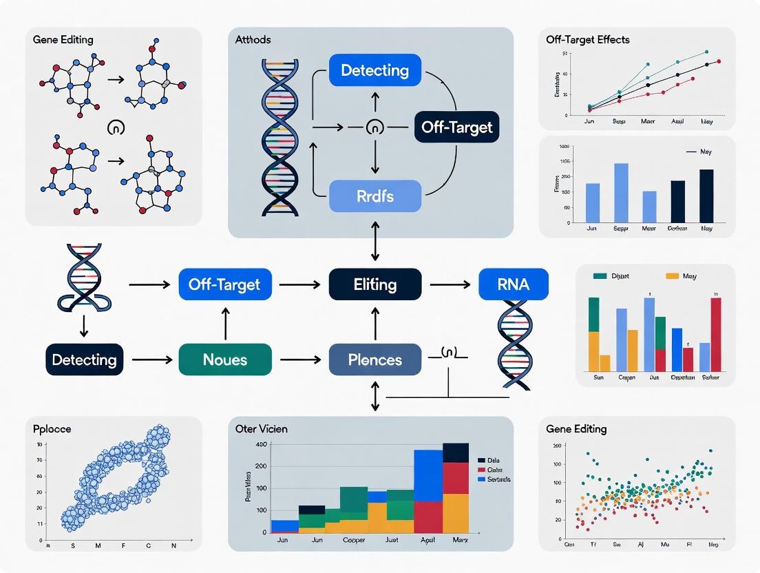
Navigating the Landscape of Gene Editing Off-Target Detection: From Foundational Concepts to Clinical Validation
As CRISPR-based gene editing rapidly advances toward clinical applications, the precise detection of off-target effects has become a critical pillar for ensuring therapeutic safety and efficacy.
Navigating the Landscape of Gene Editing Off-Target Detection: From Foundational Concepts to Clinical Validation
Abstract
As CRISPR-based gene editing rapidly advances toward clinical applications, the precise detection of off-target effects has become a critical pillar for ensuring therapeutic safety and efficacy. This article provides a comprehensive guide for researchers, scientists, and drug development professionals, detailing the entire workflow of off-target assessment. It covers foundational principles explaining why off-target effects occur, a detailed analysis of in silico, biochemical, and cellular detection methodologies, strategic troubleshooting to enhance editing precision, and essential frameworks for rigorous validation and comparative analysis to meet evolving regulatory standards. The content synthesizes the latest technological advancements and practical considerations to empower the development of safer gene therapies.
Understanding the Roots: Why Off-Target Effects Occur in Gene Editing
The CRISPR-Cas9 Mechanism and the Source of Imperfection
Troubleshooting Guides
FAQ: Addressing Common CRISPR-Cas9 Experimental Challenges
1. My knockout efficiency is low. What can I optimize? Low knockout efficiency is a common challenge often stemming from suboptimal sgRNA design, inefficient delivery of CRISPR components, or high activity of DNA repair mechanisms in your cell line [1] [2].
- Solution: Systematically optimize your experiment using the following checklist:
| Troubleshooting Strategy | Specific Actions & Reagents | Key Performance Indicators |
|---|---|---|
| sgRNA Design & Selection [1] [2] | Use multiple algorithms (e.g., Benchling, CCTop) to design 3-5 candidate sgRNAs. Prefer sgRNAs with high predicted on-target scores. Chemically synthesize sgRNAs with 2'-O-methyl-3'-thiophosphonoacetate modifications for enhanced stability. | INDEL percentage (target >80%); Verification via T7EI assay, ICE, or TIDE analysis [1]. |
| Delivery Method & Conditions [3] [1] [2] | Use ribonucleoprotein (RNP) complexes for transient delivery. For hard-to-transfect cells, use electroporation. Optimize cell-to-sgRNA ratio (e.g., 5 μg sgRNA for 8×10^5 cells). Consider stable Cas9-expressing cell lines for consistent expression. | Transfection efficiency; Cell survival rate post-nucleofection; Cas9 expression levels. |
| Validation & Screening [1] [2] | Employ robust genotyping (T7EI assay, Sanger sequencing analyzed by ICE). Perform functional validation via Western blot to confirm protein loss. Use high-throughput screening to select the most effective sgRNA-cell line pair. | INDEL percentage correlation between ICE and sequencing; Absence of target protein on Western blot. |
2. How can I minimize off-target effects in my experiments? Off-target effects occur when Cas9 cuts at unintended genomic sites with sequences similar to your target, potentially due to mismatches or DNA/RNA bulges [4] [5].
- Solution: Implement a multi-layered strategy to enhance specificity:
| Mitigation Strategy | Technical Approach | Key Mechanism of Action |
|---|---|---|
| Enhanced sgRNA Design [4] | Use truncated sgRNAs with shorter complementarity regions. Employ online tools (e.g., CCTop, Cas-OFFinder) to predict and avoid sgRNAs with high-risk off-target sites. | Reduces tolerance for mismatches between the sgRNA and DNA. |
| High-Fidelity Cas9 Variants [3] [4] | Use engineered Cas9 proteins like SpCas9-HF1 or eSpCas9. | Contains mutations that enforce stricter proofreading of the sgRNA-DNA match. |
| Computational Prediction [5] [6] | Utilize advanced prediction tools like CCLMoff, a deep learning framework that uses an RNA language model. | Accurately identifies potential off-target sites for a given sgRNA across diverse datasets, informing better sgRNA selection. |
| Alternative Nucleases [4] | Use Cas9 nickase (makes single-strand breaks) paired with two adjacent sgRNAs, or dCas9-FokI fusions that require dimerization to cut. | Increases the specificity required for a double-strand break, dramatically reducing off-target cleavage. |
3. I suspect cell toxicity from the CRISPR system. How can I address this? Cytotoxicity can result from high concentrations of CRISPR components or prolonged Cas9 activity [3] [2].
- Solution:
- Titrate Components: Start with lower concentrations of Cas9 and sgRNA and gradually increase to find a balance between editing efficiency and cell viability [3].
- Use RNP Complexes: Direct delivery of pre-formed RNP complexes leads to faster editing and clearance, reducing prolonged exposure and toxicity [1] [2].
- Inducible Systems: Use doxycycline-inducible Cas9 (iCas9) systems to control the timing and duration of Cas9 expression, minimizing stress on cells [1].
Experimental Protocols for Detecting Off-Target Effects
A core part of a robust CRISPR workflow is the empirical detection of off-target effects. Below are detailed protocols for key methodologies.
Protocol 1: Digenome-seq (In Vitro Detection) Digenome-seq is a sensitive, genome-wide method that detects Cas9 cleavage sites in purified genomic DNA digested in vitro [4].
- Workflow:
- Genomic DNA Isolation: Extract high-molecular-weight genomic DNA from your target cell type.
- In Vitro Digestion: Incubate the purified genomic DNA with Cas9 ribonucleoprotein (RNP) complexes programmed with your sgRNA of interest.
- Whole-Genome Sequencing (WGS): Subject the digested DNA (and an undigested control) to high-coverage next-generation sequencing.
- Bioinformatic Analysis: Map the sequencing reads to a reference genome and identify sites with a concentration of DNA breaks (cleavage sites) that are present only in the digested sample. These sites represent both on-target and off-target activity [4].
The following diagram illustrates the Digenome-seq workflow:
Protocol 2: BLESS (In Vivo Detection) BLESS (Direct in situ breaks labelling, streptavidin enrichment and next-generation sequencing) detects double-strand breaks (DSBs) directly in fixed cells, providing a snapshot of nuclease activity in a more native cellular context [4].
- Workflow:
- Cell Fixation: Fix cells that have been transfected with your CRISPR/Cas9 system.
- DSB Labeling In Situ: Label the unrepaired DSBs in situ within the nucleus using biotinylated linkers.
- Genomic DNA Extraction & Fragmentation: Extract the genomic DNA and fragment it.
- Enrichment of Biotinylated Fragments: Capture the biotin-labeled DNA fragments (which contain the DSBs) using streptavidin-coated magnetic beads.
- Sequencing & Analysis: Prepare a sequencing library from the enriched fragments and identify off-target sites by mapping the sequences back to the genome [4].
The following diagram illustrates the BLESS workflow:
Protocol 3: GUIDE-Seq (In Vivo Detection via Integration) GUIDE-seq (Genome-wide, unbiased identification of DSBs enabled by sequencing) utilizes the integration of a short, double-stranded oligodeoxynucleotide (dsODN) tag into DSB sites in vivo to mark them for sequencing [5].
- Workflow:
- Co-delivery of CRISPR and Tag: Co-transfect cells with your CRISPR/Cas9 components and a defined, short dsODN tag.
- Tag Integration: The dsODN tag is captured and integrated into DSB sites (both on-target and off-target) by the cell's endogenous repair machinery.
- Genomic DNA Extraction & Shearing: Extract genomic DNA and shear it.
- Enrichment of Tag-Containing Fragments: Use PCR with primers specific to the dsODN tag to enrich for fragments that contain the integrated tag.
- Sequencing & Analysis: Sequence the enriched library to identify the genomic locations where the tag integrated, revealing the spectrum of Cas9 cleavage sites [5].
The following diagram illustrates the GUIDE-seq workflow:
The Scientist's Toolkit: Research Reagent Solutions
This table details key materials and reagents essential for implementing the troubleshooting strategies and detection protocols discussed above.
| Item | Function & Application | Specific Examples / Notes |
|---|---|---|
| High-Fidelity Cas9 Variants [3] [4] | Engineered for reduced off-target cleavage while maintaining high on-target activity. | SpCas9-HF1, eSpCas9, xCas9. |
| Cas9 Nickase [4] | A mutant Cas9 that cuts only one DNA strand; requires two adjacent sgRNAs for a DSB, dramatically improving specificity. | Useful for precise editing and reducing off-target effects. |
| Chemically Modified sgRNA [1] | Enhanced stability within cells, leading to increased editing efficiency and potentially reduced variability. | sgRNAs with 2'-O-methyl-3'-thiophosphonoacetate modifications at the 5' and 3' ends. |
| dsODN Tag [5] | A short, double-stranded oligodeoxynucleotide used as a tag for DSB sites in the GUIDE-seq protocol. | Essential reagent for the GUIDE-seq method to mark and later identify cleavage sites. |
| Computational Prediction Tools [5] [6] | In silico platforms for predicting potential off-target sites during the sgRNA design phase. | CCLMoff (deep learning-based), Cas-OFFinder (alignment-based), CCTop (formula-based). |
| Stable Inducible Cas9 Cell Lines [1] | Cell lines with integrated, inducible Cas9 (e.g., via a Tet-On system) for controlled, uniform expression, minimizing toxicity and variability. | Doxycycline-inducible SpCas9 hPSC lines (hPSCs-iCas9). |
| Ethyl 3,4,5-Trimethoxybenzoate | Ethyl 3,4,5-Trimethoxybenzoate, CAS:6178-44-5, MF:C12H16O5, MW:240.25 g/mol | Chemical Reagent |
| Prostaglandin D2-1-glyceryl ester | Prostaglandin D2-1-glyceryl ester, MF:C23H38O7, MW:426.5 g/mol | Chemical Reagent |
Frequently Asked Questions (FAQs)
1. What are the primary molecular factors that cause CRISPR/Cas9 off-target effects? The primary factors are mismatches between the sgRNA and DNA sequence, flexibility in Protospacer Adjacent Motif (PAM) recognition, and the presence of DNA or RNA bulges. The CRISPR/Cas9 system can tolerate imperfect complementarity, leading to cleavage at unintended genomic sites that resemble the intended target [4] [7] [8]. Mismatches in the seed region (PAM-proximal 10–12 nucleotides) are particularly impactful, though mismatches in the PAM-distal region can also be tolerated [4] [7].
2. How does the position of a mismatch in the sgRNA influence its impact? The impact of a mismatch is highly position-dependent. The seed region, located closest to the PAM, is the most critical for specific recognition and cleavage [4]. Mismatches in this PAM-proximal region are less likely to be tolerated and can prevent efficient binding. In contrast, mismatches near the distal end (further from the PAM) are more likely to be tolerated and result in off-target activity [4] [7].
3. What is PAM flexibility, and how does it contribute to off-target effects? While the commonly used SpCas9 nuclease is designed to recognize a canonical "NGG" PAM sequence, it has been shown to tolerate non-canonical variants like "NAG" and "NGA" [4] [8]. This flexibility means that many more potential off-target sites exist throughout the genome where Cas9 can bind and cleave, even if the PAM sequence is not a perfect match [4]. The development of PAM-free or less restrictive systems, such as SpRY, further expands the targetable genome but may also increase the potential for off-target effects [4].
4. What are DNA/RNA bulges, and why are they problematic? DNA/RNA bulges refer to extra nucleotide insertions that arise due to imperfect complementarity between the sgRNA and the target DNA [4]. Even in the presence of such bulges, where the sequences do not perfectly align, Cas9 can still cleave the DNA at these mismatched sites, resulting in off-target effects [4].
5. What strategies can be used to minimize off-target effects driven by these factors? Several strategies have been developed to enhance specificity:
- High-Fidelity Cas9 Variants: Using engineered Cas9 proteins like SpCas9-HF1 or eSpCas9, which are designed to be less tolerant of mismatches [4] [9].
- Optimized sgRNA Design: Truncating the sgRNA length by 1-2 nucleotides can increase specificity and reduce mismatch tolerance [4] [8]. Using chemically modified synthetic sgRNAs can also improve stability and specificity [9] [10].
- Alternative CRISPR Systems: Employing Cas9 nickases (which create single-strand breaks) or dCas9-FokI fusions, which require two adjacent binding events for a double-strand break, significantly improves precision [4] [9].
- Ribonucleoprotein (RNP) Delivery: Delivering pre-assembled Cas9 protein and sgRNA complexes (RNPs) has been shown to reduce off-target effects compared to plasmid-based delivery, as it shortens the exposure time of cells to the editing machinery [10] [8].
Quantitative Data on Off-Target Factors
Table 1: Impact of Mismatch Position on Cleavage Efficiency
| Mismatch Position Relative to PAM | Tolerance Level & Impact on Cleavage |
|---|---|
| Seed Region (PAM-proximal, ~nt 1-12) | Low tolerance. Mismatches, especially in positions 1-8, are most likely to prevent efficient binding and cleavage [4] [7]. |
| PAM-distal Region (~nt 18-20) | Higher tolerance. Off-target cleavage can occur even with up to six base mismatches in this region [4] [7]. |
Table 2: PAM Sequence Specificity of Different Cas9 Variants
| Cas9 Variant | Recognized PAM Sequence | Implication for Off-Target Risk |
|---|---|---|
| SpCas9 (Standard) | NGG | Moderate risk due to tolerance of non-canonical PAMs like NAG and NGA [4] [8]. |
| SaCas9 | NNGRRT | Longer, more complex PAM reduces occurrence frequency in the genome, narrowing the target range and improving specificity [4]. |
| SpCas9-NG | NG | Less restrictive PAM than SpCas9, expanding target range but potentially increasing off-target risk [4]. |
| SpRY | NRN > NYN | Nearly PAM-free, offering great targeting flexibility but requiring careful off-target assessment [4]. |
Experimental Protocols for Detection
Protocol 1: Genome-Wide Unbiased Identification of DSBs Enabled by Sequencing (GUIDE-seq) GUIDE-seq is a cellular method that detects double-strand breaks (DSBs) directly in living cells, providing high biological relevance [11] [12].
- Transfection: Co-deliver CRISPR/Cas9 components (e.g., as plasmid or RNP) and a double-stranded oligodeoxynucleotide (dsODN) tag into cells.
- Tag Integration: The dsODN tag is incorporated into CRISPR-induced DSBs via the cellular non-homologous end joining (NHEJ) repair pathway.
- Genomic DNA Extraction: Harvest cells and isolate genomic DNA.
- Library Preparation & Sequencing: Fragment the DNA and perform PCR amplification using primers specific to the integrated dsODN tag. Prepare libraries for next-generation sequencing (NGS).
- Data Analysis: Map the sequenced tags to the reference genome to identify all sites of DSB formation, both on-target and off-target [11] [12].
Protocol 2: Circularization for In Vitro Reporting of Cleavage Effects by Sequencing (CIRCLE-seq) CIRCLE-seq is a sensitive, biochemical, cell-free method that can detect rare off-target sites by enriching for cleaved fragments [11] [12].
- DNA Purification and Shearing: Isolate genomic DNA and shear it into fragments.
- Circularization: Ligate the sheared DNA fragments into circular molecules.
- In Vitro Cleavage: Incubate the circularized DNA library with pre-assembled Cas9/sgRNA ribonucleoprotein (RNP) complexes.
- Exonuclease Digestion: Treat the reaction with an exonuclease that digests linear DNA but not circular DNA. This step enriches for linear DNA fragments generated by Cas9 cleavage.
- Library Preparation & Sequencing: Break the circles at the cleavage sites, add sequencing adapters, and perform NGS.
- Data Analysis: Identify sequencing reads with breaks that align to the reference genome, revealing potential off-target cleavage sites [11] [12].
Signaling Pathways and Workflow Visualizations
Diagram 1: Molecular pathway of on-target and off-target CRISPR/Cas9 activity, showing key decision points where mismatches, bulges, and PAM flexibility lead to errors.
Diagram 2: Decision workflow for selecting the appropriate off-target detection method based on research goals and context.
The Scientist's Toolkit: Research Reagent Solutions
Table 3: Essential Reagents and Tools for Off-Target Analysis
| Reagent / Tool | Function & Application | Key Considerations |
|---|---|---|
| High-Fidelity Cas9 Variants (e.g., SpCas9-HF1, eSpCas9) | Engineered nucleases with reduced mismatch tolerance; used to minimize off-target cleavage from the outset [4] [9]. | Balance between enhanced specificity and potential reduction in on-target efficiency. |
| Chemically Modified Synthetic sgRNA | Improved stability and reduced innate immune response; certain modifications can also reduce off-target editing [9] [10]. | Modifications like 2'-O-methyl (2'-O-Me) at terminal residues are common. Prefer over in vitro transcribed (IVT) guides for sensitive applications. |
| Ribonucleoprotein (RNP) Complexes | Pre-complexed Cas9 protein and sgRNA; shortens editing exposure time in cells, reducing off-target effects compared to plasmid delivery [10] [8]. | Ideal for "DNA-free" editing and protocols requiring high precision and low toxicity. |
| Tagmented Oligos (for GUIDE-seq) | Double-stranded oligodeoxynucleotides that are incorporated into DSBs, enabling genome-wide mapping of cleavage sites in cells [11] [12]. | Critical for cellular, unbiased detection methods. Efficiency of tag integration can affect assay sensitivity. |
| Deep Learning Prediction Tools (e.g., CCLMoff, DNABERT-Epi) | Computational models that predict potential off-target sites by analyzing sgRNA sequence and epigenetic context; used for pre-screening and guide selection [5] [13]. | Models trained on diverse datasets (in vitro and in cellula) generally have better generalization and accuracy. |
| 2-Benzoylbenzoic Acid | 2-Benzoylbenzoic Acid, CAS:85-52-9, MF:C14H10O3, MW:226.23 g/mol | Chemical Reagent |
| 1,3,7-Trihydroxy-2-methoxyxanthone | 1,3,7-Trihydroxy-2-methoxyxanthone|Research-Chemical | High-purity 1,3,7-Trihydroxy-2-methoxyxanthone for research applications. Explore its bioactivity for your studies. This product is For Research Use Only (RUO). Not for human or veterinary use. |
The Critical Role of the Seed Region in DNA Recognition
FAQs: Core Concepts of the Seed Region
Q1: What is the seed region in nucleic acid-guided systems? The seed region is a short, crucial sequence at the 5' end of a guide RNA or single-guide RNA (sgRNA). In RNA silencing, the core seed sequence typically spans nucleotides 2–7 of the guide strand, and to a lesser extent, can include nucleotide 8 [14]. This region serves as a primary anchor site for initial target recognition. In CRISPR-Cas9 systems, the seed region is also the portion of the sgRNA that exhibits the least tolerance for mismatches when binding to its DNA target, making it critical for specificity [9].
Q2: Why is the seed region so critical for specificity and off-target effects? The seed region is critical because it provides an enhanced-affinity anchor site that nucleates target recognition. Association of the guide strand with the PIWI/MID domain of an Argonaute protein in RISC can enhance the interaction affinity over the seed sequence by up to 300-fold [14]. In CRISPR systems, the Cas9 nuclease can tolerate between three and five base pair mismatches in other parts of the guide sequence, but mismatches within the seed region, particularly at position 3, are amplified and lead to significantly reduced off-target activity [14] [9]. This amplified discrimination means that proper seed binding is essential for accurate targeting.
Q3: How do protein interactions enhance seed region function? Structural studies show that the seed region of the guide strand is anchored via its phosphodiester backbone to the PIWI/MID domain of effector proteins like Argonaute or Cas9 [14] [15]. This association generates an enhanced affinity anchor site by reducing the entropy penalty for interaction, likely through immobilization or preordering of the guide strand [14]. For PAM-relaxed SpCas9 variants like SpRY, this preference is mediated by specific interactions with amino acid residues such as A61R and A1322R [15].
Q4: Are seed region rules consistent across different editing platforms? While the fundamental principle of a privileged 5' anchor region is conserved, specific characteristics vary. In RNAi/miRNA systems, the seed region (nucleotides 2-7/8) of the guide RNA is paramount for target recognition [14]. In CRISPR-Cas9, the seed region's importance is maintained even in PAM-relaxed variants like SpG and SpRY, which exhibit a preference for the seed region to compensate for relaxed PAM recognition [15]. This demonstrates how different nucleic acid-guided systems have evolutionarily converged on the strategic use of a seed region for efficient target searching.
Troubleshooting Guide: Common Seed Region Experimental Problems
Problem: High Off-Target Editing Rates
Symptoms: Unintended mutations at sites with homology to your target sequence, particularly in regions with similar seed sequences but divergent 3' regions.
| Potential Cause | Solution | Verification Method |
|---|---|---|
| Suboptimal gRNA design with high similarity to multiple genomic sites | Redesign gRNA using in silico tools (e.g., Cas-OFFinder, CRISPOR) to select guides with unique seed sequences and high on-target/off-target scores [16] [9] [17]. | Use GUIDE-seq or CIRCLE-seq to experimentally profile off-target sites genome-wide [16] [17]. |
| Using wild-type Cas9 with high mismatch tolerance | Switch to high-fidelity Cas9 variants (e.g., HiFi Cas9, eSpCas9, SpCas9-HF1) that enforce stricter seed recognition [9] [17]. | Compare editing profiles of wild-type and high-fidelity Cas9 using targeted sequencing of predicted off-target sites. |
| Prolonged expression of editing components | Use Cas9 ribonucleoprotein (RNP) complexes for transient delivery rather than plasmid-based expression to limit activity duration [9]. | Measure indel frequencies over time; RNP delivery typically shows reduced off-targets compared to plasmid transfection. |
| High GC content outside seed compromising specificity | Design gRNAs with optimal (40-60%) GC content overall, ensuring perfect complementarity in the seed region [9]. | Test multiple candidate gRNAs with varying GC content in a reporter assay to assess specificity. |
Problem: Low On-Target Editing Efficiency
Symptoms: Poor gene modification rates at the intended target site despite apparently good gRNA design.
| Potential Cause | Solution | Verification Method |
|---|---|---|
| Chromatin inaccessibility at target site | Select target sites in open chromatin regions using ATAC-seq or DNase-seq data; consider using chromatin-modulating compounds [16]. | Perform Cas9 ChIP-seq to verify binding accessibility; use FACS or Western blot to quantify editing efficiency. |
| Suboptimal PAM or seed sequence context | For CRISPR, choose different PAM sites; ensure no secondary structure in seed region; use algorithms that predict on-target efficiency [15] [3]. | Test multiple gRNAs targeting the same gene; use T7E1 assay or Sanger sequencing to quantify editing efficiency. |
| Ineffective delivery of editing components | Optimize delivery method (electroporation, lipofection, viral vectors) for your specific cell type; validate RNP complex formation [3]. | Use immunofluorescence to detect Cas9 nuclear localization; measure guide RNA expression levels by qRT-PCR. |
Problem: Inconsistent Editing Outcomes Across Cell Types
Symptoms: The same gRNA produces different editing efficiencies and specificities in different biological models.
| Potential Cause | Solution | Verification Method |
|---|---|---|
| Cell-type specific chromatin organization | Validate gRNA performance in your specific experimental model rather than relying solely on predictions from other cell types [16] [17]. | Perform ATAC-seq in your specific cell type to identify accessible regions; use Western blot to confirm Cas9 expression. |
| Variable DNA repair machinery activity | Consider cell cycle synchronization; use different Cas9 formats (nickase, base editors) that leverage alternative repair pathways [17]. | Assess repair outcomes by sequencing; measure cell cycle distribution by FACS. |
| Differential expression of key pathway components | Use consistent, controlled delivery methods (RNP preferred); select cell models with robust DNA repair capabilities [3]. | Perform RNA-seq to characterize DNA repair pathway expression; use isogenic cell lines for comparisons. |
Table 1: Seed Region Binding Affinity and Discrimination Power
| Parameter | Value | Experimental System | Reference |
|---|---|---|---|
| Binding affinity enhancement | Up to 300-fold | AfPiwi-guide RNA complex [14] | PMC2642989 |
| Mismatch discrimination | Amplified at position 3 | Archaeoglobus fulgidus PIWI/MID domain [14] | PMC2642989 |
| Cas9 mismatch tolerance | 3-5 mismatches outside seed region | Streptococcus pyogenes Cas9 [9] | Synthego Blog |
| Core seed sequence length | Nucleotides 2-7 (extends to nt 8) | microRNA/RNA silencing [14] | PMC2642989 |
Table 2: Detection Methods for Seed Region-Dependent Off-Target Effects
| Method | Principle | Sensitivity | Key Consideration for Seed Analysis |
|---|---|---|---|
| GUIDE-seq | Integrates dsODNs into DSBs for sequencing | High (low false positive rate) | Identifies off-targets with seed similarity [16] [17] |
| CIRCLE-seq | Circularized genomic DNA + Cas9 RNP in vitro | Highly sensitive | Detects seed-matched off-targets without cellular context [16] [17] |
| Digenome-seq | In vitro Cas9 digestion of purified genomic DNA | Highly sensitive | Requires high sequencing coverage; identifies seed-mediated cleavage [16] |
| CHANGE-seq | In vitro method with sequencing adapter integration | Highly sensitive | Unbiased detection of seed-dependent off-targets [17] |
| LAM-HTGTS | Detects chromosomal translocations from DSBs | Targeted (requires bait sites) | Identifies pathogenic rearrangements from seed-mediated off-targets [16] [17] |
Experimental Protocols
Protocol 1: Assessing Seed Region-Dependent Off-Target Effects Using GUIDE-seq
Purpose: To genome-widely identify off-target editing sites, particularly those driven by seed region homology.
Reagents Needed:
- GUIDE-seq dsODN tag (commercially available)
- Cas9 nuclease and sgRNA (as RNP or expression plasmid)
- PCR and NGS library preparation reagents
- Cells of interest and appropriate culture media
- Transfection reagent (lipofectamine CRISPRMAX or electroporation system)
Procedure:
- Design and synthesize sgRNA: Ensure optimal seed region sequence with minimal off-target potential using prediction tools like Cas-OFFinder [16].
- Co-deliver editing components: Transfect cells with Cas9-sgRNA complex along with GUIDE-seq dsODN tag. For human primary T cells, use ~100,000 cells, 100pmol Cas9, 100pmol sgRNA, and 100pmol dsODN [16] [17].
- Harvest genomic DNA: Extract high molecular weight genomic DNA 72 hours post-transfection.
- Library preparation and sequencing:
- Shear gDNA to ~500bp fragments
- Prepare sequencing libraries with GUIDE-seq-specific primers
- Perform high-throughput sequencing (Illumina recommended)
- Data analysis:
- Map sequenced reads to reference genome
- Identify GUIDE-seq tag integration sites
- Filter and validate bona fide off-target sites
- Particularly note sites with seed region homology but 3' mismatches
Troubleshooting Tips:
- Low tag integration: Optimize dsODN concentration and transfection efficiency
- High background: Include proper negative controls (transfection without Cas9)
- Validation: Confirm top off-target sites by targeted amplicon sequencing
Protocol 2: In Vitro Cleavage Specificity Profiling with CIRCLE-seq
Purpose: Highly sensitive, cell-free identification of potential seed region-dependent off-target sites without cellular constraints.
Reagents Needed:
- Purified genomic DNA from target cells
- Cas9 nuclease protein and in vitro transcribed sgRNA
- CIRCLE-seq library preparation kit or components
- DNA shearing equipment (sonicator or nebulizer)
- ATP-dependent DNA degradase (plasmid-safe ATP-dependent DNase)
Procedure:
- Genomic DNA preparation:
- Extract high-quality genomic DNA
- Shear DNA to ~300bp fragments using covaris sonicator
- DNA circularization:
- End-repair and ligate sheared DNA into circular molecules using T4 DNA ligase
- Treat with plasmid-safe ATP-dependent DNase to linearize DNA
- In vitro cleavage:
- Incubate circularized DNA library with preassembled Cas9-sgRNA RNP complex
- Use titration of RNP complex (e.g., 100nM, 50nM, 10nM) to assess cleavage efficiency
- Library preparation and sequencing:
- End-repair cleaved fragments
- Add sequencing adapters and amplify with indexed primers
- Sequence on appropriate platform (Illumina recommended)
- Bioinformatic analysis:
- Map cleavage sites to reference genome
- Identify sites with seed region complementarity
- Rank off-target sites by read count and mismatch position
Technical Notes:
- This method is particularly sensitive for detecting seed-matched off-target sites that might be missed in cellular assays due to chromatin inaccessibility [16]
- Always include a no-Cas9 control to identify background cleavage
- Validation of top candidate sites in cellular models is recommended
Visualization: Seed Region Function and Off-Target Detection
Diagram 1: Seed-Centric gRNA Design and Validation Workflow - This workflow illustrates the critical steps for designing and validating gRNAs with optimal seed region properties to minimize off-target effects, incorporating both computational and experimental approaches.
Research Reagent Solutions
Table 3: Essential Reagents for Seed Region Studies
| Reagent Category | Specific Examples | Function in Seed Region Studies |
|---|---|---|
| High-Fidelity Cas9 Variants | HiFi Cas9, eSpCas9, SpCas9-HF1 [17] | Reduce seed region-dependent off-target effects while maintaining on-target activity |
| Chemical Modified gRNAs | 2'-O-methyl-3'-phosphonoacetate, bridged nucleic acids [17] | Enhance stability and specificity of seed region binding |
| Off-Target Detection Kits | GUIDE-seq, CIRCLE-seq, SITE-seq kits [16] [17] | Experimental validation of seed-mediated off-target effects |
| In Silico Prediction Tools | Cas-OFFinder, CRISPOR, FlashFry [16] [9] | Computational prediction of seed region-dependent off-target sites |
| Cell Delivery Systems | Lipofectamine CRISPRMAX, Neon Electroporation System [18] [3] | Efficient RNP delivery to minimize duration of nuclease activity and off-target editing |
This technical support center provides troubleshooting guides and FAQs to help researchers address challenges related to chromatin state and genetic variation in gene editing experiments, framed within the broader context of detecting off-target effects.
FAQs: Chromatin and Genetic Variation in Gene Editing
How does chromatin state influence CRISPR-Cas9 editing efficiency? Chromatin state significantly impacts CRISPR-Cas9 editing efficiency. Heterochromatin (transcriptionally inactive, tightly packed DNA) presents a substantial barrier to Cas9 access and cutting, leading to reduced editing efficiency compared to euchromatin (open, active DNA) [19] [20]. The local chromatin environment at the cut site also influences the DNA repair pathway balance, with heterochromatic regions more frequently repaired via error-prone microhomology-mediated end joining (MMEJ) [19].
What specific chromatin modifications are linked to variable editing outcomes? Enhancer-associated histone modifications, such as H3K27ac and H3K4me1, show the highest degree of variability across individuals [21]. This natural variation can lead to differences in how accessible a genomic region is to gene editing tools. The repressive mark H3K27me3 is also highly variable, particularly in "poised" or bivalent regulatory elements [21].
How can genetic variation between individuals lead to unexpected editing results? Genetic variations, like single nucleotide polymorphisms (SNPs), can create or disrupt potential off-target sites by altering the DNA sequence [16] [22]. A SNP at your intended target site might reduce on-target efficiency by creating a mismatch with your guide RNA (gRNA). Conversely, a SNP elsewhere in the genome might create a novel, unintended sequence that perfectly matches your gRNA, leading to an off-target cut [22].
What practical steps can I take to improve editing in refractory chromatin regions? Pretreating cells with specific chromatin-modifying drugs, such as histone deacetylase (HDAC) inhibitors, can loosen chromatin compaction and improve Cas9 access [19]. The effectiveness of these drugs is highly dependent on the local chromatin context. For example, HDAC inhibitor PCI-24781 improved editing efficiency across all heterochromatin types, while apicidin was only effective in euchromatin and H3K27me3-marked regions [19].
Troubleshooting Guides
Problem: Low Editing Efficiency in Heterochromatin
Potential Cause: The target site is located within tightly packed, transcriptionally inactive heterochromatin, physically blocking Cas9 binding [19] [20].
Solutions:
- Epigenetic Pretreatment: Treat cells with chromatin-modifying drugs prior to editing.
- Protocol: Dose cells with an HDAC inhibitor (e.g., PCI-24781 at 10 µM, 1 µM, or 100 nM) 24 hours before transfection with CRISPR components. Re-dose 24 hours post-transfection and culture for an additional 48-72 hours before analysis [19].
- Validation: Use qPCR or a CUT&RUN assay on treated vs. untreated samples to confirm an increase in active chromatin marks (e.g., H3K27ac) at the target locus [23].
- gRNA Redesign: If possible, redesign gRNAs to target exons within the same gene that are located in more accessible euchromatic regions. Use epigenome browsers (like Ensembl or WashU Epigenome Browser) to identify regions with high H3K27ac or low H3K27me3 signals [24] [21].
Problem: High Off-Target Editing Due to Genetic Variation
Potential Cause: Common genetic variations in your cell line or population (e.g., SNPs) create novel, unintended off-target sites with high complementarity to your gRNA [16] [22].
Solutions:
- Use Population-Aware In Silico Prediction:
- Protocol: When designing your gRNA, use prediction tools like Cas-OFFinder that allow you to input a custom genome sequence or a panel of genomes representing the genetic diversity of your model system. This helps identify off-target sites specific to your experimental context [16].
- Follow-up: Empirically test all high-scoring potential off-target sites, especially those created by SNPs, using amplicon sequencing.
- Employ High-Fidelity Cas9 Variants: Use engineered Cas9 variants like eSpCas9 or SpCas9-HF1, which have reduced tolerance for gRNA-DNA mismatches, to minimize cleavage at these SNP-generated off-target sites [16] [22].
- Validate with an Empirical Method: Use highly sensitive, unbiased detection methods like GUIDE-seq or CIRCLE-seq.
Problem: Inconsistent Editing Outcomes Across Cell Lines or Donors
Potential Cause: Underlying genetic and epigenetic variation between the cell lines or individual donors leads to differences in chromatin accessibility and gRNA binding [21] [25].
Solutions:
- Characterize Cellular Context:
- Adapt gRNA Design: Avoid designing gRNAs where the seed sequence (PAM-proximal 10-12 bases) overlaps with a known common SNP [22].
- Consider Epigenome Editing: If the goal is gene regulation, consider using a dCas9-epigenetic effector system (e.g., dCas9-p300 for activation, dCas9-KRAB for repression) instead of cutting. The outcomes of these systems can also be context-dependent but offer an alternative perturbation method [23].
Experimental Protocols & Data
Protocol: Testing the Impact of an HDAC Inhibitor on Editing
This protocol systematically evaluates how a chromatin-modifying drug affects Cas9 editing efficiency and repair outcomes in different chromatin contexts [19].
Protocol: Unbiased Off-Target Detection with GUIDE-seq
This protocol provides a robust method for empirically identifying off-target sites in your specific cellular context [16] [22].
Quantitative Data on Chromatin and Editing
Table 1: Variability of Chromatin Features Across Individuals [21] This table summarizes the extent of natural variation found in different chromatin marks and features in human lymphoblastoid cell lines, which can predispose certain genomic regions to variable editing outcomes.
| Chromatin Feature | Relative Variability (vs. Gene Expression) | Functional Correlation |
|---|---|---|
| Enhancer Marks (H3K27ac, H3K4me1) | Highest | Individual-specific active/repressed states; enriched for motif-disrupting SNPs. |
| Promoter Marks (H3K4me3) | High | More variable at enhancers than at core promoters. |
| Repressive Marks (H3K27me3) | High | Most variable in "poised" or bivalent states. |
| Gene Body Marks (H3K36me3) | Low | Relatively stable across individuals. |
| Gene Expression | Lowest (Baseline) | Remains stable despite enhancer variability; changes only when >60% of a gene's enhancers vary. |
Table 2: Chromatin-Dependent Effects of Selected Epigenetic Drugs on Cas9 Editing [19] This table provides examples of drugs that modulate editing efficiency in a manner that depends on the local chromatin environment.
| Drug Example | Target | Impact on Cas9 Editing Efficiency | Chromatin Context Specificity |
|---|---|---|---|
| PCI-24781 | HDAC inhibitor | Improves efficiency | Effective across all types of heterochromatin. |
| Apicidin | HDAC inhibitor | Improves efficiency | Only effective in euchromatin and H3K27me3-marked regions. |
| NU7441 | DNA-PKcs inhibitor (NHEJ inhibitor) | Alters repair outcome (MMEJ:NHEJ ratio) | Used as a positive control for NHEJ inhibition. |
| Mirin | MRE11 inhibitor (MMEJ inhibitor) | Alters repair outcome (MMEJ:NHEJ ratio) | Used as a positive control for MMEJ inhibition. |
The Scientist's Toolkit
Table 3: Key Research Reagents for Investigating Context-Dependent Effects
| Reagent / Tool | Function | Example Use Case |
|---|---|---|
| HDAC Inhibitors (e.g., PCI-24781) | Loosen chromatin compaction by increasing histone acetylation. | Improving Cas9 access and cutting efficiency in heterochromatic regions [19]. |
| High-Fidelity Cas9 (e.g., eSpCas9) | Engineered Cas9 variant with reduced tolerance for gRNA-DNA mismatches. | Minimizing off-target effects at sites with high sequence similarity, including those created by SNPs [16] [22]. |
| dsODN Tag (for GUIDE-seq) | Short, double-stranded DNA molecule that integrates into DSBs. | Experimental, genome-wide identification of off-target cleavage sites in living cells [16] [22]. |
| Chromatin-Modifying Effectors (e.g., dCas9-p300) | Fusions of catalytically dead Cas9 with epigenetic writer domains. | Systematically studying the causal role of specific chromatin marks (e.g., H3K27ac) on transcription and editing [23]. |
| In Silico Prediction Tools (e.g., Cas-OFFinder) | Algorithmic nomination of potential off-target sites based on sequence similarity. | Initial, sgRNA-dependent assessment of off-target risk, which can be customized for user-provided genomes or genetic variants [16]. |
| 3,4-Dihydroxy-2-methoxyxanthone | 3,4-Dihydroxy-2-methoxyxanthone, MF:C14H10O5, MW:258.23 g/mol | Chemical Reagent |
| Emixustat Hydrochloride | Emixustat Hydrochloride | Emixustat hydrochloride is a potent RPE65 inhibitor for researching retinal diseases like Stargardt disease and geographic atrophy. For Research Use Only. Not for human consumption. |
Troubleshooting Guides & FAQs
What are CRISPR off-target effects and why are they a primary concern in therapeutic development?
A: CRISPR off-target effects refer to unintended edits at locations in the genome that are genetically similar to the intended target site [9]. These are a major concern because they can confound research results and, in a clinical setting, pose critical patient safety risks [8] [9]. Unintended mutations can disrupt essential genes, including tumor suppressor genes or oncogenes, potentially leading to genomic instability or carcinogenesis [8] [26]. Regulatory agencies like the FDA and EMA require comprehensive off-target characterization for therapies moving into clinical trials [8] [26].
What types of unintended genetic alterations beyond small indels should I be worried about?
A: Beyond small insertions or deletions (indels), CRISPR editing can lead to larger, more complex structural variations (SVs) [26]. These include:
- Kilobase- to megabase-scale deletions at the on-target site [26].
- Chromosomal translocations between different chromosomes [26].
- Chromosomal losses or truncations [26].
- Chromothripsis, a catastrophic event where chromosomes are shattered and rearranged [26]. These SVs are a pressing safety concern as they can delete critical regulatory elements or genes with profound consequences [26].
My editing efficiency is high, but my phenotypic results are inconsistent. Could off-target effects be the cause?
A: Yes. In functional genomics studies, off-target editing can make it difficult to determine if an observed phenotype is the result of the intended on-target edit or due to unintended mutations at other genomic loci [9]. It is crucial to use carefully designed gRNAs with low predicted off-target activity and to verify the genotype of your cell lines through comprehensive sequencing.
Do high-fidelity Cas9 variants completely eliminate the risk of structural variations?
A: No. While high-fidelity Cas9 variants (e.g., HiFi Cas9) or paired nickase strategies are excellent for reducing off-target cleavage activity, they can still introduce substantial on-target aberrations, including structural variations [26]. Therefore, using these improved nucleases does not eliminate the need for thorough genomic integrity screening.
How do strategies to enhance HDR, like using DNA-PKcs inhibitors, impact genomic integrity?
A: Strategies that inhibit key components of the NHEJ pathway, such as the DNA-PKcs inhibitor AZD7648, to enhance HDR can have unintended consequences. Recent studies show these inhibitors can aggravate genomic aberrations, leading to a significant increase in large deletions and chromosomal translocations [26]. This can also lead to an overestimation of HDR efficiency in standard assays, as large deletions may remove primer binding sites used in short-read sequencing, making the aberrant events "invisible" [26].
Experimental Protocols for Off-Target Detection
A thorough off-target assessment strategy often combines in silico prediction with empirical methods.
Protocol 1: Genome-Wide Off-Target Detection Using GUIDE-seq
GUIDE-seq (Genome-wide Unbiased Identification of DSBs Enabled by sequencing) is a sensitive, cell-based method for identifying off-target sites in vivo [27].
Detailed Methodology:
- Oligonucleotide Tag Integration: Co-deliver your CRISPR-Cas9 components (e.g., Cas9 mRNA and sgRNA) along with a short, double-stranded oligonucleotide tag (dsODN) into the target cells.
- Tag Capture at DSBs: When a double-strand break (DSB) occurs—whether on-target or off-target—the cellular repair machinery integrates the dsODN tag into the break site.
- Genomic DNA Preparation and Enrichment: Harvest cells 2-3 days post-transfection. Extract genomic DNA and shear it by sonication. Enrich for tag-integrated fragments using PCR.
- Library Preparation and Sequencing: Prepare a next-generation sequencing (NGS) library from the enriched fragments.
- Data Analysis: Map the sequencing reads back to the reference genome. Off-target sites are identified as genomic locations where the dsODN tag has been integrated, indicating a DSB occurred at that site [27].
The workflow for this protocol is summarized in the diagram below:
Protocol 2: In Vitro Off-Target Detection Using CIRCLE-seq
CIRCLE-seq (Circularization for In Vitro Reporting of Cleavage Effects by Sequencing) is a highly sensitive in vitro method that can detect potential off-target sites without the constraints of cellular context [8] [27].
Detailed Methodology:
- Genomic DNA Isolation and Shearing: Extract high-molecular-weight genomic DNA from your cell type of interest and shear it to a desired fragment size.
- DNA Circularization: Ligate the sheared genomic DNA into circular molecules.
- In Vitro Cleavage: Incubate the circularized DNA library with the Cas9-gRNA ribonucleoprotein (RNP) complex. The Cas9 nuclease will introduce DSBs at sites complementary to the gRNA.
- Linear Fragment Enrichment: Treat the reaction with an exonuclease to degrade all non-cleaved, circular DNA. The linear fragments resulting from Cas9 cleavage are protected and enriched.
- Library Preparation and Sequencing: Prepare an NGS library from the enriched linear fragments and sequence them.
- Bioinformatic Analysis: Map the sequencing reads to the reference genome to identify sequences that have been cleaved by Cas9, revealing a comprehensive profile of potential off-target sites [8] [27].
The workflow for this protocol is summarized in the diagram below:
Comparison of Off-Target Detection Methods
The choice of detection method depends on your experimental needs, balancing sensitivity, throughput, and biological context. The table below summarizes key characteristics of major techniques.
| Method | Principle | Key Advantage | Key Limitation | Best For |
|---|---|---|---|---|
| GUIDE-seq [27] | Integration of a dsODN tag into DSBs in vivo. | Unbiased, genome-wide profiling in a cellular context. | Requires efficient delivery of the dsODN into cells. | Identifying biologically relevant off-target sites in cell cultures. |
| CIRCLE-seq [8] [27] | In vitro cleavage of circularized genomic DNA by Cas9 RNP. | Extremely high sensitivity; not limited by cell viability or delivery. | Purely in vitro; may detect sites not accessible in cells. | Comprehensive, ultra-sensitive screening of gRNA specificity before cellular experiments. |
| Digenome-seq [27] | In vitro cleavage of purified genomic DNA followed by whole-genome sequencing. | Unbiased, genome-wide; no cloning required. | Lower sensitivity compared to CIRCLE-seq; in vitro context. | Genome-wide off-target identification. |
| DISCOVER-seq [27] | Relies on the recruitment of DNA repair factors (e.g., MRE11) to DSBs. | Identifies off-targets in vivo; applicable to any organism. | Relies on the endogenous repair machinery. | Detecting off-target edits in vivo, including in animal models. |
| Whole Genome Sequencing (WGS) [8] | Direct sequencing of the entire genome before and after editing. | Most comprehensive method; can detect all mutation types, including SVs. | Expensive; may miss low-frequency events due to sequencing depth. | Gold-standard safety assessment for clinical therapies; detecting large SVs. |
The Scientist's Toolkit: Key Research Reagents & Solutions
| Item | Function & Application | Key Consideration |
|---|---|---|
| High-Fidelity Cas9 Variants (e.g., HiFi Cas9) [8] [26] | Engineered Cas9 proteins with reduced mismatch tolerance, lowering off-target cleavage. | Often trade reduced off-target activity for slightly lower on-target efficiency. |
| Chemically Modified sgRNAs [9] | sgRNAs with 2'-O-methyl and/or phosphorothioate modifications to increase stability and reduce off-target effects. | Modifications can improve gRNA performance by enhancing nuclease resistance and editing specificity. |
| Cas9 Nickase (nCas9) [26] | A Cas9 variant that creates single-strand breaks instead of DSBs. Used in pairs to mimic a DSB, drastically reducing off-target activity. | Requires careful design of two adjacent gRNAs. Off-target nicking can still occur. |
| DNA-PKcs Inhibitors (e.g., AZD7648) [26] | Small molecules that inhibit the NHEJ pathway to promote HDR. | Can exacerbate large structural variations and chromosomal translocations; use with caution. |
| CAST-Seq Assay [26] | A method specifically designed to identify and quantify chromosomal rearrangements (translocations, large deletions) resulting from CRISPR editing. | Critical for a comprehensive genotoxicity assessment beyond small indels. |
| Bioinformatics Tools (e.g., CRISPOR, GuideScan) [8] [9] | Computational software for designing sgRNAs and predicting potential off-target sites in silico before experiments. | Essential first step for gRNA selection; predictions should be validated empirically. |
| 42-(2-Tetrazolyl)rapamycin | 42-(2-Tetrazolyl)rapamycin, CAS:221877-56-1, MF:C52H79N5O12, MW:966.2 g/mol | Chemical Reagent |
| 1-Cinnamoyl-3-hydroxypyrrolidine | 1-Cinnamoyl-3-hydroxypyrrolidine | Research-grade 1-Cinnamoyl-3-hydroxypyrrolidine for antifungal studies. This product is For Research Use Only (RUO). Not for human consumption. |
Mechanisms and Consequences of Off-Target Effects
Understanding how off-target effects occur and their potential downstream impacts is crucial for risk assessment. The following diagram illustrates the key pathways from CRISPR delivery to functional consequences.
The Detection Toolkit: In Silico, Biochemical, and Cellular Methods for Off-Target Identification
In CRISPR/Cas9 gene editing, off-target effects occur when the system acts on untargeted genomic sites, creating cleavages that can lead to unintended and potentially adverse outcomes [16]. These effects are a significant concern, particularly in clinical applications, as they can confound experimental results and pose critical safety risks to patients if mutations arise in critical genes, such as oncogenes [9].
In silico prediction tools are essential for nominating potential off-target sites during the guide RNA (gRNA) design phase. They are typically open-source online software that provides a convenient and efficient first pass for assessing off-target risk based primarily on sequence homology [16]. This guide focuses on three types of predictors: the versatile algorithm Cas-OFFinder, the user-friendly CCTop, and state-of-the-art deep learning models such as CCLMoff.
## Frequently Asked Questions (FAQs)
1. What are the key differences between traditional tools (like Cas-OFFinder and CCTop) and newer deep learning models (like CCLMoff) for off-target prediction?
Traditional tools largely rely on sequence alignment and predefined rules about mismatch tolerance. In contrast, deep learning models can automatically learn complex sequence features and patterns from large, comprehensive datasets, often leading to superior performance and generalization [5] [28].
The table below summarizes the core characteristics of each tool type:
| Tool Type | Examples | Underlying Principle | Key Advantages |
|---|---|---|---|
| Alignment-Based | Cas-OFFinder [16], CasOT | Exhaustive genome-wide scanning for sites with sequence similarity to the gRNA [16]. | Highly versatile; allows custom adjustment of PAM sequences, mismatch numbers, and bulges [16] [29]. |
| Scoring-Based | CCTop [29], MIT Score, CFD Score | Assigns weights/penalties based on mismatch position (e.g., proximity to PAM) and type to generate an off-target score [16] [29]. | Provides an intuitive user interface and ranks potential off-target sites, facilitating gRNA selection [29]. |
| Deep Learning-Based | CCLMoff [5], CRISPR-Net, Crispr-SGRU | Uses deep neural networks to automatically extract relevant features from gRNA and target site sequences [5] [28]. | Superior generalization to unseen gRNA sequences; captures complex, non-linear sequence relationships [5] [6]. |
| Di-O-demethylcurcumin | Di-O-demethylcurcumin, CAS:60831-46-1, MF:C19H16O6, MW:340.3 g/mol | Chemical Reagent | Bench Chemicals |
| Azido-PEG1-methyl ester | Azido-PEG1-methyl ester, CAS:1835759-80-2, MF:C6H11N3O3, MW:173.17 g/mol | Chemical Reagent | Bench Chemicals |
2. How do I choose the right prediction tool for my experiment?
The choice depends on your specific needs for accuracy, speed, and user support. The following table provides a comparative overview to aid in selection.
| Tool Name | Type | Key Features | Best For |
|---|---|---|---|
| Cas-OFFinder [30] [29] | Alignment-Based | No limit on mismatches; customizable PAM; fast searching [29]. | Researchers needing a flexible, first-pass scan of potential off-targets across a whole genome. |
| CCTop [29] | Scoring-Based | Intuitive interface; ranks off-targets by score; provides full output documentation [29]. | Beginners and experts seeking a user-friendly tool with clear candidate ranking for various editing applications. |
| CCLMoff [5] [6] | Deep Learning | Incorporates a pre-trained RNA language model; trained on 13 genome-wide detection datasets; high accuracy. | Projects requiring the highest prediction accuracy and robust performance on novel gRNA sequences, especially for therapeutic development. |
3. A deep learning model predicted a high-risk off-target site, but my validation experiment (e.g., GUIDE-seq) did not detect editing there. Why might this happen?
Discrepancies between in silico predictions and experimental results are common and can arise from several factors:
- Cellular Context: Deep learning models like CCLMoff are primarily trained on sequence data. They may not fully account for intracellular factors that influence Cas9 binding and cleavage, such as chromatin accessibility, epigenetic modifications (e.g., DNA methylation, histone marks), and nuclear localization [16] [5]. A site that is accessible in silico might be buried in heterochromatin in a specific cell type.
- Training Data Bias: A model's performance is tied to the data it was trained on. If the model was not trained on data from a cell type or experimental condition similar to yours, its predictions may be less accurate [28].
- Sensitivity of Experimental Methods: Even highly sensitive methods like GUIDE-seq have limitations in detection efficiency and can be influenced by factors like transfection efficiency [16]. Very low-frequency off-target events might fall below the detection limit.
4. What should I do if different in silico tools give me conflicting off-target predictions?
Lack of consensus among predictors is a known challenge, as demonstrated in a study on Mucopolysaccharidosis type I where different tools identified vastly different numbers of off-target sites with low agreement [31]. To address this:
- Use a Consensus Approach: Rely on sites that are nominated by multiple, diverse tools (e.g., one alignment-based and one deep learning-based) [31].
- Prioritize by Score: Within each tool, pay closest attention to the top-ranked off-target sites with the highest predicted activity or lowest number of mismatches, particularly in the seed region near the PAM site [16] [31].
- Validate Experimentally: Treat all in silico predictions as a guide for targeted off-target validation. Use methods like GUIDE-seq or Digenome-seq to experimentally confirm or refute the computationally predicted sites in your specific experimental system [16] [9].
## Troubleshooting Common Problems
Problem: The off-target site validated in my experiment was not predicted by any in silico tool.
- Potential Cause 1: Unaccounted for Bulges. Early prediction tools and some scoring models only consider mismatches and may not account for DNA or RNA bulges (insertions or deletions), which Cas9 can tolerate [5].
- Solution: Use tools that specifically include bulge prediction in their algorithm. When setting up your search, enable options for "bulges" or "indels." Cas-OFFinder, for instance, allows for the specification of bulge numbers [29].
- Potential Cause 2: Non-Canonical PAM Sequences. The tool may be restricted to the standard NGG PAM for SpCas9, while the nuclease might be interacting with a non-canonical PAM (e.g., NAG or NGA) [31].
- Solution: If using a tool that allows it (like Cas-OFFinder), expand the PAM search parameters to include other common non-canonical sequences [29].
- Potential Cause 3: Cell-Type Specific Genetic Variations. The reference genome used by the prediction tool might not contain a sequence variant present in your specific cell line, creating a novel off-target site [31].
- Solution: If available, use a personalized or population-specific genome reference for prediction. Always check known genetic variants in your cell line at the predicted off-target loci.
Problem: My chosen gRNA has high on-target efficiency but also many high-scoring off-target predictions.
- Potential Cause: The gRNA sequence has high homology to multiple genomic loci.
- Solution:
- Re-design gRNAs: Go back to your gRNA design tool (e.g., CHOPCHOP, CRISPOR) and select an alternative gRNA with a higher specificity score (e.g., a lower CFD off-target score) [9] [29].
- Use High-Fidelity Cas9 Variants: Switch from wild-type SpCas9 to a high-fidelity engineered variant (e.g., eSpCas9, SpCas9-HF1) that has reduced tolerance for mismatches [9].
- Modify the gRNA: Truncating the gRNA length (to 17-18 nucleotides) or adding specific chemical modifications (e.g., 2'-O-methyl analogs) can reduce off-target binding without completely abolishing on-target activity [9].
- Solution:
## The Scientist's Toolkit: Key Research Reagent Solutions
The table below lists essential materials and resources used in the field of CRISPR off-target prediction and analysis.
| Item | Function/Description | Example Use Case |
|---|---|---|
| Cas-OFFinder [30] [29] | A fast, versatile algorithm for exhaustive search of potential off-target sites with customizable parameters. | Initial genome-wide screening for sequences with homology to a candidate gRNA. |
| CCTop [29] | An online predictor that identifies and ranks candidate sgRNA target sequences based on their off-target score. | Rapidly identifying high-quality target sites for gene inactivation, HDR, and NHEJ experiments. |
| CCLMoff Model [5] [6] | A deep learning framework using an RNA language model for highly accurate and generalizable off-target prediction. | Selecting optimal sgRNAs with minimal off-target risk for preclinical therapeutic development. |
| GUIDE-seq [16] [9] | An experimental method that captures DSBs in cells by integrating double-stranded oligodeoxynucleotides (dsODNs). | Unbiased, genome-wide experimental validation of predicted off-target sites in a relevant cell model. |
| CRISPOR [9] [29] | A web tool for gRNA design that ranks candidates using multiple on-target and off-target scoring algorithms. | Designing and selecting gRNAs, with comprehensive support from cloning to off-target analysis. |
| LPA1 receptor antagonist 1 | LPA1 receptor antagonist 1, MF:C28H26N4O4, MW:482.5 g/mol | Chemical Reagent |
| Drim-7-ene-11,12-diol acetonide | Drim-7-ene-11,12-diol acetonide, MF:C18H30O2, MW:278.4 g/mol | Chemical Reagent |
## Experimental Workflow for Comprehensive Off-Target Assessment
The following diagram illustrates a robust, multi-step protocol integrating in silico prediction with experimental validation, forming a core methodology for detecting off-target effects in gene editing research.
## Decision Framework for Tool Selection
Use the workflow below to select the most appropriate prediction tool based on your project's specific requirements and stage.
Frequently Asked Questions (FAQs)
1. What are the primary advantages of using in vitro biochemical methods like CIRCLE-seq over cell-based methods for off-target nomination?
In vitro biochemical methods offer several key advantages for initial, genome-wide off-target discovery. They provide ultra-high sensitivity and can identify a broader spectrum of potential off-target sites because they are not limited by cellular delivery efficiency, chromatin states, or cell fitness effects [32] [11]. By using purified genomic DNA and high concentrations of Cas9 ribonucleoprotein (RNP), these methods can reveal rare cleavage events that might be missed in cell populations [32]. This makes them excellent for comprehensive, unbiased screening during the early sgRNA selection and risk assessment phase [11].
2. A common critique is that biochemical assays may overestimate biologically relevant off-target effects. How can I validate my findings?
While biochemical methods are highly sensitive for nominating off-target sites, their findings should be interpreted as a list of potential off-targets. It is recommended to validate bona fide off-target sites using complementary, cell-based methods [11]. Techniques like GUIDE-seq or amplicon sequencing can confirm whether the nominated sites are actually cleaved and edited in a cellular context, which accounts for factors like chromatin accessibility and DNA repair [32] [16] [11]. This two-tiered approach—broad discovery with a biochemical method followed by validation in a biologically relevant system—is considered a robust strategy for off-target assessment [11].
3. I have a limited amount of genomic DNA. Which method is most suitable?
CIRCLE-seq and CHANGE-seq are highly sensitive methods that require only nanogram amounts of purified genomic DNA, making them suitable for situations where DNA is scarce [11]. In contrast, Digenome-seq typically requires microgram quantities of input DNA [11] [12].
4. What is the key technological improvement of CHANGE-seq over CIRCLE-seq?
CHANGE-seq is described as an improved version of CIRCLE-seq that incorporates a tagmentation-based library prep process [11]. This enhancement reduces bias and improves the sensitivity of the assay, allowing for the detection of even rarer off-target events while also simplifying and streamlining the workflow [11].
Comparison of Key Biochemical Off-Target Detection Methods
The following table summarizes the core characteristics of CIRCLE-seq, Digenome-seq, and CHANGE-seq to help you select the most appropriate method for your research needs.
Table 1: Summary of Biochemical Off-Target Assays
| Feature | Digenome-seq [11] [12] | CIRCLE-seq [32] [11] | CHANGE-seq [11] |
|---|---|---|---|
| General Description | Treats purified genomic DNA with nuclease, then detects cleavage sites by whole-genome sequencing. | Uses circularized genomic DNA and exonuclease digestion to enrich for nuclease-induced breaks. | Improved version of CIRCLE-seq with tagmentation-based library prep. |
| Sensitivity | Moderate; requires deep sequencing to detect off-targets. | High sensitivity; lower sequencing depth needed compared to Digenome-seq. | Very high sensitivity; can detect rare off-targets with reduced false negatives. |
| Input DNA | Micrograms of purified genomic DNA. | Nanogram amounts of purified genomic DNA. | Nanogram amounts of purified genomic DNA. |
| Key Enrichment Step | None (direct WGS of digested DNA). | Circularization of DNA → exonuclease removes linear DNA, enriching cleavage products. | DNA circularization + tagmentation → efficient capture of nuclease cuts. |
| Sequencing Depth | High (~400-500 million reads for human genome) [32] [12]. | Lower (~100-fold fewer reads than Digenome-seq) [32]. | High sensitivity with optimized sequencing. |
Experimental Workflows
The diagrams below illustrate the core procedural steps for each method, highlighting the key differences in their approaches to enriching for nuclease-cleaved DNA fragments.
CIRCLE-seq Workflow
Digenome-seq Workflow
CHANGE-seq Workflow
Research Reagent Solutions
Table 2: Essential Reagents for Biochemical Off-Target Detection Assays
| Reagent / Material | Function in the Experimental Workflow |
|---|---|
| Purified Genomic DNA | The substrate for in vitro cleavage. High-quality, high-molecular-weight DNA is essential [11] [12]. |
| Cas9 Nuclease (High Purity) | The active enzyme that creates double-strand breaks. Used as a purified protein to form the RNP complex [16] [12]. |
| In vitro Transcribed or Synthetic sgRNA | Guides the Cas9 nuclease to its target and potential off-target sequences [16]. |
| DNA Circularization Enzymes | Critical for CIRCLE-seq and CHANGE-seq. Enzymes like circligases are used to form covalently closed DNA circles [32] [11]. |
| Exonucleases | Used in CIRCLE-seq to degrade linear DNA fragments, thereby enriching for circularized DNA that was linearized by Cas9 cleavage [32] [11]. |
| Tagmentation Enzyme Mix | A key reagent for CHANGE-seq, which combines fragmentation and adapter ligation into a single step, streamlining library preparation [11]. |
| Next-Generation Sequencing Library Prep Kit | Required for preparing the enriched DNA fragments for high-throughput sequencing on platforms like Illumina [32] [11]. |
This technical support guide details the use of three key cellular methods—GUIDE-seq, DISCOVER-seq, and BLESS—for detecting off-target effects of CRISPR-Cas9 genome editing. These techniques are essential for identifying biologically relevant off-target activity in living cells or native tissue contexts, capturing the influence of chromatin structure, DNA repair pathways, and cellular environment on editing outcomes. This resource provides troubleshooting guides, FAQs, and detailed protocols to support researchers and drug development professionals in ensuring the safety and specificity of gene editing therapies [11].
The table below summarizes the core characteristics, strengths, and limitations of each method for easy comparison.
| Method | General Description & Principle | Input Material | Key Strengths | Key Limitations | Primary Detection |
|---|---|---|---|---|---|
| GUIDE-seq [11] [33] | Incorporates a double-stranded oligonucleotide tag into double-strand breaks (DSBs) in vivo, followed by enrichment and sequencing. | Cellular DNA from edited, tagged cells. [11] | High sensitivity for off-target DSB detection; reflects true cellular activity. [11] | Requires efficient delivery of oligonucleotide tag; may miss rare sites. [11] | DSBs via tag integration [11] |
| DISCOVER-seq+ [34] | Chromatin immunoprecipitation (ChIP) of the DNA repair protein MRE11 recruited to CRISPR-Cas-targeted sites. Often combined with DNA-PKcs inhibition (DISCOVER-Seq+) to boost signal. | Cellular DNA; ChIP-seq of MRE11 binding. [11] | High sensitivity in native chromatin; suitable for in vivo and primary cells; captures real nuclease activity. [11] [34] | Technically complex (ChIP protocol); requires specific antibodies. [11] | DSBs via MRE11 binding [11] [34] |
| BLESS [11] [4] | Direct in situ labeling of DSB ends in fixed cells with biotinylated linkers, followed by capture and sequencing. | Fixed/permeabilized cells or nuclei. [11] | Preserves genome architecture; captures breaks in situ. [11] | Technically complex; lower throughput; variable sensitivity. [11] | DSBs via direct end-labeling [11] [4] |
Experimental Protocols
GUIDE-seq (Genome-wide, Unbiased Identification of DSBs Enabled by Sequencing)
Detailed Methodology:
- Cell Transfection: Co-deliver into living cells the Cas9 nuclease (as plasmid, mRNA, or ribonucleoprotein complex) and sgRNA, along with the proprietary double-stranded GUIDE-seq oligonucleotide tag [11].
- Genomic DNA Extraction: Harvest cells 2-3 days post-transfection and isolate genomic DNA using standard methods.
- Library Preparation & Sequencing: Fragment the DNA and perform next-generation sequencing (NGS) library preparation. The tag-containing fragments are enriched, amplified, and sequenced [11].
- Data Analysis: Process sequencing reads through a bioinformatics pipeline (e.g., the original GUIDE-seq algorithm) to map the genomic locations of tag integration, which correspond to both on-target and off-target DSBs [11] [33].
DISCOVER-seq+ (Discovery ofin situCas Off-targets with Verification and Sequencing)
Detailed Methodology:
- Gene Editing & Inhibition: Deliver CRISPR-Cas9 components into target cells or in vivo. To enhance sensitivity (DISCOVER-Seq+), inhibit DNA-dependent protein kinase catalytic subunit (DNA-PKcs) using compounds like Ku-60648 or Nu7026 to accumulate MRE11 at break sites [34].
- Cell Fixation and Cross-linking: Fix cells to preserve protein-DNA interactions.
- Chromatin Immunoprecipitation (ChIP): Lyse cells, shear chromatin, and immunoprecipitate DNA fragments bound to MRE11 using a specific anti-MRE11 antibody [11] [34].
- Library Preparation & Sequencing: Reverse cross-links, purify the enriched DNA, and construct an NGS library for sequencing [34].
- Data Analysis: Analyze sequencing data with a dedicated pipeline (e.g., BLENDER) to identify significant peaks of MRE11 binding, which indicate Cas9 cleavage sites genome-wide [34].
BLESS (Breaks Labeling, Enrichment on Streptavidin, and Sequencing)
Detailed Methodology:
- Cell Fixation and Permeabilization: Fix cells or nuclei promptly to "freeze" DSBs in their native genomic context and permeabilize them to allow reagent access [4].
- In Situ Breaks Labeling: In fixed and permeabilized cells, label the ends of DSBs using biotinylated linkers or adapters [11] [4].
- Genomic DNA Extraction & Enrichment: Extract genomic DNA and capture the biotin-labeled fragments containing DSBs using streptavidin-coated magnetic beads [4].
- Library Preparation & Sequencing: Process the enriched DNA fragments into an NGS library and sequence [11] [4].
- Data Analysis: Map the sequenced reads back to the reference genome to identify the precise locations of DSBs [4].
Troubleshooting Guides & FAQs
GUIDE-seq
FAQ: Why is my GUIDE-seq oligonucleotide not being efficiently incorporated?
- A: Low tag incorporation is a common issue. Ensure the oligonucleotide is delivered at an optimal concentration. Using electroporation for delivery (especially in hard-to-transfect cells like primary T cells or stem cells) can significantly improve efficiency compared to lipid-based methods. Test different delivery conditions and tag concentrations.
FAQ: The experiment identified a very high number of off-target sites. Is this normal?
- A: GUIDE-seq is highly sensitive. However, an unusually high number may indicate that the sgRNA has low specificity. Verify your sgRNA design using multiple in silico prediction tools (e.g., CRISPOR) to select guides with minimal predicted off-targets. Consider testing a high-fidelity Cas9 variant to reduce off-target cleavage [9].
DISCOVER-seq
FAQ: How does DNA-PKcs inhibition improve DISCOVER-Seq+ sensitivity?
- A: Inhibiting DNA-PKcs blocks the non-homologous end joining (NHEJ) repair pathway. This causes a buildup of the MRE11 repair complex at the DSB sites, prolonging its residence time and thereby increasing the signal captured during ChIP-seq, which can discover up to fivefold more off-target sites [34].
FAQ: What is the recommended control for DISCOVER-seq experiments?
- A: Always perform a control experiment under identical conditions (including inhibitor treatment) but without the Cas9 nuclease. This identifies background MRE11 binding signals unrelated to CRISPR editing. The final set of off-target sites is determined by subtracting sites found in the "no Cas9" control from those in the experimental sample [34].
BLESS
FAQ: The signal-to-noise ratio in my BLESS experiment is low. How can I improve it?
- A: Low signal can stem from inefficient in situ labeling or degradation of DNA ends. Ensure fixation is performed quickly after sampling to preserve break ends. Optimize the permeabilization and labeling reaction times and temperatures. Using fresh reagents and including positive control samples with known DSBs can help troubleshoot this issue [4].
FAQ: Can BLESS detect transient DSBs?
- A: Yes, a key advantage of BLESS is that it provides a "snapshot" of DSBs at the moment of fixation, making it suitable for detecting transient breaks. However, its sensitivity may be limited by the efficiency of the in situ labeling reaction [11] [4].
Research Reagent Solutions
The table below lists essential materials and their functions for implementing these methods.
| Reagent / Solution | Function | Example Methods |
|---|---|---|
| DNA-PKcs Inhibitor (e.g., Ku-60648) | Boosts MRE11 residence at DSBs by blocking NHEJ, enhancing ChIP-seq signal sensitivity [34]. | DISCOVER-seq+ |
| Anti-MRE11 Antibody | Specifically binds to MRE11 protein for chromatin immunoprecipitation of Cas9-targeted sites [11] [34]. | DISCOVER-seq |
| Biotinylated Linker / Adapter | Labels DSB ends in situ for subsequent capture and enrichment [11] [4]. | BLESS |
| Double-stranded Oligonucleotide Tag | Integrates into DSBs, serving as a molecular barcode for PCR amplification and sequencing of break sites [11]. | GUIDE-seq |
| Streptavidin Magnetic Beads | Captures and enriches biotin-labeled DNA fragments containing DSBs [4]. | BLESS |
| High-Fidelity Cas9 Variant | Engineered nuclease with reduced off-target activity; a critical negative control or tool to mitigate risk [9]. | All (as control) |
Method Workflow Diagrams
GUIDE-seq Workflow
DISCOVER-seq+ Workflow
BLESS Workflow
The application of CRISPR-Cas9 and other genome editing tools has revolutionized biological research and therapeutic development. However, a significant challenge remains the occurrence of off-target effects—unintended modifications at sites other than the intended on-target location [16] [35]. These off-target events can confound experimental results and raise substantial safety concerns for clinical applications [36]. Next-Generation Sequencing (NGS) has emerged as the gold-standard method for comprehensively identifying and quantifying these unintended edits, providing the precision and sensitivity required for confident off-target assessment [37]. Two primary NGS approaches are employed: Whole Genome Sequencing (WGS) and Targeted Amplicon Sequencing. This guide details their applications, provides troubleshooting support, and outlines best practices for their implementation in gene editing research.
Core Technology Comparison: WGS vs. Targeted Amplicon Sequencing
The choice between WGS and Targeted Amplicon Sequencing is fundamental and depends on the research objective, scale, and available resources. The table below summarizes their key characteristics for off-target detection.
Table 1: Comparison of WGS and Targeted Amplicon Sequencing for Off-Target Analysis
| Feature | Whole Genome Sequencing (WGS) | Targeted Amplicon Sequencing |
|---|---|---|
| Coverage Scope | Unbiased, comprehensive profiling of the entire genome [38] [37] | Focused analysis of specific, pre-identified regions of interest [38] [37] |
| Primary Application in Off-Target Detection | Unbiased discovery and nomination of novel off-target sites ("hotspots") across the genome [37] | Targeted verification and quantification of editing efficiency at known on-target and nominated off-target sites [37] |
| Cost & Resource Requirements | Higher cost and computational complexity [38] [39] | Highly cost-effective for targeted studies [38] |
| Typical Turnaround Time | Longer (e.g., 5-7 weeks reported by service providers) [39] | Shorter (e.g., 3-4 weeks reported by service providers) [38] [39] |
| Ideal Use Case | Initial, unbiased discovery phase to find where off-target edits might occur [37] | Validation and routine monitoring phase to quantify how often editing occurs at known sites [37] |
Experimental Protocols for Off-Target Detection
A robust off-target analysis strategy often involves a two-phase approach: an initial genome-wide discovery step followed by targeted validation and quantification [37] [27].
Genome-Wide Discovery Methods for Off-Target Nomination
Before you can quantify off-target effects, you must first identify where in the genome they might occur. The following methods are used to nominate these "hotspot" sites.
Table 2: Empirical Methods for Genome-Wide Off-Target Site Discovery
| Method | Core Principle | Key Advantage | Key Consideration |
|---|---|---|---|
| GUIDE-seq [37] [27] | Integrates double-stranded oligodeoxynucleotides (dsODNs) into DNA double-strand breaks (DSBs) in cells, followed by sequencing. | Highly sensitive, cost-effective, and has a low false-positive rate [16] [37]. | Limited by transfection efficiency [16]. |
| CIRCLE-seq [37] [27] | An in vitro method that circularizes sheared genomic DNA, incubates it with Cas9/sgRNA, and sequences linearized DNA fragments. | Extremely high sensitivity; performed in a test tube without cell culture [16] [37]. | An in vitro method that may not fully reflect cellular context [16]. |
| DISCOVER-seq [37] [27] | Utilizes the DNA repair protein MRE11 to mark DSB sites for chromatin immunoprecipitation and sequencing (ChIP-seq) in vivo. | Unbiased detection in vivo; uses endogenous repair machinery [37]. | Potential for false positives [16]. |
| Digenome-seq [37] [27] | Digests purified genomic DNA with Cas9/sgRNA ribonucleoprotein (RNP) complex, followed by whole-genome sequencing. | Highly sensitive; does not require a reference genome for initial detection [16]. | Expensive and requires high sequencing coverage [16]. |
The workflow for these discovery methods often follows a logical progression from sample preparation to data analysis, as illustrated below.
Targeted Validation and Quantification Using Amplicon Sequencing
Once potential off-target sites are nominated, Targeted Amplicon Sequencing is the preferred method for sensitive and economical quantification of editing events. This process involves a series of critical steps, from initial primer design to final data interpretation.
Successful off-target analysis requires a suite of specialized reagents and tools. The following table details key components.
Table 3: Research Reagent Solutions for CRISPR Off-Target Analysis
| Reagent / Tool | Function | Example / Note |
|---|---|---|
| CRISPR-Cas9 System | Creates targeted double-strand breaks in the genome. | Alt-R S.p. Cas9 Nuclease V3 is an engineered variant known for high on-target activity [37]. |
| High-Fidelity Cas9 | A engineered nuclease variant designed to minimize off-target cleavage while maintaining strong on-target activity. | Alt-R HiFi Cas9 is optimized for reduced off-target effects [37]. |
| Targeted Amplicon Seq Kit | An end-to-end solution for designing primers, preparing libraries, and analyzing sequencing data for on- and off-target sites. | The rhAmpSeq CRISPR Analysis System uses multiplexed PCR for efficient target enrichment [37]. |
| In Silico Prediction Tools | Computational software to nominate potential off-target sites based on sgRNA sequence similarity. | Cas-OFFinder, CCTop, and IDT's own design checker are commonly used [16] [27]. |
Troubleshooting Guides and FAQs
Frequently Asked Questions (FAQs)
Q1: Is it absolutely necessary to perform an off-target effect analysis for our edited cells? Yes, it is highly recommended. Unexpected off-target cleavages can lead to false-positive or false-negative results in your downstream analysis and are a critical safety consideration, especially for therapeutic development [39].
Q2: What is the best stage in my experiment to perform off-target analysis? The ideal strategy is complementary: use prediction tools or discovery methods (like GUIDE-seq) before editing to inform your experimental design, and use verification methods (like targeted amplicon sequencing) after editing to confirm the results [39] [37].
Q3: My research uses Cas12a (Cpf1). Are these NGS methods still applicable? Yes. While some discovery methods like GUIDE-seq were developed for Cas9, others like DISCOVER-Seq have been applied to Cas12a. However, you may need to empirically evaluate which discovery technique works best for your specific Cas enzyme [37].
Q4: What type of samples can I submit for off-target analysis? Both cells and purified genomic DNA are generally acceptable. The required amount depends on the specific assay, so you should confirm with your service provider or protocol beforehand [39].
Troubleshooting Common NGS Preparation Issues
Problems during library preparation can compromise your entire off-target sequencing experiment. Below are common issues and their solutions.
Table 4: Troubleshooting Common NGS Library Preparation Problems
| Problem Symptom | Potential Root Cause | Corrective Action |
|---|---|---|
| Low Library Yield | Poor input DNA quality (degraded or contaminated with salts, phenol, etc.) [40]. | Re-purify the input sample using clean columns/beads. Use fluorometric quantification (e.g., Qubit) instead of UV absorbance for accuracy [40]. |
| High Adapter Dimer Peaks | Suboptimal adapter ligation conditions; overly aggressive purification; incorrect adapter-to-insert molar ratio [40]. | Titrate the adapter-to-insert ratio. Ensure fresh ligase and buffer are used. Optimize bead cleanup parameters to retain target fragments [40]. |
| Uneven Coverage Across Targets | Poor primer design for multiplex PCR; suboptimal PCR conditions; primer interference in the pool [38] [41]. | Use vendor-validated primer pools. Consider technologies like anchored multiplex PCR to reduce primer interference [41]. |
| Sequencing Instrument: Chip Not Detected | Chip not properly seated; clamp not closed; damaged chip [42]. | Open the clamp, remove and re-seat the chip, ensuring it is properly positioned. Inspect for physical damage and replace if necessary [42]. |
| Sequencing Instrument: Low Key Signal | Problem with library or template preparation; control particles not added [42]. | Verify the quantity and quality of the library and template. Confirm that required control particles were added during preparation [42]. |
For researchers, scientists, and drug development professionals working in gene editing, accurately detecting off-target effects is crucial for assessing the safety and reliability of CRISPR-based therapies. The FDA now recommends using multiple methods, including genome-wide analysis, to measure off-target editing events [11]. This guide provides a structured framework for selecting the appropriate off-target detection assay based on your specific experimental goals, system complexity, and research context.
FAQ: Addressing Common Experimental Challenges
1. My initial in silico prediction identified very few off-target sites. Should I proceed to experimental validation?
While in silico tools (e.g., Cas-OFFinder, CRISPOR) are fast and inexpensive for guide RNA design and prediction, they have a significant limitation: they rely solely on sequence similarity and PAM rules [11]. They do not account for biological context, such as chromatin structure, DNA accessibility, or cellular repair mechanisms [11]. Therefore, a low number of in silico predictions does not guarantee a lack of off-target activity. Proceeding to unbiased, genome-wide experimental methods (biochemical or cellular) is often necessary, especially for pre-clinical therapeutic development, to capture a more comprehensive profile of off-target effects [11].
2. My biochemical assay (e.g., CIRCLE-seq) detected many potential off-target sites, but my cellular validation found very few. Why this discrepancy?
This is a common and expected outcome due to the fundamental difference between these approaches. Biochemical assays are performed on purified, naked DNA, which lacks the protective and regulatory structure of chromatin found in living cells [11]. This allows the nuclease to access and cleave sites that would be physically blocked or less accessible in a cellular environment. Consequently, biochemical assays are ultra-sensitive but may overestimate biologically relevant cleavage [11]. The results from cellular assays (e.g., GUIDE-seq, DISCOVER-seq) are generally considered more physiologically relevant as they occur in a native cellular context [11].
3. I am not detecting any cleavage bands in my genomic cleavage detection assay. What could be wrong?
Several technical issues could be at play. Consult the troubleshooting table below for common problems and solutions [43].
Table: Troubleshooting Guide for Cleavage Detection Assays
| Problem | Possible Cause | Recommendation |
|---|---|---|
| No cleavage band visible | Low transfection efficiency | Optimize transfection protocol for your cell line [43]. |
| Nuclease cannot access target site | Design a new gRNA targeting a different, more accessible nearby sequence [43]. | |
| Overall genomic modification too low | Use antibiotic selection or FACS to enrich for successfully transfected cells [43]. | |
| Smear in DNA bands | Lysate is too concentrated | Dilute the lysate 2- to 4-fold and repeat the PCR reaction [43]. |
| No PCR product | Poor PCR primer design or GC-rich region | Redesign primers to be 18–22 bp with 45–60% GC content. For GC-rich regions, add a GC enhancer [43]. |
| Nonspecific cleavage bands | Too much detection enzyme or over-digestion | Reduce the amount of enzyme or incubation time. Redesign PCR primers for a clearer banding pattern [43]. |
4. For a novel therapeutic development program, what assay strategy is recommended to meet regulatory standards?
The evolving regulatory landscape, as seen in the FDA's review of the first CRISPR-based therapy, emphasizes comprehensive off-target assessment. A robust strategy should include:
- Use of Unbiased Methods: Move beyond purely in silico or biased methods to include genome-wide assays during pre-clinical development [11].
- Use of Biologically Relevant Cells: Conduct studies in cells that are physiologically similar to the intended target cells (e.g., hematopoietic stem cells for blood disorders) [11].
- A Tiered Approach: Start with a highly sensitive biochemical assay (e.g., CHANGE-seq) for broad discovery, then use cellular assays (e.g., GUIDE-seq) in relevant cell types to validate which sites are actually edited in a biological system [11].
Comparative Analysis of Off-Target Detection Assays
Selecting the right method depends on your research goal, whether it's initial gRNA design, broad discovery of potential sites, or validation of biologically relevant edits.
Table: Comparison of Major Off-Target Analysis Approaches
| Approach | Example Assays | Input Material | Strengths | Limitations | Best For |
|---|---|---|---|---|---|
| In silico | Cas-OFFinder, CRISPOR, MIT CRISPR tool | Genome sequence & computational models | Fast, inexpensive; useful for initial gRNA design [11]. | Predictions only; lacks biological context (chromatin, repair) [11]. | Initial gRNA screening and risk prediction [11]. |
| Biochemical | CIRCLE-seq, CHANGE-seq, DIGENOME-seq | Purified genomic DNA | Ultra-sensitive, comprehensive; works with any DNA source; standardized [11]. | Uses naked DNA, may overestimate cleavage; lacks cellular context [11]. | Broad, unbiased discovery of all possible cleavage sites [11]. |
| Cellular | GUIDE-seq, DISCOVER-seq, UDiTaS | Living cells (edited) | Captures effects of native chromatin & repair; identifies biologically relevant edits [11]. | Requires efficient delivery; less sensitive than biochemical methods; may miss rare sites [11]. | Validating the biological relevance of off-target sites found in biochemical assays [11]. |
| In situ | BLISS, BLESS | Fixed cells or nuclei | Preserves genome architecture; captures breaks in their native location [11]. | Technically complex; lower throughput; variable sensitivity [11]. | Studying off-target effects in the context of 3D nuclear organization [11]. |
Detailed Experimental Protocols
Biochemical Assay: CIRCLE-seq
CIRCLE-seq (Circularization for In vitro Reporting of Cleavage Effects by Sequencing) is a highly sensitive method for comprehensive off-target discovery [11].
Workflow Diagram: CIRCLE-seq Protocol
Key Considerations:
- Input: Requires only nanogram amounts of purified genomic DNA [11].
- Sensitivity: Can detect very rare off-target events due to the circularization and exonuclease enrichment steps [11].
- Context: Remember that results represent potential, not necessarily biologically relevant, cleavage sites.
Cellular Assay: GUIDE-seq
GUIDE-seq (Genome-wide, Unbiased Identification of DSBs Enabled by sequencing) uses a short, double-stranded oligonucleotide tag that is incorporated into double-strand breaks (DSBs) in living cells, providing a genome-wide map of nuclease activity in a cellular context [11].
Workflow Diagram: GUIDE-seq Protocol
Key Considerations:
- Delivery: Requires efficient co-delivery of the Cas9/gRNA ribonucleoprotein (RNP) and the GUIDE-seq oligonucleotide into the cell nucleus [11].
- Advantage: Directly captures the cellular repair outcome and is considered one of the most sensitive cellular methods for detecting off-target DSBs [11].
- Throughput: Lower throughput than biochemical methods due to the requirement of cell culture and transfection.
The Scientist's Toolkit: Essential Research Reagent Solutions
The following table details key reagents and their functions for setting up critical off-target detection experiments.
Table: Essential Reagents for Off-Target Effect Research
| Reagent / Material | Function / Application | Key Considerations |
|---|---|---|
| Purified Genomic DNA | Input material for biochemical assays (e.g., CIRCLE-seq, CHANGE-seq) [11]. | DNA quality and purity are critical for assay sensitivity and reducing background noise. |
| Cas9 Nuclease (WT) | The core enzyme for creating double-strand breaks at target (and off-target) sites [11]. | Using high-quality, recombinant Cas9 protein for RNP formation can improve editing efficiency and reduce off-target effects compared to plasmid delivery. |
| Synthetic sgRNA | Guides the Cas9 nuclease to specific genomic loci [11]. | Careful design is critical. Avoid homology with other genomic regions to minimize off-target risks. Use modified sgRNAs (e.g., with chemical modifications) to enhance stability and specificity [43]. |
| GUIDE-seq Oligo | A short, double-stranded DNA oligonucleotide that tags double-strand breaks in living cells for the GUIDE-seq assay [11]. | Must be designed to be efficiently captured during cellular DNA repair. Optimal concentration is key to avoid cellular toxicity while ensuring high tagging efficiency. |
| NGS Library Prep Kit | Prepares the final DNA fragments from cleavage assays for high-throughput sequencing [11]. | Choose kits compatible with your specific assay's output (e.g., tagmentation-based kits for CHANGE-seq). Consider throughput, workflow (manual vs. automated), and cost. |
| Transfection Reagent | Delivers CRISPR components (RNP, plasmid) and/or detection oligonucleotides into cells [43]. | Optimization is essential. Efficiency varies by cell line. Use specialized reagents (e.g., Lipofectamine 3000) for best results, especially in hard-to-transfect cells [43]. |
| 3-(Hydroxymethyl)cyclopentanol | 3-(Hydroxymethyl)cyclopentanol, MF:C6H12O2, MW:116.16 g/mol | Chemical Reagent |
| 5-Carboxymethylaminomethyluridine | 5-Carboxymethylaminomethyluridine|CAS 69181-26-6 | Research-grade 5-carboxymethylaminomethyluridine (cmnm⁵U), a key tRNA wobble modification. For Research Use Only. Not for human, veterinary, or household use. |
Selecting the optimal assay for detecting CRISPR off-target effects is not a one-size-fits-all process. It requires a clear understanding of the trade-offs between sensitivity and biological relevance. A strategic, multi-faceted approach—often starting with sensitive in vitro biochemical assays for broad discovery and following up with physiologically relevant cellular assays for validation—provides the most robust safety profile for research and therapeutic applications. As the field moves towards standardization, aligning your assay choice with your experimental goals and system complexity is paramount for generating reliable, actionable data.
Strategies for Enhanced Precision: Minimizing and Managing Off-Target Risks
In CRISPR-Cas9 gene editing, the single-guide RNA (sgRNA) is responsible for directing the Cas9 nuclease to a specific DNA target sequence. A significant challenge in this process is the occurrence of off-target effects, where the Cas9 complex cleaves unintended genomic sites with sequences similar to the intended target [8]. These off-target mutations can compromise experimental results and pose serious safety risks in therapeutic applications, including potential genomic instability and oncogenesis [8].
This technical support article focuses on two key strategies for enhancing sgRNA specificity: using truncated gRNAs (tru-gRNAs) and implementing enhanced specificity motifs such as extended gRNAs (x-gRNAs). Within the broader context of off-target effect detection in gene editing research, optimizing sgRNA design represents the first and most crucial step in ensuring precise genomic modifications [44]. The following sections provide detailed troubleshooting guides, experimental protocols, and frequently asked questions to assist researchers in implementing these specificity-enhancing strategies.
sgRNA Specificity Enhancement Strategies
Truncated gRNAs (tru-gRNAs)
Truncated gRNAs involve shortening the guide sequence from the conventional 20 nucleotides to 17-18 nucleotides at the 5' end [8] [44]. This reduction in length decreases the stability of interactions between the sgRNA and DNA, making the system less tolerant to mismatches and thus improving specificity [44].
Mechanism of Action: Shortening the sgRNA spacer reduces the number of base-pairing interactions with off-target sites. Since off-target binding typically involves sequences with mismatches, the decreased binding stability makes it less likely for Cas9 to cleave at these imperfect matches while maintaining activity at the perfectly matched on-target site [44].
Extended gRNAs with Specificity-Enhancing Motifs
Extended gRNAs (x-gRNAs) represent an alternative approach that involves adding short nucleotide extensions (typically 6-16 nucleotides) to the 5' end of the sgRNA spacer [44]. A specialized category called hairpin-gRNAs (hp-gRNAs) contains extensions designed to form secondary structures that interfere with off-target interactions while preserving on-target activity [44].
Mechanism of Action: The 5' extensions, particularly those forming secondary structures, are believed to create steric hindrance or alter the binding dynamics of the Cas9-sgRNA complex in a way that disproportionately affects off-target sites where binding is already less stable due to mismatches [44]. Research has demonstrated that properly designed x-gRNAs can increase specificity by up to 200-fold compared to standard gRNAs [44].
Table 1: Comparison of sgRNA Optimization Strategies
| Strategy | Mechanism | Specificity Improvement | Key Considerations |
|---|---|---|---|
| Truncated gRNAs (tru-gRNAs) | Reduced sgRNA-DNA interaction stability decreases mismatch tolerance | Significant reduction in off-target activity [44] | May reduce on-target efficiency in some cases [44] |
| Extended gRNAs (x-gRNAs) | 5' extensions create steric hindrance at off-target sites | Up to 200-fold improvement with optimal designs [44] | Requires screening to identify effective extension sequences |
| Hairpin-gRNAs (hp-gRNAs) | Structured extensions preferentially disrupt off-target binding | 50-fold average improvement across targets [44] | Structure prediction needed for optimal design |
Experimental Protocols
In Vitro Cleavage Assay for Validating sgRNA Efficiency and Specificity
This protocol allows researchers to quantitatively assess both on-target efficiency and off-target specificity of designed sgRNAs before moving to cellular experiments [45].
Materials and Reagents:
- Purified Cas9 nuclease
- In vitro transcribed or synthetic sgRNAs
- DNA templates containing both on-target and potential off-target sequences (can be PCR-amplified genomic DNA or synthetic dsDNA fragments) [45]
- Reaction buffer (typically provided with Cas9)
- Agarose gel electrophoresis equipment
Procedure:
- DNA Template Preparation: PCR-amplify an 1800 bp sequence of your target gene, ensuring the cut site is positioned asymmetrically within the fragment to enable clear visualization of cleavage products [45]. Alternatively, use synthetic double-stranded DNA fragments (e.g., gBlocks) replicating both native and potential off-target sequences [45].
RNP Complex Formation: Combine sgRNA with Cas9 enzyme to form ribonucleoprotein (RNP) complexes. Use a molecular ratio calculation to ensure equal molar amounts of different sgRNA lengths are used in comparative assays [45].
Cleavage Reaction: Add DNA templates to the RNP complexes and incubate at 37°C for 1-2 hours in a thermal cycler [45].
Product Analysis: Run the resulting cleavage products on a 2% agarose gel. Include controls such as DNA template alone (no Cas9) and a well-characterized sgRNA known to have high cleavage efficiency [45].
Efficiency Quantification: Analyze gel bands to determine cleavage efficiency by comparing the intensity of cleaved versus uncleaved fragments [45].
Figure 1: Workflow for In Vitro Cleavage Assay to Validate sgRNA Designs
SECRETS Protocol for Screening Optimal x-gRNAs
The Selection of Extended CRISPR RNAs with Enhanced Targeting and Specificity (SECRETS) protocol provides a high-throughput method for identifying x-gRNAs that maintain robust on-target activity while minimizing off-target effects [44].
Materials and Reagents:
- E. coli strain compatible with three-plasmid system
- High-copy plasmid with arabinose-inducible ccdB toxin and target sequence
- Medium-copy plasmid with aTc-inducible Cas9 and chloramphenicol resistance
- Low-copy plasmid with kanamycin resistance and off-target sequence
- x-gRNA library with randomized 5' extensions
- Antibiotics: chloramphenicol, kanamycin
- Inducers: anhydrotetracycline (aTc), arabinose
Procedure:
- Plasmid Preparation: Transform E. coli with the three-plasmid system [44].
Selection Phase: Induce Cas9 and x-gRNA expression with aTc for 1 hour, then plate on LB agar containing aTc, arabinose, chloramphenicol, and kanamycin [44].
Overnight Growth: Incubate plates overnight at 37°C [44].
Colony Analysis: Sequence surviving colonies to identify x-gRNA sequences that enable survival through efficient on-target cleavage (eliminating ccdB toxin plasmid) while avoiding off-target cleavage (preserving kanamycin resistance plasmid) [44].
Validation: Test identified x-gRNA candidates using in vitro cleavage assays to confirm specificity profiles [44].
Figure 2: SECRETS Protocol for High-Throughput x-gRNA Screening
Troubleshooting Guides and FAQs
Frequently Asked Questions
Q1: How do I decide between using truncated versus extended gRNAs for my specific application?
A: The choice depends on your experimental constraints and goals. Tru-gRNAs are simpler to design and implement, making them suitable for initial specificity improvements [44]. x-gRNAs typically offer greater specificity enhancements (up to 200-fold) but require more extensive screening to identify optimal sequences [44]. For therapeutic applications where maximum specificity is critical, investing in x-gRNA screening is recommended. For general laboratory use where moderate specificity improvements are sufficient, tru-gRNAs may be adequate.
Q2: What is the optimal length for truncated gRNAs?
A: Research indicates that reducing sgRNA length to 17-18 nucleotides provides the best balance between maintained on-target activity and reduced off-target effects [44]. Shorter truncations (below 17 nt) may significantly compromise on-target efficiency, while longer truncations (19 nt) may not provide sufficient specificity enhancement [44].
Q3: Can these sgRNA optimization strategies be combined with high-fidelity Cas9 variants?
A: Yes, optimized sgRNAs can be used in combination with high-fidelity Cas9 variants like eCas9 for additive or synergistic improvements in specificity [44]. Research has demonstrated that properly designed x-gRNAs can outperform eCas9 with standard sgRNAs in terms of specificity [44].
Q4: How many potential off-target sites should I test when validating sgRNA specificity?
A: It is recommended to test multiple off-target sites with varying degrees of similarity to the target sequence. Studies typically evaluate 2-4 off-target sites containing 2-4 nucleotide mismatches to comprehensively assess specificity [45] [44]. Computational prediction tools can help identify the most likely off-target sites for experimental validation.
Troubleshooting Common Issues
Problem: Low On-Target Efficiency with Optimized sgRNAs
- Potential Cause: Over-optimization for specificity at the expense of efficiency.
- Solution: Test a range of sgRNA lengths or extension sequences to identify designs that balance both criteria. For x-gRNAs, screen larger libraries to identify variants that maintain robust on-target activity [44].
Problem: Inconsistent Specificity Improvements
- Potential Cause: Sequence-dependent effects that vary across different genomic targets.
- Solution: Always validate specificity improvements for each new target sequence. What works for one genomic locus may not translate directly to others [44].
Problem: Difficulty Identifying Functional x-gRNAs
- Potential Cause: Insufficient library diversity or suboptimal selection pressure in screening protocols.
- Solution: Implement the SECRETS protocol with appropriate controls and ensure adequate library size (tens to hundreds of thousands of variants) [44].
Table 2: Experimental Performance Metrics for sgRNA Optimization Strategies
| sgRNA Type | Spacer Length | On-Target Efficiency | Off-Target Reduction | Key Findings |
|---|---|---|---|---|
| Standard sgRNA | 20 nt | Baseline | Baseline (reference) | Conventional design [45] |
| Truncated gRNA | 17-18 nt | Variable (may decrease) | Significant reduction | Improves specificity by destabilizing off-target binding [44] |
| Extended gRNA | 20+5-16 nt | Maintains ~70-100% of standard | Up to 200-fold improvement | 5' extensions enhance specificity [44] |
| Hairpin-gRNA | 20+structured extension | Maintains high efficiency | 50-fold average improvement | Structured extensions most effective [44] |
Table 3: Mass of sgRNA Required for In Vitro Cleavage Assays
| sgRNA Length (bp) | Mass RNA (ng) | Calculation Basis |
|---|---|---|
| 19 | 47.5 | 0.4 ng per bp [45] |
| 20 | 50.0 | 0.4 ng per bp [45] |
| 30 | 75.0 | 0.4 ng per bp [45] |
| 40 | 100.0 | 0.4 ng per bp [45] |
| 53 | 132.5 | 0.4 ng per bp [45] |
Research Reagent Solutions
Table 4: Essential Reagents for sgRNA Optimization Experiments
| Reagent/Category | Specific Examples | Function/Application |
|---|---|---|
| sgRNA Formats | Synthetic sgRNA, In vitro transcribed (IVT) sgRNA, Plasmid-expressed sgRNA | Direct Cas9 to target DNA sequences [46] |
| Specificity-Enhanced Cas9 Variants | eCas9, High-fidelity Cas9 | Engineered for reduced off-target activity [8] |
| Detection Methods | GUIDE-seq, Digenome-seq, SITE-seq, CIRCLE-seq | Identify and quantify off-target effects [47] |
| Delivery Methods | Ribonucleoprotein (RNP) complexes, Plasmid vectors, Viral vectors | Introduce CRISPR components into cells [10] |
| Design Tools | ZiFiT Targeter, CAS-OFFinder, CHOPCHOP, Synthego tool | Predict sgRNA efficiency and potential off-target sites [45] [46] |
Frequently Asked Questions (FAQs)
Q1: What are high-fidelity Cas9 variants and why are they important? High-fidelity Cas9 variants are engineered forms of the standard SpCas9 nuclease designed to drastically reduce off-target editing while maintaining robust on-target activity. They are crucial for applications where specificity is critical, such as in functional genomics studies, the development of cell lines, and preclinical therapeutic development, where off-target edits could confound experimental results or pose significant safety risks [48] [49].
Q2: What are the key mechanistic differences between eSpCas9(1.1) and SpCas9-HF1? Although both aim to increase specificity, they achieve this through different structural mechanisms:
- eSpCas9(1.1): Contains mutations (K848A/K1003A/R1060A) that reduce the protein's affinity for the non-target DNA strand. This promotes the reinvasion of this strand into the RNA-DNA hybrid helix, thereby destabilizing mismatched complexes and increasing the proofreading capability of the nuclease [48] [50].
- SpCas9-HF1: Contains mutations (N497A/R661A/Q695A/Q926A) that weaken interactions between the Cas9 protein and the target DNA strand's phosphate backbone. This makes the energy threshold for DNA cleavage more stringent, so the complex is less tolerant of imperfect guide RNA matches [48] [50].
Q3: I'm getting low on-target editing efficiency with high-fidelity Cas9 variants. What could be the cause? Low on-target activity is a common trade-off with high-fidelity variants. The most common cause is the use of suboptimal guide RNA (gRNA) formats. These enzymes perform best with perfectly matching 20-nucleotide spacers. Modifications often made to comply with the U6 promoter's requirement for a 5' guanine (G)—such as using a 21-nt guide, truncating a short guide, or altering the first nucleotide to a G—can significantly diminish their activity. Notably, adding a matching 5' G extension is more detrimental than adding a mismatched one [48].
Q4: Do high-fidelity variants also reduce off-target binding, or just off-target cleavage? It is critical to understand that most high-fidelity Cas9 variants are engineered to reduce off-target cleavage, but not necessarily off-target binding [48] [9]. If you are using a catalytically dead Cas9 (dCas9) fused to an effector domain (e.g., for transcriptional activation or repression), these high-fidelity mutations may not reduce off-target effects, as the dCas9 can still bind to imperfectly matched sites [49].
Q5: When should I use a high-fidelity variant over wild-type SpCas9? You should prioritize high-fidelity variants in the following scenarios:
- For clinical or therapeutic development: Where patient safety is paramount and off-target edits could be dangerous [9] [49].
- When generating clonal cell lines: Where a single off-target edit in your chosen clone could confound all downstream experiments [49].
- When your gRNA design has high sequence similarity to other genomic loci, increasing off-target risk [49].
- When using sensitive detection methods reveals significant off-target activity with wild-type SpCas9.
Q6: Besides using a high-fidelity nuclease, what other strategies can minimize off-target effects? A multi-faceted approach is most effective:
- Optimize gRNA Design: Use design tools (e.g., CRISPOR) to select guides with minimal off-target potential. Use chemically synthesized, modified gRNAs with features like 2'-O-methyl analogs to improve stability and specificity [9] [10].
- Use a Cas9 Nickase Pair: Employ two gRNAs with Cas9 nickase (Cas9n) to create single-strand breaks on opposite strands. A DSB only occurs when both nickases bind in close proximity, dramatically increasing specificity [50].
- Utilize Ribonucleoprotein (RNP) Delivery: Delivering pre-complexed Cas9 protein and gRNA as an RNP leads to a short, sharp burst of activity, reducing off-target effects compared to plasmid-based delivery [10].
- Employ High-Fidelity Cas12a: For targets in AT-rich genomes, consider Cas12a (Cpf1), which has different off-target profiles and can be a good alternative [10].
Troubleshooting Guides
Problem: Low On-Target Editing Efficiency
Potential Causes and Solutions:
- Cause 1: Non-optimal guide RNA format.
- Solution: Redesign your gRNA to be a 20-nucleotide spacer that natively begins with a 'G' to be compatible with the U6 promoter. Avoid 5' extensions or truncations. If this is not possible, consider using alternative promoters (e.g., T7, U1) that do not require a 5' G [48].
- Cause 2: The high-fidelity variant has intrinsically lower activity for your specific target.
- Cause 3: Low expression or delivery efficiency.
Problem: Suspected Persistent Off-Target Effects
Potential Causes and Solutions:
- Cause 1: The chosen high-fidelity variant does not effectively eliminate cleavage at a particular off-target site.
- Solution: Different high-fidelity variants can have distinct off-target profiles. A site resistant to one variant may be effectively suppressed by another. If off-targets are suspected, profile your editing experiment with an unbiased detection method (see below) and consider switching to a different high-fidelity nuclease [48].
- Cause 2: Off-target effects are from dCas9 binding, not cleavage.
- Solution: If using dCas9 fusions (for activation, repression, or base editing), the high-fidelity mutations from eSpCas9 or SpCas9-HF1 will not prevent off-target binding. For these applications, focus on optimal gRNA design with minimal off-target binding potential and consider using a dual-guide system if possible [9] [49].
Experimental Protocol: Validating Specificity with GUIDE-seq
For comprehensive, unbiased off-target detection, GUIDE-seq (Genome-wide, Unbiased Identification of DSBs Enabled by Sequencing) is a highly sensitive, cell-based method [16] [11].
Detailed Methodology:
Co-delivery: Transfect your cells with the following components:
- Plasmid encoding your high-fidelity Cas9 variant or Cas9-gRNA RNP complex.
- Plasmid expressing your target-specific sgRNA.
- dsODN Tag: A short, double-stranded oligonucleotide tag (e.g., 34-36 bp) that is phosphorylated and protected from exonuclease activity [11].
Tag Integration: When a double-strand break (DSB) occurs—either on-target or off-target—the cell's repair machinery incorporates the dsODN tag into the break site via the NHEJ pathway.
Genomic DNA Extraction: Harvest cells 2-4 days post-transfection and extract genomic DNA.
Library Preparation & Sequencing:
- Fragment the genomic DNA.
- Perform PCR amplification using one primer specific to the integrated dsODN tag and a second primer that binds to an adaptor ligated to the genomic fragments. This enriches for sequences that contain the tag.
- Prepare sequencing libraries from the amplified products for high-throughput sequencing.
Bioinformatic Analysis:
- Map the sequenced reads to the reference genome.
- Identify genomic locations where the dsODN tag has been integrated. These sites represent putative Cas9 cleavage events.
- Filter and analyze these sites to generate a genome-wide list of on-target and off-target sites.
Quantitative Comparison of High-Fidelity Cas9 Variants
The table below summarizes key characteristics of several high-fidelity SpCas9 variants.
| Variant Name | Key Mutations | Proposed Mechanism | Key Strengths | Reported Limitations |
|---|---|---|---|---|
| eSpCas9(1.1) | K848A, K1003A, R1060A | Reduces non-target strand binding, promoting DNA re-annealing to reject mismatches [48] [50] | High specificity for many targets; less sensitive to 5' mismatches than SpCas9-HF1 [48] | Performance is target-dependent; sensitive to 5' G extensions on sgRNA [48] |
| SpCas9-HF1 | N497A, R661A, Q695A, Q926A | Weakers Cas9-DNA phosphate backbone interactions, increasing energetic threshold for cleavage [48] [50] | Extremely high fidelity; near complete elimination of off-targets with multiple mismatches [48] | Can have significant reduction in on-target efficiency for some guides; highly sensitive to sgRNA 5' modifications [48] |
| HypaCas9 | N692A, M694A, H698A | Enhances proofreading by stabilizing the pre-cleavage state, improving mismatch discrimination [49] [50] | High fidelity with robust on-target activity maintained [50] | Specificity and activity can be guide-dependent. |
| evoCas9 | M495V, Y515N, K526E, R661Q | Laboratory evolution to generate a more stringent "active" conformation [49] [50] | Very low off-target activity, even for challenging guides. | May have lower on-target activity than wild-type SpCas9, requiring validation. |
| Sniper-Cas9 | F539S, M763I, K890N | Identified through directed evolution; mechanism involves improved selectivity [50] | High on-target activity with reduced off-target effects; works well with truncated gRNAs [50] | Trade-off between on-target efficiency and fidelity may vary per target. |
The Scientist's Toolkit: Essential Reagents for High-Fidelity Editing
| Reagent / Tool | Function | Key Considerations |
|---|---|---|
| Chemically Modified sgRNA | Synthetic guide RNA with modifications (e.g., 2'-O-methyl, 3' phosphorothioate) to increase nuclease stability and editing efficiency while reducing immune responses [9] [10]. | Superior to in vitro transcribed (IVT) or plasmid-derived gRNAs for performance and specificity in sensitive applications. |
| Ribonucleoprotein (RNP) Complex | Pre-complexed Cas9 protein and sgRNA. Provides immediate activity upon delivery, reduces off-target effects by shortening exposure time, and enables "DNA-free" editing [10]. | The preferred delivery cargo for therapeutic development and difficult-to-transfect cells. |
| Cas9 Nickase (Cas9n) | A Cas9 variant (e.g., D10A mutation) that cuts only one DNA strand. Used in pairs with two sgRNAs to create a DSB, dramatically increasing specificity [49] [50]. | Requires two proximal binding events for a DSB, making it much less likely to cause off-target mutations. |
| Unbiased Off-Target Detection Assay (e.g., GUIDE-seq) | A genome-wide method to empirically identify all nuclease-induced DSBs in a cellular context, providing a true measure of editing specificity [16] [11]. | Critical for preclinical safety assessment. More reliable than in silico prediction alone. |
| Bioinformatics Design Tools (e.g., CRISPOR) | Software to design sgRNAs with high on-target efficiency scores and predict potential off-target sites based on sequence similarity [16] [49]. | An essential first step for selecting the best possible gRNA before any experiments begin. |
Workflow Diagram for Nuclease Selection and Validation
The following diagram outlines a logical workflow for selecting and validating the appropriate high-fidelity nuclease for a gene editing experiment.
FAQ: Cas12a and Base Editor Systems
Q1: What are the primary advantages of using Cas12a over Cas9 for genome editing? Cas12a offers several key advantages: it requires only a single CRISPR RNA (crRNA) for guidance, unlike the two-component guide system of Cas9 [51]. It creates staggered-ended double-strand breaks (DSBs) with a 5' overhang, which can be more favorable for certain repair pathways [52]. Furthermore, Cas12a recognizes a T-rich protospacer adjacent motif (PAM), significantly expanding the range of targetable genomic sites compared to the G-rich PAM of SpCas9 [22].
Q2: How do base editors fundamentally reduce the risks associated with standard CRISPR nucleases? Base editors operate through chemical modification of DNA bases without creating a DSB. They use a catalytically impaired Cas nuclease (such as a nickase) fused to a deaminase enzyme. This system directly converts one base pair into another (e.g., C•G to T•A or A•T to G•C) without relying on the error-prone non-homologous end joining (NHEJ) repair pathway. By avoiding DSBs, base editors significantly reduce the risk of introducing small insertions or deletions (indels) and large chromosomal rearrangements that can occur with Cas9 or Cas12a nucleases [52] [9].
Q3: Our lab is observing unexpected nicking in our Cas12a experiments. What could be the cause? Unexpected nicking is a recognized characteristic of Cas12a. High-throughput studies have revealed that Cas12a orthologs (such as FnCas12a, LbCas12a, and AsCas12a) exhibit pervasive sequence-specific nicking activity on dsDNA substrates containing up to four mismatches with the guide RNA, where full linearization does not always occur [51]. Furthermore, upon activation by binding to its target DNA, Cas12a can display robust non-specific trans-nicking activity against dsDNA. To troubleshoot, verify the specificity of your crRNA and consider trying a different Cas12a ortholog, as nicking activity depends on the ortholog, crRNA sequence, and the type and position of mismatches [51].
Q4: What are the critical controls for a base editing experiment to confirm on-target efficiency and rule out off-target effects? A comprehensive base editing experiment should include:
- Untreated Negative Control: Cells that do not receive the base editor to establish the baseline genotype and sequencing error rate.
- Delivery Control: Cells transfected with only the delivery vehicle to account for effects of the transfection process.
- dCas9-Deaminase Control: A construct containing a catalytically dead Cas9 (dCas9) fused to the deaminase. This controls for any background noise caused by the editor binding without catalyzing base conversion and helps identify guide-independent off-target effects [52].
- Targeted Deep Sequencing: This is the gold standard for validating both on-target efficiency and nominated off-target sites. It provides quantitative data on editing frequency [52].
Troubleshooting Common Experimental Challenges
Problem: Low On-Target Editing Efficiency with Cas12a
| Potential Cause | Solution |
|---|---|
| Inefficient crRNA | Redesign crRNA, ensuring it is specific and has minimal self-complementarity. Use design tools that are specifically validated for Cas12a. |
| Suboptimal PAM recognition | Confirm that your target site is adjacent to a correct PAM sequence for your specific Cas12a ortholog (e.g., TTTV for AsCas12a and LbCas12a). |
| Low expression of Cas12a or crRNA | Use a high-activity promoter to drive expression. Validate protein and RNA expression levels via western blot or qPCR. |
| Inefficient delivery | Optimize delivery method (e.g., electroporation for RNPs, viral transduction) for your specific cell type. |
Problem: High Off-Target RNA Editing by DNA Base Editors
| Potential Cause | Solution |
|---|---|
| Deaminase activity on single-stranded RNA | This is a known issue with some base editor architectures. The deaminase domain can exhibit promiscuous activity on cellular RNA [52]. |
| Prolonged expression of base editor | Use transient delivery methods (e.g., RNP or mRNA) instead of plasmid DNA to limit the window of editor activity. |
| High editor concentration | Titrate the amount of base editor delivered to find the lowest dose that achieves sufficient on-target editing. |
| Solution | Consider using next-generation base editors engineered with mutated deaminase domains that have reduced RNA off-target activity [52]. |
Problem: Unwanted Indels at the Target Site with Base Editors
| Potential Cause | Solution |
|---|---|
| Nickase-induced NHEJ | The single-strand break (nick) introduced by the base editor nickase can be repaired via NHEJ, leading to indels. |
| Ung-mediated excision | Repair of the edited base by cellular uracil DNA glycosylase (UNG) can lead to error-prone repair and indels. |
| Solution | Utilize "high-fidelity" base editor designs that incorporate an engineered UNG inhibitor (e.g., UGI) to suppress this pathway and reduce indel formation [52]. |
Quantitative Comparison of Nuclease Systems
The table below summarizes key characteristics of different gene-editing systems.
Table 1: Comparison of CRISPR-Based Gene Editing Systems
| Feature | Cas9 Nuclease | Cas12a Nuclease | DNA Base Editors | Prime Editors |
|---|---|---|---|---|
| DNA Break Type | Blunt-ended DSB | Staggered-ended DSB | Typically, single-strand nick or no break | Single-strand nick |
| Primary Repair Pathway | NHEJ (indels) / HDR (precise) | NHEJ (indels) / HDR (precise) | Base excision repair | DNA synthesis & repair |
| DSB Risk | High | High | Very Low | None |
| Primary Edit Type | Knockout (indels) | Knockout (indels) | Point mutations (C>T, A>G) | All 12 possible base substitutions, small insertions/deletions |
| Guide RNA | sgRNA (tracrRNA + crRNA) | crRNA only | sgRNA or crRNA | Prime Editing Guide RNA (pegRNA) |
| PAM (Example, SpCas9) | NGG | TTTV (for As/LbCas12a) | NGG (for SpCas9-derived) | NGG (for SpCas9-derived) |
| Potential Off-target | DSBs at off-target sites | DSBs & pervasive nicking [51] | Off-target point mutations; RNA editing [52] | Off-target point mutations |
Experimental Protocol: Off-Target Assessment with CIRCLE-Seq
CIRCLE-seq is a highly sensitive in vitro method for identifying potential nuclease off-target sites genome-wide [16] [52].
Workflow:
Detailed Steps:
- Genomic DNA Isolation: Extract high-molecular-weight genomic DNA from the cell type of interest (e.g., using a blood or cell culture DNA kit).
- DNA Fragmentation: Shear the genomic DNA using sonication or enzymatic digestion to generate fragments of 0.5-1 kb.
- Circularization: Purify the sheared DNA and perform intramolecular ligation in a large volume to favor the formation of circular DNA molecules. Use an exonuclease to degrade any remaining linear DNA.
- In Vitro Cleavage: Incubate the circularized genomic DNA with the preassembled Cas protein (Cas9 or Cas12a) and guide RNA ribonucleoprotein (RNP) complex. The RNP will linearize any circular DNA molecules that contain a cognate (on-target or off-target) cleavage site.
- Enrich Linear DNA: Treat the reaction with a plasmid-safe exonuclease to degrade the remaining circular and linear DNA that was not generated by the RNP. This step enriches for RNP-linearized fragments.
- Sequencing Library Preparation: Prepare a next-generation sequencing library from the enriched linear DNA. This typically involves end-repair, adapter ligation, and PCR amplification.
- Bioinformatic Analysis:
- Map the sequencing reads to the reference genome.
- Identify sites with a significant enrichment of read starts, which correspond to RNP cleavage sites.
- Generate a list of nominated off-target sites, including sequences with mismatches and bulges.
- Validation: The top-predicted off-target sites from CIRCLE-seq must be validated in an in vivo context. This is typically done by amplifying the nominated loci from edited cellular DNA and analyzing them using targeted deep sequencing.
The Scientist's Toolkit: Essential Reagents
Table 2: Key Research Reagents for Off-Target Analysis
| Reagent / Tool | Function | Example / Note |
|---|---|---|
| High-Fidelity Cas Variants | Engineered nucleases with reduced off-target cleavage activity. | eSpCas9, SpCas9-HF1 [52] |
| Cas12a Orthologs | Alternative nucleases with different PAM requirements and fidelity profiles. | AsCas12a, LbCas12a, FnCas12a [51] |
| Base Editor Systems | Enable precise point mutations without inducing DSBs. | ABE (Adenine Base Editor), CBE (Cytosine Base Editor) [52] |
| CRISPR gRNA Design Tools | In silico prediction of on-target efficiency and off-target sites. | Cas-OFFinder, CRISPOR, CHOPCHOP [16] [9] |
| Next-Generation Sequencing | Gold standard for quantifying on-target and off-target editing frequencies. | Targeted amplicon sequencing (e.g., rhAmpSeq system) [53] or Whole Genome Sequencing [9] |
| In Vitro Off-Target Screening Kits | Detect nuclease cleavage sites in a cell-free system. | Commercial kits based on CIRCLE-seq or GUIDE-seq principles. |
| Anti-Cas9 Antibody | Validates delivery and nuclear localization of Cas protein via immunocytochemistry or western blot [54]. | |
| T7 Endonuclease I (T7EI) | Rapid, gel-based method for initial assessment of editing efficiency, but lacks single-base resolution [54] [53]. |
Troubleshooting Guides
Troubleshooting Guide: Addressing Off-Target Effects in CRISPR Experiments
Problem: High off-target activity detected in pre-clinical models. Question: What are the primary factors contributing to high off-target effects, and how can they be systematically addressed?
Answer: High off-target effects can arise from multiple factors within your experimental design. The table below outlines common causes and their respective solutions.
| Contributing Factor | Description | Recommended Solution |
|---|---|---|
| Suboptimal gRNA Design | gRNA with high similarity to multiple genomic sites [9]. | Use design tools (e.g., CRISPOR) to select gRNAs with high on-target/off-target activity ratios and higher GC content [9]. |
| Cas Nuclease Choice | Use of wild-type SpCas9, which can tolerate 3-5 base pair mismatches [9]. | Switch to high-fidelity Cas9 variants (e.g., SpCas9-HF1, eSpCas9) [4] or alternative nucleases (Cas12, Cas13) that create single-stranded breaks [9]. |
| Delivery & Cargo Persistence | Long-lasting activity of CRISPR components increases off-target chances [9]. | Use transient delivery methods (e.g., Cas9 ribonucleoprotein (RNP) complexes) over plasmid vectors to shorten activity windows [9]. |
| Genetic Background | Single Nucleotide Polymorphisms (SNPs) can create novel, unexpected off-target sites [4]. | Use patient-derived cell lines or specific animal models during validation and employ genome-wide detection methods (e.g., GUIDE-seq) to identify cell line-specific off-target sites [4]. |
Problem: Inconsistent editing outcomes between cell lines. Question: Why does the same CRISPR construct perform differently in HEK293 cells versus primary T-cells?
Answer: Variability between cell lines is common and often linked to intrinsic cellular factors.
- DNA Repair Machinery: Different cell types have varying efficiencies of DNA repair pathways (e.g., Non-Homologous End Joining vs. Homology-Directed Repair), which can influence the final edit outcome.
- Chromatin Accessibility: The target DNA site may be in a more open or "heterochromatic" (closed) state in different cell types, affecting how easily the Cas9 complex can access and bind to it.
- Solution: Optimize delivery and dosage for each cell type. Using Cas9 RNP delivery can help bypass transcription and translation steps, potentially reducing variability. For hard-to-transfect cells like primary T-cells, optimize electroporation conditions [9].
Troubleshooting Guide: Selecting an Off-Target Detection Method
Problem: Choosing the right method to validate off-target effects for a regulatory submission. Question: What are the key differences between off-target detection methods, and how do I select one?
Answer: The choice of method depends on your application's stage (discovery vs. pre-clinical), budget, and required comprehensiveness. The table below summarizes key methodologies.
| Method Name | Key Principle | Key Advantages | Limitations | Best For |
|---|---|---|---|---|
| In Silico Prediction | Computational algorithms scan a reference genome for sites homologous to the gRNA [4]. | Fast, inexpensive, performed during gRNA design [9]. | Relies on a reference genome; may miss sites affected by genetic variation or atypical PAMs [4]. | Initial gRNA screening and risk assessment [9]. |
| GUIDE-seq | Uses a short, double-stranded oligo that integrates into double-strand breaks (DSBs) genome-wide, which are then sequenced [4]. | Unbiased, genome-wide, highly sensitive [4]. | Requires delivery of an extra component (the oligo) into cells. | Comprehensive, discovery-stage profiling in relevant cell types [4] [9]. |
| CIRCLE-seq | In vitro digestion of purified genomic DNA with Cas9 RNP, followed by sequencing to map all cleavage sites [9]. | Highly sensitive, works without a cellular context, can be performed with any genome. | Purely in vitro; does not account for cellular factors like chromatin structure [9]. | Ultra-sensitive, pre-clinical safety assessment without cell culture limitations. |
| Digenome-seq | Similar to CIRCLE-seq; genomic DNA is digested in vitro with Cas9 RNP and sequenced to find cleavage sites [4]. | Sensitive, in vitro method. | Does not account for cellular context or chromatin accessibility [4]. | In vitro profiling of nuclease cleavage preferences. |
| Whole Genome Sequencing (WGS) | Sequences the entire genome of edited and unedited cells to identify all mutations [9]. | Most comprehensive method; can detect off-target edits and large chromosomal aberrations [9]. | Very expensive and computationally intensive; requires complex data analysis. | Final, thorough safety assessment of clinical candidate cells [9]. |
Frequently Asked Questions (FAQs)
FAQ 1: What is the single most impactful change I can make to reduce off-target effects in a therapeutic design? Answer: The most impactful step is to use high-fidelity Cas9 variants (e.g., SpCas9-HF1, eSpCas9) [4] in combination with optimized gRNA design. This two-pronged approach addresses the core issue: the innate promiscuity of wild-type Cas9 and the specificity of the guide RNA. Delivering the nuclease as a pre-formed Ribonucleoprotein (RNP) complex further reduces the risk by shortening its active window in cells [9].
FAQ 2: For an IND (Investigational New Drug) application, what level of off-target analysis is typically required by regulators? Answer: Regulatory agencies like the FDA expect a thorough characterization of off-target effects. This includes:
- In silico analysis to predict potential off-target sites during gRNA selection [9].
- Empirical validation using sensitive, unbiased methods (like GUIDE-seq or CIRCLE-seq) in biologically relevant cell types to identify potential off-target sites [4] [9].
- Sequencing of top predicted and empirically identified off-target sites in your final therapeutic product (e.g., edited cells) to confirm the absence of edits [9]. The guidance emphasizes that individuals with rare genetic variants may be at higher risk, so assessments should be comprehensive [9].
FAQ 3: How do base editing and prime editing compare to standard CRISPR-Cas9 in terms of off-target effects? Answer: Base editing and prime editing generally present a lower risk of off-target effects because they do not rely on creating full double-strand breaks (DSBs) in the DNA, which are a major driver of unwanted genomic rearrangements [9]. Instead, base editors use a catalytically impaired Cas9 (nCas9) to perform single-nucleotide changes, while prime editing uses an nCas9 fused to a reverse transcriptase. However, it is crucial to note that these systems can still exhibit off-target activity at the DNA or RNA level, so comprehensive profiling remains necessary [9].
FAQ 4: Can I rely solely on in silico prediction tools to rule out off-target effects? Answer: No, in silico predictions are not sufficient on their own for clinical applications. While they are an excellent first step for gRNA screening, these tools rely on a reference genome and may miss off-target sites caused by factors they cannot model, such as genetic variation (SNPs), chromatin structure, or non-canonical PAM interactions [4] [9]. A combination of in silico prediction and empirical, genome-wide validation is considered the gold standard for therapeutic development.
The Scientist's Toolkit: Research Reagent Solutions
| Item | Function & Rationale |
|---|---|
| High-Fidelity Cas9 Nuclease | Engineered protein variants (e.g., SpCas9-HF1) with reduced tolerance for gRNA:DNA mismatches, significantly lowering off-target cleavage while maintaining on-target activity [4]. |
| Chemically Modified gRNAs | Synthetic guide RNAs with 2'-O-methyl and phosphorothioate backbone modifications increase stability and reduce off-target editing by improving binding specificity [9]. |
| Cas9 Ribonucleoprotein (RNP) | Pre-complexed Cas9 protein and gRNA. RNP delivery leads to rapid editing and rapid degradation, shortening the window for off-target activity and improving reproducibility [9]. |
| GUIDE-seq Kit | An all-in-one reagent kit for performing GUIDE-seq, enabling unbiased, genome-wide profiling of off-target sites in your specific cell model, which is critical for pre-clinical safety data [4] [9]. |
| ICE Analysis Tool (Synthego) | A free, online software tool (Inference of CRISPR Edits) that uses Sanger sequencing data to quickly analyze on-target editing efficiency and identify potential off-target edits, streamlining initial validation [9]. |
Frequently Asked Questions (FAQs)
Q1: Why is it critical to distinguish between benign and critical off-target edits?
Unexpected CRISPR-Cas9 activity at off-target sites can cause mutations with varying functional impacts. Benign off-targets, often in non-coding or intronic regions, may have no detectable phenotypic consequence. In contrast, critical off-targets can disrupt protein-coding regions, tumor suppressor genes, or oncogenes, potentially leading to loss of function, genomic instability, or even carcinogenesis, posing significant safety risks in therapeutic applications [9] [8]. Distinguishing between them is essential for accurate data interpretation and patient safety.
Q2: What are the primary molecular characteristics of a high-risk off-target event?
High-risk off-target events are typically defined by their functional consequence and genomic context. Key characteristics include:
- Location in an Exon: Editing within a protein-coding sequence is more likely to be disruptive [9].
- Indel Mutations: Small insertions or deletions (indels) in a coding region can cause frameshift mutations, leading to non-sense mediated mRNA decay and gene silencing [16].
- Impact on Critical Genes: Unwanted edits in oncogenes or tumor suppressor genes carry the highest risk [8].
- Chromosomal Rearrangements: Large structural variations, such as chromosomal translocations induced by dual DSBs, are particularly dangerous [9].
Q3: After detecting potential off-target sites, what is the recommended workflow for validation and risk assessment?
A robust risk assessment workflow proceeds from detection to functional validation, as illustrated in the following diagram.
Q4: What experimental methods are used to validate the functional impact of a critical off-target edit?
Once a potentially critical off-target site is identified and its mutation confirmed, functional assays are necessary:
- RNA Sequencing (RNA-seq): Assesses changes in the transcriptome to determine if the off-target edit has altered gene expression of the affected gene or nearby genes [8].
- Phenotypic Screening: If the off-target site is suspected to affect a critical pathway, cell-based assays can be used to screen for phenotypic changes, such as altered proliferation or morphology [8].
Quantitative Data for Off-Target Risk Assessment
The following table summarizes key metrics that influence the potential risk of a detected off-target event, helping to prioritize sites for further validation.
Table 1: Key Metrics for Prioritizing Detected Off-Target Sites
| Metric | Description | Interpretation for Risk Assessment |
|---|---|---|
| Read Support | The number of sequencing reads containing the indel mutation [16]. | A higher read count indicates a higher frequency of editing at that site, suggesting greater potential impact. |
| Variant Allele Frequency (VAF) | The percentage of sequencing reads showing the variant versus the wild-type sequence. | A high VAF suggests the edit is present in a large fraction of cells, increasing its potential to cause a phenotypic effect. |
| Genomic Context | The location of the off-target site (e.g., exon, intron, intergenic, promoter) [9]. | Edits in exons or essential regulatory regions pose a higher functional risk than those in introns or intergenic regions. |
| In Silico Score | Computational scores (e.g., CFD, MIT) predicting the likelihood of off-target cleavage [16] [9]. | A high prediction score provides additional evidence that the site is a bona fide off-target and not a random mutation. |
Essential Reagents and Kits for Off-Target Analysis
Table 2: Research Reagent Solutions for Off-Target Effect Analysis
| Item | Function in Analysis |
|---|---|
| High-Fidelity Cas9 Variants | Engineered Cas9 proteins (e.g., SpCas9-HF1, eSpCas9) with reduced mismatch tolerance, used to minimize off-target cleavage during validation experiments [8]. |
| GUIDE-seq dsODN Tag | A short, double-stranded oligonucleotide tag that is incorporated into DNA double-strand breaks (DSBs) by NHEJ, enabling genome-wide profiling of off-target sites via sequencing [16] [27]. |
| Digenome-seq / CIRCLE-seq Kits | Commercialized kits that provide optimized reagents for these sensitive in vitro methods, which use purified genomic DNA or circularized DNA to map Cas9 cleavage sites biochemically [16] [27]. |
| rhAmpSeq CRISPR Analysis System | A targeted sequencing system that uses RNase H-dependent PCR (rhPCR) to amplify and sequence a predefined set of on- and off-target sites with high specificity and sensitivity [27]. |
| Next-Generation Sequencing (NGS) Kits | Library preparation kits for whole genome sequencing (WGS) or amplicon sequencing, which are essential for identifying and quantifying edits from validation experiments [39]. |
Step-by-Step Protocol for Validating Critical Off-Targets
Protocol: Off-Target Site Validation via Amplicon Sequencing
This protocol details the steps to confirm and quantify editing at specific nominated off-target sites.
1. Design PCR Primers:
- Design primers to flank each potential off-target site, generating an amplicon of 200-300 bp for optimal NGS efficiency.
- Verify primer specificity using a tool like BLAST to ensure unique amplification from the genome.
2. Amplify and Prepare NGS Libraries:
- Perform PCR on genomic DNA from CRISPR-edited cells and a wild-type control using the designed primers.
- Attach NGS platform-specific barcodes and adapters to the amplicons via a second round of PCR or during the library prep process to enable multiplexing.
3. Sequence and Analyze Data:
- Sequence the pooled libraries on an appropriate NGS platform to achieve high coverage (>10,000x) for confident variant calling.
- Align the sequencing reads to the reference genome.
- Use a specialized analysis tool (e.g., Inference of CRISPR Edits (ICE) or CRISPResso2) to quantify the insertion and deletion (indel) frequencies at each off-target site [9].
The final step in the analytical workflow is to synthesize all data into a clear risk assessment, as shown below.
From Discovery to Deployment: Validation Frameworks and Regulatory Considerations
Frequently Asked Questions
What is the main difference between biased and unbiased off-target detection methods? Unbiased methods (e.g., GUIDE-seq, CIRCLE-seq) are discovery-phase tools that screen the entire genome for off-target effects without prior assumptions. Biased methods (e.g., targeted amplicon sequencing) are validation-phase tools used to confirm and monitor specific, pre-identified off-target sites. A complete confirmation pipeline starts with unbiased discovery and transitions to targeted validation [11].
Why is it important to use genome-wide methods during pre-clinical studies? Genome-wide methods are crucial because they can reveal unexpected off-target sites that computational predictions, which rely on sequence similarity, might miss. This is especially important for clinical safety. For example, during the review of the first approved CRISPR therapy, the FDA highlighted concerns that databases used for in silico predictions might not adequately represent the genetic diversity of all patient populations [11].
My editing efficiency is high, but I suspect off-target effects. What is the first step I should take? Begin by using one of the highly sensitive, unbiased biochemical assays like CIRCLE-seq or CHANGE-seq on purified genomic DNA from your edited cells. These in vitro methods are excellent for broad discovery as they can reveal a wide spectrum of potential off-target sites, including rare ones, without the constraints of cellular context [4] [11].
How can I reduce off-target effects from the start of my experiment? You can employ several strategies:
- Use High-Fidelity Cas9 Variants: Engineered nucleases like eSpCas9 or SpCas9-HF1 have reduced off-target activity [9] [4].
- Optimize gRNA Design: Select gRNAs with high specificity scores using design tools and consider chemical modifications (e.g., 2'-O-methyl analogs) to improve accuracy [9].
- Choose the Right Delivery Method: Using pre-assembled Cas9-gRNA ribonucleoprotein (RNP) complexes can shorten activity time in cells, thereby reducing the window for off-target editing [9] [55].
Troubleshooting Guides
Problem: Low Detection Sensitivity in Unbiased Assays
- Potential Cause 1: Inefficient delivery of detection reagents.
- Solution: For cellular assays like GUIDE-seq, optimize the transfection of the double-stranded oligodeoxynucleotide (dsODN) tag to ensure efficient integration at double-strand break sites. Titrate the amount of dsODN and confirm its delivery [11].
- Potential Cause 2: Insufficient sequencing depth.
Problem: Discrepancy Between In Vitro and Cellular Off-Target Results
- Potential Cause: Lack of biological context in biochemical assays.
- Solution: Biochemical assays (e.g., SITE-seq, DIGENOME-seq) use purified DNA and lack cellular factors like chromatin structure and DNA repair pathways. They may overestimate cleavage. Always follow up findings from biochemical assays with a cellular assay (e.g., GUIDE-seq, DISCOVER-seq) to identify which off-target sites are actually edited in a biologically relevant system [11].
Problem: Difficulty Detecting Chromosomal Rearrangements
- Potential Cause: Standard assays are not designed to detect large structural variations.
Experimental Protocols for Key Methods
This section provides detailed methodologies for foundational unbiased discovery and targeted validation assays.
Protocol 1: GUIDE-seq (Genome-wide, Unbiased Identification of DSBs Enabled by Sequencing) - A Cellular Discovery Assay
GUIDE-seq is a highly sensitive, cellular method that detects double-strand breaks (DSBs) directly in living cells by capturing the integration of a tagged oligonucleotide [11].
1. Key Research Reagent Solutions
| Reagent | Function |
|---|---|
| Cas9 Nuclease | Creates double-strand breaks at target and off-target sites. |
| sgRNA Complex | Guides the Cas9 nuclease to specific genomic loci. |
| dsODN Tag | A double-stranded oligodeoxynucleotide that integrates into DSBs, serving as a tag for amplification and sequencing. |
| PCR Reagents | Amplify genomic DNA fragments containing the integrated dsODN tag. |
| NGS Library Prep Kit | Prepares the amplified fragments for next-generation sequencing. |
2. Workflow Diagram
The following diagram illustrates the key steps in the GUIDE-seq protocol:
Protocol 2: CIRCLE-seq (Circularization for In vitro Reporting of Cleavage Effects by Sequencing) - A Biochemical Discovery Assay
CIRCLE-seq is an ultra-sensitive in vitro method that uses circularized genomic DNA and exonuclease digestion to enrich for nuclease-induced breaks, allowing for comprehensive off-target discovery [11].
1. Key Research Reagent Solutions
| Reagent | Function |
|---|---|
| Purified Genomic DNA | The substrate for in vitro cleavage. |
| Cas9 RNP Complex | The editing machinery used to digest the DNA in a test tube. |
| Circularization Ligase | Joins the ends of genomic DNA fragments to form circles. |
| Exonuclease | Digests linear DNA, enriching for circularized molecules that were protected from cleavage. |
| NGS Library Prep Kit | Prepares the enriched fragments for sequencing. |
2. Workflow Diagram
The following diagram illustrates the key steps in the CIRCLE-seq protocol:
Comparison of Off-Target Analysis Methods
Quantitative Data Table: Biochemical vs. Cellular Unbiased Assays
The following table summarizes the key characteristics of major genome-wide off-target detection methods to help you select the right tool for your confirmation pipeline [11].
| Assay | Approach | Input Material | Key Strength | Key Limitation |
|---|---|---|---|---|
| DIGENOME-seq | Biochemical | Purified Genomic DNA (μg) | Moderate sensitivity; direct WGS of digested DNA | Requires deep sequencing; may overestimate cleavage |
| CIRCLE-seq | Biochemical | Purified Genomic DNA (ng) | High sensitivity; lower sequencing depth needed | Lacks biological context (chromatin, repair) |
| CHANGE-seq | Biochemical | Purified Genomic DNA (ng) | Very high sensitivity; reduced false negatives | Lacks biological context (chromatin, repair) |
| GUIDE-seq | Cellular | Living Cells (edited) | Captures true cellular activity with native chromatin | Requires efficient delivery of tag and RNP |
| DISCOVER-seq | Cellular | Living Cells (edited) | Captures real nuclease activity via MRE11 recruitment | Lower throughput; complex workflow |
| UDiTaS | Cellular | Genomic DNA from edited cells | High sensitivity for indels and rearrangements | Amplicon-based; not fully genome-wide |
Quantitative Data Table: Transitioning from Discovery to Validation
This table outlines the purpose and common methods used at each stage of building a robust confirmation pipeline [9] [11].
| Pipeline Stage | Primary Goal | Example Methods | Application Context |
|---|---|---|---|
| Unbiased Discovery | Identify potential off-target sites genome-wide without prior assumptions. | GUIDE-seq, CIRCLE-seq, DISCOVER-seq | Pre-clinical safety assessment; guide RNA characterization |
| Targeted Validation | Deeply sequence and quantify editing frequency at specific, pre-identified sites. | Targeted Amplicon Sequencing (e.g., of in silico or discovery-based candidates) | Lot-release testing; long-term monitoring in clinical trials |
The Scientist's Toolkit: Essential Reagents for Off-Target Analysis
| Item | Function in the Pipeline |
|---|---|
| High-Fidelity Cas9 Nuclease | Engineered variants (e.g., SpCas9-HF1) with reduced off-target cleavage activity, used to minimize the problem from the start [9] [4]. |
| Chemically Modified Synthetic gRNA | gRNAs with modifications (e.g., 2'-O-methyl analogs) that increase stability and editing efficiency while reducing off-target effects [9]. |
| Ribonucleoprotein (RNP) Complex | The pre-assembled complex of Cas9 protein and gRNA. RNP delivery leads to high editing efficiency and short activity time, reducing off-target effects [55]. |
| Tagmented dsODN | A key reagent for the GUIDE-seq protocol, serving as a marker that is incorporated into double-strand breaks for genome-wide identification [11]. |
| GMP-Grade Reagents | Cas nuclease and gRNA manufactured under current Good Manufacturing Practice regulations. These are essential for ensuring the purity, safety, and efficacy of therapies entering clinical trials [56]. |
FAQs on Cross-Platform Off-Target Detection
Why is cross-platform verification critical for assessing CRISPR off-target effects?
Cross-platform verification is essential because individual off-target detection assays have inherent limitations and biases. Relying on a single method can yield an incomplete or misleading picture. Using multiple, complementary assays provides a more comprehensive and confident assessment of a gene editing tool's true off-target profile, which is crucial for therapeutic safety [11]. The FDA has highlighted shortcomings of approaches that rely only on limited, pre-knowledge-based (biased) databases and has recommended genome-wide analysis [11].
What are the main categories of off-target detection assays?
Assays are broadly categorized as biased (in silico) or unbiased (genome-wide). Unbiased methods are further divided based on their approach [11]:
- Biochemical: Uses purified genomic DNA in a test tube. Highly sensitive but may overestimate cleavage as it lacks cellular context.
- Cellular: Conducted in living cells. Captures biologically relevant edits but can be less sensitive and requires efficient delivery.
- In situ: Performed on fixed cells. Preserves genome architecture but is technically complex.
How do I choose the right combination of assays for my study?
Select assays from different categories to balance discovery and validation. A common strategy is to pair a sensitive, broad-discovery biochemical method (e.g., CIRCLE-seq) with a biologically relevant cellular method (e.g., GUIDE-seq) to confirm which predicted sites are actually edited in your specific cell type [11]. The table below provides a detailed comparison to guide your selection.
What are common reasons for low signal or failed detection in off-target assays?
Several factors can lead to failed detection [3]:
- Low editing efficiency: Caused by poor gRNA design, ineffective delivery, or low nuclease expression.
- Inadequate assay sensitivity: The chosen method may not be sensitive enough to detect rare off-target events.
- Cell toxicity: High concentrations of CRISPR components can cause cell death, reducing the viable pool of edited cells for analysis.
Troubleshooting Guides
Issue 1: High Off-Target Activity
Problem: Initial in silico prediction or preliminary testing indicates unacceptably high levels of off-target editing, compromising experimental results and potential therapeutic safety [3].
Solutions:
- Redesign the sgRNA: Ensure the guide RNA (gRNA) is highly specific. Use multiple online prediction tools (e.g., Cas-OFFinder, CRISPOR) to find a gRNA sequence with minimal homology to other genomic sites [3].
- Use High-Fidelity Cas9 Variants: Switch from wild-type Cas9 to engineered high-fidelity variants (e.g., eSpCas9, SpCas9-HF1) that have reduced off-target cleavage while maintaining robust on-target activity [3].
- Optimize Delivery Conditions: Deliver ribonucleoproteins (RNPs - Cas9 protein complexed with gRNA) instead of plasmids. RNPs have a shorter cellular lifetime, reducing the window for off-target cleavage [3].
- Modify Concentration: Titrate down the amount of delivered Cas9 and gRNA. High concentrations can exacerbate off-target effects; find the minimum dose that achieves efficient on-target editing [3].
Issue 2: Discrepant Results Between Assays
Problem: Different off-target detection assays report conflicting sets of off-target sites, creating uncertainty about which results to trust.
Solutions:
- Understand and Leverage Assay Strengths: Recognize that discrepancies are expected. Biochemical assays (e.g., CIRCLE-seq) are ultra-sensitive and may report many potential sites. Cellular assays (e.g., GUIDE-seq) report biologically relevant sites edited in a specific cellular context. Treat the union of these results as a high-confidence list for further validation [11].
- Employ Orthogonal Validation: Use an independent, targeted method (such as amplicon sequencing) to confirm the top candidate off-target sites identified by the primary discovery assays. This confirms their presence and quantifies their frequency [11].
- Standardize Input Material: Ensure that the same source of genomic DNA (from the same batch of edited cells) is used across different assays where possible to minimize variability arising from biological samples.
Comparison of Off-Target Detection Assays
The tables below summarize key methods to help you select the right assays.
Table 1: Summary of General Off-Target Analysis Approaches
| Approach | Key Assays/Tools | Input Material | Strengths | Limitations |
|---|---|---|---|---|
| In silico | Cas-OFFinder, CRISPOR, CCTop | Genome sequence & computational models | Fast, inexpensive; useful for initial guide RNA design [11] | Predictions only; lacks biological context for validation [11] |
| Biochemical | CIRCLE-seq, CHANGE-seq, SITE-seq | Purified genomic DNA | Ultra-sensitive, comprehensive; works for any cell type [11] | Uses naked DNA, may overestimate cleavage; lacks cellular context [11] |
| Cellular | GUIDE-seq, DISCOVER-seq, UDiTaS | Living cells (edited) | Captures true cellular activity with native chromatin and repair [11] | Requires efficient delivery; less sensitive; may miss rare sites [11] |
| In situ | BLISS, GUIDE-tag | Fixed cells or nuclei | Preserves 3D genome architecture; captures breaks in their native location [11] | Technically complex; lower throughput; variable sensitivity [11] |
Table 2: Detailed Comparison of Key NGS-Based Off-Target Assays
| Assay | General Description | Sensitivity | Key Detections | Reference |
|---|---|---|---|---|
| CIRCLE-seq | Uses circularized genomic DNA and exonuclease digestion to enrich nuclease-induced breaks for sequencing [11] | High (lower sequencing depth needed) | Cleavage sites in purified DNA | Tsai et al., Nat Methods 2017 [11] |
| CHANGE-seq | Improved CIRCLE-seq with tagmentation-based library prep for higher sensitivity and reduced bias [11] | Very High (detects rare off-targets) | Cleavage sites in purified DNA | Lazzarotto et al., Nat Biotechnol 2020 [11] |
| GUIDE-seq | Incorporates a double-stranded oligonucleotide tag into DSBs in living cells, followed by enrichment and sequencing [11] | High for DSB detection | Off-target double-strand breaks in cells | Tsai et al., Nat Biotechnol 2015 [11] |
| DISCOVER-seq | Uses ChIP-seq of the DNA repair protein MRE11 to map nuclease cleavage sites in cells [11] | High (captures real nuclease activity) | Cleavage sites in cells via repair machinery | Wienert et al., Science 2019 [11] |
| UDiTaS | An amplicon-based NGS assay to quantify indels and translocations at targeted loci from genomic DNA [11] | High for indels at targeted loci | Indels, translocations, vector integration | Giannoukos et al., BMC Genomics 2018 [11] |
Experimental Protocols
Protocol 1: A Tiered Cross-Platform Verification Workflow
This protocol outlines a robust strategy combining discovery and validation.
1. Hypothesis: A comprehensive off-target profile requires multiple, complementary detection methods. 2. Experimental Workflow:
3. Materials and Reagents:
- Cells: Relevant cell line for your study.
- CRISPR Components: Cas9 protein (or plasmid/mRNA) and synthesized sgRNA.
- Kits: CIRCLE-seq kit (or components for genomic DNA extraction, Cas9 cleavage, library prep), GUIDE-seq kit (including tag oligo), amplicon sequencing library prep kit.
- Sequencing: Next-Generation Sequencer (e.g., Illumina).
4. Procedure:
- Step 1 - In silico Prediction: Input your target sequence into at least two prediction tools. Discard sgRNAs with numerous high-scoring off-target hits.
- Step 2 - Biochemical Discovery:
- Isolate genomic DNA from your cell line.
- Perform the CIRCLE-seq or CHANGE-seq protocol according to published methods [11]. This involves in vitro cleavage of the DNA with Cas9 RNP, processing the cleaved ends, and preparing an NGS library.
- Sequence the library and bioinformatically identify all potential cleavage sites.
- Step 3 - Cellular Discovery:
- Co-transfect your cells with the Cas9 RNP and the GUIDE-seq tag oligo [11].
- Extract genomic DNA after 2-3 days.
- Prepare an NGS library specific to the tagged integration sites and sequence.
- Bioinformatically identify off-target sites that were cut and tagged in the cellular environment.
- Step 4 - Data Integration: Combine the site lists from Steps 1, 2, and 3. Prioritize sites that appear in multiple datasets.
- Step 5 - Orthogonal Validation:
- Design PCR primers to amplify the top ~20-50 prioritized genomic loci.
- Perform targeted deep amplicon sequencing on genomic DNA from edited cells (not used in the discovery steps) and control cells.
- Use bioinformatic tools (e.g., CRISPResso2) to quantify insertion/deletion (indel) frequencies at each site. Sites with indel frequencies significantly above background in the treated sample are confirmed off-targets.
Protocol 2: Orthogonal Validation via Targeted Amplicon Sequencing
This is a detailed method for the critical validation step in the workflow above.
1. Hypothesis: Off-target sites identified by discovery assays can be definitively confirmed and quantified by a highly sensitive, targeted method. 2. Workflow for Validation:
3. Materials and Reagents:
- Genomic DNA: From cells treated with CRISPR-Cas9 and negative control cells.
- Primers: Oligonucleotides designed to flank each candidate off-target site.
- PCR Reagents: High-fidelity DNA polymerase, dNTPs.
- Library Prep Kit: For NGS (e.g., Illumina DNA Prep).
- Size Selection Beads: e.g., SPRIselect beads.
4. Procedure:
- Step 1 - Primer Design: Design primers to generate amplicons of 200-300bp centered on the predicted cut site. Ensure specificity.
- Step 2 - PCR Amplification: Perform the first PCR to amplify each target locus from sample and control DNA. Use a high-fidelity polymerase to minimize errors.
- Step 3 - Library Preparation and Barcoding: Clean the PCR products. In a second, limited-cycle PCR, add Illumina sequencing adapters and unique dual indices (UDIs) to each amplicon to allow for sample multiplexing.
- Step 4 - Pooling and Sequencing: Quantify the final libraries, pool them in equimolar ratios, and sequence on a MiSeq or similar platform with sufficient read depth (e.g., >100,000x per amplicon).
- Step 5 - Analysis: Align sequences to the reference genome and use a tool like CRISPResso2 to quantify the percentage of reads containing indels at the target site. Compare to the control sample to establish a baseline.
The Scientist's Toolkit: Research Reagent Solutions
Table 3: Essential Reagents for Off-Target Detection
| Item | Function | Example/Note |
|---|---|---|
| High-Fidelity Cas9 Nuclease | Reduces off-target cleavage while maintaining on-target activity for more specific editing [3] | eSpCas9(1.1), SpCas9-HF1 |
| Synthetic sgRNA | Chemically synthesized guide RNA; offers high purity and consistency compared to plasmid-based expression [57] | Often supplied as modified (e.g., 2'-O-methyl) for stability |
| Ribonucleoprotein (RNP) Complex | Pre-complexed Cas9 protein and sgRNA; short cellular half-life reduces off-target effects, improves editing efficiency [3] | The preferred delivery form for many applications |
| GUIDE-seq Tag Oligo | A short, double-stranded DNA oligonucleotide that is incorporated into double-strand breaks during cellular discovery phase [11] | Core component of the GUIDE-seq assay protocol |
| CIRCLE-seq Kit | A commercial kit providing optimized reagents for performing the biochemical CIRCLE-seq assay [11] | Simplifies a complex multi-step protocol |
| UDiTaS Kit | A commercial system for targeted, amplicon-based sequencing of genomic rearrangements and edits [11] | Designed for efficient and sensitive orthogonal validation |
| Genomic DNA Extraction Kit | For high-quality, high-molecular-weight DNA, critical for all subsequent analysis steps. | |
| High-Sensitivity DNA Assay Kits | For accurate quantification of DNA libraries and intermediates (e.g., Qubit, Bioanalyzer). | Essential for NGS preparation |
Amplicon sequencing is a targeted genetic analysis technique that uses PCR amplification and next-generation sequencing (NGS) to analyze specific genomic regions with high precision and efficiency [58]. In the context of CRISPR gene editing, this method has become indispensable for quantifying both on-target editing efficiency and identifying off-target effects, which remain a primary concern for therapeutic development [16] [17]. By focusing sequencing power on predefined regions of interest, researchers can detect genetic variations with unmatched accuracy while reducing costs and complexity compared to whole-genome sequencing [58].
The fundamental process involves designing primers to flank the target genomic region, performing PCR amplification to create amplicons, and then sequencing these products using high-throughput technologies [58]. For CRISPR applications, this typically means targeting the edited locus and potential off-target sites predicted by in silico tools. The resulting data provides quantitative information about insertion and deletion (indel) frequencies, homology-directed repair (HDR) efficiency, and the presence of unintended mutations at off-target sites [59].
Frequently Asked Questions (FAQs)
Q1: How does amplicon sequencing specifically quantify CRISPR editing efficiencies?
Amplicon sequencing quantifies editing efficiency by sequencing PCR-amplified target regions from edited cells and comparing them to a reference sequence [59]. Specialized analysis tools like ampliCan then perform nuclease-optimized alignments, filter experimental artifacts, and quantify different types of editing events including insertions, deletions, and HDR repair [59]. The tool normalizes for background noise and genetic variants by comparing to control samples, ensuring only genuine CRISPR-induced mutations are counted [59]. This approach provides both the overall mutation frequency and the specific spectrum of indels present in the sample.
Q2: Why is amplicon sequencing preferred over whole genome sequencing for routine off-target assessment?
Amplicon sequencing offers several advantages over whole genome sequencing (WGS) for off-target assessment. It is significantly more cost-effective and time-efficient, with shorter library preparation times and lower data storage requirements due to smaller datasets [58]. While WGS provides a comprehensive analysis of the entire genome, its high cost makes it less practical for routine screening [9]. Amplicon sequencing provides sufficient depth to detect rare off-target events that might be missed by WGS at standard sequencing depths [60]. However, for ultimate comprehensive safety assessment, especially in clinical applications, WGS may still be necessary to detect chromosomal rearrangements and truly unexpected off-target sites [9] [17].
Q3: What are the key considerations when designing amplicon sequencing panels for off-target detection?
Effective amplicon panel design requires careful consideration of several factors. First, you must include all potential off-target sites nominated by in silico prediction tools like Cas-OFFinder or CCTop [16] [17]. Second, ensure adequate primer design to cover challenging genomic regions, including those with high GC content where amplicon sequencing excels [58]. Third, the panel should have high multiplexing capability to process hundreds to thousands of amplicons in a single reaction [58]. Finally, always include the on-target site and appropriate control regions to distinguish genuine editing events from background noise or genetic variants [59].
Q4: How does rhAmpSeq technology improve upon conventional amplicon sequencing?
While the search results don't specifically detail rhAmpSeq technology, they do establish that advanced amplicon sequencing methods generally improve upon conventional approaches by offering enhanced specificity and the ability to sequence challenging genomic regions [58]. These technologies typically achieve this through proprietary primer designs that minimize artifacts and improve coverage of difficult sequences.
Detailed Experimental Protocols
Protocol 1: Basic Amplicon Sequencing for CRISPR Editing Efficiency
This protocol describes how to validate CRISPR edits using amplicon sequencing, from sample preparation to data analysis [61] [59].
Step 1: Sample Preparation and DNA Extraction
- Harvest cells after CRISPR editing (typically 3-7 days post-transfection)
- Extract genomic DNA using standard methods, ensuring DNA quality and concentration
- Include appropriate controls: unedited cells and, if possible, a positive control with known editing
Step 2: Target Amplification and Library Preparation
- Design primers to amplify the target region(s), ensuring at least 200 base pairs flank the edit site [61]
- Perform PCR amplification using high-fidelity polymerases to minimize errors
- For multiplexed approaches, incorporate barcodes to allow sample pooling
- Purify PCR products and quantify using fluorometric methods
Step 3: Sequencing and Data Analysis
Table 1: Key Metrics for CRISPR Editing Quantification
| Metric | Description | Calculation Method |
|---|---|---|
| Indel Frequency | Percentage of reads with insertions or deletions | (Reads with indels / Total reads) × 100 |
| Knockout Score | Proportion of cells with frameshift mutations | Percentage of indels not multiples of 3 [62] |
| HDR Efficiency | Percentage of reads with precise knock-in | (Reads with correct HDR / Total reads) × 100 [59] |
| Mutation Spectrum | Distribution of different indel types | Frequency of each specific indel pattern |
Protocol 2: Comprehensive Off-Target Assessment Workflow
This workflow integrates in silico prediction with experimental validation for thorough off-target profiling [16] [4] [17].
Step 1: In Silico Off-Target Prediction
Step 2: Amplicon Panel Design
Step 3: Experimental Validation and Analysis
- Perform amplicon sequencing as described in Protocol 1
- Analyze data using tools that can detect low-frequency events (sensitivity to 0.1% or better) [60]
- Validate bona fide off-target sites through orthogonal methods if needed
Troubleshooting Common Experimental Issues
Table 2: Troubleshooting Guide for Amplicon Sequencing in CRISPR Applications
| Problem | Potential Causes | Solutions |
|---|---|---|
| Low Editing Efficiency | Poor gRNA design, inefficient delivery, low nuclease activity | Test multiple gRNAs, optimize delivery method, use quality-controlled reagents [61] |
| High Background Noise | PCR artifacts, genetic variants in cell population, misalignment | Use high-fidelity polymerase, include proper controls, optimize alignment parameters [59] |
| Inconsistent Results Between Replicates | Cell population heterogeneity, variable transfection efficiency | Use clonal cell lines, enrich for transfected cells (e.g., FACS sorting), normalize cell numbers [61] |
| Failure to Detect Predicted Off-Targets | Low sequencing depth, chromatin inaccessibility, false positive predictions | Increase sequencing coverage, consider chromatin context in predictions, use orthogonal validation [16] [17] |
| Poor Coverage in GC-Rich Regions | Suboptimal primer design, PCR amplification bias | Use specialized polymerases for GC-rich templates, optimize annealing temperature, try rhAmpSeq technology [58] |
The Scientist's Toolkit: Essential Research Reagents
Table 3: Key Research Reagents for Amplicon Sequencing-Based CRISPR Validation
| Reagent/Tool | Function | Examples/Alternatives |
|---|---|---|
| High-Fidelity Polymerase | Accurate amplification of target regions for sequencing | Q5 High-Fidelity, KAPA HiFi HotStart ReadyMix |
| NGS Library Prep Kit | Preparation of sequencing libraries from amplicons | Illumina DNA Prep, Swift Accel-NGS Amplicon Panels |
| CRISPR Analysis Software | Quantification of editing efficiency and mutation spectra | ampliCan, CRISPResso, ICE, TIDE [59] [62] [61] |
| Off-Target Prediction Tools | In silico nomination of potential off-target sites | Cas-OFFinder, CCTop, CCLMoff [16] [5] [17] |
| gRNA Design Tools | Selection of optimal gRNAs with high on-target and low off-target activity | CRISPOR, CHOPCHOP [9] [61] |
| Positive Control gRNAs | Validation of experimental workflow and reagents | Synthego Positive Control Kit, commercially validated gRNAs |
Amplicon sequencing represents a powerful, cost-effective approach for quantifying CRISPR editing efficiencies and screening for off-target effects in gene editing research [58]. When properly implemented with appropriate controls and analysis tools, it provides the sensitivity and specificity needed for robust experimental validation [59]. As CRISPR therapeutics advance toward clinical applications, methods like rhAmpSeq and comprehensive amplicon panels will play an increasingly important role in ensuring safety and efficacy by thoroughly characterizing editing outcomes [17].
Future developments in this field are likely to focus on improving the scalability of amplicon sequencing to cover even more potential off-target sites, integrating epigenetic data into prediction algorithms [5], and developing more sophisticated analysis tools that can better distinguish between technical artifacts and genuine biological signals [59]. For drug development professionals, establishing standardized amplicon sequencing workflows early in therapeutic development will be crucial for regulatory compliance and successful translation of CRISPR-based therapies to the clinic [17].
Frequently Asked Questions (FAQs)
Regulatory and Clinical Trial Design
What are the key regulatory designations that facilitated the development of exa-cel (Casgevy)? Exa-cel was granted multiple regulatory designations to accelerate its development and review [63] [64]:
- Regenerative Medicine Advanced Therapy (RMAT)
- Fast Track
- Orphan Drug
- Priority Medicines (PRIME) in Europe
What long-term follow-up is required for patients receiving genome-edited therapies? The FDA recommends long-term monitoring for patients who receive gene therapies. For approved products like Casgevy and Lyfgenia, patients are followed in a long-term study to evaluate the product's safety and effectiveness [64]. The standard follow-up period can be up to 15 years to monitor for potential delayed adverse effects [65] [66].
What specific nonclinical safety assessments are recommended for oligonucleotide-based therapeutics? The FDA's draft guidance recommends addressing several key areas in nonclinical safety assessment [67]:
- Pharmacology: Primary pharmacology studies to investigate the mode of action and duration of effect.
- Pharmacokinetics: Understanding absorption, distribution, metabolism, and excretion parameters in a pharmacologically relevant species.
- General Toxicity: Studies typically conducted in two species (one rodent and one nonrodent).
- Specific Considerations: Genotoxicity, reproductive and developmental toxicity, carcinogenicity, immunotoxicity, and photosafety assessments.
Troubleshooting Experimental Design
What are the main types of CRISPR/Cas9 off-target effects?
- sgRNA-dependent off-target effects: Occur when Cas9 acts on genomic sites with partial complementarity to the sgRNA, tolerating up to 3-6 mismatches, particularly away from the PAM-proximal region [16] [22].
- sgRNA-independent off-target effects: Can occur when Cas9 binds to PAM-like sequences or due to cellular factors like genetic diversity (SNPs, insertions, deletions) that create novel off-target sites [16] [22].
Which Cas9 variants offer improved specificity? Several high-fidelity Cas9 variants have been developed to reduce off-target effects while maintaining on-target activity [22]:
- SpCas9-HF1
- eSpCas9
- xCas9
- High-fidelity Cas9 (shown to dramatically reduce off-target activity in FDA-funded research [68])
Technical and Methodological Considerations
What factors influence CRISPR/Cas9 targeting accuracy?
- PAM Sequence Specificity: The Protospacer Adjacent Motif (PAM) requirement varies by Cas9 variant. SpCas9 recognizes "NGG", while other variants like SaCas9 ("NNGRRT") and NmCas9 ("NNNNGATT") have longer PAM sequences that can improve specificity [22].
- Seed Region: The PAM-proximal 10-12 nucleotide region of the sgRNA is crucial for specific recognition and cleavage [22].
- sgRNA Design: Carefully designed crRNA target oligos and avoiding homology with other genomic regions are critical for minimizing off-target effects [69].
What methods are available for detecting off-target effects? Comprehensive comparison of off-target detection methods [16] [22]:
Table 1: Methods for Detecting Off-Target Effects
| Method Type | Examples | Key Characteristics | Advantages | Disadvantages |
|---|---|---|---|---|
| In silico Prediction | Cas-OFFinder, CCTop, DeepCRISPR | Computational nomination of off-target sites based on sequence similarity | Convenient, accessible via internet | Biased toward sgRNA-dependent effects; insufficient consideration of epigenetic states |
| In vitro Detection | Digenome-seq, CIRCLE-seq, SITE-seq | Cell-free methods using purified genomic DNA | Highly sensitive; genome-wide coverage | Expensive; requires high sequencing coverage |
| Cell Culture-Based Detection | GUIDE-seq, BLISS, BLESS | Uses cells in culture to detect DSBs | Highly sensitive; low false positive rate | Limited by transfection efficiency |
| In vivo Detection | DISCOVER-seq, GUIDE-tag | Detects off-target sites in living organisms | Highly sensitive in physiological context | Lower incorporation rates of markers |
Troubleshooting Guides
Problem: High Background in Off-Target Detection Experiments
Possible Causes and Solutions [16] [69] [22]:
- Cause: Plasmid contamination in cell culture-based methods.
- Solution: Pick single clones when culturing cleavage selection plasmids; reduce the amount of vector included in transfection.
- Cause: Nonspecific cleavage by detection enzymes for certain target loci.
- Solution: Redesign PCR primers to amplify target sequence; use lysate from mock transfected cells as negative control.
- Cause: Intricate mutations at the target site.
- Solution: Use high-fidelity Cas9 variants; optimize sgRNA design to minimize off-target potential.
Problem: Poor CRISPR/Cas9 Editing Efficiency
Possible Causes and Solutions [69]:
- Cause: Low transfection efficiency.
- Solution: Optimize transfection conditions; use Lipofectamine 3000 or 2000 reagent for best results.
- Cause: Cell line-dependent issues.
- Solution: Test with control cell lines (e.g., 293FT cells) to verify cleavage activity; consider adding antibiotic selection or FAC sorting to enrich for transfected cells.
- Cause: Poor sgRNA design.
- Solution: Use algorithm-based tools to select optimal guide RNA sequences with high on-target activity scores.
Problem: Difficulty in Analyzing Gel Data from Cleavage Detection Assays
Possible Causes and Solutions [69]:
- Cause: Smear appearance on gels.
- Solution: Dilute lysate 2- to 4-fold and repeat the PCR reaction (lysate may be too concentrated).
- Cause: Faint bands.
- Solution: Double the amount of lysate in the PCR reaction (but do not use more than 4 μL).
- Cause: Disparity in band intensity between amplicons.
- Solution: Purify PCR products and use the same quantity of DNA in each cleavage assay (50-100 ng is sufficient).
Experimental Protocols
GUIDE-seq (Genome-wide, Unbiased Identification of DSBs Enabled by Sequencing)
Detailed Methodology [16] [22]:
- Transfection: Co-transfect cells with Cas9/sgRNA expression plasmids and double-stranded oligodeoxynucleotides (dsODNs).
- Integration: During repair of CRISPR-induced double-strand breaks, the dsODNs integrate into break sites.
- Genomic DNA Extraction: Harvest cells 2-3 days post-transfection and extract genomic DNA.
- Library Preparation:
- Fragment DNA by sonication.
- Ligate sequencing adapters.
- Enrich dsODN-integrated fragments via PCR using primers specific to the dsODN sequence.
- Sequencing and Analysis: Perform next-generation sequencing and map integration sites to the genome to identify on-target and off-target cleavage sites.
GUIDE-seq Experimental Workflow
Digenome-seq (In Vitro Digested Genomic DNA Sequencing)
Detailed Methodology [16] [22]:
- DNA Extraction: Purify genomic DNA from cells of interest.
- In Vitro Cleavage: Incubate purified genomic DNA with preassembled Cas9/sgRNA ribonucleoprotein (RNP) complexes in a test tube.
- Sequencing Library Preparation:
- Fragment the DNA (if not already fragmented by cleavage).
- Prepare next-generation sequencing library from the entire digested genome.
- Whole Genome Sequencing: Sequence the entire library at high coverage (typically 50-100x).
- Bioinformatic Analysis:
- Map sequencing reads to the reference genome.
- Identify sites with significant clusters of read ends, indicating Cas9 cleavage sites.
- Compare to in silico predictions to validate off-target sites.
Digenome-seq Experimental Workflow
The Scientist's Toolkit: Essential Research Reagents
Table 2: Key Reagents for Off-Target Assessment Experiments
| Reagent/Category | Specific Examples | Function/Application |
|---|---|---|
| Cas9 Nuclease Variants | Wild-type SpCas9, SpCas9-HF1, eSpCas9, xCas9 | Creates double-strand breaks at target DNA sites; high-fidelity variants reduce off-target effects |
| sgRNA Design Tools | Cas-OFFinder, CCTop, DeepCRISPR, FlashFry | Computational prediction of potential off-target sites based on sequence alignment and scoring models |
| Detection Enzymes | GeneArt Genomic Cleavage Detection Kit enzymes | Detect and validate cleavage events at specific genomic loci |
| Transfection Reagents | Lipofectamine 3000, Lipofectamine 2000 | Deliver CRISPR components into cells with high efficiency |
| Sequencing Platforms | Illumina, PacBio, Oxford Nanopore | Perform whole genome sequencing or targeted deep sequencing of potential off-target sites |
| Control Cell Lines | 293FT cells | Verify cleavage activity and optimize experimental conditions |
| DNA Purification Kits | PureLink HQ Mini Plasmid Purification Kit, PureLink PCR Purification Kit | Ensure high-quality DNA for cloning, sequencing, and analysis |
Regulatory Pathway for Genome-Edited Therapies
Gene Therapy Regulatory Pathway
Table 3: Key Efficacy and Safety Data from Exa-cel (Casgevy) Clinical Trials
| Parameter | Result | Context |
|---|---|---|
| Freedom from severe VOCs | 93.5% (29/31 patients) | For at least 12 consecutive months during 24-month follow-up [64] |
| Successful engraftment | 100% (44/44 patients) | No graft failure or rejection reported [64] |
| Off-target sites with high-fidelity Cas9 | Minimal (9 sites out of >2,000 assayed) | Detected in FDA-UCSF research using transient delivery in primary cells [68] |
| Recommended monitoring period | 15 years | Post-treatment follow-up for potential adverse events [65] [66] |
| Common side effects | Low platelets/white blood cells, mouth sores, nausea, musculoskeletal pain | Most frequent adverse events observed in clinical trials [64] |
For researchers, scientists, and drug development professionals working with gene editing technologies, the accurate detection of off-target effects represents a critical challenge in therapeutic development. Inconsistent results from different detection methods can hinder reproducibility and regulatory confidence. This technical support center outlines the current initiatives, primarily led by the National Institute of Standards and Technology (NIST), that are addressing these challenges through standardization, and provides practical guidance for troubleshooting common experimental issues.
Frequently Asked Questions
FAQ 1: What is the main reason my lab's off-target detection results differ from published data on the same gRNA?
Differences in off-target detection results often stem from variability in experimental workflows rather than a failure of the assay itself. Key sources of this variability include:
- Reagent Quality: Differences in the synthesis and purification of guide RNAs (gRNAs) and Cas nuclease can affect activity [70].
- Delivery Efficiency: The method used to deliver editing components (e.g., electroporation, viral vectors) can influence editing outcomes and what is detected [70].
- Cell Type Context: The same gRNA can exhibit different off-target profiles in different cell types due to variations in chromatin accessibility, DNA repair mechanisms, and transcriptional activity [11].
- Assay Sensitivity and Bias: Each detection method has inherent limitations. In silico and biochemical methods (like CIRCLE-seq) may over-predict off-target sites that are not edited in a cellular context, while cellular methods (like GUIDE-seq) depend on robust delivery for reliable results [11].
FAQ 2: My unbiased, genome-wide off-target assay failed. What are the first things I should check?
When a genome-wide off-target assay fails, systematically check these critical points:
- DNA/Input Quality: For biochemical assays (e.g., CIRCLE-seq, CHANGE-seq), ensure purified genomic DNA has high integrity and is not degraded. For cellular assays (e.g., GUIDE-seq), verify the successful integration of the tag into the genome, which requires efficient delivery of both the editing components and the oligonucleotide tag [11].
- Controls: Always include a positive control, such as a gRNA with known on-target and off-target profiles. NIST is actively working to develop such qualified control materials to help labs benchmark their performance [71] [72].
- Sequencing Depth: Confirm that you have achieved sufficient sequencing depth to detect rare off-target events, which may occur at very low frequencies [11].
- Bioinformatics Analysis: Use the analysis pipeline recommended by the assay's developers and ensure parameters are correctly set for your data. Inconsistent bioinformatics is a major source of variability [71].
FAQ 3: How can I determine which off-target detection assay is best for my specific application, such as pre-clinical therapy development?
Selecting the right assay depends on your application's stage and requirements. The following table compares the primary approaches to guide your decision.
| Approach | Example Assays | Best For | Key Limitations |
|---|---|---|---|
| In silico | Cas-OFFinder, CRISPOR [11] | Initial gRNA design and candidate screening [11]. | Purely predictive; misses sites with low sequence homology but favorable chromatin context [11]. |
| Biochemical | CIRCLE-seq, CHANGE-seq, SITE-seq [71] [11] | Broad, ultra-sensitive discovery of potential off-target sites in purified DNA [11]. | Uses naked DNA; lacks cellular context, so may overestimate biologically relevant off-target activity [11]. |
| Cellular | GUIDE-seq, DISCOVER-seq, UDiTaS [71] [11] | Validating biologically relevant off-target edits in living cells; crucial for pre-clinical safety assessment [11]. | Requires efficient delivery into cells; less sensitive than biochemical methods for detecting very rare events [11]. |
| In situ | BLISS, GUIDE-tag [11] | Mapping DNA breaks while preserving spatial genome architecture [11]. | Technically complex, lower throughput, and variable sensitivity [11]. |
For a comprehensive pre-clinical safety assessment, the FDA recommends using multiple methods, including a genome-wide analysis [11]. A common strategy is to use a sensitive biochemical method for broad discovery, followed by validation of top candidate sites in physiologically relevant cells using a cellular method.
FAQ 4: I am getting inconsistent results when quantifying AAV vectors for gene therapy. Which measurement method is most reliable?
A recent 2025 interlaboratory study by NIST, NIIMBL, and USP evaluated methods for quantifying adeno-associated virus (AAV) vectors and found significant performance differences [73]. The following table summarizes the key findings.
| Method | Reported Accuracy & Precision | Key Findings and Recommendations |
|---|---|---|
| SEC-MALS | Most accurate and precise [73]. | Recommended as a general method for quantifying AAV vector concentration [73]. |
| SV-AUC | Less accurate/precise than SEC-MALS [73]. | Considered a "gold standard" for detailed analysis of content distribution; better for "mapping" than quantification alone. Standard Operating Procedures (SOPs) are under development to improve its reproducibility [73]. |
| PCR-ELISA | Problematic - low accuracy and poor reproducibility [73]. | Should not be used for quantitative AAV measurements without further development and harmonization [73]. |
| A260/A280 | Has significant limitations [73]. | Cannot distinguish between full and partial AAV capsids; generally not reliable for highly accurate measurements [73]. |
The study's principal investigator emphasized, "All the different methods we tested have their limitations and uncertainties... What's important is that you understand what your measurement technique can and cannot tell you." [73].
FAQ 5: Where can I find standardized definitions for genome editing terminology?
The international standard ISO 5058-1:2021, "Biotechnology — Genome editing — Part 1: Vocabulary" provides a harmonized lexicon [74]. The NIST Genome Editing Consortium is also actively working on a metadata schema and expanding this vocabulary to ensure consistent data reporting and interpretation across the field [75].
Troubleshooting Guides
Issue 1: High Variability in Off-Target Detection Across Labs
Problem: Different laboratories testing the same gene editing system report different off-target profiles, making it difficult to validate safety.
Solution:
- Adopt Standardized Protocols: Follow published, detailed protocols for your chosen assay. NIST and partners are working to qualify assays like CHANGE-seq and GUIDE-seq and develop Standard Operating Procedures (SOPs) for them [71] [73].
- Use Reference Materials: Whenever possible, incorporate benchmark materials. NIST is developing physical control materials, including engineered cell lines and DNA mixtures, which can be used to calibrate assays and compare data across labs [72].
- Report Comprehensive Metadata: Use the emerging metadata norms from the NIST Consortium to ensure all critical experimental parameters (e.g., cell type, delivery method, sequencing depth, analysis pipeline version) are documented alongside your results [75]. This transparency is key to identifying sources of discrepancy.
Issue 2: Choosing a Method for a Pre-clinical Safety Package
Problem: Determining the right combination of off-target assays to satisfy regulatory requirements for an Investigational New Drug (IND) application.
Solution:
- Implement a Tiered Strategy:
- Step 1 (Prediction): Use multiple in silico tools to predict potential off-target sites during gRNA selection [11].
- Step 2 (Discovery): Perform an unbiased, genome-wide biochemical assay (e.g., CHANGE-seq) on purified genomic DNA to identify a broad list of potential off-target sites [71] [11].
- Step 3 (Validation): Test the top candidate sites from Step 2 in a physiologically relevant cell model using a targeted method (e.g., amplicon sequencing). If possible, supplement this with an unbiased cellular method (e.g., GUIDE-seq) to confirm you haven't missed biologically relevant sites [11].
- Justify Your Choices: In your regulatory submission, clearly explain why the chosen methods and cell models are appropriate for your therapy, addressing the FDA's guidance to use multiple methods, including genome-wide analysis [11].
Diagram 1: A tiered strategy for comprehensive off-target analysis in pre-clinical development.
Issue 3: Inconsistent Quantification of Genome Editing Reagents
Problem: Measurements of key reagents, such as AAV vector concentration or gRNA quality, are not reproducible, leading to variable editing efficiencies.
Solution:
- For AAV Vector Titering: Move away from PCR-ELISA and A260/A280 methods for critical measurements. Instead, adopt SEC-MALS as a primary method for precise quantification, and use SV-AUC for detailed analysis of capsid content, especially once its SOPs are finalized [73].
- For Genome Editing Components: Adhere to developing quality control standards. The NIST Consortium has a subgroup focused on identifying sources of variability and building consensus for qualifying the quality of genome editing components like gRNAs and Cas nucleases [75]. Follow published best practices and use controls to benchmark reagent performance between lots.
Research Reagent Solutions
The following table details key materials and tools essential for robust and reproducible off-target effect analysis.
| Reagent / Material | Function in Off-Target Analysis | Examples & Notes |
|---|---|---|
| Qualified Control gRNAs | Positive controls for assay validation; have known on-target and off-target profiles. | NIST is working with partners to qualify controls for off-target assays [71]. |
| Engineered Cell Line Controls | Physical benchmarks for NGS pipeline validation; contain known engineered variants at defined frequencies. | NIST is developing clonal cell lines with an allelic series of variants as part of its "Engineered Cell Controls" project [72]. |
| Synthetic "Alien" RNA Sequences | External spike-in controls for gene expression assays (e.g., qRT-PCR) to account for technical variability. | NIST has previously developed reference materials with 12 synthetic sequences not found in any known genome [76]. |
| Characterized Genomic DNA | Renewable, well-genotyped reference material for test development, validation, and quality control. | The CDC's GeT-RM program provides over 450 cell line-based genomic DNA samples characterized for thousands of loci [77]. |
| Standardized Oligo Pools | Defined mixtures of oligonucleotides for calibrating sequencing runs and multiplexed assays. | Quality is ensured by standards like ISO 20688-1, which outlines requirements for synthesized oligonucleotide production [74]. |
How to Engage with Standardization Efforts
The field of genome editing is dynamic, and standards are evolving. Researchers and professionals can actively participate in and stay informed about these critical initiatives. The NIST Genome Editing Consortium is a public-private partnership that welcomes engagement from the community [71]. You can participate in working groups, contribute to interlaboratory studies, and attend public workshops to help shape the development of standards, reference materials, and best practices [75]. Following the outputs of this consortium, as well as relevant ISO standards (e.g., ISO 5058-1 on vocabulary), is crucial for maintaining alignment with the latest scientific norms [74].
Diagram 2: The structure and key outputs of the NIST Genome Editing Program, which underpins standardization efforts.
Conclusion
The journey toward safe and effective clinical gene editing hinges on a multi-faceted and rigorous approach to off-target assessment. A robust strategy now integrates predictive in silico design with sensitive, genome-wide experimental discovery, followed by meticulous validation in biologically relevant models. The field is moving beyond single-method reliance, embracing orthogonal verification and standardized practices to build comprehensive safety profiles. Future directions will be shaped by the continuous development of more precise editing tools, the integration of advanced AI and deep learning models for prediction, and the establishment of universally accepted validation standards. By systematically addressing the challenge of off-target effects, the scientific community can fully unlock the transformative potential of gene editing for treating a wide spectrum of human diseases.
References
- 1. Optimization of gene knockout approaches and sgRNA ... [https://www.nature.com/articles/s41598-025-24184-4]
- 2. Troubleshooting Low Knockout Efficiency in CRISPR ... [https://www.biosynsis.com/blog/troubleshooting-low-knockout-efficiency-in-crispr-experiments/]
- 3. Troubleshooting Guide for Common CRISPR-Cas9 Editing ... [https://synapse.patsnap.com/article/troubleshooting-guide-for-common-crispr-cas9-editing-problems]
- 4. Methods for detecting off-target effects of CRISPR/Cas9 [https://www.sciencedirect.com/science/article/abs/pii/S0734975025002368]
- 5. A versatile CRISPR/Cas9 system off-target prediction tool ... [https://www.nature.com/articles/s42003-025-08275-6]
- 6. Deep learning predicts CRISPR off-target effects [https://crisprmedicinenews.com/news/deep-learning-predicts-crispr-off-target-effects/]
- 7. Off-target interactions in the CRISPR-Cas9 Machinery [https://pmc.ncbi.nlm.nih.gov/articles/PMC12272595/]
- 8. CRISPR Off-Target Effects: Mechanisms and Solutions [https://lifesciences.danaher.com/us/en/library/crispr-off-target-effects.html]
- 9. CRISPR Off-Target Editing: Prediction, Analysis, and More [https://www.synthego.com/blog/crispr-off-target-editing/]
- 10. 5 tips when starting a CRISPR-Cas9 experiment | IDT [https://www.idtdna.com/page/support-and-education/decoded-plus/quick-tips-when-starting-a-crispr-experiment/]
- 11. BLOG: Selecting the Right Gene Editing Off-Target Assay [https://seqwell.com/guide-to-selecting-right-gene-editing-off-target-assay/]
- 12. Off-target effects in CRISPR/Cas9 gene editing [https://www.frontiersin.org/journals/bioengineering-and-biotechnology/articles/10.3389/fbioe.2023.1143157/full]
- 13. Improved CRISPR/Cas9 off-target prediction with DNABERT ... [https://pmc.ncbi.nlm.nih.gov/articles/PMC12611124/]
- 14. Enhancement of the Seed-Target Recognition Step in RNA ... [https://pmc.ncbi.nlm.nih.gov/articles/PMC2642989/]
- 15. PAMless SpRY exhibits a preference for the seed region ... [https://www.sciencedirect.com/science/article/pii/S2211124724005539]
- 16. Off-target effects in CRISPR/Cas9 gene editing - PMC [https://pmc.ncbi.nlm.nih.gov/articles/PMC10034092/]
- 17. evaluating and mitigating off-target effects in CRISPR gene ... [https://www.frontiersin.org/journals/bioengineering-and-biotechnology/articles/10.3389/fbioe.2023.1339189/full]
- 18. Genome Editing Resource Library [https://www.thermofisher.com/ng/en/home/life-science/genome-editing/genome-editing-learning-center/genome-editing-resource-library.html]
- 19. Chromatin context-dependent effects of epigenetic drugs ... [https://pmc.ncbi.nlm.nih.gov/articles/PMC11347147/]
- 20. Why Are Some Genes Difficult to CRISPR Edit? [https://www.synthego.com/blog/genes-difficult-crispr-edit/]
- 21. Extensive Variation in Chromatin States Across Humans [https://pmc.ncbi.nlm.nih.gov/articles/PMC4075767/]
- 22. Methods for detecting off-target effects of CRISPR/Cas9 [https://www.sciencedirect.com/science/article/pii/S0734975025002368]
- 23. Systematic epigenome editing captures the context ... [https://www.nature.com/articles/s41588-024-01706-w]
- 24. Irregular Protein Expression After CRISPR Edits [https://www.synthego.com/blog/crispr-irregular-protein-expression/]
- 25. Mapping the Effects of Genetic Variation on Chromatin ... [https://www.sciencedirect.com/science/article/pii/S193459092030343X]
- 26. The hidden risks of CRISPR/Cas: structural variations and ... [https://www.nature.com/articles/s41467-025-62606-z]
- 27. Identifying CRISPR editing off-target sites | IDT [https://www.idtdna.com/pages/support/faqs/how-do-i-identify-crispr-editing-off-target-sites]
- 28. Deep Learning Based Models for CRISPR/Cas Off-Target ... [https://pubmed.ncbi.nlm.nih.gov/40468633/]
- 29. Tools for sgRNA Target Finding and Evaluation ... [https://www.creative-biogene.com/support/tools-for-sgrna-target-finding-and-evaluation-and-prediction-of-off-target-effects.html]
- 30. Cas-OFFinder [http://www.rgenome.net/cas-offinder/]
- 31. In silico analysis of potential off-target sites to gene editing ... [https://pmc.ncbi.nlm.nih.gov/articles/PMC8786118/]
- 32. CIRCLE-seq: a highly sensitive in vitro screen for genome- ... [https://pmc.ncbi.nlm.nih.gov/articles/PMC5924695/]
- 33. A versatile CRISPR/Cas9 system off-target prediction tool ... [https://pmc.ncbi.nlm.nih.gov/articles/PMC12144082/]
- 34. Improving the sensitivity of in vivo CRISPR off-target ... [https://www.nature.com/articles/s41592-023-01840-z]
- 35. Off-target Effects in CRISPR/Cas9-mediated Genome ... [https://www.sciencedirect.com/science/article/pii/S216225311630049X]
- 36. Off-target effects in CRISPR-Cas genome editing for ... [https://www.sciencedirect.com/science/article/pii/S2162253125001908]
- 37. CRISPR off-target detection with NGS | IDT [https://www.idtdna.com/pages/technology/crispr/crispr-genome-editing/crispr-detection/next-generation-sequencing]
- 38. Amplicon Sequencing vs. Other NGS Techniques [https://www.paragongenomics.com/amplicon-sequencing-vs-other-ngs-techniques-choosing-the-right-approach-for-your-research/]
- 39. CRISPR Off-Target Effects Analysis [https://www.creative-biogene.com/crispr-cas9/faqs/frequently-asked-questions-crispr-off-target-effects-analysis.html]
- 40. How to Troubleshoot Sequencing Preparation Errors (NGS ... [https://www.cd-genomics.com/resource-sequencing-preparation-troubleshooting.html]
- 41. Target Enrichment Approaches for Next-Generation ... [https://pmc.ncbi.nlm.nih.gov/articles/PMC9318977/]
- 42. NGS Sequencing Support-Troubleshooting [https://www.thermofisher.com/us/en/home/technical-resources/technical-reference-library/next-generation-sequencing-support-center/ngs-support/ngs-support-troubleshooting.html]
- 43. CRISPR-Based Genome Editing Support—Troubleshooting [https://www.thermofisher.com/us/en/home/technical-resources/technical-reference-library/genome-editing-support-center/crispr-based-genome-editing-support/crispr-based-genome-editing-support-troubleshooting.html]
- 44. Selection of extended CRISPR RNAs with enhanced ... [https://www.nature.com/articles/s42003-024-05776-8]
- 45. Optimizing sgRNA length to improve target specificity and ... [https://pmc.ncbi.nlm.nih.gov/articles/PMC6903732/]
- 46. The Complete Guide to Understanding CRISPR sgRNA [https://www.synthego.com/guide/how-to-use-crispr/sgrna/]
- 47. Comprehensive Methods for Off - Target in Detection Gene Editing [https://www.cd-genomics.com/biomedical-ngs/resource/comprehensive-methods-off-target-detection-gene-editing.html]
- 48. Crossing enhanced and high fidelity SpCas9 nucleases to optimize specificity and cleavage | Genome Biology | Full Text [https://genomebiology.biomedcentral.com/articles/10.1186/s13059-017-1318-8]
- 49. CRISPR 101: Off-Target Effects [https://blog.addgene.org/crispr-101-off-target-effects]
- 50. CRISPR Guide [https://www.addgene.org/guides/crispr/]
- 51. CRISPR-Cas12a has widespread off-target and dsDNA- ... [https://pmc.ncbi.nlm.nih.gov/articles/PMC7186167/]
- 52. Assessing and advancing the safety of CRISPR-Cas tools [https://www.nature.com/articles/s41467-023-35886-6]
- 53. How to confirm CRISPR gene editing success | IDT [https://www.idtdna.com/pages/technology/crispr/crispr-genome-editing/crispr-detection]
- 54. TALEN and CRISPR Validation Tools [https://www.thermofisher.com/us/en/home/life-science/genome-editing/crispr-validation.html]
- 55. RNAi vs. CRISPR: Guide to Selecting the Best Gene ... [https://www.synthego.com/blog/rnai-vs-crispr-guide/]
- 56. CRISPR Cell and Gene Therapies: Key Challenges in ... [https://www.synthego.com/blog/crispr-cell-gene-therapy-obstacles/]
- 57. How To Use CRISPR: Your Guide to Successful Genome ... [https://www.synthego.com/guide/how-to-use-crispr/]
- 58. Amplicon Sequencing Simplified Guide [https://www.paragongenomics.com/amplicon-sequencing-simplified-a-comprehensive-guide-to-targeted-genetic-analysis/]
- 59. Accurate analysis of genuine CRISPR editing events with ... [https://pmc.ncbi.nlm.nih.gov/articles/PMC6499316/]
- 60. Off-Target Analysis in Gene Editing and Applications for ... [https://pmc.ncbi.nlm.nih.gov/articles/PMC8525399/]
- 61. CRISPR 101: Validating Your Genome Edit [https://blog.addgene.org/crispr-101-validating-your-genome-edit]
- 62. How To Use ICE: A Detailed Guide for Analyzing CRISPR ... [https://www.synthego.com/guide/how-to-use-crispr/ice-analysis-guide/]
- 63. CRISPR Therapeutics and Vertex Announce Global exa- ... [https://crisprtx.com/about-us/press-releases-and-presentations/crispr-therapeutics-and-vertex-announce-global-exa-cel-regulatory-submissions-for-sickle-cell-disease-and-beta-thalassemia-in-2022]
- 64. FDA Approves First Gene Therapies to Treat Patients with ... [https://www.fda.gov/news-events/press-announcements/fda-approves-first-gene-therapies-treat-patients-sickle-cell-disease]
- 65. Food and Drug Administration Guidance on Design of Clinical Trials for Gene Therapy Products with Potential for Genome Integration or Genome Editing and Associated Long-Term Follow-Up of Research Subjects - PMC [https://pmc.ncbi.nlm.nih.gov/articles/PMC10068672/]
- 66. SCDAA Statement on Exa-cel Gene Therapy [https://www.sicklecelldisease.org/2023/10/26/scdaa-statement-on-exa-cel-gene-therapy/]
- 67. FDA Issues New Guidance: Nonclinical Safety Assessment Of Oligonucleotide-Based Therapeutics [https://www.advancingrna.com/doc/fda-issues-new-guidance-nonclinical-safety-assessment-of-oligonucleotide-based-therapeutics-0001]
- 68. An Unbiased Analysis of Identification and Assessment of Cas9/gRNA Potential Off-Target Sites in Clinical Development of Ex Vivo Manufactured Genome Edited Cell Products | FDA [https://www.fda.gov/science-research/advancing-regulatory-science/unbiased-analysis-identification-and-assessment-cas9grna-potential-target-sites-clinical-development]
- 69. CRISPR-Based Genome Editing Support—Troubleshooting [https://www.thermofisher.com/br/en/home/technical-resources/technical-reference-library/genome-editing-support-center/crispr-based-genome-editing-support/crispr-based-genome-editing-support-troubleshooting.html]
- 70. Gaining Confidence in Gene Editing Off-Target Analysis [https://seqwell.com/gaining-confidence-in-gene-editing-off-target-analysis/]
- 71. NIST Genome Editing Program [https://www.nist.gov/programs-projects/nist-genome-editing-program]
- 72. NIST Genome Editing Consortium: Working Group 1 [https://www.nist.gov/programs-projects/nist-genome-editing-consortium-working-group-1-specificity-measurements]
- 73. Study Highlights Need for Standardized Measurement ... [https://www.nist.gov/news-events/news/2025/01/study-highlights-need-standardized-measurement-methods-gene-therapy]
- 74. Standards, Guides and Protocols | NIST [https://www.nist.gov/mml/bbd/standards-guides-and-protocols]
- 75. Genome Editing Consortium Workshop | NIST [https://www.nist.gov/news-events/events/2025/06/genome-editing-consortium-workshop]
- 76. RNA Universal expression reference materials for gene [https://pubmed.ncbi.nlm.nih.gov/15155546/]
- 77. Testing Genetic Coordination Program | CDC Reference Materials [https://www.cdc.gov/lab-quality/php/get-rm/index.html]