A Comprehensive CRISPR-Cas9 Protocol: From sgRNA Design to Validation for Biomedical Research
This detailed guide provides a complete, step-by-step protocol for performing CRISPR-Cas9 gene editing, tailored for researchers, scientists, and drug development professionals.
A Comprehensive CRISPR-Cas9 Protocol: From sgRNA Design to Validation for Biomedical Research
Abstract
This detailed guide provides a complete, step-by-step protocol for performing CRISPR-Cas9 gene editing, tailored for researchers, scientists, and drug development professionals. It covers foundational principles, a meticulous methodological workflow, common troubleshooting and optimization strategies, and robust validation techniques. The article is designed to help users successfully execute knock-out and knock-in experiments, analyze results, and apply CRISPR-Cas9 effectively in their research and therapeutic development pipelines.
CRISPR-Cas9 Essentials: Understanding the Mechanism and Prerequisites for Successful Editing
Components of the CRISPR-Cas9 System
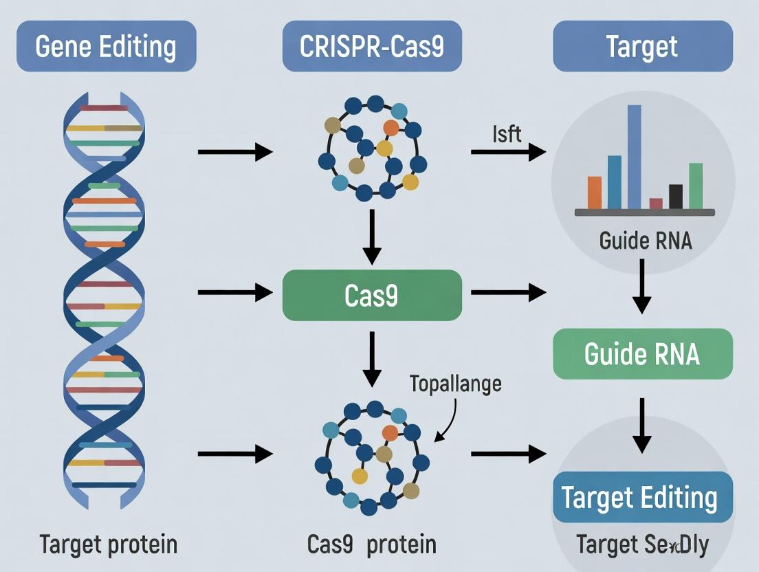
The CRISPR-Cas9 system comprises two core molecular components: the Cas9 endonuclease and a single guide RNA (sgRNA). The sgRNA is a chimeric RNA molecule that combines the functions of the naturally occurring CRISPR RNA (crRNA) and trans-activating crRNA (tracrRNA).
Table 1: Core Components of the CRISPR-Cas9 System
| Component | Description | Key Function |
|---|---|---|
| Cas9 Nuclease | A large, multi-domain protein (typically ~160 kDa from S. pyogenes). | Molecular scissors that creates double-strand breaks (DSBs) in DNA. Contains RuvC and HNH nuclease domains. |
| Single Guide RNA (sgRNA) | A synthetic ~100-nucleotide RNA molecule. | Guides Cas9 to the specific genomic target via Watson-Crick base pairing with the target DNA sequence. |
| Protospacer Adjacent Motif (PAM) | A short (2-6 bp) DNA sequence immediately downstream of the target site. | Essential for Cas9 recognition and binding. For SpCas9, the PAM is 5'-NGG-3'. |
| Target DNA Sequence | The 20-nucleotide genomic sequence preceding the PAM. | Specifies the site of Cas9 cleavage. The sgRNA is designed to be complementary to this sequence. |
Molecular Mechanism of Action
The mechanism can be broken down into three sequential phases: recognition, cleavage, and DNA repair.
- Recognition & Binding: The Cas9-sgRNA ribonucleoprotein (RNP) complex scans the genome. The sgRNA's 5' spacer region probes for complementary sequences adjacent to a valid PAM sequence. Base pairing between the sgRNA and target DNA triggers a conformational change in Cas9, activating its nuclease domains.
- Cleavage: Upon successful binding, the HNH nuclease domain cleaves the DNA strand complementary to the sgRNA (the "target" strand), while the RuvC-like domain cleaves the non-complementary strand (the "non-target" strand). This results in a blunt-ended double-strand break (DSB) typically 3-4 nucleotides upstream of the PAM.
- DNA Repair & Editing Outcome: The cellular DNA repair machinery processes the DSB, leading to one of two primary outcomes:
- Non-Homologous End Joining (NHEJ): An error-prone repair pathway that often results in small insertions or deletions (indels) at the break site, leading to gene knockouts via frameshift mutations.
- Homology-Directed Repair (HDR): In the presence of an exogenously supplied donor DNA template with homology arms, this precise repair pathway can be co-opted to introduce specific point mutations or insert new sequences (e.g., reporter genes).
Key Protocol: Delivery, Transfection, and Analysis of CRISPR-Cas9 in Mammalian Cells
This protocol outlines a standard workflow for performing CRISPR-Cas9-mediated gene knockout in adherent mammalian cell lines.
Protocol 3.1: sgRNA Design and RNP Complex Formation
Materials:
- Target genomic DNA sequence.
- sgRNA design software (e.g., CRISPick, CHOPCHOP).
- Chemically synthesized sgRNA (or in vitro transcription kit).
- Purified recombinant Cas9 protein (e.g., S. pyogenes SpCas9).
- Nuclease-Free Duplex Buffer (IDT) or equivalent.
- Optional: Fluorescently labeled tracrRNA for complex tracking.
Method:
- Design: Identify the target exon of your gene of interest. Use design tools to select sgRNAs with high on-target and low predicted off-target scores. The target sequence must be 20 nucleotides directly 5' of an NGG PAM.
- Complex Formation: Resuspend sgRNA to 100 µM in nuclease-free duplex buffer. In a microcentrifuge tube, combine:
- 2 µL of 100 µM sgRNA
- 2 µL of 100 µM fluorescent tracrRNA (optional)
- 16 µL of nuclease-free buffer
- Heat at 95°C for 5 minutes, then cool to room temperature.
- RNP Assembly: To the annealed RNA, add 3.2 µL of 62 µM Cas9 protein (final molar ratio ~1:1.2 Cas9:sgRNA). Mix gently and incubate at room temperature for 10-20 minutes before use.
Protocol 3.2: Delivery via Electroporation (for HEK293T cells)
Materials:
- Neon Transfection System (Thermo Fisher) or other electroporator.
- Neon Tip (100 µL)
- Resuspension Buffer R (Thermo Fisher)
- HEK293T cells in log-phase growth.
- Complete growth medium (DMEM + 10% FBS).
- Prepared RNP complex (from Protocol 3.1).
Method:
- Harvest and count cells. For one electroporation, wash 5 x 10⁵ cells with 1x PBS.
- Resuspend the cell pellet in Resuspension Buffer R to a final density of 1.1 x 10⁷ cells/mL.
- In a sterile tube, mix 9 µL of cell suspension (~100,000 cells) with 1 µL of the assembled RNP complex.
- Electroporate using the Neon system with the following parameters: 1,350 V, 10 ms, 3 pulses.
- Immediately transfer the electroporated cells into a pre-warmed 24-well plate containing 500 µL of complete medium.
- Incubate cells at 37°C, 5% CO₂ for 48-72 hours before analysis.
Protocol 3.3: Analysis of Editing Efficiency via T7 Endonuclease I Assay
Materials:
- Genomic DNA extraction kit.
- PCR reagents: High-fidelity DNA polymerase, primers flanking the target site (~300-500 bp product).
- T7 Endonuclease I (T7E1) or Surveyor Mutation Detection Kit.
- NEBuffer 2.1 (for T7E1).
- Agarose gel electrophoresis system.
Method:
- Harvest & Extract: 72 hours post-transfection, harvest cells and extract genomic DNA.
- PCR Amplify: Amplify the target locus from 100 ng of genomic DNA. Run a PCR clean-up step.
- Heteroduplex Formation: Dilute purified PCR product to ~50 ng/µL. Denature and reanneal in a thermocycler: 95°C for 5 min, ramp down to 85°C at -2°C/sec, then to 25°C at -0.1°C/sec.
- Digestion: In a 20 µL reaction, mix:
- 200 ng of reannealed PCR product
- 2 µL NEBuffer 2.1
- 1 µL T7 Endonuclease I
- Nuclease-free water to volume. Incubate at 37°C for 30 minutes.
- Analysis: Run the digested product on a 2% agarose gel. Cleavage of heteroduplex DNA (containing mismatches from indels) yields two smaller bands. Editing efficiency is estimated using band intensity analysis software.
The Scientist's Toolkit: Essential Reagents and Materials
Table 2: Key Research Reagent Solutions for CRISPR-Cas9 Experiments
| Reagent/Material | Function & Explanation | Example Vendor/Product |
|---|---|---|
| Recombinant Cas9 Nuclease | High-purity, endotoxin-free protein for RNP assembly. Essential for direct delivery methods, reducing off-target risks and temporal control compared to plasmid DNA. | Integrated DNA Technologies (IDT) Alt-R S.p. Cas9 Nuclease. |
| Chemically Modified sgRNA | Synthetic guide RNA with phosphorothioate bonds and 2'-O-methyl modifications at terminal nucleotides. Increases stability, reduces innate immune response, and improves editing efficiency. | Synthego sgRNA EZ Kit. |
| Electroporation System & Buffer | Enables high-efficiency delivery of RNP complexes into hard-to-transfect cells (e.g., primary cells, iPSCs). Buffer composition is critical for cell viability. | Thermo Fisher Neon System & Resuspension Buffer R. |
| HDR Donor Template | Single-stranded oligodeoxynucleotide (ssODN) or double-stranded DNA (dsDNA) donor with homologous arms. Provides the template for precise editing via the HDR pathway. | IDT Ultramer DNA Oligos. |
| Genomic DNA Extraction Kit | For reliable, PCR-ready genomic DNA isolation from small numbers of transfected cells. | Qiagen DNeasy Blood & Tissue Kit. |
| Mutation Detection Enzyme | Enzyme that cleaves mismatched heteroduplex DNA (e.g., T7 Endonuclease I, Surveyor Nuclease). Used for initial, rapid quantification of indel formation efficiency. | New England Biolabs T7 Endonuclease I. |
| Next-Generation Sequencing (NGS) Library Prep Kit | For comprehensive, unbiased analysis of on-target editing efficiency and genome-wide off-target profiling. | Illumina CRISPResso2 Library Prep. |
| Cell Viability Assay | To monitor cytotoxicity associated with CRISPR delivery (e.g., electroporation, lipofection). | Promega CellTiter-Glo Luminescent Assay. |
Within the comprehensive framework of a thesis on CRISPR-Cas9 gene editing protocols, a critical first step is the precise definition of the experimental goal. The choice between creating a gene knockout (disruption) or a knock-in (precise insertion) dictates every subsequent decision, from guide RNA design to the selection of the DNA repair pathway to be harnessed. This application note delineates the fundamental mechanisms of Non-Homologous End Joining (NHEJ) and Homology-Directed Repair (HDR), provides comparative data, and outlines detailed protocols for achieving each outcome.
Core Mechanisms: NHEJ vs. HDR
The CRISPR-Cas9 system induces a double-strand break (DSB) at a target genomic locus. The cellular repair of this break determines the editing outcome.
- Non-Homologous End Joining (NHEJ): The dominant, error-prone pathway active throughout the cell cycle. It directly ligates the broken ends, often resulting in small insertions or deletions (indels). These indels can cause frameshift mutations, leading to premature stop codons and effective gene knockout.
- Homology-Directed Repair (HDR): A precise, template-dependent pathway active primarily in the S/G2 phases. It uses a homologous DNA template (donor template) to repair the DSB, enabling precise nucleotide changes or insertion of sequences (e.g., tags, reporters, pathogenic variants) for knock-in.
Comparative Data and Decision Matrix
Table 1: Key Characteristics of NHEJ and HDR Pathways
| Feature | Non-Homologous End Joining (NHEJ) | Homology-Directed Repair (HDR) |
|---|---|---|
| Primary Goal | Gene Disruption (Knockout) | Precise Insertion/Modification (Knock-in) |
| Template Required | No | Yes (ssODN or dsDNA donor) |
| Repair Fidelity | Error-prone (generates indels) | High-fidelity (precise) |
| Cell Cycle Phase | Active throughout, dominant in G0/G1 | Primarily active in S/G2 |
| Relative Efficiency | High (≥80% indels common) | Low (Typically 0.5%-20%) |
| Key Applications | Functional gene screens, modeling loss-of-function, therapeutic gene disruption. | Protein tagging, disease modeling (SNPs), gene correction, reporter knock-in. |
Table 2: Quantitative Comparison of Editing Outcomes in Common Mammalian Cell Lines
| Cell Type | Typical NHEJ (Indel %) Range* | Typical HDR (Precise Edit %) Range* | Preferred Donor Template |
|---|---|---|---|
| HEK293T | 70% - 90% | 5% - 30% | ssODN (<200 nt) |
| HCT116 | 60% - 85% | 1% - 10% | ssODN or dsDNA |
| iPSCs | 40% - 70% | 0.5% - 5% | dsDNA with long homologies |
| Primary T Cells | 50% - 80% | 0.5% - 10% | ssODN or AAV6-delivered dsDNA |
| *Ranges are approximate and highly dependent on locus, guide efficiency, and delivery method. |
Detailed Experimental Protocols
Protocol A: Gene Knockout via NHEJ-Promoted Indel Formation
Objective: To disrupt the coding sequence of a target gene by generating frameshift mutations.
Materials: See "Scientist's Toolkit" section.
Procedure:
- Design & Cloning: Design a gRNA targeting an early exon of the gene. Clone into a Cas9/gRNA expression plasmid (e.g., pSpCas9(BB)).
- Cell Transfection: Seed HEK293T cells to reach 70-80% confluency at transfection. Transfect with 1 µg of plasmid using a suitable transfection reagent.
- Validation & Analysis:
- 48-72h post-transfection: Harvest genomic DNA.
- PCR Amplification: Amplify the target region (amplicon size: 300-500 bp).
- Assessment: Analyze indels by:
- T7 Endonuclease I (T7E1) or Surveyor Assay: Digest heteroduplexed PCR products; analyze fragments by gel electrophoresis.
- Sanger Sequencing & Decomposition Analysis: Sequence the PCR product and use tools like ICE (Inference of CRISPR Edits) or TIDE to quantify indel percentages.
Protocol B: Gene Knock-in via HDR Using a ssODN Donor Template
Objective: To introduce a precise point mutation (e.g., a disease-relevant SNP) into the target locus.
Materials: See "Scientist's Toolkit" section.
Procedure:
- Design:
- gRNA: Design to cut as close as possible to the intended edit site.
- ssODN Template: Design a single-stranded oligodeoxynucleotide (ssODN, ~120-200 nt) with the desired mutation flanked by homology arms (40-90 nt each). Incorporate silent blocking mutations in the PAM or seed region to prevent re-cutting.
- Nucleofection: For high efficiency in difficult cells (e.g., iPSCs), use nucleofection. For 1x10^6 cells, prepare a mix containing 2 µg Cas9 RNP (complex of recombinant Cas9 protein and synthetic gRNA) and 100 pmol of purified ssODN.
- Transfection & Synchronization: To enrich for HDR, synchronize cells at the S/G2 boundary using thymidine or aphidicolin treatment prior to transfection.
- Screening & Validation:
- Post-transfection (72h-96h): Harvest genomic DNA.
- PCR & Restriction Digest (if introduced): If the edit creates/destroys a restriction site, digest the PCR product.
- Clonal Isolation: For homozygous edits, single-cell sort transfected cells and expand clonal populations.
- Validation: Perform Sanger sequencing of the target locus from clonal genomic DNA to confirm precise incorporation.
Visualizations
Diagram 1: CRISPR-Cas9 Editing Pathways: NHEJ vs. HDR
Diagram 2: Experimental Workflow for Knockout vs. Knock-in
The Scientist's Toolkit: Research Reagent Solutions
Table 3: Essential Materials for CRISPR-Cas9 Editing Experiments
| Item | Function & Description | Example Product/Catalog |
|---|---|---|
| Cas9 Expression Vector | Plasmid encoding SpCas9 nuclease for transient expression. | pSpCas9(BB)-2A-Puro (Addgene #62988) |
| Recombinant Cas9 Protein | Purified Cas9 for rapid, transient activity with reduced off-target effects; used in RNP formation. | Alt-R S.p. Cas9 Nuclease V3 (IDT) |
| Synthetic gRNA (crRNA + tracrRNA) | Chemically synthesized guide RNA components for complexing with recombinant Cas9 protein (RNP). | Alt-R CRISPR-Cas9 crRNA & tracrRNA (IDT) |
| HDR Donor Template (ssODN) | Ultramer DNA oligonucleotide with homology arms for precise knock-in via HDR. | Alt-R HDR Donor Oligo (IDT) |
| Transfection Reagent | Lipid-based reagent for plasmid or RNP delivery into adherent cell lines. | Lipofectamine CRISPRMAX (Thermo) |
| Nucleofector System | Electroporation-based system for high-efficiency delivery into hard-to-transfect cells (T cells, iPSCs). | 4D-Nucleofector (Lonza) |
| NHEJ Detection Kit | Enzyme-based assay to detect and quantify indel formation from bulk populations. | T7 Endonuclease I (NEB) |
| Cell Cycle Inhibitor | Small molecule to synchronize cells in S-phase to favor HDR over NHEJ. | Aphidicolin (Sigma) |
| Cloning/Digestion Kit | For isolating and validating single-cell clones. | Zero-Blunt TOPO Cloning Kit (Thermo) |
Within a comprehensive thesis on CRISPR-Cas9 gene editing, the design of the single-guide RNA (sgRNA) is the critical first determinant of experimental success. This pre-experimental planning stage dictates the efficiency, specificity, and overall functionality of the genome editing system. An optimal sgRNA ensures high on-target activity while minimizing off-target effects. This protocol details the principles, tools, and validation steps essential for robust sgRNA design.
Core Design Principles for Optimal sgRNA
Sequence Composition Rules
The efficacy of an sgRNA is governed by specific sequence features. The following parameters must be evaluated for every candidate sgRNA.
Table 1: Quantitative sgRNA Design Parameters and Optimal Ranges
| Parameter | Optimal Range / Feature | Rationale & Impact |
|---|---|---|
| GC Content | 40-60% | Low GC (<20%) reduces stability; high GC (>80%) may increase off-target binding. |
| sgRNA Length | 17-20 nt (SpCas9) | Shorter guides increase specificity but may reduce activity; 20 nt is standard. |
| Protospacer Adjacent Motif (PAM) | NGG (for SpCas9) | Must be present immediately 3' of the target DNA sequence. Cas9 variant-specific. |
| On-Target Efficiency Score | >50 (tool-specific) | Predicts cleavage likelihood. Benchmarked algorithms (e.g., Doench '16, Moreno-Mateos). |
| Specificity (Off-Target) | 0-3 mismatches screened | Fewer potential off-target sites with ≤3 mismatches indicates higher specificity. |
| Poly-T/TTTT | Avoid | Four consecutive T's act as a termination signal for Pol III promoters (U6). |
| Self-Complementarity | Avoid | Secondary structure in sgRNA can impede Cas9 binding. |
| 5' Nucleotide (U6) | G for U6, A for T7 | U6 promoters require a 5' G for transcription initiation; T7 requires 5' GG. |
Specificity and Off-Target Prediction
A comprehensive off-target analysis is non-negotiable. Tools must search the relevant genome for sequences with the highest homology to the sgRNA spacer, allowing for up to 3-4 mismatches, with particular attention to mismatches in the "seed" region (positions 1-12 proximal to PAM).
Protocol: A Step-by-Step sgRNA Design andIn SilicoValidation Workflow
Pre-Design Phase: Target Definition
- Step 1: Identify Genomic Target Region. Define the exact genomic coordinates (e.g., from Ensembl, UCSC Genome Browser) of the exon, regulatory element, or specific nucleotide to be edited.
- Step 2: Select Cas9 Variant. Choose the nuclease (e.g., SpCas9, SaCas9, HiFi Cas9) based on PAM requirement and fidelity needs. This determines the PAM sequence to search for (e.g., SpCas9: NGG).
- Step 3: Retrieve Genomic Sequence. Use a genome browser or database to extract a ~500 bp sequence flanking the target site. Verify the assembly and version (e.g., GRCh38/hg38).
Primary Design Phase: Candidate sgRNA Generation
- Step 4: Input Sequence into Design Tools. Submit the target sequence to multiple reputable sgRNA design platforms (see Table 2).
- Step 5: Generate and Filter Candidates. Tools will output all possible sgRNAs with the correct PAM. Apply the filters from Table 1:
- Eliminate any sgRNA with a 5' TTTT (poly-T) tract.
- Filter for GC content between 40-60%.
- Rank by highest on-target efficiency score.
- Step 6: Select Top 3-5 Candidates. Based on the initial ranking, select the top 3-5 candidates for downstream analysis.
Specificity Validation Phase: Off-Target Analysis
- Step 7: Perform Genome-Wide Off-Target Search. Input each top candidate sequence into an off-target prediction tool. Use tools that employ the latest algorithms (e.g., cutting frequency determination [CFD] score) and allow for bulges.
- Step 8: Evaluate and Compare Off-Target Hits. For each candidate, review the list of potential off-target sites. Prioritize sgRNAs with:
- Fewer total predicted off-target sites.
- Mismatches located distal to the PAM (positions >12).
- Lower CFD scores for off-target sites (indicating lower probability of cleavage).
- No off-targets within coding exons or functional genomic elements of high concern.
Final Selection andIn SilicoValidation
- Step 9: Cross-Reference with Multiple Tools. Validate the final 2-3 candidates by checking their scores and off-target profiles across a second, independent design tool.
- Step 10: Design Oligonucleotides for Cloning. For the final selected sgRNA(s), design forward and reverse oligonucleotides with the appropriate 5' and 3' overhangs for your chosen cloning system (e.g., BbsI sites for Addgene's pSpCas9(BB) backbone).
Diagram 1: sgRNA Design & Selection Workflow
Title: sgRNA Design and Selection Protocol
Essential Tools for sgRNA Design and Analysis
A combination of tools is required for comprehensive design.
Table 2: Key sgRNA Design and Analysis Tools (Current as of 2023-2024)
| Tool Name (Provider) | Primary Function | Key Metric/Algorithm | Access (URL) |
|---|---|---|---|
| CRISPOR (Haeussler et al.) | Integrated Design & Off-Target | Doench '16, Moreno-Mateos scores; CFD for off-targets | http://crispor.tefor.net |
| ChopChop (Harvard) | Target Site Finder & Scoring | Efficiency scores, specificity, and off-targets | https://chopchop.cbu.uib.no |
| Broad Institute GPP Portal (Broad) | sgRNA Design & Ranking | Rule Set 2 (Doench '16), Saporito score | https://portals.broadinstitute.org/gpp/public |
| CRISPRscan (Moreno-Mateos) | Efficiency Scoring (zebrafish-focused but broadly applicable) | Algorithm for predicting sgRNA activity | https://www.crisprscan.org |
| Cas-OFFinder (Bae et al.) | Genome-Wide Off-Target Search | Searches for bulges & mismatches | http://www.rgenome.net/cas-offinder |
| UCSC Genome Browser (UCSC) | Genomic Context Visualization | View target in genomic, regulatory, conservation context | https://genome.ucsc.edu |
Diagram 2: Tool Utilization Logic for Optimal Design
Title: Interplay of Key sgRNA Design Tools
The Scientist's Toolkit: Research Reagent Solutions
Table 3: Essential Reagents and Materials for sgRNA Design & Validation
| Item | Function & Application | Example/Notes |
|---|---|---|
| High-Fidelity DNA Polymerase | Amplification of target genomic regions for cloning and validation. | Q5 (NEB), KAPA HiFi. |
| Restriction Enzymes (BbsI/BsaI) | Golden Gate or standard cloning of annealed oligos into sgRNA expression vectors. | Esp3I (BbsI isothermmal). |
| T4 DNA Ligase | Ligation of sgRNA insert into digested plasmid backbone. | Quick ligation variants reduce time. |
| sgRNA Expression Vector | Backbone with Pol III promoter (U6/H1) for sgRNA transcription. | pSpCas9(BB) (Addgene #48139), pX330. |
| Desalted Oligonucleotides | Forward and reverse oligos encoding the sgRNA spacer sequence. | 25-30 nt, desalted purification is sufficient. |
| Gel Extraction Kit | Purification of digested vector and PCR products. | Critical for reducing re-ligation background. |
| Competent E. coli | Transformation of ligated plasmid for amplification. | DH5α, Stbl3 (for repetitive sequences). |
| Plasmid Miniprep Kit | Isolation of purified sgRNA plasmid for sequencing. | Confirm insert by Sanger sequencing with U6-F primer. |
| Sanger Sequencing Service | Final validation of cloned sgRNA sequence. | Use vector-specific forward primer. |
Within a comprehensive CRISPR-Cas9 gene editing protocol, the selection of an appropriate delivery vector is a critical determinant of editing efficiency, specificity, and therapeutic safety. This application note compares the two principal vector classes—viral and non-viral—providing structured data and experimental protocols to guide researchers and drug development professionals in their system selection for in vitro and in vivo applications.
Comparative Vector Analysis
Quantitative Performance Metrics
Table 1: Key Performance Characteristics of Viral vs. Non-Viral Vectors for CRISPR-Cas9 Delivery
| Characteristic | Viral Vectors (Lentivirus/AAV) | Non-Viral Vectors (LNPs/Electroporation) |
|---|---|---|
| Typical Payload Capacity | AAV: ~4.7 kb; Lentivirus: ~8 kb | High (LNPs: >10 kb; Can deliver Cas9 mRNA + gRNA) |
| In Vivo Delivery Efficiency | High (Titer-dependent, often >70% transduction in vitro) | Variable (LNP: Moderate-High in liver; Electroporation: High ex vivo) |
| Immunogenicity Risk | High (Pre-existing immunity, adaptive immune response) | Lower (LNP components can be immunogenic, but often tunable) |
| Insertional Mutagenesis Risk | Low for AAV; Moderate for Lentivirus (random integration) | None (Typically transient expression) |
| Manufacturing Complexity & Cost | High (Biosafety concerns, upstream/downstream processing) | Lower (Scalable, synthetic chemistry) |
| Expression Kinetics | Persistent (Weeks to months for AAV) | Transient (Days to weeks for mRNA/LNPs) |
| Tropism & Targeting Flexibility | Moderate (Engineered capsids possible) | High (LNPs can be conjugated with targeting ligands) |
Table 2: Selection Guide Based on Experimental Goals
| Research Goal | Recommended Primary System | Rationale & Key Consideration |
|---|---|---|
| Long-term in vivo gene knockout (e.g., CNS) | AAV (serotype specific to target tissue) | Sustained Cas9/gRNA expression required for post-mitotic cells. |
| High-throughput in vitro screening | Lentiviral Vector | Stable genomic integration enables permanent modification in cell pools. |
| Ex vivo cell therapy (e.g., CAR-T editing) | Electroporation (RNP delivery) | High efficiency, rapid kinetics, minimal off-targets, clinical translatability. |
| Systemic in vivo delivery to hepatocytes | Lipid Nanoparticles (LNPs) | High delivery efficiency to liver, transient expression reduces off-target risk. |
| Base/Prime editing in vivo | AAV or dual AAVs (if payload large) | Requires longer expression window for slow-converting editors; monitor size limits. |
Detailed Protocols
Protocol 1: Lentiviral Production and Transduction for Stable Cell Line Generation
Application: Creating pools of cells with stable genomic integration of gRNA and/or Cas9 for long-term studies. Materials: See "The Scientist's Toolkit" below. Method:
- Day 1-2: Plasmid Transfection: Seed HEK293T cells in a 10cm dish to reach 70-80% confluency. Co-transfect using PEI Max with the following plasmid mix: 10 µg transfer plasmid (gRNA+Cas9, if all-in-one), 7.5 µg psPAX2 (packaging), and 2.5 µg pMD2.G (envelope). Change media 6 hours post-transfection.
- Day 3-4: Harvest Virus: Collect supernatant at 48 and 72 hours post-transfection. Filter through a 0.45 µm PES filter. Concentrate via ultracentrifugation (25,000 rpm, 2h, 4°C) or using PEG-it virus precipitation solution. Resuspend pellet in cold PBS, aliquot, and store at -80°C. Determine functional titer (TU/mL) via transduction of HeLa cells and flow cytometry.
- Day 5: Target Cell Transduction: Plate target cells. Add viral supernatant with polybrene (8 µg/mL). Spinfect at 1000 x g for 30-60 min at 32°C. Replace media after 24h.
- Day 6-7: Selection/Purification: Begin antibiotic selection (e.g., puromycin) 48h post-transduction. Maintain selection for 5-7 days before expanding and validating edited pools.
Protocol 2: Lipid Nanoparticle (LNP) Formulation for Cas9 mRNA/gRNA DeliveryIn Vivo
Application: Systemic delivery of CRISPR-Cas9 components to murine liver. Materials: See "The Scientist's Toolkit" below. Method:
- Aqueous Phase Preparation: Dilute Cas9 mRNA and chemically modified sgRNA in sodium acetate buffer (50 mM, pH 5.0) to a final total nucleic acid concentration of 0.2 mg/mL. Maintain an mRNA:sgRNA molar ratio of 1:3.
- Lipid Phase Preparation: Dissolve ionizable lipid (e.g., DLin-MC3-DMA), DSPC, Cholesterol, and PEG-lipid (e.g., DMG-PEG 2000) in ethanol at a molar ratio of 50:10:38.5:1.5. The total lipid concentration should be 12.5 mM.
- Microfluidic Mixing: Using a precision microfluidic mixer (or a turbulent mixing device), combine the aqueous and lipid phases at a 3:1 volumetric ratio (aqueous:ethanol) with a total flow rate of 12 mL/min. The resulting mixture forms LNPs.
- Buffer Exchange & Characterization: Dialyze the LNP suspension against PBS (pH 7.4) for 4 hours at 4°C using a 10kDa MWCO dialysis cassette. Filter sterilize (0.22 µm). Characterize particle size (~80-100 nm) via DLS and measure encapsulation efficiency (>90%) using a Ribogreen assay.
- In Vivo Administration: Administer LNPs intravenously via tail vein at a dose of 0.5-1.0 mg mRNA/kg mouse body weight. Analyze editing efficiency in target tissues (e.g., liver) by NGS 7-14 days post-injection.
Visualizations
Title: Lentiviral Workflow for Stable Cell Line Generation
Title: Vector Selection Decision Tree for CRISPR Delivery
The Scientist's Toolkit
Table 3: Essential Research Reagents and Materials
| Item | Function/Description | Example Vendor/Product |
|---|---|---|
| PEI Max (Linear PEI, 40kDa) | High-efficiency, low-cost transfection reagent for viral producer cells. | Polysciences, Inc. |
| Lenti-X Concentrator | Chemical precipitation solution for quick lentivirus concentration. | Takara Bio |
| Polybrene (Hexadimethrine bromide) | Cationic polymer that enhances viral transduction efficiency. | Sigma-Aldrich |
| Puromycin Dihydrochloride | Selection antibiotic for cells transduced with puromycin-resistance vectors. | Thermo Fisher Scientific |
| Ionizable Cationic Lipid (DLin-MC3-DMA) | Key component of LNPs, promotes encapsulation and endosomal escape. | MedChemExpress |
| DMG-PEG 2000 | PEGylated lipid for LNP stability and circulation time modulation. | Avanti Polar Lipids |
| Nucleofector System & Kit | Electroporation device and cell-type specific buffers for high-efficiency RNP delivery. | Lonza |
| Ribogreen Assay Kit | Fluorescent quantitation of free vs. encapsulated nucleic acids in LNPs. | Thermo Fisher Scientific |
| AAVpro Purification Kit | All-in-one kit for purification and titering of AAV vectors. | Takara Bio |
| Surveyor Nuclease Assay Kit | Gel-based method for detecting CRISPR-induced indels (validation). | IDT |
Selecting an appropriate cellular model is a critical first step in any CRISPR-Cas9 gene editing experiment. The choice between primary, stem, and immortalized cell lines dictates the biological relevance, experimental feasibility, and translational potential of the research. This application note, framed within a broader thesis on CRISPR-Cas9 protocols, provides a comparative analysis and detailed methodologies for working with these distinct cell types in gene editing workflows.
Comparative Analysis of Cell Models for CRISPR-Cas9
Table 1: Key Characteristics and Considerations for CRISPR-Cas9 Gene Editing
| Characteristic | Primary Cells | Stem Cells (iPSCs/ESCs) | Immortalized Cell Lines |
|---|---|---|---|
| Physiological Relevance | Very High; native tissue genotype/phenotype | High (upon differentiation); retain developmental potential | Low to Moderate; genetically altered, adapted to culture |
| Proliferative Capacity | Limited (senescence after few passages) | High (virtually unlimited self-renewal) | High (infinite proliferation) |
| Genetic Stability | High (but degrades with passage) | High (but requires monitoring for karyotype) | Variable; often aneuploid, prone to drift |
| CRISPR Transfection Efficiency | Typically Low (10-30%) | Variable; can be low (5-40%) | Typically High (often >70% for HEK293) |
| Clonal Isolation Difficulty | High (due to limited division) | Moderate (requires careful handling) | Low (robust growth facilitates cloning) |
| Protocol Duration | Short (limited culture window) | Long (weeks for derivation, expansion, differentiation) | Short (rapid expansion and editing) |
| Cost | High (donor variability, fresh isolation) | Very High (specialized media, quality control) | Low (easy maintenance, standardized) |
| Ideal CRISPR Application | Disease modeling (oncogenic mutations in patient-derived cells), functional genomics in native context | Developmental disease modeling, isogenic control generation, regenerative medicine studies | Protocol optimization, high-throughput screens, mechanistic studies |
Detailed Protocols for CRISPR-Cas9 Across Cell Models
Protocol 1: CRISPR-Cas9 Knockout in Immortalized HEK293T Cells
This protocol is optimized for high-efficiency editing in robust, easily transfected lines.
Research Reagent Solutions & Materials:
| Item | Function |
|---|---|
| HEK293T Cells | Robust, highly transfertable immortalized line for protocol optimization. |
| Lipofectamine 3000 | Cationic lipid reagent for high-efficiency plasmid DNA delivery. |
| pSpCas9(BB)-2A-Puro (PX459) v2.0 | All-in-one plasmid expressing Cas9, sgRNA, and a puromycin selection marker. |
| Puromycin Dihydrochloride | Antibiotic for selecting successfully transfected cells (2-5 µg/mL working concentration). |
| Luria-Bertani (LB) Broth | Medium for amplifying plasmid DNA in bacterial culture. |
| DPBS, Calcium/Magnesium-Free | For washing cells during passaging. |
| 0.05% Trypsin-EDTA | Enzyme solution for dissociating adherent cells. |
| Fetal Bovine Serum (FBS) | Serum supplement to quench trypsin and support cell growth. |
Methodology:
- Design & Cloning: Design a 20-nt sgRNA sequence targeting your gene of interest. Anneal oligonucleotides and clone into the BbsI site of the PX459 plasmid. Transform into competent E. coli, culture in LB broth with ampicillin (100 µg/mL), and purify plasmid DNA.
- Cell Seeding: Seed HEK293T cells in a 6-well plate at 3 x 10^5 cells/well in DMEM + 10% FBS. Incubate at 37°C, 5% CO2 until 70-80% confluent (typically 24h).
- Transfection: For each well, prepare two mixes: A) 125 µL Opti-MEM + 2.5 µg PX459-sgRNA plasmid + 5 µL P3000 reagent. B) 125 µL Opti-MEM + 3.75 µL Lipofectamine 3000. Combine A and B, incubate 15 min at RT. Add dropwise to cells with fresh medium.
- Selection & Cloning: 24h post-transfection, replace medium with fresh medium containing 2 µg/mL puromycin. Select for 48h. Subsequently, trypsinize and dilute cells to ~1 cell/100 µL in 96-well plates for clonal isolation. Expand clones for 2-3 weeks.
- Analysis: Screen clonal populations via genomic DNA extraction, PCR amplification of the target locus, and T7 Endonuclease I assay or Sanger sequencing to identify indels.
Title: CRISPR Workflow for Immortalized Cells
Protocol 2: CRISPR-Cas9 Editing in Human Induced Pluripotent Stem Cells (iPSCs)
This protocol emphasizes maintaining pluripotency during nucleofection and single-cell cloning.
Research Reagent Solutions & Materials:
| Item | Function |
|---|---|
| Human iPSCs | Pluripotent cells capable of differentiation into any cell type; requires meticulous culture. |
| StemFlex Medium | Specialized, feeder-free culture medium for robust iPSC maintenance. |
| RevitaCell Supplement | Improves viability post-single-cell passage and nucleofection. |
| Nucleofector Device & Kit (e.g., Lonza 4D-Nucleofector) | Electroporation system for high-efficiency delivery of RNP complexes into hard-to-transfect cells. |
| Alt-R S.p. Cas9 Nuclease V3 | Recombinant, high-fidelity Cas9 protein for rapid, transient editing. |
| Alt-R CRISPR-Cas9 sgRNA | Synthetic, chemically modified sgRNA for enhanced stability and reduced immunogenicity. |
| CloneR Supplement | Enhances survival of single iPSCs during clonal outgrowth. |
| Matrigel or Vitronectin | Extracellular matrix coating for feeder-free iPSC culture. |
Methodology:
- Culture Preparation: Culture iPSCs on Matrigel-coated plates in StemFlex Medium. Passage as small clumps using 0.5 mM EDTA. Ensure cells are >95% positive for pluripotency markers (OCT4, NANOG) and have normal karyotype before editing.
- RNP Complex Formation: Resuspend 30 pmol of Alt-R Cas9 protein and 36 pmol of Alt-R sgRNA in Nucleofector solution to form RNP complexes. Incubate at room temperature for 10-20 minutes.
- Single-Cell Preparation: Dissociate iPSCs to a single-cell suspension using Accutase. Quench with medium containing 10 µM RevitaCell. Count cells.
- Nucleofection: Centrifuge 1 x 10^6 iPSCs. Resuspend cell pellet in the pre-formed RNP complex mixture. Transfer to a nucleofection cuvette and electroporate using the recommended program (e.g., CB-150 for Lonza). Immediately add pre-warmed medium with RevitaCell.
- Recovery & Cloning: Plate nucleofected cells at high density for bulk analysis or for clonal isolation. For cloning, plate ~1-2 cells/well in a 96-well plate pre-coated with Matrigel in StemFlex medium supplemented with 10 µM CloneR. Change medium every other day.
- Screening & Expansion: After 10-14 days, manually pick and expand individual colonies. Screen via PCR and sequencing. Confirm pluripotency status post-editing.
Title: CRISPR-Cas9 RNP Workflow for iPSCs
Protocol 3: CRISPR-Cas9 Editing in Primary Human T Cells
This protocol focuses on activating and editing non-dividing or slowly dividing primary immune cells.
Research Reagent Solutions & Materials:
| Item | Function |
|---|---|
| Primary Human T Cells | Isolated from PBMCs; high physiological relevance for immunology and cell therapy. |
| CD3/CD28 T Cell Activator | Magnetic beads or antibodies to stimulate T cell proliferation and increase editing efficiency. |
| IL-2 (Interleukin-2) | Cytokine essential for T cell growth and survival in culture. |
| TexMACS Medium | Serum-free medium optimized for human T cell and lymphocyte culture. |
| Neon Transfection System | Pipette-based electroporation system suitable for sensitive primary cells. |
| Cas9 mRNA/sgRNA or RNP | Transient expression systems preferred to minimize off-target effects and immune response. |
| Anti-human CD3 Antibody | For assessing activation status via flow cytometry. |
| Propidium Iodide (PI) | Viability dye for assessing post-transfection cell death. |
Methodology:
- T Cell Activation: Isolate CD3+ T cells from PBMCs using magnetic separation. Resuspend cells at 1 x 10^6 cells/mL in TexMACS medium supplemented with 5% human AB serum, 100 U/mL IL-2, and CD3/CD28 activator beads (bead:cell ratio 1:1). Activate for 48-72 hours.
- Electroporation Preparation: On day 3 post-activation, harvest cells, remove beads, and count. Prepare RNP complexes (as in iPSC protocol) or Cas9 mRNA/sgRNA mixtures.
- Electroporation: Wash cells in PBS. For the Neon system, resuspend 1 x 10^6 cells in 10 µL Resuspension Buffer R mixed with the editing components. Electroporate using a 1400V, 10ms, 3-pulse protocol. Immediately transfer cells to pre-warmed complete TexMACS + IL-2 medium.
- Post-Transfection Culture: Add IL-2 (100 U/mL) fresh every 2-3 days. Monitor viability with PI staining at 24h post-electroporation. Expect 40-60% efficiency and 50-80% viability.
- Analysis: After 5-7 days, harvest cells for genomic DNA and flow cytometry analysis. For functional assays, expand cells as needed, but note primary T cells have a limited in vitro lifespan.
Title: Primary T Cell CRISPR-Cas9 Editing Workflow
Critical Pathway: DNA Damage Response in CRISPR-Edited Cells
CRISPR-Cas9 cutting activates critical DNA repair pathways, predominantly Non-Homologous End Joining (NHEJ) and Homology-Directed Repair (HDR), which dictate editing outcomes.
Title: DNA Repair Pathways Post-CRISPR Cutting
The optimal cell model for a CRISPR-Cas9 experiment is determined by the research question's need for physiological fidelity versus experimental tractability. Immortalized lines offer speed and ease for screening and optimization. Primary cells provide unmatched relevance for ex vivo studies. Stem cells, particularly iPSCs, enable the generation of isogenic controls and disease-relevant differentiated cell types, bridging the gap between the other two models. Integrating the protocols and considerations outlined here will inform robust experimental design within a comprehensive CRISPR-Cas9 thesis.
Step-by-Step CRISPR-Cas9 Gene Editing Protocol: A Detailed Laboratory Workflow
This protocol forms Stage 1 of a comprehensive thesis on CRISPR-Cas9 gene editing, detailing the foundational design and preparation of key reagents. Successful genome editing hinges on the precise design and high-quality construction of the single-guide RNA (sgRNA) and donor DNA template. This stage involves in silico design, specificity analysis, and molecular cloning or synthesis to generate reagents for subsequent delivery and screening stages.
Research Reagent Solutions: Essential Materials
| Reagent/Material | Function & Explanation |
|---|---|
| CRISPR Design Software (e.g., CRISPOR, Benchling, CHOPCHOP) | Web-based tools to identify candidate sgRNA sequences with high on-target efficiency and low off-target potential for a given genomic locus. |
| Off-Target Prediction Databases (e.g., UCSC Genome Browser, Ensembl) | Reference genomes and browser tools to cross-check predicted sgRNA binding sites across the genome to minimize unintended edits. |
| DNA Oligonucleotides (Ultramer or Gene Fragments) | High-fidelity synthetic DNA for sgRNA template PCR or direct donor template synthesis, especially for single-stranded DNA (ssDNA) donors. |
| Cloning Vector (e.g., pSpCas9(BB)-2A-Puro, pX459) | Backbone plasmids for expressing sgRNA and Cas9 nuclease (and often a selection marker) in mammalian cells. |
| High-Fidelity DNA Polymerase (e.g., Q5, Phusion) | For error-free amplification of donor DNA template fragments or sgRNA expression cassettes. |
| T7 Endonuclease I or Surveyor Nuclease | Enzymes for mismatch cleavage assays used to validate sgRNA cutting efficiency in vitro or in preliminary cellular tests. |
| In Vitro Transcription Kit (T7 or U6 promoter-based) | For generating high-purity, capped sgRNA transcripts when using direct RNA delivery methods like ribonucleoprotein (RNP) complexes. |
Part 1: sgRNA Design and Validation Protocol
Target Selection andIn SilicoDesign
- Identify Target Sequence: Define the genomic region for editing. For gene knockouts, target early exons encoding critical protein domains. For precise knock-ins, design the cut site within 10 bp of the desired insertion site.
- sgRNA Sequence Retrieval: Use design tools (CRISPOR is recommended). Input the genomic coordinates or sequence. The tool will output a list of candidate sgRNAs (typically 20-bp protospacers) with associated predictions.
- Evaluation Criteria: Select sgRNAs based on:
- High On-Target Score: (>60 using Doench ‘16 or Moreno-Mateos scores).
- Low Off-Target Potential: Examine predicted off-target sites with ≤3 mismatches. Avoid sgRNAs with off-targets in coding regions or known oncogenes/tumor suppressors.
- Proximal to PAM: The Protospacer Adjacent Motif (NGG for SpCas9) must be present immediately 3’ of the target.
Table 1: Example sgRNA Candidate Analysis from CRISPOR for Human EMX1 Gene
| sgRNA Sequence (5'-3') | On-Target Efficiency (Doench '16) | Predicted Off-Targets (≤3 Mismatches) | Recommended? |
|---|---|---|---|
| GAGTCCGAGCAGAAGAAGAA | 68 | 2 (both intergenic) | Yes |
| GTAGAACTACCATCACCCGC | 92 | 5 (1 in intron of PARP1) | With caution |
| TGCAGAAGCACCTCCACCCG | 45 | 0 | No (Low efficiency) |
sgRNA Expression Construct Preparation
Method A: Cloning into a Plasmid Vector (Common)
- Order Oligos: Design complementary oligonucleotides encoding your selected 20-bp guide sequence, with 5' overhangs compatible with your chosen vector (e.g., BbsI for pSpCas9(BB)).
- Forward oligo: 5'-CACCg[20-nt guide sequence]-3'
- Reverse oligo: 5'-AAACc[reverse complement of guide]-3'
- Annealing & Phosphorylation: Mix oligos, anneal, and phosphorylate using T4 PNK to form a double-stranded insert.
- Digestion & Ligation: Digest the destination vector with BbsI. Ligate the annealed oligo duplex into the vector using T4 DNA Ligase.
- Transformation & Verification: Transform into competent E. coli, isolate plasmid DNA, and verify by Sanger sequencing using a U6 promoter primer.
Method B: In Vitro Transcription (for RNP Delivery)
- Template Preparation: Perform PCR using a T7-promoter template containing the guide sequence or use a dsDNA fragment.
- Transcription: Use the HiScribe T7 High Yield RNA Synthesis Kit. Include anti-reverse cap analog (ARCA) for co-transcriptional capping.
- Purification: Purify sgRNA using phenol-chloroform extraction and isopropanol precipitation or a commercial RNA clean-up kit. Verify integrity via denaturing PAGE or Bioanalyzer.
3In VitroCleavage Assay for Efficiency Validation
- Generate Target DNA: PCR-amplify a ~500-800 bp genomic region encompassing the target site from the cell line of interest.
- Set Up Cleavage Reaction:
- Combine 200 ng of target PCR product.
- Add 100-200 ng of purified Cas9 protein.
- Add in vitro transcribed sgRNA (or use sgRNA from plasmid if testing multiple guides via T7 in vitro transcription) at a 1:2 molar ratio (Cas9:sgRNA).
- Incubate in NEBuffer 3.1 at 37°C for 1 hour.
- Analyze Products: Run products on a 2% agarose gel. Efficient cleavage yields two smaller fragments. Quantify cleavage efficiency using gel analysis software: % cleavage = (1 - (intensity of parent band / total intensity)) * 100.
Part 2: Donor DNA Template Design and Preparation
Design Principles for Homology-Directed Repair (HDR) Templates
- Template Type Selection:
- ssDNA Oligo Donor: Optimal for short insertions (<100 bp). Fast to synthesize, high HDR efficiency.
- dsDNA Plasmid Donor: Required for large insertions (>1 kb). Allows for inclusion of long homology arms (500-1000 bp) and selection cassettes.
- Key Design Features:
- Homology Arms: Sequence identical to genomic flanking regions. For plasmid donors, use 500-800 bp arms. For ssDNA donors, 50-90 bp total homology (25-45 bp each arm) is sufficient.
- Silent Mutations: Introduce synonymous mutations within the PAM sequence or seed region of the sgRNA binding site in the donor to prevent re-cleavage of the edited allele.
- Left Homology Arm – Edited Sequence – Right Homology Arm: The desired edit (SNP, tag, cassette) is placed centrally.
Table 2: Donor DNA Template Design Specifications by Type
| Parameter | ssDNA Oligo Donor | dsDNA Plasmid Donor |
|---|---|---|
| Total Length | 100-200 nt | >2 kb |
| Homology Arm Length | 25-45 nt each | 500-1000 bp each |
| Optimal Symmetry | Symmetric arms | Symmetric arms |
| Key Modification | Include PAM/seed disruption | Include PAM/seed disruption & selection marker if needed |
| Synthesis/Purification | HPLC or PAGE-purified | Maxiprep, endotoxin-free |
Donor Template Preparation Protocol
For ssDNA Oligo Donors:
- Ordering: Specify the required sequence with homology arms and edit. Request HPLC purification.
- Resuspension: Resuspend lyophilized oligo in nuclease-free TE buffer to a stock concentration of 100 µM. Store at -80°C.
For dsDNA Plasmid Donors:
- Construct Assembly: Assemble the donor plasmid using Gibson Assembly or a similar method, inserting homology arms and the payload into a standard backbone.
- Large-Scale Preparation: Transform assembled plasmid, inoculate a large culture, and prepare endotoxin-free plasmid using a maxiprep kit suitable for transfection.
- Linearization (Optional): Linearize the plasmid outside the homology arm region prior to transfection to enhance HDR efficiency and reduce random integration. Verify by gel electrophoresis.
Visualization: sgRNA and Donor Design Workflow
Title: CRISPR Stage 1 Workflow: sgRNA and Donor Design Paths
Visualization: Donor Template Structure for HDR
Title: HDR Donor Template Alignment and Editing Outcome
In the systematic framework of a step-by-step CRISPR-Cas9 gene editing thesis, the selection and execution of a delivery method constitute a critical, rate-limiting step. This stage translates the in vitro design into a functional intracellular complex. The choice among non-viral physical/chemical methods (lipofection, electroporation) and viral vectors (lentivirus) is dictated by cell type, efficiency requirements, and desired perturbation duration. The following Application Notes and Protocols provide detailed methodologies for these three cornerstone techniques.
The quantitative performance metrics of each method vary significantly, necessitating informed selection as summarized in Table 1.
Table 1: Quantitative Comparison of CRISPR-Cas9 Delivery Methods
| Parameter | Lipofection | Electroporation (Neon System Example) | Lentiviral Transduction |
|---|---|---|---|
| Primary Mechanism | Lipid-nucleic acid complex endocytosis | Electrical field-induced membrane pore formation | Viral envelope-mediated fusion |
| Typical Efficiency | 40-80% in easy-to-transfect cell lines | 70-95% in primary & hard-to-transfect cells | >90% in dividing & non-dividing cells |
| Onset of Expression | Rapid (24-48 hrs) | Rapid (24-48 hrs) | Delayed (48-72 hrs post-transduction) |
| Payload Capacity | Moderate (~10 kb) | High (>20 kb) | Limited (~8 kb with standard systems) |
| Cellular Toxicity | Moderate | High (requires optimization) | Low (pseudotyping reduces toxicity) |
| Integration Risk | None (transient) | None (transient) | Yes (random integration of cDNA) |
| Best For | Easy-to-transfect adherent lines, high-throughput screens | Immune cells, stem cells, neurons, other sensitive primary cells | Creating stable knockouts/knockdowns, in vivo delivery, hard-to-transfect cells |
Detailed Experimental Protocols
Protocol 1: Lipofection of CRISPR RNP Complexes into HEK293T Cells
This protocol details the delivery of pre-assembled Cas9-gRNA Ribonucleoprotein (RNP) complexes using a commercial lipid transfection reagent, minimizing genomic integration risk and enabling rapid editing.
- Materials: HEK293T cells, DMEM+10% FBS, Opti-MEM, Cas9 Nuclease (IDT, 10 µM), synthetic crRNA:tracrRNA duplex or sgRNA (IDT, 10 µM), Lipofectamine CRISPRMAX or Lipofectamine 2000.
- Day 1: Seed Cells. Seed 1.0-1.5 x 10⁵ cells per well in a 24-well plate in 500 µL antibiotic-free growth medium. Incubate overnight to reach ~70-80% confluency.
- Day 2: Prepare Complexes.
- RNP Complex: In a tube, combine 1.5 µL of 10 µM Cas9 nuclease and 1.5 µL of 10 µM gRNA (final 3 µL total). Mix and incubate at room temperature for 10-20 minutes.
- Lipid Mix: In a separate tube, dilute 1.5 µL of CRISPRMAX reagent into 25 µL Opti-MEM. Mix gently.
- Combine: Add the 3 µL RNP complex to the diluted lipid mix. Mix by pipetting. Incubate at RT for 10-20 minutes.
- Transfection: Add the 30 µL RNP-lipid complex dropwise to the cells. Gently swirl the plate.
- Analysis: Assay editing efficiency 48-72 hours post-transfection via T7E1 assay, ICE analysis, or NGS.
Protocol 2: Electroporation of CRISPR Plasmid DNA into Human Primary T Cells
This protocol utilizes the Neon Transfection System (Thermo Fisher) for high-efficiency delivery into sensitive primary cells.
- Materials: Isolated human PBMCs/CD3+ T cells, pre-activated for 48-72 hours with CD3/CD28 beads, RPMI+10% FBS+IL-2 (100 U/mL), Neon System with 10 µL Kit, CRISPR-Cas9 plasmid(s) (maxiprep quality, endotoxin-free <1 EU/µg).
- Day 0: Cell Preparation. Isolate and activate T cells. Culture in IL-2 containing medium.
- Day 3: Electroporation.
- Harvest 1-2 x 10⁶ activated T cells. Wash once with PBS.
- Resuspend cell pellet in "Resuspension Buffer R" to a density of 1-2 x 10⁷ cells/mL.
- For each reaction, mix 10 µL cell suspension (~1-2 x 10⁵ cells) with 1-3 µg of plasmid DNA in a sterile tube.
- Load mixture into a 10 µL Neon Pipette.
- Electroporate using optimized parameters: Pulse Voltage: 1600 V; Pulse Width: 10 ms; Pulse Number: 3.
- Immediately transfer electroporated cells into pre-warmed, antibiotic-free complete medium in a 24-well plate.
- Recovery & Analysis: Culture cells, replenishing IL-2 as needed. Assess editing and phenotype 4-7 days post-electroporation via flow cytometry and genomic analysis.
Protocol 3: Lentiviral Production and Transduction for Stable Knockout Generation
This protocol describes a third-generation, split-component system for producing replication-incompetent lentivirus encoding SaCas9 or a gRNA, followed by target cell transduction.
- Materials: HEK293T/17 packaging cells, DMEM+10% FBS, polyethylenimine (PEI), psPAX2 (packaging plasmid), pMD2.G (VSV-G envelope plasmid), transfer plasmid (e.g., lentiCRISPRv2 or pLV-U6-sgRNA-PGK-Puro), 0.45 µm PVDF filter, polybrene (4-8 µg/mL).
- Day 1: Seed Packaging Cells. Seed 3 x 10⁶ HEK293T cells per 10 cm dish.
- Day 2: Transfection for Virus Production.
- In a tube, mix 10 µg transfer plasmid, 7.5 µg psPAX2, and 2.5 µg pMD2.G in 1 mL serum-free DMEM.
- Add 60 µL of 1 mg/mL PEI. Vortex immediately. Incubate 15-20 min at RT.
- Add mixture dropwise to cells. Replace medium after 6-8 hours.
- Day 3/4: Harvest Virus. Collect supernatant at 48 and 72 hours post-transfection. Pool, filter through a 0.45 µm filter. Aliquot and store at -80°C or use immediately.
- Day 5: Transduction of Target Cells.
- Seed target cells (e.g., HeLa) at 30% confluency in a 24-well plate.
- Thaw virus supernatant. Mix 500 µL virus with 500 µL fresh medium containing polybrene (final 8 µg/mL). Add to cells.
- Centrifuge plate at 800 x g for 30 min at 32°C (spinoculation) to enhance infection.
- Replace medium after 24 hours.
- Selection & Analysis: Begin puromycin selection (1-5 µg/mL, titered) 48 hours post-transduction. After 3-5 days, harvest polyclonal population for genomic DNA extraction and downstream validation.
Visualization of Method Selection and Workflows
Flowchart: CRISPR Delivery Method Selection Guide
Workflow: Lentiviral CRISPR Component Production & Transduction
The Scientist's Toolkit: Key Research Reagent Solutions
Table 2: Essential Materials for CRISPR Delivery Experiments
| Reagent/Material | Supplier Examples | Function & Application Note |
|---|---|---|
| Lipofectamine CRISPRMAX | Thermo Fisher Scientific | Lipid reagent optimized for RNP delivery; offers high efficiency with reduced cytotoxicity. |
| Neon Transfection System | Thermo Fisher Scientific | Electroporation device with fixed pipette tips; ideal for high-efficiency delivery in primary cells. |
| Polyethylenimine (PEI) Max | Polysciences | Low-cost, high-efficiency polymer for viral packaging cell transfection. |
| psPAX2 & pMD2.G Plasmids | Addgene | Second-generation packaging and VSV-G envelope plasmids for safe lentivirus production. |
| LentiCRISPRv2 Vector | Addgene | All-in-one lentiviral plasmid expressing SpCas9, sgRNA, and a puromycin resistance marker. |
| Opti-MEM I Reduced Serum Medium | Thermo Fisher Scientific | Low-serum medium for diluting lipids/DNA during lipofection complexes formation. |
| Polybrene | Sigma-Aldrich | Cationic polymer that enhances viral transduction efficiency by neutralizing charge repulsion. |
| Recombinant IL-2 | PeproTech | Critical cytokine for maintaining primary T-cell viability and proliferation post-electroporation. |
Within a comprehensive CRISPR-Cas9 gene editing workflow, post-transfection culture and selection are critical for isolating clonal cell populations with the desired genetic modification. Following delivery of the Cas9 nuclease, guide RNA (gRNA), and potentially a donor template, cells must be maintained, enriched for edits, and clonally derived. This stage ensures the elimination of non-edited cells and the establishment of genetically homogeneous lines for downstream validation and functional studies, a prerequisite for robust research and therapeutic development.
Application Notes and Protocols
Post-Transfection Culture and Recovery
- Purpose: To allow cells to recover from transfection stress, express selection markers or reporter genes, and initiate the DNA repair process that incorporates the edit.
- Protocol:
- Culture Initiation: 24-48 hours post-transfection, passage cells at a lower density (e.g., 1:5 to 1:10) into fresh, pre-warmed complete growth medium. Do not apply selection at this stage.
- Monitoring: Culture for an additional 48-72 hours, monitoring cell viability and confluence. This recovery period is crucial for allowing the expression of antibiotic resistance genes or fluorescent reporters linked to the edit.
- Medium Refreshment: Refresh medium 24 hours after the initial post-transfection passage.
Selection with Antibiotics
- Purpose: To enrich for cells that have successfully incorporated a plasmid expressing an antibiotic resistance gene, often co-delivered with the CRISPR-Cas9 components or as part of a donor template.
- Critical Parameters: Antibiotic selection is only effective when a resistance cassette is part of the intended genetic payload. It does not select for specific homologous recombination events unless strategically linked.
- Protocol:
- Determine Kill Curve: Prior to the main experiment, perform a kill curve to establish the minimum antibiotic concentration that kills 100% of non-transfected (wild-type) cells within 5-7 days. See Table 1.
- Initiate Selection: After the 3-5 day recovery period, passage the cells and plate them at an appropriate density (e.g., 25-30% confluence). Add the pre-determined optimal concentration of antibiotic to the culture medium.
- Maintain Selection: Culture the cells, refreshing antibiotic-containing medium every 2-3 days. Massive cell death should be observed in the first week.
- Isolate Colonies: Continue selection for 10-14 days until distinct, healthy colonies (each potentially derived from a single edited cell) are visible (typically 1-3 mm in diameter).
- Colony Picking: Manually pick isolated colonies using cloning cylinders or via automated pickers, and transfer them to a multi-well plate for expansion.
Table 1: Common Antibiotics for Selection in Mammalian Cell Culture
| Antibiotic | Common Working Concentration Range | Mechanism of Action | Resistance Gene |
|---|---|---|---|
| Puromycin | 1 - 5 µg/mL | Inhibits protein synthesis | puromycin N-acetyltransferase (pac) |
| Geneticin (G418) | 200 - 1000 µg/mL | Disrupts protein synthesis | aminoglycoside phosphotransferase (neo/aph) |
| Hygromycin B | 50 - 200 µg/mL | Inhibits protein synthesis | hygromycin B phosphotransferase (hph) |
| Blasticidin S | 2 - 10 µg/mL | Inhibits protein synthesis | blasticidin S deaminase (bsr) |
Enrichment via Fluorescence-Activated Cell Sorting (FACS)
- Purpose: To physically isolate cells based on the expression of a fluorescent reporter (e.g., GFP, RFP) that is either co-expressed with Cas9/gRNA or knocked into the target locus via homologous recombination. This method allows for enrichment prior to single-cell cloning.
- Protocol:
- Sample Preparation: After a 72-96 hour recovery/post-transfection expression period, harvest cells using a gentle dissociation reagent (e.g., EDTA-based, not trypsin for sensitive cells).
- Staining (if needed): For viability sorting, resuspend cells in a FACS buffer (PBS + 1-2% FBS) containing a viability dye (e.g., DAPI, propidium iodide).
- FACS Setup: Use a wild-type, non-fluorescent control to set the gate for negative fluorescence. Use a strongly positive control (if available) to set the positive gate.
- Sorting Strategy: Sort the top 0.5-5% of fluorescent-positive, viable cells directly into a well containing pre-warmed growth medium. Two strategies are common:
- Bulk Enrichment: Sort 10,000-50,000 positive cells into a T-25 flask for expansion before single-cell cloning.
- Direct Single-Cell Deposition: Sort individual cells directly into each well of a 96- or 384-well plate.
- Post-Sort Culture: Culture sorted cells under optimal conditions. For direct single-cell sorting, use conditioned medium or a supplemental reagent (e.g., CloneR, FACS boost) to enhance single-cell survival.
Single-Cell Cloning
- Purpose: To derive a genetically homogeneous cell population from a single progenitor, ensuring that subsequent analyses are performed on a pure clonal line.
- Protocol (Limited Dilution):
- Cell Preparation: After enrichment (via antibiotic selection or FACS), harvest and count the polyclonal cell pool. Serially dilute the cell suspension in growth medium to a final density of 0.5 - 1 cell per 100 µL.
- Plating: Seed 100 µL of the diluted suspension into each well of a 96-well plate. To improve survival, plates can be pre-filled with 50-100 µL of conditioned medium.
- Cloning Validation: Statistically, a density of 0.5 cells/well results in ~39% of wells receiving a single cell, based on the Poisson distribution. 24-48 hours after plating, microscopically scan each well and mark those containing exactly one cell.
- Clonal Expansion: Monitor marked wells, refreshing medium carefully every 3-4 days. Allow colonies to expand until they are 30-50% confluent before transferring them to progressively larger vessels (e.g., 96-well → 24-well → 6-well plate).
- Archive: Cryopreserve a portion of each expanding clonal line as early as possible to prevent genetic drift and loss.
The Scientist's Toolkit: Key Reagents & Materials
Table 2: Essential Materials for Post-Transfection Selection and Cloning
| Reagent/Material | Function/Application | Key Considerations |
|---|---|---|
| Selection Antibiotics | Eliminates non-transfected cells. See Table 1. | Concentration is cell-line dependent; always perform a kill curve. |
| FACS Buffer (PBS + 1% FBS) | Suspension medium for cell sorting. | Maintains cell viability and prevents clumping during sort. |
| Viability Stain (e.g., DAPI) | Labels dead cells for exclusion during FACS. | Use at low concentration to avoid cytotoxicity. |
| CloneR or FACS Boost | Supplement to enhance single-cell survival after sorting/dilution. | Contains factors that reduce apoptosis in low-density cultures. |
| Conditioned Medium | Spent medium from a healthy, fast-growing culture of the same cell line. | Provides growth factors and mitigates the "culture shock" of low-density plating. |
| 96-well & 384-well Plates | Vessels for single-cell cloning via limiting dilution or direct FACS deposition. | Use tissue-culture treated, sterile plates. |
| Cloning Cylinders | Small cylinders coated with silicone grease used to physically isolate colonies from a monolayer for picking. | Requires manual skill; less common than FACS or limiting dilution. |
Visualizations
Title: Post-Transfection Selection & Cloning Workflow
Title: Limiting Dilution Single-Cell Probability
Following the successful transfection/electroporation of CRISPR-Cas9 components (Stage 3) and a period of cell culture to allow for editing events and potential phenotypic selection, the extraction of high-quality genomic DNA (gDNA) is a critical step. This stage provides the foundational template for downstream analysis of editing efficiency (e.g., via T7E1 assay, Sanger sequencing, or next-generation sequencing). The integrity, purity, and yield of the extracted gDNA directly impact the reliability of all subsequent genotyping results, making the choice of extraction protocol paramount within the overall gene-editing workflow.
Application Notes & Key Considerations
- Cell Lysis Efficiency: Complete lysis is essential. Protocols must be robust enough to break down the nuclear membrane, especially in primary cells or difficult-to-lyse cell types.
- Inhibition Removal: Contaminants like salts, proteins, RNA, and cellular metabolites must be thoroughly removed to prevent inhibition in downstream enzymatic applications (e.g., PCR, restriction digests).
- DNA Integrity: While many genotyping assays work well with moderately fragmented DNA, long-range PCR or Southern blotting may require high-molecular-weight DNA. Mechanical shearing should be minimized.
- Scalability & Throughput: Methods should be chosen based on sample number. Manual silica-membrane spin columns are ideal for low-to-medium throughput, while magnetic bead-based systems enable automation for high-throughput screening.
- Post-Editing Timing: Extract gDNA after sufficient expansion of the edited cell population (typically 3-7 days post-transfection) to ensure an accurate representation of the edited genomic landscape.
Detailed Protocol: Column-Based gDNA Extraction
This protocol is optimized for adherent or pelleted mammalian cell populations (approx. 1x10^6 - 5x10^6 cells).
Materials & Reagents:
- Phosphate-Buffered Saline (PBS), ice-cold.
- Cell Lysis Buffer (e.g., containing Tris-HCl, EDTA, SDS, and Proteinase K).
- RNAse A (optional, but recommended).
- Binding Buffer/Equilibration Buffer (typically high-salt, chaotropic agent-containing buffer).
- Wash Buffers (low-salt ethanol-containing buffers).
- Elution Buffer (10 mM Tris-HCl, pH 8.5-9.0, or nuclease-free water).
- Silica-membrane spin columns and collection tubes.
- Microcentrifuge, water bath or heat block (set to 56°C and 70°C), pipettes.
Methodology:
- Cell Harvest & Lysis:
- Wash cultured cells with ice-cold PBS and trypsinize if adherent.
- Pellet 1x10^6 - 5x10^6 cells by centrifugation at 300 x g for 5 min. Discard supernatant.
- Resuspend cell pellet thoroughly in 200 µL of PBS.
- Add 20 µL of Proteinase K (20 mg/mL) and 200 µL of Cell Lysis Buffer. Mix by vortexing.
- Incubate at 56°C for 10-30 minutes until the lysate is clear. Briefly centrifuge to remove droplets from lid.
RNA Digestion (Optional):
- Add 2 µL of RNase A (100 mg/mL) to the lysate. Mix by inverting.
- Incubate at room temperature for 2-5 minutes.
DNA Binding:
- Add 200 µL of absolute ethanol to the lysate and mix thoroughly by vortexing.
- Transfer the entire mixture to a prepared silica-membrane spin column placed in a 2 mL collection tube.
- Centrifuge at ≥11,000 x g for 1 minute. Discard flow-through and place column back in the same tube.
Washing:
- Add 500 µL of Wash Buffer 1 (with chaotropic salts) to the column. Centrifuge at 11,000 x g for 1 min. Discard flow-through.
- Add 500 µL of Wash Buffer 2 (ethanol-based) to the column. Centrifuge at 11,000 x g for 1 min. Discard flow-through.
- Perform a second wash with 500 µL of Wash Buffer 2. Centrifuge at 11,000 x g for 2 minutes to dry the membrane. Discard flow-through and collection tube.
DNA Elution:
- Place the column in a clean, labeled 1.5 mL microcentrifuge tube.
- Apply 50-100 µL of pre-warmed (70°C) Elution Buffer directly to the center of the membrane.
- Let it stand for 3-5 minutes at room temperature.
- Centrifuge at 11,000 x g for 2 minutes to elute the purified gDNA.
- Store gDNA at -20°C or -80°C for long-term storage.
Table 1: Comparison of Genomic DNA Extraction Methods
| Method | Typical Yield (from 10^6 cells) | Average A260/A280 Ratio | Time to Process 12 Samples | Suitability for Downstream NGS | Approx. Cost per Sample |
|---|---|---|---|---|---|
| Silica Spin Column | 5 - 20 µg | 1.7 - 1.9 | 45 - 60 min | High | $2 - $5 |
| Magnetic Beads | 8 - 25 µg | 1.8 - 2.0 | 30 - 40 min (manual); <10 min (automated) | Very High | $3 - $8 |
| Phenol-Chloroform | 10 - 30 µg | 1.6 - 1.8 | 90 - 120 min | Moderate (requires careful cleanup) | $1 - $3 |
| Salt Precipitation | 4 - 15 µg | 1.5 - 1.7 | 60 - 90 min | Low to Moderate | < $1 |
Table 2: Troubleshooting Common gDNA Extraction Issues
| Problem | Potential Cause | Solution |
|---|---|---|
| Low DNA Yield | Incomplete cell lysis, insufficient binding, or over-drying of membrane. | Ensure complete lysis; adjust ethanol concentration in binding step; reduce dry spin time. |
| Low A260/A280 Ratio (<1.7) | Protein or phenol contamination. | Ensure complete removal of Wash Buffer 1; repeat proteinase K digestion; use fresh lysis reagents. |
| High A260/A280 Ratio (>2.0) | RNA contamination or degraded DNA. | Include RNase A treatment in protocol. |
| DNA not Amplifying in PCR | Residual ethanol or chaotropic salts inhibiting polymerase. | Perform an additional dry spin step; re-elute with fresh buffer or re-precipitate DNA. |
| Viscous/Difficult-to-Pipette Lysate | Genomic DNA is very high molecular weight and sheared. | Pass lysate through a wide-bore pipette tip before binding; include a brief incubation step post-elution. |
The Scientist's Toolkit: Essential Research Reagent Solutions
Table 3: Key Reagents for Genomic DNA Extraction
| Item | Function | Key Consideration |
|---|---|---|
| Proteinase K | Serine protease that digests nucleases and structural proteins, enabling efficient lysis and protecting DNA. | Must be active in the presence of SDS and EDTA. Quality varies by supplier. |
| Chaotropic Salts (e.g., Guanidine HCl) | Disrupt hydrogen bonding, denature proteins, and facilitate binding of DNA to silica surfaces. | Concentration is critical for efficient binding. Can inhibit downstream reactions if carryover occurs. |
| Silica Membrane / Magnetic Beads | Solid phase that selectively binds DNA under high-salt conditions and releases it under low-salt/water conditions. | Bead size and surface chemistry affect yield and fragment size selection. |
| RNase A | Ribonuclease that degrades contaminating RNA to ensure pure gDNA and accurate spectrophotometry. | Should be DNase-free. Can be added during or after lysis. |
| Ethanol (Molecular Biology Grade) | Used in binding and wash buffers to promote DNA binding to silica and to remove salts during washing. | Must be nuclease-free. Concentration (usually 70-80%) is critical for effective washing. |
Workflow & Logical Relationship Diagrams
Title: Genomic DNA Extraction and Quality Control Workflow
Title: gDNA Extraction Position in CRISPR Thesis Workflow
Application Notes
This protocol is presented within the context of a broader thesis investigating step-by-step optimization of CRISPR-Cas9 gene editing for therapeutic target validation. The objective is to provide a robust, standardized methodology for achieving high-efficiency, frameshift-inducing gene knockout in mammalian cells via the Non-Homologous End Joining (NHEJ) DNA repair pathway. Success is measured by the rate of insertion/deletion (indel) formation at the target locus, with a benchmark of >70% efficiency in easily transfected cell lines. This protocol is critical for initial functional genomics screens and preclinical drug target discovery, where complete gene disruption is required to model loss-of-function phenotypes.
Key Principles for Maximizing NHEJ
- Guide RNA (gRNA) Design: Target Cas9 to exonic regions near the 5' end of the coding sequence to maximize the probability of a frameshift and premature stop codon.
- Cas9 Delivery: Use a constitutively expressed, high-activity nuclease (e.g., SpCas9) to ensure persistent double-strand break (DSB) formation.
- Cell Cycle Context: NHEJ is active throughout the cell cycle but is dominant in G0/G1. No synchronization is typically required, but rapidly dividing cells may exhibit higher editing rates.
- Inhibition of Competing Pathways: To bias repair toward error-prone NHEJ and away from precise Homology-Directed Repair (HDR), small molecule inhibitors of key HDR factors can be employed.
Table 1: Comparison of NHEJ-Enhancing Reagents
| Reagent (Target) | Recommended Concentration | Reported Avg. Indel Increase vs. Control | Key Consideration |
|---|---|---|---|
| Scr7 (DNA Ligase IV) | 1-10 µM | 1.5 - 3.0 fold | Can be cytotoxic with prolonged exposure (>72h). |
| NU7026 (DNA-PKcs) | 10-20 µM | 1.8 - 2.5 fold | More potent than Scr7 in many cell lines. |
| RS-1 (Rad51 stimulator) | 5-10 µM | Supports HDR | Inhibits NHEJ. Used here as a negative control. |
| Control (DMSO) | Vehicle | 1.0 fold (baseline) | Essential for normalization. |
Table 2: Expected Performance Metrics by Delivery Method
| Delivery Method | Typical Transfection Efficiency | Expected Indel Efficiency (Robust gRNA) | Optimal Assay Timepoint (Post-Transfection) |
|---|---|---|---|
| Lipid-based Transfection | 70-95% | 60-80% | 72-96 hours |
| Electroporation (Nucleofection) | 50-90%* | 50-85%* | 96-120 hours |
| Lentiviral Transduction | >90% (stable) | 70-95% (after selection) | 10-14 days (post-selection) |
*Highly cell-type dependent.
Detailed Experimental Protocol
Part 1: gRNA Design & Cloning
- Design: Identify a 20-nt protospacer sequence within the first 50% of the target gene's coding exon, immediately 5' of an NGG Protospacer Adjacent Motif (PAM). Use tools like CHOPCHOP or Benchling to minimize off-target potential.
- Oligos: Order forward and reverse oligonucleotides corresponding to your target: Forward: 5'-CACCG[20-nt GUIDE SEQUENCE]-3', Reverse: 5'-AAAC[20-nt GUIDE SEQUENCE REVERSE COMPLEMENT]C-3'.
- Cloning into Expression Vector:
- Use a U6-promoter driven gRNA scaffold plasmid (e.g., pSpCas9(BB)).
- Digest plasmid with BbsI (or BsaI) and purify.
- Anneal oligos (95°C for 5 min, ramp down to 25°C at 5°C/min).
- Ligate annealed duplex into digested vector using T4 DNA Ligase.
- Transform competent E. coli, sequence-validate clones.
Part 2: Cell Transfection & NHEJ Enhancement
- Day 0: Seed adherent cells in a 24-well plate to reach 70-80% confluence at the time of transfection.
- Day 1: Transfection.
- For each well, prepare two mixes:
- DNA Mix: 500 ng Cas9 expression plasmid + 250 ng gRNA plasmid in 50 µL Opti-MEM.
- Lipid Mix: 1.5 µL Lipofectamine 3000 reagent in 50 µL Opti-MEM. Incubate 5 min.
- Combine mixes, incubate 15-20 min at RT.
- Add complexes dropwise to cells in 500 µL complete medium.
- For each well, prepare two mixes:
- Day 1: NHEJ Enhancer Treatment (Optional).
- 6 hours post-transfection, replace medium with fresh complete medium containing the NHEJ inhibitor (e.g., 10 µM NU7026) or DMSO vehicle control.
- Critical: Include a transfection-only (DMSO) control and a no-treatment control.
Part 3: Harvest & Analysis (72-96 Hours Post-Transfection)
- Harvest: Aspirate medium, wash with PBS, and lyse cells directly in the well with 100-200 µL of lysis buffer (e.g., QuickExtract DNA Solution). Incubate at 65°C for 15 min, then 98°C for 10 min. Cool and store at -20°C.
- PCR Amplification: Design primers ~200-300 bp flanking the target site. Perform PCR using high-fidelity polymerase on 2 µL of lysate.
- Indel Analysis by T7 Endonuclease I (T7EI) Assay:
- Purify PCR product.
- Hybridize: Denature/reanneal 200 ng purified PCR product (95°C, 5 min; ramp to 85°C at -2°C/s; ramp to 25°C at -0.1°C/s) to form heteroduplex DNA.
- Digest: Add 1 µL T7EI (NEB) to hybridized DNA in NEBuffer 2.1. Incubate at 37°C for 30-60 min.
- Analyze: Run products on a 2% agarose gel. Cleaved bands indicate presence of indels.
- Quantify: Use densitometry. % Indel = (1 - sqrt(1 - (b+c)/(a+b+c))) * 100, where a is undigested band intensity, and b & c are digested band intensities.
- Validation (Optional but Recommended): Clone purified PCR products into a TA-vector. Sanger sequence 20-50 clones to determine precise indel spectra and frameshift percentage.
Visualizations
Title: CRISPR-Cas9 DSB Repair Pathway Decision
Title: Gene Knockout Protocol Workflow
The Scientist's Toolkit: Research Reagent Solutions
Table 3: Essential Materials for NHEJ-Maximized Knockout
| Item | Example Product/Catalog # | Function in Protocol |
|---|---|---|
| Cas9 Expression Vector | pSpCas9(BB)-2A-Puro (Addgene #62988) | Constitutively expresses SpCas9 nuclease and a puromycin resistance gene. |
| gRNA Cloning Vector | pU6-(BbsI)_CBh-Cas9-T2A-mCherry (Addgene #64324) | U6 promoter drives gRNA expression; allows mCherry-based transfection tracking. |
| NHEJ Enhancer | NU7026 (Selleckchem, S2893) | DNA-PKcs inhibitor that biases DSB repair toward error-prone NHEJ. |
| Transfection Reagent | Lipofectamine 3000 (Invitrogen, L3000015) | Lipid-based reagent for efficient co-delivery of plasmid DNA to many mammalian cell lines. |
| T7 Endonuclease I | T7EI (NEB, M0302S) | Surveyor nuclease that cleaves heteroduplex DNA at mismatch sites, enabling indel detection. |
| High-Fidelity Polymerase | Q5 Hot Start (NEB, M0493S) | For error-free amplification of the target genomic locus from crude cell lysates. |
| Rapid DNA Lysis Buffer | QuickExtract DNA Extraction Solution (Lucigen, QE09050) | Rapid, single-tube solution for direct PCR-ready DNA extraction from cultured cells. |
Application Notes
Homology-Directed Repair (HDR) using CRISPR-Cas9 and exogenous donor templates enables precise gene knock-in, essential for functional genomics, disease modeling, and therapeutic development. Efficiency is inherently limited by competing Non-Homologous End Joining (NHEJ) pathways and cell cycle dependency. This protocol, part of a broader thesis on systematic CRISPR-Cas9 optimization, details strategies to maximize HDR rates in mammalian cells through donor template design, cell cycle synchronization, and pharmacological inhibition of NHEJ. Recent data (2023-2024) underscores the impact of synchronized delivery timelines and modified donor structures.
Key Quantitative Data Summary
Table 1: Impact of Donor Template Design on HDR Efficiency (%)
| Donor Template Type | HDR Efficiency Range | Key Advantage |
|---|---|---|
| Single-Stranded Oligodeoxynucleotide (ssODN) | 5-25% | Rapid synthesis, high cellular uptake |
| Double-Stranded DNA (dsDNA) Plasmid | 1-10% | Large insertion capacity (>1kb) |
| Adeno-Associated Virus (AAV) | 10-40% | High transduction efficiency, nuclear stability |
| PCR Fragment with Homology Arms | 2-15% | No bacterial backbone, reduced toxicity |
Table 2: Pharmacological Modulators of DNA Repair Pathways
| Compound | Target Pathway | Typical Concentration | Effect on HDR | Key Consideration |
|---|---|---|---|---|
| SCR7 | DNA Ligase IV (NHEJ) | 1-10 µM | Increase up to 3-5 fold | Potential off-target effects |
| NU7441 | DNA-PKcs (NHEJ) | 1 µM | Increase ~2-3 fold | Cytotoxic at higher doses |
| RS-1 | Rad51 (HDR enhancer) | 5-10 µM | Increase up to 2-4 fold | Optimize timing carefully |
| Alt-R HDR Enhancer | Proprietary | 0.5-2 µM | Increase ~1.5-3 fold | Compatible with lipofection |
Experimental Protocols
Protocol 1: Design and Preparation of ssODN Donor Templates
- Design: Flank the knock-in sequence with homology arms (50-90 bp each) complementary to the target locus. Incorporate silent mutations in the PAM sequence within the homology arm to prevent Cas9 re-cleavage.
- Synthesis: Order HPLC-purified ssODNs. Resuspend in nuclease-free TE buffer to a stock concentration of 100 µM.
- Complex Formation: For RNP delivery, pre-complex 60 pmol Cas9 nuclease, 60 pmol sgRNA, and 200 pmol ssODN in opti-MEM at room temperature for 10 minutes. Add transfection reagent per manufacturer's instructions.
Protocol 2: Cell Cycle Synchronization to Enhance HDR
- Culture: Seed HEK293T or other target cells 24 hours prior.
- Synchronize: Treat cells with 2 mM thymidine for 18 hours. Wash twice with PBS and release into fresh medium for 9 hours.
- Transfect: At the peak of S-phase (post-release), perform transfection with CRISPR-Cas9 RNP and donor template using electroporation (e.g., Neon System: 1400V, 20ms, 2 pulses).
- Analyze: Harvest cells 48-72h post-transfection for downstream analysis (flow cytometry, sequencing).
Protocol 3: Co-treatment with NHEJ Inhibitors
- Transfection: Deliver CRISPR-Cas9 components (RNP or plasmid) and donor template via standard method.
- Pharmacological Treatment: 1 hour post-transfection, add pre-warmed medium containing SCR7 (final conc. 5 µM) or Alt-R HDR Enhancer (final conc. 1 µM).
- Incubation: Incubate cells with inhibitor for 24 hours, then replace with standard growth medium.
- Validation: Allow cells to recover for 48-72 hours before assaying knock-in efficiency via NGS or phenotypic screening.
Visualizations
Title: HDR Knock-in Experimental Workflow
Title: DNA Repair Pathway Competition & Modulation
The Scientist's Toolkit: Essential Research Reagents
Table 3: Key Reagents for HDR Knock-in Experiments
| Reagent | Function & Rationale | Example Product |
|---|---|---|
| Alt-R S.p. HiFi Cas9 Nuclease V3 | High-fidelity Cas9 variant; reduces off-target cleavage, improving specificity for HDR. | IDT, Cat# 1081060 |
| Chemically Modified sgRNA | Enhanced stability and binding affinity; increases cleavage efficiency. | Synthego, Gene Knock-in Kit |
| ssODN with Phosphorothioate Bonds | Donor template; backbone modifications increase nuclease resistance and cellular persistence. | IDT Ultramer DNA Oligo |
| HDR Enhancer (Small Molecule) | Pharmacologically modulates DNA repair machinery to favor HDR over NHEJ. | MilliporeSigma, Cat# SCR7 |
| Neon Transfection System | Electroporation platform for high-efficiency RNP/donor delivery into hard-to-transfect cells. | Thermo Fisher, MPK5000 |
| Cell Cycle Synchronization Reagent | Arrests cells at G1/S boundary; release enriches S-phase population for HDR. | Thymidine, MilliporeSigma, T9250 |
| NGS-based HDR Analysis Kit | Validates knock-in precision and quantifies HDR efficiency at target locus. | Illumina, xGen Hybridization Capture |
Troubleshooting CRISPR-Cas9 Experiments: Solving Low Efficiency and Off-Target Effects
Application Notes
Low editing efficiency in CRISPR-Cas9 experiments remains a primary bottleneck in therapeutic development. This document outlines a systematic approach to diagnose root causes—often poor delivery or insufficient Homology-Directed Repair (HDR) activation—and provides targeted protocols to enhance outcomes. Within the broader thesis of standardizing CRISPR-Cas9 protocols, these notes provide actionable solutions.
The following tables compile recent data on delivery systems and HDR enhancement strategies.
Table 1: Comparison of CRISPR-Cas9 Delivery Methods and Efficiencies
| Delivery Method | Typical Editing Efficiency (Indel %) | Key Advantages | Key Limitations | Ideal Use Case |
|---|---|---|---|---|
| Lipid Nanoparticles (LNPs) | 60-85% (in vitro) | High efficiency, low immunogenicity, clinical relevance | Variable cargo size, batch variability | Primary cells, in vivo delivery |
| AAV (Adeno-Associated Virus) | 30-70% (in vivo) | High tropism, long-term expression | Cargo size limit (<4.7kb), immunogenicity | In vivo tissue-specific targeting |
| Electroporation (Nucleofection) | 70-90% (ex vivo) | High efficiency in hard-to-transfect cells | High cell mortality, requires expertise | Immune cells (T-cells, stem cells) |
| Polymer-Based Transfection | 40-75% (in vitro) | Low cytotoxicity, scalable | Lower efficiency in some cell types | Standard cell lines (HEK293, HeLa) |
Table 2: HDR Enhancement Strategies and Outcomes
| Strategy | Mechanism | Reported Increase in HDR/Indel Ratio | Key Considerations |
|---|---|---|---|
| NHEJ Inhibition (e.g., SCR7) | Inhibits DNA Ligase IV | 2-8 fold | Can be cytotoxic; timing is critical |
| Cell Cycle Synchronization (at S/G2 phases) | Increases availability of HDR machinery | 3-7 fold | Requires precise drug treatment (e.g., nocodazole) |
| HDR Enhancer Molecules (e.g., RS-1) | Stimulates Rad51 nucleoprotein filament activity | 2-5 fold | Concentration optimization required |
| Modified Donor Template Design (ssODN vs dsDNA) | Provides homologous repair template | ssODN: up to 4-fold vs dsDNA | ssODN optimal for point mutations; dsDNA for large inserts |
| Temperature Modulation (32°C post-editing) | Slows cell cycle, favors HDR | 2-3 fold | Simple but cell-type dependent |
Detailed Experimental Protocols
Protocol A: Diagnosing Delivery Failure in Primary T-Cells
Objective: To determine if low editing stems from poor ribonucleoprotein (RNP) delivery. Materials:
- Primary human T-cells
- Cas9 protein and synthetic sgRNA
- Cy5-labeled Cas9 or fluorescent-tagged sgRNA
- Nucleofector Device & Kit (e.g., Lonza P3)
- Flow cytometer
Methodology:
- Complex Formation: Assemble RNP by incubating Cy5-labeled Cas9 with sgRNA (3:1 molar ratio) at 25°C for 10 minutes.
- Delivery: Mix 2x10^5 T-cells with 2 µg of fluorescent RNP complex. Electroporate using the recommended program (e.g., EH-115).
- Control: Include a non-fluorescent RNP control.
- Analysis: 6 hours post-nucleofection, wash cells and analyze by flow cytometry. Calculate delivery efficiency as the percentage of Cy5-positive cells.
- Diagnosis: If <70% of cells are Cy5+, delivery is the primary issue. Proceed to optimize RNP concentration, nucleofection program, or switch to a different LNP formulation.
Protocol B: Enhancing HDR for Precise Knock-In in HEK293 Cells
Objective: To improve precise knock-in efficiency using cell cycle synchronization and HDR enhancers. Materials:
- HEK293 cells
- Cas9 mRNA or protein + sgRNA
- Single-stranded oligodeoxynucleotide (ssODN) donor template (with homology arms ~60bp)
- Nocodazole (cell cycle inhibitor)
- RS-1 (HDR enhancer)
- Transfection reagent (e.g., lipofectamine CRISPRMAX)
Methodology:
- Synchronization: Treat cells with 100 ng/mL nocodazole for 16 hours to arrest cells at G2/M. Wash thoroughly with fresh medium to release.
- Complex Formation: Co-complex Cas9 RNP with ssODN donor (1:3 molar ratio).
- Co-treatment: Add 7.5 µM RS-1 to the transfection mix.
- Transfection: Transfer the complex into synchronized cells per manufacturer's protocol.
- Incubation: Maintain cells at 32°C for 48-72 hours post-transfection to slow cell division.
- Analysis: Harvest genomic DNA and assess knock-in efficiency via next-generation sequencing (NGS) or droplet digital PCR (ddPCR). Compare to unsynchronized, non-RS-1 treated controls.
Visualization Diagrams
Title: Decision Tree for Diagnosing Low CRISPR Efficiency
Title: Strategies to Bias Repair from NHEJ to HDR
The Scientist's Toolkit
Table 3: Essential Research Reagent Solutions for CRISPR Efficiency Optimization
| Item | Function & Purpose | Example Product/Catalog |
|---|---|---|
| Chemically Modified sgRNA | Increases stability and reduces immune response in primary cells; improves RNP half-life. | Synthego sgRNA EZ (2'-O-methyl, phosphorothioate) |
| Lipid Nanoparticle (LNP) Kit | For in vitro and in vivo delivery of Cas9 mRNA/sgRNA or RNP with high efficiency and low toxicity. | Invitrogen Lipofectamine CRISPRMAX |
| Nucleofection Kit for Primary Cells | Electroporation solution optimized for difficult-to-transfect cell types like T-cells and stem cells. | Lonza P3 Primary Cell 4D-Nucleofector Kit |
| Recombinant HiFi Cas9 Protein | High-fidelity Cas9 variant reduces off-target effects while maintaining robust on-target activity. | IDT Alt-R HiFi S.p. Cas9 Nuclease V3 |
| HDR Enhancer (Small Molecule) | Stimulates the cellular Rad51 protein to increase the rate of homologous recombination. | Sigma RS-1 (Rad51 stimulator) |
| NHEJ Inhibitor | Temporarily inhibits the dominant NHEJ pathway to favor HDR, used in a pulse treatment. | XcessBio SCR7 (DNA Ligase IV inhibitor) |
| Fluorescent Cas9/SgRNA | Allows quantitative tracking of delivery efficiency via flow cytometry or microscopy. | TriLink Cy5-labeled Cas9 protein |
| Purified ssODN Donor Template | High-purity single-stranded DNA donor for point mutations or small insertions with minimal toxicity. | IDT Ultramer DNA Oligo |
| Cell Cycle Synchronization Agent | Arrests cells at specific phases (e.g., S/G2) where HDR machinery is most active. | Cayman Chemical Nocodazole (G2/M arrest) |
Within the broader thesis on CRISPR-Cas9 gene editing step-by-step protocol research, a paramount challenge is the mitigation of off-target effects. These unintended modifications at genomic loci with sequence similarity to the on-target site can confound experimental results and pose significant safety risks in therapeutic applications. This application note details practical strategies and protocols for employing advanced Cas9 variants, such as HiFi Cas9, to achieve high-precision editing.
The following table summarizes key performance metrics for wild-type SpCas9 and engineered high-fidelity variants, as reported in recent literature.
Table 1: Comparison of Wild-Type and High-Fidelity Cas9 Nucleases
| Cas9 Variant | On-Target Efficiency (Relative to WT) | Off-Target Reduction (Fold vs. WT) | Primary Engineering Strategy | Key Reference |
|---|---|---|---|---|
| Wild-Type SpCas9 | 100% (Baseline) | 1x (Baseline) | N/A | Jinek et al., 2012 |
| SpCas9-HF1 | 60-85% | >10x | Weakened Nonspecific DNA Contacts | Kleinstiver et al., 2016 |
| eSpCas9(1.1) | 70-90% | >10x | Weakened Nonspecific DNA Contacts | Slaymaker et al., 2016 |
| HiFi Cas9 | 70-95% | >50x | Mutations to Reduce Energetic Flexibility | Vakulskas et al., 2018 |
| HypaCas9 | 70-90% | >50x | Enhanced Fidelity Conformation | Chen et al., 2017 |
Detailed Experimental Protocol: Assessing Off-Target Activity with HiFi Cas9
This protocol is designed to compare the off-target profile of HiFi Cas9 to wild-type SpCas9 at a defined genomic locus.
Part A: Design and Synthesis of Targeting Components
- sgRNA Design: Using a validated tool (e.g., CHOPCHOP, Broad Institute GPP Portal), design a 20-nt spacer sequence for your target gene. In silico predict the top 10-15 potential off-target sites allowing for up to 5 mismatches.
- sgRNA Cloning: Synthesize and clone the sgRNA sequence into an appropriate expression vector (e.g., pX330-derived for WT SpCas9, or a plasmid expressing HiFi Cas9).
- Control Preparation: Prepare separate plasmid constructs for:
- WT SpCas9 + target sgRNA
- HiFi Cas9 + target sgRNA
- A non-targeting control sgRNA.
Part B: Cell Transfection and Editing
- Cell Culture: Seed HEK293T or other relevant cells in a 24-well plate to reach 70-80% confluence at transfection.
- Transfection: For each condition (WT, HiFi, Control), transfect 500 ng of the respective plasmid complex using a lipofectamine-based reagent (e.g., Lipofectamine 3000). Perform in triplicate.
- Harvest: Incubate cells for 72 hours post-transfection. Harvest genomic DNA using a commercial kit (e.g., DNeasy Blood & Tissue Kit).
Part C: Analysis of On- and Off-Target Editing
On-Target Analysis:
- Design PCR primers flanking the on-target site (amplicon size: 300-500 bp).
- Amplify the locus from harvested genomic DNA.
- Purify PCR products and subject to Sanger sequencing.
- Quantify indels using decomposition tools (e.g., ICE from Synthego, TIDE).
Off-Target Analysis (Targeted Deep Sequencing):
- Design multiplex PCR primers for the predicted off-target loci and the on-target site.
- Perform a two-step PCR: 1) Amplify each locus with barcoded primers. 2) Add sequencing adapters.
- Pool amplicons and perform high-throughput sequencing (Illumina MiSeq).
- Data Analysis: Align sequences to the reference genome. Quantify indel frequencies at each locus using CRISPResso2. Compare frequencies between WT and HiFi Cas9 conditions.
Visualization of Experimental Workflow and Strategy
Title: HiFi Cas9 Off-Target Assessment Workflow
Title: Strategic Approaches to Mitigate CRISPR Off-Target Effects
The Scientist's Toolkit: Research Reagent Solutions
Table 2: Essential Reagents for High-Fidelity CRISPR-Cas9 Experiments
| Reagent/Material | Function/Purpose | Example Product (Supplier) |
|---|---|---|
| HiFi Cas9 Expression Plasmid or mRNA | Provides the high-fidelity nuclease protein with reduced off-target activity. | Alt-R HiFi S.p. Cas9 Nuclease V3 (IDT) |
| Chemically Modified Synthetic sgRNA | Enhances stability and can further reduce off-target effects; used for RNP formation. | Alt-R CRISPR-Cas9 sgRNA (IDT), Synthego sgRNA EZ Kit |
| Lipofectamine CRISPRMAX | A lipid-based transfection reagent optimized for the delivery of Cas9 RNP complexes. | Lipofectamine CRISPRMAX Transfection Reagent (Thermo Fisher) |
| NEBNext Ultra II FS DNA Library Prep Kit | For preparing high-quality NGS libraries from amplicons for deep sequencing of target sites. | NEBNext Ultra II FS DNA Library Prep Kit (NEB) |
| CRISPResso2 Analysis Software | A standardized, open-source tool for quantifying genome editing outcomes from NGS data. | CRISPResso2 (Broad Institute) |
| Genomic DNA Purification Kit | Reliable isolation of high-quality, PCR-ready genomic DNA from transfected cells. | DNeasy Blood & Tissue Kit (Qiagen) |
| SITE-Seq Kit | For unbiased, genome-wide identification of Cas9 off-target sites. | GUIDE-seq or SITE-Seq Reagents (Integrated DNA Technologies) |
Common Pitfalls in Clonal Isolation and Expansion and How to Avoid Them
Within a comprehensive CRISPR-Cas9 gene editing protocol, the steps following transfection and selection—clonal isolation and expansion—are critical for establishing isogenic, genetically modified cell lines. This phase is fraught with technical challenges that can compromise experimental validity and reproducibility. These Application Notes detail common pitfalls and provide optimized protocols to ensure the derivation of high-quality clonal cell lines for downstream functional analysis and drug development research.
Table 1: Common Pitfalls, Their Impact, and Frequency
| Pitfall | Consequence | Estimated Occurrence in Initial Screens | Primary Mitigation Strategy |
|---|---|---|---|
| Pseudo-Clonality (non-single cell origin) | Genetic heterogeneity, misleading data | 15-30% of picked colonies | Automated single-cell deposition & matrix verification |
| Cell Stress/Death Post-Picking | Loss of potential clones, bottlenecking | 40-70% in difficult lines | Conditioned media, ROCK inhibitors, optimal seeding density |
| Genotypic Misidentification (PCR false negatives/positives) | Incorrect clone selection, wasted resources | 10-25% without confirmation | Multi-allelic PCR, duplicate screening, sequencing validation |
| Contamination (Mycoplasma, etc.) | Loss of entire culture, unreliable data | 5-15% of expansions | Rigorous aseptic technique, regular testing |
| Phenotypic Drift During Expansion | Altered biology not due to edit | N/A (time-dependent) | Early cryopreservation, limited passages |
Table 2: Recommended Reagent Solutions for Clonal Work
| Reagent/Solution | Function & Rationale |
|---|---|
| Cloning Cylinders (or silicone grease) | Physical isolation of adherent colonies to ensure single-origin pickup. |
| Conditioned Media | Supernatant from parent cell line; provides growth factors & mitigates stress. |
| ROCK Inhibitor (Y-27632) | Enhances single-cell survival post-dissociation by inhibiting apoptosis (10µM for 24-48h). |
| 96-/384-Well Plate Pre-filled with Media | Ensures immediate nutrient access for deposited single cells. |
| High-Quality, Low-BSA Fetal Bovine Serum (FBS) | Supports robust growth while minimizing variables. |
| Mycoplasma Elimination Reagent (e.g., Plasmocin) | Prophylactic or treatment agent to maintain clean cultures. |
| Matrigel or Laminin Coating | For sensitive cells (e.g., iPSCs, primary cells); improves attachment. |
Detailed Protocols
Protocol 1: Verifiable Single-Cell Cloning by Limiting Dilution
Objective: To ensure clonality through statistical and physical verification.
- Post-selection Preparation: 7-10 days after CRISPR-Cas9 transfection and antibiotic/fluorescence selection, visualize plates for distinct, well-separated colonies.
- Cell Harvest & Counting: Trypsinize and resuspend cells in complete medium supplemented with 10µM ROCK inhibitor. Obtain an accurate cell count using an automated counter.
- Serial Dilution: Dilute cell suspension to a final concentration of 10 cells/mL. Perform a secondary dilution to 1 cell/mL in a separate tube.
- Plating: Seed cells in a 96-well plate. A: Add 100µL of the 10 cells/mL suspension to 48 wells (theoretical 1 cell/well). B: Add 100µL of the 1 cells/mL suspension to the remaining 48 wells (theoretical 0.1 cell/well).
- Matrix Verification & Expansion: After 7 days, microscopically map wells containing a single colony. Mark wells with >1 colony. Only expand from wells in Group B that show growth, and which were documented as having a single colony at day 7. This statistical method ensures >95% probability of clonality (Poisson distribution).
- Transfer: Expand verified single colonies to larger vessels.
Protocol 2: Robust Genotype Screening by Multi-Amplicon PCR
Objective: To accurately identify homozygous, heterozygous, and bi-allelic editing events while avoiding false negatives.
- Genomic DNA Extraction: At ~50% confluence in a 24-well plate, extract gDNA using a quick alkaline lysis method or commercial kit. Elute in 50µL TE buffer.
- PCR Primer Design: Design two independent primer pairs flanking the target cut site. Amplicon sizes should differ (e.g., 300bp and 500bp) to allow multiplexing and detection of potential large deletions.
- Multiplex PCR Setup:
- 2x PCR Master Mix: 12.5 µL
- Primer Mix (4 primers, 2µM each): 2.5 µL
- gDNA template: 2 µL
- Nuclease-free H₂O: 8 µL
- Total: 25 µL
- Thermocycling: Standard cycling with an annealing temperature suitable for all primers. Include positive (wild-type) and negative (no template) controls.
- Analysis: Run products on a 2% agarose gel or capillary electrophoresis system. Interpretation: Compare banding patterns from both amplicons. Sequence all potential hits to confirm exact edits. A failed PCR for one amplicon but success with the other indicates a large deletion, which would be missed by a single-plex assay.
Visualizations
Title: Clonal Isolation & Expansion Workflow with Critical Checkpoints
Title: Statistical Verification of Clonality via Limiting Dilution
Title: Multi-Amplicon PCR Strategy to Prevent Genotyping Errors
Optimizing Donor Template Design for Higher Knock-in Success
Application Notes
Precise genomic integration of exogenous DNA via homology-directed repair (HDR) remains a key challenge in CRISPR-Cas9 applications. The design of the donor template is a critical determinant of knock-in efficiency. This protocol, part of a comprehensive thesis on CRISPR-Cas9 workflows, details evidence-based strategies for donor template optimization for mammalian cells.
Key Quantitative Data Summary
Table 1: Impact of Homology Arm Length on HDR Efficiency in Mammalian Cells
| Cell Type | Homology Arm Length (each side) | Relative HDR Efficiency (%) | Primary Reference |
|---|---|---|---|
| HEK293T | 35-50 bp | 100 (Baseline) | (Richardson et al., 2016) |
| HEK293T | 200-500 bp | 200-400 | (Yao et al., 2017) |
| iPSCs | 800-1000 bp | Maximal for large inserts | (Yoshimi et al., 2016) |
| Primary T Cells | 30-40 bp (ssODN) | Optimal for point mutations | (Roth et al., 2018) |
Table 2: Effects of Donor Template Modifications on Knock-in Outcomes
| Donor Modification | Proposed Function | Effect on HDR Efficiency | Effect on NHEJ/Undesired Events |
|---|---|---|---|
| 5' & 3' Phosphorylation (ssODN) | Prevents exonuclease degradation | Increase up to 2x | Moderate reduction |
| Silent CRISPR Blocking Mutations (PAM or seed) | Prevents re-cleavage of donor/edited locus | Increase up to 5x | Significant reduction |
| Chemically Modified Nucleotides (e.g., phosphorothioates) | Enhances nuclease resistance & stability | Increase 1.5-3x | Variable |
| Asymmetric Homology Arms (longer 5' arm) | Aids in replication fork initiation | Increase 2-3x for large plasmids | Not significant |
Detailed Experimental Protocol: Donor Design & Evaluation for a GFP Knock-in
Aim: To integrate a GFP-P2A-puromycinR cassette into the human ROSA26 safe harbor locus in HEK293 cells.
I. Reagent Preparation
- sgRNA Design: Design sgRNA targeting the ROSA26 locus with high on-target score (using tools like CHOPCHOP or CRISPick). Select a cut site close to the desired insertion point.
- Donor Template Construction (Plasmid-based): a. Homology Arms: Amplify ~800 bp genomic sequences upstream (5' arm) and downstream (3' arm) of the cut site from genomic DNA. b. Insert Cassette: Assemble GFP-P2A-puromycinR from standard plasmids. c. Cloning: Clone the 5' arm -> Insert -> 3' arm into a standard cloning vector (e.g., pUC19) using Gibson Assembly or Golden Gate cloning. d. Modification: Introduce 2-3 silent mutations in the sgRNA protospacer or PAM sequence within the donor homology arms using site-directed mutagenesis.
- Control: Prepare a circular non-homologous plasmid donor as a negative control.
II. Cell Transfection and Selection
- Seed HEK293 cells in a 24-well plate at 70% confluency.
- Co-transfect using a suitable reagent (e.g., Lipofectamine CRISPRMAX):
- Test condition: 500 ng Cas9 expression plasmid (or 250 ng Cas9 RNP) + 250 ng sgRNA plasmid (or 50 pmol synthetic sgRNA) + 500 ng optimized donor plasmid.
- Control condition: Same RNP components + 500 ng non-homologous donor.
- At 48 hours post-transfection, begin puromycin selection (1-2 µg/mL) for 5-7 days.
III. Analysis of Knock-in Efficiency
- Flow Cytometry: Harvest cells post-selection, resuspend in PBS, and analyze for GFP positivity using a flow cytometer. Calculate % GFP+ cells.
- PCR Genotyping: Perform two PCRs on genomic DNA.
- 5' Junction PCR: Forward primer upstream of 5' homology arm, reverse primer within GFP.
- 3' Junction PCR: Forward primer within puromycinR, reverse primer downstream of 3' homology arm.
- Sanger Sequencing: Purify PCR products and sequence to confirm precise, seamless integration at both junctions.
Visualizations
Diagram Title: Donor Template Design and Optimization Workflow
Diagram Title: DNA Repair Pathway Competition After Cas9 Cleavage
The Scientist's Toolkit: Research Reagent Solutions
Table 3: Essential Reagents for Donor Template Experiments
| Reagent / Material | Function & Importance | Example Vendor(s) |
|---|---|---|
| High-Fidelity DNA Polymerase | Error-free amplification of homology arms and insert fragments. Critical for donor construction. | Thermo Fisher, NEB |
| Gibson Assembly or HiFi DNA Assembly Master Mix | Enables seamless, multi-fragment cloning of long homology arms and inserts in a single reaction. | NEB, Takara Bio |
| Phosphorylated & Chemically Modified ssODNs | Single-stranded donors with enhanced stability against nucleases for point mutations/small insertions. | IDT, Sigma-Aldrich |
| Cas9 Nuclease (WT) Protein | For RNP formation. Allows rapid delivery and reduces donor exposure time to cellular nucleases. | IDT, Thermo Fisher |
| Electroporation System (e.g., Neon, Nucleofector) | Essential for high-efficiency delivery of RNP and donor into hard-to-transfect cells (e.g., primary cells). | Thermo Fisher, Lonza |
| Puromycin Dihydrochloride | Selection antibiotic for cells that have successfully integrated a puromycin resistance cassette. | Thermo Fisher, Sigma-Aldrich |
| Junction PCR Primer Pairs | Validate precise 5' and 3' integration events. Must span genomic-donor boundaries. | Designed in-house, ordered from any oligo supplier. |
| Next-Generation Sequencing (NGS) Library Prep Kit | For unbiased, quantitative measurement of knock-in efficiency and purity at sequence level. | Illumina, Twist Bioscience |
This application note, framed within a broader thesis on CRISPR-Cas9 step-by-step protocol research, details the critical experimental parameters for successful gene editing. Optimizing cell confluence, reagent ratios, and timing is paramount for achieving high editing efficiency while minimizing off-target effects and cellular toxicity, directly impacting downstream research and drug development workflows.
Table 1: Optimization Matrix for Key Experimental Parameters
| Parameter | Typical Range | Optimal Point (HeLa Cells) | Impact of Deviation | Key Reference (2023-2024) |
|---|---|---|---|---|
| Cell Confluence at Transfection | 50-90% | 70-80% | <70%: Reduced viability; >80%: Increased cytotoxicity, lower efficiency | CRISPR Journal, 2023 |
| Lipid:DNA Ratio (Lipofectamine) | 2:1 - 5:1 (µL:µg) | 3:1 | Lower: Poor delivery; Higher: Significant toxicity | Nat. Protoc., 2024 |
| Cas9 RNP:gRNA Molar Ratio | 1:1 - 1:3 | 1:1.5 (Cas9:gRNA) | Lower: Incomplete complex; Higher: Waste of reagent, potential for off-targets | Cell Rep. Methods, 2023 |
| Time to Analysis (Post-Transfection) | 48-96 hours | 72 hours | <72h: Insufficient editing; >96h: Edited cell dilution, clonal overgrowth | Sci. Adv., 2024 |
| Serum Starvation Duration | 0-6 hours | 2 hours (pre-transfection) | Longer periods induce stress, reducing recovery and editing outcomes | J. Biol. Chem., 2024 |
Detailed Experimental Protocols
Protocol 1: Optimizing Cell Seeding for Confluence
Objective: To achieve 70-80% confluence at the time of transfection. Materials: Cultured cells, hemocytometer, appropriate growth medium, multi-well plate. Method:
- Harvest & Count: Trypsinize and count cells using a hemocytometer or automated counter.
- Calculate Seeding Density: Use the formula:
Cells per well = (Target Confluence % * Growth Area (cm²) * Saturation Density) / 100. For a 24-well plate (1.9 cm²/well) and a saturation density of 1.0e5 cells/cm² targeting 75% confluence:(0.75 * 1.9 * 1.0e5) = ~1.43e5 cells/well. - Seed Cells: Plate the calculated number of cells in complete growth medium. Incubate for the precise time required (typically 18-24 hours) to reach the target confluence.
- Verify: Prior to transfection, visually confirm confluence using phase-contrast microscopy.
Protocol 2: Transfection with Optimized Lipid:DNA Ratios
Objective: To deliver CRISPR-Cas9 plasmids or RNP complexes with maximal efficiency and minimal toxicity. Materials: Lipofectamine CRISPRMAX or similar, Opti-MEM, CRISPR-Cas9 plasmid or RNP complex, sterile tubes. Method (for plasmid delivery in a 24-well plate):
- Dilute Reagents: In Tube A, dilute 1 µg of total plasmid DNA (e.g., 0.8 µg Cas9 plasmid + 0.2 µg gRNA plasmid) in 50 µL Opti-MEM. In Tube B, dilute 3 µL of Lipofectamine reagent in 50 µL Opti-MEM.
- Prepare Complexes: Combine Tube A and Tube B directly. Mix gently by pipetting. Incubate at room temperature for 10-15 minutes.
- Transfect: Add the 100 µL complex dropwise to cells in 500 µL of complete medium. Gently swirl the plate.
- Incubate & Exchange: Incubate cells at 37°C for 6-8 hours, then replace medium with fresh complete medium to reduce toxicity.
Protocol 3: Timing for Editing Analysis via Flow Cytometry
Objective: To harvest cells at the peak of editing efficiency for accurate assessment. Materials: PBS, trypsin, fixation/permeabilization buffer, antibodies for target protein detection, flow cytometer. Method:
- Schedule Transfection: Designate transfection as Day 0.
- Monitor & Harvest: At 48, 72, and 96 hours post-transfection, harvest cells from identical wells using trypsin.
- Process for Analysis: Wash cells with PBS. Fix and permeabilize according to antibody protocol. Stain with a fluorescent antibody against the target protein or a surrogate reporter (e.g., GFP if co-delivered).
- Analyze: Acquire data on a flow cytometer. Plot editing efficiency (% fluorescence loss or gain) against time to identify the peak (typically at 72h).
Visualizing the Optimization Workflow
Diagram Title: CRISPR-Cas9 Parameter Optimization Feedback Cycle
The Scientist's Toolkit: Research Reagent Solutions
Table 2: Essential Materials for Parameter Optimization
| Item | Function & Relevance to Parameter Optimization |
|---|---|
| Lipofectamine CRISPRMAX | A lipid-based transfection reagent specifically optimized for CRISPR-Cas9 delivery. Critical for determining the optimal lipid:DNA or lipid:RNP ratio to balance efficiency and cytotoxicity. |
| Accutase / Gentle Dissociation Reagent | Provides gentle, enzyme-free cell detachment for accurate cell counting and re-seeding, essential for achieving precise cell confluence targets prior to transfection. |
| Opti-MEM Reduced Serum Medium | A low-serum medium used for diluting lipids and nucleic acids during complex formation. Essential for creating stable transfection complexes with minimal interference. |
| Recombinant Cas9 Nuclease & Synthetic gRNA | For Ribonucleoprotein (RNP) assembly. Allows precise control over the Cas9:gRNA molar ratio, a key parameter for on-target efficiency and reducing off-target effects. |
| Cell Viability Dye (e.g., Trypan Blue, Propidium Iodide) | Used to assess cytotoxicity resulting from suboptimal confluence or transfection reagent ratios, enabling informed parameter adjustment. |
| Digital Cell Imager / Automated Counter | Provides accurate, reproducible cell confluence measurements and counts, removing subjectivity from the critical seeding step. |
| Surrogate Reporter Plasmid (e.g., GFP) | Co-transfected with CRISPR components to enable rapid, flow cytometry-based assessment of delivery efficiency and timing, informing harvest schedule. |
| Nucleofector System (e.g., Lonza 4D) | An electroporation-based alternative for hard-to-transfect cells. Requires optimization of different parameters (pulse code, cell number) but follows the same conceptual framework. |
1. Introduction & Thesis Context Within the broader research thesis "A Step-by-Step Protocol for CRISPR-Cas9 Mediated Gene Editing," a critical preliminary chapter focuses on sgRNA validation. The efficiency and specificity of the designed sgRNA directly determine the success of downstream knockout, knock-in, or screening experiments. This application note details the use of in vitro mismatch detection assays as a rapid, cell-free method to pre-validate sgRNA activity before committing to lengthy and costly cell-based experiments.
2. Core Principle: The In Vitro Cleavage Assay The assay leverages purified Cas9 nuclease, the in vitro transcribed target sgRNA, and a synthetic DNA substrate containing the target sequence. Successful cleavage by the Cas9:sgRNA ribonucleoprotein (RNP) complex indicates functional sgRNA assembly and activity. To assess specificity, mismatched DNA substrates can be included in parallel.
3. Detailed Experimental Protocol
3.1. Materials & Reagents
- Target DNA Template: A PCR-amplified or synthesized double-stranded DNA fragment (200-500 bp) containing the protospacer adjacent motif (PAM) and target sequence.
- Control DNA Template: A similar fragment with a scrambled or non-target sequence.
- Purified Cas9 Nuclease: Commercially available (e.g., EnGen Spy Cas9 NLS, IDT).
- In Vitro Transcription Kit: For sgRNA synthesis from a DNA template with a T7 promoter.
- Nuclease-Free Duplex Buffer (e.g., IDT).
- Agarose Gel Electrophoresis system and a high-resolution gel matrix (e.g., 2-4% agarose or PAGE gel).
- DNA Stain (e.g., SYBR Gold).
3.2. Step-by-Step Methodology
- sgRNA Synthesis: Transcribe the sgRNA in vitro from a DNA oligonucleotide template using a T7 RNA polymerase kit. Purify the sgRNA using RNA clean-up beads or columns.
- DNA Substrate Preparation: Prepare the target DNA substrate via PCR. Include a control DNA substrate lacking the target sequence. Quantify DNA concentration.
- RNP Complex Formation:
- For a 20 µL reaction, combine:
- Nuclease-Free Water (to 20 µL)
- 10 µM purified sgRNA: 2 µL
- 10 µM purified Cas9 protein: 2 µL
- 10X Cas9 Reaction Buffer: 2 µL
- Incubate at 37°C for 10 minutes to form the RNP complex.
- For a 20 µL reaction, combine:
- In Vitro Cleavage Reaction:
- Add 100-200 ng of target DNA substrate (in 1 µL) to the 6 µL pre-formed RNP complex.
- Mix gently and incubate at 37°C for 30-60 minutes.
- Include controls: DNA only, DNA + Cas9 (no sgRNA), DNA + non-targeting sgRNA.
- Reaction Termination & Analysis:
- Add Proteinase K (or a stop solution containing SDS/EDTA) and incubate at 56°C for 10-15 minutes to degrade Cas9 and stop the reaction.
- Load the products onto a high-percentage agarose gel (2-4%) or a polyacrylamide gel for higher resolution.
- Run electrophoresis, stain with SYBR Gold, and image.
- Quantification:
- Use gel analysis software to quantify the band intensities of the uncut (full-length) and cut (cleaved fragments) DNA.
- Calculate cleavage efficiency: (Intensity of Cut Bands) / (Intensity of Cut + Uncut Bands) × 100%.
4. Data Presentation: Cleavage Efficiency Metrics
Table 1: Example Results from an *In Vitro Cleavage Assay for Three Candidate sgRNAs*
| sgRNA ID | Target Gene | Cleavage Efficiency (%) | Observed Specificity (Cleavage of Mismatched Substrate) | Validation Outcome |
|---|---|---|---|---|
| sgRNA-A | MYC | 85 ± 4 | No cleavage | Validated - Proceed |
| sgRNA-B | EGFR | 45 ± 7 | Weak cleavage (<10%) | Marginal - Redesign |
| sgRNA-C | TP53 | 12 ± 3 | No cleavage | Inefficient - Discard |
Table 2: Key Reagent Solutions for sgRNA Validation Assays
| Reagent / Material | Function & Rationale | Example Vendor/Product |
|---|---|---|
| Spy Cas9 Nuclease (Purified) | The core effector enzyme; high-purity, nuclease-free protein ensures reliable in vitro activity. | NEB (M0386T), IDT (1081058), Thermo Fisher (A36498) |
| T7 In Vitro Transcription Kit | Enables rapid, cost-effective synthesis of multiple sgRNA candidates from oligonucleotide templates. | NEB (E2040S), Thermo Fisher (AM1334) |
| Nuclease-Free Duplex Buffer | Optimal ionic conditions for annealing complementary oligonucleotides or forming RNP complexes. | IDT (11-01-03-01) |
| SYBR Gold Nucleic Acid Gel Stain | High-sensitivity, stable dye for visualizing dsDNA and RNA fragments post-electrophoresis. | Thermo Fisher (S11494) |
| High-Resolution Gel Matrix | Agarose (2-4%) or polyacrylamide gels to resolve small differences between cleaved and uncleaved DNA. | Lonza (SeaKem LE Agarose), Bio-Rad (Precise Protein Gels) |
5. Visualization of Workflow and Logic
Title: sgRNA Pre-Validation Workflow Using In Vitro Cleavage
Title: Logical Rationale for Pre-Experimental sgRNA Validation
Validating CRISPR Edits: Essential Assays and Comparative Analysis of Outcomes
1. Introduction & Context within CRISPR-Cas9 Thesis Research
Within the comprehensive workflow of a CRISPR-Cas9 gene editing thesis, primary validation of edited clones is a critical step following transfection and single-cell cloning. This stage confirms the presence and nature of intended genetic modifications at the target locus before embarking on resource-intensive downstream functional assays. Sanger sequencing of PCR-amplified target regions remains the gold standard for definitive sequence verification. The TIDE (Tracking of Indels by Decomposition) and TIDER (TIDE for long reads with indels and substitutions) computational methods provide a rapid, quantitative, and cost-effective initial analysis of Sanger sequencing chromatograms from polyclonal or clonal populations, enabling efficient screening and identification of correctly edited clones before confirmatory Next-Generation Sequencing (NGS).
2. Application Notes
- Purpose: TIDE/TIDER analysis deconvolutes complex Sanger sequencing traces from edited samples by comparing them to a control (unmodified) trace. It identifies the spectrum of Insertions and Deletions (Indels) present in the sample and quantifies their frequencies.
- Key Advantages: Fast (results in minutes), low-cost relative to NGS, and quantitative for indel efficiencies in polyclonal pools. TIDER extends this capability to analyze more complex edits involving long indels and point substitutions.
- Limitations: Sensitivity is lower than NGS (~5% detection limit). It is less reliable for detecting heterozygous single-nucleotide variants and complex rearrangements. Best applied as a primary screen to prioritize clones for deep sequencing.
- Typical Data Output: The analysis yields a table of identified indels, their sequences, and their relative frequencies (%). A quality score (R²) indicates the reliability of the decomposition fit.
Table 1: Comparison of Genotyping Methods for Primary Validation
| Method | Throughput | Sensitivity | Cost per Sample | Key Output | Best For |
|---|---|---|---|---|---|
| TIDE/TIDER | High | ~5% | Very Low | Indel spectrum & frequencies | Rapid screening of polyclonal pools/clones |
| Sanger (Manual) | Low | ~15-20% | Low | Qualitative sequence | Quick check for homozygous, large indels |
| Fragment Analysis | Medium | ~1% | Medium | Size distribution of indels | Quantifying indel efficiency, large insertions/deletions |
| Next-Generation Sequencing | Very High | <0.1% | High | Comprehensive variant catalogue | Definitive validation, detecting complex edits |
3. Experimental Protocol: Genotyping Edited Clones
A. Genomic DNA (gDNA) Extraction from Clonal Populations
- Reagent: QuickExtract DNA Extraction Solution or column-based kits.
- Protocol:
- Grow clonal cell populations in 96- or 24-well plates until ~80% confluent.
- Aspirate media and wash with PBS.
- For QuickExtract: Add 50-100 µL of solution, incubate at 65°C for 10 min, then 98°C for 5 min. Dilute 1:5 with nuclease-free water.
- For column-based kits: Lyse cells per manufacturer's protocol and elute in 30-50 µL.
B. PCR Amplification of the Target Locus
- Objective: Amplify a 300-500 bp fragment surrounding the CRISPR target site.
- Protocol:
- Design primers ~150-250 bp upstream and downstream of the cut site.
- Set up a 25-50 µL PCR reaction: 1x High-Fidelity PCR Master Mix, 0.5 µM each primer, 2-5 µL gDNA template.
- Cycling Conditions: Initial denaturation: 98°C, 30 sec; 35 cycles: 98°C (10 sec), 60-65°C (15 sec), 72°C (30 sec/kb); Final extension: 72°C, 2 min.
- Verify PCR product on a 1.5% agarose gel. Purify amplicons using a PCR cleanup kit.
C. Sanger Sequencing
- Protocol:
- Submit purified PCR product for sequencing using one of the PCR primers. Using the same primer for all samples (control and edited) is critical for TIDE analysis.
- Ensure sequencing trace quality (chromatogram with low noise, clear peaks).
D. TIDE/TIDER Analysis Workflow
- Obtain Control Sequence: Use the Sanger sequencing trace (.ab1 file) from an unedited, wild-type control sample.
- Obtain Edited Sample Sequence: Use the .ab1 file from the cloned or polyclonal sample.
- Access Web Tool: Navigate to the publicly available TIDE web tool.
- Upload Files: Upload the control and test .ab1 files.
- Set Parameters:
- Decomposition window: Set to span the editing window (typically ~15 bp upstream and downstream of the expected cut site, usually 3 bp upstream of PAM).
- Indel size range: Set to -30 to +30 for TIDE. Use TIDER for larger ranges or substitutions.
- Run Analysis: Execute the decomposition. Review the fit quality (R² > 0.8 is good).
- Interpret Results: The output shows the predominant indels, their sequences, and percentages. A successful biallelic knockout clone, for example, will show one or two predominant indels summing to near 100%.
4. Visualization: Workflow Diagram
Title: CRISPR Clone Genotyping & TIDE Analysis Workflow
5. The Scientist's Toolkit: Research Reagent Solutions
Table 2: Essential Materials for Genotyping Edited Clones
| Item | Function & Rationale | Example/Notes |
|---|---|---|
| QuickExtract DNA Solution | Rapid, plate-based gDNA extraction for PCR. Ideal for high-throughput screening of clones without column purification. | Lucigen. Quick and cost-effective for 96-well formats. |
| High-Fidelity DNA Polymerase | PCR amplification with low error rates to avoid introducing sequencing artifacts. Essential for accurate genotyping. | Phusion (Thermo), Q5 (NEB). |
| PCR Purification Kit | Clean-up of amplification products to remove primers, dNTPs, and enzymes prior to Sanger sequencing. | QIAquick PCR Purification Kit (Qiagen). |
| Sanger Sequencing Service | Generation of sequencing chromatograms (.ab1 files) for the target region. Requires precise primer specification. | In-house core facility or commercial providers (Eurofins, Genewiz). |
| TIDE/TIDER Web Tool | Free, online bioinformatics tool for decomposing Sanger traces and quantifying editing efficiencies. | https://tide.nki.nl |
| NGS Validation Service | Definitive, high-sensitivity validation of selected clones after TIDE screening. Detects low-frequency and complex edits. | Amplicon-EZ service (Genewiz) or in-house Illumina MiSeq run. |
This Application Note, framed within a thesis on CRISPR-Cas9 step-by-step protocols, details the critical downstream validation required after successful genomic editing. Following the generation of putative knockout (KO) cell lines via CRISPR-Cas9, confirmation of the loss of target protein expression and function is essential. This document provides protocols for Western blotting and functional assays, which together provide robust evidence of a functional knockout.
The Scientist's Toolkit: Essential Research Reagents
| Reagent / Material | Function in Validation |
|---|---|
| Validated Primary Antibody | Binds specifically to the target protein for detection via Western blot. Crucial for confirming protein absence. |
| HRP-conjugated Secondary Antibody | Binds to the primary antibody, enabling chemiluminescent detection of the target protein. |
| RIPA Lysis Buffer | A robust buffer for efficient extraction of total cellular protein, including membrane-bound proteins. |
| Protease/Phosphatase Inhibitor Cocktail | Added to lysis buffer to prevent post-lysis protein degradation and dephosphorylation. |
| ECL or SuperSignal Substrate | Chemiluminescent substrate that reacts with HRP to produce light for imaging protein bands. |
| Cell Viability/Proliferation Assay Kit (e.g., MTT, CellTiter-Glo) | Measures metabolic activity as a proxy for cell health and proliferation, often impacted by gene knockout. |
| Apoptosis Detection Kit (e.g., Annexin V) | Detects early and late apoptotic cells, relevant for knockouts affecting survival pathways. |
| Positive Control siRNA/shRNA | A known inhibitor of the target gene, used as a comparative control in functional assays. |
| Loading Control Antibodies (e.g., β-Actin, GAPDH) | Binds to constitutively expressed proteins to verify equal protein loading across Western blot lanes. |
Protocol 1: Western Blot Validation of Protein Knockout
Detailed Methodology
A. Protein Extraction and Quantification
- Harvest parental (wild-type, WT) and CRISPR-edited cell lines (e.g., 3-5 clonal lines) at 70-90% confluence.
- Wash cells with ice-cold PBS and lyse directly in the culture dish using RIPA buffer supplemented with 1X protease inhibitors (e.g., 100 µL per well of a 6-well plate).
- Scrape lysates, transfer to microcentrifuge tubes, and incubate on ice for 15-30 minutes.
- Centrifuge at >12,000 x g for 15 minutes at 4°C to pellet cellular debris.
- Transfer the supernatant (protein lysate) to a new tube. Determine protein concentration using a BCA or Bradford assay. Normalize all samples to the same concentration (e.g., 2 µg/µL) using lysis buffer.
B. SDS-PAGE and Immunoblotting
- Prepare samples with 1X Laemmli buffer, boil at 95°C for 5 minutes. Load equal amounts of protein (20-40 µg) per lane alongside a pre-stained protein ladder.
- Resolve proteins by SDS-PAGE (8-12% gel, depending on target protein size) at 100-150V for 1-2 hours.
- Transfer proteins from gel to a PVDF or nitrocellulose membrane using wet or semi-dry transfer apparatus.
- Block membrane with 5% non-fat milk or BSA in TBST for 1 hour at room temperature.
- Incubate with primary antibody (diluted in blocking buffer per manufacturer's recommendation) overnight at 4°C.
- Wash membrane 3x with TBST, 5 minutes each.
- Incubate with HRP-conjugated secondary antibody (1:2000-1:10000 in blocking buffer) for 1 hour at room temperature.
- Wash membrane 3x with TBST. Develop using an ECL substrate and image with a chemiluminescence detector.
Table 1: Example Western Blot Densitometry Analysis for Target X Knockout
| Cell Line | Target Protein Band Intensity (Normalized) | Loading Control (β-Actin) Band Intensity | Relative Target Expression (vs. WT) | Interpretation |
|---|---|---|---|---|
| Parental (WT) | 1.00 | 1.00 | 1.00 | Full expression |
| CRISPR Clone A1 | 0.05 | 0.98 | 0.05 | Confirmed Knockout |
| CRISPR Clone B3 | 0.85 | 1.02 | 0.83 | Incomplete editing (heterozygous/mosaic) |
| CRISPR Clone D5 | 0.02 | 1.05 | 0.02 | Confirmed Knockout |
| Positive Control (siRNA) | 0.15 | 0.97 | 0.15 | Partial knockdown |
Western Blot Validation Workflow
Protocol 2: Functional Assay to Confirm Phenotype
A functional assay is selected based on the known biological role of the target gene (e.g., proliferation, apoptosis, migration, or a pathway-specific readout).
Example: Cell Proliferation/Viability Assay (MTT)
Detailed Methodology
- Seed WT and confirmed knockout clones (from Western blot) in a 96-well plate at a density optimized for linear growth (e.g., 2,000-5,000 cells/well in 100 µL media). Include a media-only background control. Use at least 5-6 replicate wells per cell line.
- Allow cells to adhere overnight in a standard incubator (37°C, 5% CO2).
- At the desired time points (e.g., Day 0, 1, 2, 3), add 10 µL of MTT reagent (5 mg/mL in PBS) directly to each well. Incubate for 2-4 hours.
- Carefully remove the media and add 100 µL of DMSO or SDS-based solubilization solution to dissolve the formed formazan crystals. Shake gently for 10-15 minutes.
- Measure the absorbance at 570 nm with a reference wavelength of 630-650 nm using a plate reader.
- Subtract the background absorbance (media-only wells) from all readings. Plot the mean absorbance vs. time for each cell line.
Table 2: Functional Proliferation Assay Data at 72 Hours
| Cell Line | Mean Absorbance (570 nm) | Std. Deviation | % Viability vs. WT | p-value (vs. WT) | Phenotype Conclusion |
|---|---|---|---|---|---|
| Parental (WT) | 1.00 | 0.08 | 100% | — | Normal proliferation |
| KO Clone A1 | 0.45 | 0.05 | 45% | <0.001 | Severe proliferation defect |
| KO Clone D5 | 0.48 | 0.06 | 48% | <0.001 | Severe proliferation defect |
| Clone B3 (Mosaic) | 0.92 | 0.07 | 92% | 0.12 | No significant defect |
Functional Knockout Confirmation Pathway
The combination of quantitative data from Western blot (demonstrating loss of protein) and functional assays (demonstrating loss of function) provides definitive evidence for a successful functional knockout. Clones exhibiting >90% reduction in target protein and a statistically significant functional phenotype (e.g., p < 0.01) are prime candidates for further thesis research. This two-tiered validation strategy mitigates the risk of pursuing clones with only partial edits or compensatory adaptations.
The precise integration of exogenous DNA sequences via homology-directed repair (HDR) following CRISPR-Cas9 cleavage represents a cornerstone of advanced genome engineering. Within the broader thesis on step-by-step CRISPR-Cas9 protocols, validation of successful and precise knock-in events is a critical, non-negotiable step. Initial screening methods like fluorescence or drug selection only indicate potential integration. Definitive confirmation requires molecular validation of both the correct genomic insertion site and the integrity of the inserted sequence, free of unintended mutations, deletions, or vector backbone integration. This application note details two orthogonal and complementary validation methodologies: Junction PCR and Long-Range Sequencing.
Junction PCR is a rapid, accessible first-pass assay to confirm the correct location and orientation of the knock-in. It uses primers spanning the novel junctions created between the host genome and the inserted donor DNA.
Long-Range Sequencing (using platforms like PacBio SMRT or Oxford Nanopore) provides definitive, base-pair resolution of the entire edited locus, revealing the precise sequence of the integrated donor and its flanking genomic regions.
Table 1: Comparison of Knock-in Validation Methods
| Method | Primary Purpose | Resolution | Throughput | Key Limitation |
|---|---|---|---|---|
| Junction PCR | Confirm correct integration site & orientation. | ~1-3 kb around junctions. | High (many clones). | Does not assess internal donor integrity or distant off-target integrations. |
| Sanger Sequencing | Validate sequence of PCR amplicons. | Single-base. | Medium. | Limited amplicon size (~1 kb). |
| Long-Range Sequencing | Full characterization of the edited locus & surrounding context. | Single-base, across multi-kb regions. | Low to Medium. | Higher cost and bioinformatics requirement. |
| ddPCR/qPCR | Copy number assessment. | Quantitative, but not sequence data. | High. | Cannot distinguish precise from random integration. |
Detailed Protocol: Junction PCR Assay
Principle
Design primer pairs where one primer binds in the native genomic sequence outside the homology arm of the donor template, and the other primer binds inside the inserted donor sequence. A successful, precise knock-in yields a product of expected size.
Materials & Reagent Solutions
Table 2: Research Reagent Solutions for Junction PCR
| Reagent | Function | Example/Note |
|---|---|---|
| High-Fidelity Polymerase Mix | Amplifies long genomic targets with low error rates. | KAPA HiFi HotStart, PrimeSTAR GXL. |
| Genomic DNA Isolation Kit | Pure, high-molecular-weight DNA template. | DNeasy Blood & Tissue Kit, Phenol-chloroform extraction. |
| Electrophoresis System | Size fractionation and visualization of PCR products. | 0.8-1.2% agarose gel, TAE buffer. |
| Gel Extraction/PCR Cleanup Kit | Purify amplicons for sequencing. | QIAquick Gel Extraction Kit. |
| Sanger Sequencing Service | Confirm amplicon sequence. | In-house or commercial provider. |
Step-by-Step Protocol
- Genomic DNA Extraction: Isolate genomic DNA from edited clonal cell lines using a silica-membrane or precipitation-based method. Ensure DNA concentration is >50 ng/µL and A260/A280 ~1.8.
- Primer Design:
- 5'-Junction Assay: Forward primer (in upstream genomic region) + Reverse primer (in donor, near 5' end).
- 3'-Junction Assay: Forward primer (in donor, near 3' end) + Reverse primer (in downstream genomic region).
- Internal Positive Control: Design primers for a stable, unedited genomic locus.
- Criteria: Tm ~60-65°C, length 18-22 bp, amplicon size 500-1500 bp.
- PCR Setup (50 µL Reaction):
- Genomic DNA (100-200 ng): 2 µL
- 2X High-Fidelity PCR Master Mix: 25 µL
- Forward Primer (10 µM): 2 µL
- Reverse Primer (10 µM): 2 µL
- Nuclease-free H₂O: 19 µL
- Thermocycling Conditions (KAPA HiFi):
- 95°C for 3 min (initial denaturation)
- 35 cycles of:
- 98°C for 20 sec (denaturation)
- 65°C for 15 sec (annealing) Optimize Tm
- 72°C for 60 sec/kb (extension)
- 72°C for 5 min (final extension)
- Hold at 4°C.
- Analysis:
- Run 10 µL of product on an agarose gel. A specific band at the expected size indicates a correct junction.
- Purify positive amplicons and submit for Sanger sequencing. Align sequence data to the expected junction sequence.
Title: Junction PCR Validation Workflow
Detailed Protocol: Long-Range Sequencing
Principle
Amplify the entire edited locus (including extensive upstream and downstream flanking regions) using long-range PCR. This multi-kilobase amplicon is then sequenced on a long-read platform to generate a single, contiguous read spanning the entire region of interest.
Materials & Reagent Solutions
Table 3: Research Reagent Solutions for Long-Range Sequencing
| Reagent | Function | Example/Note |
|---|---|---|
| Ultra-Long Range Polymerase | Amplifies targets >10 kb from genomic DNA. | PrimeSTAR GXL, KAPA HiFi with added enhancer. |
| Long-Read Sequencing Kit | Library prep for PacBio or Nanopore. | PacBio SMRTbell Prep Kit, Oxford Nanopore Ligation Sequencing Kit. |
| Size Selection Beads | Isolate and purify the correct long-range amplicon. | SPRIselect or AMPure XP beads. |
| Bioinformatics Tools | Align reads and analyze variants. | Minimap2, pbmm2 (PacBio), Medaka, SnapGene. |
Step-by-Step Protocol
- Long-Range PCR (LR-PCR):
- Design primers in conserved genomic regions 1-2 kb outside both homology arms.
- Reaction (25 µL): 100-200 ng gDNA, 1X GXL buffer, 200 µM dNTPs, 0.3 µM each primer, 1.25 units PrimeSTAR GXL polymerase.
- Thermocycling: 98°C for 2 min; 35 cycles of (98°C for 10 sec, 60°C for 15 sec, 68°C for 2 min/kb); 68°C for 10 min.
- Amplicon Purification & Quality Control:
- Run the entire reaction on a low-melt agarose gel (0.6-0.8%).
- Gel-extract the band at the exact expected size (wild-type and knock-in alleles will differ).
- Quantify using fluorometry (Qubit).
- Library Preparation & Sequencing:
- Follow manufacturer protocols for the chosen long-read platform.
- For PacBio: Use the SMRTbell Express Template Prep Kit. Perform size selection to remove primer dimers.
- For Nanopore: Use the Ligation Sequencing Kit, prioritizing the Native Barcoding kit for multiplexing.
- Sequence on an appropriate instrument (Sequel IIe for PacBio, PromethION or MinION for Nanopore).
- Data Analysis:
- Alignment: Map reads to a reference sequence containing both the genomic locus and the donor sequence using a long-read aligner (e.g.,
minimap2). - Variant Calling: Use platform-specific polishing tools (
medakafor Nanopore,pbmm2&ccsfor PacBio HiFi) to generate a consensus. - Visualization: Load aligned data (.bam) into a viewer like IGV or use SnapGene to confirm precise integration, sequence fidelity, and absence of unintended structural variants.
- Alignment: Map reads to a reference sequence containing both the genomic locus and the donor sequence using a long-read aligner (e.g.,
Title: Long-Range Sequencing Validation Pathway
Integrated Validation Strategy
For conclusive validation within a CRISPR-Cas9 thesis, a sequential approach is recommended:
- Screen clones via Junction PCR (5' and 3' assays).
- For junction-positive clones, perform LR-PCR to generate an amplicon spanning the entire modified locus.
- Subject the LR-PCR product to long-read sequencing for definitive, single-experiment validation of precision.
This combined protocol ensures that knock-ins are not only correctly targeted but are also flawless at the sequence level, meeting the stringent requirements for research and therapeutic development.
Assessing Karyotype and Genomic Stability Post-Editing
Application Notes
The successful introduction of a desired genetic modification via CRISPR-Cas9 is only the first step. Unintended, large-scale chromosomal aberrations—including deletions, translocations, and aneuploidy—can arise from on-target editing events like large deletions or chromothripsis, or from off-target double-strand breaks (DSBs). These aberrations pose significant risks for clinical applications, potentially leading to oncogenic transformation or functional deficits. Therefore, a robust, multi-faceted assessment of karyotypic and genomic stability is a critical component of any gene editing research pipeline, particularly for therapeutic development. This protocol integrates established and next-generation sequencing (NGS) methods to provide a comprehensive stability profile.
Table 1: Comparison of Genomic Stability Assessment Methods
| Method | Target Aberration | Resolution | Throughput | Key Advantage | Key Limitation |
|---|---|---|---|---|---|
| Karyotyping (G-banding) | Aneuploidy, large translocations/inversions (>5-10 Mb) | ~5-10 Mb | Low | Gold standard for whole-chromosome view; low cost. | Low resolution; requires metaphase cells. |
| Fluorescence In Situ Hybridization (FISH) | Specific translocations, aneuploidy for target chromosomes | 50 kb - 2 Mb | Low-medium | High sensitivity for known, specific aberrations. | Requires prior knowledge of target; limited probe multiplexing. |
| Multiplex-FISH (mFISH/SKY) | Complex rearrangements, translocations across all chromosomes | 1-2 Mb | Low | Comprehensive view of all inter-chromosomal rearrangements. | Lower resolution; complex analysis. |
| Array Comparative Genomic Hybridization (aCGH) | Copy Number Variations (CNVs), deletions/duplications | 50-100 kb | High | Genome-wide, high-resolution CNV profile. | Cannot detect balanced rearrangements (e.g., translocations). |
| ddPCR/qPCR for CNV | Targeted CNV assessment at specific loci | Single exon level | High-medium | Absolute quantification; high sensitivity for known edits. | Limited to pre-defined target regions. |
| Karyo-Seq / Optical Genome Mapping | Structural Variants (SVs), CNVs, aneuploidy | 1-10 kb (NGS), ~500 bp (OGM) | High | Genome-wide, high-resolution, unbiased SV detection. | Higher cost; complex bioinformatics. |
| Long-read Sequencing (PacBio, ONT) | Complex SVs, precise breakpoint mapping | Single-base (for SVs) | Medium | Resolves complex rearrangements and phased haplotypes. | High cost per sample; high DNA input. |
Protocols
Protocol 1: Integrated Workflow for Post-Editing Genomic Stability Assessment
- Sample: Clonal cell population derived from single-cell expansion post-editing (minimum 2 weeks of culture).
- Materials: Cell culture reagents, metaphase arrest agent (e.g., KaryoMAX Colcemid), hypotonic solution (0.075M KCl), fixative (3:1 methanol:acetic acid), Giemsa stain, NGS library prep kits, PCR reagents.
A. Primary Screening: Karyotyping and Targeted CNV ddPCR
- Metaphase Spread Preparation: Treat ~70% confluent cells with Colcemid (0.1 µg/mL, 2-4h). Harvest by trypsinization, incubate in pre-warmed hypotonic solution (15-20 min), and fix with cold fixative (3 changes). Drop cells onto clean slides and air dry.
- G-banding: Age slides (65°C overnight or 90°C, 1h). Treat with Trypsin-EDTA solution (0.025%, 30-60 sec). Stain with Giemsa (5%, 5-10 min). Rinse, air dry, and mount.
- Analysis: Image 20-50 metaphase spreads per clone using a microscope with karyotyping software. Score for ploidy and visible structural rearrangements.
- Targeted CNV ddPCR: Design TaqMan probes spanning the on-target edit site and control loci on the same and different chromosomes. Extract gDNA from the same clone. Perform ddPCR according to manufacturer's protocol (Bio-Rad QX200). Calculate copy number from the ratio of positive droplets for target vs. reference assays.
B. In-Depth Analysis for Aberration-Prone Targets: Karyo-Seq
- Library Preparation: Fragment high-quality gDNA (≥1 µg) to ~300-500 bp via sonication. Prepare sequencing libraries using a kit compatible with paired-end, high-depth sequencing (e.g., Illumina TruSeq). Include a uniquely dual-indexed adapter for each sample.
- Sequencing: Perform high-coverage whole-genome sequencing (≥30x coverage) on an Illumina NovaSeq or equivalent platform.
- Bioinformatic Analysis:
- Alignment: Map reads to the reference genome (GRCh38) using BWA-MEM or similar.
- CNV Calling: Use tools like CNVkit or Control-FREEC to generate genome-wide copy number profiles.
- SV Calling: Use Manta, Delly, or LUMPY to detect structural variants (deletions, duplications, inversions, translocations).
- Filtering & Annotation: Filter calls against database of common polymorphisms (e.g., DGV). Annotate SVs intersecting the on-target site and known cancer or haploinsufficiency genes.
The Scientist's Toolkit: Research Reagent Solutions
| Item | Function | Example/Note |
|---|---|---|
| KaryoMAX Colcemid Solution | Arrests cells in metaphase by inhibiting microtubule polymerization, facilitating chromosome spread preparation. | Thermo Fisher Scientific. Alternative: Nocodazole. |
| Giemsa Stain | Creates characteristic G-banding patterns on chromosomes for identification and aberration detection. | Sigma-Aldrich. Requires buffer for correct pH. |
| ddPCR Supermix for Probes | Enables absolute quantification of target DNA sequences without a standard curve, ideal for CNV analysis. | Bio-Rad (#1863024). No dUTP/UNG if using probe-based assays. |
| QIAamp DNA Mini Kit | Reliable extraction of high-quality, high-molecular-weight genomic DNA for all downstream assays. | Qiagen. Elute in low-EDTA TE buffer for long-term storage. |
| Illumina DNA PCR-Free Prep | Library prep method that reduces GC bias and provides uniform coverage for accurate CNV/SV detection by WGS. | Illumina (#20041705). Recommended for input >100ng. |
| Human Reference Genomes | Essential for accurate alignment in NGS analyses; GRCh38 (hg38) is recommended over hg19. | UCSC or GENCODE. Always specify version used. |
Visualizations
Title: Post-Editing Genomic Stability Assessment Workflow
Title: DSB Repair Pathways and Genomic Risk Outcomes
This document provides detailed application notes and protocols within a broader thesis research context on CRISPR-Cas9 gene editing optimization. It focuses on comparing editing efficiency, precision, and outcomes across different delivery methods and biologically relevant cell lines.
In CRISPR-Cas9 research, editing outcomes are not solely determined by guide RNA design. The choice of delivery method (e.g., RNP vs. plasmid) and the intrinsic biological properties of the target cell line (e.g., DNA repair pathway dominance, transfection efficiency, karyotype) critically influence the final result. This protocol systematizes the comparison of these variables to enable robust, reproducible experimental design.
Key Experimental Protocol: Parallel Transfection and Analysis
Objective: To compare CRISPR-Cas9 editing outcomes for a single target locus across multiple delivery methods in two distinct mammalian cell lines (e.g., HEK293T and a relevant cancer cell line like HCT-116).
Materials & Reagents:
- Cell Lines: HEK293T (easy-to-transfect, adherent), HCT-116 (cancer, adherent, more robust karyotype).
- Target: A well-characterized genomic locus (e.g., AAVS1 safe harbor).
- CRISPR Components:
- RNP Complex: Recombinant S.p. Cas9 nuclease and synthetic sgRNA.
- Plasmid DNA (px459): Expressing both Cas9 and sgRNA.
- mRNA/sgRNA: Cas9 mRNA and synthetic sgRNA.
- Delivery Reagents: Lipofectamine CRISPRMAX (for RNP/plasmid) and a dedicated mRNA transfection reagent.
- Culture Media: Appropriate complete growth media and antibiotic-free media for transfection.
- Analysis Reagents: Lysis buffer for genomic DNA extraction, PCR primers flanking the target site, T7 Endonuclease I or Surveyor nuclease, agarose gel electrophoresis supplies, or materials for next-generation sequencing (NGS) library prep.
Detailed Methodology:
Day 0: Cell Seeding
- Culture HEK293T and HCT-116 cells in recommended media.
- For each cell line, seed two 24-well plates at a density of 1.5 x 10^5 cells/well in 500 µL of antibiotic-free complete medium. Cells should be ~70-80% confluent at the time of transfection (Day 1).
Day 1: Transfection Prepare the following conditions for EACH cell line (HEK293T and HCT-116) in triplicate:
- RNP Delivery:
- Complex 30 pmol of Cas9 protein with 36 pmol of sgRNA in a sterile tube. Incubate 10 min at RT.
- Dilute 1.5 µL of CRISPRMAX reagent in 25 µL Opti-MEM. Incubate 5 min.
- Combine RNP complex with diluted reagent. Incubate 10-20 min.
- Add complex dropwise to one well. Gently swirl.
- Plasmid Delivery:
- Dilute 500 ng of px459 plasmid in 25 µL Opti-MEM.
- Dilute 1.5 µL of Lipofectamine 3000 (or similar) in a separate 25 µL Opti-MEM. Incubate 5 min.
- Mix diluted plasmid and reagent. Incubate 15-20 min.
- Add complex to one well.
- mRNA/sgRNA Delivery:
- Complex 200 ng of Cas9 mRNA and 30 pmol of sgRNA with a dedicated mRNA transfection reagent per manufacturer's instructions.
- Add complex to one well.
- Include untreated control wells for each cell line.
- Return plates to incubator (37°C, 5% CO2).
Day 2-3: Media Change & Recovery
- ~24 hours post-transfection, aspirate transfection mix and replace with fresh complete medium.
- Allow cells to recover and express/modify the target for 72 hours total post-transfection.
Day 4: Harvest and Analysis
- Aspirate media. Wash cells with PBS.
- Lyse cells directly in the well using 50-100 µL of direct lysis buffer (e.g., 50mM NaOH, then neutralized with Tris-HCl). Heat at 95°C for 10 min. Vortex and use 2 µL as PCR template.
- Primary Analysis (T7E1/Surveyor Assay):
- Perform PCR on the target region from all samples.
- Purify PCR products.
- Hybridize: Denature/reanneal PCR products to form heteroduplexes.
- Digest with T7 Endonuclease I. Run products on agarose gel.
- Quantify indel % using gel image densitometry.
- Secondary Analysis (NGS Validation - for selected conditions):
- For conditions showing high activity, prepare amplicon NGS libraries from the initial PCR products using barcoded primers.
- Sequence on a MiSeq or similar platform.
- Analyze sequences against the reference allele to determine precise indel spectra, precise HDR rates if a donor was included, and the distribution of repair outcomes.
Table 1: Comparison of Editing Efficiency by Method and Cell Line
| Delivery Method | Cell Line | Average Indel Efficiency (T7E1) (%) | HDR Efficiency (NGS) (%)* | Predominant Indel Type (NGS) | Transfection Viability (%) |
|---|---|---|---|---|---|
| RNP (CRISPRMAX) | HEK293T | 78 ± 5 | 32 ± 8 (with donor) | 1-bp deletions | 92 ± 3 |
| Plasmid (px459) | HEK293T | 65 ± 7 | 18 ± 6 (with donor) | Larger deletions (>10 bp) | 75 ± 5 |
| mRNA/sgRNA | HEK293T | 70 ± 6 | 25 ± 7 (with donor) | Mixed spectrum | 88 ± 4 |
| RNP (CRISPRMAX) | HCT-116 | 45 ± 8 | 8 ± 3 (with donor) | 1-bp insertions | 85 ± 5 |
| Plasmid (px459) | HCT-116 | 30 ± 10 | <2 (with donor) | Microhomology-mediated deletions | 70 ± 8 |
| mRNA/sgRNA | HCT-116 | 40 ± 9 | 5 ± 2 (with donor) | 1-bp deletions | 82 ± 6 |
*HDR efficiency measured with co-delivery of a single-stranded DNA donor template.
Table 2: Summary of Outcome Characteristics by Method
| Delivery Method | Onset of Editing | Duration of Cas9 Activity | Risk of Genomic Integration (Vector DNA) | Ease of Use | Relative Cost |
|---|---|---|---|---|---|
| RNP | Immediate (minutes) | Short (<24-48h) | Very Low | Moderate | High |
| Plasmid DNA | Delayed (hours-days) | Prolonged (days) | High | Easy | Low |
| mRNA | Delayed (hours) | Moderate (days) | Low | Moderate | Moderate |
Visualization of Experimental Workflow
Title: CRISPR Editing Comparison Workflow
Title: DNA Repair Pathways Shaping Editing Outcomes
The Scientist's Toolkit: Essential Research Reagents
Table 3: Key Reagent Solutions for Comparative Editing Studies
| Reagent/Material | Function/Application in Protocol | Key Consideration |
|---|---|---|
| Recombinant S.p. Cas9 Nuclease | Core enzyme for RNP complex formation. Immediate activity upon delivery. | High purity and concentration are critical for efficient RNP formation. |
| Synthetic sgRNA (chemically modified) | Guides Cas9 to target locus. Used in RNP and mRNA co-delivery methods. | Chemical modifications (e.g., 2'-O-methyl) enhance stability and reduce immunogenicity. |
| CRISPR-Cas9 Expression Plasmid (e.g., px459) | All-in-one vector for Cas9 and sgRNA expression. Cost-effective and easy to use. | Risk of persistent Cas9 expression and random genomic integration. Contains antibiotic selection marker. |
| Cas9 mRNA | Template for transient Cas9 protein expression. Lower immunogenicity than plasmid. | Requires specialized handling and high-quality, capped/tailed transcripts. |
| Lipofectamine CRISPRMAX | Lipid-based transfection reagent optimized for RNP delivery. | Different reagents are optimized for DNA, RNA, or RNP; using the correct one is vital. |
| T7 Endonuclease I / Surveyor Nuclease | Mismatch-specific nucleases for detecting indels at target site via gel electrophoresis. | Robust, inexpensive primary screen. Semi-quantitative; less sensitive for low efficiency or complex outcomes. |
| Next-Generation Sequencing (NGS) Library Prep Kit | For preparing barcoded amplicon libraries from target PCR products. | Enables precise, quantitative, and comprehensive analysis of editing outcomes (indel spectra, HDR%). |
| Single-Stranded DNA Donor Template (ssODN) | Homology-directed repair template for introducing precise edits (point mutations, tags). | Must have sufficient homology arms (typically 60-120 bp each); should be PAGE-purified. |
Within a comprehensive CRISPR-Cas9 gene editing research thesis, the generation of a knockout or knock-in cell line is merely the initial step. Validation of the edit via sequencing, PCR, or Western blot confirms genetic integrity but does not reveal the biological consequence. This application note details the subsequent, critical phase: in-depth phenotypic characterization to elucidate functional impacts. We outline protocols for assessing common phenotypic endpoints, moving from validation to functional understanding in a drug discovery context.
Table 1: Core Phenotypic Assays for Characterizing Edited Cell Lines
| Phenotypic Category | Key Assay(s) | Quantitative Readout | Typical Timeline | Information Gained |
|---|---|---|---|---|
| Cell Growth & Viability | ATP-based Viability (e.g., CellTiter-Glo) | Luminescence (RLU); IC50/EC50 curves | 24-72 hours | Impact on metabolic activity and cell health. |
| Proliferation | Real-Time Cell Analysis (RTCA) | Cell Index; Doubling Time (hours) | 24-96 hours | Kinetic growth profiles, mitogenic effects. |
| Cell Cycle | Flow Cytometry (PI staining) | % Cells in G0/G1, S, G2-M phases | 24-48 hours | Arrest or progression defects. |
| Apoptosis | Annexin V / PI Flow Cytometry | % Early (Annexin V+/PI-) and Late (Annexin V+/PI+) Apoptotic Cells | 6-48 hours | Induction of programmed cell death. |
| Migration/Invasion | Transwell (Boyden Chamber) Assay | Number of cells migrated/invaded per field (count) | 6-24 hours | Metastatic or wound-healing potential. |
| Morphology | High-Content Imaging (HCI) | Cell area, nuclear size, texture, neurite length (pixels/µm) | 24-48 hours | Gross structural changes, differentiation. |
Detailed Experimental Protocols
Protocol 1: Kinetic Proliferation Analysis via Real-Time Cell Analysis (RTCA)
Objective: To continuously monitor the proliferation dynamics of edited cells compared to wild-type controls.
Materials:
- Edited and wild-type/isogenic control cell lines.
- xCELLigence RTCA or equivalent impedance-based system.
- E-Plate 16 or 96.
- Complete growth medium.
- Sterile PBS.
Method:
- Background Measurement: Add 50 µL of medium per well (E-Plate 96) to the E-Plate. Perform a background scan on the RTCA station.
- Cell Seeding: Trypsinize, count, and resuspend cells. Seed 50 µL of cell suspension into each well at an optimized density (e.g., 5,000 cells/well for adherent lines). Gently tap the plate to ensure even distribution.
- Initial Adhesion Monitoring: Allow cells to settle at room temperature for 30 minutes. Place the E-Plate on the RTCA station in the incubator (37°C, 5% CO2). Initiate scanning every 15 minutes for the first 3-4 hours to monitor adhesion.
- Proliferation Monitoring: After 4-24 hours, add an additional 100 µL of pre-warmed medium to each well. Continue scheduled scans (e.g., every 15-60 minutes) for the duration of the experiment (72-144 hours).
- Data Analysis: Export the normalized Cell Index (CI) vs. time data. Calculate key parameters: Doubling Time from the exponential growth phase, Slope of growth, and Area Under the Curve (AUC) for overall growth.
Protocol 2: Multiparametric Apoptosis Detection via Flow Cytometry
Objective: To quantify early and late apoptotic populations in edited cells under basal or stressed conditions.
Materials:
- Edited and control cells.
- Annexin V Binding Buffer (1X).
- FITC-conjugated Annexin V.
- Propidium Iodide (PI) solution.
- Flow cytometry tubes.
- Flow cytometer with 488 nm excitation.
Method:
- Induction & Harvest: Treat cells (if desired) with a relevant apoptotic inducer (e.g., 1 µM Staurosporine) or vehicle for 6-24 hours. Harvest both adherent and floating cells using mild trypsinization. Wash twice with cold PBS.
- Staining: Resuspend ~1x10^5 cells in 100 µL of 1X Annexin V Binding Buffer. Add 5 µL of FITC-Annexin V and 5 µL of PI (or as per manufacturer's protocol). Vortex gently and incubate for 15 minutes at room temperature in the dark.
- Analysis: Add 400 µL of Annexin V Binding Buffer to each tube. Analyze on the flow cytometer within 1 hour. Use unstained and single-stained controls to set compensation and quadrants.
- Gating: Define populations: Viable (Annexin V-/PI-), Early Apoptotic (Annexin V+/PI-), Late Apoptotic (Annexin V+/PI+), and Necrotic/Debris (Annexin V-/PI+).
The Scientist's Toolkit: Key Reagent Solutions
Table 2: Essential Materials for Phenotypic Characterization
| Reagent / Kit | Supplier Examples | Function |
|---|---|---|
| CellTiter-Glo Luminescent Cell Viability Assay | Promega | Quantifies cellular ATP as a biomarker for metabolically active cells. |
| xCELLigence RTCA Systems | Agilent / ACEA Biosciences | Label-free, real-time monitoring of cell proliferation, adhesion, and morphology via electrical impedance. |
| Annexin V-FITC Apoptosis Detection Kit | Thermo Fisher, BioLegend, BD Biosciences | Detects externalized phosphatidylserine on the cell membrane, a hallmark of early apoptosis. |
| Transwell Permeable Supports (with/without Matrigel) | Corning | Polyester/carbonate membranes for quantifying cell migration (no coating) or invasion (Matrigel coating). |
| High-Content Imaging Systems (Opera Phenix, ImageXpress) | Revvity, Molecular Devices | Automated microscopy and image analysis for multiparametric morphological profiling. |
| Guava Muse Cell Analyzer | Luminex | Bench-top flow cytometry for rapid cell cycle and apoptosis analysis. |
| Bulk & Single-Cell RNA-Seq Kits | 10x Genomics, Illumina | Transcriptomic profiling to identify downstream pathway alterations and heterogeneity. |
Experimental Workflow & Pathway Analysis
Diagram 1: Phenotypic Characterization Workflow Post-CRISPR Edit
Diagram 2: Key Signaling Pathways Interrogated by Phenotypic Assays
Conclusion
This comprehensive protocol underscores that successful CRISPR-Cas9 gene editing hinges on meticulous planning at the foundational stage, precise execution of the methodological workflow, diligent troubleshooting, and rigorous multi-layered validation. By integrating the principles and steps outlined across all four intents, researchers can reliably generate genetically modified cell models for mechanistic studies, target validation, and therapeutic development. The future of CRISPR in biomedical research points toward enhanced precision with base and prime editing, improved in vivo delivery systems, and standardized protocols for clinical-grade manufacturing, paving the way for transformative gene and cell therapies.