A Step-by-Step CRISPR Protocol for Efficient PD-1 Knockout in Primary Human T Cells: From Design to Functional Validation
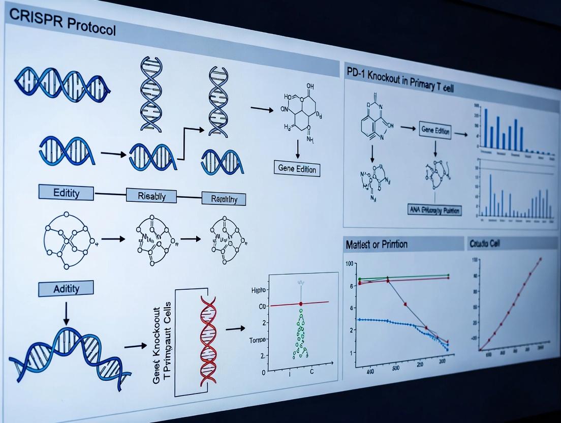
This comprehensive guide details a robust and optimized protocol for generating PD-1-deficient primary T cells using CRISPR-Cas9 genome editing.
A Step-by-Step CRISPR Protocol for Efficient PD-1 Knockout in Primary Human T Cells: From Design to Functional Validation
Abstract
This comprehensive guide details a robust and optimized protocol for generating PD-1-deficient primary T cells using CRISPR-Cas9 genome editing. Targeted at researchers and therapeutic developers, it covers the foundational rationale for PD-1 disruption in immuno-oncology, a detailed step-by-step methodology for nucleofection and culture, critical troubleshooting for low efficiency and viability, and rigorous validation techniques including flow cytometry, sequencing, and functional assays. The article synthesizes best practices to enable reliable production of engineered T cells for advanced cellular therapy research.
Why Knock Out PD-1 in T Cells? Unlocking the Therapeutic Rationale and Key Considerations
The programmed cell death protein 1 (PD-1) and its ligand PD-L1 constitute a primary immune checkpoint pathway that tumors exploit to evade immune destruction. Engagement of PD-1 on T cells by PD-L1 (or PD-L2) on tumor or antigen-presenting cells delivers an inhibitory signal, suppressing T cell receptor (TCR) signaling and promoting an exhausted T cell phenotype. This pathway is a major therapeutic target in oncology. The following tables summarize key quantitative data.
Table 1: Clinical Response Rates to FDA-Approved Anti-PD-1/PD-L1 Monotherapies (Select Cancers)
| Cancer Type | Drug (Target) | Approx. Overall Response Rate (ORR) | Key Clinical Trial(s) |
|---|---|---|---|
| Metastatic Melanoma | Pembrolizumab (PD-1) | 33-45% | KEYNOTE-006, KEYNOTE-002 |
| Metastatic Melanoma | Nivolumab (PD-1) | 40-44% | CheckMate 067, CheckMate 037 |
| NSCLC (1L, PD-L1 ≥50%) | Pembrolizumab (PD-1) | ~39-45% | KEYNOTE-024, KEYNOTE-042 |
| NSCLC (2L+) | Nivolumab (PD-1) | ~20% | CheckMate 017, 057 |
| RCC (1L) | Pembrolizumab + Axitinib (PD-1) | ~59% (ORR) | KEYNOTE-426 |
| Classical Hodgkin Lymphoma | Nivolumab (PD-1) | ~69% | CheckMate 205, 039 |
Table 2: Biomarker Prevalence and Correlation with Response
| Biomarker | Typical Measurement Method | Prevalence in Solid Tumors | Correlation with Anti-PD-1/PD-L1 Response |
|---|---|---|---|
| PD-L1 IHC (TPS ≥1%) | Immunohistochemistry (IHC) | Varies widely (e.g., ~20-30% NSCLC) | Positive association; not absolute predictor |
| Tumor Mutational Burden (TMB) High | NGS panels (mutations/Mb) | ~10-20% across solid tumors | Positive association; predictive in some cancers (e.g., TMB-H ≥10 mut/Mb) |
| Microsatellite Instability-High (MSI-H) | PCR or NGS | ~2-4% across solid tumors | Strong predictor; agnostic approval basis |
| CD8+ T-cell Infiltrate | IHC, RNA-seq, multiplex IF | Variable | Positive association with response |
Detailed Signaling Pathway of the PD-1/PD-L1 Axis
The following diagram details the molecular signaling events triggered upon PD-1 engagement.
Application Note: CRISPR-Cas9-Mediated PD-1 Knockout in Primary Human T Cells
Objective: To generate PD-1 deficient primary human T cells for in vitro and in vivo functional studies of T cell exhaustion and anti-tumor efficacy.
Background: Knocking out PD-1 in T cells removes a key intrinsic brake, potentially enhancing their proliferative capacity, cytokine production, and persistence in chronic antigen exposure settings, such as the tumor microenvironment. This protocol is designed for research use within a broader thesis investigating engineered T cell therapies.
The Scientist's Toolkit: Key Reagent Solutions
Table 3: Essential Materials for PD-1 KO in Primary T Cells
| Item Name | Function/Benefit | Example Supplier/Product |
|---|---|---|
| Primary Human T Cells | Source material; isolated from PBMCs or leukopaks. | STEMCELL Technologies (Pan T Cell Kit), Miltenyi Biotec |
| CRISPR Ribonucleoprotein (RNP) | Cas9 protein + sgRNA complex for direct, transient editing. | Synthego (sgRNA), IDT (Alt-R S.p. Cas9 Nuclease V3) |
| PD-1 Target sgRNA | Guides Cas9 to exon 1 or 2 of PDCD1 gene for frameshift KO. | Designed via CRISPick; Synthego or IDT synthesis |
| Electroporation System | For high-efficiency RNP delivery (nucleofection). | Lonza 4D-Nucleofector X Unit, SF Cell Line Kit |
| T Cell Activation & Expansion Media | Stimulates proliferation and supports growth post-editing. | TexMACS + IL-2 (Miltenyi), ImmunoCult-XF (STEMCELL) |
| Activation Beads (αCD3/αCD28) | Mimics APC engagement for robust T cell activation pre-edit. | Gibco Dynabeads, Miltenyi TransAct |
| Flow Cytometry Antibodies (Anti-hPD-1) | Validates surface PD-1 protein knockout efficiency. | BioLegend (clone EH12.2H7), BD Biosciences |
| T7 Endonuclease I or ICE Assay | Assesses genomic editing efficiency at target locus. | NEB T7E1, Synthego ICE Analysis Tool |
| In Vitro Suppression/Re-stimulation Assay | Functional validation using PD-L1 expressing cells. | aAPCs, Tumor cell lines engineered for PD-L1 OE |
Detailed Protocol: PD-1 Knockout via Electroporation of CRISPR RNP
Workflow Overview:
Step-by-Step Methodology:
Day -2 to 0: T Cell Isolation and Activation
- Isolate untouched human T cells from PBMCs using a negative selection kit.
- Count cells and resuspend in pre-warmed, serum-free T cell expansion medium (e.g., TexMACS) supplemented with 100 IU/mL recombinant IL-2.
- Activate T cells using anti-CD3/CD28 activation beads at a 1:1 bead-to-cell ratio. Culture at 1-2 x 10^6 cells/mL in a 37°C, 5% CO2 incubator for 48-72 hours.
Day 0: RNP Complex Preparation and Nucleofection Materials: Lonza SF Cell Line 4D-Nucleofector X Kit, Alt-R Cas9 nuclease, synthetic sgRNA (targeting PDCD1), P3 Primary Cell 4D-Nucleofector Solution.
- RNP Complex Assembly: For one reaction targeting 1-2e6 cells, combine 5 µg (≈37 pmol) Alt-R Cas9 protein with 6 µL of 100 µM synthetic sgRNA (60 pmol, 1.5:1 sgRNA:Cas9 molar ratio) in a sterile tube. Incubate at room temperature for 10-20 minutes.
- Cell Preparation: Harvest activated T cells, count, and remove beads magnetically. Wash cells once with PBS. Pellet 1-2e6 cells per condition.
- Nucleofection Mix: Resuspend cell pellet in 100 µL of pre-warmed P3 Primary Cell Solution. Add the prepared RNP complex. Mix gently by pipetting. Transfer the entire volume to a nucleofection cuvette.
- Electroporation: Place cuvette in the 4D-Nucleofector X Unit and run the pre-optimized program for primary human T cells (e.g., EO-115). Immediately after the pulse, add 500 µL of pre-warmed, IL-2 supplemented medium to the cuvette.
Day 0-10: Recovery, Expansion, and Analysis
- Transfer cells from the cuvette to a pre-warmed culture plate. Return to the incubator.
- Day 1 Post-Nucleofection: Assess viability (expect 50-70%). Expand culture, maintaining cell density between 0.5-2e6 cells/mL with fresh medium and IL-2 every 2-3 days.
- Day 3-5: Initial Validation: Harvest a sample (≈0.5e6 cells) for analysis.
- Flow Cytometry: Stain for surface PD-1 expression to assess knockout efficiency. Include isotype and untransfected controls.
- Genomic Analysis: Isolate genomic DNA. Perform PCR amplification of the target region. Use T7E1 assay or ICE analysis to calculate indel percentage.
- Day 7-10: Functional Assay: Co-culture PD-1 KO and control T cells with PD-L1 expressing target cells (e.g., antigen-pulsed, PD-L1+ aAPCs or tumor cells) at various E:T ratios.
- Measure after 24-48h: IFN-γ secretion (ELISA), T cell proliferation (CFSE dilution), and cytolytic activity (LDH or Incucyte killing assays).
Critical Parameters & Troubleshooting:
- Activation State: 48-72 hour pre-activation is critical for high editing efficiency in primary T cells.
- RNP Ratio & Quality: Optimize sgRNA:Cas9 ratio (1.5:1 to 2:1). Use HPLC-purified sgRNAs.
- Cell Health: Do not exceed 2e6 cells per nucleofection. Use fresh, high-viability cells and pre-warmed solutions throughout.
- Controls: Always include a non-targeting sgRNA RNP control and an untransfected cell control.
This document provides application notes and detailed protocols, framed within a broader thesis utilizing CRISPR/Cas9 for PD-1 knockout in primary T cells. The objective is to translate foundational research, such as checkpoint disruption, into the development of enhanced, next-generation adoptive cell therapies like CAR-T and TCR-T cells. These protocols integrate gene editing with cell engineering workflows.
Application Notes: Quantitative Data on Edited T-Cell Therapies
Live search data indicates current clinical and preclinical benchmarks for engineered T cells.
Table 1: Key Performance Metrics of Edited vs. Non-Edited T-Cell Therapies
| Metric | Non-Edited CAR-T (axicabtagene ciloleucel) | TCR-T Cells (NY-ESO-1) | CAR-T with PD-1 Knockout (Preclinical) | Protocol Reference Section |
|---|---|---|---|---|
| Objective Response Rate (ORR) | 83% (LBCL) | 55% (Synovial Sarcoma) | N/A (Preclinical) | N/A |
| Complete Response (CR) Rate | 58% (LBCL) | 20% (Synovial Sarcoma) | N/A | N/A |
| Median Duration of Response | 11.1 months | 44.2 months | N/A | N/A |
| % PD-1+ Cells (Post-Exhaustion) | >60% | ~40-50% | <10% | Protocol 2.3 |
| Cytokine Production (IFN-γ) Fold Change | 1x (Baseline) | 1.5x | 3-5x Increase | Protocol 2.4 |
| In Vivo Tumor Clearance (Mouse Model) | Partial | Significant | Enhanced & Sustained | Protocol 3.1 |
Table 2: Common CRISPR Delivery Methods for Primary T-Cell Engineering
| Delivery Method | Editing Efficiency (PD-1 Locus) | Cell Viability (Day 3) | Relative Cost | Best For |
|---|---|---|---|---|
| Electroporation (RNP) | 70-85% | 60-75% | $$ | Clinical-grade protocols |
| Lentiviral (saCas9) | 40-60% | >80% | $$$ | Long-term expression |
| AAV6 (Donor Template) | N/A (HDR) | 70-80% | $$$$ | Knock-in strategies |
| mRNA Electroporation | 50-70% | 50-65% | $ | Rapid, transient expression |
Detailed Protocols
Protocol 1: CRISPR/Cas9-Mediated PD-1 Knockout in Primary Human T Cells
Objective: Generate PD-1 deficient T cells as a foundational step for producing resistant CAR-T/TCR-T cells.
Materials: See "Scientist's Toolkit" below. Workflow:
- T-Cell Isolation & Activation: Isolate CD3+ T cells from PBMCs using negative selection beads. Activate with CD3/CD28 Dynabeads (1:1 bead:cell ratio) in TexMACS medium + 100 IU/mL IL-2.
- RNP Complex Formation: For a 100µL reaction, complex 10µg of high-fidelity Cas9 protein with 6µg of synthetic sgRNA (targeting human PDCD1 exon 1) by incubating at 25°C for 10 minutes.
- Electroporation: At 48 hours post-activation, wash cells. Resuspend 1-2e6 cells in 20µL P3 buffer. Add RNP complex plus 2µL of 100µM electroporation enhancer. Electroporate using 4D-Nucleofector (program EO-115). Immediately add pre-warmed medium.
- Recovery & Expansion: Culture cells in IL-2 (100 IU/mL) containing medium. Replace medium every 2-3 days. Remove activation beads on Day 5.
- Analysis: Assess editing efficiency at Day 5-7 via flow cytometry (loss of PD-1 surface expression) and T7E1 assay or NGS on genomic DNA.
Protocol 2: Engineering Next-Gen CAR-T Cells with PD-1 Knockout
Objective: Integrate a CAR construct into PD-1 knockout T cells to create exhaustion-resistant therapy.
Workflow:
- Sequential Editing-Engineering: Perform Protocol 1. On Day 3 post-electroporation, transduce cells with a lentiviral vector encoding the CAR (e.g., anti-CD19 scFv-4-1BB-CD3ζ) at an MOI of 3-5 in the presence of 8µg/mL polybrene by spinfection.
- CAR+ Cell Selection: Culture for 7-10 days. Enrich CAR+ cells via magnetic bead selection (e.g., biotinylated protein L + streptavidin beads) if necessary.
- Functional Validation (Cytotoxicity): Co-culture engineered CAR-T cells with target-positive (e.g., NALM-6) and target-negative cells at various E:T ratios for 24h. Measure specific lysis via LDH release or luciferase-based assays.
- Exhaustion Resistance Assay: Subject CAR-T cells to repetitive antigen stimulation (e.g., irradiated target cells every 7 days). Compare control (PD-1 WT) vs. PD-1 KO CAR-T cells for persistence (cell counts), sustained cytokine production (multiplex ELISA), and expression of exhaustion markers (Tim-3, LAG-3) by flow cytometry weekly.
Protocol 3: In Vivo Potency Assessment in NSG Mouse Model
Objective: Evaluate the superior antitumor activity of PD-1 edited CAR-T/TCR-T cells.
Workflow:
- Tumor Engraftment: Inject 5e6 luciferase-expressing tumor cells (e.g., Raji for CD19+ lymphoma) subcutaneously into NSG mice.
- Therapy Administration: At Day 7 (tumor palpable), randomize mice into groups. Inject 5e6 control CAR-T or PD-1 KO CAR-T cells intravenously.
- Monitoring: Measure tumor bioluminescence weekly. Monitor mouse survival. For endpoint analysis, harvest tumors and infiltrating T cells for flow cytometry to characterize phenotype and exhaustion status.
Visualization: Diagrams & Workflows
Title: Workflow for Engineering Next-Gen Cell Therapies with PD-1 Knockout
Title: PD-1 Signaling Disruption by CRISPR Enhances T Cell Function
The Scientist's Toolkit: Research Reagent Solutions
Table 3: Essential Materials for PD-1 Knockout & T-Cell Engineering
| Item | Function & Role in Protocol | Example Product/Catalog |
|---|---|---|
| Human T-Cell Isolation Kit | Negative selection for untouched, high-purity CD3+ T cells from PBMCs. | Miltenyi Pan T Cell Isolation Kit |
| CD3/CD28 T Cell Activator | Polyclonal activation for priming T cells for editing and expansion. | Gibco Dynabeads CD3/CD28 |
| Recombinant IL-2 | Supports T-cell survival and proliferation during culture post-editing. | PeproTech Proleukin (rhIL-2) |
| High-Fidelity Cas9 Nuclease | Minimizes off-target editing for clinically relevant protocols. | IDT Alt-R S.p. HiFi Cas9 |
| Synthetic sgRNA (PDCD1) | Targets exon 1 of human PD-1 gene for knockout. | Synthego PDCD1 sgRNA (crRNA+tracrRNA) |
| Nucleofector System & Kit | Enables high-efficiency RNP delivery into primary T cells. | Lonza 4D-Nucleofector X Kit, P3 buffer |
| Lentiviral CAR/TCR Vector | Delivers therapeutic transgene for antigen-specific targeting. | Custom construct with 4-1BB/CD3ζ signaling |
| Flow Antibody: Anti-human PD-1 | Critical for assessing knockout efficiency via surface staining. | BioLegend clone EH12.2H7 |
| Cell Culture Medium | Optimized, serum-free medium for robust human T-cell growth. | Miltenyi TexMACS GMP Medium |
| Cytotoxicity Assay Kit | Quantifies target cell lysis by engineered T cells. | Promega LDH-Glo Cytotoxicity Assay |
The use of primary human T cells over immortalized T cell lines (e.g., Jurkat, HuT78) has become a cornerstone for enhancing the translational relevance of immunology and immuno-oncology research. This is particularly critical in the context of developing CRISPR-based cellular therapies, such as PD-1 knockout T cells for cancer immunotherapy. Primary T cells recapitulate the physiological heterogeneity, signaling, metabolic state, and functional responses of T cells in patients, while cell lines, though convenient, possess transformed phenotypes that can lead to misleading conclusions.
Comparative Advantages: Primary T Cells vs. Cell Lines
Table 1: Key Comparative Features
| Feature | Primary Human T Cells | Immortalized T Cell Lines (e.g., Jurkat) | Translational Impact |
|---|---|---|---|
| Genetic & Epigenetic Landscape | Normal karyotype; donor-specific epigenetic programming. | Aneuploidy; aberrant epigenetic regulation from immortalization. | Predicts clinical efficacy and safety of gene-edited products. |
| Signaling Pathways | Intact, physiological TCR signaling and co-stimulatory/inhibitory networks. | Often dysregulated (e.g., constitutive pathways, mutated PTEN). | Accurate assessment of interventions like PD-1 knockout on signaling. |
| Metabolic Profile | Reliance on oxidative phosphorylation in naïve/memory states; can shift to glycolysis upon activation. | Primarily glycolytic (Warburg effect), typical of transformed cells. | Critical for predicting in vivo persistence and function of therapeutic T cells. |
| Phenotypic Heterogeneity | Diverse subsets (Naïve, Effector, Memory, Exhausted) present. | Clonal, homogeneous population lacking subset diversity. | Enables study of editing effects across relevant T cell subsets for therapy. |
| Functional Assays | Physiological cytokine production, cytolytic activity, and proliferation in response to antigen. | Often hyper-responsive or hyporesponsive; may not require antigen presentation. | Data directly correlate with expected patient T cell behavior post-therapy. |
| Clinical Relevance | Direct ex vivo model of patient material. | Model of limited physiological relevance. | Reduces the risk of late-stage translational failure. |
Table 2: Quantitative Comparison of Key Functional Parameters
| Parameter | Primary T Cells (Avg. Range) | Jurkat Cell Line | Source/Notes |
|---|---|---|---|
| Doubling Time (activated) | ~20-30 hours | ~24-30 hours | Primary cells require activation. |
| PD-1 Expression (basal/induced) | 1-5% (basal), >50% (upon TCR activation) | Negligible to low (non-inducible) | Primary cells model physiological regulation. |
| IFN-γ Secretion upon TCR stimulation | 500-5000 pg/mL per 10^6 cells | Minimal without exogenous manipulation | Critical for efficacy readout. |
| Cytotoxic Activity (specific lysis) | 20-80% (E:T = 10:1) | Not applicable | Primary CTLs are functionally cytotoxic. |
| Transfection/Efficiency | 30-70% (electroporation) | >80% (electroporation) | Primary cells are more challenging to manipulate. |
Application Notes: CRISPR-Cas9 PD-1 Knockout in Primary T Cells
Critical Considerations for Experimental Design
- Donor Variability: Use cells from ≥3 healthy donors to account for genetic and immunological diversity.
- T Cell Subset Selection: Isolate specific subsets (e.g., CD8+, naïve, or memory) using magnetic or flow sorting based on the research question.
- Activation Requirement: Primary T cells require activation (e.g., anti-CD3/CD28 beads) for efficient gene editing and expansion.
- Timeline: The protocol from isolation to functional assay typically takes 10-14 days.
Detailed Protocol: PD-1 Knockout in Primary Human T Cells Using CRISPR-Cas9 RNP Electroporation
Materials & Reagents
Table 3: The Scientist's Toolkit - Key Research Reagent Solutions
| Item | Function/Benefit | Example/Note |
|---|---|---|
| Human PBMCs or Leukopak | Source of primary T cells. | Commercial vendors or donor blood draws. |
| Pan T Cell Isolation Kit (Human) | Negative selection for untouched T cells. | Maintains cell health and avoids activation. |
| Recombinant Human IL-2 | Supports T cell growth and survival post-activation/editing. | Use at 50-100 IU/mL for expansion. |
| Anti-CD3/CD28 Dynabeads | Polyclonal T cell activator mimicking APC engagement. | Critical for inducing proliferation and PD-1 expression. |
| Alt-R S.p. HiFi Cas9 Nuclease V3 | High-fidelity Cas9 protein for RNP complex formation. | Reduces off-target editing. |
| Alt-R CRISPR-Cas9 sgRNA (targeting PDCD1) | Synthetic, chemically modified sgRNA for high stability and efficiency. | Target exon 1 or 2 of the PDCD1 gene. |
| Electroporation System & Buffer | For efficient RNP delivery. | Neon (Thermo) or 4D-Nucleofector (Lonza) systems. |
| Flow Antibodies: Anti-CD3, CD8, PD-1 | For assessing editing efficiency and phenotype. | Use clone EH12.2H7 for human PD-1. |
| Cellular DNA Extraction Kit | Genomic DNA isolation for sequencing validation. | |
| T7 Endonuclease I or ICE Analysis | For initial assessment of indel formation. | Surveyor or ICE assay. |
Protocol Steps
Day 0: T Cell Isolation and Activation
- Isolate PBMCs from fresh blood or leukopak via density gradient centrifugation (Ficoll-Paque).
- Isolate untouched T cells using a negative selection magnetic bead kit. Resuspend in complete medium (RPMI-1640, 10% FBS, 1% Pen/Strep).
- Activate T cells at a density of 1x10^6 cells/mL with anti-CD3/CD28 beads at a 1:1 bead-to-cell ratio. Add IL-2 to 50 IU/mL.
- Incubate at 37°C, 5% CO2 for 48 hours.
Day 2: CRISPR RNP Complex Assembly and Electroporation
- Design sgRNA: Use a validated sgRNA sequence targeting PDCD1 (e.g., exon 1: GAGCACAGGCAGCATGTGGA).
- Prepare RNP Complex: For a 10 µL Neon tip reaction, combine 3 µg Alt-R Cas9 protein with 1.5 nmol of sgRNA in sterile buffer. Incubate at room temperature for 10-20 minutes.
- Harvest Activated T Cells: Remove beads magnetically. Wash cells once with PBS.
- Electroporation: Resuspend 1-2x10^6 cells in 10 µL of Neon Electroporation Buffer R. Mix with the pre-formed RNP complex. Electroporate using manufacturer's optimized conditions for primary T cells (e.g., Neon: 1600V, 10ms, 3 pulses). Immediately transfer cells to pre-warmed complete medium with IL-2.
- Control: Include a non-targeting sgRNA control.
Days 2-10: Post-Electroporation Expansion
- Culture edited and control cells at 0.5-1x10^6 cells/mL in complete medium with IL-2 (50-100 IU/mL).
- Perform a 1:2 split or feed with fresh medium+IL-2 every 2-3 days.
- Monitor cell density and viability (expected >70%).
Day 7-10: Analysis
- Flow Cytometry for PD-1 Surface Expression: Stimulate a sample of cells with PMA/lonomycin for 6 hours to induce maximal PD-1 expression. Stain with anti-CD3, CD8, and PD-1 antibodies. Compare knockout to control populations to assess editing efficiency.
- Genomic Validation: Extract genomic DNA from edited bulk population. PCR amplify the target region. Assess indel percentage via T7E1 assay or, preferably, by Sanger sequencing followed by ICE analysis.
- Functional Assay: Co-culture PD-1 KO and control T cells with PD-L1-expressing target cells (e.g., cancer cells). Measure IFN-γ secretion (ELISA) or tumor cell killing (incucyte or flow-based cytotoxicity assay).
Visualizations
Diagram Title: CRISPR PD-1 KO Workflow in Primary T Cells
Diagram Title: T Cell Activation vs. PD-1 Inhibitory Signaling
The genetic knockout of Programmed Cell Death Protein 1 (PD-1) in primary T cells is a cornerstone approach in developing enhanced cellular therapies for cancer. Selecting the optimal gene-editing tool is critical for efficiency, specificity, and clinical translatability. This analysis, framed within a thesis on optimizing CRISPR for PD-1 knockout, compares CRISPR-Cas9 to other major editing platforms.
Table 1: Quantitative Comparison of Gene-Editing Tools for PD-1 Knockout in Primary T Cells
| Tool | Editing Mechanism | Typical Editing Efficiency (PD-1 Locus) | Key Advantages | Key Limitations for PD-1 KO |
|---|---|---|---|---|
| CRISPR-Cas9 (RNP) | Nuclease creates DSB, repaired by NHEJ. | 60-85% | High efficiency, rapid protocol, minimal off-target with engineered Cas9, easily multiplexed. | Potential for chromosomal translocations in multiplexing. |
| TALENs | Nuclease creates DSB, repaired by NHEJ. | 20-40% | High sequence specificity, lower off-target risk than wild-type SpCas9. | Low efficiency in primary cells, complex protein design/cloning. |
| ZFNs | Nuclease creates DSB, repaired by NHEJ. | 15-35% | Smaller delivery footprint than TALENs. | Lower efficiency, significant off-target effects, difficult to design. |
| CRISPRa/i (Interference) | Epigenetic silencing via dCas9-effectors. | ~90% repression (not knockout) | Reversible, no DNA cleavage. | Transcriptional silencing only, not a permanent knockout. |
| mRNA Electroporation | Overexpression of dominant-negative PD-1. | N/A (protein overexpression) | No genomic editing required. | Transient effect, may not fully block signaling. |
Why CRISPR-Cas9 is Preferred:
- High Efficiency in Primary Cells: CRISPR-Cas9 ribonucleoprotein (RNP) delivery achieves knockout rates sufficient for clinical manufacturing without prolonged ex vivo culture.
- Speed and Simplicity: Designing and synthesizing gRNAs is faster and more cost-effective than engineering TALE or Zinc-finger proteins.
- Multiplexing Capability: Allows concurrent knockout of PD-1 with other checkpoints (e.g., CTLA-4) or insertion of transgenes using a single delivery system.
- Enhanced Specificity: High-fidelity Cas9 variants (e.g., HiFi Cas9, eSpCas9) reduce off-target effects to levels comparable or superior to TALENs.
- Clinical Traction: Multiple ongoing clinical trials utilize CRISPR-Cas9 for PD-1 knockout in T cells (e.g., NCT03538613, NCT03399448), establishing a regulatory precedent.
Detailed Application Notes & Protocol: PD-1 Knockout in Human Primary T Cells Using CRISPR-Cas9 RNP
Thesis Context: This protocol is optimized as part of a systematic investigation into maximizing knockout efficiency while preserving T-cell viability and function for adoptive cell therapy.
Research Reagent Solutions:
| Reagent/Material | Function | Example Catalog # |
|---|---|---|
| Human T Cells (CD3+) | Primary cell target for PD-1 knockout. | Isolated from PBMCs. |
| CRISPR-Cas9 Nuclease | S. pyogenes Cas9 protein. Forms active complex with sgRNA. | TrueCut Cas9 Protein v2. |
| Synthetic sgRNA | Targets Cas9 to exon 1 or 2 of the PDCD1 gene. | Synthego, IDT Alt-R. |
| Electroporation System | For RNP delivery (e.g., Neon, Nucleofector). | Neon Transfection System 100µL Kit. |
| TexMACS GMP Medium | Serum-free culture medium supporting T-cell activation/expansion. | Miltenyi Biotec. |
| CD3/CD28 Activator | Activates T cells, inducing cell cycle and PD-1 expression. | Gibco Dynabeads. |
| IL-2, IL-7, IL-15 | Cytokines for T-cell survival and expansion post-editing. | PeproTech. |
| Flow Antibodies (anti-CD3, anti-PD-1) | For assessing knockout efficiency and phenotype. | BioLegend clones OKT3, EH12.2H7. |
| T7 Endonuclease I / NGS Kit | For assessing on- and off-target editing. | Guide-it Assay, Illumina MiSeq. |
Experimental Workflow Protocol:
Day -1: T-Cell Isolation and Activation
- Isolate CD3+ T cells from PBMCs using negative selection magnetic beads.
- Count cells and assess viability (target >95%).
- Activate cells at 1e6 cells/mL in TexMACS medium supplemented with CD3/CD28 activator beads (bead:cell ratio 1:1) and 100 IU/mL IL-2.
- Incubate at 37°C, 5% CO2 for 24 hours.
Day 0: RNP Complex Formation and Electroporation
- sgRNA Resuspension: Resuspose synthetic sgRNA (target: PDCD1 exon2, sequence: 5'-GUUUAACAAGCUAGACCAGU-3') in nuclease-free buffer.
- RNP Complexing: Combine 6 µg Cas9 protein with 3 µg sgRNA (3:1 mass ratio). Incubate at room temperature for 10-20 minutes.
- Cell Preparation: Collect activated T cells, remove beads magnetically, wash with PBS, and resuspend in Neon Resuspension Buffer R at 5e7 cells/mL.
- Electroporation: Mix 2e6 cells (40 µL) with pre-complexed RNP (total volume ≤10 µL). Electroporate using the Neon System (1400V, 10ms, 3 pulses). Immediately transfer cells to pre-warmed complete medium.
- Controls: Include a non-electroporated control and an RNP complex with a non-targeting sgRNA.
Day 1-12: Post-Editing Culture and Expansion
- After 24h, dilute cells to 5e5 cells/mL in fresh TexMACS medium with IL-2 (100 IU/mL), IL-7 (10 ng/mL), and IL-15 (10 ng/mL).
- Feed or split cells every 2-3 days, maintaining density between 5e5 and 2e6 cells/mL.
- Monitor viability and growth daily.
Day 5-7: Assessment of Knockout Efficiency
- Harvest 2-5e5 cells from experimental and control groups.
- Stain for surface markers (anti-CD3, anti-PD-1) and a viability dye.
- Analyze by flow cytometry. PD-1 knockout efficiency = % of live CD3+ cells that are PD-1 negative in edited sample minus % in non-targeting control.
Downstream Validation (Thesis Core Analysis)
- Genomic Cleavage: Isolate genomic DNA. Use T7E1 assay or NGS to confirm indels at the PDCD1 target site.
- Functional Assay: Re-stimulate edited and control T cells with anti-CD3. Assess IFN-γ production (ELISA) or proliferation as a functional readout of PD-1 signaling ablation.
- Off-Target Analysis: Perform targeted NGS on top 3-5 predicted off-target sites (via in silico tools like GuideScan) for HiFi Cas9 experiments.
Visualizations
Title: Experimental Workflow for PD-1 Knockout.
Title: PD-1 Signaling and Knockout Effect.
Title: Tool Selection Logic for PD-1 Knockout.
This Application Note outlines the critical preparatory steps for a research project framed within a broader thesis on CRISPR-mediated PD-1 knockout in primary human T cells for enhancing anti-tumor immunotherapy. Success hinges on rigorous planning in three domains: navigating institutional approvals, designing highly efficient and specific gRNAs, and selecting appropriate experimental controls.
Part 1: Institutional Approvals and Biosafety
Before any bench work, researchers must secure formal approvals. The requirements and timelines are summarized below.
Table 1: Typical Institutional Approval Requirements and Timelines
| Approval Body | Primary Concern | Typical Review Timeline | Key Submission Documents |
|---|---|---|---|
| Institutional Biosafety Committee (IBC) | Risk of gene editing, use of viral vectors (e.g., lentivirus for RNP/donor delivery). | 4-8 weeks | Biosafety Protocol, Risk Assessment, SOPs for handling. |
| Institutional Review Board (IRB) / Ethics Committee | Sourcing and use of human primary cells (e.g., donor blood, leukapheresis products). | 8-12 weeks | Study Protocol, Informed Consent Forms, Donor Privacy Plan. |
| Stem Cell/Embryonic Research Oversight | Only if using human embryonic stem cell-derived T cells. | 12+ weeks | Detailed scientific justification, ethical review document. |
| Animal Care and Use Committee (IACUC) | If subsequent in vivo engraftment of edited T cells is planned. | 8-10 weeks | Animal Use Protocol, Justification of Species/Numbers, Pain Management Plan. |
Protocol 1.1: Initiating the IBC Protocol Submission
- Identify Applicable Regulations: Confirm if your work falls under the NIH Guidelines for Research Involving Recombinant or Synthetic Nucleic Acid Molecules (Section III-A, III-B, III-C, etc.).
- Draft the Protocol: Detail the biological agents (e.g., S. pyogenes Cas9, gRNA sequences, lentiviral constructs), the specific knockout procedure, and all waste decontamination methods.
- Complete Training: Ensure all personnel complete mandatory biosafety (Bloodborne Pathogens, Cell Culture) and gene editing training.
- Submit and Revise: Submit the draft to your institution's IBC office. Be prepared to answer questions and revise the protocol during committee review.
Part 2: gRNA Design and Validation for PD-1 Knockout
The PD-1 gene (PDCD1) is located on chromosome 2 in humans. Effective gRNAs target early exons to cause frameshift mutations.
Table 2: Candidate gRNAs for Human PDCD1 Exon 1
| gRNA ID | Target Sequence (5' to 3') | PAM | Exon | Predicted Efficiency (Doench 2016 Score) | Predicted Off-Target Sites (CFD Score < 0.5) |
|---|---|---|---|---|---|
| PD1-g1 | GAGTCCAACAGAACCACAGC | AGG | 1 | 0.85 | 2 |
| PD1-g2 | CACAGAGTTCACCCCCATGG | TGG | 1 | 0.92 | 1 |
| PD1-g3 | TGCAGCTCCCCAGAGACAAG | GGG | 1 | 0.78 | 4 |
| PD1-g4 | ACCACAGCACAGAGTTCACC | CGG | 1 | 0.88 | 1 |
Protocol 2.1: In Silico gRNA Design and Selection
- Identify Target Region: Access the PDCD1 genomic sequence (ENSG00000188389) from Ensembl. Focus on exon 1 or 2.
- Generate gRNA Candidates: Use the CHOPCHOP or Broad Institute's sgRNA Designer tool. Input the FASTA sequence for a ~500bp region around the target exon.
- Filter for Efficiency and Specificity: Select candidates with efficiency scores >0.6. Run off-target analysis using the Cas-OFFinder tool, allowing up to 3 mismatches. Prioritize gRNAs with minimal predicted off-targets, especially in other coding regions.
- Final Selection: Choose 2-3 top-ranked gRNAs for empirical validation. PD1-g2 and PD1-g4 from Table 2 are strong candidates based on high efficiency and low predicted off-targets.
Protocol 2.2: In Vitro Validation of gRNA Efficiency via T7E1 Assay Materials: Synthetic gRNAs, purified Cas9 protein, Nuclease-Free Duplex Buffer, T7 Endonuclease I, PCR reagents.
- Synthesize gRNAs: Order chemically modified, crRNA:tracrRNA duplexes or sgRNAs for top candidates.
- Form RNP Complexes: For each gRNA, complex 30 pmol with 20 pmol of Cas9 protein in buffer. Incubate 10 min at 25°C.
- Amplify Target Locus: Design PCR primers ~300-500bp flanking the gRNA cut site in PD-1. Perform PCR using genomic DNA from Jurkat or unstimulated primary T cells.
- Cleavage Reaction: Mix 200ng of purified PCR product with 2µL of pre-formed RNP. Incubate at 37°C for 1 hour.
- T7E1 Digestion: Purify the cleavage reaction product. Add T7E1 enzyme to the purified DNA and incubate at 37°C for 30 minutes.
- Analyze on Gel: Run digested products on a 2% agarose gel. Cleavage efficiency (%) = (1 - sqrt(1 - (b+c)/(a+b+c))) * 100, where a is the integrated intensity of the undigested PCR product, and b & c are the digested fragment intensities.
Part 3: Control Selection Strategy
A comprehensive control strategy is non-negotiable for interpreting knockout specificity and functional outcomes.
Table 3: Essential Control Conditions for PD-1 KO Experiments
| Control Type | Purpose | Example for PD-1 KO Study |
|---|---|---|
| Unedited | Baseline phenotype and function. | T cells electroporated with Cas9 protein only (no gRNA). |
| Non-Targeting gRNA | Control for cellular responses to RNP electroporation/gRNA presence. | T cells electroporated with Cas9 + a scrambled gRNA with no genomic target. |
| Targeting Control (Essential Gene) | Control for editing efficiency and cytotoxicity. | T cells edited with a gRNA targeting a housekeeping gene (e.g., PPIB). Monitor cell viability. |
| On-Target Positive Control | Confirm system functionality. | A well-validated gRNA for a different, easy-to-detect target (e.g., TRAC) can be run in parallel. |
| Off-Target Negative Control | Assess specificity. | Include a gRNA known to have high off-target effects as a negative control for assays measuring immune synapse function or exhaustion markers. |
Protocol 3.1: Setting Up Control Electroporations
- Prepare T Cells: Isolate and activate primary human CD3+ T cells from healthy donor PBMCs using anti-CD3/CD28 beads for 48-72 hours.
- Prepare RNP Complexes: Formulate RNP complexes for each condition: a) No gRNA (Cas9 only), b) Non-targeting control gRNA, c) PD-1-targeting gRNA(s), d) Positive control gRNA (e.g., TRAC).
- Electroporate: Use a validated electroporation system (e.g., Lonza Nucleofector) with primary T cell kits. Electroporate 1-2e6 cells per condition with pre-formed RNPs.
- Culture and Analyze: Culture cells in IL-2 containing media. Assess editing efficiency at the genomic DNA level (T7E1 or NGS) at 72-96 hours post-electroporation. Assess PD-1 surface protein loss by flow cytometry at day 5-7 post-editing.
The Scientist's Toolkit: Research Reagent Solutions
Table 4: Essential Materials for PD-1 KO in Primary T Cells
| Item | Function | Example Product/Supplier |
|---|---|---|
| Primary Human T Cells | Target cell for gene editing. | Isolated from donor PBMCs using CD3+ selection kits (e.g., Miltenyi Biotec). |
| Recombinant S. pyogenes Cas9 Nuclease | Engineered nuclease for creating double-strand breaks. | Alt-R S.p. HiFi Cas9 Nuclease V3 (Integrated DNA Technologies). |
| Chemically Modified sgRNA | Guides Cas9 to the PD-1 locus; chemical modifications enhance stability. | Alt-R CRISPR-Cas9 sgRNA, custom sequence (Integrated DNA Technologies). |
| Electroporation System | Device for delivering RNP complexes into primary T cells. | Nucleofector 4D with SF Cell Line Kit (Lonza). |
| T Cell Activation Beads | Stimulates T cell proliferation and editing susceptibility. | Gibco Human T-Activator CD3/CD28 Dynabeads (Thermo Fisher). |
| Recombinant Human IL-2 | Cytokine for maintaining T cell viability and expansion post-editing. | PeproTech. |
| Flow Cytometry Antibodies | Validation of PD-1 surface protein knockout and phenotyping. | Anti-human CD279 (PD-1) APC (clone EH12.2H7, BioLegend). |
| Genomic DNA Extraction Kit | For isolating DNA to assess editing efficiency. | Quick-DNA Miniprep Kit (Zymo Research). |
| T7 Endonuclease I | Enzyme for detecting indels via mismatch cleavage. | EnGen Mutation Detection Kit (NEB). |
Visualizations
Approval Workflow for PD-1 KO Research
gRNA Design and Validation Workflow
Experimental Control Strategy Overview
Hands-On Protocol: A Detailed Workflow for CRISPR-Cas9 Mediated PD-1 Knockout in Primary T Cells
Research Reagent Solutions: Essential Toolkit for PD-1 Knockout in Primary T Cells
The following table details the key reagents, kits, and materials essential for successful CRISPR-Cas9-mediated PD-1 knockout in primary human T cells. This toolkit is curated for high-efficiency editing, viability, and subsequent functional assays.
| Category | Product Name / Reagent | Supplier (Example) | Function & Brief Explanation |
|---|---|---|---|
| T Cell Isolation | Human CD3+ T Cell Isolation Kit (negative selection) | Miltenyi Biotec / STEMCELL | Enriches untouched, viable primary T cells from PBMCs, minimizing activation prior to editing. |
| T Cell Activation & Culture | TexMACS Medium | Miltenyi Biotec | Serum-free, GMP-compliant medium optimized for human T cell culture and expansion. |
| ImmunoCult Human CD3/CD28/CD2 T Cell Activator | STEMCELL | Provides robust, consistent activation via TCR and co-stimulatory signals, essential for nucleofection efficiency. | |
| CRISPR Components | Cas9 Nuclease, HiFi (or similar high-fidelity variant) | IDT / Thermo Fisher | Creates double-strand breaks at target locus. HiFi variants reduce off-target effects. |
| PDCD1 (PD-1) CRISPR RNA (crRNA) & tracrRNA | Synthego / IDT | Target-specific guide RNA (crRNA) and universal tracrRNA. Multiple guides targeting exon 1 or 2 of the PDCD1 gene are common. | |
| Synthetic, chemically modified sgRNA (Alt-R) | IDT | Pre-complexed, modified single guide RNA; enhances stability and editing efficiency. | |
| Nucleofection System | P3 Primary Cell 4D-Nucleofector X Kit S | Lonza | Gold-standard reagent kit and cuvettes specifically optimized for primary human T cell nucleofection. |
| 4D-Nucleofector Unit (or X Unit) | Lonza | Electroporation device for high-efficiency, low-toxicity delivery of RNP complexes. | |
| Post-Editing Analysis | PD-1 (CD279) Antibody, APC | BioLegend | Flow cytometry antibody to assess surface PD-1 protein knockout efficiency 3-5 days post-nucleofection. |
| Genomic DNA Extraction Kit | Qiagen / Thermo Fisher | Isolates DNA for downstream knockout confirmation. | |
| T7 Endonuclease I or ICE Analysis | NEB / Synthego | Detects insertion/deletion (indel) mutations at the target site to quantify editing efficiency. | |
| Supplementary Reagents | Recombinant Human IL-2 (or IL-7/IL-15) | PeproTech | Cytokines for T cell survival and expansion post-nucleofection. |
| Antibiotic-Antimycotic | Thermo Fisher | Prevents contamination in culture medium. | |
| DPBS, no calcium, no magnesium | Thermo Fisher | For cell washing and dilution steps. |
Application Notes & Protocols
Detailed Protocol: CRISPR-Cas9 RNP Nucleofection of Primary Human T Cells for PD-1 Knockout
Objective: To achieve high-efficiency knockout of the PDCD1 gene in activated primary human CD3+ T cells using CRISPR-Cas9 ribonucleoprotein (RNP) complex delivery via 4D-Nucleofection.
Materials from Toolkit:
- Human CD3+ T Cell Isolation Kit
- TexMACS Medium
- ImmunoCult Human CD3/CD28/CD2 T Cell Activator
- Recombinant Human IL-2
- Alt-R Cas9 HiFi Protein
- Alt-R CRISPR-Cas9 sgRNA targeting PDCD1 (e.g., target sequence: GGAGTCCAAGAGCCTAACCA)
- P3 Primary Cell 4D-Nucleofector X Kit S
- 4D-Nucleofector Unit with X Module
Workflow:
- T Cell Isolation & Activation: Isolate CD3+ T cells from PBMCs using negative selection. Activate 1x10^6 cells/mL in TexMACS medium supplemented with 1X ImmunoCult activator and 200 IU/mL IL-2. Culture for 48 hours.
- RNP Complex Formation: For 1x10^6 cells, combine 3.6 µL of 60 µM sgRNA (216 pmol) with 1.5 µg (≈9 pmol) of Cas9 HiFi protein in a sterile tube. Bring total volume to 20 µL with Duplex Buffer. Incubate at room temperature for 10-20 minutes.
- Cell Preparation: Harvest activated T cells, count, and centrifuge. Resuspend cells in pre-warmed TexMACS medium at a density of 1-2 x 10^7 cells/mL.
- Nucleofection Setup: For each reaction, add 20 µL of P3 Nucleofector Solution to the RNP complex. Add 20 µL of the cell suspension (2-4 x 10^5 cells). Gently mix and transfer into a 16-well Nucleocuvette Strip. Avoid air bubbles.
- Nucleofection: Insert strip into the 4D-Nucleofector X Module and run the recommended program for primary human T cells: Program EO-115.
- Recovery: Immediately after pulsing, add 80 µL of pre-warmed TexMACS medium to the cuvette. Gently transfer cells to a pre-warmed plate containing 1 mL of complete medium (with IL-2). Return to incubator.
- Post-Transfection Culture: 24 hours post-nucleofection, carefully replace medium with fresh TexMACS medium + IL-2. Expand cells as needed for analysis.
- Analysis: Assess viability and cell count at 24h. Analyze PD-1 surface expression by flow cytometry at day 3-5. Harvest genomic DNA for indel analysis at day 5-7.
Critical Notes:
- Cell viability post-nucleofection is typically 50-70%.
- Optimal RNP concentrations and cell numbers may require titration.
- Including a non-targeting sgRNA control is essential for interpreting functional assay results.
Protocol: Assessment of Knockout Efficiency
A. Flow Cytometry for PD-1 Surface Expression
- Harvest 1-2x10^5 cells from the edited and control cultures.
- Wash with FACS buffer (DPBS + 2% FBS).
- Stain with anti-human CD279 (PD-1)-APC antibody and a viability dye (e.g., 7-AAD) for 20-30 minutes on ice in the dark.
- Wash, resuspend in buffer, and analyze on a flow cytometer.
- Quantitative Data: The percentage of PD-1-positive cells in the edited sample compared to the non-targeting control provides the knockout efficiency. Efficiencies of >70% are commonly achieved with optimized protocols.
B. Molecular Confirmation by Indel Analysis (T7E1 Assay)
- Extract genomic DNA from ≥5x10^5 cells using a commercial kit.
- PCR-amplify a ~500-800bp region surrounding the CRISPR target site using high-fidelity polymerase.
- Purify PCR product.
- Heteroduplex Formation: Denature and reanneal the purified amplicon (95°C for 5 min, ramp down to 25°C at 0.1°C/sec).
- Digest with T7 Endonuclease I (NEB) for 15-60 minutes at 37°C.
- Run digested products on a 2% agarose gel.
- Quantitative Data: Indel % = 100 × (1 - √(1 - (b+c)/(a+b+c))), where a is integrated intensity of undigested band, and b+c are digested fragment bands. Indel frequencies often correlate with flow cytometry data.
Visualizations
Title: CRISPR PD-1 KO in T Cells Workflow
Title: PD-1 Signaling & CRISPR Knockout Effect
Application Note: This protocol initiates a comprehensive workflow for generating PD-1 knockout primary human T cells via CRISPR-Cas9. Efficient isolation and robust activation are critical first steps, determining the viability, expansion potential, and subsequent gene-editing efficiency of the T cell population. This note details a standardized method for obtaining and preparing T cells from healthy donor peripheral blood mononuclear cells (PBMCs).
Detailed Protocol: Isolation and Activation of Human T Cells
Objective: To isolate untouched human T cells from PBMCs and activate them using anti-CD3/CD28 stimulation in preparation for CRISPR-Cas9 nucleofection.
Materials & Reagents:
- Blood from healthy human donors (e.g., leukapheresis pack or buffy coat).
- Density gradient medium (e.g., Ficoll-Paque PLUS).
- Phosphate-Buffered Saline (PBS), 1X, sterile.
- Complete T Cell Medium: RPMI-1640 supplemented with 10% heat-inactivated Fetal Bovine Serum (FBS), 2mM L-glutamine, 1% Penicillin-Streptomycin, and 10mM HEPES buffer. Pre-warm to 37°C.
- Human T Cell Isolation Kit (negative selection, untouched).
- MACS Separator and LS Columns.
- Anti-human CD3/CD28 Dynabeads or similar soluble antibody/bead conjugate.
- Recombinant human IL-2.
- Hemocytometer or automated cell counter.
- Trypan Blue solution (0.4%).
- CO₂ incubator (37°C, 5% CO₂).
Procedure:
Part A: PBMC Isolation via Density Gradient Centrifugation
- Dilute blood 1:1 with sterile PBS.
- Carefully layer 25 mL of diluted blood over 15 mL of Ficoll-Paque in a 50 mL conical tube.
- Centrifuge at 400 × g for 30 minutes at room temperature (RT), with the brake OFF.
- Aspirate the upper plasma layer. Carefully collect the mononuclear cell layer at the interface using a pipette and transfer to a new 50 mL tube.
- Wash cells with PBS: Fill tube with PBS, centrifuge at 300 × g for 10 minutes at RT. Aspirate supernatant.
- Perform a second wash. Resuspend cell pellet in 10 mL of complete T cell medium.
- Count cells using Trypan Blue to determine PBMC yield and viability.
Part B: Negative Selection of T Cells
- Centrifuge required number of PBMCs at 300 × g for 10 minutes. Aspirate supernatant completely.
- Resuspend cell pellet in cold PBS + 2% FBS at a concentration of up to 1×10⁸ cells per mL.
- Add the provided Biotin-Antibody Cocktail (50 µL per 1×10⁷ cells). Mix well and incubate for 10 minutes at 4°C.
- Add cold PBS + 2% FBS (up to 10 mL per 1×10⁸ cells) to wash. Centrifuge at 300 × g for 10 minutes. Aspirate supernatant.
- Resuspend cells in cold PBS + 2% FBS at 1×10⁸ cells/mL. Add Anti-Biotin MicroBeads (100 µL per 1×10⁷ cells). Mix and incubate for 15 minutes at 4°C.
- Prepare an LS Column on the MACS separator. Wash with 3 mL of cold PBS + 2% FBS.
- Apply cell suspension to the column. Collect flow-through containing unlabeled, untouched T cells.
- Wash column twice with 3 mL of buffer. Collect total flow-through and centrifuge at 300 × g for 10 minutes.
- Resuspend purified T cell pellet in pre-warmed complete T cell medium. Perform a cell count and viability assessment.
Part C: T Cell Activation
- Adjust concentration of purified T cells to 1×10⁶ cells/mL in complete T cell medium.
- Add anti-CD3/CD28 activator (beads or antibody) at a recommended cell-to-bead ratio of 1:1.
- Add recombinant human IL-2 to a final concentration of 100 IU/mL.
- Transfer cell suspension to culture plates or flasks. Incubate at 37°C, 5% CO₂ for 24-48 hours prior to nucleofection.
The Scientist's Toolkit: Key Research Reagent Solutions
| Item | Function in This Protocol |
|---|---|
| Ficoll-Paque PLUS | Density gradient medium for the isolation of PBMCs from whole blood based on buoyant density. |
| Human T Cell Isolation Kit | Enables negative selection (untouched) of T cells, preserving their native state and activation potential. |
| Anti-CD3/CD28 Dynabeads | Provides a robust, consistent stimulus mimicking antigen presentation, initiating T cell activation, proliferation, and cytokine production. |
| Recombinant Human IL-2 | A critical T-cell growth factor that supports the survival, expansion, and functional differentiation of activated T cells. |
| Complete T Cell Medium | A nutrient-rich, serum-supplemented base media optimized for the ex vivo culture of primary human T lymphocytes. |
Table 1: Typical Yield and Purity Metrics from T Cell Isolation
| Process Step | Typical Cell Yield | Typical Viability (Trypan Blue) | Typical T Cell Purity (by flow cytometry) |
|---|---|---|---|
| PBMCs from Leukopak | 5–10 × 10⁸ cells | >95% | 30–50% CD3⁺ |
| Post-Negative Selection | 1.5–3 × 10⁸ cells | >98% | >95% CD3⁺ |
| Post 48h Activation | 1.8–3.6 × 10⁸ cells* | >95% | >95% CD3⁺ |
*Indicates onset of proliferation. Expected doubling time post-activation is ~24 hours.
Table 2: Common Activation Parameters and Outcomes
| Activation Method | Concentration/ Ratio | Key Outcome (24-48h post-stimulation) |
|---|---|---|
| Anti-CD3/CD28 Beads | 1 bead : 1 cell | Upregulation of activation markers (CD25, CD69), initiation of proliferation, increased cell size (blastogenesis). |
| Recombinant IL-2 | 100 – 300 IU/mL | Promotion of T cell survival and sustained proliferation. Essential for clonal expansion post-CRISPR editing. |
| Culture Vessel | 1×10⁶ cells/mL | Optimal seeding density for gas exchange and nutrient availability during activation. |
Visualization: Experimental Workflow Diagram
Title: Workflow for T Cell Isolation and Activation from Blood.
Visualization: T Cell Activation Signaling Pathway
Title: Key Signaling Pathways in Anti-CD3/CD28 T Cell Activation.
Within the broader thesis on CRISPR-Cas9-mediated PD-1 knockout for enhancing T cell anti-tumor function, the preparation of Ribonucleoprotein (RNP) complexes is a critical step. This method directly delivers pre-assembled complexes of Cas9 protein and PDCD1-targeting single-guide RNA (sgRNA) into primary T cells, enabling rapid, DNA-free, and transient nuclease activity with high editing efficiency and reduced off-target effects compared to plasmid-based delivery.
Key Research Reagent Solutions
| Reagent / Material | Function / Role in RNP Preparation |
|---|---|
| Recombinant S. pyogenes Cas9 Nuclease | The effector protein that creates double-strand breaks (DSBs) at the genomic locus specified by the sgRNA. High-purity, endotoxin-free protein is essential for primary cell work. |
| Chemically Synthesized sgRNA | A synthetic RNA molecule combining the crRNA (target-specific sequence) and tracrRNA (Cas9-binding scaffold). Synthetic sgRNAs offer high consistency and are free of immunostimulatory contaminants. |
| Target-specific crRNA Sequence | The 20-nucleotide sequence complementary to the target site within exon 1 or 2 of the human PDCD1 gene (e.g., 5'-GACCATGCAGATCCCACAG-3'). |
| Nuclease-Free Duplex Buffer (e.g., IDT) | A low-salt buffer optimized for the efficient annealing of sgRNA components or the formation of the RNP complex itself. |
| RNase Inhibitor | Protects the sgRNA from degradation during complex assembly and subsequent electroporation steps. |
| Electroporation Buffer (P3 or SE Cell Line Solution) | A cell-type specific, low-conductivity buffer used to resuspend the RNP complex and cells for nucleofection, maximizing cell viability and editing efficiency. |
Table 1: Standardized Parameters for PDCD1-Targeting RNP Assembly and Validation.
| Parameter | Typical Range / Value | Notes |
|---|---|---|
| Molar Ratio (Cas9:sgRNA) | 1:1.2 to 1:2.5 | A slight molar excess of sgRNA ensures complete saturation of Cas9 protein. |
| RNP Complex Incubation | 10-20 minutes at 25°C | Allows for proper folding and stable complex formation. Prolonged incubation is not recommended. |
| Final RNP Concentration for Electroporation | 2 - 6 µM (Cas9 protein) | Must be optimized for specific T cell donor and electroporation device. Higher concentrations can increase toxicity. |
| Electrophoretic Mobility Shift Assay (EMSA) Gel | 6% Native Polyacrylamide Gel | Used to visually confirm complex formation via a mobility shift. |
| Key PDCD1 Target Exons | Exon 1 or Exon 2 | Targeting early exons promotes frameshift mutations and complete knockout of the PD-1 protein. |
Detailed Experimental Protocol
A. Preparation of Synthetic sgRNA
- Resuspension: Centrifuge lyophilized, chemically synthesized sgRNA (complete, two-part hybrid) at 3,000 x g for 1 minute. Resuspend in nuclease-free duplex buffer to a stock concentration of 100 µM.
- Aliquoting: Vortex thoroughly, pulse-centrifuge, and prepare small, single-use aliquots (e.g., 5 µL). Store at -80°C to prevent degradation.
B. Formation of RNP Complexes
- Thaw Reagents: Thaw Cas9 protein (stored at -80°C) and sgRNA aliquot on ice.
- Prepare Working Stocks: Dilute Cas9 protein and sgRNA separately in nuclease-free duplex buffer to a 2X final desired concentration (e.g., 8 µM Cas9, 10 µM sgRNA for a 1:1.25 ratio).
- Complex Assembly: In a sterile, nuclease-free microcentrifuge tube, combine equal volumes of the diluted Cas9 and sgRNA. Mix gently by pipetting. Do not vortex.
- Incubation: Incubate the mixture at 25°C (room temperature) for 10 minutes to allow RNP formation. Proceed immediately to electroporation or store the assembled RNP on ice for up to 1 hour.
C. Validation of RNP Assembly (Optional but Recommended) Protocol: Native Gel Electrophoretic Mobility Shift Assay (EMSA)
- Prepare 6% Native PAGE Gel: Mix 2 mL 30% 29:1 acrylamide/bis, 5 mL 0.5X TBE buffer, 3 mL nuclease-free water, 50 µL 10% APS, and 5 µL TEMED. Cast gel and allow to polymerize.
- Prepare Samples: Mix 2 µL of assembled RNP (or controls: Cas9 alone, sgRNA alone) with 2 µL of 2X native gel loading dye.
- Run Gel: Load samples onto the pre-chilled gel. Run in 0.5X TBE buffer at 100 V for 45-60 minutes on ice or at 4°C.
- Stain & Visualize: Stain the gel with SYBR Gold nucleic acid stain for 15 minutes. Image using a gel documentation system. Successful complex formation is indicated by a shifted band (RNP) with reduced mobility compared to free sgRNA.
Workflow and Pathway Diagrams
Diagram 1: RNP Complex Assembly Workflow
Diagram 2: RNP Mechanism of Action in T Cells
This application note details the critical optimization of nucleofection parameters for CRISPR-Cas9-mediated PD-1 knockout in primary human T cells. This step is pivotal within a broader thesis focused on developing a robust protocol for generating PD-1-deficient T cells for adoptive cell therapy and immunology research. Achieving high editing efficiency while maintaining superior cell viability is essential for downstream functional assays and therapeutic applications.
Key Optimization Parameters & Quantitative Data
Live search data indicates that optimization primarily revolves around three interdependent variables: the nucleofection program, the composition of the nucleofection solution (kit), and the ratio of CRISPR components. The following table consolidates current best practices and experimental outcomes from recent literature.
Table 1: Optimized Nucleofection Parameters for Primary T Cells
| Parameter | Options Tested | Recommended Optimal Setting (for PD-1 KO) | Typical Outcome Range | Key Consideration |
|---|---|---|---|---|
| Nucleofector Program | EH-100, FI-115, FI-120, DS-137, CM-137 | Program EH-100 or FI-115 | Editing: 60-80% Viability: 50-70% | EH-100 balances efficiency and viability for activated T cells. |
| Nucleofection Kit | P3 Primary Cell Kit, SG Cell Line Kit, 4D-Nucleofector X Kit S/L | P3 Primary Cell Kit (Lonza) | Viability impact: Low (P3) vs. High (SG) | P3 kit is specifically formulated for sensitive primary cells. |
| Cell Number per Reaction | 0.5e6 - 5e6 | 1-2 x 10^6 cells | Lower cell numbers often improve viability. | Must be balanced with need for sufficient material for analysis. |
| RNP Amount (pmol) | 10 - 100 pmol sgRNA : Cas9 complex | 50-60 pmol pre-complexed RNP | Saturation occurs ~60 pmol; higher amounts increase toxicity. | RNP (ribonucleoprotein) is superior to plasmid DNA for primary T cells. |
| Cell Health & Activation Status | Resting vs. 24-72h post-activation (αCD3/αCD28) | Cells activated for 48 hours | Activated cells tolerate electroporation better and edit more efficiently. | Critical pre-conditioning step. |
| Post-Nucleofection Recovery Media | IL-2 (50-300 IU/mL), IL-7/IL-15, Small Molecule Enhancers (e.g., Vpx) | Complete RPMI + 200 IU/mL IL-2 | Can improve viability by 10-20%. | Cytokines are essential for recovery and expansion. |
Table 2: Impact of Program Selection on Outcome (Representative Data)
| Program | Avg. Editing Efficiency (% indels) | Avg. Viability (Day 3) | Notes |
|---|---|---|---|
| EH-100 | 75% ± 8% | 65% ± 10% | Best balance for activated CD3+ T cells. |
| FI-115 | 80% ± 6% | 55% ± 12% | Higher efficiency, but higher stress. |
| DS-137 | 40% ± 15% | 75% ± 8% | High viability, lower efficiency. |
| CM-137 | 35% ± 10% | 80% ± 5% | Gentle program, suitable for fragile subsets. |
Detailed Experimental Protocol
Protocol: Optimized Nucleofection for PD-1 Knockout in Primary Human T Cells
I. Pre-Nucleofection: T Cell Activation
- Isolate PBMCs from leukapheresis or buffy coat using density gradient centrifugation.
- Isolate untouched human T cells using a negative selection kit.
- Activate T cells in complete RPMI-1640 medium (10% FBS, 1% Pen/Strep) supplemented with soluble anti-CD3 (1 µg/mL) and anti-CD28 (1 µg/mL) antibodies.
- Culture cells for 48 hours at 37°C, 5% CO₂.
II. Preparation of CRISPR-Cas9 RNP Complex
- Resusguide RNA (targeting human PDCD1 exon) to 100 µM in nuclease-free duplex buffer.
- Complex Formation: Mix 5.5 µL of 100 µM sgRNA (550 pmol) with 6.6 µL of 40 µM Alt-R S.p. Cas9 Nuclease V3 (264 pmol) in a sterile microcentrifuge tube. This creates a ~3:1 molar ratio (sgRNA:Cas9).
- Incubate at room temperature for 10 minutes to form the RNP complex.
- Optional: Add 1.1 µL of 100 µM Alt-R Cas9 Electroporation Enhancer (110 pmol) to the RNP mix to boost editing efficiency.
III. Nucleofection Procedure (Using Lonza 4D-Nucleofector)
- Harvest Activated T Cells: Collect activated T cells, wash once with PBS, and count. Ensure >95% viability pre-nucleofection.
- Prepare Cell/RNP Mixture: For each reaction, aliquot 1 x 10^6 cells in 20 µL of PBS into a new tube. Pellet and completely remove supernatant. Resuspend the cell pellet in 20 µL of P3 Primary Cell Nucleofector Solution.
- Add 12.2 µL of the prepared RNP complex (containing ~60 pmol RNP) directly to the cell suspension. Mix gently by pipetting. Do not vortex.
- Transfer the entire mixture (~32 µL) to a certified 16-well Nucleocuvette strip. Avoid introducing air bubbles.
- Place the strip in the 4D-Nucleofector X Unit and run using the optimized program: EH-100.
- Immediately after nucleofection, add 80 µL of pre-warmed (37°C) complete RPMI medium directly to the cuvette well.
- Using the provided plastic pipette, gently transfer the cells (~112 µL) to a 24-well plate containing 1.5 mL of pre-warmed recovery medium (complete RPMI + 200 IU/mL recombinant human IL-2).
- Place cells in the incubator (37°C, 5% CO₂).
IV. Post-Nucleofection Culture & Analysis
- Day 1 Post-Nucleofection: Gently resuspend cells and assess preliminary viability.
- Day 3 Post-Nucleofection:
- Perform flow cytometry analysis for viability (using 7-AAD or Annexin V/Propidium Iodide).
- Harvest genomic DNA for assessment of editing efficiency via T7 Endonuclease I assay or next-generation sequencing (NGS) of the PDCD1 target site.
- Confirm PD-1 knockout at the protein level by flow cytometry staining for anti-PD-1 antibody.
Visualizations
Diagram 1: PD-1 KO T Cell Nucleofection Workflow
Diagram 2: Key Optimization Parameters for Outcome
The Scientist's Toolkit
Table 3: Essential Research Reagent Solutions
| Item | Product Example (Vendor) | Function in Protocol |
|---|---|---|
| T Cell Isolation Kit | Human Pan T Cell Isolation Kit (Miltenyi) | Negative selection for high-purity, untouched primary T cells. |
| T Cell Activation Beads | Human T-Activator CD3/CD28 Dynabeads (Thermo) | Provides strong, consistent activation signal for pre-conditioning. |
| Nucleofector Device & Kits | 4D-Nucleofector X Unit, P3 Primary Cell Kit (Lonza) | Gold-standard system for efficient nucleic acid delivery into primary cells. |
| Cas9 Nuclease | Alt-R S.p. Cas9 Nuclease V3 (IDT) | High-activity, recombinant Cas9 protein for RNP formation. |
| sgRNA | Alt-R CRISPR-Cas9 sgRNA (IDT) or custom synthesis | Synthetic, chemically modified sgRNA for high stability and reduced immunogenicity. |
| Electroporation Enhancer | Alt-R Cas9 Electroporation Enhancer (IDT) | Improves editing efficiency by modulating intracellular processes post-nucleofection. |
| Cytokines | Recombinant Human IL-2 (PeproTech) | Critical for T cell survival, recovery, and proliferation post-nucleofection. |
| Editing Analysis | T7 Endonuclease I (NEB) or NGS Service | Enables quantification of indel formation at the target genomic locus. |
| Viability Assay | Annexin V Apoptosis Detection Kit (BioLegend) | Accurately assesses apoptotic and dead cells post-nucleofection. |
Application Notes
Following CRISPR-Cas9 ribonucleoprotein (RNP) electroporation for PD-1 knockout, the post-editing culture phase is critical for T cell recovery, phenotypic validation, and expansion to therapeutic scales. This protocol is optimized for primary human T cells and focuses on maintaining cell viability, promoting proliferation, and enabling downstream functional assays.
Key considerations include:
- Cytokine Support: IL-2 (100-300 IU/mL) is standard, but IL-7/IL-15 (10-20 ng/mL each) may better promote stem cell memory or central memory phenotypes.
- Culture Duration: A 48-hour rest period post-electroporation is essential before stimulation to allow for genomic editing and initial recovery.
- Activation & Expansion: Anti-CD3/CD28 stimulation is required for robust proliferation. The expansion timeline typically spans 10-14 days to achieve clinically relevant cell numbers.
- Monitoring: Daily cell counts and viability assessments are mandatory to track expansion rates and culture health.
Table 1: Post-Editing Culture & Expansion Parameters
| Parameter | Typical Range | Optimal Setting (This Protocol) | Purpose/Rationale |
|---|---|---|---|
| Rest Period | 24-72 hours | 48 hours | Allows for Cas9 cleavage, repair, and membrane recovery post-electroporation. |
| Base Medium | X-VIVO 15, TexMACS, RPMI-1640 | X-VIVO 15 | Serum-free, formulated for human immune cell culture. |
| IL-2 Concentration | 50-600 IU/mL | 200 IU/mL | Supports survival and expansion of activated T cells. |
| IL-7/IL-15 | 5-50 ng/mL each | 10 ng/mL each | Supports naive/memory subset survival and expansion. |
| Stimulation | Soluble/bead αCD3/αCD28 | Dynabeads (1:1 bead:cell ratio) | Provides strong, consistent TCR co-stimulation for activation. |
| Seeding Density | 0.5-2.0 x 10^6 cells/mL | 1.0 x 10^6 cells/mL | Maintains optimal cell-cell contact and nutrient availability. |
| Feed/Re-feed Schedule | Every 2-3 days | Every 2-3 days | Replenishes cytokines and nutrients, splits culture to maintain density. |
| Target Expansion Fold | 10- to 50-fold | 20- to 30-fold | Achieved over 10-14 days for therapeutic-scale manufacturing. |
Detailed Protocol
Materials & Reagents
- Edited T cells (from Step 3: Electroporation).
- Pre-warmed complete T cell medium (e.g., X-VIVO 15 supplemented with 5% human AB serum or proprietary serum-free supplements).
- Recombinant human IL-2 (200 IU/mL final).
- Recombinant human IL-7 and IL-15 (10 ng/mL each final).
- Anti-CD3/CD28 Dynabeads or similar TransAct.
- Cell culture plates/flasks (e.g., 24-well plate, G-Rex flasks).
- Hemocytometer or automated cell counter (e.g., Countess II).
- Trypan Blue or AO/PI staining solution.
- Benchtop centrifuge.
Procedure
Day 0: Post-Electroporation Rest
- Immediately after electroporation, transfer cells to a pre-warmed 24-well plate containing 2 mL of complete medium supplemented with IL-7/IL-15 (10 ng/mL each). Do not add IL-2 or stimulatory beads at this stage.
- Incubate cells at 37°C, 5% CO2 for 48 hours.
Day 2: Activation & Expansion Initiation
- Gently resuspend cells, remove a 20 µL aliquot for counting and viability assessment.
- Centrifuge the remaining cells at 300 x g for 5 minutes.
- Prepare fresh complete medium supplemented with IL-2 (200 IU/mL) and IL-7/IL-15 (10 ng/mL each).
- Resuspend cell pellet at 1 x 10^6 viable cells/mL in the new cytokine medium.
- Add pre-washed anti-CD3/CD28 Dynabeads at a 1:1 bead-to-viable-cell ratio.
- Transfer cell-bead suspension to an appropriately sized culture vessel (e.g., 1 mL/well in 24-well plate).
- Return to incubator.
Day 5, 8, 11: Monitoring and Re-feeding
- Every 2-3 days, gently resuspend culture and take a sample for cell count and viability. Monitor bead proliferation visually.
- Centrifuge cells and resuspend in fresh, pre-warmed complete medium with cytokines (IL-2, IL-7, IL-15) at 0.5-1.0 x 10^6 cells/mL.
- Remove beads (if applicable): Once visible clusters form (typically by Day 5-7), and if using magnetic beads, use a magnet to remove beads during the re-feed process per manufacturer's instructions.
- Scale culture vessel size as needed to maintain optimal density.
Day 14: Harvest for Analysis
- Perform final cell count and viability check. Expected viability >85%.
- Harvest cells by centrifugation for downstream applications:
- Genotyping: Assess PD-1 knockout efficiency by T7E1 assay or NGS.
- Flow Cytometry: Confirm PD-1 surface protein loss and assess immunophenotype (e.g., CD4/CD8, memory subsets).
- Functional Assays: Perform in vitro suppression or tumor co-culture assays to validate enhanced function.
The Scientist's Toolkit
Table 2: Essential Research Reagent Solutions
| Item | Function in Protocol | Example Product/Catalog |
|---|---|---|
| Serum-Free T Cell Medium | Provides defined, consistent base nutrients for cell growth and expansion. | X-VIVO-15 (Lonza), TexMACS (Miltenyi) |
| Recombinant Human IL-2 | Key mitogenic cytokine driving activated T cell proliferation and survival. | PeproTech, BioLegend |
| Recombinant Human IL-7/IL-15 | Cytokines that promote the survival and expansion of memory-phenotype T cells. | PeproTech, R&D Systems |
| Anti-CD3/CD28 Beads | Artificial antigen-presenting cells providing primary (CD3) and co-stimulatory (CD28) signals for robust T cell activation. | Dynabeads CD3/CD28 (Thermo Fisher), TransAct (Miltenyi) |
| Cell Counting Kit | Accurately determines cell concentration and viability to monitor expansion and guide feeding schedules. | Countess II FL (Thermo Fisher), NC-200 (Chemometec) |
| Cell Culture Vessels | Scalable flasks designed for high-density lymphocyte expansion with efficient gas exchange. | G-Rex Flasks (Wilson Wolf), Traditional T-Flasks |
Diagrams
T Cell Expansion Workflow Post-CRISPR
Signaling in T Cell Activation & Expansion
Application Notes: CRISPR-Mediated PD-1 Knockout in Primary Human T Cells This protocol details a standardized workflow for generating PD-1 knockout in primary human T cells using CRISPR-Cas9 ribonucleoprotein (RNP) electroporation, framed within research investigating checkpoint inhibition for enhanced T cell therapies. The timeline assumes donor material availability on Day 0.
Day-by-Day Protocol
Day 0: Peripheral Blood Mononuclear Cell (PBMC) Isolation & T Cell Activation
- Objective: Isplate and activate primary T cells.
- Protocol:
- Isolate PBMCs from leukapheresis product or buffy coat using density gradient centrifugation (e.g., Ficoll-Paque).
- Wash cells twice with DPBS + 2% FBS. Count and assess viability via Trypan Blue exclusion.
- Resuspend cells in complete T cell media (RPMI-1640, 10% FBS, 1% Penicillin-Streptomycin, 2mM L-Glutamine).
- Activate T cells using Human T-Activator CD3/CD28 Dynabeads at a 1:1 bead-to-cell ratio.
- Add recombinant human IL-2 to a final concentration of 100 IU/mL.
- Culture cells in a humidified incubator at 37°C, 5% CO2.
Day 1: sgRNA Preparation & RNP Complex Formation
- Objective: Prepare CRISPR-Cas9 RNP complexes targeting the PDCD1 (PD-1) gene.
- Protocol:
- Reconstitute and aliquot synthetic crRNA and tracrRNA (or use predesigned sgRNA). A common target sequence is within exon 2 of PDCD1.
- For a single RNP reaction, combine 6 µL of crRNA (100 µM) and 6 µL of tracrRNA (100 µM) in a nuclease-free tube. Incubate at 95°C for 5 minutes, then ramp down to 25°C to form guide RNA (gRNA).
- Complex the gRNA with Cas9 protein by adding 3 µL of Cas9 nuclease (10 µg/µL, e.g., IDT Alt-R S.p. Cas9) to the gRNA mixture. Mix gently and incubate at room temperature for 20 minutes to form the RNP complex.
Day 2: T Cell Electroporation
- Objective: Deliver RNP complexes into activated T cells via electroporation.
- Protocol:
- Harvest activated T cells, remove Dynabeads using a magnet, and wash once with DPBS.
- Count cells and resuspend in appropriate electroporation buffer (e.g., P3 buffer for Lonza 4D-Nucleofector) at 1 x 10^7 cells per 100 µL.
- For each reaction, combine 100 µL cell suspension with pre-formed RNP complexes. Include a non-targeting control (NTC) RNP.
- Transfer mixture to a certified cuvette. Electroporate using a pre-optimized program for primary human T cells (e.g., Lonza 4D-Nucleofector, program EH-115 or FF-120).
- Immediately add pre-warmed complete media + IL-2 (100 IU/mL) to the cuvette and transfer cells to a culture plate.
- Return cells to the incubator.
Day 3-5: Recovery and Expansion
- Objective: Allow cells to recover from electroporation and expand.
- Protocol:
- Day 3: Perform a half-media change, replenishing IL-2 to 100 IU/mL.
- Day 4-5: Monitor cell density and viability. Split cells as needed to maintain a density between 0.5-1.5 x 10^6 cells/mL. Maintain IL-2.
Day 6: Assessment of Editing Efficiency (Genomic)
- Objective: Quantify indel frequency at the PDCD1 locus.
- Protocol:
- Harvest a sample of cells (≥1x10^5) from both edited and NTC cultures.
- Extract genomic DNA using a commercial kit.
- Amplify the target region by PCR using high-fidelity polymerase.
- Purify PCR products and submit for Sanger sequencing.
- Analyze sequencing traces using decomposition software (e.g., TIDE, ICE) to calculate indel percentage.
Day 7: Functional Assay Setup
- Objective: Initiate co-culture assay to assess functional consequence of PD-1 knockout.
- Protocol:
- Harvest and count edited and control T cells.
- Seed T cells into assay plates. For a PD-1/PD-L1 blockade assay, use target cells expressing PD-L1 (e.g., antigen-pulsed, PD-L1+ tumor cells or artificial antigen-presenting cells).
- Establish co-cultures at appropriate effector-to-target (E:T) ratios (e.g., 1:1, 5:1). Include controls for target cells alone and T cells alone.
- Culture for 24-48 hours.
Day 8: Endpoint Functional Assay
- Objective: Quantify T cell effector function.
- Protocol:
- Cytokine Release: Collect supernatant from Day 7 co-culture. Quantify IFN-γ and/or IL-2 by ELISA.
- Cytotoxicity: Measure target cell killing using a real-time assay (e.g., impedance-based) or endpoint assay (e.g., LDH release).
- Surface Marker Analysis: Harvest T cells from co-culture, stain for activation markers (e.g., CD69, CD25) and analyze by flow cytometry.
Summary of Key Quantitative Metrics
Table 1: Expected Benchmarks and Assay Readouts
| Day | Parameter | Target Benchmark | Measurement Method |
|---|---|---|---|
| 0 | PBMC Viability | >95% | Trypan Blue Exclusion |
| 0 | T Cell Purity (CD3+) | >70% (post-isolation) | Flow Cytometry |
| 2 | Electroporation Viability | 60-80% (24h post-nucleofection) | Flow Cytometry (Viability Dye) |
| 6 | Indel Frequency at PDCD1 | 60-85% | TIDE/ICE Analysis |
| 8 | IFN-γ Increase (vs. NTC) | 2- to 5-fold | ELISA |
| 8 | Cytotoxicity Enhancement | 20-50% increase at E:T 5:1 | LDH/Impedance Assay |
The Scientist's Toolkit: Key Research Reagent Solutions
Table 2: Essential Materials and Reagents
| Item | Function | Example |
|---|---|---|
| CD3/CD28 Dynabeads | Polyclonal T cell activator mimicking TCR/CD28 engagement. | Gibco Human T-Activator CD3/CD28 Dynabeads |
| Recombinant IL-2 | Supports T cell survival and expansion post-activation. | PeproTech human IL-2 |
| Alt-R CRISPR-Cas9 System | Synthetic, modified gRNA components and high-activity Cas9 nuclease for RNP formation. | Integrated DNA Technologies (IDT) Alt-R S.p. Cas9, crRNA, tracrRNA |
| Nucleofector Kit & Device | Electroporation system optimized for high efficiency delivery into primary immune cells. | Lonza 4D-Nucleofector X Unit with P3 Primary Cell Kit |
| PD-L1 Expressing Target Cells | Cell line to model PD-1/PD-L1 interaction in functional assays. | CHO or K562 cells engineered to stably express PD-L1 and target antigen. |
| IFN-γ ELISA Kit | Quantitative measurement of a key T cell effector cytokine. | BioLegend Max ELISA Kit |
Visualization: Experimental Workflow
Diagram Title: CRISPR PD-1 KO T Cell Workflow Timeline
Visualization: Key Signaling Pathway Targeted
Diagram Title: PD-1 Inhibitory Pathway Disrupted by CRISPR
Solving Common Problems: Expert Troubleshooting for Low Efficiency, Viability, and Off-Targets
Application Notes
Successful PD-1 knockout in primary human T cells via CRISPR-Cas9 is critical for advancing next-generation cellular immunotherapies. However, researchers frequently encounter suboptimal knockout efficiency. This document systematically analyzes three primary failure domains: gRNA design, RNP complex quality, and nucleofection parameters, providing diagnostic workflows and optimized protocols.
Quantitative Impact of Key Variables on KO Efficiency
Table 1: Common Pitfalls and Their Quantitative Impact on PD-1 Knockout Efficiency
| Variable | Suboptimal Condition | Typical Efficiency Range | Optimized Condition | Typical Efficiency Range |
|---|---|---|---|---|
| gRNA Design | On-target score <60, high off-target risk | 10-30% | On-target score >80, validated specificity | 60-80% |
| RNP Molar Ratio | Cas9:gRNA < 1:1 or > 1:3 | 20-40% | Cas9:gRNA = 1:1.5 to 1:2.5 | 65-75% |
| RNP Complexation | Incubation < 5 min, room temp | 25-45% | Incubation 10-20 min at 37°C | 65-75% |
| Cell Health | Viability pre-nucleofection <85% | 15-35% | Viability pre-nucleofection >95% | 60-75% |
| Nucleofection | Program/Kit not T-cell optimized | 10-25% | T-cell specific program (e.g., EH-115/DS-137) | 60-80% |
| Post-Txn Culture | No rest, immediate cytokine activation | 30-50% | 24-hour rest in IL-2/IL-7/IL-15 | 70-80% |
Table 2: Recommended "Research Reagent Solutions" for PD-1 Knockout in Primary T Cells
| Reagent/Material | Function & Importance | Example Product/Catalog # |
|---|---|---|
| High-Fidelity Cas9 Nuclease | Minimizes off-target editing; essential for therapeutic-grade knockouts. | Alt-R S.p. HiFi Cas9 Nuclease V3 |
| Chemically Modified sgRNA | Enhances RNP stability and reduces immune sensing in primary cells. | Alt-R CRISPR-Cas9 sgRNA, 2'-O-methyl analogs |
| T Cell Nucleofector Kit | Optimized buffer for primary T cell electroporation. | Lonza P3 Primary Cell 96-well Kit |
| Recombinant Human IL-2/IL-7/IL-15 | Maintains T cell viability and fitness post-electroporation. | PeproTech Recombinant Cytokines |
| PEI Selection Agent | For post-editing selection to enrich knockout population. | Santa Cruz Biotechnology, Polyethylenimine |
| Genomic DNA Extraction Kit | High-yield, pure DNA for downstream knockout validation. | QIAamp DNA Micro Kit |
| T7 Endonuclease I | Detects indel formation for initial efficiency assessment. | NEB T7E1 Enzyme |
| Flow Antibodies (anti-PD-1, viability dye) | For direct surface protein knockout validation and dead cell exclusion. | BioLegend Anti-human CD279 (PD-1) APC |
Detailed Experimental Protocols
Protocol 1: Design and Validation of High-Efficiency gRNAs Targeting Human PD-1
Objective: To select and validate gRNAs with high on-target and minimal off-target activity for the PDCD1 gene.
Materials: NCBI Gene database, CRISPR design tools (e.g., CRISPick, IDT Design Tool), genomic DNA extraction kit, PCR reagents, T7 Endonuclease I kit.
Procedure:
- Target Identification: Identify the exon 1 or early exons (e.g., exon 2) of the human PDCD1 gene (NCBI Reference Sequence: NG_012139.2). Avoid regions with high homology to other genes.
- gRNA Design: Using a design tool, input the target sequence. Select 3-5 candidate gRNAs with:
- On-target efficiency score >80.
- Minimal off-target sites (0-3 with 1-3 mismatches).
- A protospacer adjacent motif (PAM) sequence (5'-NGG-3').
- Example target in exon 2: 5'-GAGTACAACTGCTGGGATTA-TGG-3' (Target sequence bold, PAM underlined).
- Synthesis: Order chemically modified sgRNAs (e.g., with 2'-O-methyl 3' phosphorothioate termini).
- In Silico Validation: Perform BLASTn analysis against the human genome to confirm specificity.
- Empirical Validation (T7E1 Assay): a. Transfect: Introduce RNP complexes (see Protocol 2) into a cell line (e.g., Jurkat) for initial screening. b. Harvest DNA: 72 hours post-transfection, extract genomic DNA. c. PCR Amplify: Amplify a ~500-800bp region flanking the target site. d. Heteroduplex Formation: Denature and reanneal PCR products. e. Digest: Treat with T7 Endonuclease I, which cleaves mismatched DNA. f. Analyze: Run on agarose gel. Calculate indel percentage: % Indel = 100 × (1 - sqrt(1 - (b+c)/(a+b+c))), where a=uncut band intensity, b and c=cut band intensities.
Protocol 2: Preparation and Quality Control of RNP Complexes
Objective: To form stable, active Cas9-ribonucleoprotein complexes.
Materials: Alt-R HiFi Cas9 (100 µM), Alt-R modified sgRNA (100 µM), Nuclease-Free Duplex Buffer, 37°C incubator.
Procedure:
- Preparation: a. Thaw Cas9 nuclease and sgRNA on ice. b. Briefly centrifuge vials.
- Complex Formation: a. In a sterile LoBind tube, combine: * 1.5 µL of 100 µM sgRNA (150 pmol) * 1.0 µL of 100 µM HiFi Cas9 (100 pmol) * 2.5 µL Nuclease-Free Duplex Buffer b. Critical: Gently pipette to mix. Do not vortex. c. Incubate at 37°C for 15 minutes. This step is crucial for proper folding and complex stability.
- Quality Control: The RNP can be used immediately. For consistency, prepare fresh for each experiment. Verify activity using the Protocol 1 validation step.
Protocol 3: Optimized Nucleofection of Primary Human T Cells
Objective: To efficiently deliver RNP complexes into primary human T cells with high viability.
Materials: Isolated primary human T cells, P3 Primary Cell Nucleofector Kit (Lonza), Recombinant IL-2, IL-7, IL-15, pre-warmed TexMACS or X-VIVO 15 medium, 96-well U-bottom plate.
Procedure:
- T Cell Preparation: a. Istaind purity (>95%) from PBMCs using a negative selection kit. b. Activate with CD3/CD28 beads for 48-72 hours. Optimal editing is typically 48-72 hours post-activation. c. On the day of nucleofection, ensure cell viability is >95%. Count cells.
- Nucleofection Setup: a. Pre-warm culture medium (with 5% human AB serum, 100 U/mL IL-2) at 37°C. b. Preheat a 96-well plate with 150 µL pre-warmed medium per well. b. For each reaction, prepare RNP complex as in Protocol 2 (using 5 µL total volume).
- Cell/RNP Mix Preparation: a. Centrifuge required T cells (2.0 × 10^5 to 1 × 10^6 per reaction). Aspirate supernatant. b. Resuspend cell pellet in 20 µL of P3 Primary Cell Solution from the kit. c. Add the 5 µL RNP complex directly to the cell suspension. Mix gently by pipetting. Do not delay.
- Nucleofection: a. Transfer the entire 25 µL cell/RNP mix into a well of a 96-well Nucleocuvette Plate. b. Insert plate into the 4D-Nucleofector X unit and run the recommended program: EO-115 for unstimulated/resting T cells or DS-137 for pre-activated T cells. c. Immediately after program completion, add 150 µL of pre-warmed medium to the cuvette well.
- Recovery and Culture: a. Gently transfer cells (~200 µL) to the pre-prepared 96-well plate with 150 µL medium. b. Place plate in incubator (37°C, 5% CO2). c. Critical: After 6 hours, carefully replace 50% of the medium with fresh medium containing IL-2 (100 U/mL), IL-7 (5 ng/mL), and IL-15 (5 ng/mL). A 24-hour rest period post-nucleofection before further activation significantly improves viability and knockout efficiency.
- Analysis: Assess PD-1 knockout efficiency via flow cytometry 72-96 hours post-nucleofection, or after a subsequent activation step (e.g., PMA/Ionomycin or antigen-specific stimulation) to induce PD-1 expression.
Visualizations
Title: Diagnostic Workflow for Low Knockout Efficiency
Title: RNP Complex Formation Protocol
Application Notes
Within the context of CRISPR-mediated PD-1 knockout in primary human T cells, maximizing post-editing viability is critical to obtaining sufficient numbers of functional cells for downstream assays and therapeutic applications. Editing stress, triggered by electroporation and nuclease activity, can induce apoptosis, metabolic imbalance, and proliferative arrest. This note details strategies to mitigate these effects, framed by recent research.
Key findings indicate that post-electroporation recovery is a decisive phase. The use of small molecule inhibitors targeting key apoptosis and stress-sensing pathways significantly improves outcomes. Furthermore, the timing and composition of recovery media, coupled with an optimized seeding density, are fundamental to supporting metabolic recovery and clonal expansion.
Summarized Quantitative Data
Table 1: Impact of Media Supplements on Post-Editing Viability of Primary T Cells
| Supplement (Concentration) | Target/Pathway | Viability Improvement (vs. Control) | Key Effect on PD-1 KO T Cells |
|---|---|---|---|
| Alt-R HDR Enhancer V2 (10µM) | p53 inhibitor | +25-40% at 48h | Reduces p53-mediated apoptosis triggered by DNA damage. |
| Bcl-2 Overexpression/BCL-xL inhibitors (e.g., A-1155463) | Anti-apoptotic Bcl-2 family | +30-50% at 72h | Overexpression enhances survival; inhibitors sensitive unwanted cells. |
| Recombinant IL-7 (10ng/mL) & IL-15 (5ng/mL) | Cytokine signaling | +20-35% at 96h | Promotes homeostatic survival and memory phenotype. |
| Rho-associated kinase (ROCK) inhibitor Y-27632 (10µM) | Actin cytoskeleton, anoikis | +15-25% at 24h | Reduces dissociation-induced apoptosis post-electroporation. |
| N-acetyl-L-cysteine (NAC, 1mM) | Reactive Oxygen Species (ROS) | +10-20% at 48h | Scavenges electroporation-induced ROS. |
Table 2: Effect of Seeding Density on Recovery and Expansion
| Seeding Density (cells/well in 96-well plate) | Viability at 24h | Recovery Rate (Day 3) | Expansion Fold (Day 7) | Notes |
|---|---|---|---|---|
| 0.5 x 10^6/mL | Low (<50%) | Poor | <5x | Sub-optimal paracrine signaling; increased anoikis. |
| 1.0 x 10^6/mL | Moderate (60-70%) | Good | 10-15x | Recommended starting density for rested recovery. |
| 2.0 x 10^6/mL | High (75-85%) | Excellent | 20-30x | Optimal for cytokine-cued immediate activation post-edit. |
| >3.0 x 10^6/mL | High but declining | Reduced | <10x | Nutrient/oxygen depletion; increased waste accumulation. |
Experimental Protocols
Protocol 1: Enhanced Recovery Post-CRISPR Electroporation for PD-1 KO
- Post-Electroporation Handling: Immediately after electroporation (with Cas9 RNP targeting PDCD1), transfer cells to a pre-warmed tube containing 5mL of complete RPMI-1640 with 10% FBS and 10µM Y-27632.
- Resting Phase: Incubate cells at 37°C, 5% CO2 for 1 hour without disturbance.
- Supplemented Recovery Media Preparation: Prepare complete T-cell media (e.g., TexMACS or RPMI-1640 with 5-10% human AB serum, 100U/mL IL-2) supplemented with 10µM Alt-R HDR Enhancer V2 and 1mM NAC.
- Seeding: Wash cells once, count, and resuspend in the supplemented recovery media at a density of 2.0 x 10^6 cells/mL.
- Culture: Seed cells in a 24-well plate (1mL/well) pre-coated with recombinant human ICAM-1 (1µg/cm²) and CD3 (0.5µg/cm²) to provide gentle stimulation. Include 10ng/mL IL-7 and 5ng/mL IL-15.
- Inhibitor Duration: Culture cells with Alt-R HDR Enhancer V2 for 48 hours only, then perform a complete media change to standard cytokine-supplemented media without the p53 inhibitor.
- Monitoring: Assess viability via trypan blue exclusion or flow cytometry using a viability dye (e.g., 7-AAD) at 24, 48, and 72 hours.
Protocol 2: Density-Optimized Outgrowth Assay
- Edited Cell Preparation: Following Protocol 1 steps 1-3, prepare a single-cell suspension of PD-1 edited T cells.
- Density Gradient Setup: Prepare four dilution tubes to achieve final seeding densities of 0.5, 1.0, 2.0, and 3.0 x 10^6 cells/mL in complete, cytokine-supplemented media (IL-2, IL-7, IL-15).
- Plating: Seed 200µL of each cell suspension into 8 replicate round-bottom 96-well plates.
- Feeding: On day 3, carefully remove 100µL of spent media from each well and replace with 100µL of fresh, pre-warmed cytokine media.
- Analysis: On day 5, pool replicates for each density condition. Perform cell counts and viability assessment. Use flow cytometry to determine PD-1 knockout efficiency (via antibody staining) and immunophenotype (e.g., CD62L, CD45RO).
Visualizations
The Scientist's Toolkit: Research Reagent Solutions
Table 3: Essential Materials for Post-Editing Recovery
| Reagent / Material | Function / Purpose | Example Product/Catalog |
|---|---|---|
| Alt-R HDR Enhancer V2 | Transient p53 inhibitor to reduce DNA damage-induced apoptosis in primary cells. | Integrated DNA Technologies (IDT) |
| Recombinant Human IL-7 & IL-15 | Cytokines supporting survival and maintenance of memory-like T cell subsets post-editing. | PeproTech, Miltenyi Biotec |
| Y-27632 (ROCK inhibitor) | Reduces anoikis (cell detachment-induced death) following electroporation. | Tocris Bioscience |
| N-Acetyl Cysteine (NAC) | Antioxidant to mitigate reactive oxygen species generated during electroporation. | Sigma-Aldrich |
| Human AB Serum | Defined, low-immunogenicity serum alternative to FBS for human T cell culture. | Valley Biomedical |
| Recombinant ICAM-1 & CD3 | For plate coating; provides gentle, sustained activation signal for recovery. | Sino Biological, R&D Systems |
| Annexin V / 7-AAD Apoptosis Kit | Flow cytometry-based assay for quantifying early and late apoptotic cells. | BD Biosciences |
| LIVE/DEAD Fixable Viability Dyes | Fixable, amine-reactive dyes for accurate dead cell exclusion in flow cytometry. | Thermo Fisher Scientific |
Application Notes
This document outlines a consolidated strategy for achieving high-fidelity CRISPR-Cas9-mediated PDCD1 (PD-1) knockout in primary human T cells, a critical step in the development of enhanced cellular immunotherapies. The focus is on minimizing off-target editing, which is paramount for clinical translation. The protocol integrates rigorous in silico gRNA selection with the use of high-fidelity Cas9 variants.
1. gRNA Selection Strategy for PDCD1
The selection of a target-specific single guide RNA (sgRNA) is the first critical determinant of on-target efficiency and off-target risk. For the human PDCD1 gene (NCBI Gene ID: 5133), follow this multi-step pipeline.
- Step 1: Initial Identification: Use the UCSC Genome Browser to confirm the genomic context of PDCD1 (chr2:242,849,310-242,854,904, GRCh38/hg38). Design 5-10 candidate sgRNAs targeting early exons (e.g., Exon 2 or 3) to ensure frameshift mutations. Protospacer length should be 20 nucleotides, directly upstream of a 5'-NGG-3' Protospacer Adjacent Motif (PAM).
Step 2: In Silico Off-Target Scoring: Submit candidate sgRNA sequences to dedicated prediction tools.
- CRISPOR (http://crispor.tefor.net): Provides comprehensive off-target predictions (CFD and Doench '16 scores) across multiple genomes.
- CHOPCHOP (https://chopchop.cbu.uib.no): Offers on- and off-target efficiency scores and predicts potential off-target sites.
Quantitative Scoring Data: Prioritize sgRNAs with the highest predicted on-target efficiency and the lowest number of potential off-target sites with few or no mismatches.
Table 1: Example In Silico Analysis of Candidate PDCD1 sgRNAs
| sgRNA Sequence (5'-3') | Target Exon | On-Target Efficiency (CHOPCHOP Score) | No. of Predicted Off-Target Sites (≤3 Mismatches) | Top Off-Target CFD Score* |
|---|---|---|---|---|
| GACCTGGGCAGCAACAGCTT | Exon 2 | 85 | 2 | 0.12 |
| GAGTGGGCAGCATGGGTCAG | Exon 2 | 78 | 5 | 0.89 |
| GTCACCAGGTACTCGGATGA | Exon 3 | 82 | 1 | 0.05 |
| GGACAGGCAGCCAAGAGCCT | Exon 3 | 80 | 4 | 0.67 |
*CFD score: 1 = perfect match, lower values indicate higher specificity. The sgRNA with the lowest CFD score for its top off-target is preferred.
- Step 3: Final Selection: The ideal candidate (e.g.,
GTCACCAGGTACTCGGATGAfrom Table 1) combines a high on-target score with a minimal number of predicted off-targets, where the top off-target site has a very low sequence similarity (low CFD score).
2. High-Fidelity Cas9 Variants
Wild-type SpCas9 can tolerate mismatches in the sgRNA-DNA interface. Engineered high-fidelity variants significantly reduce this tolerance.
Table 2: Comparison of High-Fidelity Cas9 Variants
| Variant | Key Mutations | Reported Fidelity Increase (Fold over SpCas9) | Reported On-Target Efficiency in T Cells | Primary Supplier |
|---|---|---|---|---|
| SpCas9-HF1 | N497A, R661A, Q695A, Q926A | ~4-5x | Moderate (~70-80% of wt) | Addgene, Integrated DNA Technologies |
| eSpCas9(1.1) | K848A, K1003A, R1060A | ~2-3x | High (~90% of wt) | Addgene, Thermo Fisher Scientific |
| HiFi Cas9 | R691A | ~1.5-3x | Very High (~95% of wt) | Integrated DNA Technologies |
| evoCas9 | M495V, Y515N, K526E, R661Q | >10x | Low-Moderate (Context-dependent) | Addgene |
For primary T cell editing, HiFi Cas9 is often the recommended starting point due to its optimal balance of high fidelity and robust on-target activity in these hard-to-transfect cells.
Protocol: PDCD1 Knockout in Primary Human T Cells using RNP Electroporation
A. Materials & Reagent Preparation
- Primary Cells: Isolated human CD3+ T cells from leukopaks or PBMCs.
- Culture Media: X-VIVO 15, supplemented with 5% human AB serum, 100 U/mL IL-2, and anti-CD3/CD28 activator beads.
- RNP Components:
- Cas9 Protein: Recombinant HiFi Cas9 protein (100 µM stock).
- sgRNA: Chemically synthesized, target-specific sgRNA (100 µM stock) or crRNA:tracrRNA duplex.
- Electroporation Buffer: P3 Primary Cell 96-well Nucleofector Solution (Lonza).
- Analysis: Surveyor or T7 Endonuclease I kits; deep-sequencing primers for on- and off-target sites.
B. Procedure
Day -1: T Cell Activation
- Isolate CD3+ T cells using a negative selection kit.
- Resuspend cells at 1-2 x 10^6 cells/mL in complete media with IL-2 and anti-CD3/CD28 beads (bead:cell ratio = 1:1).
- Incubate at 37°C, 5% CO2 for 24 hours.
Day 0: RNP Complex Formation and Electroporation
- Prepare RNP Complex: For a single reaction, combine:
- 5 µL (50 pmol) sgRNA (100 µM)
- 3.2 µL (32 pmol) HiFi Cas9 protein (100 µM)
- 11.8 µL of nuclease-free PBS or buffer.
- Incubate at room temperature for 10-20 minutes.
- Harvest Activated T Cells: Remove beads magnetically. Count cells.
- Electroporation Setup (per reaction): Aliquot 2 x 10^5 cells into a microcentrifuge tube. Pellet and completely aspirate media.
- Resuspend cell pellet in 20 µL of pre-warmed P3 Nucleofector Solution.
- Mix the 20 µL cell suspension with the pre-formed 20 µL RNP complex. Transfer total 40 µL to a certified 96-well Nucleocuvette.
- Electroporate using the EH-115 program on the 4D-Nucleofector X Unit.
- Immediately post-pulse, add 80 µL of pre-warmed complete media (with IL-2, no beads) to the cuvette.
- Transfer cells to a 96-well plate containing 1 mL of pre-warmed complete media (with IL-2).
- Culture at 37°C, 5% CO2.
Days 3-7: Analysis of Editing
- Genomic DNA Extraction: Harvest an aliquot of cells (e.g., 5x10^4) on day 3 or 4. Extract gDNA using a silica-column based kit.
- On-Target Efficiency Assessment:
- PCR: Amplify a ~500-800 bp region surrounding the target site.
- T7 Endonuclease I (T7EI) Assay: Hybridize, digest PCR products per manufacturer's protocol. Analyze fragments on an agarose gel to estimate indel %.
- Confirmation by NGS: For definitive quantification, perform targeted deep sequencing of the PCR amplicon.
- Off-Target Assessment:
- In Silico Validated Sites: Using the top 3-5 predicted off-target sites from Table 1, design PCR primers.
- Deep Sequencing: Perform targeted NGS on these loci from the edited sample gDNA. Compare to an unedited control. Indel frequency >0.1% above background at any off-target site is considered significant.
- Functional Knockout Validation: On day 7-10, stimulate cells with PD-L1 expressing cells and measure IFN-γ production or proliferation to confirm loss of PD-1-mediated inhibition.
Visualizations
T Cell PD-1 KO Experimental Workflow
gRNA Selection & Analysis Pipeline
The Scientist's Toolkit: Essential Research Reagents
| Reagent / Solution | Function / Purpose in PD-1 KO Protocol | Example Supplier |
|---|---|---|
| High-Fidelity Cas9 Nuclease | Engineered Cas9 protein with reduced non-specific DNA binding, crucial for minimizing off-target edits. | Integrated DNA Technologies (HiFi), Altius (eSpCas9) |
| Chemically Modified sgRNA | Synthetic sgRNA with phosphorothioate bonds and 2'-O-methyl analogs; enhances RNP stability and efficiency. | Synthego, IDT, Horizon Discovery |
| P3 Primary Cell Nucleofector Kit | Optimized electroporation buffer and cuvettes for high-efficiency delivery of RNP into primary human T cells. | Lonza |
| Recombinant Human IL-2 | Critical cytokine for T cell survival, proliferation, and maintenance post-activation and electroporation. | PeproTech, Miltenyi Biotec |
| CD3/CD28 T Cell Activator Beads | Mimics antigen presentation, providing Signal 1 & 2 to activate T cells and make them amenable to CRISPR editing. | Thermo Fisher Scientific, Stemcell Technologies |
| T7 Endonuclease I Assay Kit | Fast, cost-effective method for initial quantification of indel formation at the target locus. | NEB, IDT |
| NGS-based Off-Target Analysis Kit | Comprehensive solution for targeted amplification and deep sequencing of predicted off-target loci. | Illumina (TruSeq), IDT (xGen) |
| Anti-human PD-1 APC Antibody | Flow cytometry antibody for validating surface protein knockout alongside genomic analysis. | BioLegend, BD Biosciences |
Within the broader thesis investigating CRISPR/Cas9-mediated PD-1 knockout in primary human T cells for enhancing anti-tumor immunity, a critical challenge is ensuring that the edited cells maintain robust and persistent effector function. While PD-1 knockout aims to prevent inhibitory signaling, the ex vivo activation and genetic editing process itself, combined with subsequent antigen exposure, can drive T cells toward an exhausted (Tex) or dysfunctional state. This application note provides detailed protocols for monitoring T cell phenotype and implementing strategies to prevent exhaustion following PD-1 knockout, ensuring the consistency and potency of the final cellular product.
Monitoring a curated panel of surface and intracellular markers is essential to assess the functional state of PD-1-edited T cells. The table below summarizes key markers, their functional implications, and typical measurement methods.
Table 1: Core Markers for Monitoring T Cell Phenotype and Exhaustion Post-Editing
| Marker Category | Specific Marker | Interpretation in PD-1 KO T Cells | Primary Measurement Method |
|---|---|---|---|
| Activation/Early Differentiation | CD25 (IL-2Rα), CD69 | Indicator of recent activation. Transient upregulation is expected post-activation/editing. | Flow Cytometry |
| Memory/Persistence | CD62L, CCR7, CD45RO | High CD62L+CCR7+ (Naive/Central Memory) correlates with persistence and proliferative capacity. | Flow Cytometry |
| Effector Function | IFN-γ, TNF-α, IL-2 (intracellular) | Cytokine polyfunctionality (co-production) indicates high-quality effector response. | ICS + Flow Cytometry |
| Inhibitory Receptors (Exhaustion) | TIM-3, LAG-3, TIGIT, PD-1* | Co-expression of multiple IRs defines exhaustion. PD-1 should be absent/null in KO populations. | Flow Cytometry |
| Transcription Factors | TCF1 (encoded by TCF7), TOX | TCF1+ in progenitor exhausted subset; TOX high in terminally exhausted cells. | Flow Cytometry (if Ab available) or qPCR |
| Proliferation/Ki-67 | Ki-67 | Marks actively cycling cells. Essential for tracking expansion post-stimulation. | Flow Cytometry |
*PD-1 staining remains crucial to confirm editing efficiency and identify any unedited or partially edited populations.
Detailed Protocols
Protocol 1: Multicolor Flow Cytometry Panel for Phenotypic Monitoring
Objective: To immunophenotype PD-1 KO and control T cells at various time points post-editing and expansion.
Materials (Research Reagent Solutions):
- T Cells: PD-1 KO and wild-type control primary human T cells.
- Stimulant: Cell Activation Cocktail (with Brefeldin A) for intracellular cytokine staining.
- Antibodies: See Table 2.
- Buffers: Flow Cytometry Staining Buffer, Intracellular Fixation & Permeabilization Buffer Kit.
- Instrument: Flow cytometer capable of detecting 8+ colors.
Procedure:
- Harvest Cells: Collect 0.5-1x10^6 cells per sample in FACS tubes.
- Surface Staining:
- Wash cells once with cold buffer.
- Resuspend in 50 µL buffer containing pre-titrated antibodies against surface markers (CD3, CD8, CD4, CD45RO, CD62L, PD-1, TIM-3, LAG-3). Incubate for 30 min at 4°C in the dark.
- Wash twice with buffer.
- Fixation and Permeabilization:
- Fix cells using 100 µL of Fixation Buffer for 20 min at room temp (RT), dark.
- Wash once, then resuspend in 1X Permeabilization Buffer.
- Intracellular Staining:
- Add antibodies against cytokines (IFN-γ, TNF-α) and/or Ki-67 in Permeabilization Buffer. Incubate 30 min at 4°C, dark.
- Wash twice with Permeabilization Buffer, then resuspend in staining buffer for acquisition.
- Data Acquisition & Analysis: Acquire on flow cytometer. Use fluorescence-minus-one (FMO) controls for gating. Analyze using software (e.g., FlowJo) to determine the frequency of subsets (e.g., CD62L+CCR7+ memory, TIM-3+LAG-3+ exhausted, cytokine+).
Table 2: Example Flow Cytometry Panel (Research Reagent Solutions)
| Specificity | Clone (Example) | Fluorochrome | Purpose |
|---|---|---|---|
| CD3 | OKT3 | Pacific Blue | Pan-T cell identifier |
| CD8 | RPA-T8 | APC/Cy7 | Cytotoxic T cell subset |
| CD45RO | UCHL1 | PE/Dazzle594 | Memory/effector marker |
| CD62L | DREG-56 | BV605 | Naive/Central memory marker |
| PD-1 | EH12.2H7 | PE | Confirm knockout efficiency |
| TIM-3 | F38-2E2 | APC | Exhaustion marker |
| LAG-3 | 11C3C65 | BV711 | Exhaustion marker |
| IFN-γ | 4S.B3 | FITC | Effector function |
| Ki-67 | Ki-67 | PE/Cy5 | Proliferation status |
| Viability Dye | - | Zombie NIR | Exclude dead cells |
Protocol 2:In VitroRepeated Stimulation Assay to Model Exhaustion
Objective: To functionally challenge PD-1 KO T cells and assess their susceptibility to exhaustion.
Materials:
- T Cells: Edited and control T cells.
- Stimulators: Anti-CD3/CD28 Dynabeads, Recombinant Human IL-2.
- Culture Medium: X-VIVO 15 serum-free medium supplemented with 5% human AB serum and GlutaMAX.
Procedure:
- Initial Setup: 7 days post-editing, harvest and count T cells. Seed in a 24-well plate at 0.5x10^6 cells/mL in complete medium + 100 IU/mL IL-2.
- Primary Stimulation: Add anti-CD3/CD28 beads at a 1:1 bead-to-cell ratio. Culture for 3 days.
- Rest Phase: On day 3, carefully remove beads using a magnet. Resuspend cells in fresh medium + IL-2. Allow to rest and expand for 4 days.
- Secondary Stimulation: On day 7, re-stimulate the same T cell population with fresh anti-CD3/CD28 beads (1:1 ratio) and culture for 3 more days.
- Analysis: At days 0 (pre-stimulation), 3 (post-primary), and 10 (post-secondary), sample cells for:
- Phenotyping (Protocol 1).
- Functional assays: Cytokine secretion (ELISA/Luminex) upon re-stimulation with PMA/Ionomycin.
- Proliferation tracking (CFSE dilution or via Ki-67).
Strategies to Mitigate Exhaustion Post-Editing
- Culture Optimization: Use lower, physiological doses of IL-2 (e.g., 50-100 IU/mL) or switch to IL-7/IL-15, which promote memory and less differentiated phenotypes.
- Pharmacological Inhibition: During ex vivo expansion, add small molecule inhibitors targeting exhaustion pathways (e.g., Adenosine Receptor A2aR antagonist, PI3Kδ inhibitor) to prevent the development of Tex cells.
- Combined Gene Editing: Consider multiplex CRISPR editing to knockout PDCD1 (PD-1) alongside other exhaustion-associated genes (e.g., HAVCR2 (TIM-3)) or to overexpress transcription factors like c-JUN or AP-1 components that resist exhaustion.
Visualizations
Title: Molecular Pathway of T Cell Exhaustion
Title: Post-Editing T Cell QC and Mitigation Workflow
Within the context of optimizing a CRISPR-Cas9 protocol for PD-1 knockout in primary human T cells, a systematic titration of key parameters is critical for achieving high editing efficiency while maintaining cell viability and function. This document outlines the essential variables to optimize, providing detailed protocols and data presentation tailored for researchers and drug development professionals.
Key Parameters for Titration
The following parameters must be empirically determined for each new lot of primary T cells, Cas9 system, and electroporation device.
Cell Health & Activation
Rationale: Primary T cells require precise activation and health status for efficient editing.
- Activation Duration: Titrate anti-CD3/anti-CD28 stimulation from 24 to 72 hours.
- Cell Density: Culture density pre- and post-electroporation significantly impacts recovery.
CRISPR Component Delivery & Dosage
Rationale: The molar ratio and absolute amount of Cas9 protein to sgRNA are crucial.
- RNP Complex Ratio: Titrate the sgRNA:Cas9 protein molar ratio (e.g., 1:1 to 3:1).
- RNP Total Amount: Titrate the total amount of pre-complexed RNP delivered per cell.
Electroporation Conditions
Rationale: Pulse parameters directly govern delivery efficiency and cytotoxicity.
- Pulse Code/Voltage: Device-specific programs (e.g., BTX ECM 830 "DS-130," Lonza 4D-Nucleofector "EO-115").
- Cell Number per Reaction: Affects electroporation buffer conductivity and outcome.
Table 1: Titration of sgRNA:Cas9 Molar Ratio for PD-1 Knockout
| Ratio (sgRNA:Cas9) | Editing Efficiency (% INDELs, T7E1) | Cell Viability 72h Post-Electro (% Live) | PD-1 Surface Expression Knockdown (% MFI Reduction) |
|---|---|---|---|
| 1:1 | 45% | 65% | 70% |
| 1.5:1 | 68% | 60% | 85% |
| 2:1 | 75% | 55% | 92% |
| 3:1 | 72% | 48% | 90% |
Table 2: Electroporation Parameter Optimization (Lonza 4D-Nucleofector, P3 Kit)
| Program | Cell Number (x10^6) | Viability 24h Post-Electro | Editing Efficiency (% INDELs) |
|---|---|---|---|
| EH-100 | 1 | 40% | 30% |
| EO-115 | 1 | 70% | 60% |
| DS-130 | 1 | 58% | 78% |
| EO-115 | 2 | 50% | 55% |
Detailed Experimental Protocols
Protocol 1: Activation and Preparation of Primary Human T Cells
Materials: Human PBMCs, Anti-CD3/CD28 Dynabeads, X-VIVO 15 media, IL-2 (200 IU/mL).
- Isolate CD3+ T cells from PBMCs using a negative selection kit.
- Resuspend cells at 1x10^6 cells/mL in X-VIVO 15 + 5% human AB serum + IL-2.
- Add Anti-CD3/CD28 Dynabeads at a 1:1 bead-to-cell ratio.
- Incubate at 37°C, 5% CO2 for 48 hours (titrate between 24-72h).
- Post-activation, assess viability (>95%) and resuspend in electroporation buffer.
Protocol 2: RNP Complex Assembly and Electroporation
Materials: Alt-R S.p. Cas9 Nuclease V3, Alt-R CRISPR-Cas9 sgRNA (targeting human PDCD1 exon 2), P3 Primary Cell 4D-Nucleofector X Kit S.
- RNP Complex Assembly: For a 20µL reaction, combine:
- Cas9 protein: 6 pmol (titrate 3-12 pmol)
- sgRNA: 12 pmol (titrate ratio 1:1 to 3:1 sgRNA:Cas9)
- Incubate at 25°C for 10 minutes.
- Cell Preparation: Harvest activated T cells. Wash once with PBS. Count.
- Electroporation Setup: For each reaction, mix 1x10^6 cells (titrate 0.5-2x10^6) with 20µL of P3 solution. Add pre-complexed RNP. Transfer to a 20µL Nucleocuvette.
- Pulse: Run program DS-130 on the 4D-Nucleofector Unit (titrate EO-115, EH-100).
- Recovery: Immediately add 80µL of pre-warmed media (X-VIVO 15 + IL-2) to the cuvette. Transfer cells to a 24-well plate with 1mL pre-warmed media.
- Culture at 37°C, 5% CO2.
Protocol 3: Assessment of Editing Efficiency and Phenotype
Materials: Genomic DNA extraction kit, T7 Endonuclease I, Flow cytometry antibodies (anti-CD3, anti-CD4, anti-CD8, anti-PD-1).
- Genomic Analysis (Day 3): Extract gDNA from ~2x10^5 cells. Amplify target region via PCR. Hybridize and digest PCR product with T7E1 enzyme. Run on agarose gel to calculate INDEL percentage.
- Surface Phenotyping (Day 5-7): Stain cells with viability dye and antibody panel. Analyze via flow cytometry to determine % PD-1 negative cells and MFI reduction.
Visualization: Experimental Workflow and Pathway
Diagram Title: CRISPR-PD1 KO T Cell Workflow & Titration Points
Diagram Title: PD-1 Signaling Pathway & CRISPR Knockout Site
The Scientist's Toolkit: Research Reagent Solutions
Table 3: Essential Materials for CRISPR-Mediated PD-1 Knockout in Primary T Cells
| Item | Function/Description | Example Product |
|---|---|---|
| Primary T Cells | Target cells for gene editing; source material. | Human peripheral blood CD3+ T cells (isolated via negative selection). |
| CRISPR-Cas9 Nuclease | Endonuclease that creates double-strand breaks at the target DNA site guided by sgRNA. | Alt-R S.p. Cas9 Nuclease V3 (IDT), recombinant Cas9 protein. |
| sgRNA targeting PDCD1 | Single-guide RNA specific to exon 2 of the human PD-1 gene, directs Cas9. | Alt-R CRISPR-Cas9 sgRNA, chemically modified for stability. |
| T Cell Activator | Stimulates T cells to enter cell cycle, required for HDR/NHEJ activity. | Gibco Human T-Activator CD3/CD28 Dynabeads. |
| Recombinant IL-2 | Cytokine critical for T cell survival and proliferation post-activation/electroporation. | PeproTech Human IL-2 (Proleukin). |
| Electroporation System & Kit | Device and cell-type-specific buffer for efficient, non-viral RNP delivery. | Lonza 4D-Nucleofector System with P3 Primary Cell Kit. |
| Genomic Editing Analysis Kit | Validates on-target editing efficiency by detecting INDELs. | T7 Endonuclease I (T7E1) Mismatch Detection Kit. |
| Flow Cytometry Antibodies | Confirms surface PD-1 protein knockdown and immunophenotype. | Anti-human CD279 (PD-1) APC, Anti-human CD3/CD4/CD8. |
Proving Success: How to Validate PD-1 Knockout and Compare Functional Outcomes
Within a thesis focused on CRISPR-Cas9-mediated PD-1 knockout in primary human T cells for enhancing anti-tumor immunotherapy, robust genotypic validation is paramount. Primary T cells are fragile, heterogeneous, and difficult to clone, making the accurate quantification and characterization of insertion/deletion (indel) mutations at the PDCD1 locus critical for assessing knockout efficiency and guiding downstream functional assays. This document details three core validation methodologies, each with distinct applications, throughput, and sensitivity.
Sanger Sequencing provides definitive sequence confirmation and is ideal for initial screening of editing success in pooled populations or cloned samples. Its quantitative capacity is limited but improved by decomposition tools. T7 Endonuclease I (T7E1) Assay offers a rapid, inexpensive, and gel-based semi-quantitative assessment of indel formation without requiring prior knowledge of the exact sequence change. Next-Generation Sequencing (NGS) delivers nucleotide-resolution, quantitative data on the full spectrum of indels in a mixed population, enabling precise calculation of editing efficiency and detection of rare variants.
The choice of method depends on the experimental phase: T7E1 for quick initial checks, Sanger for verification, and NGS for comprehensive, publication-grade analysis.
Table 1: Comparison of Genotypic Validation Methods for CRISPR Indels
| Parameter | Sanger Sequencing | T7E1 Assay | Next-Gen Sequencing (Amp-Seq) |
|---|---|---|---|
| Primary Use | Qualitative sequence confirmation; semi-quantitative analysis via trace decomposition. | Semi-quantitative detection of heteroduplex formation indicating indels. | Quantitative, high-resolution profiling of all indel variants. |
| Throughput | Low to medium (96 samples/run). | Low (gel-based, manual). | High (100s-1000s of amplicons/run). |
| Sensitivity | ~10-20% indel frequency for reliable trace detection. | ~5% indel frequency. | <0.1% allele frequency. |
| Quantitative Accuracy | Moderate (dependent on decomposition algorithm). | Low to Moderate (gel densitometry). | High. |
| Cost per Sample | Low | Very Low | High |
| Key Output | Chromatogram sequence file. | Gel image with cleavage bands. | Variant calling file (VCF), indel distribution tables. |
| Best For | Verifying edits in clones, initial pool screening. | Rapid, low-cost efficiency check pre-NGS. | Definitive, quantitative analysis of editing outcomes in heterogeneous populations. |
Table 2: Typical NGS Data from PD-1 KO in Primary T Cells (Hypothetical Data)
| Sample | Total Reads | % Edited Alleles | Most Common Indel (-, deletion; +, insertion) | Frequency of Top Indel |
|---|---|---|---|---|
| Untreated T Cells | 150,000 | 0.1% | -1 bp (frameshift) | 0.08% |
| CRISPR-Cas9 Treated | 145,000 | 72.5% | -1 bp (frameshift) | 31.2% |
| CRISPR-Cas9 Treated | 155,000 | 68.8% | +1 bp (frameshift) | 25.7% |
Detailed Experimental Protocols
Protocol 1: T7 Endonuclease I (T7E1) Assay for Initial PD-1 Editing Check
Principle: PCR-amplified genomic target region from edited cells is denatured and reannealed, creating heteroduplexes at sites of indels. T7E1 cleaves mismatched DNA, visualized via gel electrophoresis. Materials: See Scientist's Toolkit. Steps:
- Genomic DNA Extraction: Harvest T cells 72h post-nucleofection. Use a micro-gDNA kit. Elute in 30 µL.
- PCR Amplification: Design primers ~300-500 bp flanking the CRISPR cut site in PDCD1 exon.
- Reaction: 50 ng gDNA, Q5 High-Fidelity Master Mix, 0.5 µM primers. Cycle: 98°C 30s; 35 cycles of (98°C 10s, 65°C 30s, 72°C 20s); 72°C 2 min.
- Heteroduplex Formation: Purify PCR product. Use 200 ng in 19 µL 1X NEBuffer 2. Denature at 95°C for 5 min, then cool to 85°C at -2°C/s, then further cool to 25°C at -0.1°C/s.
- T7E1 Digestion: Add 1 µL T7E1 enzyme to annealed DNA. Incubate at 37°C for 30 min.
- Analysis: Run digest on a 2% agarose gel. Cleaved bands indicate indel presence.
- Efficiency Calculation: (1 - sqrt(1 - (b+c)/(a+b+c))) * 100, where a=uncut band intensity, b+c=cut bands intensity.
Protocol 2: Sanger Sequencing & Trace Decomposition for PD-1 Locus
Principle: Sanger sequencing of the target region from a pooled population produces complex chromatograms around the cut site if indels are present. Software deconvolutes these traces. Steps:
- PCR & Purification: Amplify target as in T7E1 Step 2. Gel-purify the amplicon.
- Sequencing Reaction: Submit purified amplicon with one PCR primer for Sanger sequencing. Request >500 bp read length.
- Data Analysis:
- Qualitative: Visually inspect chromatogram for overlapping peaks (noise) starting at the expected cut site (~3 bp upstream of PAM).
- Quantitative (Using ICE or TIDE): Upload the edited sample chromatogram and a control (untransfected) trace to the ICE (Synthego) or TIDE (Brinkman et al.) webtool. Set the cut site location. The tool reports estimated editing efficiency and likely indel distributions.
Protocol 3: Next-Generation Sequencing (Amp-Seq) for PD-1 Indel Profiling
Principle: Target-specific PCR with barcoded primers amplifies the genomic region from individual samples, which are pooled and sequenced deeply to identify all variants. Steps:
- Library Preparation (Two-Step PCR):
- Step 1 – Target Amplification: Amplify ~200-250 bp region surrounding cut site from gDNA (as in Protocol 1, Step 2) using primers with partial Illumina adapter overhangs.
- Step 2 – Indexing PCR: Use a limited-cycle PCR with full Illumina indexing primers (i5 and i7) to add unique dual indices and complete adapters to each sample's amplicon.
- Library Clean-up & QC: Pool indexed libraries at equimolar ratios. Clean with SPRI beads. Quantify via qPCR (KAPA Library Quant Kit).
- Sequencing: Run on an Illumina MiSeq (2x250 bp or 2x300 bp) to achieve >10,000x coverage per sample.
- Bioinformatic Analysis:
- Demultiplex: Assign reads to samples via indices.
- Align: Map reads to the PDCD1 reference sequence (BWA, Bowtie2).
- Call Variants: Use CRISPR-specific variant callers (CRISPResso2, Cas-Analyzer) centered on the cut site to quantify all indels and calculate overall editing efficiency.
Diagrams
Diagram 1: CRISPR PD-1 KO & Validation Workflow
Diagram 2: T7E1 Assay Biochemical Principle
The Scientist's Toolkit: Key Research Reagent Solutions
Table 3: Essential Materials for Genotypic Validation of PD-1 KO
| Item | Function & Role in Protocol | Example Product/Brand |
|---|---|---|
| Primary Human T Cell Isolation Kit | Isolate untouched CD3+ T cells from PBMCs for editing. | Miltenyi Biotec Pan T Cell Kit |
| CRISPR RNP Complex | Cas9 protein + synthetic sgRNA targeting PDCD1 exon; the editing machinery. | Alt-R S.p. Cas9 Nuclease V3 & Alt-R CRISPR-Cas9 sgRNA |
| Nucleofector & Kit | Device/reagents for delivering RNP into hard-to-transfect primary T cells. | Lonza 4D-Nucleofector, P3 Primary Cell Kit |
| Genomic DNA Micro Kit | Rapid isolation of high-quality gDNA from limited T-cell samples. | Qiagen DNeasy Blood & Tissue Kit |
| High-Fidelity PCR Master Mix | Accurate amplification of target locus for T7E1, Sanger, and NGS library prep. | NEB Q5 Hot Start, Takara PrimeSTAR GXL |
| T7 Endonuclease I | Enzyme that recognizes and cuts mismatched DNA in heteroduplexes. | NEB T7E1 (M0302S) |
| Agarose Gel Electrophoresis System | Visualize PCR and T7E1 digestion products. | Standard lab gel box, power supply, imager |
| Sanger Sequencing Service | Provide definitive sequence data for chromatogram analysis. | GENEWIZ, Eurofins |
| ICE or TIDE Analysis Tool | Web-based software for deconvolving Sanger traces to estimate indel %. | Synthego ICE, Brinkman Lab TIDE |
| Illumina-Compatible Indexing Primers | Add unique barcodes to amplicons for multiplexed NGS. | Illumina Nextera XT, IDT for Illumina UD Indexes |
| NGS Library Quantification Kit | Accurate qPCR-based measurement of library concentration for pooling. | KAPA Biosystems Library Quant Kit |
| CRISPResso2 Software | Critical bioinformatic tool for aligning NGS reads and quantifying indels from CRISPR experiments. | Pinello Lab CRISPResso2 (GitHub) |
Within the broader thesis investigating CRISPR/Cas9-mediated PD-1 knockout in primary human T cells, phenotypic validation of knockout efficiency is a critical step. While genomic sequencing confirms edits, flow cytometry provides direct, quantitative evidence of successful PD-1 protein loss on the cell surface. This application note details a robust flow cytometry protocol and optimized antibody panels for validating PD-1 knockout, enabling researchers to accurately assess the functional outcome of their genetic edits.
Key Antibody Panels for Multi-Parameter Validation
A comprehensive panel should confirm PD-1 loss while verifying T cell identity and activation status. The following tables summarize recommended panels.
Table 1: Core 4-Color Panel for PD-1 Knockout Validation
| Fluorochrome | Target | Clone | Purpose | Expected Result in KO |
|---|---|---|---|---|
| FITC | CD3 | OKT3 | Pan-T cell identifier | Unchanged |
| PE | PD-1 | EH12.2H7 | Target knockout protein | Significant Loss |
| PerCP-Cy5.5 | CD4 | RPA-T4 | T cell subset | Unchanged |
| APC | CD8 | RPA-T8 | T cell subset | Unchanged |
Table 2: Extended 8-Color Panel for Activation & Exhaustion Profiling
| Fluorochrome | Target | Clone | Purpose |
|---|---|---|---|
| BV421 | CD3 | UCHT1 | T cell identifier |
| BV510 | CD279 (PD-1) | EH12.2H7 | Primary knockout readout |
| BV605 | CD4 | SK3 | Helper T cells |
| BV650 | CD8 | SK1 | Cytotoxic T cells |
| PE | CD69 | FN50 | Early activation marker |
| PE-Cy7 | CD25 | BC96 | Activation/IL-2R |
| APC | LAG-3 | 11C3C65 | Exhaustion marker (compensatory) |
| APC-Cy7 | Viability Dye | N/A | Exclude dead cells |
Table 3: Example Flow Cytometry Data (Representative Experiment)
| Sample Condition | % PD-1+ of Live CD3+ | MFI of PD-1 (PE) | % CD69+ of Live CD4+ |
|---|---|---|---|
| Non-Transfected (Resting) | 12.3 ± 2.1 | 1,250 ± 180 | 5.1 ± 1.3 |
| Non-Transfected (Activated) | 78.5 ± 5.6 | 15,420 ± 2,100 | 92.3 ± 4.5 |
| CRISPR PD-1 KO (Activated) | 3.8 ± 1.4 | 510 ± 95 | 89.7 ± 3.8 |
| CRISPR Control (Activated) | 75.9 ± 6.1 | 14,980 ± 1,850 | 90.5 ± 4.1 |
Detailed Protocol: Flow Cytometry for PD-1 Surface Staining
Part A: Sample Preparation (CRISPR-edited T Cells)
- Cell Harvest: 48-72 hours post-activation/transfection, collect primary human T cells in a sterile flow cytometry tube.
- Cell Count: Perform a viable cell count using Trypan Blue or an automated cell counter. Aim for 0.5-1 x 10^6 cells per staining condition.
- Wash: Pellet cells at 300 x g for 5 minutes. Aspirate supernatant and resuspend in 2 mL of cold Flow Cytometry Staining Buffer (FACS Buffer: PBS + 2% FBS + 1 mM EDTA).
- Repeat Wash: Pellet again and resuspend in 100 µL of cold FACS Buffer per tube.
Part B: Surface Antigen Staining
- Antibody Master Mix: Prepare antibody cocktails in FACS Buffer based on the panels in Tables 1 or 2. Include a viability dye (e.g., Zombie NIR or LIVE/DEAD Fixable Stain) for the extended panel. Protect from light.
- Staining: Add 100 µL of the antibody master mix to each cell pellet. Gently vortex and incubate for 30 minutes at 4°C in the dark.
- Wash: Add 2 mL of cold FACS Buffer to each tube. Pellet at 300 x g for 5 minutes. Aspirate supernatant carefully.
- Fixation (Optional): For immediate analysis, resuspend in 300-500 µL of FACS Buffer. For delayed analysis, fix cells by resuspending in 200 µL of 1-2% formaldehyde in PBS for 20 min at 4°C, then wash and resuspend in 500 µL PBS.
- Filtration: Pass cell suspension through a 35 µm cell strainer cap into a FACS tube to remove clumps.
Part C: Flow Cytometer Acquisition & Analysis
- Instrument Setup: Prior to acquisition, perform daily startup and quality control using calibration beads. Optimize PMT voltages using unstained and single-stained compensation controls for each fluorochrome in the panel.
- Gating Strategy: Acquire at least 50,000 events per sample within the live cell gate. Use the following logical hierarchy:
- FSC-A vs. SSC-A to gate on lymphocytes.
- FSC-H vs. FSC-A to exclude doublets.
- Viability dye-negative to gate on live cells.
- CD3+ to gate on T cells.
- Subsequent gates on CD4+/CD8+, then analysis of PD-1 (and other markers) fluorescence.
- Analysis: Use FlowJo or similar software. Compare the PD-1 fluorescence intensity and percentage positive cells between CRISPR PD-1 KO samples and non-targeting control samples. Successful knockout is indicated by a drastic reduction in both parameters specifically in the edited population.
The Scientist's Toolkit: Research Reagent Solutions
| Item | Function & Importance |
|---|---|
| Primary Human T Cells (Isolated) | The primary cell model for physiologically relevant CRISPR editing and validation. |
| Anti-human PD-1 Antibody (Clone EH12.2H7) | High-affinity, well-validated antibody crucial for specific detection of surface PD-1 protein. |
| Multi-Color Flow Cytometry Antibody Panel | Pre-optimized panels (as in Table 2) save time and reduce compensation challenges. |
| Flow Cytometry Staining Buffer (with EDTA & FBS) | Preserves cell viability, prevents clumping, and reduces non-specific antibody binding. |
| Viability Dye (Fixable) | Essential for distinguishing live cells from dead cells, which exhibit high nonspecific antibody binding. |
| UltraComp eBeads or Similar | Critical particles for preparing accurate single-color compensation controls. |
| Flow Cytometer with 3+ Lasers | Enables simultaneous detection of the multi-parameter panels needed for thorough immunophenotyping. |
Visualizing the Experimental Workflow & Key Pathways
Title: Flow Cytometry Workflow for PD-1 Knockout Validation
Title: PD-1 Induction Pathway and CRISPR Knockout Site
Application Notes
Functional validation of CRISPR-edited T cells is critical. In the context of PD-1 knockout (KO) primary T cells, these assays confirm that gene editing successfully enhances the desired effector functions without compromising cell viability. Key applications include:
- Proliferation: PD-1 signaling inhibits T-cell proliferation. Successful PD-1 KO should result in enhanced, antigen-driven expansion, particularly in the presence of its ligand PD-L1.
- Cytokine Secretion: PD-1 engagement dampens cytokine production. PD-1 KO T cells are expected to show increased secretion of effector cytokines (e.g., IFN-γ, IL-2, TNF-α) upon stimulation.
- Cytotoxic Killing: The ultimate therapeutic goal. PD-1 KO should augment the serial killing capacity of T cells against PD-L1-expressing tumor targets in co-culture systems.
These assays provide a multi-faceted, quantitative profile of the enhanced anti-tumor potency of edited T cells, supporting preclinical proof-of-concept for adoptive cell therapies.
Protocol 1: In Vitro Proliferation Assay (CFSE Dilution)
Objective: To measure antigen-specific proliferation of PD-1 KO versus Wild-Type (WT) T cells.
Materials:
- PD-1 KO and WT primary human T cells.
- CFSE (Carboxyfluorescein succinimidyl ester)
- Antigen-presenting cells (APCs) or anti-CD3/CD28 activator beads.
- Soluble recombinant PD-L1-Fc (for inhibition control).
- Flow cytometer.
Procedure:
- CFSE Labeling: Resuspend T cells at 1-5x10^6/mL in PBS with 0.1% BSA. Add CFSE to a final concentration of 1-5 µM. Incubate 10 min at 37°C. Quench with 5 volumes of complete media, wash twice.
- Stimulation Setup: Plate 1x10^5 CFSE-labeled T cells/well in a 96-well U-bottom plate.
- Condition A: T cells + Irradiated APCs loaded with cognate antigen.
- Condition B: T cells + Anti-CD3/CD28 beads (1:1 bead:cell ratio).
- Control: T cells only (unstimulated). For PD-1 inhibition control, add soluble PD-L1-Fc (2-5 µg/mL) to relevant WT T cell wells.
- Culture: Incubate for 3-5 days at 37°C, 5% CO₂.
- Analysis: Harvest cells, stain with viability dye and optional surface markers (e.g., CD3, CD8). Acquire on flow cytometer. Analyze CFSE dilution in viable T cell population.
Diagram Title: CFSE Proliferation Assay Workflow
Quantitative Data Expectations:
| T Cell Type | Stimulation | % Divided Cells (Mean ± SD) | Proliferation Index |
|---|---|---|---|
| WT | None | < 5% | ~1.0 |
| WT | Anti-CD3/CD28 | 65% ± 8% | 2.8 ± 0.3 |
| WT | Anti-CD3/CD28 + PD-L1 | 45% ± 7% | 1.9 ± 0.2 |
| PD-1 KO | Anti-CD3/CD28 + PD-L1 | 80% ± 6% | 3.5 ± 0.4 |
Protocol 2: Cytokine Secretion Assay (ELISA/MSD)
Objective: To quantify effector cytokine secretion from stimulated PD-1 KO versus WT T cells.
Materials:
- PD-1 KO and WT T cells.
- Anti-CD3-coated plate or activator beads.
- Recombinant PD-L1-Fc.
- Cytokine capture assays (e.g., ELISA or Multiplex MSD U-PLEX kits).
- Plate reader or MSD MESO QuickPlex SQ 120.
Procedure:
- Stimulation: Plate T cells (1x10^5/well) in a 96-well plate pre-coated with anti-CD3 (1 µg/mL). Add soluble anti-CD28 (1 µg/mL). Include conditions with PD-L1-Fc (2-5 µg/mL).
- Supernatant Collection: Culture for 24-48 hours. Centrifuge plate at 300 x g for 5 min. Carefully collect 100 µL of supernatant per well without disturbing cells.
- Cytokine Quantification: Process supernatants immediately or store at -80°C. Use commercial ELISA or MSD kits according to manufacturer instructions. Key cytokines: IFN-γ, IL-2, TNF-α, Granzyme B.
- Analysis: Calculate cytokine concentration from standard curves. Normalize to cell count if required.
Diagram Title: PD-1 Signaling Modulates Cytokine Secretion
Quantitative Data Expectations (48h stimulation):
| Cytokine | WT T Cells (+PD-L1) | PD-1 KO T Cells (+PD-L1) | Fold Change |
|---|---|---|---|
| IFN-γ (pg/mL) | 850 ± 120 | 2,500 ± 300 | ~2.9x |
| IL-2 (pg/mL) | 150 ± 25 | 550 ± 75 | ~3.7x |
| TNF-α (pg/mL) | 400 ± 60 | 1,100 ± 150 | ~2.8x |
Protocol 3: Tumor Co-culture Killing Assay (Real-Time Cytotoxicity)
Objective: To assess the ability of PD-1 KO T cells to kill PD-L1+ tumor cells over time.
Materials:
- PD-1 KO and WT T cells.
- PD-L1+ target tumor cell line (e.g., A375 melanoma, NCI-H292 NSCLC).
- Real-time cell analyzer (e.g., xCELLigence RTCA) or flow cytometry-based assay (Annexin V/7-AAD).
- Culture media appropriate for tumor cells.
Procedure (xCELLigence RTCA):
- Target Cell Seeding: Seed 5x10^3 tumor cells/well in an E-Plate 96. Allow cells to adhere and establish baseline impedance overnight.
- Effector Cell Addition: Add PD-1 KO or WT T cells at various Effector:Target (E:T) ratios (e.g., 1:1, 5:1, 10:1). Include tumor-only control wells.
- Real-Time Monitoring: Place plate in the xCELLigence instrument. Monitor cell impedance (Cell Index) every 15 minutes for 72-120 hours. Killing is indicated by a decrease in Cell Index relative to tumor-only control.
- Analysis: Calculate percent cytotoxicity:
[1 - (Cell Index Co-culture / Cell Index Tumor Alone)] * 100%at specific time points.
Alternative Flow Cytometry Protocol:
- Label Targets: Label tumor cells with a cell tracker dye (e.g., CFSE, PKH26).
- Co-culture: Co-culture labeled tumor cells with T cells at specified E:T ratios in a V-bottom plate for 4-24 hours.
- Stain & Acquire: Add a viability dye (e.g., 7-AAD, DAPI) and count beads. Acquire on flow cytometer. Calculate specific lysis:
% Dead (Dye+) Target Cells in Co-culture.
Diagram Title: Real-Time Tumor Killing Assay Workflow
Quantitative Data Expectations (xCELLigence, 72h, E:T 5:1):
| Time Point (h) | WT T Cell % Cytotoxicity | PD-1 KO T Cell % Cytotoxicity |
|---|---|---|
| 24 | 15% ± 4% | 35% ± 6% |
| 48 | 35% ± 5% | 75% ± 8% |
| 72 | 50% ± 7% | 90% ± 5% |
The Scientist's Toolkit: Research Reagent Solutions
| Reagent/Material | Function & Application | Example Vendor/Catalog |
|---|---|---|
| Human T Cell Nucleofector Kit | Enables high-efficiency CRISPR RNP delivery into primary T cells. | Lonza, Nucleofector Kit for Human T Cells |
| Recombinant Human PD-L1 Fc Chimera | Provides the inhibitory ligand to test PD-1 pathway function in assays. | R&D Systems, 156-B7 |
| CellTrace CFSE Cell Proliferation Kit | Fluorescent dye for labeling and tracking sequential cell divisions. | Thermo Fisher Scientific, C34554 |
| Human IFN-γ U-PLEX Assay (MSD) | Multiplex, high-sensitivity electrochemiluminescence detection of cytokines. | Meso Scale Discovery, K151A9H-2 |
| xCELLigence RTCA DP Instrument | Label-free, real-time monitoring of cell killing, proliferation, and adhesion. | Agilent Technologies, xCELLigence RTCA DP |
| Anti-human CD3/CD28 Activator Beads | Polyclonal stimulation of T cells, mimicking antigen presentation. | Gibco, Dynabeads Human T-Activator CD3/CD28 |
| Annexin V Apoptosis Detection Kit | Flow cytometry-based detection of early and late apoptotic tumor cells. | BioLegend, 640922 |
| Mycoplasma Detection Kit | Essential for ensuring cell cultures are free of contamination before functional assays. | Lonza, MycoAlert PLUS |
This application note, framed within a broader thesis on CRISPR-Cas9-mediated PD-1 knockout in primary human T cells, provides a standardized protocol for comparing edited and wild-type T cells in functional assays. Exhaustion assays measure the dysfunctional state of T cells in chronic stimulation, while activation assays assess their effector potential. Direct comparison to an unedited control is critical to isolate the specific functional impact of PD-1 knockout from general CRISPR-related effects.
Experimental Protocols
Primary T Cell Isolation and Activation
Objective: Isolate primary human CD4+/CD8+ T cells and activate for subsequent editing. Protocol:
- Collect PBMCs from leukapheresis or buffy coats via Ficoll-Paque density gradient centrifugation.
- Isolate untouched human T cells using a negative selection magnetic bead kit (e.g., Miltenyi Biotec).
- Count cells and assess viability via trypan blue exclusion (target >95% viability).
- Activate T cells in pre-warmed X-VIVO 15 or RPMI-1640 + 10% FBS media. Use Human T-Activator CD3/CD28 Dynabeads at a 1:1 bead-to-cell ratio. Add recombinant human IL-2 to a final concentration of 100 IU/mL.
- Culture cells at 37°C, 5% CO₂ for 48 hours prior to electroporation.
CRISPR-Cas9 RNP Electroporation for PD-1 Knockout
Objective: Efficiently deliver PD-1-targeting CRISPR RNP into activated primary T cells. Protocol:
- Design: Use a synthetic crRNA targeting the first exon of human PDCD1 (e.g., 5'-GUUUAUGAGGCUGAUCCUGA-3'). Complex with tracrRNA and Cas9 protein to form RNP.
- Preparation: 24 hours post-activation, harvest and wash T cells. Resuspend at 1x10⁸ cells/mL in P3 Primary Cell Electroporation Buffer.
- Electroporation: For each 100µL reaction, mix 2µL of 60µM RNP complex with 20µL of cells. Transfer to a 16-well Nucleocuvette Strip. Electroporate using a 4D-Nucleofector (Code: EH-115).
- Recovery: Immediately add 80µL of pre-warmed, IL-2-supplemented media. Transfer cells to a 24-well plate with 1mL pre-warmed media. Incubate at 37°C.
- Control: Process wild-type (WT) control cells identically but with an RNP complex containing a non-targeting control crRNA.
Assessment of Editing Efficiency
Objective: Quantify INDEL frequency at the PDCD1 locus 72 hours post-electroporation. Protocol:
- Extract genomic DNA from 2x10⁵ edited and WT control cells.
- Perform PCR amplification of the target region (~300-500bp).
- Purify PCR products and subject to Sanger sequencing.
- Analyze sequencing traces using decomposition software (e.g., TIDE, ICE Synthego) to calculate INDEL percentage. Target efficiency >70% is desirable for downstream assays.
Chronic Stimulation Exhaustion Assay
Objective: Induce and measure T cell exhaustion in PD-1 KO vs. WT cells. Protocol:
- Stimulation: At day 5 post-electroporation, re-stimulate both PD-1 KO and WT T cells. Use either:
- CD3/CD28 Dynabeads at a 1:1 ratio.
- Antigen-presenting cells (APCs) loaded with supra-optimal antigen (e.g., 1µM SEB).
- Culture: Maintain cells in IL-2 (100 IU/mL). Re-stimulate every 3-4 days for a total of 10-14 days.
- Analysis: At endpoint, analyze by flow cytometry for exhaustion markers: surface staining for PD-1, TIM-3, LAG-3, and intracellular staining for TOX.
Acute Activation and Functional Assay
Objective: Assess effector function upon acute TCR stimulation. Protocol:
- At day 7-10 post-editing, rest PD-1 KO and WT T cells in low-dose IL-2 (10 IU/mL) for 48 hours.
- Re-stimulate cells for 6 hours with PMA/Ionomycin or anti-CD3/CD28 beads in the presence of protein transport inhibitors (e.g., Brefeldin A/Monensin).
- Harvest cells, perform surface staining, then fix, permeabilize, and stain intracellularly for IFN-γ, TNF-α, and IL-2.
- Analyze cytokine production via flow cytometry.
Data Presentation
Table 1: Summary of Key Phenotypic and Functional Metrics in Exhaustion Assay
| Metric | Wild-Type (WT) Control (Mean ± SD) | PD-1 Knockout (KO) (Mean ± SD) | P-value | Assay Details |
|---|---|---|---|---|
| INDEL Frequency (%) | <1% | 75% ± 8 | <0.001 | TIDE Analysis, Day 3 |
| PD-1+ Cells (%) | 65% ± 12 | 5% ± 3 | <0.001 | Flow, Day 10 Post-Chronic Stim. |
| TIM-3+ Cells (%) | 45% ± 10 | 25% ± 7 | <0.01 | Flow, Day 10 Post-Chronic Stim. |
| TOX (MFI) | 8500 ± 1200 | 5200 ± 900 | <0.01 | Intracellular Flow, Day 10 |
| Cell Expansion (Fold) | 15x ± 3 | 22x ± 4 | <0.05 | Count, Day 14 |
Table 2: Summary of Effector Function in Acute Activation Assay
| Cytokine | WT (% Positive Cells) | PD-1 KO (% Positive Cells) | P-value | Stimulation |
|---|---|---|---|---|
| IFN-γ | 40% ± 6 | 62% ± 9 | <0.01 | 6h, PMA/Iono |
| TNF-α | 35% ± 5 | 50% ± 7 | <0.05 | 6h, PMA/Iono |
| IL-2 | 20% ± 4 | 38% ± 6 | <0.01 | 6h, PMA/Iono |
| Dual IFN-γ+/TNF-α+ | 18% ± 4 | 32% ± 5 | <0.01 | 6h, PMA/Iono |
Visualizations
T Cell Editing and Assay Workflow
PD-1 Signaling in WT vs. KO T Cells
The Scientist's Toolkit
Table 3: Essential Research Reagents & Solutions
| Item | Function/Benefit | Example Product/Catalog |
|---|---|---|
| Human T Cell Isolation Kit | Negative selection for high-purity, untouched primary T cells. | Miltenyi Biotec, Pan T Cell Isolation Kit |
| CD3/CD28 T Cell Activator | Provides strong, consistent TCR stimulation for activation and exhaustion assays. | Gibco, Human T-Activator CD3/CD28 Dynabeads |
| Cas9 Nuclease & crRNA/tracrRNA | Forms the RNP complex for precise, high-efficiency gene editing with reduced off-target risk. | Synthego, CRISPR Cas9 2NLS Nuclease & crRNA |
| 4D-Nucleofector System & Kit | Optimized electroporation device and reagents for high-viability transfection of primary T cells. | Lonza, 4D-Nucleofector X Unit, P3 Primary Cell Kit |
| Recombinant Human IL-2 | Supports T cell survival, proliferation, and maintenance of effector function post-editing. | PeproTech, Recombinant Human IL-2 |
| Flow Cytometry Antibody Panel | Multiplexed detection of surface exhaustion markers and intracellular cytokines/transcription factors. | BioLegend, Anti-human: CD279 (PD-1), TIM-3, LAG-3, IFN-γ, TNF-α, TOX |
| Intracellular Staining Kit | Permeabilization and fixation buffers for robust staining of cytokines and nuclear factors. | BD Biosciences, Cytofix/Cytoperm Kit |
| Genomic DNA Extraction Kit | Rapid, high-yield DNA isolation for downstream editing efficiency analysis. | QIAGEN, DNeasy Blood & Tissue Kit |
Application Notes
The development of immune checkpoint therapies has revolutionized oncology. While monoclonal antibody (mAb) blockade of PD-1 has achieved clinical success, durable responses are limited. CRISPR-Cas9-mediated knockout of PD-1 in primary T cells represents a promising next-generation approach, offering the potential for a one-time, cell-intrinsic therapy that circumvents the need for repeated antibody infusions. These application notes compare PD-1 knockout strategies to conventional blockade and other multi-checkpoint edits within a research framework, highlighting key experimental data and protocols.
Quantitative Comparison of PD-1 Interventions
Table 1: Efficacy and Characteristics of PD-1-Targeting Modalities
| Parameter | Anti-PD-1 mAb (e.g., Nivolumab) | CRISPR PD-1 Knockout in T Cells | Multi-Checkpoint Knockout (e.g., PD-1 & CTLA-4) |
|---|---|---|---|
| Mode of Action | Extracellular receptor blockade | Permanent gene disruption | Permanent disruption of multiple genes |
| Target Cell | All PD-1+ cells in patient | Engineered adoptive T cells | Engineered adoptive T cells |
| Pharmacokinetics | Repeated IV doses (q2-4wks) | Single infusion of edited cells | Single infusion of edited cells |
| Response Rate (Avg. in Solid Tumors) | 20-40% | Preclinical & Phase I trials ongoing | Primarily preclinical |
| Median Duration of Response | ~2 years | Potentially permanent (cell lifetime) | Potentially permanent (cell lifetime) |
| Key Resistance Mechanisms | Upregulation of other checkpoints (e.g., LAG-3, TIM-3), T-cell exhaustion | Target cell exhaustion, tumor microenvironment suppression | May overcome compensatory checkpoint upregulation |
| Major Toxicity Concerns | Immune-related adverse events (irAEs) | Off-target editing, on-target/off-tumor autoimmunity risk | Potentiated autoimmunity risk, increased off-target burden |
Table 2: Technical Metrics for Primary T-Cell CRISPR Editing (Recent Studies)
| Editing Metric | Typical Range (Electroporation of RNP) | Protocol Goal | Key Influencing Factor |
|---|---|---|---|
| Knockout Efficiency (PDCD1) | 70-90% | >80% | gRNA design, RNP concentration, cell health |
| Cell Viability (Day 3 Post-Edit) | 50-70% | >60% | Electroporation parameters, recovery media |
| T-cell Expansion (Fold by Day 10) | 200-1000x | >500x | IL-2/IL-7/IL-15 cytokine cocktail, activation method |
| Off-Target Indel Frequency | <0.1-1% at top predicted sites | <0.5% | gRNA specificity, use of high-fidelity Cas9 |
| Transduction Efficiency (for in vivo tracking) | 60-80% (Lentiviral) | >70% | MOI, transduction enhancer |
Experimental Protocols
Protocol 1: CRISPR-Cas9 RNP Electroporation for PD-1 Knockout in Human Primary T Cells
Objective: To achieve high-efficiency knockout of the PDCD1 gene in activated human CD3+ T cells.
Materials (Research Reagent Solutions):
- Human T Cells: Isolated PBMCs or purified CD3+ T cells from healthy donor leukopaks.
- Activation Reagents: Anti-human CD3/CD28 Dynabeads or soluble antibody.
- Culture Media: X-VIVO 15 or TexMACS, supplemented with 5-10% FBS (or human AB serum) and recombinant IL-2 (100-300 IU/mL).
- CRISPR Components: Alt-R S.p. HiFi Cas9 Nuclease V3 and Alt-R CRISPR-Cas9 sgRNA targeting PDCD1 (e.g., target sequence: GACCAGCTGTGACCCTCAGT).
- Electroporation System: Lonza 4D-Nucleofector (using SF or P3 kits) or MaxCyte electroporator.
- Analysis: Flow cytometry antibodies for CD3, CD4, CD8, PD-1, viability dye; T7E1 or NGS for indel analysis.
Method:
- T Cell Isolation & Activation: Isolate CD3+ T cells using a negative selection kit. Activate cells at 1e6 cells/mL with anti-CD3/CD28 beads (bead:cell ratio 1:1) in complete media + IL-2. Culture for 48 hours.
- RNP Complex Formation: For 1e6 cells, complex 30 pmol of Alt-R HiFi Cas9 protein with 60 pmol of sgRNA in 20 µL of nucleofector buffer. Incubate at room temperature for 10-20 minutes.
- Electroporation: Harvest activated T cells, wash with PBS, and resuspend in the provided electroporation supplement. Mix cell suspension with RNP complex. Transfer to a certified cuvette. Electroporate using the recommended program (e.g., EH-115 for P3 kit).
- Recovery & Expansion: Immediately add pre-warmed complete media + IL-2 (50 IU/mL for first 24h, then 100-300 IU/mL) to the cuvette. Transfer cells to a culture plate. Remove activation beads at 24-48 hours post-electroporation. Maintain cell density between 0.5-2e6 cells/mL.
- Quality Control: At day 3-5, assess viability via trypan blue and knockout efficiency via flow cytometry for surface PD-1 protein (post-stimulation) and genomic indel frequency via T7E1 assay or next-generation sequencing.
Protocol 2: Functional Comparison: Edited T Cells vs. Antibody BlockadeIn Vitro
Objective: To compare the anti-tumor functionality of PD-1 knockout T cells versus anti-PD-1 mAb-treated wild-type T cells in a co-culture assay.
Materials:
- Effector Cells: PD-1 KO T cells (from Protocol 1) and unedited control T cells.
- Target Cells: PD-L1+ tumor cell line (e.g., NCI-H292, MDA-MB-231).
- Antibody: Clinical-grade anti-PD-1 mAb (e.g., Pembrolizumab, 10 µg/mL).
- Assay Kits: IFN-γ ELISA kit, LDH cytotoxicity detection kit, flow cytometry for activation markers.
Method:
- Co-culture Setup: Seed target tumor cells in a 96-well plate. Add effector T cells at varying Effector:Target (E:T) ratios (e.g., 1:1, 5:1, 10:1). For antibody blockade condition, add anti-PD-1 mAb to wells containing wild-type T cells and tumor cells.
- Incubation: Co-culture for 18-24 hours for cytokine measurement and 48-72 hours for cytotoxicity/proliferation assays.
- Functional Readouts:
- Cytokine Release: Collect supernatant and quantify IFN-γ by ELISA.
- Cytotoxicity: Measure LDH release in supernatant per manufacturer's protocol.
- Proliferation: Use CFSE dilution or Ki-67 staining on T cells.
- Exhaustion Profiling: After extended (5-7 day) co-culture, stain T cells for TIM-3, LAG-3, and PD-1.
- Data Analysis: Compare dose-response curves (E:T ratio vs. cytotoxicity) and maximal cytokine release between PD-1 KO, antibody-blocked, and control T cells.
Visualizations
Diagram Title: PD-1 Pathway and Intervention Mechanisms
Diagram Title: Primary T Cell CRISPR Editing and Analysis Workflow
The Scientist's Toolkit: Key Research Reagent Solutions
Table 3: Essential Materials for CRISPR-Mediated Checkpoint Editing in T Cells
| Item | Example Product/Type | Function in Protocol |
|---|---|---|
| T Cell Isolation Kit | Human CD3+ T Cell Negative Selection Kit (e.g., Miltenyi, STEMCELL) | Isletes untouched, highly pure T cells from PBMCs for optimal editing and function. |
| T Cell Activator | Anti-human CD3/CD28 Dynabeads or TransAct (Miltenyi) | Provides strong, consistent TCR and co-stimulatory signaling to activate T cells prior to editing. |
| CRISPR Nuclease | Alt-R S.p. HiFi Cas9 V3 (IDT) or Cas9 Electroporation Enhancer (Thermo) | High-fidelity Cas9 variant reduces off-target editing. Enhancer improves RNP delivery. |
| Synthetic sgRNA | Alt-R CRISPR-Cas9 sgRNA (IDT) or modified sgRNA (Synthego) | Chemically synthesized, high-purity guide RNA for specific PDCD1 targeting. |
| Electroporation Kit | P3 Primary Cell 4D-Nucleofector X Kit (Lonza) or OC-100 T Cell Kit (MaxCyte) | Optimized buffer and supplements for high viability and efficiency in primary T cells. |
| Cytokine Cocktail | Recombinant Human IL-2, IL-7, and IL-15 (PeproTech) | Supports post-electroporation recovery and promotes expansion of memory-phenotype T cells. |
| PD-1 Detection Antibody | Anti-human CD279 (PD-1) APC, Clone EH12.2H7 (BioLegend) | Validated antibody for flow cytometric assessment of PD-1 surface protein knockout. |
| Off-Target Analysis Service | NEXT-Guide NGS (IDT) or GUIDE-seq | Next-generation sequencing service to profile potential off-target indel events. |
Conclusion
This protocol establishes a foundational framework for reliably generating PD-1 knockout primary T cells, a critical tool for advancing cellular immunotherapy research. By integrating a clear understanding of the immuno-oncological rationale, a robust and optimized methodological workflow, proactive troubleshooting strategies, and rigorous multi-layered validation, researchers can produce well-characterized engineered T cells with enhanced potential. The successful application of this protocol not only facilitates basic research into T cell exhaustion but also paves the way for developing more potent and durable 'next-generation' adoptive cell therapies. Future directions will involve combining PD-1 knockout with other genetic modifications (e.g., CAR insertion, other checkpoint edits) and moving towards clinically compliant, non-viral manufacturing processes for direct therapeutic application.