Beyond the Target: Advanced CRISPR Amplification Methods for Detecting Rare Off-Target Events in Therapeutic Development
This article provides a comprehensive guide for researchers and drug development professionals on the critical role of CRISPR amplification methods in profiling rare off-target editing events.
Beyond the Target: Advanced CRISPR Amplification Methods for Detecting Rare Off-Target Events in Therapeutic Development
Abstract
This article provides a comprehensive guide for researchers and drug development professionals on the critical role of CRISPR amplification methods in profiling rare off-target editing events. It covers the foundational principles of why off-target detection is non-negotiable for clinical safety, delves into the methodologies of key amplification techniques like CIRCLE-seq, GUIDE-seq, and Digenome-seq, and offers practical troubleshooting advice for optimizing sensitivity and specificity. Finally, it presents a comparative analysis of validation strategies and emerging orthogonal validation platforms, synthesizing the current landscape and future directions for ensuring the precision and safety of CRISPR-based therapies.
The Imperative for Sensitivity: Why Detecting Rare CRISPR Off-Targets is Critical for Therapeutic Safety
Technical Support Center for CRISPR Off-Target Detection Amplification Methods
Frequently Asked Questions (FAQs)
Q1: Our assay shows consistently high background noise, masking low-frequency off-target signals. What are the primary troubleshooting steps? A: High background is often due to non-specific amplification or incomplete digestion. Follow this protocol:
- Re-optimize Digestion: Perform a digestion efficiency check. Incubate your sample with the Cas9/sgRNA RNP complex, extract DNA, and run on an agarose gel. A successful on-target digestion should show a clear band shift. If not, titrate the RNP concentration and ensure buffer conditions (Mg²⁺, temperature, time) are optimal.
- Purify Post-Digestion: Use a stringent DNA clean-up protocol (e.g., solid-phase reversible immobilization beads) after digestion to remove proteins and salts that interfere with subsequent steps.
- Increase Selectivity in Amplification: Increase the annealing temperature during the first rounds of PCR in a step-wise manner (e.g., from 60°C to 68°C) to favor primer binding to genuine off-target sites over non-specific sites. Use a "hot-start" polymerase.
- Verify Primer Specificity: Re-analyze primer sequences for secondary structures and potential off-priming sites using tools like Primer-BLAST. Consider redesigning primers.
Q2: We cannot achieve detectable amplification from low-input genomic DNA samples (<100 ng). How can we improve sensitivity? A: Sensitivity loss can occur at multiple stages.
- Minimize Sample Loss: Avoid ethanol-based precipitation. Use magnetic bead-based clean-up systems throughout the workflow and elute in a low-EDTA TE buffer or nuclease-free water.
- Optimize the Pre-Amplification Step: For methods like GUIDE-seq or CIRCLE-seq, the initial tag integration or circularization is efficiency-critical. For GUIDE-seq, titrate the concentration of the oligonucleotide tag. For CIRCLE-seq, ensure the blunting and ligation steps are performed with high-efficiency enzymes.
- Increase PCR Cycles Judiciously: Increase the number of amplification cycles for the initial, targeted enrichment PCR (e.g., from 25 to 30 cycles). Avoid over-cycling the final indexing PCR to limit chimera formation.
- Use a High-Fidelity Polymerase: Ensure you are using a polymerase optimized for amplifying complex genomic DNA with low error rates.
Q3: Our negative control shows amplification products, suggesting contamination. What is the decontamination protocol? A: Contamination is a critical issue in sensitive amplification assays.
- Physical Separation: Perform pre- and post-PCR work in separate, dedicated rooms with separate equipment (pipettes, racks, centrifuges).
- UV Irradiation: Irrogate all workspaces, pipettes, and consumables (excluding enzymes) with UV light (254 nm) for 20-30 minutes before use.
- Enzymatic Decontamination: Treat all pre-amplification reaction setups with a uracil-DNA glycosylase (UDG) system. Use dUTP in place of dTTP during PCR amplification. UDG will degrade any contaminating amplicons from previous runs before the thermal cycling begins.
- Reagent Aliquots: Prepare single-use aliquots of all critical reagents (buffers, nucleotides, primers, water).
Q4: How do we validate and interpret sequencing data to distinguish true off-target sites from experimental artifact? A: Validation requires orthogonal methods and stringent bioinformatic filtering.
- Bioinformatic Filtering Pipeline:
- Map sequencing reads to the reference genome using a tool like
BWA-MEMorBowtie2. - Identify candidate sites with read counts significantly above the median genomic background (e.g., using a Poisson-based test).
- Filter out sites present in the negative control sample (no-Cas9 or nuclease-dead dCas9 control).
- Require the presence of a protospacer adjacent motif (PAM) and sequence homology to the sgRNA.
- Map sequencing reads to the reference genome using a tool like
- Orthogonal Validation: Confirm top candidate sites using an independent, quantitative method such as:
- Targeted Deep Sequencing: Design primers around the candidate site and perform amplicon-seq from the original, untreated genomic sample.
- T7 Endonuclease I (T7E1) or ICE Analysis: Synthesize the genomic locus and treat with the RNP in vitro, then detect indels via cleavage assay.
Experimental Protocol: Detection of Rare Off-Target Events via CIRCLE-Seq
Principle: CIRCLE-seq (Circularization for In vitro Reporting of Cleavage Effects by Sequencing) is an in vitro, high-sensitivity method that uses circularized genomic libraries to enrich for cleaved fragments, enabling genome-wide, unbiased off-target detection.
Detailed Methodology:
Genomic Library Preparation:
- Extract genomic DNA (gDNA) from cells of interest (e.g., 1 µg).
- Fragment gDNA using a focused ultrasonicator (target size: 300-400 bp).
- End-repair, A-tail, and ligate with a hairpin adapter using a commercial library prep kit. This adapter is non-phosphorylated on its 3' end to prevent concatemerization.
- Purify the ligated DNA.
Circularization and Digestion of Non-Cleaved DNA:
- Treat the adapter-ligated DNA with a single-strand specific nuclease (e.g., S1 nuclease or E. coli Exonuclease I) to remove the hairpin loop, creating a linear molecule with known ends.
- Circularize the DNA using a high-concentration T4 DNA ligase under dilute conditions to favor intramolecular ligation.
- Digest the circularized library with a Cas9/sgRNA ribonucleoprotein (RNP) complex (e.g., 200 nM Cas9, 240 nM sgRNA, 3h at 37°C). Only linear DNA fragments containing an off-target site will be cleaved and linearized.
Enrichment and Amplification of Cleaved Fragments:
- Re-linearize the now-cleaved circles by digesting with a nicking enzyme specific to a site within the universal adapter.
- Perform a primer extension reaction from the nick to copy the insert.
- Amplify the products by PCR using primers complementary to the adapter sequence, adding sequencing indices.
- Purify the final library and quantify by qPCR.
Sequencing and Analysis:
- Sequence on a high-throughput platform (e.g., Illumina MiSeq/HiSeq, 2x150 bp).
- Bioinformatic processing: Trim adapters, map reads, and identify integration sites as detailed in FAQ Q4.
Research Reagent Solutions Toolkit
| Reagent / Material | Function in Off-Target Detection |
|---|---|
| High-Fidelity Cas9 Nuclease | Ensures precise cleavage at intended and off-target sites, minimizing star activity that contributes to background. |
| Chemically Modified sgRNA (e.g., with 2'-O-methyl 3' phosphorothioate ends) | Increases stability, reduces immune response in cells, and can improve specificity by promoting correct RNP folding. |
| Magnetic Bead-based Cleanup Kits (SPRI) | Enables efficient, high-recovery purification and size selection of DNA fragments throughout the multi-step workflow. |
| High-Fidelity PCR Polymerase Mix | Essential for accurate amplification of low-abundance targets from complex genomic templates with minimal errors. |
| Hairpin Adapter Oligonucleotides | Key reagent for CIRCLE-seq; their unique structure allows selective circularization and subsequent linearization of cleaved fragments. |
| Uracil-DNA Glycosylase (UDG) | Critical for contamination control in amplification-based assays by degrading carryover amplicons from previous runs. |
| S1 Nuclease or Exonuclease I | In CIRCLE-seq, removes the hairpin loop from adapter-ligated DNA to prepare it for efficient circularization. |
| Next-Generation Sequencing Library Prep Kit | Provides optimized, validated enzymes and buffers for efficient adapter ligation and indexing of enriched fragments. |
Table 1: Comparison of Key Amplification-Based Off-Target Detection Methods.
| Method | Principle | Sensitivity (Theoretical) | Requires Live Cells? | Primary Artifact/Challenge |
|---|---|---|---|---|
| GUIDE-seq | Integration of a double-stranded oligo tag into DSBs in vivo. | ~0.1% | Yes | Tag toxicity; inefficient tag integration in primary cells. |
| BLISS | Direct ligation of adapters to DSB ends in fixed cells or nuclei. | ~0.01% | No (works on fixed samples) | Background from endogenous breaks; requires precise sequencing. |
| CIRCLE-seq | In vitro cleavage of circularized genomic libraries. | <0.01% | No (uses purified gDNA) | In vitro bias; may detect sites not cut in cellular context. |
| Digenome-seq | In vitro cleavage of whole genome, then sequencing of fragment ends. | ~0.1% | No | High sequencing depth/cost; computationally intensive. |
| SITE-Seq | In vitro cleavage, biotinylation of ends, and capture. | <0.01% | No | Requires careful optimization of biotinylation and capture. |
Visualizations
Diagram 1: CIRCLE-seq Experimental Workflow
Diagram 2: Bioinformatics Pipeline for Off-Target Identification
Technical Support Center
Troubleshooting Guides & FAQs
Q1: During Sanger sequencing validation of CRISPR-Cas9 edits, my chromatogram shows overlapping peaks starting at the cut site. What does this indicate and how should I proceed? A: This is a classic symptom of heterogeneous editing outcomes or mixed cell populations. Sanger sequencing cannot deconvolute signals from multiple alleles with high sensitivity. When the editing efficiency is low (<15-20%), the background wild-type signal will dominate, masking minor variants.
- Action:
- Clone the PCR amplicon into a plasmid vector and sequence 50-100 individual colonies. This provides a semi-quantitative measure of variant frequency.
- For a more precise and sensitive measurement, transition to a Next-Generation Sequencing (NGS)-based assay. Design primers with unique molecular identifiers (UMIs) to enable accurate quantification of rare edits down to ~0.1-1% variant allele frequency (VAF).
Q2: My standard, amplicon-based NGS run for off-target analysis shows high background noise. How can I distinguish true off-target events from PCR/sequencing errors? A: Standard NGS library prep suffers from PCR amplification bias and polymerase errors, which obscure low-frequency variants (<0.5%).
- Action:
- Implement Duplex Sequencing: Use UMIs to tag both strands of each original DNA molecule. True mutations are present in both complementary strands, while polymerase errors are not. This reduces the error rate to ~10⁻⁷.
- Optimize Enzymes: Use high-fidelity polymerases during target enrichment and increase PCR cycle number cautiously.
- Bioinformatic Filtering: Apply a stringent variant-calling pipeline that requires supporting reads on both forward and reverse strands and a minimum UMI family size.
Q3: What is the typical limit of detection (LOD) for Sanger and standard NGS, and why is it insufficient for rare off-target detection in therapeutic contexts? A: The quantitative LOD for these methods is too high for comprehensive off-target profiling.
- Action: Refer to Table 1 for a comparison. To detect sub-0.1% events, you must employ specialized methods like CRISPR amplification-based enrichment (e.g., CIRCLE-seq, GUIDE-seq, or VIVO) coupled with ultra-deep, UMI-corrected NGS.
Table 1: Sensitivity Limits of Standard Detection Methods
| Method | Effective Limit of Detection (Variant Allele Frequency) | Primary Limiting Factors |
|---|---|---|
| Sanger Sequencing | ~15-20% (qualitative); ~5-10% (with decomposition tools) | Signal averaging from bulk PCR product; cannot resolve complex mixtures. |
| Standard Amplicon NGS | ~0.1-0.5% | PCR amplification artifacts (chimeras, polymerase errors), sequencing errors. |
| UMI-Corrected NGS | ~0.01-0.1% | Input DNA damage, initial PCR errors prior to UMI tagging, sequencing depth/cost. |
Q4: My negative control sample in an off-target NGS experiment shows unexpected, low-frequency variant calls. What are potential sources? A: This indicates background contamination or systematic errors.
- Action:
- Wet Lab: Use separate, dedicated pre- and post-PCR workspaces. Include no-template controls (NTCs) and wild-type controls. Use fresh, aliquoted reagents.
- Dry Lab: Filter variants present in negative controls from your experimental samples. Apply a minimum frequency threshold (e.g., 3-5x the mean frequency in the control).
Experimental Protocol: UMI-Based Amplicon Sequencing for Off-Target Validation
Purpose: To quantitatively detect rare CRISPR off-target edits with improved accuracy.
Materials:
- Genomic DNA from edited and control cells.
- Predicted off-target site primers (with overhangs for NGS adapters).
- High-fidelity DNA polymerase (e.g., Q5 Hot Start).
- UMI-containing sequencing adapters (e.g., from commercial kits like Illumina TruSeq).
- SPRI beads for cleanup.
- NGS platform (e.g., Illumina MiSeq, NovaSeq).
Procedure:
- Initial Amplification (5 cycles): Perform a limited-cycle PCR to amplify the target loci from gDNA. This minimizes early amplification bias.
- UMI Ligation: Purify the amplicon and ligate dual-indexed adapters containing unique molecular identifiers (UMIs) to both ends of each DNA fragment.
- Final Enrichment PCR (10-12 cycles): Amplify the ligated library using primers complementary to the adapter sequences.
- Library Quantification & Pooling: Quantify libraries by qPCR, normalize, and pool.
- Sequencing: Sequence on an NGS platform with paired-end reads to a depth of at least 100,000x per target.
- Bioinformatic Analysis:
- Demultiplex: Assign reads to samples based on indices.
- Consensus Building: Group reads by their UMI to create error-corrected consensus sequences (CCS).
- Alignment & Variant Calling: Align CCS reads to the reference genome and call variants. True variants must be supported by multiple independent UMIs.
Diagrams
Diagram 1: Standard NGS vs UMI-NGS Workflow for Low-Frequency Variant Detection
Diagram 2: CRISPR Amplification Method for Rare Off-Target Detection
The Scientist's Toolkit: Research Reagent Solutions
| Item | Function in CRISPR Off-Target Detection |
|---|---|
| High-Fidelity DNA Polymerase (e.g., Q5, KAPA HiFi) | Minimizes PCR-introduced errors during amplicon generation for NGS, crucial for low-frequency variant detection. |
| Unique Molecular Identifier (UMI) Adapter Kits (Illumina, IDT) | Tags individual DNA molecules pre-amplification, enabling bioinformatic error correction and accurate quantification of rare variants. |
| CRISPR-Cas9 Ribonucleoprotein (RNP) Complex | Used in in vitro cleavage assays (CIRCLE-seq) to identify potential off-target sites without cellular context bias. |
| SPRI Beads (e.g., AMPure XP) | For consistent size selection and cleanup of DNA fragments during NGS library preparation. |
| Next-Generation Sequencer (Illumina MiSeq/NextSeq) | Provides the deep, high-accuracy sequencing required to achieve the necessary coverage (>100,000x) for rare event detection. |
| Cell Line Genomic DNA Isolation Kit | Provides high-quality, high-molecular-weight DNA input for sensitive amplification-based assays. |
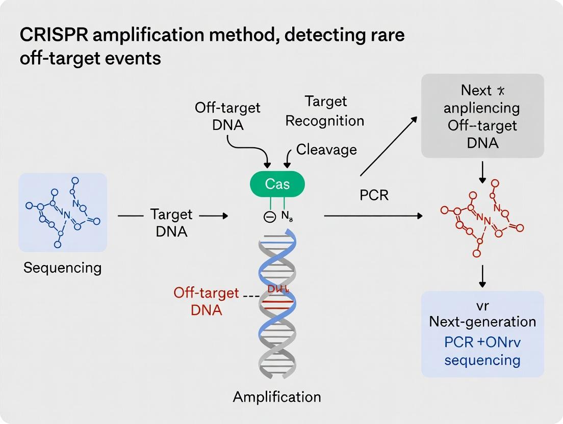
Amplification-enhanced detection is a cornerstone of modern molecular diagnostics, particularly in sensitive applications like identifying rare genomic events. Its core principle involves the selective amplification of a target signal—nucleic acid, protein, or other analyte—coupled with a detection mechanism, dramatically increasing sensitivity and specificity over direct detection methods. This is achieved through enzymatic (e.g., PCR, isothermal amplification) or signal amplification (e.g., branched DNA, rolling circle amplification) cascades. The primary advantages include unparalleled sensitivity for low-abundance targets, the ability to work with limited sample volumes, improved signal-to-noise ratios, and quantifiability. Within CRISPR-based diagnostics, amplification is often layered with the programmability of Cas enzymes (like Cas12a, Cas13) for sequence-specific detection, creating a powerful synergy for identifying rare off-target edits in therapeutic development.
Troubleshooting Guides & FAQs
FAQ 1: My amplification-enhanced CRISPR assay (e.g., DETECTR, SHERLOCK) shows high background noise or non-specific signal. What could be the cause and how do I resolve it?
- Answer: High background often stems from non-specific amplification or premature Cas enzyme cleavage. To resolve:
- Optimize Guide RNA (gRNA) Design: Ensure high on-target specificity. Use the latest algorithms (e.g., from ChopChop, CRISPOR) and include off-target prediction checks.
- Increase Stringency: Adjust the incubation temperature of the Cas/gRNA complex closer to the enzyme's optimal temperature (e.g., 37°C for Cas12a, 42°C for Cas13) to improve binding fidelity.
- Titrate Reporter Probe: A high concentration of fluorescent quenched reporter can lead to background hydrolysis. Titrate the reporter (typical range 0.1-1 µM) to find the minimal concentration giving a clean signal.
- Segregate Reactions: Physically separate the amplification step from the CRISPR detection step in a two-tube protocol to prevent amplicon contamination and premature reporter cleavage.
FAQ 2: The sensitivity of my assay is lower than published protocols for detecting rare off-target events. What steps can I take to improve it?
- Answer: Sensitivity loss can occur at the amplification or detection stage.
- Amplification Efficiency: Check primer design and template quality for your pre-amplification step (e.g., RPA, LAMP). Use fresh aliquots of enzymes and ensure magnesium concentration is optimized.
- Inhibit Carryover: Use dUTP and Uracil-DNA Glycosylase (UDG) in pre-amplification to degrade PCR products from previous runs, preventing false positives that mask rare true signals.
- Cas Enzyme Activity: Verify Cas enzyme activity with a known positive control target. Ensure it is not inhibited by components from the amplification reaction; a purification or dilution step post-amplification may help.
- Instrument Calibration: Ensure your fluorescence plate reader or lateral flow strip reader is properly calibrated for the reporter dye (e.g., FAM, HEX).
FAQ 3: How do I quantify the frequency of a rare off-target event from the signal generated in an amplification-enhanced detection assay?
- Answer: Absolute quantification requires a standard curve.
- Generate a Standard Curve: Serially dilute a synthetic DNA/RNA template containing the exact off-target sequence. Run these standards alongside your experimental samples in the same assay.
- Plot & Analyze: Plot the fluorescence intensity (or time-to-positive for real-time assays) against the log of the template copy number. Use this curve to interpolate the copy number in your unknown sample.
- Normalize: To report as a frequency, divide the estimated off-target copy number by the total genomic copy number in the input sample (e.g., determined by a separate assay for a reference gene).
Table 1: Comparison of Amplification Methods Used with CRISPR Detection
| Amplification Method | Typical Time | Temp. | Key Enzyme | Optimal for | Sensitivity (LoD) |
|---|---|---|---|---|---|
| Recombinase Polymerase Assay (RPA) | 10-20 min | 37-42°C | Recombinase, Polymerase | DNA targets, field use | ~1-10 aM (single digit copies) |
| Loop-Mediated Isothermal Amplification (LAMP) | 15-60 min | 60-65°C | Bst DNA Polymerase | DNA, high yield | ~10-100 copies/reaction |
| Reverse Transcription RPA (RT-RPA) | 15-30 min | 37-42°C | Reverse Transcriptase + RPA enzymes | RNA targets | ~10-100 copies/reaction |
| Polymerase Chain Reaction (PCR) | 60-120 min | Thermo-cycled | Taq Polymerase | DNA, gold-standard quantitation | ~1-10 copies/reaction |
Table 2: Common CRISPR-Cas Enzymes for Amplification-Enhanced Detection
| Cas Enzyme | Target | Collateral Activity Upon Binding | Typical Reporter | Key Advantage |
|---|---|---|---|---|
| Cas12a (e.g., LbCas12a) | dsDNA/ssDNA | Non-specific ssDNA cleavage | Fluorescent quenched ssDNA probe | Fast kinetics, works on DNA |
| Cas13a (e.g., LwaCas13a) | ssRNA | Non-specific ssRNA cleavage | Fluorescent quenched ssRNA probe | High specificity for RNA |
| Cas14 | ssDNA | Non-specific ssDNA cleavage | Fluorescent quenched ssDNA probe | Small size, single-stranded DNA target |
Detailed Experimental Protocol: DETECTR for Off-Target DNA Detection
This protocol detects a specific DNA sequence (e.g., a potential CRISPR-Cas9 off-target site) using RPA pre-amplification followed by Cas12a detection.
Materials:
- Genomic DNA sample.
- RPA primers specific to the off-target locus.
- TwistAmp Basic RPA kit (or equivalent).
- Purified LbCas12a enzyme.
- Designed crRNA targeting the amplicon.
- Fluorescent ssDNA reporter (e.g., FAM-TTATT-BHQ1).
- Nuclease-free water.
- 1.5 mL tubes, 0.2 mL PCR tubes.
- Fluorescence plate reader or real-time PCR machine.
Procedure:
- RPA Pre-amplification:
- In a 0.2 mL tube, mix 29.5 µL of rehydration buffer from the RPA kit with 1 µL of each primer (10 µM), 2 µL of genomic DNA template (10-100 ng), and nuclease-free water to 47.5 µL.
- Add 2.5 µL of magnesium acetate (provided in kit) to the tube lid.
- Briefly centrifuge to mix magnesium acetate into the reaction.
- Incubate at 37-42°C for 15-20 minutes.
Cas12a Detection Setup:
- Prepare a detection mix in a separate tube: 1 µL LbCas12a (100 nM final), 1 µL crRNA (100 nM final), 1 µL ssDNA reporter (500 nM final), and 5 µL of appropriate reaction buffer (e.g., NEBuffer 2.1).
- Add nuclease-free water to bring the detection mix volume to 20 µL.
Combined Reaction & Detection:
- Transfer 2 µL of the completed RPA reaction product into a well of a 96-well plate containing the 20 µL detection mix. Mix gently by pipetting.
- Immediately place the plate in a fluorescence plate reader pre-warmed to 37°C.
- Measure fluorescence (FAM channel: Ex 485nm/Em 520nm) every 30 seconds for 30-60 minutes.
Analysis:
- Plot fluorescence over time. A positive signal shows a sharp exponential increase in fluorescence. Use a threshold value (e.g., 5 standard deviations above the mean of negative controls) to determine time-to-positive.
Visualization
Amplification-Enhanced CRISPR Detection Workflow
The Scientist's Toolkit: Research Reagent Solutions
Table 3: Essential Materials for Amplification-Enhanced CRISPR Off-Target Detection
| Item | Function & Role in Experiment | Example/Supplier Note |
|---|---|---|
| High-Fidelity Polymerase | For generating initial amplicon standards and cloning. Critical for accuracy in control template prep. | Q5 High-Fidelity DNA Polymerase (NEB), KAPA HiFi. |
| Isothermal Amplification Kit | Enables rapid, instrument-free nucleic acid amplification at constant temperature. Core pre-amplification step. | TwistAmp RPA Kits (TwistDx), LAMP Kits (NEB WarmStart). |
| Purified CRISPR-Cas Enzyme | The detection core. Cas12a (for DNA) or Cas13 (for RNA) with collateral cleavage activity. | LbCas12a (IDT), LwaCas13a (from Mammoth Biosciences kits). |
| Synthetic crRNA or gRNA | Guides the Cas enzyme to the specific target sequence within the amplicon. Must be designed for the off-target site. | Custom Alt-R crRNA (IDT), Synthego synthetic guides. |
| Fluorescent Quenched Reporter | The signal-generating molecule. Cleaved upon Cas collateral activity, releasing fluorescence. | FAM-TTATT-BHQ1 ssDNA reporter for Cas12a; FAM-UUUU-BHQ1 for Cas13. |
| Digital PCR System | Gold-standard for absolute quantification of rare events. Used for orthogonal validation of assay results. | Bio-Rad QX200, Thermo Fisher QuantStudio 3D. |
| UDG/dUTP | Prevents carryover contamination from past amplicons, crucial for high-sensitivity, reproducible assays. | Included in many master mixes (e.g., from Thermo Fisher). |
| Magnetic Bead Cleanup Kits | For purifying and concentrating amplicons post-pre-amplification before CRISPR detection, reducing inhibitors. | AMPure XP beads (Beckman Coulter). |
Troubleshooting Guides & FAQs
Q1: In my GUIDE-seq experiment, I am detecting a high level of background noise (reads in negative controls). What are the primary causes and how can I mitigate this? A1: High background noise in GUIDE-seq often stems from:
- Non-specific tag integration: The dsODN tag can integrate into random double-strand breaks (DSBs) not generated by Cas9. Ensure cells are healthy and not under excessive stress from transfection/electroporation.
- PCR amplification bias: Over-amplification during library prep can exaggerate low-frequency, non-specific events. Optimize PCR cycle number and use high-fidelity polymerases. Include unique molecular identifiers (UMIs) to deduplicate reads.
- Incomplete tag purification: Residual free dsODN tag can be ligated into libraries. Implement stringent purification (e.g., solid-phase reversible immobilization beads) after genomic DNA extraction and tag cleavage.
- Solution: Always run a "no nuclease" negative control. Analytically, apply a stringent cutoff based on the read count distribution in this control (e.g., require sites to have reads >10x the 99th percentile of the control).
Q2: My CIRCLE-seq analysis shows excellent sensitivity, but I cannot validate many predicted off-target sites by amplicon sequencing. Why is there a discrepancy between detection limit and validation? A2: CIRCLE-seq operates on purified, cell-free genomic DNA, eliminating cellular context (chromatin accessibility, repair machinery). This gives it a low detection limit (e.g., 0.001% frequency) but can lead to false positives. Key factors:
- Chromatin: Predicted sites may be in heterochromatic regions inaccessible in living cells.
- Repair: The method detects DSBs, but some may be repaired without mutations.
- Coverage Bias: Ensure your amplicon sequencing has sufficient depth (≥100,000x) to detect the low-frequency variants that CIRCLE-seq identified.
Q3: For SITE-Seq, what defines the "coverage" metric, and how can I ensure I have sufficient coverage to trust my negative result? A3: In SITE-Seq, coverage refers to the sequencing depth across all potential genomic loci with even marginal similarity to the on-target site (e.g., all sites with ≤6 mismatches). Insufficient coverage risks false negatives.
- Protocol: Perform in silico digestion of the reference genome with your guide's PAM sequence to define the "search space." Your total sequencing reads must be high enough to provide >1000x coverage of this theoretical space.
- Solution: Use spike-in control DNA fragments with known off-target sequences at low frequencies (e.g., 0.1%) to empirically determine the detection limit for your specific library prep and sequencing run.
Q4: How do I interpret the "detection limit" reported in different studies, and why does it vary between methods? A4: The detection limit is the minimum variant frequency a method can reliably distinguish from technical noise. It is method-dependent.
Table 1: Comparison of Key Off-Target Detection Methods
| Method | Typical Detection Limit | Key Principle | Primary Noise Source |
|---|---|---|---|
| GUIDE-seq | 0.01% - 0.1% | dsODN integration into DSBs in living cells | Non-specific tag integration |
| CIRCLE-seq | 0.001% - 0.01% | In vitro circularization & amplification of Cas9-cleaved DNA | In vitro cleavage bias |
| SITE-Seq | ~0.1% | In vitro cleavage & biotin-streptavidin capture | Non-specific biotin binding |
| Amplicon Seq | 0.1% - 1% | Targeted PCR of predicted sites | PCR errors & base calling errors |
Experimental Protocols
Protocol 1: GUIDE-seq Library Preparation (Key Steps)
- Cell Culture & Transfection: Seed HEK293T cells in a 24-well plate. Co-transfect 500ng Cas9 expression plasmid, 250ng sgRNA plasmid, and 100pmol of dsODN tag using a recommended transfection reagent (e.g., Lipofectamine 3000).
- Genomic DNA (gDNA) Extraction: Harvest cells 72 hours post-transfection. Extract gDNA using a silica-column based kit with RNase A treatment. Elute in 50µL nuclease-free water.
- Tag Cleavage & Repair: Digest 500ng gDNA with 5U MmeI (NEB) for 2h at 37°C to cleave ~20bp adjacent to integrated tag. Purify fragments. Perform end-repair and A-tailing using the NEBNext Ultra II End Repair/dA-tailing Module.
- Adapter Ligation & PCR: Ligate Illumina-compatible adapters. Amplify with 12-14 PCR cycles using a high-fidelity polymerase and barcoded primers. Include a UMI in the forward primer.
- Sequencing: Purify the final library and quantify by qPCR. Sequence on an Illumina MiSeq or HiSeq platform (2x150bp recommended).
Protocol 2: CIRCLE-seq Endogenous Adapter Ligation
- Genomic DNA Preparation: Shear 1µg of human genomic DNA to an average size of 300bp using a focused-ultrasonicator.
- In Vitro Cleavage: Incubate sheared DNA with purified Cas9:sgRNA ribonucleoprotein (RNP) complex (100nM) for 16h at 37°C in CutSmart Buffer.
- End Repair & A-tailing: Purify DNA using AMPure XP beads. Treat with the NEBNext End Repair/dA-tailing Module (30min, 20°C, then 30min, 65°C).
- Circularization: Ligate the DNA into circles using T4 DNA Ligase (1h, 25°C) at a low DNA concentration (<10ng/µL) to promote intramolecular ligation.
- Exonuclease Digestion: Treat with Plasmid-Safe ATP-dependent DNase (16h, 37°C) to linearize and digest non-circular DNA, enriching for Cas9-cleaved, re-ligated fragments.
- Amplification: Amplify circles using phi29 polymerase (8h, 30°C) for multiple displacement amplification (MDA). Proceed to library construction for sequencing.
Diagrams
GUIDE-seq Experimental Workflow
Relationship of Key Detection Metrics
The Scientist's Toolkit: Research Reagent Solutions
Table 2: Essential Reagents for CRISPR Off-Target Detection Assays
| Reagent / Material | Function in Experiment | Key Consideration |
|---|---|---|
| dsODN Tag (for GUIDE-seq) | Double-stranded oligodeoxynucleotide that integrates into Cas9-induced DSBs, providing a universal handle for PCR amplification. | Must be PAGE-purified, phosphorothioated on last 3 bases of each end to prevent degradation. |
| High-Fidelity Polymerase (e.g., Q5, KAPA HiFi) | Amplifies tagged genomic fragments or libraries with minimal errors, crucial for detecting low-frequency events. | Essential for reducing PCR-introduced noise and maintaining sequence fidelity. |
| Phi29 Polymerase (for CIRCLE-seq) | Performs Multiple Displacement Amplification (MDA) from circularized DNA, amplifying cleaved fragments isothermally. | Provides high processivity and strand displacement, enabling whole-genome amplification from circles. |
| Cas9 Nuclease (WT) | The effector protein that creates DSBs at on- and off-target sites guided by the sgRNA. | Use purified, recombinant protein for in vitro assays (CIRCLE-seq, SITE-Seq); plasmid or mRNA for cellular assays. |
| MmeI Restriction Endonuclease | Type IIS enzyme that cuts 20/18bp downstream of its recognition site, used in GUIDE-seq to cut genomic DNA near the integrated tag. | Generates a consistent, short sequence adjacent to the tag for efficient sequencing. |
| Streptavidin Magnetic Beads | Used in SITE-Seq to capture biotinylated ends of Cas9-cleaved DNA fragments. | High binding capacity and low non-specific binding are critical for signal-to-noise ratio. |
| Unique Molecular Identifiers (UMIs) | Short random nucleotide sequences added during initial PCR to uniquely tag each original DNA molecule. | Enables bioinformatic removal of PCR duplicates, providing absolute quantification and reducing noise. |
A Deep Dive into Leading Amplification Techniques: CIRCLE-seq, GUIDE-seq, Digenome-seq, and Beyond
Technical Support Center
Troubleshooting Guides & FAQs
FAQ 1: For CRISPR-Cas9 off-target detection, when should I use an in vitro amplification method vs. an in situ approach?
- Answer: The choice depends on your primary research question and the required biological context.
- Use in vitro amplification methods (e.g., CIRCLE-seq, GUIDE-seq, SITE-seq) when you need a highly sensitive, unbiased, and genome-wide profile of potential off-target sites. These methods are excellent for identifying a comprehensive list of loci for subsequent validation, especially for rare events. They are performed on isolated genomic DNA, removing cellular context.
- Use in situ or in vivo approaches (e.g., immunostaining for DNA repair markers, live-cell imaging, transgenic animal models) when you need to understand the functional consequences and cellular response to off-target events in their native biological environment. This is crucial for assessing actual mutational outcomes and phenotypic impacts in drug development.
FAQ 2: My in vitro amplification assay (e.g., CIRCLE-seq) shows high background noise. What are the key troubleshooting steps?
- Answer: High background is often due to non-specific amplification or incomplete enzymatic steps.
- Optimize Cas9 Concentration: Too much Cas9 nuclease can lead to non-specific cleavage. Titrate the Cas9 protein amount in the initial digestion reaction.
- Verify Circularization Efficiency: Ensure the ssDNA ligase is active and the reaction conditions are optimal. Check the integrity of the splinter oligo.
- Purify DNA Rigorously: Use silica-column or bead-based cleanups between enzymatic steps to remove primers, salts, and enzymes that can inhibit subsequent reactions. Perform two-sided size selection after adapter ligation.
- Use High-Fidelity Polymerase: For the final PCR amplification, use a high-fidelity polymerase to minimize PCR errors and artifacts.
FAQ 3: I am not detecting expected off-target sites in my cell-based (in vivo) validation experiment. What could be wrong?
- Answer: Discrepancy between in vitro predictions and in vivo validation is common.
- Check Chromatin Accessibility: The predicted site may be in a heterochromatic region in your specific cell type. Consult ATAC-seq or DNase-seq data for your cell line. Consider using epigenetic modifiers or switch to a more relevant cell type.
- Confirm Guide RNA Expression: Verify that your guide RNA is being expressed efficiently in your delivery system (lentivirus, transfection) via RT-qPCR.
- Optimize Detection Sensitivity: The variant allele frequency may be very low (<0.1%). Use ultra-sensitive methods like digital PCR (dPCR) or amplicon sequencing with unique molecular identifiers (UMIs) instead of standard sequencing.
- Assay Timing: Off-target editing may occur on a different timeline than on-target editing. Harvest cells at multiple time points post-transfection.
FAQ 4: How do I choose between GUIDE-seq and CIRCLE-seq for an in vitro off-target profiling study?
- Answer:
- Choose GUIDE-seq if you are working with cell lines amenable to nucleofection and want to capture off-targets within the native cellular context (in situ), including chromatin effects. It requires delivery of an oligonucleotide tag.
- Choose CIRCLE-seq if you need maximum sensitivity from minimal input (e.g., clinical samples), are profiling nucleases not easily delivered to cells, or require a purely in vitro workflow. It is considered one of the most sensitive biochemical methods.
Comparative Data Tables
Table 1: Key Characteristics of Major Off-Target Detection Methods
| Method | Approach Type | Primary Principle | Sensitivity (Typical LOD) | Throughput | Required Input |
|---|---|---|---|---|---|
| CIRCLE-seq | In vitro | Circularization of gDNA & in vitro cleavage | ~0.01% VAF | High | Purified gDNA (≥ 1 µg) |
| GUIDE-seq | In situ (cellular) | Integration of a double-stranded tag | ~0.1% VAF | High | Live cells for nucleofection |
| SITE-seq | In vitro | Biotinylated guide RNA capture of cleaved DNA | ~0.1% VAF | Medium | Purified gDNA (≥ 5 µg) |
| Digenome-seq | In vitro | In vitro cleavage & whole genome sequencing | ~0.1% VAF | High | Purified gDNA (≥ 5 µg) |
| BLISS | In situ | Ligation of adapters to DSBs in fixed cells/samples | Single-cell | Medium | Fixed cells or tissue sections |
Table 2: Decision Matrix: Selecting a Strategy for Your Thesis on Rare Off-Target Detection
| Criteria | Recommended Approach (In Vitro) | Recommended Approach (In Vivo/In Situ) |
|---|---|---|
| Primary Goal | Unbiased, genome-wide discovery | Functional validation & phenotypic impact |
| Biological Context | Not required; uses purified DNA | Essential (specific cell type, tissue, or organism) |
| Sensitivity Need | Extremely High (detect very rare events) | Moderate to High (validate predicted events) |
| Sample Type | Bulk genomic DNA | Live cells, primary cells, or animal models |
| Cost & Speed | Lower cost per sample for screening; faster setup | Higher cost and time investment; complex setup |
| Downstream Analysis | Bioinformatics pipeline for site identification | Sequencing, imaging, or phenotypic assays |
Experimental Protocols
Protocol 1: CIRCLE-seq for In Vitro Off-Target Profiling
- Principle: Genomic DNA is circularized, digested in vitro with the RNP complex, linearized at off-target cleavage sites, and prepared for sequencing to reveal cut sites.
- Detailed Steps:
- Genomic DNA Isolation & Shearing: Extract high-molecular-weight gDNA from your target cell type. Fragment to ~300 bp using controlled sonication or enzymatic digestion.
- End Repair & A-tailing: Repair fragment ends using a mix of T4 PNK, T4 DNA polymerase, and Klenow fragment. Add a single 'A' overhang with Taq polymerase.
- Circularization: Ligate the 'A'-tailed fragments to a specially designed splinter oligo (with a 'T' overhang) using ssDNA ligase (CircLigase).
- Cas9 RNP Cleavage In Vitro: Incubate the circularized DNA library with pre-assembled Cas9-gRNA ribonucleoprotein (RNP) complex. Cleavage linearizes circles at active target sites.
- Adapter Ligation & PCR: Ligate sequencing adapters to the linearized fragments. Amplify with a limited number of PCR cycles.
- Sequencing & Analysis: Perform paired-end high-throughput sequencing. Map reads to the reference genome and identify peaks corresponding to cleavage sites using specialized bioinformatics tools (e.g., CIRCLE-seq analysis pipeline).
Protocol 2: Cell-Based (In Situ) Validation via Targeted Amplicon Sequencing
- Principle: Validate predicted off-target sites by deep sequencing of PCR amplicons from treated cells.
- Detailed Steps:
- Cell Transfection/Nucleofection: Deliver your Cas9 and gRNA constructs (as plasmid, mRNA, or RNP) into your target cell line.
- Genomic DNA Harvest: After 72-96 hours, harvest cells and extract gDNA.
- Primary PCR: Design primers flanking each predicted off-target locus (typically within a 200-300 bp window). Perform the first round of PCR to amplify each locus from sample and control gDNA.
- Indexing PCR (with UMIs): In a second PCR, add full Illumina adapters, sample indices, and Unique Molecular Identifiers (UMIs). UMIs are short random sequences that tag each original DNA molecule, allowing for correction of PCR amplification bias and errors.
- Sequencing & Analysis: Pool amplicons and sequence deeply (>100,000x coverage per site). Process data using a UMI-aware pipeline (e.g., CRISPResso2, ampliconDIVider) to accurately calculate insertion/deletion (indel) frequencies at each locus.
Visualizations
Title: Decision Flowchart for Off-Target Detection Strategy
Title: Comparative Experimental Workflows for Off-Target Detection
The Scientist's Toolkit: Research Reagent Solutions
| Item | Function in CRISPR Off-Target Research | Example/Note |
|---|---|---|
| High-Fidelity Cas9 Nuclease | Catalyzes DNA cleavage at target (and off-target) sites. Enzyme purity reduces non-specific activity. | Nuclease-free, recombinant SpCas9. |
| Chemically Modified sgRNA | Guides Cas9 to target sequence. Modifications (e.g., 2'-O-methyl) can enhance stability and alter specificity. | Synthetic sgRNA with 3' phosphorothioate bonds. |
| CircLigase ssDNA Ligase | Critical for CIRCLE-seq; efficiently circularizes single-stranded DNA fragments. | Epicentre CircLigase II. |
| Unique Molecular Identifiers (UMI) | Short random nucleotide sequences used to tag individual DNA molecules before PCR, enabling accurate quantification and error correction. | Integrated into sequencing adapters. |
| High-Sensitivity DNA Assay Kits | Accurately quantify low-concentration DNA libraries prior to sequencing (critical for low-input methods). | Qubit dsDNA HS Assay, TapeStation. |
| Epigenetic Modifier Inhibitors | Used in cell-based studies to probe the effect of chromatin state on off-target editing (e.g., HDAC inhibitors). | Trichostatin A (TSA) for histone deacetylation. |
| Ultra-Sensitive DNA Polymerase | For robust and unbiased amplification of low-abundance off-target loci from limited sample material. | KAPA HiFi HotStart Uracil+, Q5 High-Fidelity. |
| Magnetic Beads for Size Selection | For precise cleanup and size selection of DNA fragments during library preparation, reducing background. | SPRIselect or AMPure XP beads. |
Within the context of advancing CRISPR amplification methods for detecting rare off-target events, CIRCLE-seq (Circularization for In vitro Reporting of Cleavage Effects by Sequencing) stands out as a highly sensitive, in vitro technique. It is designed to comprehensively profile the off-target DNA cleavage activity of CRISPR-Cas nucleases, even capturing extremely rare events critical for therapeutic safety assessment.
Technical Support Center
Troubleshooting Guides & FAQs
Q1: My final CIRCLE-seq library shows very low complexity/diversity after PCR. What could be the cause? A: This is often due to insufficient fragmentation or suboptimal circularization. Ensure genomic DNA is sheared to an appropriate size (100-500 bp) using a calibrated Covaris or sonicator. Verify the efficiency of the end repair and A-tailing steps prior to adapter ligation, as these are critical for successful circularization. Include a positive control sample with a known nuclease.
Q2: I observe high background signal from non-specific cleavage in my negative control (no nuclease added). How can I reduce this? A: High background typically stems from residual nuclease activity in recombinant enzyme preps or non-specific DNA damage during purification. Perform additional purification steps after genomic DNA isolation, such as AMPure bead clean-ups with increased ethanol wash volumes. Ensure all reagents, especially the Cas9 nuclease buffer, are nuclease-free. Include a thorough Proteinase K digestion step to eliminate any contaminating nucleases.
Q3: The cleavage signal from my positive control gRNA is weak. What should I check? A:
- Nuclease Activity: Verify the activity of your Cas9/gRNA RNP complex using a standard plasmid cleavage assay.
- Circularization Efficiency: Check the efficiency of the circligase step by running the product on a high-sensitivity bioanalyzer chip; you should see a shift to higher molecular weight.
- 䋱 PCR Amplification: Ensure your Phi29 polymerase is active for the rolling circle amplification (RCA) step, and that subsequent PCR uses a low-cycle number (e.g., 10-14 cycles) with high-fidelity polymerase to avoid skewing.
Q4: How do I distinguish true off-target sites from sequencing artifacts or background noise in the data?
A: Implement a robust bioinformatic pipeline. True sites typically show a clustered pattern of read ends at a specific genomic locus. Use established analysis tools (e.g., CIRCLE-seq specific aligners) that require a minimum number of independent read start/end clusters per site and filter against common sequencing artifacts. Biological replication is key; events reproducible across replicates are high-confidence.
Core Methodology & Protocols
Key Experimental Protocol: CIRCLE-seq Workflow
- Genomic DNA Isolation & Shearing: Purify high-molecular-weight genomic DNA from your cell type of interest. Mechanically shear it to an average size of 200-400 bp.
- End Repair & A-tailing: Convert sheared fragments to blunt-ended, 5'-phosphorylated, 3'-dA-tailed DNA using standard kits.
- Adapter Ligation & Purification: Ligate double-stranded DNA adapters compatible with subsequent circularization. Purify to remove excess adapters.
- Circularization: Treat adapter-ligated DNA with a single-stranded DNA ligase (Circligase). This circularizes fragments where both ends are properly ligated to adapters.
- Cas9 Cleavage In Vitro: Incubate the circularized DNA library with the pre-assembled Cas9-gRNA ribonucleoprotein (RNP) complex. Only DNA with a recognition site will be linearized via cleavage.
- Exonuclease Digestion: Treat the product with an exonuclease (e.g., exonuclease III or lambda exonuclease) to degrade all non-circular, linear DNA. The only linear DNA remaining should be fragments cleaved by Cas9, which are protected from exonuclease digestion by their new ends.
- Rolling Circle Amplification (RCA): Use Phi29 polymerase to amplify the nuclease-cleaved, linearized fragments. This amplifies the signal from rare cleavage events.
- Fragmentation & Library Prep for NGS: Fragment the RCA product, add sequencing adapters via a second ligation or tagmentation, and PCR-amplify.
- High-Throughput Sequencing & Analysis: Sequence the library and align reads to the reference genome. Cleavage sites are identified as genomic positions with a significant enrichment of read termini.
Data & Reagents
Table 1: Key Quantitative Metrics for CIRCLE-seq Sensitivity
| Metric | Typical Value/Range | Significance |
|---|---|---|
| Detection Sensitivity | Can detect cleavage events at frequencies <0.1% of total reads. | Essential for identifying very rare off-target sites. |
| Required Sequencing Depth | 50-100 million reads per sample (varies by genome size). | Ensures sufficient coverage to detect low-frequency events. |
| Background Noise Level | Typically < 0.01% of total reads per genomic site in no-nuclease controls. | Low background is critical for signal-to-noise ratio. |
| Genomic DNA Input | 1 µg - 5 µg per reaction. | Higher input can improve library complexity. |
The Scientist's Toolkit: Key Research Reagent Solutions
| Item | Function in CIRCLE-seq |
|---|---|
| Circligase ssDNA Ligase | Enzymatically circularizes adapter-ligated DNA fragments; critical step for background reduction. |
| Phi29 DNA Polymerase | Performs Rolling Circle Amplification (RCA) to linearly amplify nuclease-cleaved fragments, boosting signal from rare events. |
| CRISPR-Cas9 Nuclease (e.g., S.p. Cas9) | The effector protein complexed with gRNA to perform targeted in vitro cleavage. |
| Exonuclease III (or Lambda Exonuclease) | Degrades non-circular, linear DNA post-cleavage, enriching for Cas9-cut fragments. |
| AMPure XP Beads | Used for multiple purification and size selection steps throughout the protocol. |
| High-Sensitivity DNA Assay Kit (Bioanalyzer/ TapeStation) | Essential for quality control at multiple steps (shearing size, circularization, final library). |
Visualization
Diagram 1: CIRCLE-seq Experimental Workflow
Diagram 2: Principle of Selective Capture & Amplification
Practical Guide to Implementing GUIDE-seq for Genome-Wide Integration-Based Detection
Technical Support Center
Troubleshooting Guide: Common Experimental Issues & Solutions
Q1: After PCR amplification of integration events, I get no product or a smear on the gel. What could be wrong? A: This is often due to inefficient tag integration or poor PCR efficiency.
- Check Tag Integration: Ensure the oligo tag is delivered at a sufficiently high concentration (typically 100-1000X molar excess over RNP). Verify delivery method (e.g., nucleofection) efficiency for your cell line.
- Optimize Nested PCR: Use a high-fidelity polymerase. For the primary PCR, use a touchdown or step-down cycling program to increase specificity. The secondary (nested) PCR primers must be internal to the first set. Include positive control genomic DNA spiked with a known integration site.
- Reduce Complexity: If a smear persists, the initial lysate or primary PCR product may be too complex. Ensure adequate cleanup (e.g., SPRI beads) between PCR rounds and do not over-cycle.
Q2: My sequencing data shows a very high background of non-specific integration sites or primer artifacts. How can I improve specificity? A: High background compromises detection of rare off-targets.
- Enforce Double-Strand Breaks (DSB) Dependency: Always include a "no nuclease" control and a "tag-only" control. True off-target sites should be absent in these controls.
- Bioinformatic Filtering: Rigorously filter reads that do not contain the tag sequence followed by the specific primer sequence used in the nested PCR. Use tools like GUIDE-seq analysis software or pipeGUIDE-seq to require a minimum number of unique reads (e.g., ≥ 3-5 reads) supporting each site.
- Improve Enzymatic Cleanup: Treat PCR products with Exonuclease I and Shrimp Alkaline Phosphatase (SAP) before the nested PCR to degrade excess primers and dNTPs, reducing primer-dimer artifacts.
Q3: I suspect I am missing low-frequency off-target events. How can I increase sensitivity? A: Sensitivity is critical for detecting rare off-target events in CRISPR amplification method studies.
- Increase Sequencing Depth: Aim for 10-50 million paired-end reads per sample to ensure coverage of low-abundance sites.
- Maximize Tag Capture Efficiency: Optimize the ratio of tag oligo to RNP. Test different tag designs (double-stranded vs. single-stranded, phosphorothioate modifications).
- PCR Bias Mitigation: Perform technical replicates of the entire library prep and pool them before sequencing. Use a polymerase and cycling conditions that minimize GC-bias.
Q4: How do I handle the bioinformatic analysis, and what are the key output metrics? A: Analysis requires aligning sequencing reads to the reference genome and identifying tag integration sites.
- Pipeline: Use a standardized pipeline (e.g., GUIDE-seq, CRISPResso2 with GUIDE-seq mode). Steps include: adapter trimming, alignment (BWA/Bowtie2), parsing reads for tag sequence, identifying integration junctions, and clustering nearby sites.
- Key Output Table:
| Metric | Description | Typical Target Value / Note |
|---|---|---|
| Total Reads | Raw sequencing reads per sample. | 10-50 million PE reads. |
| Tag-Aligned Reads | Reads containing the tag sequence. | Usually 20-60% of total. |
| Unique Sites | Genomic loci with identified integration. | Varies with nuclease activity. |
| Reads per Site | Depth supporting each off-target. | ≥3-5 for confidence. |
| On-Target % | Reads at the intended target site. | Often the most abundant site. |
| R2 Value | Reproducibility between replicates. | >0.8 is excellent. |
Frequently Asked Questions (FAQs)
Q: What is the core principle of GUIDE-seq in the context of CRISPR off-target detection? A: GUIDE-seq uses a short, double-stranded oligodeoxynucleotide (dsODN) tag that is captured into double-strand breaks (DSBs) created by the CRISPR-Cas nuclease in vivo. The integration site is then amplified by PCR and sequenced, providing a genome-wide, unbiased map of all DSB events, including rare off-targets.
Q: How does GUIDE-seq compare to computational prediction or in vitro methods like CIRCLE-seq? A: GUIDE-seq is an in-cell method, capturing the chromatin context, nuclear delivery, and DNA repair dynamics that influence off-target cleavage. It typically identifies fewer, but biologically relevant, sites compared to in vitro methods, which may overpredict. It is more empirical than computational prediction.
Q: What are the essential controls for a valid GUIDE-seq experiment? A: Three critical controls are mandatory:
- No-Nuclease Control: Cells treated with tag only. Identifies background genomic integration.
- No-Tag Control: Cells transfected with RNP only. Identifies DSB-independent artifacts.
- Positive Control: A known active gRNA. Validates the entire workflow.
Q: Can GUIDE-seq be used for base editors or prime editors? A: Standard GUIDE-seq detects DSBs. It is not suitable for base or prime editors, which typically do not create DSBs. Modified methods like GUIDE-tag or PE-tag using nickase-fused tags are under development for these editors.
Q: What sequencing depth and platform are recommended? A: Illumina NextSeq 550 or NovaSeq 6000 systems are standard. A minimum of 10 million paired-end (2x150 bp) reads per sample is recommended, with 20-50 million providing better sensitivity for very rare events.
Detailed Experimental Protocol
Title: Comprehensive GUIDE-seq Workflow for Detecting CRISPR-Cas9 Off-Target Events
I. dsODN Tag Preparation
- Anneal Oligos: Resuspend HPLC-purified sense and antisense oligos (with phosphorothioate linkages on 3 terminal bases) to 100 µM in nuclease-free water. Mix equal volumes.
- Heat and Cool: Heat mixture to 95°C for 5 min, then slowly cool to room temperature over 45-60 min. The dsODN tag is stable at 4°C for weeks.
II. Cell Transfection & Tag Integration
- Prepare RNP: Complex purified Cas9 protein (e.g., 100 pmol) with sgRNA (120 pmol) in nucleofection buffer. Incubate 10 min at RT.
- Add Tag: Add dsODN tag (e.g., 100 pmol) to the RNP complex.
- Nucleofection: Harvest 1-2 x 10^5 cells (e.g., HEK293T), resuspend in cell-line-specific nucleofection solution. Combine cell suspension with RNP/tag mix. Transfer to a cuvette and nucleofect using recommended program (e.g., CM-130 for HEK293).
- Culture: Immediately transfer cells to pre-warmed medium. Culture for 72 hours to allow DSB repair and tag integration.
III. Genomic DNA Extraction & Shearing
- Extract gDNA: Use a column-based or magnetic bead-based gDNA extraction kit. Elute in low-EDTA TE buffer or nuclease-free water.
- Shear DNA: Fragment 1-2 µg gDNA to ~500 bp using a focused-ultrasonicator (Covaris) or enzymatic shearing kit. Verify fragment size on a bioanalyzer.
IV. Library Preparation for Sequencing
- End-Repair & A-Tailing: Use a commercial library prep kit (e.g., NEBNext Ultra II) to repair ends and add a single 'A' overhang.
- Adaptor Ligation: Ligate sequencing adaptors with a 'T' overhang. Clean up with SPRI beads.
- Primary PCR: Amplify tag-integrated fragments using a primer specific to the dsODN tag (TagP1) and a primer complementary to the adaptor (AdaptorP1). Use 12-15 cycles.
- Cycle Program: 98°C 30s; [98°C 10s, 65°C 30s, 72°C 30s] x 15; 72°C 5min.
- Purification: Clean primary PCR product with SPRI beads (0.8X ratio).
- Nested PCR: Perform a second PCR using nested primers (TagP2 and indexed AdaptorP2) to add full Illumina sequencing handles and sample indexes. Use 12-18 cycles.
- Final Purification & QC: Clean final library with SPRI beads (0.8X). Quantify by qPCR (KAPA Library Quant Kit) and check size distribution (Bioanalyzer). Pool libraries equimolarly and sequence on an Illumina platform (2x150 bp, Mid-output).
Visualization: Experimental Workflow
Title: GUIDE-seq End-to-End Experimental Workflow
The Scientist's Toolkit: Key Research Reagent Solutions
| Reagent / Material | Function & Importance in GUIDE-seq |
|---|---|
| Phosphorothioate-Modified dsODN Tag | Core reagent. Short double-stranded DNA oligo integrated into DSBs. Phosphorothioate linkages prevent exonuclease degradation, enhancing stability and integration efficiency. |
| Purified Recombinant Cas9 Protein | For RNP formation. Higher specificity and faster kinetics than plasmid delivery. Eliminates variable Cas9 expression levels. |
| Chemically Synthesized sgRNA | For RNP formation. High purity, consistent activity, and allows for chemical modifications (e.g., 2'-O-methyl) to enhance stability. |
| Cell Line-Specific Nucleofection Kit | Critical for efficient co-delivery of large RNP complexes and dsODN tag into hard-to-transfect cell types (e.g., primary cells). |
| High-Fidelity PCR Master Mix (e.g., Q5, KAPA HiFi) | Essential for accurate, unbiased amplification of tag-integrated genomic fragments during library construction, minimizing PCR errors. |
| SPRI (Solid Phase Reversible Immobilization) Beads | Used for size selection and cleanup of DNA fragments after shearing, adapter ligation, and PCR steps. Provides reproducible recovery. |
| Illumina-Compatible Dual-Indexed Adapters | Allow multiplexing of multiple samples in a single sequencing run, reducing cost and processing time. |
| Bioinformatic Pipeline (e.g., GUIDE-seq software) | Required to process raw sequencing data, align reads, identify tag integration junctions, and filter false positives to generate a final list of off-target sites. |
Leveraging Digenome-seq and BLISS for High-Resolution, Amplified Off-Target Profiling
Technical Support Center: Troubleshooting & FAQs
FAQ 1: Why is my Digenome-seq background signal too high, obscuring potential off-target cleavages?
Answer: High background in Digenome-seq is frequently caused by incomplete in vitro cleavage or non-specific genomic DNA damage. Ensure the Cas9-gRNA RNP complex is freshly reconstituted with high-specificity activity S. pyogenes Cas9. The critical step is the subsequent purification of the cleaved genomic DNA fragments; use SPRI bead-based size selection to stringently isolate fragments under 1000 bp. Increasing the concentration of Proteinase K and extending the digestion time to 4 hours can also reduce protein-associated background.
FAQ 2: In BLISS, my library yield is low after the on-bead ligation and PCR amplification. What are the primary causes?
Answer: Low BLISS library yield typically stems from two points: inefficient biotinylated adapter ligation or poor on-bead PCR. First, verify the activity of T4 DNA Ligase and ensure the biotinylated double-stranded adapter is in a 10:1 molar excess to DNA ends. Second, after streptavidin bead capture, perform a rigorous wash (3x with high-salt buffer) to remove non-specific debris. The most common error is eluting the DNA from the beads before PCR; instead, perform the PCR directly on the beads. Use a high-fidelity, bead-compatible polymerase.
FAQ 3: How do I distinguish true, rare off-target sites from sequencing artifacts or false positives in the combined data analysis?
Answer: This requires stringent bioinformatic filtering. True sites will have: 1) A clear pileup of read starts (for BLISS) or ends (for Digenome-seq) at a single genomic coordinate. 2) Sequence homology to the on-target site, allowing for up to 5 mismatches and/or bulges. 3) Statistical significance over the local background (use tools like MACS2 for peak calling). 4) Reproducibility across experimental replicates. The following table summarizes the key quantitative thresholds for calling a valid off-target event:
| Data Feature | Digenome-seq Threshold | BLISS Threshold | Combined Criterion |
|---|---|---|---|
| Read Depth/Pileup | ≥ 5 reads at site | ≥ 10 unique cell barcodes | Must pass either threshold |
| Peak Score (p-value) | < 1e-5 | < 1e-5 | Must pass both thresholds |
| Mismatch/Bulge Allowance | ≤ 5 | ≤ 5 | Consistent alignment for both |
| Replicate Concordance | Present in ≥2/3 replicates | Present in ≥2/3 replicates | Must be present in both methods |
FAQ 4: The CRISPR amplification step is not yielding sufficient signal for rare off-targets. What optimization is needed?
Answer: The amplification of the cleaved fragments prior to sequencing is critical for detecting rare events. If signal is low, optimize the Adapter Ligation-Mediated PCR (LM-PCR) step: 1) Template Quality: Use purified, size-selected fragments (<1kb). 2) Adapter Ligation: Ensure blunt-end repair is complete before ligating asymmetric adapters. Use a thermostable ligase for higher efficiency. 3) PCR Cycles: Increase cycle number cautiously (e.g., from 12 to 16 cycles) but beware of amplifying background. 4) Polymerase Choice: Use a polymerase with high processivity and low bias (e.g., KAPA HiFi). Always include a no-Cas9 negative control to identify background amplification bands.
Detailed Experimental Protocols
Protocol 1: Integrated Digenome-seq Workflow for Amplified Detection
- Genomic DNA Isolation: Isolate high molecular weight gDNA (>50 kb) from target cells using a phenol-chloroform method.
- In Vitro Cleavage: Incubate 5 µg of gDNA with 200 nM recombinant Cas9 and 600 nM sgRNA (pre-complexed as RNP for 10 min at 25°C) in 1x Cas9 reaction buffer for 12 hours at 37°C.
- DNA Purification & Size Selection: Purify DNA with SPRI beads. Perform double-size selection (0.5x and 1.5x bead-to-sample ratio) to recover fragments between 100 bp and 1000 bp.
- Library Preparation & Amplification: Convert cleaved ends into an NGS library using a blunt-end, ligation-based kit (e.g., NEBNext Ultra II). Perform 14 cycles of PCR amplification.
- Sequencing & Analysis: Sequence on an Illumina platform (>50 million 2x150 bp reads per sample). Map reads to the reference genome and identify cleavage sites as genomic positions with a cluster of read ends using the Digenome-seq analysis pipeline (v2.0).
Protocol 2: BLISS Protocol for Single-Cell Off-Target Profiling
- Cell Fixation and Permeabilization: Fix cells (or nuclei) in 4% PFA for 10 min, then permeabilize in 0.5% Triton X-100/0.1% SDS for 30 min on ice.
- In Situ Cleavage & Adapter Ligation: Incubate fixed cells with Cas9-gRNA RNP in reaction buffer for 2 hours at 37°C. Wash and then perform in situ ligation of biotinylated double-stranded adapters to the cleaved ends using T4 DNA Ligase overnight at 16°C.
- Cell Lysis & Bead Capture: Lyse cells with Proteinase K. Capture biotinylated fragments using streptavidin-coated magnetic beads.
- On-Bead Library Construction: Wash beads thoroughly. Perform on-bead primer extension and then PCR (18-20 cycles) directly on the bead-bound DNA. Use indexed primers to multiplex samples.
- Sequencing & Analysis: Sequence (2x75 bp is sufficient). Demultiplex by cell barcode. Align reads and call cleavage sites where multiple unique cell barcodes show read starts at the same genomic coordinate, using the BLISS-analysis software.
Visualizations
Diagram 1: Integrated Workflow for Amplified Off-Target Detection
Diagram 2: Off-Target Signal Amplification Logic
The Scientist's Toolkit: Key Research Reagent Solutions
| Reagent / Material | Function & Role in Off-Target Profiling |
|---|---|
| High-Activity S. pyogenes Cas9 | Recombinant, nuclease-active protein for efficient in vitro and in situ DNA cleavage. Crucial for generating clean, specific cuts. |
| sgRNA (chemically modified) | Target-specific guide RNA with stability enhancements (e.g., 2'-O-methyl analogs) to improve RNP performance and reduce degradation. |
| Biotinylated Double-Stranded Adapter | Short DNA duplex with a 5' biotin tag for BLISS. Ligation to cleaved ends enables streptavidin-bead capture and subsequent on-bead amplification. |
| Streptavidin Magnetic Beads | Solid-phase support for capturing biotinylated DNA fragments in BLISS. Allows for stringent washing to reduce background. |
| High-Fidelity PCR Polymerase (e.g., KAPA HiFi) | Enzyme for low-bias, high-efficiency amplification of adapter-ligated fragments. Essential for detecting rare events without introducing artifacts. |
| SPRI Size Selection Beads | Magnetic beads for precise size selection of DNA fragments (e.g., 100-1000 bp). Removes uncleaved gDNA and very small fragments to lower background. |
| Proteinase K | Broad-spectrum serine protease for complete digestion of Cas9 protein and cellular proteins after cleavage, preventing interference with downstream steps. |
| Indexed NGS Primers | Primers containing unique dual indices (i7 and i5) for multiplex sequencing of multiple samples/cell barcodes in a single run, reducing cost. |
Troubleshooting Guides & FAQs
Sample Preparation & DNA Extraction
Q1: My genomic DNA yield after CRISPR-Cas9 treatment and extraction is consistently low (<50% of expected). What could be the cause? A: Low yield is often due to inefficient cell lysis or DNA shearing. Ensure lysis buffer contains fresh proteinase K and is incubated at 56°C for at least 3 hours. For rare off-target detection, avoid vortexing; instead, invert tubes gently. If using formalin-fixed samples, extend lysis time to overnight.
Q2: I observe high RNA contamination in my DNA prep prior to library amplification. How do I mitigate this? A: Include an RNase A treatment step (10 μg/mL, 37°C for 15 min) immediately after lysis but before protein precipitation. Purify using a silica-column system (e.g., Qiagen DNeasy) instead of phenol-chloroform to improve RNA removal.
CRISPR Enrichment & Amplification
Q3: The on-target amplification efficiency in my CRISPR-enriched samples is suboptimal (<60% by qPCR). How can I improve this? A: This typically indicates guide RNA (gRNA) inefficiency or Cas9 nuclease inactivity. Verify gRNA concentration (should be >100 nM final) and complex with Cas9 at a 1:2 molar ratio for 20 min at 25°C before adding to DNA. Use a positive control gRNA for a known genomic locus.
Q4: I suspect nonspecific amplification of non-target regions during the post-CRISPR PCR. What are the key parameters to adjust? A: Nonspecific amplification is common when detecting rare off-targets. Implement a touchdown PCR protocol (start 5°C above calculated Tm, decrease 1°C/cycle for 10 cycles). Increase annealing temperature incrementally by 2°C in a gradient PCR to determine optimum. Ensure primer concentrations are balanced at 0.3 μM each.
Library Preparation & Sequencing
Q5: My final NGS library shows a broad size distribution (>1000 bp fragments) after post-CRISPR amplification, unsuitable for Illumina sequencing. How do I correct this? A: This indicates incomplete size selection or adapter dimer formation. Perform a double-sided SPRI bead cleanup (e.g., 0.5X followed by 0.8X ratio) to tightly select for 300-600 bp fragments. Run an aliquot on a Bioanalyzer before the final PCR to verify size.
Q6: Sequencing data shows abnormally high duplication rates (>80%) for my off-target detection libraries. What is the fix? A: High duplication suggests insufficient starting material leading to over-amplification. For rare event detection, increase the amount of CRISPR-enriched DNA input to the library prep (aim for >250 ng). Reduce the number of PCR cycles during library indexing to 8-10.
Experimental Protocols
Protocol 1: CRISPR-Cas9 Enrichment of Putative Off-Target Sites
- Complex Formation: Combine 10 pmol of Cas9 nuclease (NEB #M0386) with 20 pmol of synthesized gRNA in 1X Cas9 buffer. Incubate 20 min at 25°C.
- Digestion: Add 1 μg of purified genomic DNA (in 50 μL total volume) to the complex. Incubate at 37°C for 2 hours.
- Purification: Add 2X volume of AMPure XP beads (Beckman Coulter), incubate 5 min, wash twice with 80% ethanol. Elute DNA in 30 μL nuclease-free water.
- Validation: Assess on-target cleavage efficiency via qPCR (see Table 1 for primer sequences) and agarose gel electrophoresis.
Protocol 2: Targeted Library Construction for Illumina Sequencing
- End Repair & A-Tailing: Using 200 ng of enriched DNA, perform end repair and dA-tailing with NEBNext Ultra II FS Module (NEB #E7805) per manufacturer's protocol.
- Adapter Ligation: Ligate unique dual-indexed adapters (Illumina) using a 15:1 molar adapter-to-insert ratio. Incubate at 20°C for 15 min.
- Post-Ligation Cleanup: Clean using 0.9X volume of AMPure XP beads. Elute in 23 μL.
- Library Amplification: Amplify with 10 cycles of PCR using Q5 Hot Start High-Fidelity Master Mix. Use index primers provided in the kit.
- Final Cleanup & QC: Perform a 0.8X SPRI bead cleanup. Quantify by Qubit dsDNA HS Assay and profile on Agilent Bioanalyzer (Target peak: 350-550 bp).
Table 1: Critical qPCR Primers for On-Target Validation
| Target Region | Forward Primer (5'->3') | Reverse Primer (5'->3') | Expected Amplicon (bp) | Optimal Annealing Temp (°C) |
|---|---|---|---|---|
| On-Target Locus A | CTAGCGAATTCGCTAGCTAC | GTACGTAGCTGCTAGCTTAC | 245 | 62 |
| On-Target Locus B | ATCGATCGATCGATCGATCG | TAGCTAGCTAGCTAGCTAGC | 198 | 60 |
| Off-Target Hotspot 1 | GATCGATCGTAGCTACGTA | TCGATCGATCGATCGATCG | 301 | 63 |
| GAPDH Control | AGGTCGGTGTGAACGGATTTG | TGTAGACCATGTAGTTGAGGTCA | 123 | 60 |
Table 2: Recommended Reagent Volumes for Critical Steps
| Step | Reagent/Kit | Input Amount | Volume/Reaction | Incubation |
|---|---|---|---|---|
| CRISPR Enrichment | Alt-R S.p. Cas9 Nuclease V3 | 1 μg gDNA | 1.5 μL (10 pmol) | 37°C, 2h |
| Post-Enrichment PCR | Q5 Hot Start Master Mix | 2 μL enriched DNA | 25 μL total | 98°C 30s, [98°C 10s, 65°C 30s, 72°C 1min] x 25 |
| Library Construction | NEBNext Ultra II FS Module | 200 ng DNA | 16.5 μL FS Mix | 65°C 15min, 80°C 15min |
| Size Selection | AMPure XP Beads | 50 μL reaction | 40 μL (0.8X) | RT, 5min |
Diagrams
Title: Integrated Workflow for CRISPR Off-Target Detection
Title: CRISPR-Cas9 DNA Cleavage and Repair Pathway
The Scientist's Toolkit: Research Reagent Solutions
| Item | Function in Workflow | Key Consideration |
|---|---|---|
| High-Fidelity Cas9 Nuclease | Catalyzes DNA double-strand breaks at gRNA-specified sites. Essential for target enrichment. | Use a high-specificity variant (e.g., HiFi Cas9, eSpCas9) to reduce false-positive off-target cleavage. |
| Synthetic, Chemically Modified gRNA | Guides Cas9 to the intended DNA sequence. | Chemical modifications (2'-O-methyl, phosphorothioate) enhance stability and reduce immune responses in cell-based assays. |
| AMPure XP / SPRI Beads | Magnetic beads for size-selective purification and cleanup of DNA fragments. | The bead-to-sample ratio (e.g., 0.6X, 0.8X, 1.2X) is critical for selecting the correct fragment size range. |
| NEBNext Ultra II FS Module | Enzymatic mix for DNA end repair, dA-tailing, and adapter ligation in library prep. | Optimized for low-input and damaged DNA, crucial for processed CRISPR-enriched samples. |
| Q5 Hot Start High-Fidelity DNA Polymerase | PCR amplification of enriched targets and final library amplification. | Ultra-high fidelity reduces PCR-introduced errors, vital for accurate mutation detection. |
| Unique Dual Index (UDI) Adapters | Attached to DNA fragments to allow multiplexing and sample identification on sequencer. | UDis minimize index hopping (plex hopping) and are mandatory for sensitive rare variant detection. |
Optimizing Your Assay: Troubleshooting Common Pitfalls in Amplification-Based Off-Target Detection
Within CRISPR off-target detection research, accurate identification of rare editing events is paramount. High-fidelity amplification is critical, as PCR artifacts and background amplification can generate false-positive signals, obscuring true off-target sites. This technical support center provides targeted troubleshooting for common amplification challenges in this sensitive application.
Troubleshooting Guides & FAQs
FAQ 1: How can I reduce non-specific amplification and primer-dimer formation in my nested PCR for off-target site validation?
Answer: Non-specific amplification often stems from low primer annealing specificity, especially with complex genomic backgrounds.
- Solution: Implement a touchdown PCR protocol or use hot-start polymerase. Increase annealing temperature gradually in initial cycles. Optimize Mg²⁺ concentration (often lower than standard, e.g., 1.5-2.5 mM). Use PCR additives like DMSO (3-5%) or Betaine (1 M) to improve specificity for GC-rich regions common near CRISPR cut sites.
- Protocol - Touchdown PCR: 1) Initial denaturation: 95°C for 2 min. 2) 10 cycles: Denature at 95°C for 30 sec, Anneal at 65°C (decrease by 0.5°C per cycle) for 30 sec, Extend at 72°C for 30 sec/kb. 3) 25 cycles: Denature at 95°C for 30 sec, Anneal at 55°C for 30 sec, Extend at 72°C for 30 sec/kb. 4) Final extension: 72°C for 5 min.
FAQ 2: What strategies minimize polymerase errors that can mimic rare nucleotide variants during off-target amplicon sequencing?
Answer: Polymerase errors are stochastic and become significant when amplifying low-abundance targets.
- Solution: Use a high-fidelity polymerase with 3'→5' exonuclease proofreading activity. Limit total cycle number. Perform technical replicates and require variant presence in multiple replicates. Utilize unique molecular identifiers (UMIs) to tag original molecules before amplification.
- Protocol - UMI Integration: 1) During reverse transcription or initial amplification, incorporate primers containing random molecular barcodes (UMIs). 2) Amplify normally. 3) During bioinformatics analysis, cluster sequences with identical UMIs to generate consensus sequences, eliminating errors introduced during PCR.
FAQ 3: How do I handle high background in multiplex PCR when screening multiple potential off-target loci?
Answer: Background arises from primer cross-hybridization and imbalanced amplification of multiple targets.
- Solution: Meticulously design primers with uniform Tm (±1°C) using specialized software. Limit multiplexity to 5-10 targets initially. Use primer concentration gradients (typically 0.1-0.5 µM each) to balance amplification. Consider a two-step PCR: first, individual locus pre-amplification with limited cycles; second, pooling and indexing.
FAQ 4: What are the best practices for minimizing contamination from amplicon carryover, which is critical for detecting rare events?
Answer: Contamination from previous PCR products is a major source of false positives.
- Solution: Implement strict physical separation of pre- and post-PCR areas. Use dedicated equipment and consumables. Incorporate dUTP and uracil-DNA glycosylase (UDG) into PCR mixes to degrade carryover amplicons from previous reactions. Always include negative controls (no template and no enzyme).
Table 1: Impact of PCR Additives on Specificity and Yield in Off-Target Amplicon Generation
| Additive | Typical Concentration | Effect on Specificity (Signal/Noise) | Effect on Yield | Recommended Use Case |
|---|---|---|---|---|
| DMSO | 3-5% (v/v) | ++ (High) | Variable | GC-rich targets (>65%) |
| Betaine | 1-1.5 M | + (Moderate) | + (Increase) | Reduces secondary structure |
| Formamide | 1-3% (v/v) | ++ (High) | -- (Decrease) | Stubborn non-specific binding |
| BSA | 0.1-0.8 µg/µL | + (Moderate) | ++ (Increase) | Inhibitor-prone samples |
| MgCl₂ (Optimized) | 1.5-2.5 mM | Critical (Low or High reduces it) | Optimal peak at ~2.0 mM | Requires titration for each primer set |
Table 2: Comparison of High-Fidelity Polymerases for Rare Variant Detection
| Polymerase | Error Rate (mutations/bp) | Proofreading | Speed | Cost per rxn | Best Suited For |
|---|---|---|---|---|---|
| Polymerase A (Common) | ~1.1 x 10⁻⁵ | No | Fast | $ | Routine genotyping |
| Polymerase B (HF) | ~4.5 x 10⁻⁶ | No | Fast | $$ | Standard off-target PCR |
| Polymerase C (Proofreading) | ~2.0 x 10⁻⁶ | Yes | Slow | $$$ | UMI-based sequencing libraries |
| Polymerase D (Ultra HF) | ~1.5 x 10⁻⁶ | Yes | Medium | $$$$ | Direct sequencing of rare alleles |
Experimental Protocols
Protocol: Two-Step Nested PCR for Validating Rare Off-Target Sites
Purpose: To specifically amplify and enrich potential off-target loci identified by primary screening methods (e.g., CIRCLE-seq, GUIDE-seq) prior to sequencing. Materials: High-fidelity polymerase, dNTPs, optimized buffer, outer and inner primer sets, template DNA (pre-amplified library or genomic DNA). Procedure:
- Outer PCR (First Round):
- Reaction Mix: 1X HF buffer, 0.2 mM dNTPs, 0.3 µM outer primers, 1 U polymerase, template DNA (≤ 50 ng), nuclease-free water to 25 µL.
- Cycling: 98°C 30s; 15 cycles of (98°C 10s, 60°C 20s, 72°C 15s/kb); 72°C 2 min.
- Purification: Dilute reaction 1:50. Purify 2 µL of dilution using a spin column PCR purification kit.
- Inner PCR (Second Round):
- Reaction Mix: 1X HF buffer, 0.2 mM dNTPs, 0.5 µM inner primers (with sequencing adapters), 1 U polymerase, 2 µL purified outer product, water to 50 µL.
- Cycling: 98°C 30s; 25 cycles of (98°C 10s, 65°C 20s, 72°C 15s/kb); 72°C 2 min.
- Analysis: Purify final product and analyze by agarose gel electrophoresis and Sanger or next-generation sequencing.
Visualizations
The Scientist's Toolkit: Research Reagent Solutions
| Item | Function in CRISPR Off-Target Amplification | Key Consideration |
|---|---|---|
| High-Fidelity DNA Polymerase | Catalyzes DNA synthesis with very low error rates, essential for accurate sequencing of rare variants. | Choose proofreading enzymes for sequencing libraries; balance fidelity with processivity. |
| dNTP Mix (including dUTP) | Building blocks for new DNA strands. dUTP can be incorporated to allow enzymatic degradation of carryover contamination. | Use balanced, high-quality mixes. For dUTP use, ensure polymerase is compatible. |
| PCR Additives (DMSO, Betaine) | Reduces secondary structure, improves primer specificity, and facilitates amplification of difficult templates (e.g., high GC%). | Requires optimization; can inhibit PCR if concentration is too high. |
| Uracil-DNA Glycosylase (UDG) | Enzymatically degrades uracil-containing DNA from previous PCRs, preventing carryover contamination. | Must be inactivated by initial heating step before amplification. |
| Unique Molecular Identifier (UMI) Adapters | Short random nucleotide sequences added to template molecules pre-amplification to bioinformatically distinguish true variants from PCR errors. | Critical for ultra-rare variant detection; increases sequencing complexity. |
| Magnetic Bead Cleanup Kits | For efficient purification and size-selection of amplicons between PCR rounds, removing primers and non-specific products. | Select beads with appropriate size cutoffs for your amplicon length. |
| Hot-Start Polymerase | Polymerase activity is chemically blocked until initial high-temperature step, reducing primer-dimer formation. | Standard for all sensitive applications to improve specificity from the first cycle. |
| Nuclease-Free Water & Buffers | Provides reaction medium free of contaminants and nucleases that could degrade primers/template. | Essential for reproducibility; avoid repeated freeze-thaw of buffers. |
Troubleshooting Guides & FAQs
Q1: My GUIDE-seq or CIRCLE-seq library shows no detectable amplification after the PCR step. What are the primary causes? A: This is often due to suboptimal enzyme selection or digestion conditions. Ensure:
- Enzyme Selection: The chosen DNA polymerase must be compatible with the fragmented, end-repaired DNA. For long-amplicon detection, use a high-fidelity polymerase with processivity >1 kb.
- Digestion Efficiency: Incomplete digestion of genomic DNA can inhibit adapter ligation. Verify digestion efficiency by running an agarose gel; a successful smear should be visible. Optimize enzyme-to-substrate ratio (typical range: 0.5-1.0 units/µg DNA) and duration (30-60 min). Ensure reaction buffer is compatible with subsequent steps.
- PCR Cycle Number: Starting with low-input material (e.g., from rare off-targets) requires more cycles but can lead to high background. Use 18-22 cycles for initial amplification, followed by 10-12 cycles for indexing.
Q2: I observe high background noise (non-specific amplification) in my SITE-Seq or DISCOVER-Seq results. How can I reduce it? A: High background typically stems from over-amplification or non-specific ligation.
- Amplification Cycles: Reduce the number of primary amplification cycles. Quantitative data suggests that limiting cycles to 18-20 reduces non-specific products by ~60% compared to 25-30 cycles.
- Enzyme Specificity: Use a "hot-start" polymerase to prevent primer-dimer formation during reaction setup.
- Ligation Conditions: Optimize ligase concentration and purity. Use a high-quality T4 DNA ligase, and ensure a precise molar ratio of adapter to target ends (recommended 10:1). Include PEG-4000 (5-10%) to enhance ligation efficiency of blunt ends.
Q3: The integration of sequencing adapters during library prep for off-target detection is inefficient. What parameters are critical? A: Adapter ligation efficiency hinges on the quality of the DNA ends and ligase selection.
- End Repair & A-Tailing: Ensure complete end-repair and single 'A' overhang addition using a validated mix (e.g., Klenow Fragment 3'→5' exo-). Incomplete repair reduces ligation yield by >90%.
- Ligase Selection: Use a thermostable DNA ligase for higher specificity if performing ligation at elevated temperatures. Standard T4 DNA ligase works at 16°C for 2-16 hours.
- Digestion Clean-up: Residual salts or enzymes from the digestion/repair step can inhibit ligation. Perform rigorous bead-based clean-up with a 1.8x SPRI ratio.
Q4: How do I determine the optimal number of PCR amplification cycles for my GUIDE-seq library to balance yield and fidelity? A: Perform a pilot qPCR assay on a small aliquot of the ligated product to determine the cycle threshold (Ct). The optimal cycle number for the bulk PCR is typically Ct + 2-4 cycles. Do not exceed 25 total cycles to minimize chimera formation.
Table 1: Optimization of Digestion Conditions for Fragmentation
| Parameter | Tested Range | Optimal Value | Effect on Yield |
|---|---|---|---|
| Enzyme (dsDNA Fragmentation) | dsDNAse I, Fragmentase, Mechanical Shearing | Fragmentase | Highest proportion of 200-500 bp fragments (85%) |
| Digestion Time | 15-60 min | 30 min | >90% digestion efficiency; longer times increase damage |
| Reaction Temperature | 25°C, 37°C | 37°C | 3-fold higher efficiency vs. 25°C |
| DNA Input | 10 ng - 1 µg | 100 ng | Balanced complexity and yield |
Table 2: PCR Cycle Optimization for Low-Input Libraries
| Starting Material | Recommended Cycles (1st PCR) | Maximum Cycles (Total) | Expected Duplication Rate |
|---|---|---|---|
| >500 ng | 12-14 | 18 | <10% |
| 100-500 ng | 15-18 | 22 | 10-20% |
| <100 ng (rare events) | 18-20 | 25 | 20-35% |
Experimental Protocols
Protocol 1: Optimized Enzymatic Digestion for Off-Target Capture
- Setup Reaction: Combine 100 ng genomic DNA, 1x Fragmentation Buffer, and 0.5 units of Fragmentase enzyme in a 50 µL volume.
- Incubate: 37°C for 30 minutes.
- Purify: Add 90 µL of AMPure XP beads (1.8x ratio), incubate 5 min, wash twice with 80% ethanol, and elute in 23 µL nuclease-free water.
- Verify: Run 5 µL on a 2% agarose gel to confirm a smear centered at 300 bp.
Protocol 2: Adapter Ligation & Amplification for Low-Input Libraries
- End Repair/A-Tailing: Take 20 µL purified fragments. Add 1x End Repair/A-Tailing Buffer, 0.5 µL enzyme mix. Incubate: 20°C for 30 min, then 65°C for 30 min.
- Ligate Adapters: Add 1x Ligation Buffer, 15% PEG-4000, 0.5 µM dual-indexed adapters, 1 µL T4 DNA Ligase. Incubate at 20°C for 15 min.
- Clean Up: Use 1.8x AMPure beads. Elute in 17 µL.
- Amplify: Perform qPCR on 2 µL to determine Ct. For the main batch, use 15 µL in a 50 µL reaction with high-fidelity polymerase. Cycle: 98°C 30s; (Ct+3) cycles of [98°C 10s, 65°C 30s, 72°C 30s]; 72°C 5 min.
- Final Clean-up: 1x SPRI bead clean-up.
Visualizations
Title: CRISPR Off-Target Detection Library Prep Workflow
Title: PCR Cycle Number Optimization Logic
The Scientist's Toolkit: Research Reagent Solutions
Table 3: Essential Reagents for CRISPR Off-Target Detection Assays
| Reagent | Function in Workflow | Critical Parameter/Note |
|---|---|---|
| Fragmentase / dsDNAse I | Enzymatically fragments genomic DNA into optimal sizes for sequencing. | Select based on desired fragment distribution; Fragmentase provides more uniform shearing. |
| High-Fidelity DNA Polymerase (e.g., Q5, KAPA HiFi) | Amplifies ligated libraries with minimal error to maintain sequence accuracy. | Must have high processivity and fidelity for amplifying complex, GC-rich regions. |
| T4 DNA Ligase (High-Concentration) | Ligates sequencing adapters to blunt-ended/A-tailed DNA fragments. | Use high-concentration, rapid versions to improve efficiency on low-input material. |
| PEG 4000 | Crowding agent added to ligation reactions to increase effective concentration of DNA ends and improve ligation rate. | Typical final concentration 5-15%. Critical for blunt-end ligation efficiency. |
| SPRI/AMPure XP Beads | Magnetic beads for size selection and clean-up of DNA between enzymatic steps. | The bead-to-sample ratio (e.g., 1.8x) is critical for size selection and yield. |
| Dual-Indexed UMI Adapters | Contain unique molecular identifiers (UMIs) to tag original molecules and reduce PCR duplicate bias. | Essential for accurate quantification of rare off-target event frequency. |
| Thermostable DNA Ligase (for some protocols) | Enables ligation at higher temperatures for increased specificity in some methods like CIRCLE-seq. | Reduces non-specific ligation artifacts compared to T4 ligase at 16°C. |
Technical Support Center
Troubleshooting Guide: Common Issues in CRISPR-Cas9 Off-Target Enrichment & Sequencing
Issue 1: Low library complexity or high duplicate reads in CIRCLE-seq/GUIDE-seq data.
- Potential Cause: Insufficient input genomic DNA or PCR amplification bias during library preparation.
- Solution: Quantify gDNA using fluorometric methods (e.g., Qubit). Optimize PCR cycle number; use unique molecular identifiers (UMIs) to distinguish true off-targets from PCR duplicates. Increase amount of sheared DNA for adapter ligation.
Issue 2: Inconsistent off-target detection between replicates.
- Potential Cause: Inefficient nuclease delivery, variable cell lysis, or incomplete tag integration (for GUIDE-seq).
- Solution: Standardize transfection/electroporation conditions. Use a positive control gRNA with known off-targets. For GUIDE-seq, titrate the oligo tag concentration. Include a spike-in control DNA with known cleavage sites.
Issue 3: High background noise or false-positive off-target calls.
- Potential Cause: Spontaneous DNA breakage during shearing, tagmentation (in BLISS), or non-specific adapter ligation.
- Solution: Include a no-nuclease (Cas9-only) negative control. Implement robust bioinformatics filters (e.g., requiring ≥2 supporting reads, statistical outlier detection). Use dual-guided Cas9 nickase to reduce background.
Issue 4: Biased detection favoring certain genomic regions (e.g., euchromatin).
- Potential Cause: Accessibility bias in in vitro methods (CIRCLE-seq) or chromatin-dependent tag integration bias in cellular methods (GUIDE-seq).
- Solution: Combine data from multiple complementary assays (in vitro + in cellulo). For in vitro assays, use a cocktail of chromatin-opening enzymes during nuclei isolation. Apply computational correction algorithms trained on chromatin accessibility data (ATAC-seq, DNase-seq).
Frequently Asked Questions (FAQs)
Q1: Our off-target screen using GUIDE-seq failed to detect known sites predicted by in silico tools. What could be wrong? A: In silico predictors often overcall. However, a true miss may be due to low tag integration efficiency at that site, often caused by local chromatin compaction. Validate via targeted deep sequencing. Consider supplementing with an in vitro method like CIRCLE-seq, which is not limited by chromatin state.
Q2: How do we determine the appropriate sequencing depth for a genome-wide off-target detection experiment? A: Depth depends on the method's sensitivity goal. For detecting ultra-rare (<0.1% frequency) events in a heterogeneous cell population, high depth (>50M reads) is needed. The table below summarizes recommended depths.
Table 1: Recommended Sequencing Depth for Key Off-Target Detection Methods
| Method | Typical Input DNA | Key Step | Recommended Sequencing Depth | Primary Bias Concern |
|---|---|---|---|---|
| GUIDE-seq | 500k-1M cells | Oligo tag integration | 30-50 million paired-end reads | Chromatin accessibility |
| CIRCLE-seq | 1-5 µg genomic DNA | Circularization & rolling-circle amp | 10-20 million single-end reads | In vitro cleavage bias |
| SITE-seq | 1-5 µg genomic DNA | Biotinylated capture of ends | 5-10 million single-end reads | Adapter ligation efficiency |
| BLISS | Single cells / tissue sections | In situ tagmentation | 50-100 thousand reads per cell | Tagmentation bias |
Q3: What are the best practices for analyzing off-target sequencing data to minimize algorithmic bias? A: 1) Use a pipeline that integrates multiple alignment tools (Bowtie2, BWA) to avoid mapper bias. 2) Apply a consensus call from at least two independent detection algorithms (e.g., CRIS.py, CRISPResso2, and custom peak-callers). 3) Normalize read counts by local mappability and GC content. 4) Always compare to the matched negative control sample.
Q4: Which method is best for detecting off-targets in primary cells, which have limited expansion capacity? A: Methods requiring less input are preferable. BLISS or Discovery CIRCLE-seq (an optimized, lower-input version) are suitable. GUIDE-seq can be used if high-efficiency delivery (e.g., electroporation) is achievable. The key is to pre-optimize delivery and viability in a test cell batch.
Experimental Protocol: Integrated CIRCLE-seq Workflow for Minimizing Bias
This protocol outlines steps to reduce coverage gaps in in vitro off-target profiling.
I. Genomic DNA Preparation & Fragmentation
- Isolate high-molecular-weight gDNA (>40 kb) from target cell type using a gentle lysis kit to avoid shearing.
- Fragment 1-5 µg gDNA to ~300 bp using a focused-ultrasonicator (Covaris). Avoid enzymatic shearing to prevent sequence bias.
- Repair ends and add dA-overhangs using a DNA End Repair & dA-Tailing Module.
II. In Vitro Cleavage and Circularization
- Incubate fragmented DNA with pre-assembled RNP complex (SpCas9:gRNA at 3:1 molar ratio) in Cas9 reaction buffer for 6 hours at 37°C.
- Heat-inactivate at 70°C for 15 min.
- Perform blunt-end ligation of cleaved fragments using a high-efficiency circularization ligase (e.g., CircLigase II). This step circularizes only fragments with Cas9-generated blunt ends.
- Digest remaining linear DNA with a mixture of Plasmid-Safe ATP-Dependent DNase and Exonuclease III for 48 hours.
III. Rolling Circle Amplification (RCA) and Library Prep
- Purify circularized DNA using AMPure XP beads.
- Perform RCA using phi29 DNA polymerase with random hexamers for 16 hours at 30°C.
- Shear the RCA product to ~500 bp and prepare sequencing libraries using a standard kit (e.g., Nextera XT). Include UMIs in the adapter sequences.
IV. Bioinformatics Analysis
- Process reads: extract UMIs, trim adapters, map to reference genome (hg38) using BWA-MEM.
- Collapse PCR duplicates using UMI information.
- Identify integration sites (junction between post-cleavage sequence and pre-cleavage sequence from circularization).
- Call off-target sites requiring ≥ 2 unique UMI-supported reads and a significant fold-change over a no-RNP control (Fisher's exact test, p < 0.001).
Visualizations
Diagram 1: Bias Assessment in Off-Target Methods
Diagram 2: CIRCLE-seq Experimental Workflow
The Scientist's Toolkit: Research Reagent Solutions
Table 2: Essential Reagents for Bias-Minimized Off-Target Detection
| Item | Function | Key Consideration for Reducing Bias |
|---|---|---|
| Recombinant High-Fidelity Cas9 Nuclease | Catalyzes target DNA cleavage. | Use high-fidelity variants (e.g., SpCas9-HF1, eSpCas9) to reduce inherent promiscuity from the start. |
| Focused-ultrasonicator (Covaris) | Provides consistent, sequence-agnostic DNA shearing. | Prevents enzymatic shearing bias in initial gDNA fragmentation. |
| CircLigase II ssDNA Ligase | Circularizes blunt-ended Cas9-cleaved fragments. | High specificity for circularization; critical for CIRCLE-seq specificity. |
| Plasmid-Safe ATP-Dependent DNase | Digests linear DNA, enriching circular molecules. | Removes background uncut/linear fragments, reducing noise. |
| phi29 DNA Polymerase | Performs Rolling Circle Amplification (RCA). | Provides uniform, high-fidelity amplification of circular templates with low bias. |
| Unique Molecular Identifiers (UMI) Adapters | Molecular barcodes ligated to DNA fragments. | Enables accurate deduplication, distinguishing PCR artifacts from true signals. |
| Chromatin Opening Enzyme Cocktail | e.g., MNase, DNase I, Tn5 transposase. | For in vitro methods, opens chromatin in isolated nuclei to better mimic cellular accessibility. |
| Synthetic Oligonucleotide Tag (GUIDE-seq) | Integrates into double-strand breaks for capture. | Must be double-stranded, phosphorothioate-modified for stability; concentration requires titration. |
Troubleshooting Guides & FAQs
Q1: My CRISPR amplification assay (e.g., GUIDE-seq, CIRCLE-seq, SITE-seq) shows hundreds of potential off-target sites, but I suspect most are false positives. How can I systematically filter them? A1: Use a multi-step bioinformatic filtering pipeline. Key sequential filters are summarized below.
Table 1: Hierarchical Filtering of Putative Off-Target Sites
| Filtering Step | Typical Threshold | Rationale & Action |
|---|---|---|
| 1. Alignment Quality | Mapping Quality (MAPQ) ≥ 30 | Eliminates reads that map to multiple genomic loci with low confidence. |
| 2. Read Depth | Site-specific reads ≥ 5 | Removes sites supported by very few sequencing reads, which are often stochastic noise. |
| 3. Mismatch Tolerance | ≤ 4-5 mismatches + bulges | Based on empirical data that Cas9 tolerates limited heterology. Sites with excessive mismatches are likely artifacts. |
| 4. Off-Target Scoring | CFD or MIT score above field-established cutoff (e.g., CFD > 0.1) | Uses predictive algorithms to rank sites by cleavage probability. |
| 5. Recurrence in Controls | Not present in negative control (no nuclease) samples | Validates that signal is nuclease-dependent, removing assay-specific background. |
| 6. Genomic Context | Exclude simple repeats, centromeres, telomeres | Many artifacts arise from non-unique or difficult-to-sequence regions. |
Protocol: Bioinformatic Filtering Workflow
- Align sequenced reads to the reference genome using
bwa memorBowtie 2. - Process alignments (PCR duplicate marking, coordinate sorting) with
samtools. - Identify integration sites or breakpoints using the original tool (e.g.,
GUIDE-seq,CIRCLE-seqanalysis package). - Apply the filters from Table 1 in sequence using custom
PythonorRscripts. - Generate a final, high-confidence list for validation.
Q2: After filtering, I have a list of candidate off-targets with very low read counts (low-frequency hits). How do I prioritize and validate them? A2: Low-frequency sites require orthogonal validation because they may represent genuine, low-efficiency cleavage or residual noise. Prioritization and validation are critical.
Table 2: Prioritization and Validation Methods for Low-Frequency Hits
| Method | Description | When to Use | Key Advantage |
|---|---|---|---|
| Amplicon Sequencing (Amplicon-Seq) | PCR-amplify each candidate locus from genomic DNA and sequence deeply (>100,000X coverage). | For validating up to ~50 candidate sites. | Extremely sensitive; can detect indels at frequencies <0.1%. |
| Targeted Locus Capture (TLC) | Biotinylated probes capture and enrich candidate regions for sequencing. | For validating tens to hundreds of sites in a single experiment. | Scalable; reduces per-locus cost compared to Amplicon-Seq. |
| In Vitro Cleavage Assay (e.g., T7E1, ICE) | PCR-amplify locus, re-anneal PCR products, digest heteroduplexes with mismatch-sensitive enzyme, or use ICE analysis of Sanger traces. | For quick, low-cost validation of a handful of top candidates. | Rapid and inexpensive; qualitative or semi-quantitative. |
Protocol: Orthogonal Validation via Amplicon Sequencing
- Primer Design: Design ~200-300 bp PCR amplicons for each candidate off-target locus and a positive control on-target locus.
- PCR Amplification: Perform PCR on genomic DNA from treated and untreated cells using high-fidelity polymerase.
- Library Prep & Barcoding: Purify amplicons, add unique barcodes per sample/locus, and pool.
- Deep Sequencing: Sequence the pool on an Illumina platform to achieve >100,000X coverage per amplicon.
- Analysis: Use tools like
CRISPResso2orICEto quantify indel frequencies. A site is validated if indel frequency is significantly higher in treated vs. untreated samples.
Q3: My negative control samples still show some "integration events" or background noise. How do I handle this? A3: This is common. Implement a rigorous background subtraction and statistical significance testing.
- Define Background: Pool all identified sites from all your negative control replicates.
- Statistical Filter: Use a tool like
MAGeCKorDESeq2(adapted for count data) to compare read counts at each site in the treatment sample versus the aggregated control. Only retain sites with a significant p-value (e.g., adjusted p-value < 0.05) and a fold-change above a threshold (e.g., >2). - Dedicated Reagent Control: Always include a "no nuclease" control and a "nuclease with inactive guide RNA" control in the same experimental batch.
Visualizations
Title: Hierarchical Bioinformatic Filtering Pipeline
Title: Orthogonal Validation Strategy Selection
The Scientist's Toolkit: Research Reagent Solutions
Table 3: Essential Reagents for CRISPR Off-Target Detection & Validation
| Reagent / Material | Function in Experiment | Example/Note |
|---|---|---|
| High-Fidelity Polymerase | Amplifies target loci with minimal error during library prep and validation PCR. | KAPA HiFi, Q5. Critical for accurate sequence representation. |
| Tagmented DNA Library Prep Kit | For fragmentation and adapter ligation in methods like GUIDE-seq. | Illumina Nextera XT. Enables efficient sequencing library construction from genomic DNA. |
| Biotinylated Oligonucleotide (GUIDE-seq) | Serves as the double-strand break tag for capturing integration events. | PAGE-purified, HPLC-purified oligo with phosphorothioate bonds for stability. |
| Structured Nuclease (e.g., SpCas9) | The effector protein that induces double-strand breaks. | Wild-type or high-fidelity variants (e.g., SpCas9-HF1) to compare off-target profiles. |
| Deep Sequencing Platform | Provides the high-depth sequencing required for detection and validation. | Illumina MiSeq/NextSeq for amplicon validation; HiSeq/NovaSeq for discovery. |
| Genomic DNA Isolation Kit | To obtain high-quality, high-molecular-weight DNA from edited cells. | DNeasy Blood & Tissue Kit (Qiagen). Purity is essential for low-background assays. |
| CRISPR Analysis Software | For initial read alignment, site identification, and indel quantification. | CRISPResso2, CIRCLE-seq Mapper, GUIDE-seq software suite. |
Best Practices for Controls and Replicates to Ensure Robust, Reproducible Results
Troubleshooting Guides & FAQs
Q1: Our CRISPR amplification assay shows high background noise in the no-template control (NTC). What could be the cause? A: High NTC signal typically indicates contamination or non-specific amplification. First, decontaminate workspaces and equipment with UV and chemical agents (e.g., 10% bleach). Ensure physical separation of pre- and post-amplification areas. Use uracil-DNA glycosylase (UDG) in your master mix to carryover amplicons. Verify that your primer sets are specific and lack significant dimerization potential using bioinformatics tools. Include a digestion control (restriction enzyme digest of the template) to distinguish between true off-target signal and background.
Q2: How many biological and technical replicates are sufficient for statistical confidence in rare off-target detection? A: For rare event detection (<0.1% frequency), a minimum of 3 biological replicates (independent cell transfections/isolations) is essential. Each biological replicate should include 3 technical replicates (qPCR or sequencing library preps) to capture procedural variance. For next-generation sequencing (NGS) validation, a single deeply sequenced library per biological replicate is often acceptable, provided the library prep itself was technically replicated and pooled.
Q3: Our positive control fails intermittently. How should we troubleshoot this? A: Follow this systematic guide:
| Observation | Potential Cause | Corrective Action |
|---|---|---|
| No amplification in positive control | Degraded or incorrect control template | Aliquot control DNA/plasmid; verify concentration via fluorometry. |
| Inhibitors in reaction mix | Use a spin column to purify the control template; include an internal control. | |
| PCR reagent failure (polymerase, buffer) | Test new aliquots of enzymes; use a commercially validated master mix. | |
| Low/ variable Ct in positive control | Pipetting inaccuracy in small volumes | Calibrate pipettes; use a master mix for all reactions; use low-retention tips. |
| Thermal cycler calibration drift | Verify block temperature uniformity with a thermal gradient test. |
Q4: What are the essential controls for a CRISPR-Cas9 off-target amplification experiment (e.g., CIRCLE-seq, GUIDE-seq)? A: The following panel is non-negotiable:
| Control Type | Description | Purpose |
|---|---|---|
| No-Template Control (NTC) | Reaction mix + water. | Detects reagent or environmental DNA contamination. |
| No-Enzyme Control | Full reaction without Cas9 nuclease. | Identifies background from non-specific adapter ligation or PCR. |
| Input Genomic DNA Control | Genomic DNA without enrichment. | Assesses background cleavage/amplification from sheared DNA. |
| On-Target Positive Control | Reaction with a known high-efficiency gRNA. | Confirms the entire enzymatic and amplification workflow is functional. |
| Spike-in Control | Known off-target sequence at low frequency added to sample. | Validates sensitivity and detection limit of the assay. |
Experimental Protocol: Modified CIRCLE-seq for Off-Target Detection
Principle: Circularization of sheared genomic DNA followed by Cas9 in vitro cleavage and amplification of linearized fragments to enrich for potential off-target sites.
Detailed Methodology:
- Genomic DNA Preparation: Isolate high-molecular-weight gDNA (>40 kb) from target cells using a phenol-chloroform method. Shear to ~300 bp fragments via focused ultrasonication.
- End Repair & A-tailing: Use T4 DNA Polymerase, Klenow Fragment, and T4 PNK for blunt-end repair. Incubate with dATP and Taq Polymerase for A-tailing.
- Circulariation: Ligate sheared, A-tailed fragments using a high-concentration circ ligase (e.g., CircLigase II) at 60°C for 1 hour. Purify circular DNA with AMPure XP beads.
- In Vitro Cleavage: Incubate 200 ng circularized DNA with 100 nM recombinant Cas9:gRNA RNP complex in 1x NEBuffer 3.1 at 37°C for 4 hours.
- Linear DNA Capture & Amplification: Treat with exonuclease to degrade remaining linear DNA (from incomplete circularization). The Cas9-cleaved products are linearized, purified, and amplified with 12 cycles of PCR using Illumina-compatible indexed primers.
- Sequencing & Analysis: Pool libraries, sequence on an Illumina platform (≥5M read pairs per sample). Map reads to reference genome using BWA-MEM. Identify cleavage sites using peak-calling algorithms (e.g., GEM, BLISS).
Visualized Workflows
CIRCLE-seq Workflow with Critical Control Points
Pillars of Reproducible Off-Target Detection
The Scientist's Toolkit: Research Reagent Solutions
| Reagent / Material | Function & Importance |
|---|---|
| Recombinant Cas9 Nuclease (High Purity) | Ensures consistent in vitro cleavage activity; reduces batch-to-batch variability in off-target enrichment assays. |
| CircLigase II (ssDNA Ligase) | Critical for efficient circularization of sheared genomic DNA in CIRCLE-seq; higher efficiency than T4 DNA ligase for this step. |
| AMPure XP SPRI Beads | For reproducible size selection and clean-up post-ligation and post-PCR; crucial for removing adapter dimers and enzymes. |
| UDG (Uracil-DNA Glycosylase) | Incorporated into PCR master mix to prevent carryover contamination from previous amplicons, critical for low-backroom NTCs. |
| ERCC (External RNA Controls Consortium) Spike-in Mix | Synthetic DNA sequences at known low abundances added to samples pre-amplification to quantitatively assess assay sensitivity and dynamic range. |
| Next-Generation Sequencing Library Prep Kit (xGen) | Commercial kits designed for low-input and difficult samples improve library complexity and uniformity, vital for rare event detection. |
| Nuclease-Free BSA (Bovine Serum Albumin) | Stabilizes enzymes (e.g., Cas9, polymerases) in reactions, reduces surface adsorption, and improves reproducibility in complex samples. |
Benchmarking and Validation: Comparing Methods and Confirming Findings for Regulatory Confidence
This technical support center is designed to aid researchers employing amplification methods within CRISPR-based rare off-target detection assays. The following guides address common experimental challenges.
FAQs & Troubleshooting
Q1: In our digital PCR (dPCR) validation of CRISPR off-targets, we are getting a high number of negative partitions, even with positive control samples. What could be the cause? A1: This typically indicates inhibitor carryover from the upstream CRISPR enrichment or extraction step. Common inhibitors include nucleases, salts, or residual proteins. Troubleshooting Steps:
- Dilute the Template: Perform a 1:5 and 1:10 dilution of your amplified product in nuclease-free water. Re-run the dPCR. A significant increase in positive partitions suggests inhibition.
- Purify Amplicons: Use a column-based or bead-based PCR purification kit before dPCR setup. Ensure you elute in a low-EDTA TE buffer or water.
- Include an Internal Control: Spike your reaction with a known concentration of an exogenous control assay (e.g., from a different species) to distinguish between true target negativity and reaction failure.
Q2: When using LAMP for rapid field detection of a known off-target site, we observe nonspecific amplification (laddering on gel) in our no-template controls. How can we improve specificity? A2: LAMP is highly sensitive to primer-dimer artifacts. This is critical for off-target detection where false positives compromise data.
- Optimize Primer Design: Re-design primers using the latest LAMP design software (e.g., PrimerExplorer V5) with stricter parameters. Increase the melting temperature (Tm) of the loop primers.
- Increase Temperature: Run the reaction at 65-67°C instead of the standard 63°C. The Bst polymerase is still active, and this can dramatically reduce off-priming.
- Use a Hot-Start Enzyme: Switch to a thermostable polymerase-based LAMP kit (e.g., using GspSSD polymerase) that allows for a hot-start, preventing activity during setup.
Q3: Our qPCR assays for quantifying off-target cleavage efficiency show poor reproducibility and high variation between technical replicates. What are the key factors to check? A3: For rare event detection, consistency is paramount.
- Master Mix Homogeneity: Always prepare a single, large-volume master mix for all replicates and controls, then aliquot. Vortex and centrifuge the master mix thoroughly before aliquoting.
- Pipette Calibration: Ensure all pipettes, especially those used for template addition, are recently calibrated. For low-volume templates (< 2 µL), use calibrated pipettes with low-retention tips.
- Template Integrity: Check the concentration and purity (260/280, 260/230 ratios) of your gDNA post-enrichment. Degraded or impure DNA leads to variable amplification efficiency.
Q4: For our droplet-based amplification (dPCR, ddLAMP), we observe low droplet count or coalesced droplets. How do we resolve this? A4: This is often a reagent or workflow issue.
- Oil Equilibrium: Ensure the droplet generation oil and your sample are at the same temperature (room temp, typically 20-25°C). A >5°C difference causes instability.
- Surfactant Freshness: If using a system requiring cartridges or reagents, ensure they are within expiration and have been stored properly. Do not use frost-coated cartridges.
- Sample Viscosity: The aqueous phase must not be too viscous. If your sample contains high concentrations of glycerol or other additives, dilute it with the recommended buffer.
Comparative Data Table
Table 1: Performance Characteristics of Amplification Methods in Rare Off-Target Detection
| Method | Theoretical Sensitivity (Limit of Detection) | Specificity (Discrimination of Mismatches) | Practicality (Speed, Cost, Throughput) | Best Use Case in CRISPR Workflow |
|---|---|---|---|---|
| Quantitative PCR (qPCR) | Moderate (0.1% variant allele frequency) | Moderate-High | High; Fast (<2 hrs), low cost, high-throughput. | Primary screening of predicted off-target sites. |
| Digital PCR (dPCR) | High (0.001% - 0.01% VAF) | High (via endpoint detection) | Moderate; Higher cost, medium throughput, absolute quantification. | Gold-standard validation and quantification of rare off-target events identified by NGS. |
| Loop-mediated Isothermal Amplification (LAMP) | High (Single copy possible) | Low-Moderate (Primer-dependent) | Very High; Very fast (<1 hr), low equipment cost, low throughput. | Rapid, point-of-need validation of a single, critical known off-target. |
| Recombinase Polymerase Amplification (RPA) | High (Single copy possible) | Low-Moderate | Very High; Fast (30 min), low equipment cost, low throughput. | Field-deployable or rapid checkpoint assays. |
| Next-Generation Sequencing (NGS) | Very High (Depends on depth; <0.001% VAF) | Very High (via sequencing) | Low; Slow (days), high cost, very high-throughput & discovery power. | Unbiased genome-wide discovery of unknown off-target sites. |
Experimental Protocols
Protocol 1: dPCR Validation of NGS-Identified Off-Target Sites Objective: To absolutely quantify the frequency of a rare off-target indel identified via unbiased CRISPR off-target discovery methods (e.g., CIRCLE-seq, GUIDE-seq). Materials: Purified post-CRISPR genomic DNA, dPCR supermix for probes (no dUTP), FAM-labeled TaqMan assay for the off-target locus, HEX-labeled reference assay for a stable genomic locus, droplet generator, droplet reader, ddPCR oil. Method:
- Prepare Reaction Mix: For each sample, combine 11 µL dPCR supermix, 1.1 µL FAM-off-target assay (20X), 1.1 µL HEX-reference assay (20X), and 50 ng of gDNA. Adjust to 22 µL with nuclease-free water.
- Generate Droplets: Pipet 20 µL of the reaction mix into the DG8 cartridge's sample well. Add 70 µL of droplet generation oil to the oil well. Place the gasket and run on the droplet generator.
- Transfer & PCR: Carefully pipette ~40 µL of generated droplets into a 96-well PCR plate. Seal with a foil heat seal. Run PCR: 95°C for 10 min (enzyme activation), then 40 cycles of 94°C for 30 sec and 60°C for 1 min, followed by 98°C for 10 min (enzyme deactivation). Ramp rate: 2°C/sec.
- Read Droplets: Place plate in droplet reader. Analyze using manufacturer's software. The off-target frequency (%) is calculated as (FAM-positive droplets / HEX-positive droplets) * 100.
Protocol 2: Rapid LAMP Check for a Predicted High-Risk Off-Target Objective: Quick confirmation of the presence/absence of a specific off-target indel in edited cell pools. Materials: Cell lysate or purified gDNA, LAMP master mix (isothermal, with fluorescent dye), custom LAMP primer set (F3, B3, FIP, BIP, LF, LB), heat block or water bath at 65°C, real-time fluorometer or plate reader (optional). Method:
- Prepare LAMP Reaction: Combine 12.5 µL of 2X LAMP master mix, 1.5 µL of primer mix (containing all 6 primers at optimized concentrations), and 2 µL of template gDNA (or 5 µL of crude cell lysate). Adjust to 25 µL with water.
- Amplify: Incubate the reaction at 65°C for 30-60 minutes. No thermal cycling is required.
- Detection:
- Real-time: Monitor fluorescence every 60 seconds.
- Endpoint: Visualize by adding 1 µL of SYBR Green I dye post-amplification. Green color indicates amplification (Caution: open tubes after amplification to avoid aerosol contamination). Alternatively, run products on a 2% agarose gel to see a characteristic ladder pattern.
Visualization
CRISPR Off-Target Detection Amplification Workflow
Amplification Method Performance Hierarchy
The Scientist's Toolkit
Table 2: Essential Research Reagents for Amplification-Based Off-Target Analysis
| Reagent/Material | Function & Importance |
|---|---|
| High-Fidelity Polymerase (for qPCR/dPCR) | Ensures accurate amplification from low-abundance templates, minimizing polymerase-introduced errors during rare target amplification. |
| Droplet Generation Oil & Surfactants | Creates stable, monodisperse water-in-oil emulsions for dPCR, partitioning individual DNA molecules for absolute quantification. |
| TaqMan MGB Probes (for qPCR/dPCR) | Provide superior mismatch discrimination compared to SYBR Green, crucial for distinguishing single-nucleotide variations at potential off-target sites. |
| Isothermal Master Mix (e.g., for LAMP/RPA) | Contains strand-displacing or recombinase-polymerase enzymes enabling rapid amplification at constant temperature, ideal for quick checks. |
| PCR Inhibitor Removal Kit | Critical for cleaning gDNA after complex sample prep (e.g., from FFPE tissue or cell lysates) to prevent false negatives in sensitive dPCR/LAMP. |
| Nuclease-Free Water & Low-Bind Tips | Prevents degradation of templates and reagents, and minimizes adsorption of precious low-concentration samples to plastic surfaces. |
Technical Support Center: Troubleshooting Orthogonal Validation in CRISPR Off-Target Analysis
FAQ & Troubleshooting Guide
Q1: Our targeted deep sequencing data shows a high number of PCR duplicates, skewing variant allele frequency (VAF) calculations for potential off-target sites. How can we mitigate this? A: This is often due to limited input DNA or over-amplification. Implement a unique molecular identifier (UMI) strategy.
- Protocol: During the initial PCR amplification of the targeted loci, use primers containing random molecular barcodes (UMIs). After sequencing, bioinformatically group reads originating from the same original DNA molecule using the UMI, collapsing PCR duplicates. True variants will be supported by multiple unique UMIs.
- Reagent Solution: Use a high-fidelity, UMI-compatible polymerase mix (e.g., KAPA HiFi HotStart Uracil+ ReadyMix) and custom double-stranded DNA UMI adapters.
Q2: With long-read sequencing (e.g., PacBio HiFi, ONT), we observe a high error rate that confounds the detection of true single-nucleotide variants (SNVs) at putative off-target loci. How do we improve accuracy? A: Leverage the circular consensus sequencing (CCS) capability of HiFi reads or adaptive sampling on Oxford Nanopore Technology (ONT) to increase coverage.
- Protocol:
- For PacBio: Prepare a SMRTbell library from your amplicon pool. Sequence to generate subreads, requiring a minimum of 3 full passes (--min-passes 3) to generate one HiFi read. A minimum quality (QV) of 30 is recommended.
- For ONT: Use the "ReadUntil" API for adaptive sampling to enrich for targets, increasing on-target coverage. Apply a robust basecaller (e.g., Dorado) and polish consensus sequences using tools like
Medaka.
- Key Parameter Table:
| Platform | Recommended Mode | Minimum Coverage for SNV Calling | Target Accuracy |
|---|---|---|---|
| PacBio | HiFi (CCS) Reads | 20x | >99.9% (QV30) |
| ONT | Duplex or Super Accuracy Mode | 50x | >99% (Q20) |
| Illumina | Targeted Deep Seq (w/ UMIs) | 1000x+ | >99.9% (Q30) |
Q3: How do we reconcile discordant results between long-read and targeted deep sequencing validation? A: Discordance often points to technical artifacts or complex variations. Follow this diagnostic workflow:
Diagram Title: Decision Path for Resolving Validation Discordance
Q4: What is the optimal wet-lab workflow to prepare samples for orthogonal validation? A: A robust, two-branch workflow minimizes cross-contamination and bias.
Diagram Title: Orthogonal Validation Wet-Lab Workflow
Q5: What are the critical bioinformatics parameters for analyzing each data type? A: Use tailored pipelines for each data modality. Key parameters are summarized below:
| Analysis Step | Targeted Deep Seq (Illumina) | Long-Read Seq (PacBio/ONT) |
|---|---|---|
| Alignment | BWA-MEM2 (-K 100000000). Strict mapping quality filter (MAPQ > 50). |
Minimap2 (-ax map-hifi or -ax map-ont). Consider secondary alignments for complex loci. |
| Duplicate Handling | Mandatory: UMI-based deduplication (e.g., fgbio). |
Not Required for HiFi/Duplex reads. For standard ONT, consider calibration over deduplication. |
| Variant Calling | DeepVariant or GATK HaplotypeCaller with --min-base-quality-score 30. |
DeepVariant in PacBio/ONT mode. For ONT, use Clair3. |
| Frequency Threshold | Minimum VAF: 0.1% with ≥3 unique UMI families. | Minimum VAF: 1% with ≥3 supporting HiFi reads. |
| Key Metric | UMI Family Depth per amplicon. | Consensus Quality and read depth. |
The Scientist's Toolkit: Research Reagent Solutions
| Item | Function in Orthogonal Validation | Example/Note |
|---|---|---|
| High-Fidelity DNA Polymerase with UMI Handling | Ensures accurate amplification for deep sequencing while preserving UMI information for deduplication. | KAPA HiFi HotStart Uracil+, Q5 High-Fidelity. |
| Double-Stranded DNA UMI Adapters | Provides unique barcodes to each original DNA molecule prior to amplification. | IDT Duplex Seq adapters, Custom designs. |
| CRISPR Off-Target Enrichment Panel | Biotinylated oligonucleotides for hybrid capture of predicted off-target loci. | xGen Lockdown Probes, SureSelectXT. |
| SMRTbell Prep Kit 3.0 | Creates PCR-free libraries optimized for PacBio HiFi sequencing, preserving long fragments. | Pacific Biosciences. Critical for complex indel assessment. |
| Ligation Sequencing Kit (ONT) | Prepares genomic DNA libraries for nanopore sequencing; SGK-LSK114 for highest accuracy. | Oxford Nanopore. Use with Native Barcodes for multiplexing. |
| AMPure XP / PB Beads | For precise size selection and cleanup of both NGS and long-read libraries. | Beckman Coulter. Ratios are critical for library quality. |
| High-Sensitivity DNA Assay | Accurate quantification of library concentration and size profile prior to sequencing. | Agilent Bioanalyzer/TapeStation, Qubit dsDNA HS Assay. |
Technical Support Center: CRISPR Amplification for Rare Off-Target Detection
FAQs & Troubleshooting
Q1: In our NGS data from GUIDE-seq, we are detecting a high background of non-specific amplification products. What could be the cause and solution?
A: High background often stems from inefficient tag integration or non-specific primer binding during the PCR amplification of the tag-integrated genomic DNA.
- Troubleshooting Steps:
- Optimize Tag Concentration: Ensure the dsODN tag is in optimal molar excess over the RNP complex. Titrate from 50 to 5000-fold molar excess over RNP.
- Verify Tag Quality: Use HPLC-purified dsODN tags. Check for duplex integrity via native PAGE.
- Employ Touchdown PCR: Use a touchdown PCR protocol (e.g., start 5°C above calculated Tm, decrease 1°C every cycle for 10 cycles) to increase specificity during library amplification.
- Add DMSO: Include 2-4% DMSO in the PCR mix to reduce secondary structures.
Q2: When using CIRCLE-seq, our negative control (no enzyme) shows high levels of signal. How do we address this?
A: Signal in the no-enzyme control indicates background from non-circularized or self-ligated linear genomic fragments.
- Troubleshooting Steps:
- Purify Circularized DNA Rigorously: Use exonuclease digestion (Exo I/III) followed by size-exclusive purification (e.g., AMPure XP beads at a 2:1 ratio) to remove all linear DNA.
- Optimize Circligase Conditions: Ensure the ATP and Mg2+ concentrations are optimal. Perform a time-course experiment (1-4 hours).
- Fragment DNA Mechanically: Use acoustic shearing instead of enzymatic fragmentation (e.g., non-specific endonucleases) to generate cleaner ends for circularization.
Q3: Our SITE-Seq experiment shows low reproducibility between technical replicates. What key factors should we check?
A: Reproducibility issues often arise from inconsistent in vitro cleavage reaction conditions or NGS library preparation bias.
- Troubleshooting Steps:
- Standardize RNP Reconstitution: Always use fresh, aliquoted Cas9 buffer. Pre-complex guide RNA and Cas9 protein at a fixed molar ratio (e.g., 1.2:1) for 10 minutes at room temperature before use.
- Control Reaction Temperature: Perform the in vitro cleavage reaction in a thermal cycler with a heated lid, not in a water bath.
- Use Unique Molecular Identifiers (UMIs): Incorporate UMIs in the initial adapter ligation step to control for PCR duplication bias and improve quantitative accuracy.
Q4: For FDA/EMA submissions, what are the current sensitivity benchmarks for off-target detection methods?
A: Regulatory expectations are method-agnostic but require demonstration of sufficient sensitivity to rule out risks. Common benchmarks are summarized below.
Table 1: Sensitivity Benchmarks for Key Off-Target Detection Methods
| Method | Typical Sensitivity Range | Key Parameter for Sensitivity | Best Use Case for Submission |
|---|---|---|---|
| GUIDE-seq | ~0.1% - 0.01% of reads | dsODN tag integration efficiency | In-cellulo, genome-wide profiling |
| CIRCLE-seq | < 0.001% in vitro | Sequencing depth, circularization efficiency | Ultra-sensitive in vitro screening |
| SITE-Seq | ~0.1% - 0.01% | In vitro cleavage efficiency, background subtraction | Biochemical, chromatin-aware |
| DISCOVER-Seq | ~1% - 0.1% | MRE11 binding/recruitment | In-cellulo, leveraging DNA repair |
Experimental Protocols
Protocol 1: Optimized GUIDE-seq Workflow for Preclinical Batches
- Cell Transfection: Co-deliver 20 pmol of Cas9 RNP complex and 100 pmol of HPLC-purified dsODN tag (GUIDE-seq tag) into 1e6 cells (e.g., HEK293T) via nucleofection.
- Genomic DNA (gDNA) Extraction: Harvest cells 72 hours post-transfection. Extract gDNA using a silica-column based kit with RNAse A treatment. Elute in 10mM Tris-HCl, pH 8.5.
- Tag-Specific PCR: Perform primary PCR on 500ng gDNA using Q5 High-Fidelity DNA Polymerase and GUIDE-seq primer set 1. Use the following program:
- 98°C for 30s
- 18 cycles: 98°C for 10s, 65°C for 30s, 72°C for 30s
- 72°C for 2 min.
- Nested PCR & Indexing: Perform a secondary, indexing PCR using 2µL of a 1:50 dilution of the primary PCR product.
- 8 cycles: 98°C for 10s, 65°C for 30s, 72°C for 30s.
- Sequencing: Pool libraries, quantify by qPCR, and sequence on an Illumina platform (≥ 50 million paired-end 150bp reads per sample).
Protocol 2: High-Sensitivity CIRCLE-seq Library Preparation
- gDNA Isolation & Shearing: Extract high-molecular-weight gDNA from target cells. Fragment 1µg gDNA to ~300bp using a Covaris S220 focused-ultrasonicator.
- End-Repair & A-Tailing: Use a commercial end-prep module (e.g., NEBNext Ultra II) following manufacturer instructions.
- Adapter Ligation: Ligate hairpin adapters (containing UMIs) to blunted ends using T4 DNA Ligase at 20°C for 1 hour.
- Circularization: Purify DNA with AMPure XP beads (0.8x ratio). Perform circularization with Circligase II in 1x reaction buffer at 60°C for 2 hours.
- Exonuclease Digestion: Treat with Plasmid-Safe ATP-dependent DNase and Exonuclease I/III mix at 37°C for 1 hour to degrade linear DNA.
- In Vitro Cleavage: Incubate 200ng of purified circular DNA with 100nM pre-complexed Cas9 RNP in 1x Cas9 buffer at 37°C for 16 hours.
- Linearization & PCR: Digest with USER enzyme to linearize cleaved circles. Amplify with PCR (12 cycles) using Illumina P5/P7 primers.
- Sequencing: Sequence to a depth of ≥ 200 million reads to achieve ultra-sensitive detection.
Diagrams
CIRCLE-seq Experimental Workflow
Strategy for Regulatory Off-Target Analysis
The Scientist's Toolkit: Key Research Reagent Solutions
Table 2: Essential Reagents for CRISPR Off-Target Detection Assays
| Reagent | Function & Specification | Critical Note for Submissions |
|---|---|---|
| Recombinant Cas9 Nuclease | High-purity (>95%), endotoxin-free protein for consistent RNP activity. | Use the same GMP-grade material intended for therapeutic use for in vitro assays. |
| Synthetic Guide RNA | Chemically modified, HPLC-purified sgRNA or crRNA:tracrRNA duplex. | Document sequence, modifications, and QC data (HPLC/MS, endotoxin). |
| dsODN GUIDE-seq Tag | HPLC-purified double-stranded oligodeoxynucleotide with phosphorothioate modifications. | Essential for reproducibility. Include certificate of analysis. |
| Hairpin Adapters (with UMIs) | Double-stranded adapters with a hairpin loop and unique molecular identifiers. | UMIs are critical for distinguishing PCR duplicates from unique cleavage events. |
| Circligase II ssDNA Ligase | Enzyme for circularizing single-stranded DNA ends. | Critical for CIRCLE-seq sensitivity. Optimize lot-to-lot. |
| High-Fidelity DNA Polymerase | Polymerase with ultra-low error rate for NGS library amplification (e.g., Q5, KAPA HiFi). | Minimizes introduction of sequencing errors mistaken for variants. |
| ATP-Dependent DNase | Degrades linear double-stranded DNA post-circularization (e.g., Plasmid-Safe). | Reduces background in CIRCLE-seq to achieve <0.001% sensitivity. |
Technical Support Center: Troubleshooting CRISPR Off-Target Validation Pipelines
This support center addresses common issues encountered when integrating single-cell sequencing and computational prediction tools into CRISPR off-target validation workflows for rare event detection.
FAQs & Troubleshooting Guides
Q1: Our computational prediction tool (e.g., CIRCLE-seq, GUIDE-seq analysis pipeline) identifies thousands of potential off-target sites. How do we prioritize which sites to validate experimentally?
A: This is a common challenge. Prioritize based on a combination of factors:
- Mismatch Score & Location: Sites with fewer mismatches, especially in the seed region (nucleotides 1-12 proximal to the PAM), are higher risk.
- Predicted Cleavage Score: Use the tool's in-built scoring model (e.g., CFD score for MIT's tools).
- Genomic Context: Prioritize sites in coding regions, regulatory elements, or known fragile sites.
- Empirical Data Integration: Cross-reference with results from initial low-throughput assays (e.g., targeted amplicon sequencing of top candidates).
- Troubleshooting: If the list is unmanageably large, increase the stringency of your initial prediction parameters (e.g., allow fewer mismatches). Use a consolidated table from multiple prediction tools (see Table 1).
Q2: After performing single-cell RNA sequencing (scRNA-seq) on edited cells, we cannot detect transcriptional signatures of rare off-target effects. What could be the reason?
A: Several factors could be at play:
- Sensitivity Limit: The off-target event may be present in a cell population below the detection limit of your scRNA-seq run (typically >0.1% of cells). Consider enriching for edited cells prior to sequencing.
- Cell State Masking: The transcriptional consequence may be subtle or masked by normal cell-to-cell variation. Utilize computational "perturbation detection" tools (e.g., Mixscape) designed to extract signal from single-cell perturbation data.
- Apoptotic Cell Loss: Cells with severe off-target effects in critical genes may undergo rapid apoptosis and be lost during sample preparation. Consider incorporating a viability dye and checking for significant cell loss.
Q3: When using a hybrid capture-based single-cell DNA sequencing method to validate off-target loci, we get high background noise and low on-target coverage. How can we improve this?
A: This often relates to probe design and library preparation.
- Probe Design: Ensure hybridization probes are designed against the predicted off-target loci with high specificity. Avoid repetitive regions. Use a pool of tiling probes for each locus (80-120bp each) to increase capture efficiency.
- Library Complexity: Low input or over-amplification can cause high duplication rates and noise. Follow a strict protocol for cell lysis and whole-genome amplification (WGA) specific to your single-cell DNA platform (e.g., MALBAC, DOP-PCR).
- Sequencing Depth: Off-target sites require deep sequencing. Aim for a minimum of 500,000x total reads per cell pool, with a target of >100x mean coverage per interrogated locus.
Q4: Our computational pipeline for analyzing GUIDE-seq or CIRCLE-seq data fails to align a significant portion of reads to the reference genome. What are the key steps to check?
A: Follow this diagnostic checklist:
- Adapter Trimming: Verify that all sequencing adapter remnants have been properly trimmed using tools like
cutadaptorTrimmomatic. - Quality Filtering: Apply quality score filtering (e.g., Phred score >20).
- Alignment Parameters: For tools like
BWA-MEMorBowtie2, adjust parameters for shorter reads (-Lfor seed length) and increase the number of allowable mismatches (-N) as the integration sites may be mismatched. - Reference Genome: Confirm you are using the correct reference genome build (e.g., GRCh38/hg38) and that it matches the build used in the guide RNA design.
Table 1: Comparison of Computational Off-Target Prediction Tools
| Tool Name | Method | Key Output | Typical Runtime* | Key Consideration for Validation |
|---|---|---|---|---|
| Cas-OFFinder | Genome-wide search for sites with mismatches/ bulges | List of potential off-target sites | 30 min - 2 hrs (CPU) | Provides raw sites; requires filtering and scoring. |
| CHOPCHOP | Integrates multiple prediction algorithms & efficiency scores | Ranked list with scores (CFD, MIT, etc.) | < 1 hr (Web) | Good for initial guide design and top candidate selection. |
| CIRCLE-Seq Analysis Pipeline | Analysis of in vitro circularized & sequenced genomic DNA | Empirical off-target sites with read counts | 2-4 hrs (CPU) | High sensitivity; wet-lab intensive but provides experimental data. |
| GUIDE-Seq Analysis Pipeline | Analysis of double-stranded oligodeoxynucleotide tag integration | Empirical off-target sites with unique integration sites | 1-3 hrs (CPU) | In situ method; can miss sites in inaccessible chromatin. |
*Runtime depends on genome size and hardware.
Table 2: Single-Cell Sequencing Platforms for Off-Target Validation
| Platform | Modality | Key Strength for Off-Target | Estimated Cost per Cell* | Best Suited For |
|---|---|---|---|---|
| 10x Genomics Chromium | scRNA-seq, Multiome (ATAC+RNA) | Detecting transcriptional perturbations in heterogeneous populations | $0.50 - $1.00 | Profiling consequences of off-target edits in mixed cell types. |
| Smart-seq2 | Full-length scRNA-seq | Higher sensitivity for isoform detection & lowly expressed genes | $2 - $5 | Deep investigation of specific, rare edited cells sorted by FACS. |
| Mission Bio Tapestri | Targeted scDNA-seq | High-throughput targeted sequencing of 100s-1000s of genomic loci | $5 - $10 | Validating a panel of predicted off-target loci across thousands of single cells. |
| sNuc-Seq | Single-nucleus RNA-seq | For cells difficult to isolate (e.g., neurons, frozen tissues) | $0.80 - $1.50 | Detecting off-target effects in complex primary tissues. |
*Cost estimates are approximate and include library prep reagents.
Experimental Protocols
Protocol 1: Targeted Single-Cell DNA Sequencing for Off-Target Loci Validation (using Tapestri Platform) Objective: To quantify the frequency and spectrum of mutations at predicted off-target loci in thousands of single cells. Materials: See "Research Reagent Solutions" table. Method:
- Cell Preparation: Harvest CRISPR-edited cells. Prepare a single-cell suspension at 0.5-1 x 10^6 cells/mL in PBS + 0.04% BSA. Filter through a 40µm flow cytometry strainer.
- Probe Panel Design: Design a custom AmpliSeq panel (~400 amplicons) targeting your top 50-100 predicted off-target loci, the on-target locus, and several control genomic regions.
- Microfluidic Partitioning & Lysis: Load cells, lysis buffer, and hydrogel beads containing cell barcodes and primers into the Tapestri instrument. Each cell is co-encapsulated with a bead in a droplet.
- In-Droplet PCR: Droplets are thermally cycled. Lysis releases genomic DNA, and targeted loci are amplified with cell- and sample-specific barcodes.
- Library Preparation & Sequencing: Droplets are broken, amplicons are purified, and a sequencing library is constructed via a second PCR to add Illumina adapters. Pool and sequence on an Illumina MiSeq or HiSeq (2x150bp).
- Analysis: Use the Tapestri Pipeline (or custom
Pythonscripts) for demultiplexing, alignment, variant calling, and generating a matrix of genotypes per cell per locus.
Protocol 2: Integrating CIRCLE-seq with scRNA-seq for Functional Validation Objective: To first empirically identify off-target sites in vitro, then probe their functional consequences in single cells. Method:
- Perform CIRCLE-seq: Isolate genomic DNA from your cell type of interest. Shear, repair, and circularize with a splint oligo. Digest circularized DNA with Cas9 RNP in vitro, linearizing only DNA with active cleavage sites. Add adapters, amplify, and sequence. Analyze with the CIRCLE-seq pipeline to get a high-confidence list of empirical off-target sites.
- Design Validation Experiment: Transfect cells with the same CRISPR RNP. After 72 hours, sort single cells into 96- or 384-well plates.
- Smart-seq2 Library Prep: Lyse cells, reverse transcribe with oligo-dT primers and template-switching oligos. Amplify cDNA by PCR. Quality check with Bioanalyzer.
- Targeted Amplicon Sequencing: From the same cDNA or a separate aliquot of genomic DNA, perform multiplex PCR amplifying the top CIRCLE-seq identified off-target loci. Sequence amplicons to confirm edit status per cell.
- scRNA-seq Library Prep & Analysis: Prepare sequencing libraries from the amplified cDNA. Sequence. Align reads (STAR), quantify gene expression (featureCounts), and perform differential expression (DESeq2) comparing off-target edited vs. non-edited single cells.
Diagrams
Title: CRISPR Off-Target Validation Pipeline Workflow
Title: CIRCLE-seq Experimental Procedure
The Scientist's Toolkit: Research Reagent Solutions
| Item | Function in Validation Pipeline |
|---|---|
| High-Fidelity Cas9 Nuclease | Ensures precise cutting; reduces spurious cleavage during in vitro assays like CIRCLE-seq. |
| Recombinant Guide RNA (sgRNA) | Chemically synthesized, high-purity guides ensure consistent on-target activity and reduce confounding effects. |
| Proteinase K | Essential for complete cell lysis and gDNA release in single-cell WGA and CIRCLE-seq protocols. |
| Template Switch Oligo (TSO) | Critical for cDNA amplification in Smart-seq2 and related full-length scRNA-seq protocols. |
| Cell Barcoded Beads (10x or Tapestri) | Enable multiplexing of thousands of single cells for sequencing. |
| AMPure XP Beads | For size selection and clean-up of sequencing libraries and amplicons. |
| Next-Generation Sequencing Kit (Illumina) | For final high-depth sequencing of amplicon or single-cell libraries. |
| CRISPOR Web Tool | Key in silico resource for guide design, efficiency prediction, and off-target site compilation. |
Technical Support Center: Troubleshooting Guides & FAQs
Frequently Asked Questions (FAQs)
Q1: In our GUIDE-seq experiment, we are getting a high number of reads but very few bona fide off-target sites. What could be the cause? A: This is often due to inefficient integration of the oligonucleotide tag. Ensure the dsODN is present in a 150-200:1 molar ratio relative to the RNP complex during transfection. Validate dsODN quality via gel electrophoresis and use a fresh, non-freeze-thawed aliquot. High background can also stem from excessive PCR cycles during library preparation; reduce to 12-14 cycles.
Q2: Our CIRCLE-seq results show an unexpectedly high global background of DNA breaks in the in vitro digested genomic control, even without the Cas9 nuclease. How can we reduce this? A: This indicates non-specific nuclease activity or mechanical DNA shearing. First, titrate the digestion enzyme (e.g., NlaIII) concentration and strictly limit digestion time to 2 hours. Perform all post-digestion DNA handling with wide-bore tips to prevent shear. Include a no-enzyme control in the workflow to diagnose the issue source.
Q3: When integrating SITE-seq data with flow cytometry data from a functional T-cell activation assay, the correlation is poor. How should we interpret this? A: Discrepancy is common. In vitro amplification methods like SITE-seq detect potential cleavage sites, while functional assays reveal biologically consequential events. Prioritize off-targets detected by multiple amplification methods. Then, filter by location: sites in promoters or exons of immunologically relevant genes are more likely to yield a functional signal. Re-validate these top candidate loci with targeted deep sequencing in the functional assay cell population.
Q4: For our safety dossier, what is the minimum sequencing depth required for reliable off-target site identification from amplicon sequencing of targeted loci? A: Depth depends on variant frequency. For a rigorous safety assessment:
- Detection Threshold: Aim for a minimum of 100,000x read depth per amplicon.
- Statistical Power: This depth allows for reliable detection of indels at a frequency of ~0.1% with >95% confidence.
- Replication: Perform sequencing across at least 3 biological replicates.
Table 1: Recommended Sequencing Depth for Off-Target Validation
| Application | Minimum Recommended Depth | Key Rationale |
|---|---|---|
| Primary Discovery (GUIDE-seq, CIRCLE-seq) | 30-50 Million reads per sample | Balanced coverage for genome-wide unbiased identification. |
| Targeted Validation (Amplicon-Seq) | 100,000x per amplicon | Enables detection of low-frequency (0.1%) events. |
| Functional Assay Integration (e.g., RNA-seq) | 40 Million reads per sample | Captures transcriptome-wide changes beyond direct editing. |
Experimental Protocols
Protocol 1: Enhanced GUIDE-seq for Primary Cell Systems Method: This protocol modifies standard GUIDE-seq for sensitive application in transfected T-cells.
- RNP Complex Formation: Complex 3 µg of chemically modified sgRNA with 10 pmol of high-fidelity Cas9 protein in serum-free media. Incubate 10 min at 25°C.
- Electroporation: Mix RNP complex with 100 pmol of HPLC-purified dsODN tag. Electroporate 1x10^6 cells using a system-specific protocol (e.g., Neon: 1400V, 10ms, 3 pulses).
- Genomic DNA Extraction: After 72 hours, extract gDNA using a silica-column method. Elute in 50 µL of low-EDTA TE buffer.
- Library Preparation & Sequencing: Digest 500 ng gDNA with MboI. Ligate adapters, perform PCR enrichment (14 cycles max). Purify and sequence on a 150bp paired-end run.
Protocol 2: Two-Step Validation via Amplicon Sequencing Method: Orthogonal validation of candidate off-targets from amplification assays.
- Primer Design: Design 180-220 bp amplicons flanking each candidate site using Primer-BLAST to ensure specificity.
- Primary PCR: Amplify from 100 ng of sample gDNA using a high-fidelity polymerase. Use a touchdown PCR program (65°C–55°C annealing).
- Indexing PCR: Add Illumina indices in a second, limited-cycle (8 cycles) PCR.
- Pooling & Clean-up: Pool amplicons equimolarly, clean with double-sided SPRI beads, and quantify by qPCR.
- Sequencing: Sequence on a MiSeq or iSeq with a 20% PhiX spike-in for low-diversity libraries.
Visualizations
GUIDE-seq Experimental Workflow
Safety Dossier Data Integration Pathway
The Scientist's Toolkit: Research Reagent Solutions
Table 2: Essential Reagents for Amplified Off-Target Detection
| Reagent / Material | Function / Application | Critical Quality Consideration |
|---|---|---|
| High-Fidelity Cas9 Nuclease | Ensures minimal non-specific cleavage during in vitro (CIRCLE-seq) or cellular assays. | Use recombinant, endotoxin-free protein with lot-specific activity validation. |
| Chemically Modified sgRNA | Increases stability and reduces immune activation in primary cell assays. | Incorporate 2'-O-methyl 3' phosphorothioate at first 3 and last 3 nucleotides. |
| HPLC-Purified dsODN Tag (GUIDE-seq) | Serves as the donor template for integration at DSB sites. Must be blunt, phosphorylated. | HPLC purification is essential to remove incomplete ssDNA strands that cause background. |
| Restriction Enzyme MboI / NlaIII | Used in GUIDE-seq or CIRCLE-seq to digest genomic DNA, fragmenting non-integrated DNA. | Use a high-fidelity (HF) version, titrate to optimize digestion and minimize star activity. |
| High-Sensitivity DNA Assay Kits | Accurate quantification of low-concentration DNA libraries prior to sequencing. | Fluorometric assays (e.g., Qubit) are mandatory over spectrophotometry for accuracy. |
| SPRI Beads (Double-Sided Size Selection) | For precise cleanup and size selection of sequencing libraries to remove adapter dimers. | Calibrate bead-to-sample ratio for your target amplicon size range. |
| Pooled Positive Control gDNA | gDNA from a cell line with known, validated on- and off-target edits. | Essential as a positive control for the entire workflow, from tag integration to analysis. |
Conclusion
The development of sophisticated CRISPR amplification methods has fundamentally transformed our ability to profile rare off-target events, moving from a blind spot to a quantifiable safety parameter. Mastering these techniques—from foundational principles through methodological execution, optimization, and rigorous validation—is essential for advancing any CRISPR-based therapeutic toward the clinic. The future lies in the integration of these amplified in vitro assays with advanced in vivo tracking and ever-more-predictive computational models, ultimately converging on a multi-layered, highly confident safety assessment framework. This progress will be pivotal in fulfilling the therapeutic promise of CRISPR by ensuring not only efficacy but also the highest possible standard of precision and patient safety.