Boosting CRISPR Efficiency: A Scientist's Guide to Troubleshooting Low Editing Rates
This guide provides researchers, scientists, and drug development professionals with a comprehensive framework for diagnosing and resolving low CRISPR-Cas9 editing efficiency.
Boosting CRISPR Efficiency: A Scientist's Guide to Troubleshooting Low Editing Rates
Abstract
This guide provides researchers, scientists, and drug development professionals with a comprehensive framework for diagnosing and resolving low CRISPR-Cas9 editing efficiency. Covering foundational principles to advanced validation techniques, it synthesizes the latest research on cell-type-specific repair mechanisms, optimized sgRNA design, delivery methods, and chemical enhancers. The article offers actionable strategies for improving knockout and knock-in outcomes, ensuring reliable and reproducible genome editing for both basic research and therapeutic applications.
Understanding the Core Principles of CRISPR-Cas9 Editing and Efficiency Barriers
Frequently Asked Questions (FAQs)
Q1: What are the primary DNA repair pathways activated after a CRISPR-Cas9-induced double-strand break (DSB), and how do they influence the editing outcome?
After CRISPR-Cas9 creates a DSB, the cell primarily activates two repair pathways [1]:
- Non-Homologous End Joining (NHEJ): This is the dominant and most error-prone pathway. It directly ligates the broken DNA ends, often resulting in small insertions or deletions (indels). For knockout experiments, this is the desired outcome, as indels can disrupt the gene's reading frame.
- Homology-Directed Repair (HDR): This is a precise repair pathway that uses a DNA template (such as a donor DNA molecule) to faithfully repair the break. HDR is less efficient than NHEJ and is restricted to certain cell cycle phases (S/G2), making it challenging for precise knock-in experiments [2].
Q2: Beyond small indels, what are the more complex, unintended on-target consequences of CRISPR-Cas9 editing?
Recent studies reveal that CRISPR-Cas9 can cause significant on-target structural variations (SVs) that are often undetected by standard short-read sequencing [1]. These include:
- Large Deletions: Ranging from kilobases to megabases, which can remove entire genes or critical regulatory elements.
- Chromosomal Translocations: Occur when simultaneous breaks on different chromosomes are incorrectly joined.
- Chromothripsis: A catastrophic event where a chromosome is shattered and reassembled incorrectly.
These SVs raise substantial safety concerns for therapeutic applications, as they could disrupt tumor suppressor genes or activate oncogenes [1].
Q3: How does the choice of cell type, particularly dividing versus non-dividing cells, affect CRISPR repair outcomes?
DNA repair is not universal across cell types. Postmitotic cells, such as neurons and cardiomyocytes, repair DSBs differently than rapidly dividing cells [2]:
- Repair Kinetics: Indels accumulate over a much longer period (up to two weeks) in neurons, unlike in dividing cells where repair plateaus within days.
- Pathway Preference: Neurons exhibit a narrower distribution of repair outcomes, heavily favoring small indels typical of NHEJ and showing a significantly lower prevalence of the larger deletions associated with microhomology-mediated end joining (MMEJ), a pathway more active in dividing cells [2].
Q4: What strategies can be used to minimize off-target editing?
Several strategies can enhance the specificity of CRISPR-Cas9 [3] [4]:
- High-Fidelity Cas Variants: Use engineered Cas9 nucleases (e.g., HiFi Cas9) designed to reduce off-target cleavage while maintaining on-target activity.
- Optimized gRNA Design: Carefully select gRNAs with high specificity using bioinformatic tools to avoid off-target sites. Chemical modifications (e.g., 2'-O-methyl analogs) on synthetic gRNAs can also reduce off-target activity.
- Alternative Editors: Consider using base editors or prime editors that do not create DSBs, thereby lowering the risk of off-target effects.
- Delivery Method: Use delivery methods that result in transient expression of CRISPR components (e.g., Cas9 ribonucleoprotein, RNP) to limit the window of opportunity for off-target cutting [3].
Troubleshooting Common Experimental Issues
Issue 1: Low Knockout Efficiency
Low knockout efficiency is a prevalent problem where an insufficient percentage of cells show gene disruption, leading to weak phenotypes and unreliable data [5].
Potential Causes and Solutions:
- Suboptimal sgRNA Design: The chosen sgRNA may have low activity or form secondary structures that hinder function [5].
- Solution: Use bioinformatics tools (e.g., CRISPR Design Tool, Benchling) to design and select 3-5 highly specific sgRNAs with optimal GC content. Test them empirically to identify the most effective one [5].
- Low Transfection Efficiency: The CRISPR components are not successfully delivered to a high percentage of cells [5].
- Solution: Optimize the delivery method. For hard-to-transfect cells, use electroporation or high-efficiency viral vectors (e.g., lentivirus). Lipid-based transfection reagents (e.g., DharmaFECT, Lipofectamine 3000) can be optimized for other cell types [5].
- Inefficient DNA Repair: The cell's DNA repair machinery may be efficiently repairing the DSBs.
- Solution: Use stably expressing Cas9 cell lines to ensure consistent nuclease activity. The choice of cell line matters, as some (e.g., HeLa) have highly efficient DNA repair systems that can reduce knockout success [5].
- Suboptimal sgRNA Design: The chosen sgRNA may have low activity or form secondary structures that hinder function [5].
Validation Protocol:
- Genotypic Validation: Use T7 Endonuclease I or Surveyor assays to detect indels. Confirm with Sanger sequencing and analyze editing efficiency with tools like ICE (Inference of CRISPR Edits) [3].
- Phenotypic Validation: Perform Western blotting to confirm the absence of the target protein or use reporter assays to demonstrate loss of gene function [5].
Issue 2: High Off-Target Activity
Off-target activity occurs when Cas9 cuts at genomic sites similar but not identical to the intended target, potentially confounding experimental results and posing a critical safety risk in therapies [3].
Potential Causes and Solutions:
- Promiscuous sgRNA: The sgRNA has high similarity to multiple genomic sites.
- Solution: Redesign the sgRNA using prediction tools to minimize off-target potential. Select gRNAs with a high on-target/off-target ratio and consider shorter guide lengths (17-20 nt) to increase specificity [3].
- Prolonged Cas9 Expression: Persistent activity of the Cas9 nuclease increases the chance of off-target cleavage.
- Solution: Deliver CRISPR components as a pre-assembled ribonucleoprotein (RNP) complex. RNP delivery leads to rapid degradation and a shorter activity window, significantly reducing off-target effects [3].
- Use of Wild-Type Cas9: The standard SpCas9 has a known tolerance for mismatches.
- Solution: Switch to high-fidelity Cas9 variants (e.g., SpCas9-HF1, eSpCas9) or alternative Cas nucleases with inherently higher specificity (e.g., Cas12a) [3].
- Promiscuous sgRNA: The sgRNA has high similarity to multiple genomic sites.
Detection and Analysis Protocol:
- Prediction: Use tools like CRISPOR to identify potential off-target sites during the gRNA design phase [3].
- Targeted Detection: For candidate site sequencing, design primers flanking the top predicted off-target loci and sequence them after editing. For a more comprehensive profile, use methods like GUIDE-seq or CIRCLE-seq [3].
- Comprehensive Analysis: Use whole genome sequencing (WGS) for the most thorough, unbiased detection of off-target edits and chromosomal aberrations [3].
Issue 3: Unintended On-Target Structural Variations
As discussed in the FAQs, Cas9 cutting can lead to large, unintended on-target mutations that are difficult to detect with standard methods [1].
Potential Causes and Solutions:
- Inhibition of Canonical NHEJ: Using DNA-PKcs inhibitors (e.g., AZD7648) to enhance HDR efficiency can dramatically increase the frequency of kilobase- to megabase-scale deletions and chromosomal translocations [1].
- Solution: Avoid or carefully titrate NHEJ-inhibiting compounds. Explore alternative HDR-enhancing strategies that do not disrupt core NHEJ factors, such as transient 53BP1 inhibition, which has not been associated with increased translocation frequencies in some studies [1].
- Standard Amplicon Sequencing: Typical analysis methods that use short-read sequencing and PCR amplification fail to detect large deletions that span primer binding sites.
- Solution: Employ long-read sequencing technologies (e.g., PacBio, Oxford Nanopore) or specialized assays like CAST-Seq (chromosomal translocation sequencing) and LAM-HTGTS that are designed to detect and quantify these large SVs [1].
- Inhibition of Canonical NHEJ: Using DNA-PKcs inhibitors (e.g., AZD7648) to enhance HDR efficiency can dramatically increase the frequency of kilobase- to megabase-scale deletions and chromosomal translocations [1].
Detection Protocol:
- CAST-Seq: This method is highly effective for identifying and quantifying CRISPR-induced translocations and other complex rearrangements. It involves a targeted enrichment and NGS-based workflow specifically designed for SV detection, making it suitable for preclinical safety assessment [1].
Research Reagent Solutions
The following table details key reagents and their functions for optimizing CRISPR-Cas9 experiments.
| Reagent / Tool | Function / Application | Key Consideration |
|---|---|---|
| High-Fidelity Cas9 Variants [3] | Engineered nucleases with reduced off-target activity. | Ideal for applications requiring high specificity; may have slightly reduced on-target efficiency. |
| Cas9 Ribonucleoprotein (RNP) [3] | Pre-complexed Cas9 protein and guide RNA for direct delivery. | Reduces off-target effects due to short activity window; improves editing efficiency in many cell types. |
| Stably Expressing Cas9 Cell Lines [5] | Cell lines with constitutive Cas9 expression. | Ensures consistent editing and improves experimental reproducibility; avoids transfection variability. |
| DNA-PKcs Inhibitors (e.g., AZD7648) [1] | Small molecules that inhibit NHEJ to enhance HDR rates. | Can cause severe genomic aberrations (large deletions, translocations); use with extreme caution. |
| Virus-Like Particles (VLPs) [2] | Engineered particles for efficient protein delivery to hard-to-transfect cells (e.g., neurons). | Enables editing in postmitotic cells; high transduction efficiency with minimal immunogenicity. |
| Chemically Modified sgRNAs [3] | Synthetic guides with modifications (e.g., 2'-O-Me, PS bonds) to improve stability and performance. | Increases editing efficiency and reduces off-target effects; essential for in vivo therapeutic applications. |
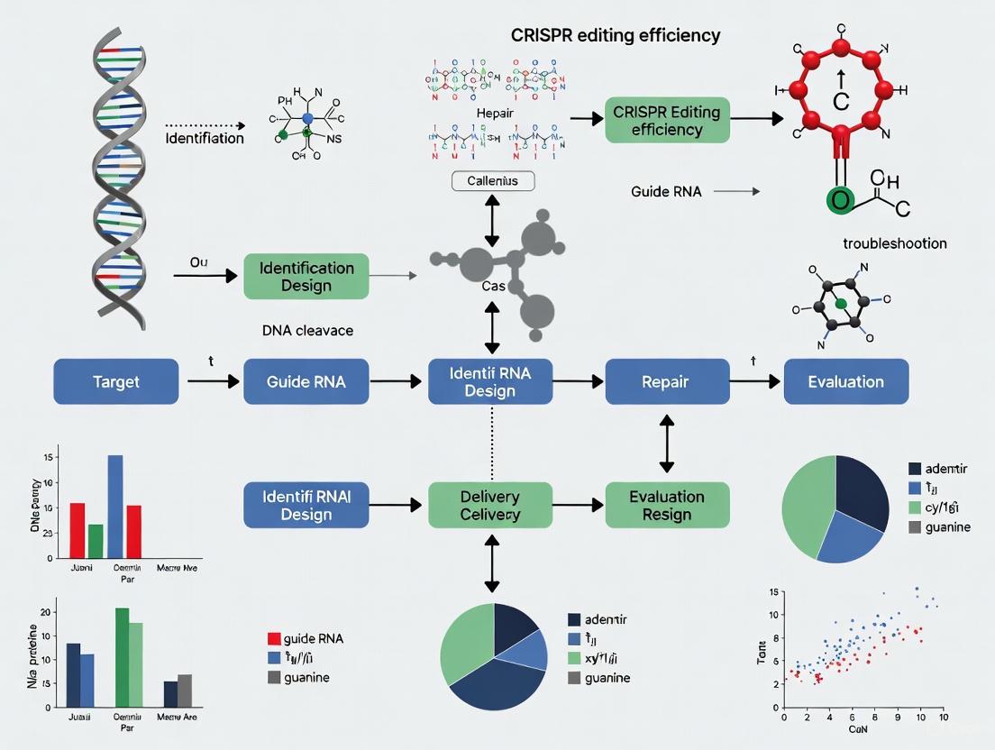
Visualizing the CRISPR-Cas9 Workflow and Repair Pathways
The following diagram illustrates the core mechanism of CRISPR-Cas9, from the creation of a double-strand break to the potential repair outcomes and associated technical challenges.
This troubleshooting guide provides a foundational framework for diagnosing and resolving common issues in CRISPR-Cas9 experiments. As the field evolves, staying informed on novel Cas variants, refined delivery methods, and advanced detection techniques will be crucial for achieving precise and efficient genome editing.
Frequently Asked Questions (FAQs)
Q1: What are the most common factors that lead to low editing efficiency in CRISPR experiments?
Low editing efficiency is most commonly caused by suboptimal guide RNA (gRNA) design, inefficient delivery methods, and low expression or activity of the Cas nuclease. The design of the gRNA is paramount; guides must be highly specific to the target and avoid off-target sites. Delivery is another critical bottleneck—whether using viral vectors, lipid nanoparticles, or electroporation, the CRISPR machinery must efficiently reach the cell nucleus. Furthermore, the choice of promoter driving Cas9 and gRNA expression must be suitable for your specific cell type to ensure adequate levels of the nuclease and its guide [4] [6].
Q2: How can I improve the specificity of my edits and reduce off-target effects?
To enhance specificity and minimize off-target effects, researchers should:
- Use high-fidelity Cas9 variants that have been engineered for greater precision.
- Employ modified, chemically synthesized guide RNAs, which are more stable and can elicit a lower immune response, improving editing accuracy.
- Utilize ribonucleoprotein (RNP) complexes for delivery. Delivering pre-assembled Cas9-gRNA complexes leads to a faster, more short-lived editing window, reducing the opportunity for off-target cuts [4] [6].
- Apply anti-CRISPR proteins. A newly developed system uses a cell-permeable protein to rapidly deactivate Cas9 after editing is complete, significantly reducing off-target activity [7].
Q3: My edits are successful but inconsistent, resulting in a mix of edited and unedited cells (mosaicism). How can I address this?
Mosaicism often arises from the timing of delivery and the cell cycle stage. To achieve a more homogeneous edited population:
- Synchronize your cell population to ensure delivery occurs at the optimal cell cycle stage for your desired repair pathway (e.g., S/G2 phases for homology-directed repair).
- Use inducible Cas9 systems to control the timing of nuclease activity.
- Isolate single-cell clones after editing. Following the editing process, perform single-cell dilution cloning and screen the resulting colonies to isolate clonal populations with uniform edits [4].
Q4: Are there new delivery technologies that can boost editing efficiency?
Yes, delivery technology is a rapidly advancing area. A significant recent development is the lipid nanoparticle spherical nucleic acid (LNP-SNA). This nanostructure wraps the CRISPR machinery in a protective, DNA-coated shell that cells absorb much more efficiently. In lab tests, this system tripled gene-editing success rates and improved precision compared to standard lipid nanoparticles [8]. Furthermore, researchers are continuously engineering new biodegradable ionizable lipids for LNPs that improve mRNA delivery to target organs like the liver, which is a common target for CRISPR therapies [9].
Troubleshooting Common Problems
The table below summarizes common issues, their potential causes, and recommended solutions.
| Problem | Potential Cause | Recommended Solution |
|---|---|---|
| Low Editing Efficiency [4] [6] | Poor gRNA design, low transfection efficiency, suboptimal Cas9/gRNA expression. | Test 2-3 gRNAs; optimize delivery method (e.g., electroporation, lipofection); use RNPs; verify promoter suitability and codon-optimize Cas9. |
| High Off-Target Effects [4] [7] | Cas9 activity lingering in cells, gRNA homology with other genomic sites. | Use high-fidelity Cas9 variants; design specific gRNAs with prediction tools; employ anti-CRISPR shut-off systems; deliver via RNP complexes. |
| Cell Toxicity [4] | High concentrations of CRISPR components. | Titrate component concentrations (start low); use RNPs or modified gRNAs to reduce immune stimulation. |
| Mosaicism [4] | Editing occurring at different cell cycles, delayed Cas9 expression. | Synchronize cell population; use inducible Cas9 systems; perform single-cell cloning post-editing. |
| Inability to Detect Edits [4] | Insensitive genotyping methods. | Use robust detection methods (T7EI assay, Surveyor assay, Sanger sequencing, or NGS). |
| Unsuccessful Cloning of gRNA [10] | Incorrectly designed oligonucleotides, degraded ds oligonucleotides. | Verify oligo design includes required cloning sequences (e.g., GTTTT, CGGTG); avoid repeated freeze-thaw cycles of oligonucleotides. |
Experimental Protocols for Efficiency Optimization
Protocol 1: Testing and Validating Guide RNA Efficiency
A critical first step is empirically determining the most effective gRNA for your target.
- Design: Select 2-3 bioinformatically predicted gRNAs for your target gene using reputable design tools [6] [11].
- Delivery: Transferd your chosen gRNAs and Cas9 (as plasmid, mRNA, or RNP) into your target cells. Include a positive control (a well-validated gRNA) and a negative control (a non-targeting gRNA) [4].
- Incubation: Allow 48-72 hours for editing to occur.
- Analysis: Extract genomic DNA. Amplify the target region by PCR and analyze the products using one of the following methods:
- Enzymatic Mismatch Assay: Use T7 Endonuclease I (T7EI) or Surveyor assay on the PCR products. Cleaved bands indicate successful editing. Analyze by gel electrophoresis [4] [6].
- Sequencing: For the most accurate results, subject the PCR products to Sanger or Next-Generation Sequencing (NGS). This reveals the exact sequences and spectrum of indels [6].
Protocol 2: Delivering CRISPR Components as Ribonucleoproteins (RNPs)
RNP delivery can increase efficiency and reduce off-target effects.
- Complex Formation: In vitro, complex purified Cas9 protein with your synthesized, modified gRNA at a molar ratio of 1:2 to 1:3 (Cas9:gRNA). Incubate at room temperature for 10-20 minutes to form the RNP complex [6].
- Cell Delivery: Deliver the pre-formed RNP complexes into your cells using a method suitable for your cell type, such as electroporation or lipofection (e.g., using Lipofectamine 3000 reagent) [10] [6].
- Advantages: This method leads to rapid editing, reduces the risk of immune activation and genomic integration of DNA, and shortens the window for Cas9 activity, thereby lowering off-target effects [6].
Key Factors and Optimization Workflow
The diagram below outlines the logical relationship between key factors, common issues, and optimization strategies in a CRISPR experiment.
The Scientist's Toolkit: Essential Reagents and Materials
The table below details key reagents and their functions for successful and efficient CRISPR genome editing.
| Research Reagent | Function & Application |
|---|---|
| High-Fidelity Cas9 Variants | Engineered versions of Cas9 with reduced off-target effects, crucial for therapeutic applications [4]. |
| Modified Synthetic gRNAs | Chemically synthesized guide RNAs with modifications (e.g., 2'-O-methyl) that enhance stability, improve editing efficiency, and reduce immune response compared to in vitro transcribed (IVT) gRNAs [6]. |
| Ribonucleoprotein (RNP) Complexes | Pre-assembled complexes of Cas9 protein and gRNA. The preferred delivery method for many applications due to high efficiency, rapid action, and reduced off-target effects [6]. |
| Lipid Nanoparticles (LNPs) | A non-viral delivery vehicle, ideal for in vivo delivery. Recent advances include LNP-SNAs (Spherical Nucleic Acids), which dramatically improve cellular uptake and editing efficiency [8] [9]. |
| Anti-CRISPR Proteins (e.g., LFN-Acr/PA) | Proteins used to precisely "turn off" Cas9 activity after editing is complete. This new technology minimizes the time Cas9 is active in the cell, greatly reducing off-target effects [7]. |
| T7 Endonuclease I (T7EI) / Surveyor Assay Kits | Enzymatic mismatch detection kits used for initial, rapid assessment of editing efficiency at the target site by detecting DNA heteroduplexes [4] [6]. |
Advanced Strategies: Signaling and Workflow for Precision
For advanced applications requiring high precision, such as homology-directed repair (HDR), the cellular signaling pathways and experimental workflow become critical. The following diagram illustrates a strategy that combines optimal delivery with a safety switch to maximize on-target editing.
The efficiency and outcome of CRISPR-Cas9 genome editing are profoundly influenced by the physiological state of the target cell. Recent research reveals that fundamentally different DNA repair mechanisms operate in dividing versus non-dividing (postmitotic) cells, leading to dramatic variations in editing results [2]. This cellular context dependency presents both significant challenges and opportunities for therapeutic genome editing, particularly for diseases affecting non-dividing tissues such as neurons and cardiomyocytes [2] [12].
Understanding these differences is crucial for troubleshooting low CRISPR editing efficiency. While dividing cells like immortalized cell lines (HEK293, HeLa) efficiently utilize certain repair pathways, therapeutically relevant primary cells and differentiated cells often exhibit slower editing kinetics and distinct repair outcomes [13] [2]. This technical guide provides troubleshooting strategies and FAQs to help researchers navigate these complexities and optimize editing protocols for their specific experimental systems.
Key Biological Differences: Why Cell Type Matters
DNA Repair Pathway Utilization
The core challenge stems from differential activation of DNA repair pathways across cell states. Dividing cells actively cycle through cell cycle phases, enabling them to utilize a broader repertoire of repair mechanisms, including homology-directed repair (HDR) and microhomology-mediated end joining (MMEJ) [2]. In contrast, non-dividing cells predominantly rely on non-homologous end joining (NHEJ) and exhibit upregulated non-canonical DNA repair factors [2] [12].
Table 1: DNA Repair Pathway Activity in Different Cell States
| DNA Repair Pathway | Dividing Cells | Non-Dividing Cells | Cell Cycle Dependence |
|---|---|---|---|
| Non-homologous end joining (NHEJ) | High | Very High | No |
| Homology-directed repair (HDR) | High | Very Low | Yes (S/G2 phases) |
| Microhomology-mediated end joining (MMEJ) | High | Low | Yes (S/G2 phases) |
| Alternative end joining | Variable | upregulated in neurons [2] | No |
Editing Kinetics and Outcome Distributions
The timeline for achieving maximal editing efficiency varies dramatically between cell types. In dividing cells, CRISPR-induced indels typically plateau within 1-2 days post-transfection. However, in non-dividing cells such as neurons and cardiomyocytes, indel accumulation can continue increasing for up to 2 weeks after Cas9 delivery [2]. This prolonged timeline reflects fundamental differences in how these cell types manage DNA damage response.
Table 2: Quantitative Comparison of Editing Outcomes Between Cell Types
| Editing Parameter | iPSCs (Dividing) | Neurons (Non-dividing) | Experimental Evidence |
|---|---|---|---|
| Time to maximal indel accumulation | 1-2 days | 14-16 days [2] | VLP delivery of Cas9 RNP to isogenic cells |
| Predominant repair pathway | MMEJ | NHEJ [2] | Sequencing of editing outcomes at multiple loci |
| Distribution of outcomes | Broad range of indels | Narrow distribution [2] | Deep sequencing analysis |
| Insertion-to-deletion ratio | Lower | Significantly higher [2] | Analysis of multiple sgRNAs |
Diagram Title: Differential CRISPR Repair in Dividing vs. Non-Dividing Cells
Troubleshooting Guide: FAQs for Experimental Challenges
FAQ 1: Why is my editing efficiency so low in primary neurons and cardiomyocytes?
Cause: Non-dividing cells exhibit inherently slower editing kinetics and limited repair pathway availability compared to immortalized cell lines [2].
Solutions:
- Extend your assessment timeline: Allow at least 2 weeks post-transduction before evaluating editing outcomes in non-dividing cells [2].
- Optimize delivery methods: Use virus-like particles (VLPs) pseudotyped with VSVG and BaEVRless (BRL) glycoproteins, which achieve >95% transduction efficiency in human neurons [2].
- Manipulate DNA repair pathways: Apply chemical or genetic perturbations to direct repair toward desired outcomes in non-dividing cells [2] [12].
FAQ 2: Why do I see different mutation patterns in my stem cells versus differentiated cells?
Cause: Genetically identical cells at different differentiation stages employ distinct DNA repair machinery, resulting in divergent editing outcomes [2].
Solutions:
- Pre-validate sgRNAs in your target cell type: sgRNA performance varies significantly between cell types, even when targeting the same genomic locus [13] [11].
- Select target loci carefully: For knockout experiments in cells with multiple isoforms, design sgRNAs targeting exons common to all relevant isoforms [13].
- Anticipate pathway-specific outcomes: Recognize that MMEJ-associated larger deletions predominate in dividing cells, while NHEJ-associated small indels are more common in non-dividing cells [2].
FAQ 3: How can I improve editing precision in non-dividing cells for therapeutic applications?
Cause: The natural repair pathway bias in non-dividing cells favors error-prone NHEJ over precise HDR [2] [14].
Solutions:
- Consider alternative editors: Base editors or prime editors can achieve precise modifications without requiring DSBs or HDR, working efficiently in non-dividing cells [2].
- Modulate repair pathways: Chemical inhibition of NHEJ factors or activation of alternative repair pathways can shift outcome distributions [2] [12].
- Use RNP complexes: Preassembled Cas9-gRNA ribonucleoprotein complexes often show higher editing efficiency and specificity than plasmid-based delivery, especially when delivered via electroporation or nanoparticles [15] [16].
Essential Protocols for Cell-Type Specific Editing
Protocol: Rapid Assessment of Editing Outcomes Using Fluorescent Reporters
This protocol enables high-throughput screening of editing efficiency across different cell types using an eGFP to BFP conversion assay [14].
Materials:
- eGFP-positive cell lines (e.g., HEK293T, Hepa 1-6, IMR90, HepG2)
- SpCas9-NLS protein
- sgRNA targeting eGFP locus: GCUGAAGCACUGCACGCCGU
- Optimized BFP mutation HDR template
- Delivery reagent (Polyethylenimine or ProDeliverIN CRISPR)
- Flow cytometer with appropriate filters
Step-by-Step Workflow:
- Cell Preparation: Culture eGFP-positive cells to 70-80% confluency in appropriate complete medium [14].
- RNP Complex Formation: Combine SpCas9 protein with sgRNA at molar ratio 1:1.2 and incubate 10 minutes at room temperature.
- Transfection: Deliver RNP complexes with or without HDR template using your preferred method. For HEK293T cells, polyethylenimine (PEI) provides efficient delivery.
- Incubation: Maintain cells for 3-7 days post-transfection to allow editing and fluorescence conversion.
- Analysis: Analyze cells by flow cytometry, measuring eGFP loss (NHEJ) and BFP gain (HDR) to quantify editing outcomes.
Diagram Title: Fluorescent Reporter Workflow for Editing Assessment
Protocol: Optimizing Delivery for Challenging Non-Dividing Cells
Efficient delivery remains a primary challenge in non-dividing cells. This protocol outlines VLP-based delivery optimized for neurons and cardiomyocytes [2].
Materials:
- HIV-based or FMLV-based VLP systems
- VSVG and BaEVRless (BRL) pseudotyping proteins
- Cas9 RNP complexes
- iPSC-derived neurons or cardiomyocytes
- Appropriate neuronal or cardiac culture media
Step-by-Step Workflow:
- VLP Production: Produce VLPs containing Cas9 RNP using appropriate packaging cells and pseudotyping with VSVG/BRL glycoproteins [2].
- Cell Preparation: Differentiate iPSCs to target cell type (neurons/cardiomyocytes), confirming purity (>95% NeuN+ for neurons) and postmitotic state (Ki67-negative) [2].
- Transduction: Apply VLPs to cells at optimized MOI, determined through pilot experiments.
- Extended Incubation: Maintain cells for 14-16 days post-transduction, refreshing media as needed.
- Outcome Assessment: Harvest cells at multiple timepoints and analyze editing outcomes using next-generation sequencing to capture the prolonged editing kinetics.
Research Reagent Solutions: Essential Tools for Cell-Type Specific Editing
Table 3: Key Reagents for Optimizing CRISPR Across Cell Types
| Reagent/Category | Specific Examples | Function & Application | Considerations |
|---|---|---|---|
| Delivery Systems | Virus-like particles (VLPs) pseudotyped with VSVG/BRL [2] | Efficient Cas9 RNP delivery to non-dividing cells | Achieves >95% transduction in human neurons |
| Lipid nanoparticles (LNPs) [16] | Non-viral RNP delivery for in vivo applications | Tissue-specific targeting possible | |
| Editing Cargo | Preassembled RNP complexes [15] [16] | Highest editing efficiency with minimal off-target effects | Ideal for sensitive primary cells |
| High-fidelity Cas9 variants [15] | Reduced off-target effects | Important for therapeutic applications | |
| Reporter Systems | eGFP-BFP conversion system [14] | Rapid quantification of HDR vs NHEJ outcomes | Enables high-throughput optimization |
| Cell Models | iPSC-derived neurons & cardiomyocytes [2] | Physiologically relevant non-dividing models | Genetically matched to iPSC controls available |
The dramatic differences in CRISPR repair outcomes between dividing and non-dividing cells underscore the critical importance of cell-type specific optimization in genome editing experiments. By understanding the distinct DNA repair environments in these cells, researchers can develop more effective troubleshooting strategies and design better experiments.
Future directions in this field include developing small molecule modulators of cell-type specific repair factors, engineering novel Cas variants with reduced dependence on endogenous repair pathways, and optimizing delivery platforms that account for the unique biology of non-dividing cells. As CRISPR-based therapeutics advance toward clinical applications, acknowledging and addressing these fundamental cellular differences will be essential for success, particularly for neurological and cardiac diseases where non-dividing cells are the primary therapeutic targets.
Frequently Asked Questions (FAQs)
Q1: Why does non-homologous end joining (NHEJ) dominate over homology-directed repair (HDR) in postmitotic cells like neurons?
A1: The dominance of NHEJ in postmitotic cells is fundamentally due to cell cycle restrictions. HDR is strictly confined to the S and G2 phases of the cell cycle because it requires a sister chromatid as a template for repair [2] [17]. Postmitotic cells, such as mature neurons and cardiomyocytes, have permanently exited the cell cycle. Consequently, they lack this essential template and cannot activate the HDR machinery effectively [18] [17]. In contrast, NHEJ is active throughout all cell cycle phases and does not require a template, making it the primary and most readily available pathway for repairing double-strand breaks in non-dividing cells [17].
Q2: We are observing very low CRISPR knockout efficiency in our primary neuronal cultures. Is this a delivery problem or a repair problem?
A2: While efficient delivery is always a consideration, recent evidence strongly suggests that the inherently slow kinetics of DNA repair in postmitotic cells is a major contributing factor. Research shows that while indels in dividing cells plateau within days, they can continue to accumulate in neurons for up to two weeks after Cas9 delivery [2] [19]. Before troubleshooting delivery, ensure you are allowing a sufficiently long time for editing outcomes to manifest. Furthermore, confirm that your experimental system is truly postmitotic, as the DNA repair pathway balance differs significantly between dividing and non-dividing cells [2].
Q3: Are the risks of large structural variations (like chromosomal translocations) different when editing postmitotic cells?
A3: The risk of structural variations (SVs) is a critical safety concern for all CRISPR therapies. While the unique repair environment of postmitotic cells may influence SV profiles, the use of certain NHEJ-inhibiting small molecules to enhance HDR in other cell types has been shown to drastically increase the frequency of kilobase- to megabase-scale deletions and chromosomal translocations [1]. This underscores the importance of using advanced sequencing methods (e.g., CAST-Seq, LAM-HTGTS) that can detect these large aberrations, as they are often missed by standard short-read amplicon sequencing [1].
Troubleshooting Guide: Low Knock-in Efficiency via HDR
Problem: Failure to achieve precise gene insertion or correction via HDR in postmitotic cells.
Solution: Given the near impossibility of performing standard HDR in non-cycling cells, consider switching to alternative precision editing tools that do not rely on the HDR pathway.
- Utilize Advanced Editors: Consider Prime Editing or Base Editing systems. These technologies can mediate precise changes without requiring a double-strand break or a donor template, making them much more effective in postmitotic cells. For example, prime editing has been used successfully in human iPSC-derived cardiomyocytes to correct disease-causing mutations with up to 34.8% efficiency [20].
- Explore Novel Nuclease Platforms: Investigate platforms like ARCUS nucleases, which have been reported to trigger high-frequency homologous recombination and achieve precise edits in non-dividing cells [20].
- If HDR is Absolutely Necessary: If you must use HDR, strategies involve artificially manipulating the cell cycle or DNA repair pathways, but this is typically only feasible in ex vivo settings with dividing cells. For postmitotic cells, HDR remains a major challenge, and alternative editors are strongly recommended [18] [17].
Key Data and Experimental Comparisons
The tables below summarize core quantitative findings and methodological details from recent key studies.
Table 1: Comparison of CRISPR-Cas9 Repair Kinetics and Outcomes in Dividing vs. Postmitotic Cells
| Feature | Dividing Cells (e.g., iPSCs) | Postmitotic Cells (e.g., Neurons) | Key References |
|---|---|---|---|
| Primary Repair Pathway | Microhomology-Mediated End Joining (MMEJ) & NHEJ | Classical Non-Homologous End Joining (cNHEJ) | [2] |
| HDR Efficiency | Low, but possible in S/G2 phase | Extremely Low / Theoretically impossible | [18] [17] |
| Time to Indel Plateau | A few days | Up to 2 weeks | [2] [19] |
| Indel Distribution | Broad, larger deletions (MMEJ-like) | Narrow, small indels (NHEJ-like) | [2] |
| Therapeutic Example | Ex vivo editing of hematopoietic stem cells | Gene inactivation for dominant neurodegenerative diseases | [2] [21] |
Table 2: Small Molecules for Modulating CRISPR Editing Efficiency
| Small Molecule | Target | Effect on Editing | Reported Efficiency Increase | Notes and Risks |
|---|---|---|---|---|
| Repsox | TGF-β pathway (SMAD2/3/4) | Enhances NHEJ-mediated knockout | Up to 3.16-fold (in porcine cells) | Mechanism involves downregulation of SMAD proteins [22]. |
| AZD7648 | DNA-PKcs (NHEJ) | Inhibits NHEJ to enhance HDR | N/A (HDR increase reported) | Risks: Significantly increases large structural variations and chromosomal translocations [1]. |
| Various Inhibitors | 53BP1, Ligase IV | Inhibits NHEJ to enhance HDR | Varies | Transient 53BP1 inhibition did not increase translocations in one study, but general suppression of NHEJ carries risks [1]. |
Experimental Protocol: Analyzing DNA Repair in Postmitotic Neurons
This protocol is adapted from a 2025 Nature Communications study that directly compared repair outcomes in iPSCs and iPSC-derived neurons [2].
Workflow Title: Comparing CRISPR Repair in Dividing and Postmitotic Cells
Step-by-Step Methodology:
Cell Line Preparation:
Validation of Postmitotic State:
CRISPR Delivery via Virus-Like Particles (VLPs):
- Rationale: Standard transfection is inefficient in neurons. VLPs engineered from Friend murine leukemia virus (FMLV) or HIV, pseudotyped with VSVG/BRL glycoproteins, can achieve >95% transduction efficiency in human neurons [2].
- Procedure: Produce VLPs loaded with pre-assembled Cas9 ribonucleoprotein (RNP) complexed with your target sgRNA.
Transduction and Time-Course Experiment:
Analysis of Editing Outcomes:
- Amplify the target genomic locus by PCR and perform next-generation sequencing (NGS).
- Use computational tools (e.g., CRISPResso2) to quantify the spectrum and frequency of insertion/deletion mutations (indels).
- Key Analysis: Compare the kinetics of indel accumulation and the distribution of indel types (e.g., ratio of small vs. large deletions) between the two cell types [2].
The Scientist's Toolkit: Essential Research Reagents
Table 3: Key Reagents for Studying DNA Repair in Postmitotic Cells
| Reagent / Tool | Function | Application in Postmitotic Cells |
|---|---|---|
| iPSC-Derived Neurons | Provides a genetically defined, human-relevant model of postmitotic cells. | Essential for creating isogenic pairs with dividing iPSCs to isolate cell cycle effects on DNA repair [2]. |
| Virus-Like Particles (VLPs) | Efficient delivery of Cas9 protein (as RNP) into hard-to-transfect cells. | Superior to plasmids for transient, efficient Cas9 delivery to neurons, minimizing off-target effects from prolonged expression [2] [19]. |
| NHEJ-Enhancing Molecules (e.g., Repsox) | Small molecules that inhibit specific pathways to bias repair toward NHEJ. | Can boost NHEJ-mediated knockout efficiency in challenging cell types, as demonstrated in porcine cells [22]. |
| Prime Editing System (e.g., PE4) | A "search-and-replace" system that directly writes new genetic information without DSBs. | Enables precise single-base changes or small insertions/deletions in postmitotic cells where HDR is ineffective [20]. |
| Advanced Sequencing (CAST-Seq) | Detects large structural variations and chromosomal translocations. | Critical for comprehensive safety profiling, as standard amplicon-seq misses large deletions from CRISPR editing [1]. |
Technical Support Center
Troubleshooting Guides and FAQs
Q: Why does CRISPR-induced indel formation happen so slowly in my postmitotic cells, such as neurons and cardiomyocytes, compared to standard dividing cell lines?
A: Slow accumulation of insertions and deletions (indels) is a fundamental characteristic of how non-dividing cells respond to CRISPR-Cas9-induced DNA damage. Unlike rapidly proliferating cells, which resolve double-strand breaks (DSBs) quickly to avoid cell death during division, postmitotic cells lack this pressure and repair DNA over a much longer, weeks-long timeline [2]. The primary cause is the different DNA repair pathway preferences: dividing cells frequently use faster, more mutagenic pathways like microhomology-mediated end joining (MMEJ), while neurons rely more heavily on the non-homologous end joining (NHEJ) pathway, which can proceed at a slower pace and even result in a higher ratio of small insertions to deletions [2].
Key Differences in DNA Repair Between Dividing and Non-Dividing Cells
| Feature | Dividing Cells (e.g., iPSCs) | Non-Dividing Cells (e.g., Neurons, Cardiomyocytes) |
|---|---|---|
| Primary DSB Repair Pathway | MMEJ-like (larger deletions predominant) [2] | NHEJ-like (smaller indels predominant) [2] |
| Typical Indel Accumulation Timeline | Plateaus within a few days [2] | Continues to increase for up to 16 days or more [2] |
| Ratio of Insertions to Deletions | Lower [2] | Significantly higher [2] |
| Pressure for Fast Repair | High (to pass cell cycle checkpoints) [2] | Low (no replication checkpoints) [2] |
Q: What experimental evidence supports this prolonged editing timeline in neurons?
A: A key 2025 study used virus-like particles (VLPs) to deliver Cas9 ribonucleoprotein (RNP) to both human induced pluripotent stem cells (iPSCs) and genetically identical iPSC-derived neurons [2]. The researchers tracked the formation of indels over time and found that while editing in iPSCs reached a maximum within a few days, indels in neurons continued to accumulate for at least two weeks post-delivery [2]. This same prolonged timeline was also observed in iPSC-derived cardiomyocytes, confirming it is a trait of postmitotic cells [2].
Detailed Experimental Protocol: Tracking Indel Kinetics
- Cell Models: Human iPSCs and iPSC-derived cortical-like excitatory neurons (confirmed postmitotic by Ki67-negative and NeuN-positive staining) [2].
- CRISPR Delivery: Cas9 RNP delivered via VSVG-pseudotyped or VSVG/BRL-co-pseudotyped Virus-Like Particles (VLPs), achieving up to 97% transduction efficiency [2].
- Time-Course Experiment:
- Transduce cells with Cas9 VLPs.
- Harvest cells at multiple time points post-transduction (e.g., days 1, 2, 4, 7, 11, 16).
- Extract genomic DNA.
- Amplify the target genomic region by PCR and perform next-generation sequencing (NGS).
- Use bioinformatics tools (e.g., CRISPResso2) to quantify the percentage of reads containing indels at each time point [2].
- Validation: The extended timeline was replicated using multiple sgRNAs and two different VLP platforms, ruling out delivery delays as the sole cause [2].
Diagram 1: Divergent CRISPR repair timelines in dividing and non-dividing cells.
Q: How can I troubleshoot and potentially improve editing efficiency in these challenging cell types?
A: While the slow timeline may be intrinsic, you can optimize your experiment for the best possible outcome.
- Optimize Delivery: Ensure your CRISPR components are efficiently delivered. VLPs pseudotyped with VSVG and BaEVRless (BRL) have shown high efficiency (up to 97%) in human neurons [2]. For other non-dividing cells like resting T cells, electroporation of Cas9 RNP can be effective [2] [5].
- Validate sgRNA Activity: A poorly designed sgRNA is a common cause of low efficiency [4] [5]. Use bioinformatics tools (e.g., CRISPR Design Tool, Benchling) to design highly specific sgRNAs with optimal GC content (40-60%) and test 3-5 different sgRNAs per gene to identify the most effective one [5] [23].
- Confirm Successful Transduction and Cutting: Use immunocytochemistry to confirm the presence of Cas9-induced DSBs by co-staining for markers like γH2AX and 53BP1 in your target cells [2].
- Allow Sufficient Time for Editing: Plan your experiment with the extended timeline in mind. Harvesting cells too early (e.g., before 7 days) will capture only a fraction of the eventual indels. Monitor editing over a 2-week period to capture the full effect [2].
- Consider Alternative Editors: If precise nucleotide change is the goal, base editing or prime editing might be more efficient than Cas9 nuclease in neurons, as they do not rely on the formation of a DSB and can sometimes show comparable or even higher efficiency within a shorter time frame [2].
Q: Are there specific reagents or tools that can help study this phenomenon?
A: Yes, the table below lists key reagents and tools used in the foundational research on this topic.
Research Reagent Solutions
| Item | Function | Example/Specification |
|---|---|---|
| Virus-Like Particles (VLPs) | Protein-based delivery of Cas9 RNP to hard-to-transfect postmitotic cells [2]. | VSVG-pseudotyped HIV VLPs or VSVG/BRL-co-pseudotyped FMLV VLPs [2]. |
| iPSC-Derived Neurons | A genetically defined, clinically relevant model for studying DNA repair in human neurons [2]. | Cortical-like excitatory neurons; >95% NeuN-positive, >99% Ki67-negative [2]. |
| Cas9 RNP Complex | The active editing complex; direct delivery of RNP reduces off-target effects and allows for transient activity [2]. | Pre-complexed purified Cas9 protein and sgRNA [2]. |
| sgRNA Design Tools | Bioinformatics software to predict and select high-activity, specific guide RNAs [5]. | CRISPR Design Tool, Benchling, GuideScan [5] [23]. |
| Antibodies for ICC | Validate DSB formation and repair protein recruitment [2]. | Anti-γH2AX and anti-53BP1 [2]. |
Diagram 2: A logical troubleshooting workflow for slow indel accumulation.
Strategic Guide and Donor Design for Knockout and Knock-in Experiments
FAQ: Addressing Key Challenges in sgRNA Design and Workflow
What are the most critical factors for designing an sgRNA for gene knockout? The primary factors are on-target activity and minimizing off-target effects. Successful design depends on:
- sgRNA Sequence: The nucleotide sequence at specific positions in the guide region significantly influences efficiency. For example, certain bases in the "seed" region (positions 1-10) and the non-seed region (positions 15-18) are critical [24].
- GC Content: Optimal GC content (typically 40-60%) improves stability and binding.
- Off-target Potential: The sgRNA should have minimal similarity to other genomic sequences, especially in the seed region, to prevent unintended cuts [5] [25].
- Target Location: For knockouts, target an exon near the 5' end of the gene's coding sequence to maximize the chance of a frameshift mutation [5].
Why is my homology-directed repair (HDR) efficiency so low, and how can I improve it? HDR is inherently less efficient than error-prone repair pathways like non-homologous end joining (NHEJ) in mammalian cells [26] [18]. You can improve HDR by:
- Modulating DNA Repair Pathways: Inhibiting key NHEJ factors (e.g., KU70, KU80, LIG4) or activating HDR factors (e.g., CDK1, CtIP) can shift the balance toward HDR. One study showed that simultaneously activating CDK1 and repressing KU80 increased HDR rates by an order of magnitude [26].
- Using High-Fidelity Cas9 Variants: Enzymes like HiFi Cas9 can reduce off-target effects, but be aware that some can cause a guide-dependent loss of on-target efficiency [24].
- Cell Cycle Timing: HDR is most active in the S and G2 phases. Transfecting cells during these phases or using cell cycle synchronization can enhance HDR [26].
Important Safety Note: Strategies that inhibit the NHEJ pathway (e.g., using DNA-PKcs inhibitors) to enhance HDR can carry a hidden risk. They have been shown to significantly increase the frequency of large, on-target structural variations, including megabase-scale deletions and chromosomal translocations, which traditional short-read sequencing often misses [27].
My knockout worked in one cell line but not another. What could be the reason? Cell line specificity is a major challenge in CRISPR experiments. Causes for variable efficiency include:
- DNA Repair Machinery: Different cell lines have varying levels of DNA repair enzymes. Some, like HeLa cells, have highly efficient repair systems that can quickly fix Cas9-induced breaks, reducing knockout success [5].
- Cell State: Post-mitotic cells, such as neurons, repair DNA with unique kinetics and favor different repair pathways (like NHEJ over MMEJ) compared to dividing cells, leading to a narrower range of edits and an extended editing window [28].
- Transfection Efficiency: Delivery of CRISPR components can vary dramatically between cell types. Hard-to-transfect cells may require optimized methods like electroporation or the use of stable Cas9-expressing cell lines [5].
How many sgRNAs should I test per gene? It is strongly recommended to test multiple sgRNAs per gene, typically 3 to 5 [5]. This is because sgRNA efficiency is highly variable and sequence-dependent. Testing multiple guides ensures that at least one will be highly effective, safeguarding your experiment against the failure of a single sgRNA.
Optimized sgRNA Design Parameters
The table below summarizes key design rules for maximizing on-target activity and specificity, synthesized from large-scale empirical studies [25].
Table 1: Key sgRNA Sequence Features for Optimal On-Target Activity
| Feature | Optimal Characteristic | Rationale & Impact |
|---|---|---|
| Seed Region (pos 1-10) | Specific nucleotide preferences (e.g., G at pos 1, C at pos 19 for some variants) | Critical for initial DNA binding; specific bases are enriched in highly active guides [25] [24]. |
| Non-Seed Region (pos 15-18) | Avoid sequences leading to loss of efficiency in high-fidelity Cas9 variants [24]. | Interacts with the REC3 domain of Cas9; mutations in high-fidelity variants can make efficiency dependent on these positions. |
| GC Content | 40% - 60% | Guides with very high or very low GC content can form stable secondary structures or have poor binding affinity [5]. |
| Off-target Score | Select sgRNAs with the lowest predicted off-target activity. | Minimizes unintended genomic alterations. Tools like MOFF score can predict off-target potential [25] [24]. |
Experimental Protocol: A Workflow for Validating sgRNA Efficiency and Knockout
This protocol outlines a robust method for generating and validating knockout hPSC lines using an inducible Cas9 system, incorporating optimizations for high efficiency [29].
Design and Synthesis:
- Design 3-5 sgRNAs per target gene using a reliable algorithm (e.g., Benchling, CCTop, or GuideVar for high-fidelity Cas9 variants) [29] [24].
- Synthesize chemically modified sgRNAs (CSM-sgRNA) with 2'-O-methyl-3'-thiophosphonoacetate modifications at both ends to enhance stability within cells, which has been shown to improve editing rates [29].
Cell Preparation and Nucleofection:
- Culture your engineered cell line (e.g., hPSCs with doxycycline-inducible Cas9).
- Induce Cas9 expression with doxycycline 24 hours before nucleofection.
- Dissociate cells and electroporate using a high-efficiency program (e.g., program CA137 on a Lonza 4D-Nucleofector). A critical optimized parameter is using 5 µg of sgRNA for 8×10^5 cells [29].
- For difficult targets, a second nucleofection with the same sgRNA can be performed 3 days after the first to boost indel rates.
Assessing Editing Efficiency (INDELs):
- Harvest cells 3-5 days post-nucleofection and extract genomic DNA.
- Amplify the target region by PCR and submit the product for Sanger sequencing.
- Analyze the sequencing chromatograms using algorithms like ICE (Inference of CRISPR Edits) or TIDE (Tracking of Indels by Decomposition) to quantify the percentage of insertions and deletions (INDELs) [29]. This optimized system can achieve INDEL rates of 82-93% [29].
Functional Validation of Knockout:
- Perform Western blotting to confirm the absence of the target protein. This is a crucial step, as high INDEL percentages do not always equate to complete protein loss. Some reading frame shifts may not be disruptive, resulting in "ineffective sgRNAs" and retained protein expression [29].
- For further phenotypic characterization, you can perform downstream assays like flow cytometry, reporter assays, or functional cellular assays specific to your target [5].
Diagram 1: Experimental workflow for sgRNA validation and knockout confirmation.
Advanced Strategy: Enhancing HDR with CRISPRa/i
For precise editing requiring HDR, a powerful strategy is to use catalytically dead guide RNAs (dgRNAs) to reprogram the cell's DNA repair machinery at the transcriptional level. This method uses a single active Cas9 for cutting and dgRNAs for regulation [26].
Diagram 2: CRISPRa/i mechanism for HDR enhancement.
Workflow:
- Co-deliver three components into your cells:
- A plasmid expressing active Cas9 and a target-specific sgRNA to create the DSB.
- Plasmids expressing dgRNAs that recruit activator complexes (like MS2-P65-HSF1) to promoters of HDR-related genes (e.g., CDK1, CtIP).
- Plasmids expressing dgRNAs that recruit repressor complexes (like COM-KRAB) to promoters of NHEJ-related genes (e.g., KU70, KU80, LIG4) [26].
- The dgRNAs activate HDR pathway components and repress NHEJ components, steering the repair of the Cas9-induced break toward the precise HDR pathway.
- This synergistic approach has been shown to increase HDR efficiency by up to 15-fold compared to standard methods [26].
Table 2: Key Research Reagents and Tools for CRISPR sgRNA Design and Validation
| Reagent / Tool | Function / Description | Example & Utility |
|---|---|---|
| Bioinformatics Tools | Algorithms to predict sgRNA on-target efficiency and off-target effects. | Benchling was identified as providing the most accurate predictions in one study [29]. GuideVar is a framework specifically for predicting performance with high-fidelity Cas9 variants [24]. |
| Stable Cas9 Cell Lines | Cell lines engineered for consistent Cas9 nuclease expression. | Eliminates variability from transient transfection, enhancing reproducibility and knockout efficiency (e.g., hPSCs-iCas9) [5] [29]. |
| High-Fidelity Cas9 Variants | Engineered Cas9 proteins with reduced off-target activity. | HiFi Cas9 and LZ3 are promising variants, but their efficiency can be sgRNA-sequence-dependent [24]. |
| Chemically Modified sgRNA | Synthetic sgRNAs with altered backbone for increased nuclease resistance. | Enhances sgRNA stability within cells, leading to higher editing efficiency [29]. |
| Analysis Software | Tools to quantify editing outcomes from Sanger sequencing data. | ICE (Synthego) and TIDE are widely used to calculate INDEL percentages from mixed sequencing traces [29]. |
Within the broader context of troubleshooting low CRISPR editing efficiency, selecting the appropriate Cas protein is a critical first step. The choice between wild-type, high-fidelity, and newly engineered variants directly impacts the success of your experiments, influencing on-target activity, off-target rates, and compatibility with your delivery system. This guide provides targeted FAQs and troubleshooting advice to help you navigate this complex decision.
FAQs: Cas Protein Selection
Q1: What are the primary limitations of wild-type SpCas9 that would lead me to consider an alternative?
Wild-type Streptococcus pyogenes Cas9 (SpCas9), while a workhorse, has three key limitations that can cause low experimental efficiency or confounding results:
- Off-target effects: Its complementarity requirements are not very stringent, allowing it to recognize and cleave non-specific DNA sequences with similar protospacer adjacent motifs (PAMs), such as NAG or NGA, leading to unwanted mutations [30].
- Large size: At about 4.2 kb, the SpCas9 coding sequence is difficult to package into delivery vectors with limited cargo capacity, such as adeno-associated viruses (AAVs), hindering certain in vivo applications [30].
- Restrictive PAM requirement: The canonical NGG PAM sequence limits the genomic regions that can be targeted, reducing the flexibility of your experimental design [30].
Q2: When should I use a high-fidelity Cas9 variant?
High-fidelity variants are engineered to minimize off-target cleavage while retaining robust on-target activity. They are essential for applications where specificity is paramount.
- SpCas9-HF1 is an early high-fidelity variant that harbors four alanine substitutions (N497A, R661A, Q695A, Q926A) designed to reduce non-specific interactions with the DNA phosphate backbone [31]. GUIDE-seq experiments showed it rendered all or nearly all off-target events undetectable for standard non-repetitive target sites while maintaining comparable on-target activity for over 85% of sgRNAs tested [31].
- eSpOT-ON (ePsCas9) is a more recently engineered high-fidelity nuclease that achieves exceptionally low off-target editing without the common trade-off of reduced on-target activity. It is derived from Parasutterella secunda Cas9 and is commercially available in both recombinant protein and mRNA formats [30].
Q3: I need to deliver CRISPR components via AAV. What are my best options?
For AAV delivery, compact Cas proteins are necessary. The table below summarizes key small-sized variants.
| Cas Protein | Origin | Size (amino acids) | PAM Sequence | Key Features |
|---|---|---|---|---|
| SaCas9 [30] | Staphylococcus aureus | 1053 | 3'-NNGRRT | Popular choice; used in neuronal studies, hepatitis B research, and plant genomes; has high-fidelity (SaCas9-HF) and broad-PAM (KKH-SaCas9) variants. |
| CjCas9 [30] | Campylobacter jejuni | ~984 | 3'-NNNNRYAC | Another naturally small Cas9 ortholog suitable for viral delivery. |
| hfCas12Max [30] | Engineered from Cas12i | 1080 | 5'-TN | High-fidelity nuclease; broader PAM recognition than SpCas9; uses a shorter crRNA. |
| Cas12g [32] | Type V-G System | 767 | None identified | RNA-guided ribonuclease (RNase) with collateral RNase and single-strand DNase activities; thermostable. |
Q4: Are there Cas variants that can target a wider range of genomic sequences?
Yes, several variants overcome the restrictive NGG PAM of SpCas9.
- ScCas9: Isolated from Streptococcus canis, it recognizes a less stringent 5′-NNG-3′ PAM, significantly expanding the potential target space [30].
- Cas12 Nucleases: Many Type V effectors, such as Cas12c, Cas12h, and Cas12i, recognize different PAM sequences. For instance, Cas12i prefers a 5'-TTN PAM [32].
- hfCas12Max: This engineered variant recognizes a simple 5'-TN PAM, enabling targeting of regions inaccessible to SpCas9 [30].
Q5: What is the role of AI in the future of Cas protein design?
Artificial intelligence is now being used to design novel Cas proteins that bypass the functional trade-offs of naturally derived systems. For example, OpenCRISPR-1 is an AI-generated gene editor. Its sequence is over 400 mutations away from any known natural Cas protein, yet it demonstrates comparable or improved activity and specificity relative to SpCas9 while being compatible with base editing. This approach can generate a massive expansion of functional diversity, creating editors with optimal properties for specific applications [33].
Troubleshooting Guide: Low Editing Efficiency
Problem: Suspected Off-Target Effects
- Solution 1: Switch to a high-fidelity Cas variant like SpCas9-HF1 or eSpOT-ON. These are specifically engineered to minimize non-specific DNA cleavage [31] [30].
- Solution 2: Use bioinformatics tools (e.g., CRISPR Design Tool, Benchling) to design sgRNAs with maximal specificity. Predict and screen potential off-target sites for your sgRNA design [5].
- Solution 3: For wild-type SpCas9, consider using truncated sgRNAs with shortened complementarity regions, which can improve specificity [31].
Problem: Low On-Target Editing Efficiency
- Solution 1: Optimize sgRNA Design. Ensure your sgRNA has optimal GC content (typically 40-60%), avoids stable secondary structures, and is targeted to a genomically accessible region. Test 3-5 different sgRNAs for your target to identify the most effective one [5].
- Solution 2: Improve Delivery Efficiency. Low transfection efficiency means only a subset of cells receive the editing components.
- Chemical Transfection: Optimize lipid-based transfection reagents (e.g., Lipofectamine, DharmaFECT) for your specific cell type [5].
- Physical Methods: Use electroporation for hard-to-transfect cell lines [5] [4].
- Viral Delivery: For primary cells or in vivo work, use lentivirus or AAV (with a suitably small Cas protein like SaCas9) [30] [5].
- Solution 3: Use Stably Expressing Cas9 Cell Lines. Transient transfection can lead to variable expression levels. Using a validated, stable cell line that constitutively expresses Cas9 ensures consistent nuclease presence and can improve editing efficiency and reproducibility [5].
- Solution 4: Consider Cell Line Specificity. Be aware that different cell lines have varying DNA repair activity. Cell lines like HeLa with highly efficient DNA repair mechanisms can show reduced knockout efficiency. You may need to adjust protocols or model systems accordingly [5].
Problem: Difficulty with Viral Vector Packaging
- Solution: Select a compact Cas protein. The best-established option is SaCas9. Alternatively, explore newer small variants like hfCas12Max or Cas12g [30] [32].
Experimental Protocol: Assessing Cas9 Fidelity with GUIDE-seq
The following detailed protocol is adapted from the validation of the SpCas9-HF1 variant [31].
1. Principle: Genome-wide unbiased identification of DSBs enabled by sequencing (GUIDE-seq) detects double-strand breaks (DSBs) by capturing the integration of a transfected double-stranded oligodeoxynucleotide (dsODN) tag. This allows for a genome-wide, unbiased profile of both on-target and off-target nuclease activity.
2. Reagents and Materials:
- Human cells (e.g., HEK293T)
- Plasmids: Wild-type SpCas9 and high-fidelity variant (e.g., SpCas9-HF1) expression plasmids; sgRNA expression plasmid
- GUIDE-seq dsODN tag
- Transfection reagent (e.g., Lipofectamine 3000)
- Lysis buffer for genomic DNA extraction
- PCR reagents and primers for on-target and potential off-target sites
- Next-generation sequencing library preparation kit
3. Procedure:
- Step 1: Co-transfect cells with the Cas9 expression plasmid, sgRNA plasmid, and the GUIDE-seq dsODN tag.
- Step 2: Incubate for 48-72 hours to allow for editing and tag integration.
- Step 3: Harvest cells and extract genomic DNA.
- Step 4: Verify on-target activity using a restriction fragment length polymorphism (RFLP) assay or T7 Endonuclease I (T7EI) assay on the target site amplicon.
- Step 5: Perform GUIDE-seq library construction.
- Shear genomic DNA to an average fragment size of 500 bp.
- Prepare sequencing libraries using adapters. The GUIDE-seq dsODN tag is designed to serve as a primer binding site during PCR amplification, enriching for sequences that have incorporated the tag.
- Step 6: Sequence the libraries on a next-generation sequencing platform.
- Step 7: Bioinformatic Analysis. Map the sequenced reads to the reference genome and identify genomic locations where the dsODN tag has been integrated, indicating a DSB site.
4. Data Analysis:
- Compare the number and sequence of off-target sites identified for wild-type SpCas9 versus the high-fidelity variant.
- Confirm key off-target sites identified by GUIDE-seq using targeted deep sequencing of PCR amplicons from transfected cells that did not receive the dsODN tag.
Cas Protein Selection Workflow
The following diagram outlines a logical decision process for selecting the most appropriate Cas protein for your experiment.
Research Reagent Solutions
The table below lists essential materials and reagents for working with Cas protein variants, as featured in the cited experiments.
| Research Reagent | Function & Application |
|---|---|
| High-Fidelity Cas9 Variants (e.g., SpCas9-HF1, eSpOT-ON) | Engineered nucleases for reducing off-target effects in sensitive applications like therapeutic development [31] [30]. |
| Compact Cas Proteins (e.g., SaCas9, hfCas12Max, CjCas9) | Small-sized nucleases enabling efficient delivery via size-limited vectors like AAVs [30]. |
| Stably Expressing Cas9 Cell Lines | Cell lines with consistent Cas9 expression, improving editing efficiency and reproducibility over transient transfection [5]. |
| Lipid Nanoparticles (LNPs) | Non-viral delivery system for encapsulating and delivering CRISPR ribonucleoproteins (RNPs) or mRNA in vivo [30]. |
| GUIDE-seq dsODN Tag | A double-stranded oligodeoxynucleotide used to capture and sequence genome-wide double-strand breaks for comprehensive off-target profiling [31]. |
| Bioinformatics Tools (e.g., Benchling, CRISPR Design Tool) | Software for designing and optimizing highly specific sgRNA sequences and predicting potential off-target sites [5]. |
A key step of any CRISPR workflow is successfully delivering the guide RNA (gRNA) and Cas nuclease into your target cells. The choice of delivery system is frequently the primary variable determining the success or failure of an experiment. When editing efficiency is low, the delivery method is often the culprit. This guide provides a structured, troubleshooting-focused comparison of three primary delivery systems—plasmids, viral vectors, and ribonucleoprotein (RNP) electroporation—to help you diagnose problems and improve your results.
The questions and answers below are framed within the context of a broader thesis on troubleshooting low CRISPR editing efficiency, guiding you from problem identification to solution implementation.
Frequently Asked Questions
FAQ 1: My editing efficiency is low across multiple cell lines. What is the most common factor I should check first?
The most common factor is the format of the CRISPR components and their delivery method. Each format has a different cellular journey that impacts how quickly and efficiently editing occurs, which in turn affects off-target effects and cytotoxicity.
- Problem: Using a DNA-based format (plasmid or viral vector) that requires transcription and/or translation before editing can begin. This delays editing, increasing the window for off-target effects and cellular stress.
- Solution: Switch to a pre-complexed Ribonucleoprotein (RNP) format. The RNP complex is active immediately upon delivery and degrades quickly, leading to faster editing, higher efficiency, and reduced off-target effects [34] [35].
- Workflow: The diagram below illustrates the simplified, efficient pathway of RNP delivery compared to DNA-based methods.
FAQ 2: I need to edit hard-to-transfect cells like primary T cells or iPSCs. Plasmids and lipofection are not working. What is a more effective strategy?
For sensitive and hard-to-transfect cells, RNP-based Electroporation, particularly Nucleofection, is the gold standard. Physical delivery methods outperform chemical ones for these cell types.
- Problem: Lipid-based transfection (lipofection) of plasmids or RNPs struggles to cross the membrane of difficult cells and can be cytotoxic. Plasmid delivery also requires nuclear entry, which is inefficient in non-dividing cells.
- Solution: Use electroporation to deliver pre-assembled RNPs. For highest efficiency in primary cells and stem cells, use Nucleofector systems, which are optimized for nuclear delivery [34] [35]. To further boost efficiency, consider adding electroporation enhancers, which are carrier molecules that improve RNP delivery and cell viability [35].
- Protocol: Optimized RNP Electroporation for Difficult Cells
- Complex Formation: Incubate purified Cas9 protein with synthetic gRNA at a molar ratio of 1:2 to 1:3 for 10-20 minutes at room temperature to form the RNP complex.
- Cell Preparation: Harvest and wash your cells (e.g., primary T cells or iPSCs). Resuspend them in the appropriate electroporation buffer recommended by the system manufacturer for your specific cell type. Keep cells on ice.
- Electroporation: Mix the cell suspension with the pre-formed RNP complex and any electroporation enhancer. Electroporate using a pre-optimized program on a system like the Lonza Nucleofector or Thermo Fisher Neon.
- Recovery: Immediately transfer the electroporated cells to pre-warmed, enriched culture medium. Analyze editing efficiency 48-72 hours post-delivery.
FAQ 3: My experiment requires long-term, stable gene expression (e.g., for a genetic disease model or CRISPRa/i). Which delivery method should I use?
For long-term, stable expression, viral vectors, particularly lentiviral vectors (LVs), are the most suitable choice.
- Problem: Plasmids and RNPs are transient; their effects fade as cells divide, making them unsuitable for long-term expression needs.
- Solution: Use integrating viral vectors like LVs, which permanently insert the CRISPR machinery into the host genome, ensuring stable, long-term expression [34] [36].
- Caution: The prolonged expression from viral vectors increases the risk of off-target effects and immune responses. A common strategy to mitigate this is to create a stable cell line expressing only Cas9, then transiently deliver guide RNAs targeting different genes as needed [34].
FAQ 4: I am working on an in vivo model and need systemic delivery to the liver. What are my best options?
For in vivo delivery, especially to the liver, viral vectors and lipid nanoparticles (LNPs) are the leading technologies, each with distinct advantages.
- Problem: The delivery vehicle must survive in the bloodstream, home to the correct tissue, and efficiently enter the target cells.
- Solution:
- Adeno-Associated Viruses (AAVs): Excellent for liver-targeted in vivo delivery due to their natural tropism and ability to sustain long-term expression. A key limitation is their small ~4.7 kb packaging capacity, which is too small for the standard SpCas9 and its gRNA. Strategies to overcome this include using smaller Cas9 orthologs (e.g., SaCas9) or splitting the components across two AAVs [36] [37].
- Lipid Nanoparticles (LNPs): These are synthetic particles that efficiently encapsulate and deliver CRISPR cargo (like mRNA or RNP) to the liver. A major advantage is the potential for re-dosing, which is difficult with viral vectors due to immune responses. LNPs were successfully used in the first personalized in vivo CRISPR therapy [21].
Delivery System Comparison at a Glance
The following table provides a quantitative summary of the key characteristics of each delivery system to aid in your selection and troubleshooting.
Table 1: Quantitative Comparison of CRISPR Delivery Systems
| Feature | Plasmid DNA | Viral Vectors (AAV/LV) | RNP Electroporation |
|---|---|---|---|
| Typical Editing Efficiency | Variable, often low to moderate [36] | High [34] [36] | High, consistently among the highest [34] [35] |
| Time to Onset of Editing | Slow (requires nuclear entry, transcription, translation) [34] | Slow (requires transcription/translation) [34] | Fast (minutes to hours); immediately active [34] [35] |
| Duration of Expression/Activity | Transient (days) | Stable/Long-term (weeks to months) [34] [36] | Very Transient (hours to days) [34] [38] |
| Risk of Off-Target Effects | High (prolonged Cas9 expression) [36] | High (prolonged Cas9 expression) [36] | Low (short Cas9 exposure) [36] [35] |
| Cargo Size Capacity | Very High (limited only by transfection) | Limited (AAV: <4.7 kb; LV: ~8 kb) [36] [37] | Limited only by electroporation efficiency |
| Ideal Cell Types | Easy-to-transfect immortalized lines (HEK293, HeLa) [34] | Broad range, including hard-to-transfect and in vivo targets [36] | Difficult cells (primary, stem, immune cells) [34] [35] |
| Key Advantage | Cost-effective, high throughput possible [34] | High efficiency, stable expression, excellent in vivo delivery [36] | High efficiency & precision, low toxicity, works in many cell types [35] |
| Primary Disadvantage | Low efficiency in difficult cells, cytotoxicity [34] | Cargo size limits (AAV), immunogenicity, risk of genomic integration [36] | Lower throughput, requires specialized equipment, can be harsh on cells [34] |
The Scientist's Toolkit: Essential Reagents for Success
Table 2: Key Research Reagent Solutions
| Reagent / Material | Function in CRISPR Delivery | Example Products / Notes |
|---|---|---|
| Pre-complexed RNP | The active CRISPR editing complex; delivers Cas protein and gRNA directly into cells for fast, precise editing with minimal off-target effects. | Alt-R CRISPR-Cas9 System (IDT) [35] |
| Electroporation Enhancer | Single-stranded DNA molecules that act as carriers during electroporation, improving RNP delivery into cells, enhancing editing efficiency, and improving cell viability. | Alt-R Electroporation Enhancer (IDT) [35] |
| Chemical Transfection Reagent | Lipid-based reagents that form complexes with nucleic acids or RNPs, enabling them to fuse with and cross the cell membrane. | Lipofectamine CRISPRMAX, RNAiMAX (Thermo Fisher) [34] [35] |
| Adeno-Associated Virus (AAV) | A viral vector for highly efficient in vivo or difficult-to-transfect in vitro delivery; known for a strong safety profile but limited cargo capacity. | Serotypes like AAV5, AAV8, AAV9 for specific tissue tropism [21] [37] |
| Lentivirus (LV) | A viral vector that integrates into the host genome, enabling long-term, stable expression of CRISPR components. Ideal for creating stable cell lines. | Third-generation, replication-incompetent for safety [36] |
| Nucleofector System | Specialized electroporation technology optimized for nuclear delivery, critical for high-efficiency editing in primary cells and stem cells. | Nucleofector (Lonza) [34] |
Decision Workflow for Selecting a Delivery System
Use the following decision diagram to systematically select the best delivery method for your experimental goals and cell type, a critical step in preemptively troubleshooting efficiency issues.
Designing the Donor Template for Efficient Homology-Directed Repair (HDR)
Frequently Asked Questions (FAQs)
What is Homology-Directed Repair (HDR) and why is it used in CRISPR genome editing? Homology-Directed Repair (HDR) is a precise cellular mechanism for repairing DNA double-strand breaks (DSBs) by using a homologous DNA sequence as a template [39]. In CRISPR-based genome engineering, researchers leverage this pathway by providing an exogenous donor template containing desired edits (e.g., insertions, mutations). When a CRISPR-Cas9-induced DSB occurs, this donor template can be used by the cell's repair machinery to generate precise, site-specific modifications, enabling applications like gene knock-ins, reporter tagging, and correction of pathogenic mutations [40].
Why is my HDR efficiency low even with a well-designed gRNA? Low HDR efficiency is a common challenge, primarily because the competing Non-Homologous End Joining (NHEJ) pathway is more active in most cells, especially post-mitotic cells like neurons [28] [40]. Beyond gRNA design, key factors affecting HDR efficiency include:
- Distance from Cut Site: The desired edit should be as close as possible to the Cas9 cut site, ideally within 10 nucleotides [41] [42].
- Donor Template Type and Design: The choice between single-stranded oligodeoxynucleotides (ssODNs) and double-stranded DNA (dsDNA) donors must match the size of your insertion, with appropriately sized homology arms [42].
- Cell Cycle Stage: HDR is primarily active in the S and G2 phases of the cell cycle, so dividing cells typically show higher HDR efficiency [40].
- Template Delivery: The method of delivering the donor template and CRISPR components into the cell can significantly impact results [5].
How can I prevent Cas9 from re-cleaving the genome after a successful HDR event? To prevent repeated cutting, you must disrupt the CRISPR target site within the donor template. This can be achieved by [41] [42]:
- Introducing silent mutations in the Protospacer Adjacent Motif (PAM) sequence or within the seed region of the gRNA binding site.
- Designing the insertion itself to split the target sequence or the PAM. This ensures that after successful HDR, the genomic locus is no longer recognized and cleaved by the Cas9-gRNA complex.
Troubleshooting Guides
Problem 1: Low Knock-in Efficiency
Potential Causes and Solutions:
| Cause | Solution | Reference |
|---|---|---|
| Inefficient gRNA | Use bioinformatics tools to select a gRNA with high predicted activity and specificity. An NHEJ-mediated efficiency of at least 25% is recommended. Test multiple gRNAs. | [41] [5] |
| Suboptimal donor template design | Ensure the modification site is <10 nt from the cut site. Use the correct donor type and homology arm lengths (see Table 1). | [41] [42] |
| Low HDR pathway activity | Use small molecule HDR enhancers (e.g., Alt-R HDR Enhancer) or consider transiently inhibiting key NHEJ factors to favor the HDR pathway. | [42] [40] |
| Poor delivery of CRISPR components | Optimize transfection methods (e.g., electroporation, lipofection) for your specific cell type. Consider using Cas9 ribonucleoprotein (RNP) complexes for faster editing and reduced off-target effects. | [5] [43] |
Problem 2: High Background of Random Integration
Potential Causes and Solutions:
| Cause | Solution | Reference |
|---|---|---|
| Using dsDNA plasmid donors | Random integration is more common with double-stranded DNA templates. For inserts under 200 bp, switch to single-stranded oligodeoxynucleotides (ssODNs), which are less genotoxic and show higher HDR efficiency. | [39] [42] |
| Homology arms are too short | When using dsDNA donors (e.g., for large insertions), ensure homology arms are sufficiently long, typically 500-1000 bp for plasmids and 200-300 bp for long dsDNA fragments. | [41] [42] |
| Lack of selection or enrichment | Employ antibiotic selection or Fluorescence-Activated Cell Sorting (FACS) to enrich for successfully edited cells, thereby reducing the background of unedited cells and random integration events. | [10] [5] |
Quantitative Data for Donor Template Design
The design of the donor template is critical for HDR success. The table below summarizes key quantitative parameters based on current best practices.
Table 1: Donor Template Design Specifications
| Template Type | Ideal Insert Size | Homology Arm Length | Key Considerations | |
|---|---|---|---|---|
| ssODN (single-stranded oligo) | 1 - 50 bp (up to ~200 bp total) | 30 - 60 nt | Highest HDR efficiency for small edits. Total length often kept under 200 nt. | [39] [42] |
| dsDNA Donor Block (linear dsDNA) | 200 bp - 3 kb | 200 - 300 bp | Less toxic than plasmid donors. Suitable for medium to large insertions. | [42] |
| Plasmid Donor | Large insertions (e.g., fluorescent reporters, selection cassettes) | 500 - 1000 bp | Can have low HDR efficiency; consider linearizing the plasmid or using self-cleaving designs to improve efficiency. | [41] [39] |
Experimental Protocols
Protocol 1: Designing and Using an ssODN Donor for a Point Mutation
This protocol is adapted from best practices for introducing small changes like point mutations or short tags [39] [42].
Design the ssODN:
- Sequence: The donor should contain your desired edit (e.g., mutation) flanked by homology arms.
- Homology Arms: Use 30-60 nucleotide (nt) arms on each side. The total length of the ssODN will be the sum of both arms plus the insert.
- Silent Mutations: Introduce silent mutations in the PAM sequence or the gRNA binding site within one of the homology arms to prevent re-cleavage.
- Strand Selection: Design the ssODN to be homologous to the strand that Cas9 does not cleave (the non-target strand), as this has been reported to improve efficiency.
Co-deliver Components:
- Transfect your cells with the Cas9 nuclease (as plasmid, mRNA, or protein), the specific gRNA, and the purified ssODN donor template simultaneously.
- The recommended molar ratio for RNP complexes to donor can vary; a common starting point is 1:5 or 1:10.
Enhance HDR (Optional):
- Treat cells with a small molecule HDR enhancer according to the manufacturer's instructions.
Validate Editing:
- Allow 48-72 hours for repair and expression.
- Extract genomic DNA and amplify the target region by PCR.
- Confirm precise editing via Sanger sequencing or next-generation sequencing (NGS).
Protocol 2: Using Cas9 RNP Complexes for HDR in Challenging Cell Types
Using pre-assembled Cas9 ribonucleoprotein (RNP) complexes can increase editing efficiency and reduce off-target effects, making it suitable for difficult-to-transfect cells [43].
Assemble RNP Complexes:
- In vitro, complex purified Cas9 protein with synthetic sgRNA at a molar ratio of 1:2 (Cas9:sgRNA) and incubate at room temperature for 10-20 minutes to form the RNP.
Prepare the Donor Template:
- For large insertions, use a linear dsDNA donor fragment with homology arms of 200-300 bp.
Co-electroporation:
- Mix the assembled RNP complexes and the donor template. Deliver this mixture into the target cells using electroporation. Optimization of voltage and pulse time for your specific cell type is crucial.
Analysis:
- Culture the cells and analyze editing outcomes as described in Protocol 1. Studies in fungi have shown that using two RNP complexes to create a double-cut at the target locus can significantly improve gene deletion efficiency [43].
Conceptual Framework of HDR
The following diagram illustrates the core conceptual and experimental workflow for achieving successful HDR-based editing, highlighting the critical decision points.
The Scientist's Toolkit: Research Reagent Solutions
Table 2: Essential Reagents for HDR Experiments
| Item | Function | Example/Best Practice |
|---|---|---|
| gRNA Design Tools | Bioinformatics platforms to predict gRNA efficiency and specificity, minimizing off-target effects. | CRISPR Design Tool, Benchling [5]. Alt-R HDR Design Tool [42]. |
| HDR Donor Templates | Single- or double-stranded DNA containing the desired edit and homology arms. | ssODNs for small edits (<200 bp); dsDNA "Donor Blocks" or plasmids for large insertions [39] [42]. |
| Cas9 Delivery Format | The form in which the nuclease is introduced. | Plasmid, mRNA, or Recombinant Protein (for RNP complexes). RNP delivery is fast-acting and can reduce off-target effects [43]. |
| HDR Enhancers | Small molecule compounds that inhibit the NHEJ pathway or promote the HDR pathway. | Alt-R HDR Enhancer V2 [42]. |
| Stable Cas9 Cell Lines | Cell lines engineered to constitutively express Cas9, eliminating the need for repeated transfection and improving reproducibility. | Commercially available or generated in-house for consistent editing platforms [5]. |
| Genotype Validation Kits | Kits to detect and confirm successful editing at the target locus. | T7 Endonuclease I Assay, Genomic Cleavage Detection Kit [10], or sequencing services. |
Frequently Asked Questions (FAQs)
Q1: My editing efficiency is consistently low. Where should I start troubleshooting? Begin by verifying your sgRNA design and delivery system. Use bioinformatics tools like CRISPR Design Tool or Benchling to ensure your sgRNA has high on-target and low off-target activity [5]. Then, optimize your transfection method; for hard-to-transfect cells, consider electroporation over lipid-based methods [5]. Always include a positive control, such as a well-validated sgRNA, to distinguish between guide RNA failures and delivery issues [44].
Q2: How can I quickly and affordably quantify editing efficiency in my cells? For a cost-effective method, use Sanger sequencing followed by analysis with a tool like Synthego's ICE (Inference of CRISPR Edits). ICE can use Sanger data to provide quantitative, NGS-quality analysis of editing efficiency, including indel percentages and a knockout score, at a fraction of the cost of NGS [45].
Q3: What are the best methods to detect and quantify off-target effects? Employ a combination of in silico prediction and sequencing-based validation. Use web-based tools to predict potential off-target sites during the sgRNA design phase [46]. For experimental validation, use high-throughput whole-genome sequencing (WGS) and analyze the data with specialized pipelines like CRISPR-detector, which co-analyses treated and control samples to identify true off-target mutations with high accuracy [47].
Q4: My edited cell population is a mosaic of edited and unedited cells. How can I achieve a homogeneous edit? Mosaicism often results from delayed CRISPR component activity after the target cell has divided. To overcome this:
- Use single-cell cloning (dilution cloning) to isolate and expand fully edited clonal cell lines [4].
- Consider using a stably expressing Cas9 cell line to ensure the nuclease is present and active immediately upon cell division [5].
- For some applications, the delivery of pre-assembled Cas9-gRNA ribonucleoprotein (RNP) complexes can lead to faster editing and reduced mosaicism [46].
Q5: I suspect my cell type is the problem. How can I optimize editing for a difficult-to-edit cell line? Systematic optimization of delivery parameters is key. One approach involves testing a large number of conditions in parallel. For example, Synthego's platform tests up to 200 electroporation parameters to identify the ideal conditions for a given cell line, which can increase editing efficiency from a baseline of 7% to over 80% in challenging cells like THP-1 [44]. Key parameters to optimize include voltage, pulse length, and the concentration of CRISPR components.
Troubleshooting Guide: Low Editing Efficiency
The table below outlines common issues, their root causes, and validated solutions to improve CRISPR editing efficiency.
| Problem | Potential Causes | Recommended Solutions & Tools |
|---|---|---|
| Suboptimal sgRNA Design [5] | Low on-target activity, high GC content, stable secondary structures, high off-target potential. | Solution: Use bioinformatics tools for design.Tools: CRISPR Design Tool, Benchling. Design & test 3-5 sgRNAs per gene [5] [44]. |
| Inefficient Delivery [5] [4] | Low transfection efficiency; method unsuitable for cell type (e.g., primary cells). | Solution: Optimize delivery method.Methods: Lipid-based transfection (e.g., Lipofectamine), electroporation for hard-to-transfect cells, viral vectors. Use a positive control sgRNA [5] [44]. |
| High Off-Target Effects [5] [46] | sgRNA binds and cleaves at unintended genomic sites with sequence similarity. | Solution: Use high-fidelity Cas9 variants and optimized sgRNA design. Validate with off-target screening.Tools: CRISPR-detector for WGS data analysis [47]. |
| Cell Line-Specific Issues [5] [44] | Robust DNA repair machinery; low Cas9/sgRNA expression; inherent resistance to transfection. | Solution: Use stably expressing Cas9 cell lines. Perform large-scale optimization of delivery parameters (e.g., 200-point optimization) [5] [44]. |
| Inadequate Analysis Method [45] | Insensitive genotyping assay fails to detect a diverse range of indels. | Solution: Use sensitive, quantitative analysis tools.Tools: ICE for Sanger data analysis; NGS-based amplicon sequencing for high-resolution profiling [45]. |
Experimental Protocols for Validation
Protocol 1: Validating Editing Efficiency with ICE This protocol uses Synthego's ICE tool to analyze Sanger sequencing data for knockout experiments [45].
- Sample Preparation: Extract genomic DNA from CRISPR-edited and control (wild-type) cells. Perform PCR to amplify the target region and prepare the product for Sanger sequencing.
- Data Upload: Navigate to the ICE web tool. Upload the Sanger sequencing trace files (.ab1) for both the edited and control samples.
- Parameter Input: Enter the 20-nucleotide sgRNA target sequence (excluding the PAM) and select the nuclease used (e.g., SpCas9, Cas12a) from the dropdown menu.
- Analysis and Interpretation: Initiate the analysis. ICE will output several key metrics:
- Indel Percentage: The overall editing efficiency.
- Knockout Score: The proportion of cells with a frameshift or large indel likely to disrupt gene function.
- Model Fit (R²): Indicates the quality and confidence of the ICE analysis (values >0.9 are excellent).
- Indel Breakdown: A detailed list of all detected insertion and deletion sequences and their relative abundances.
Protocol 2: High-Throughput Optimization of Transfection Parameters This protocol outlines a systematic approach to optimize delivery for challenging cell lines, as demonstrated by Synthego [44].
- Selection of Parameters: Choose key variables to test, such as voltage, pulse length, and Cas9-gRNA RNP concentration. The goal is to create a matrix of ~200 different conditions.
- Electroporation Setup: Use a high-throughput electroporation system to deliver a positive control sgRNA and a fluorescent reporter into the target cell line across all conditions.
- Cell Culture and Harvest: Culture the transfected cells for a suitable period (e.g., 72 hours) to allow for expression and editing. Harvest the cells and extract genomic DNA.
- Genotyping and Analysis: Amplify the target locus by PCR and use either NGS or the ICE tool to genotype each condition. The primary readout is the percentage of editing efficiency for each condition.
- Condition Selection: Identify the transfection parameters that yield the highest editing efficiency while maintaining acceptable cell viability. Use this optimized protocol for all future experiments with that specific cell line.
Experimental Workflow for CRISPR Troubleshooting
The following diagram illustrates a logical, step-by-step workflow for diagnosing and resolving common CRISPR efficiency issues.
The table below lists key tools and resources crucial for setting up efficient and well-controlled CRISPR experiments.
| Tool / Resource | Function & Application |
|---|---|
| Bioinformatics Design Tools (e.g., Benchling, CRISPR Design Tool) [5] [46] | Design and select optimal sgRNA sequences by predicting on-target efficiency and potential off-target effects. |
| Stably Expressing Cas9 Cell Lines [5] | Cell lines engineered for consistent Cas9 expression, improving reproducibility and editing efficiency by eliminating transfection variability. |
| Positive Control sgRNAs [44] | Well-validated sgRNAs (e.g., targeting human safe-harbor genes) used to confirm that the entire experimental system (delivery, nuclease activity) is working. |
| High-Fidelity Cas9 Variants [4] | Engineered Cas9 proteins (e.g., SpyFi Cas9) with reduced off-target cleavage activity while maintaining high on-target efficiency. |
| Analysis Software (e.g., ICE, CRISPR-detector) [47] [45] | Software for quantifying editing outcomes from Sanger (ICE) or NGS data (CRISPR-detector), including indel percentage and off-target analysis. |
| Ribonucleoprotein (RNP) Complexes [46] | Pre-assembled complexes of Cas9 protein and sgRNA. RNP delivery can increase editing speed, reduce off-target effects, and be ideal for hard-to-transfect cells. |
Proven Tactics to Diagnose and Solve Low Efficiency Problems
Troubleshooting Guides
Why is my knockout efficiency low?
Low knockout efficiency is a common challenge in CRISPR experiments. The table below outlines frequent causes and their solutions.
| Problem Area | Specific Issue | Recommended Solution |
|---|---|---|
| sgRNA Design | Suboptimal sequence, low intrinsic activity [5] | Use bioinformatics tools (e.g., CRISPOR, CHOPCHOP) to design sgRNAs with high predicted efficiency. Test 3-5 sgRNAs per gene to identify the best performer [5] [48]. |
| GC Content | Excessively high or low GC content [49] [23] | Aim for a GC content between 40% and 60%. This stabilizes the DNA:RNA duplex without promoting misfolding [49] [23]. |
| Transfection | Low delivery efficiency of CRISPR components [5] | Optimize delivery method. Use lipid-based transfection reagents (e.g., Lipofectamine) or electroporation for hard-to-transfect cells [5]. |
| Cas9 Expression | Variable expression from transient transfection [5] | Use stably expressing Cas9 cell lines to ensure consistent and reliable Cas9 expression [5]. |
| Cell Line | High levels of DNA repair activity in certain cell lines (e.g., HeLa) [5] | Acknowledge cell-specific differences; may require further optimization of the above parameters [5]. |
How can I predict and minimize off-target effects?
Off-target effects occur when Cas9 cuts at unintended sites in the genome, which is a major concern for therapeutic applications [23] [3]. The following table summarizes key strategies.
| Strategy Category | Specific Method | How It Works |
|---|---|---|
| sgRNA Optimization | Careful in silico design [48] [3] | Select sgRNAs with minimal homology to other genomic sites. Use tools like CRISPOR that provide off-target scores [3]. |
| Truncated sgRNAs [50] [23] | Shortening the sgRNA sequence by 2-3 nucleotides reduces its tolerance for mismatches, increasing specificity [50] [23]. | |
| Chemical modifications [3] | Adding 2'-O-methyl analogs (2'-O-Me) and 3' phosphorothioate bonds (PS) to synthetic sgRNAs can reduce off-target editing [3]. | |
| Nuclease Engineering | High-fidelity Cas9 variants [23] [51] | Use engineered variants like eSpCas9(1.1) or SpCas9-HF1, which have mutated residues to reduce non-specific binding to DNA [51]. |
| Cas9 nickase [50] [3] | Using a Cas9 that cuts only one DNA strand requires two adjacent sgRNAs to create a double-strand break, dramatically improving specificity [50] [3]. | |
| Delivery Control | Regulate expression duration [23] [3] | Using transient delivery methods (like RNP complexes) instead of plasmid DNA limits the time Cas9 is active in the cell, reducing off-target opportunities [23] [3]. |
Should I test more than one sgRNA?
Yes, testing multiple sgRNAs is a critical and recommended practice. Even the most sophisticated prediction algorithms cannot account for all variables that affect sgRNA efficiency in a biological system, such as local chromatin structure [5] [52].
- Improved Success Rate: Designing and testing 3 to 5 distinct sgRNAs for your target gene significantly increases the probability of finding one with high knockout efficiency [5].
- Boosting Efficiency: Using multiple sgRNAs against the same target locus can increase the frequency of double-strand breaks (DSBs), which in some cases enhances gene targeting efficiency [52].
- Empirical Validation: The top-ranked sgRNA in silico may not be the most effective in practice. Empirical testing is necessary to identify the best performer for your specific experimental conditions [3].
Frequently Asked Questions (FAQs)
What is the ideal length for an sgRNA?
For the commonly used Streptococcus pyogenes Cas9 (SpCas9), the standard and effective guide length is 20 nucleotides [48] [49]. While the natural system uses a 20nt guide, slightly truncated guides (17-18nt), known as truncated sgRNAs, are sometimes used to increase specificity, though they may come with a trade-off in on-target efficiency [50] [23].
What PAM sequence does my Cas nuclease need?
The PAM sequence is essential for Cas9 to recognize and bind to the DNA. It is specific to the type of Cas nuclease you are using.
- SpCas9 (Wild-type): 5'-NGG-3' (where "N" is any nucleotide) [48] [50].
- SaCas9: 5'-NNGRRT-3' (or 5'-NNGRR(N)-3') [48] [50].
- hfCas12Max: 5'-TN-3' and/or 5'-(T)TNN-3' [48].
- PAM-free variants: Newer engineered variants like SpRY have less restrictive PAM requirements, greatly expanding the targetable range in the genome [50].
Remember, the PAM sequence itself is not part of the sgRNA and should not be included in its design [48].
How can I experimentally detect off-target effects?
While computational tools predict potential off-target sites, experimental validation is crucial, especially for therapeutic development. Here are key methods:
| Method | Principle | Key Characteristic |
|---|---|---|
| GUIDE-seq [53] [50] | Uses a short, double-stranded oligonucleotide tag that integrates into DSBs, followed by sequencing to map integration sites. | High sensitivity; works in cells. |
| CIRCLE-seq [53] [50] | An in vitro method that uses circularized genomic DNA digested with Cas9-sgRNA complexes, then sequenced. | Highly sensitive; cell-free system. |
| Digenome-seq [50] [23] | Cas9-sgRNA complexes digest purified genomic DNA in vitro; whole-genome sequencing then reveals cleavage sites. | Genome-wide; cell-free. |
| BLESS [50] | Direct in situ labeling of DSBs in fixed cells, followed by enrichment and sequencing. | Captures breaks in their native chromatin context. |
| Whole Genome Sequencing (WGS) [3] | Sequencing the entire genome of edited and control cells to identify all mutations. | Most comprehensive but expensive and may miss low-frequency events. |
Experimental Protocols
Protocol 1: Testing Multiple sgRNAs for Optimal Efficiency
This protocol outlines the steps for empirically determining the most effective sgRNA for your target gene.
- Design: Use a bioinformatics tool like CRISPOR or CHOPCHOP to select 3-5 candidate sgRNAs for your gene of interest [5] [48].
- Clone: Clone each sgRNA sequence into your chosen delivery vector (e.g., a plasmid with a U6 promoter).
- Transfert: Deliver the sgRNA constructs along with Cas9 (if not stably expressed) into your target cells. Include a non-targeting control sgRNA.
- Harvest: Culture cells for 3-5 days to allow editing to occur, then harvest genomic DNA.
- Analyze: Assess editing efficiency. This can be done by:
- T7 Endonuclease I Assay or similar mismatch detection assays.
- Sanger Sequencing followed by analysis with tools like ICE (Inference of CRISPR Edits) to calculate indel percentages [3].
- Next-Generation Sequencing (NGS) for the most accurate and quantitative measurement.
Protocol 2: Workflow for Validating sgRNA Specificity
A comprehensive workflow to ensure your chosen sgRNA edits only the intended target.
- Computational Prediction: Input your candidate sgRNA sequences into multiple off-target prediction tools (e.g., Cas-OFFinder, COSMID) to generate a list of potential off-target sites [50] [49].
- Prioritize Sites: Rank the predicted off-target sites based on factors like the number of mismatches and their proximity to the PAM sequence.
- Experimental Detection:
- Data Analysis: Align sequencing data to the reference genome and use specialized software (often provided with the detection method) to identify and quantify off-target editing events.
Research Reagent Solutions
The table below lists essential reagents and tools for optimizing sgRNA design and execution.
| Item | Function / Application | Example Products / Tools |
|---|---|---|
| sgRNA Design Tools | Predicts on-target efficiency and potential off-target sites to guide sgRNA selection. | CRISPOR, CHOPCHOP, GuideScan, Synthego Design Tool [48] [49] [23] |
| Off-Target Prediction Software | Scans the reference genome to find sites with sequence similarity to your sgRNA. | Cas-OFFinder, COSMID, CCTop [50] [49] |
| High-Fidelity Cas9 Variants | Engineered nucleases with reduced off-target effects for more precise editing. | eSpCas9(1.1), SpCas9-HF1, HypaCas9 [23] [51] |
| Synthetic sgRNA | Chemically synthesized, high-purity guides; can include specificity-boosting chemical modifications. | Synthego sgRNAs [48] [3] |
| Transfection Reagents | Deliver CRISPR components (RNP, plasmid) into a wide range of cell types. | Lipofectamine (e.g., 3000), DharmaFECT [5] |
| NGS-Based Analysis | Gold standard for quantitatively assessing on-target and off-target editing efficiency. | Illumina MiSeq/HiSeq platforms [50] |
| Editing Analysis Tool | Free software to analyze Sanger sequencing data and calculate editing efficiency from bulk cells. | ICE (Inference of CRISPR Edits) [3] |
Workflow and Strategy Diagrams
sgRNA Design and Optimization Workflow
Multiple sgRNA Testing Strategy
Boosting Transfection and Delivery Efficiency in Hard-to-Transfect Cells
For researchers working with CRISPR-based gene editing, achieving high transfection efficiency is a critical yet often challenging step, especially in hard-to-transfect cells such as primary cells, stem cells, and neurons. Low delivery efficiency directly translates to low editing rates, complicating data interpretation and hindering experimental progress. This guide provides targeted troubleshooting strategies and FAQs to help you identify and overcome the most common barriers to efficient transfection in your CRISPR experiments.
Frequently Asked Questions
Q1: What are the primary causes of low transfection efficiency in hard-to-transfect cells?
Low transfection efficiency can stem from several factors related to the cells, the delivery method, and the nucleic acids themselves.
- Cell Health and Type: Poor cell viability before transfection, the use of senescent cells, or simply the innate resistance of certain primary and stem cell types to standard transfection methods are common culprits [54].
- Suboptimal Transfection Conditions: An incorrect ratio of transfection reagent to nucleic acid, inappropriate cell confluency at the time of transfection, or excessive cytotoxicity from the transfection reagent can drastically reduce efficiency [54] [55].
- Nucleic Acid Quality and Accessibility: Impure or degraded DNA/RNA, large plasmid sizes, and target genes located in tightly packed, inaccessible heterochromatin regions can all impede successful transfection and editing [56] [54].
Q2: My CRISPR knockout efficiency is low despite good cell health. What should I investigate first?
When cell health is not the issue, focus on the core components of your CRISPR system.
- sgRNA Design: The single-guide RNA is the targeting component. Suboptimal sgRNA design with low specificity or potential for forming secondary structures can lead to inefficient binding and cleavage of the target DNA [5].
- Delivery Method Efficiency: Successful delivery of the Cas9 nuclease and sgRNA (as plasmid, RNA, or Ribonucleoprotein complexes) is paramount. The transfection method that works for one cell type may be inefficient for another. Transfection efficiency must be distinguished from editing efficiency [5].
- Cell-Line Specificity: Some cell lines possess highly efficient DNA repair mechanisms that can rapidly repair the Cas9-induced double-strand breaks, effectively reversing the knockout before a permanent mutation is established [5].
Q3: How can I improve delivery into particularly sensitive cells like stem cells or neurons?
Sensitive cells require optimized reagents and conditions to maintain viability while achieving delivery.
- Use Specialized Reagents: Choose transfection reagents specifically validated for sensitive cell types. For example, Lipofectamine Stem is optimized for stem cells, while Lipofectamine MessengerMAX is designed for neurons and primary cells [57].
- Minimize Toxicity: Reduce the amount of transfection reagent or the incubation time with the transfection complexes. Using the minimal effective dose of nucleic acids can also prevent over-stressing the cells [54] [55].
- Consider Alternative Methods: For extremely hard-to-transfect cells, physical methods like electroporation or microinjection can be more effective. Electroporation uses electrical pulses to create pores in the cell membrane, while microinjection delivers CRISPR components directly into the nucleus, offering high precision and efficiency [58] [59].
Troubleshooting Guide: Optimizing Transfection Workflow
Systematic optimization is key to successful transfection. The workflow below outlines a strategic path to diagnose and resolve efficiency problems.
Step 1: Assess Cell Health and Confluency
Always begin by ensuring your cells are in an optimal state.
- Cell Health: Use healthy, actively dividing cells with high baseline viability. Avoid using cells that are contaminated, have been passaged too many times, or are under stress [54].
- Cell Confluency: For most adherent cell lines, a confluency of 70–90% at the time of transfection is ideal. However, some primary cells or sensitive lines may require lower density (e.g., 60–80%). It is recommended to test a range of densities to find the optimum for your specific cell type [55].
Step 2: Verify Nucleic Acid Quality and Purity
The integrity of your genetic material is fundamental.
- Purity: Use high-purity nucleic acids that are free from contaminants like endotoxins, salts, or proteins. Low-quality preparations can severely impact complex formation with transfection reagents and trigger cellular immune responses [55].
- Format: Consider the format of your CRISPR components. Transfection of Cas9-sgRNA Ribonucleoprotein (RNP) complexes is often faster and more efficient than plasmid DNA, as it bypasses the need for transcription and translation, reducing off-target effects and cytotoxicity [58] [59].
Step 3: Screen Transfection Reagents and Methods
If the problem persists, systematically evaluate your delivery strategy. The table below compares common transfection methods.
| Method | Mechanism | Best For | Advantages | Limitations |
|---|---|---|---|---|
| Lipid-Based (Lipofection) [54] | Cationic lipids form complexes with nucleic acids, fusing with cell membrane. | A broad range of adherent and suspension cells. | High efficiency for many lines; easy to use. | Can have moderate cytotoxicity; cost. |
| Polymer-Based [54] | Cationic polymers condense nucleic acids for endocytosis. | Difficult-to-transfect cells. | Cost-effective; scalable. | Can have higher cytotoxicity. |
| Electroporation [54] [59] | High-voltage pulses create pores in the cell membrane. | Hard-to-transfect cells, stem cells, primary cells. | High efficiency; applicable to many cell types. | Can cause significant cell death; requires specialized equipment. |
| Microinjection [58] [59] | Direct mechanical injection into cytoplasm or nucleus. | Single-cell applications, zygotes, very valuable cells. | Maximum control over delivered dose; high precision. | Technically challenging; low throughput; labor-intensive. |
Step 4: Optimize Key Parameters
Fine-tuning the protocol is often the key to success. The most critical parameters to optimize are:
- Ratio of Transfection Reagent to Nucleic Acid: This is the most crucial parameter. Test a gradient of ratios (e.g., 1:1, 2:1, 3:1 of reagent:DNA) to find the balance that gives the highest efficiency with the lowest cytotoxicity [55]. For instance, a study with Hep G2 cells found a 2:1 ratio to be optimal [55].
- Incubation Time: The duration cells are exposed to transfection complexes needs to be balanced. Too short (e.g., <4h) and uptake is insufficient; too long (e.g., >24h) and cytotoxicity increases. Test multiple time points (e.g., 4–8 hours) and replace the medium promptly afterward [54] [55].
Step 5: Validate with Functional Assays
Finally, confirm that successful delivery leads to the desired biological outcome.
- For CRISPR Knockout: Transfection efficiency (e.g., percentage of GFP-positive cells) is not the same as knockout efficiency. Validate successful gene editing by checking for indels at the target site using sequencing (e.g., Sanger sequencing with a tool like Synthego's ICE Analysis) and by confirming the loss of target protein expression via Western blotting [56] [5].
- For CRISPR Knock-in: Use a combination of PCR genotyping and functional assays to confirm the precise insertion of the donor DNA template and its functional expression [5].
Case Study: Optimizing CRISPR Delivery in Bovine Zygotes
A 2025 study directly compared three methods for delivering CRISPR-Cas9 RNP into hard-to-transfect bovine zygotes [59]. The goal was to achieve high gene editing rates while maintaining good embryo development.
- Methods Compared: Lipofection (Lipofectamine CRISPRMAX) vs. two electroporation systems (NEPA21 and Neon).
- Key Findings: The table below summarizes the quantitative outcomes, demonstrating the trade-off between editing efficiency and embryo viability.
| Delivery Method | Editing Efficiency | Blastocyst Development Rate | Key Takeaways |
|---|---|---|---|
| Lipofection (CRISPRMAX) | 27.3% - 36.4% | 27.0% (Not significantly different from control) | Feasible, no special equipment needed, lower editing efficiency. |
| Electroporation (NEPA21) | 42.9% | 31.3% | Good balance of efficiency and embryo health. |
| Electroporation (Neon) | 65.2% | 14.3% (Significantly reduced) | Highest editing efficiency, but high cytotoxicity. |
- Conclusion: The study highlights that the optimal delivery method must balance high editing efficiency with acceptable cell viability. For critical applications where cell survival is paramount, a method like lipofection or NEPA21 electroporation may be preferable, whereas the Neon system could be chosen for maximum editing power when some cell death is acceptable [59].
The Scientist's Toolkit: Key Reagent Solutions
Selecting the right tools is essential. Below is a table of common reagent types and their applications for transfecting hard-to-transfect cells.
| Reagent / Tool | Function | Example Applications |
|---|---|---|
| Lipofectamine Stem [57] | A cationic lipid reagent optimized for co-delivery of DNA, RNA, and Cas9 RNP into stem cells. | Transfection of pluripotent stem cells (PSCs), neural stem cells (NSCs) with minimal differentiation. |
| Lipofectamine MessengerMAX [57] | A reagent designed for high-efficiency delivery of mRNA into sensitive cells. | Transfection of neurons and a broad spectrum of primary cells. |
| Lipofectamine CRISPRMAX [59] | A lipid nanoparticle reagent specifically formulated for the delivery of CRISPR-Cas9 RNP complexes. | Gene editing in a wide range of cell types, including bovine and porcine zygotes. |
| Lipofectamine 3000 [57] | A versatile cationic lipid reagent for superior transfection performance in a wide range of difficult-to-transfect and common cell types. | General transfection of DNA and RNA into challenging immortalized cell lines. |
| Electroporation Systems (e.g., Neon, NEPA21) [59] | Physical method using electrical pulses to create transient pores in cell membranes for nucleic acid entry. | Genome editing in hard-to-transfect cells, including primary cells, stem cells, and zygotes. |
| Single-Cell Seeding & Microinjection [58] | An automated system that isolates single cells and uses microinjection to deliver RNP directly into the nucleus. | Precise gene editing of hard-to-transfect cells (e.g., primary cells), ensuring 100% monoclonality. |
Harnessing Small Molecules to Enhance NHEJ and HDR Efficiency
Troubleshooting Guides
FAQ: Enhancing CRISPR Editing Efficiency with Small Molecules
Q1: Why is my CRISPR knock-in (HDR) efficiency low, and how can small molecules help?
Low Homology-Directed Repair (HDR) efficiency is common because the competing Non-Homologous End Joining (NHEJ) pathway is more active in most cells. Small molecules can help by inhibiting key proteins in the NHEJ or alternative repair pathways, thereby shifting the cellular repair machinery toward HDR [60]. For instance, a newly developed HDR Enhancer Protein has been shown to facilitate an up to two-fold increase in HDR efficiency in challenging cells like iPSCs and HSPCs [61].
Q2: Which small molecules can improve CRISPR-mediated NHEJ gene knockout efficiency?
Recent research has identified several small molecules that can enhance NHEJ-mediated gene knockout. The table below summarizes effective molecules and their performance in porcine cells, demonstrating that significant improvements are achievable [22].
Table 1: Small Molecules for Enhancing NHEJ-Mediated Gene Knockout
| Small Molecule | Delivery System | Fold Increase in NHEJ Efficiency vs. Control |
|---|---|---|
| Repsox | Cas9-sgRNA RNP | 3.16-fold |
| Zidovudine | Cas9-sgRNA RNP | 1.17-fold |
| GSK-J4 | Cas9-sgRNA RNP | 1.16-fold |
| IOX1 | Cas9-sgRNA RNP | 1.12-fold |
| Repsox | CRISPR/Cas9 Plasmid | 1.47-fold |
| GSK-J4 | CRISPR/Cas9 Plasmid | 1.23-fold |
| IOX1 | CRISPR/Cas9 Plasmid | 1.21-fold |
| Zidovudine | CRISPR/Cas9 Plasmid | 1.15-fold |
Q3: Are there universal strategies to boost HDR for knock-in without extensive sgRNA screening?
Yes, combining small molecules that modulate different repair pathways can create a more universal HDR-enhancing strategy. A 2025 study in mouse embryos developed "ChemiCATI," a highly effective method that combines:
- Polq knockdown: To inhibit the MMEJ repair pathway.
- AZD7648 treatment: A potent DNA-PKcs inhibitor that shifts repair from NHEJ toward MMEJ [62].
This dual approach validated at over ten genomic loci, achieved knock-in efficiencies of up to 90%, making it a powerful and more universal strategy [62].
Q4: What is the mechanism by which Repsox enhances NHEJ efficiency?
Research indicates that Repsox increases NHEJ efficiency by targeting the TGF-β signaling pathway. Experimental results showed that Repsox reduces the expression levels of SMAD2, SMAD3, and SMAD4, which are key components of the TGF-β pathway. This suppression is the identified mechanism for boosting CRISPR/Cas9-mediated NHEJ gene editing [22].
Q5: Where can I find a reliable protocol for screening HDR-enhancing chemicals?
A detailed protocol for identifying chemicals that enhance HDR efficiency using high-throughput screening (HTS) is available. The protocol describes [60]:
- Designing 96-well plates for the screening assay.
- Executing the high-throughput screen.
- Performing data analysis to identify hit compounds.
This method is designed to discover reliable HDR enhancers and can be adapted for various research needs [60].
Experimental Protocols
Protocol 1: Enhancing NHEJ with Small Molecules
This protocol is adapted from a 2025 study that tested small molecules in porcine (PK15) cells [22].
- Cell Culture: Maintain PK15 cells in appropriate medium under standard conditions.
- CRISPR Delivery:
- Option A (RNP Delivery): Complex the Cas9 protein with sgRNA to form a Ribonucleoprotein (RNP). Deliver the RNP into cells via nucleofection.
- Option B (Plasmid Delivery): Transfect cells with a plasmid expressing Cas9 and the sgRNA.
- Small Molecule Treatment:
- Prepare stock solutions of the small molecules (e.g., Repsox, Zidovudine, GSK-J4, IOX1).
- Treat cells with the optimal, non-toxic concentration of each molecule immediately after CRISPR delivery. The optimal concentration must be determined via a viability assay for each cell type.
- Analysis:
- Harvest cells 48-72 hours post-treatment.
- Extract genomic DNA and amplify the target region by PCR.
- Analyze editing efficiency using next-generation sequencing or the T7E1 assay to calculate the frequency of NHEJ-induced indels.
Protocol 2: High-Throughput Screening for HDR Enhancers
This protocol outlines the steps for screening chemicals to enhance HDR efficiency [60].
- Experimental Design:
- Design a 96-well plate layout, including positive controls (e.g., cells with CRISPR but no compound), negative controls (e.g., wild-type cells), and test wells for each candidate compound.
- Cell Seeding and Transfection:
- Seed cells expressing Cas9 and a reporter construct into all wells of the 96-well plate.
- Transfect cells with an HDR donor template and sgRNA.
- Compound Library Treatment:
- Immediately after transfection, treat cells with a library of small molecule compounds from a commercial source or a custom collection.
- High-Throughput Data Acquisition:
- After 3-5 days, measure the HDR efficiency using a flow cytometry-based reporter assay or a luminescence readout.
- Data Analysis:
- Normalize the HDR signal from each well to the cell viability signal.
- Identify "hit" compounds that significantly increase HDR efficiency compared to the positive control without compromising cell health.
Signaling Pathways and Workflows
NHEJ Enhancement via the TGF-β Pathway
The diagram below illustrates the mechanism by which the small molecule Repsox enhances NHEJ efficiency by inhibiting the TGF-β pathway [22].
A Universal Strategy for Enhanced Knock-In (HDR)
This workflow, dubbed "ChemiCATI," shows how combining Polq knockdown and AZD7648 treatment creates a powerful, universal strategy for enhancing HDR-mediated knock-in [62].
The Scientist's Toolkit: Research Reagent Solutions
Table 2: Key Reagents for Enhancing CRISPR Editing Efficiency
| Reagent / Tool | Type | Primary Function | Example Use Case |
|---|---|---|---|
| Repsox | Small Molecule | Inhibits TGF-β pathway to enhance NHEJ efficiency. | Improving knockout efficiency in porcine cells [22]. |
| AZD7648 | Small Molecule | Potent and selective DNA-PKcs inhibitor; shifts DSB repair away from NHEJ. | Universal knock-in strategy in mouse embryos when combined with Polq knockdown [62]. |
| Alt-R HDR Enhancer Protein | Recombinant Protein | Increases HDR efficiency by shifting repair pathway balance toward HDR. | Achieving precise knock-ins in difficult-to-edit cells (iPSCs, HSPCs) [61]. |
| GSK-J4 | Small Molecule | Enhances NHEJ-mediated gene editing (mechanism not specified in source). | Used in a plasmid delivery system to boost knockout rates [22]. |
| Zidovudine | Small Molecule | Enhances NHEJ-mediated gene editing (mechanism not specified in source). | Effective in both RNP and plasmid CRISPR delivery systems [22]. |
| Polq siRNA/shRNA | RNAi Molecule | Knocks down DNA Polymerase Theta (Polθ), a key mediator of the MMEJ pathway. | Combined with AZD7648 to create the ChemiCATI method for high-efficiency knock-in [62]. |
Utilizing Stably Expressing Cas9 Cell Lines for Consistent Results
Frequently Asked Questions (FAQs)
Q1: What are the main advantages of using stably expressing Cas9 cell lines over transient transfection methods?
Stably expressing Cas9 cell lines offer several key advantages for CRISPR editing efficiency and experimental consistency. These cell lines are engineered to constitutively express the Cas9 nuclease, which eliminates the variability in expression levels common with transient transfection methods and ensures robust, consistent Cas9 availability for genome editing. This stable integration minimizes the need for repeated transfection or co-transduction of Cas9, making these lines ideal for high-throughput sgRNA screening applications. Furthermore, the consistent Cas9 expression enhances the reliability and reproducibility of gene editing outcomes across experiments [5] [63].
Q2: My knockout efficiency remains low despite using a Cas9 stable cell line. What are the primary factors I should investigate?
When troubleshooting low knockout efficiency in Cas9 stable cell lines, you should systematically investigate these critical factors:
- sgRNA Design Quality: Inefficient sgRNA design is a predominant cause of poor editing. Suboptimal GC content, secondary structure formation, or distance from transcription start sites can significantly reduce cleavage efficiency [5].
- Delivery Efficiency of sgRNA: Successful delivery of sgRNA into cells is crucial. Only cells that receive the sgRNA will undergo editing, so inefficient transfection directly limits knockout rates [5].
- Cell Line-Specific Factors: Different cell lines exhibit varying DNA repair capabilities. Some lines, like HeLa cells, possess elevated levels of DNA repair enzymes that can fix Cas9-induced double-strand breaks, thereby reducing knockout success [5].
- Off-Target Effects: Unintended cuts by Cas9 can produce NHEJ products that don't consistently yield functional knockouts, potentially contributing to false outcomes [5].
Q3: How long should I wait after sgRNA transfection to assess editing outcomes in my Cas9 stable cell line?
The optimal timeline for assessing editing outcomes depends significantly on your cell type. For dividing cells, indels typically plateau within a few days post-transfection. However, recent research reveals that in nondividing cells (such as neurons or cardiomyocytes), indels can continue to accumulate for up to two weeks or longer after Cas9 delivery. This extended timeframe reflects fundamental differences in DNA repair kinetics between dividing and postmitotic cells [2].
Q4: What types of controls are essential when performing CRISPR experiments with stable Cas9 cell lines?
Proper controls are fundamental for interpreting CRISPR experiments accurately. The essential controls include:
- Positive Editing Control: A validated sgRNA with known high editing efficiency (e.g., targeting human TRAC, RELA, or CDC42BPB genes) confirms your transfection conditions are optimized [64].
- Negative Editing Controls:
- sgRNA Only: Deliver sgRNA without Cas9 to control for potential effects of the guide RNA itself.
- Cas9 Only: Deliver Cas9 without sgRNA to identify effects of Cas9 presence alone.
- Scramble sgRNA: Use a nonspecific sgRNA that doesn't target any genomic sequence [64].
- Transfection Control: A fluorescent reporter (e.g., GFP mRNA) helps visualize and quantify delivery efficiency [64].
- Mock Control: Subject cells to transfection conditions without delivering any nucleic acids to account for stress responses from the transfection process itself [64].
Troubleshooting Guides
Problem 1: Consistently Low Knockout Efficiency Across Multiple sgRNAs
Potential Causes and Solutions:
Cause: Suboptimal sgRNA Design
Cause: Inefficient sgRNA Delivery
- Solution: Optimize transfection protocol by:
- Using modified, chemically synthesized sgRNAs with stability-enhancing modifications (e.g., 2'-O-methyl at terminal residues) to improve cellular persistence and editing efficiency [6].
- Testing alternative delivery methods: Lipid-based transfection reagents (DharmaFECT, Lipofectamine) work well for many cell types, while electroporation may be superior for hard-to-transfect cells [5].
- Validating delivery efficiency with fluorescent reporter controls [64].
- Solution: Optimize transfection protocol by:
Cause: Cell Line-Specific High DNA Repair Activity
- Solution: Consider cell line alternatives with demonstrated high editing efficiency. If alternative lines aren't feasible, verify Cas9 functionality in your specific stable cell line through reporter assays or sequencing of control target genes [5].
Problem 2: High Variability in Editing Outcomes Between Experimental Replicates
Potential Causes and Solutions:
Cause: Inconsistent Cell Culture Conditions
- Solution: Maintain strict standardization of passage number, cell density at transfection, and culture conditions. Use low-passage cells and ensure consistent confluency at time of sgRNA delivery [5].
Cause: Variable sgRNA Transfection Efficiency
- Solution: Implement rigorous transfection control using fluorescent reporters to monitor and normalize for delivery efficiency across replicates. Consider using ribonucleoprotein (RNP) complexes of sgRNA and Cas9 protein, which can provide more consistent editing than separate component delivery [6].
Cause: Genetic Heterogeneity in Cell Population
- Solution: Use clonal Cas9 stable cell lines rather than polyclonal populations to ensure uniform genetic background and consistent Cas9 expression levels across experiments [63].
Problem 3: Unexpected Phenotypes or Questionable Editing Validation
Potential Causes and Solutions:
Cause: Off-Target Effects
Cause: Incomplete Editing Validation
- Solution: Implement multi-modal validation:
Cause: Large Structural Variations
- Solution: Be aware that CRISPR editing can sometimes cause large structural variations (kilobase- to megabase-scale deletions, chromosomal translocations) that may not be detected by standard PCR-based validation methods. For critical applications, consider using specialized detection methods like CAST-Seq or LAM-HTGTS if large structural variations are suspected [1].
Experimental Protocols & Workflows
Protocol 1: Optimizing sgRNA Delivery in Cas9 Stable Cell Lines
Materials Needed:
- Cas9 stable cell line (select from validated options below)
- Chemically synthesized, modified sgRNAs
- Appropriate transfection reagent (lipid-based or electroporation system)
- Fluorescent reporter control (e.g., GFP mRNA)
- Validation reagents (T7EI assay, PCR primers, sequencing reagents)
Step-by-Step Workflow:
Expected Outcomes: Within 3-5 days, you should achieve >70% delivery efficiency (based on fluorescent control) and detectable editing via T7EI assay. Optimal sgRNAs typically show >20% indel formation in initial testing [5] [6].
Protocol 2: Comprehensive Validation of Knockout Cell Lines
Materials Needed:
- Candidate knockout cells
- DNA extraction kit
- PCR reagents and target-specific primers
- T7 endonuclease I or sequencing reagents
- Western blot equipment and antibodies
- Functional assay reagents (context-dependent)
Validation Workflow:
Key Considerations: Always include appropriate controls (wild-type cells, negative editing controls) in your validation workflow. For complete knockouts, single-cell cloning followed by comprehensive characterization is recommended [5] [64].
Research Reagent Solutions
Table: Essential Reagents for CRISPR Experiments with Stable Cas9 Cell Lines
| Reagent Type | Specific Examples | Function & Application | Key Considerations |
|---|---|---|---|
| Stable Cas9 Cell Lines | HEK293-Cas9, HeLa-Cas9, Jurkat-Cas9, K562-Cas9 [63] [65] | Constitutive Cas9 expression eliminates transfection variability | Select cell line relevant to your biological context; verify Cas9 functionality |
| sgRNA Design Tools | CRISPR Design Tool, Benchling [5] | Bioinformatics prediction of optimal sgRNA sequences | Test 3-5 sgRNAs per gene; consider specificity and off-target potential |
| sgRNA Formats | Chemically synthesized with 2'-O-methyl modifications [6] | Enhanced stability and reduced immune stimulation | Modified guides show improved editing efficiency over IVT or unmodified guides |
| Transfection Reagents | Lipid-based (DharmaFECT, Lipofectamine) [5] | Delivery of sgRNA into Cas9 stable cells | Optimize ratio for specific cell type; use fluorescent controls to validate efficiency |
| Validation Reagents | T7 Endonuclease I assay, NGS kits [63] [6] | Detection and quantification of indel formation | T7EI provides rapid screening; NGS offers comprehensive mutation profiling |
| Editing Controls | Positive control sgRNAs (TRAC, RELA, ROSA26) [64] | Benchmark editing efficiency and optimize workflow | Essential for troubleshooting and experimental validation |
Table: Editing Efficiency Expectations and Technical Specifications
| Parameter | Typical Range | Optimal Performance | Technical Notes |
|---|---|---|---|
| Delivery Efficiency | 50-97% [2] | >80% | Measured via fluorescent reporters; varies by cell type and delivery method |
| Time to Peak Editing | 2-16 days [2] | Cell-type dependent | Dividing cells: 2-3 days; Non-dividing cells: up to 16 days |
| Expected Indel Efficiency | 20-80% [5] | >50% | Varies by sgRNA design and target locus |
| sgRNA Testing Recommendations | 3-5 per gene [5] [6] | Multiple designs | Essential for identifying high-performing guides |
| Structural Variation Risk | Kilobase to megabase deletions [1] | Context dependent | Higher with DNA-PKcs inhibitors; requires specialized detection methods |
Advanced Technical Considerations
Cell Type-Specific DNA Repair Mechanisms
Different cell types employ distinct DNA repair pathways that significantly impact CRISPR outcomes. Dividing cells (like iPSCs) frequently utilize microhomology-mediated end joining (MMEJ), producing larger deletions. In contrast, nondividing cells (neurons, cardiomyocytes) predominantly employ classical non-homologous end joining (cNHEJ), resulting in smaller indels and requiring longer timeframes for complete editing [2]. Understanding these fundamental biological differences is crucial when working with various Cas9 stable cell lines.
Safety Considerations for Therapeutic Applications
Recent research reveals that CRISPR editing can induce large structural variations (SVs) including chromosomal translocations and megabase-scale deletions, particularly when using DNA-PKcs inhibitors to enhance HDR efficiency [1]. These findings highlight the importance of comprehensive genomic integrity assessment beyond standard indel detection, especially for therapeutic applications. Traditional short-read sequencing often misses these large alterations, potentially leading to overestimation of precise editing outcomes.
A primary challenge in CRISPR-based genome editing is the variability of editing outcomes across different cell lines. A key factor driving this inconsistency is the inherent DNA repair capacity of the cells being modified. Different cell lines possess varying levels and activities of DNA repair machinery, which directly compete with the intended CRISPR edits. This guide addresses how to account for and overcome these cell line-specific differences to achieve robust and reproducible editing efficiency.
## Frequently Asked Questions (FAQs)
1. Why does the same CRISPR construct work well in one cell line but poorly in another? Different cell lines exhibit elevated levels of specific DNA repair enzymes. When Cas9 creates a double-strand break, the cell's repair mechanisms, primarily non-homologous end joining (NHEJ) or homology-directed repair (HDR), are activated. Cell lines with highly efficient NHEJ pathways may rapidly repair Cas9-induced breaks in a way that does not result in a functional knockout, drastically reducing editing efficiency. For instance, studies show that HeLa cells possess strong DNA repair abilities, leading to reduced knockout efficiency compared to other lines [5].
2. What are the practical consequences of high DNA repair capacity in my cell line? High DNA repair capacity can lead to several experimental challenges:
- Low Knockout Efficiency: Efficient repair of double-strand breaks can result in a high rate of perfect repair or in-frame mutations that do not disrupt gene function [5].
- Mosaicism: In a population of cells, you may end up with a mixture of edited and unedited cells, as editing and repair outcomes vary from cell to cell [4].
- Reduced Knock-in Efficiency: For edits requiring HDR and a donor template, competing, highly efficient NHEJ pathways can dominate the repair process, leading to low rates of precise gene insertion or correction [66].
3. Beyond DNA repair, what other cell-intrinsic factors affect CRISPR efficiency? While DNA repair is a major factor, other cell line-specific properties are critical:
- Innate Cellular Toxicity: High concentrations of CRISPR components can trigger cell death, particularly in sensitive primary cells [4].
- Delivery Efficiency: The ability to successfully deliver ribonucleoprotein (RNP) complexes or plasmids into cells varies greatly with cell type (e.g., suspension vs. adherent cells), impacting how many cells even receive the editing machinery [5].
- Cas9/Guide RNA Expression: The performance of promoters driving Cas9 or gRNA expression can be cell line-dependent [4].
## Troubleshooting Guide: Overcoming High DNA Repair Capacity
Problem: Low Knockout Efficiency Due to Efficient DNA Repair
Diagnosis: Confirm the issue by using a well-validated, positive-control sgRNA. If efficiency remains low despite a working control, and you have verified successful transfection, high DNA repair activity is a likely culprit. Sanger sequencing followed by analysis with a tool like Synthego's ICE can reveal a high rate of in-frame indels or wild-type sequence, indicating successful but unproductive repair [45].
Solutions:
- Optimize sgRNA Design: Use multiple bioinformatically optimized sgRNAs (3-5 per gene) to increase the probability of effective cleavage. Software like CRISPR Design Tool or Benchling can help select sgRNAs with high on-target activity [5].
- Modulate the Cellular State: Synchronize cells to the S/G2 phase of the cell cycle, where HDR is more active, which can be beneficial for certain edit types. Alternatively, consider using small molecule inhibitors to transiently suppress key NHEJ pathway proteins [4].
- Utilize Advanced Editors: Switch to base editing or prime editing systems. These technologies do not rely on creating double-strand breaks, thereby bypassing the NHEJ repair pathway entirely. This is particularly effective in cell lines with hyperactive NHEJ [67] [68].
Problem: Low Knock-in (HDR) Efficiency
Diagnosis: Knock-in efficiency is low despite confirmation of donor template delivery. Analysis shows a high frequency of indels at the target site instead of the desired precise insertion.
Solutions:
- Time Delivery with Cell Cycle: The HDR pathway is most active in the S and G2 phases. Deliver CRISPR components when a high proportion of cells are in these phases, or use cell cycle synchronization protocols [66].
- Employ NHEJ Inhibitors: Co-deliver small molecule inhibitors of key NHEJ factors (e.g., KU-0060648) to tilt the repair balance toward the HDR pathway [66].
- Use HDR-Enhanced Cas9 Variants: Utilize engineered Cas9 fusion proteins that are designed to recruit HDR-related factors to the cut site, thereby enhancing the likelihood of precise editing [66].
The following diagram illustrates the core strategic options for tackling high DNA repair capacity in a cell line.
Quantitative Data on Editing Outcomes Across Cell Lines
Table 1: Hypothetical Editing Efficiencies in Different Cell Lines with Varying Repair Phenotypes. Data is for illustrative purposes based on common experimental observations.
| Cell Line | Known Repair Phenotype | NHEJ Knockout Efficiency (%) | HDR Knock-in Efficiency (%) | Recommended Strategy |
|---|---|---|---|---|
| HEK293T | Moderate NHEJ, Competent HDR | High (70-90%) | Moderate (20-40%) | Standard CRISPR-Cas9 protocols often effective [68] |
| HeLa | High NHEJ | Low-Moderate (20-50%) | Low (<10%) | Use base/prime editors or NHEJ inhibitors [5] [68] |
| HAP1 | HDR-efficient | High (80-95%) | High (30-60%) | Ideal for complex knock-in experiments |
| Primary T-cells | High NHEJ, Low HDR | Variable (30-70%) | Very Low (<5%) | Use NHEJ inhibitors or switch to base editing [66] |
## The Scientist's Toolkit: Essential Reagents for Optimization
Table 2: Key Research Reagent Solutions for Managing DNA Repair Capacity.
| Reagent / Tool | Function in Optimization | Example Use Case |
|---|---|---|
| Stable Cas9 Cell Lines | Ensures consistent, high-level Cas9 expression, overcoming variability from transient delivery and improving the odds of successful cutting before repair [5]. | Generating clonal knockout populations in a difficult-to-transfect cell line. |
| NHEJ Inhibitors (e.g., KU-0060648) | Chemically suppresses the non-homologous end joining pathway, favoring homology-directed repair for knock-ins [66]. | Increasing the rate of precise gene insertion when a donor template is used. |
| HDR Enhancers (e.g., RS-1) | Boosts the activity of the homology-directed repair pathway by stimulating key HDR factors like Rad51 [66]. | Co-delivery with CRISPR components to improve knock-in efficiency. |
| Base Editor Plasmids | Enables direct, irreversible conversion of one base pair to another without inducing a double-strand break, thus avoiding NHEJ [67] [68]. | Introducing a point mutation in a cell line with extremely high NHEJ activity. |
| Prime Editor Plasmids | A "search-and-replace" system that can install all possible base substitutions, small insertions, and deletions without DSBs or a donor DNA template [67] [68]. | Performing precise edits in cell lines where both NHEJ and HDR are problematic. |
| Cell Synchronization Agents (e.g., Nocodazole) | Arrests cells at specific phases of the cell cycle (e.g., S/G2) where the HDR machinery is more active [4]. | Improving HDR-mediated knock-in efficiency by timing editing with the cell cycle. |
## Experimental Protocol: Validating and Overcoming Repair Capacity Issues
Objective: To assess DNA repair capacity in a new cell line and apply a targeted optimization strategy.
Step 1: Baseline Efficiency Assessment
- Transfect your target cell line with a validated CRISPR-Cas9 knockout construct (e.g., targeting a safe-harbor locus) using your standard method.
- Include a control cell line (e.g., HEK293T) with known good editing efficiency as a benchmark.
- Harvest cells 72 hours post-transfection. Extract genomic DNA.
- Analyze Editing: Amplify the target region by PCR and submit for Sanger sequencing. Use the ICE Analysis tool (Synthego ICE) to calculate the indel percentage and visualize the distribution of edits [45].
Step 2: Interpret Results and Select Strategy
- If ICE analysis shows high indel % but many in-frame edits: This suggests highly active but "error-free" NHEJ. Your strategy should be to bypass the repair pathway.
- Action: Switch to a base editor (for point mutations) or prime editor (for small insertions/deletions) [68].
- If ICE analysis shows low indel % with high wild-type sequence: This suggests highly efficient perfect repair. Your strategy should be to overwhelm or modulate the system.
- Action 1 (Overwhelm): Test multiple, highly efficient sgRNAs. Use a stably expressing Cas9 cell line to ensure persistent editor expression [5].
- Action 2 (Modulate): If performing knock-ins, repeat the experiment with the addition of an NHEJ inhibitor and/or synchronize your cells to enrich for S/G2 phase [4] [66].
Step 3: Validation
- For Knockouts: Always perform a functional validation, such as Western blotting, to confirm the loss of the target protein [5].
- For Knock-ins/Precise Edits: Use a combination of sequencing (to confirm the sequence) and a functional assay specific to the inserted or corrected gene.
Assessing Editing Success and Benchmarking Tools for Confident Results
In CRISPR genome editing, verifying the presence and type of genetic modifications is a critical step in the research workflow. Accurate genetic validation ensures that experimental outcomes are correctly interpreted, which is particularly crucial when troubleshooting low editing efficiency. The primary methods for this validation are Sanger sequencing and Next-Generation Sequencing (NGS), each with distinct advantages and limitations. Sanger sequencing, when coupled with computational analysis tools like ICE (Inference of CRISPR Edits), provides a cost-effective method for quantifying editing efficiency. In contrast, NGS offers a more comprehensive, albeit more expensive, view of the editing landscape, capable of detecting complex mutations that other methods might miss. This guide provides a detailed comparison of these methods and practical protocols for their implementation to help researchers diagnose and resolve issues with low CRISPR editing efficiency.
Sanger Sequencing & ICE Analysis
FAQ: Sanger Sequencing and ICE Fundamentals
Q: What is ICE analysis and how does it help with low editing efficiency? A: ICE (Inference of CRISPR Edits) is a computational tool that analyzes Sanger sequencing data from CRISPR-edited samples. It deconvolutes the complex sequencing traces that result from a heterogeneous mixture of edited and unedited cells to provide a quantitative readout of editing efficiency. If you are observing low editing efficiency, ICE provides precise Indel Percentage and Knockout Score metrics, allowing you to objectively confirm whether your editing experiment was successful or if optimization is needed. It is a cost-effective alternative to NGS, offering similar quantitative capabilities at a fraction of the price [45] [69].
Q: My ICE analysis result has a low R² value. What does this mean and how can I fix it? A: A low Model Fit (R²) Score indicates that the sequencing data does not align well with the algorithm's prediction of indel distribution. This reduces confidence in the ICE results. Common causes and fixes include:
- Poor Quality Sequencing Data: Ensure your Sanger sequencing chromatograms have clean, low-noise traces.
- Complex Edits Beyond Tool Capability: The sample may contain very large or complex indels that are difficult for the algorithm to model. If you suspect this, consider using NGS for validation [70].
- Incorrect gRNA Sequence Input: Double-check that the gRNA sequence and selected nuclease (e.g., SpCas9, Cas12a) entered into the ICE tool match what was used in your experiment [45] [69].
Q: Can I use ICE for knock-in experiments? A: Yes. The ICE tool can analyze knock-in edits by incorporating a donor template sequence (up to 300 bp) during the analysis setup. The key metric for success is the Knock-in Score, which represents the proportion of sequences containing the desired precise knock-in edit [45] [69].
Troubleshooting Guide: Sanger and ICE
| Problem | Potential Cause | Recommended Solution |
|---|---|---|
| Low Indel Percentage in ICE | Inefficient gRNA, poor RNP delivery, or suboptimal transfection. | 1. Check gRNA Efficiency: Use in silico tools to pre-screen gRNA quality [71].2. Optimize Delivery: Perform a transfection optimization with multiple parameters (e.g., Synthego's 200-point optimization) [44]. |
| Failed ICE Analysis (Red Error) | Incorrect file format, severe sample contamination, or major primer issues. | 1. Verify Sanger File: Ensure the uploaded file is a valid .ab1 chromatogram.2. Re-run PCR and Sequencing: Check PCR specificity and ensure a clean genomic DNA template [45]. |
| Discrepancy between ICE and functional data | ICE may miss very large deletions or complex structural variations. | Validate with NGS: Use amplicon sequencing to detect large deletions that Sanger sequencing might not capture [27] [72]. |
Experimental Protocol: Genotyping with Sanger Sequencing and ICE
This protocol outlines the steps for preparing and analyzing samples to quantify indel efficiency.
Key Research Reagent Solutions:
- Lysis Buffer: For genomic DNA extraction (e.g., 10 mM Tris-HCl pH 8, 0.1 mM EDTA, 0.2% Triton-X, 200 mM NaCl, and 0.2 mg/mL proteinase K) [70].
- PCR Master Mix: A high-fidelity polymerase is recommended to avoid PCR errors (e.g., KOD One PCR Master Mix) [70].
- Sanger Sequencing Service/Kit: Standard services or kits for capillary sequencing.
- ICE Web Tool: Accessible online via Synthego or EditCo websites [45] [69].
Methodology:
- Genomic DNA Extraction: Lyse cells and isolate crude genomic DNA. For cultured cells or embryos, incubation in a lysis buffer with proteinase K at 55°C for 2-3 hours, followed by heat inactivation at 95°C for 10 minutes, is sufficient for PCR [70].
- PCR Amplification of Target Locus: Design primers that flank the CRISPR target site. The amplicon size should be appropriate for robust PCR and Sanger sequencing (typically 300-700 bp).
- Primer Design Tools: Use NCBI Primer-BLAST or Primer3 [71].
- Purification and Sequencing: Purify the PCR product and submit it for Sanger sequencing, ensuring you request sequencing with the same primer used for the PCR amplification.
- ICE Analysis:
- Upload the Sanger sequencing file (.ab1) from the edited sample.
- Upload a control (unedited) sample file for comparison.
- Input the 20nt gRNA target sequence (excluding the PAM) and select the nuclease used.
- For knock-in analysis, provide the donor template sequence.
- Run the analysis and review the Indel Percentage, Knockout/Knock-in Score, and R² value [45] [69].
The following workflow summarizes the key steps and decision points in this protocol:
Next-Generation Sequencing (NGS) for CRISPR Validation
FAQ: NGS for Comprehensive Analysis
Q: When should I use NGS over Sanger/ICE for validation? A: NGS is recommended in several key scenarios when troubleshooting persistent low efficiency:
- Detecting Complex Mutations: To identify large structural variations (e.g., megabase-scale deletions, chromosomal translocations) that are invisible to Sanger and ICE [27].
- Verifying Suspected Overestimation: When HDR efficiency seems high based on Sanger data but functional validation fails, NGS can reveal that large deletions have removed primer binding sites, leading to an overestimation of true HDR rates [27].
- Analyzing Heterogeneous Pools: When you need a complete, sequence-resolved profile of every unique indel in a mixed cell population [72].
Q: What are the main challenges with NGS for CRISPR validation? A: The primary challenges are cost and data complexity. NGS is significantly more expensive than Sanger sequencing and requires specialized bioinformatics expertise to process and interpret the data accurately [72]. However, for critical validation steps, its comprehensiveness is unmatched.
Q: How can NGS be used to improve knock-in efficiency? A: A powerful application is an NGS-based enrichment strategy. When knock-in efficiency is very low (<1%), researchers can use low-density seeding of edited cells (e.g., hiPSCs) and pool many clones for NGS screening. This "footprint-free" method allows for the rapid identification of rare, correctly edited clones without the need for labor-intensive manual screening of hundreds of individual colonies [72].
Experimental Protocol: Amplicon Sequencing for Indel Detection
This protocol uses NGS to deeply sequence the PCR-amplified target region from a pool of edited cells.
Methodology:
- Primer Design: Design primers with overhangs containing Illumina adapter sequences to flank the CRISPR target site.
- Library Preparation: Amplify the target locus from purified genomic DNA using the designed primers. The resulting PCR products are then indexed with unique barcodes for each sample.
- Sequencing: Pool the barcoded libraries and sequence them on an NGS platform (e.g., Illumina MiSeq).
- Bioinformatic Analysis:
- Demultiplexing: Assign sequences to samples based on their barcodes.
- Alignment: Map the sequencing reads to the reference genome.
- Variant Calling: Use specialized algorithms (e.g., CRISPResso2) to identify and quantify insertions, deletions, and other sequence variations centered on the target site.
Method Comparison and Selection Guide
Choosing the right validation method is critical for accurate interpretation of your CRISPR experiments. The table below provides a direct comparison of Sanger/ICE and NGS.
Comparison of Genetic Validation Methods
| Feature | Sanger Sequencing + ICE | Next-Generation Sequencing (NGS) |
|---|---|---|
| Cost | Low (~100x cheaper than NGS) [45] [69] | High |
| Throughput | Low to Medium (Batch analysis of hundreds of samples) [45] | High |
| Data Output | Indel percentage, KO/KI score, model fit (R²) [45] [69] | Sequence-resolved data for every indel in the population |
| Detection of Large Structural Variations | No | Yes (Critical for safety assessment) [27] |
| Ease of Use | High (User-friendly web tool) | Low (Requires bioinformatics expertise) |
| Best For | Routine efficiency checks, initial optimization | Comprehensive safety profiling, detecting complex edits, validating ambiguous results |
How to Select the Appropriate Method
The following decision tree will help you select the most efficient validation path based on your experimental context and goals:
Advanced Considerations: Addressing Hidden CRISPR Risks
FAQ: Structural Variations and Genomic Integrity
Q: What are "hidden risks" in CRISPR editing, and how can I detect them? A: Beyond small indels, CRISPR-Cas9 can induce large structural variations (SVs), including kilobase- to megabase-scale deletions and chromosomal translocations [27]. These are often undetected by standard Sanger and ICE analysis because they can delete the primer binding sites used for PCR. NGS-based methods like CAST-Seq or LAM-HTGTS are required to profile these potentially genotoxic events, which is especially important for therapeutic applications [27].
Q: Can strategies to boost HDR efficiency create new risks? A: Yes. Using small molecule inhibitors like DNA-PKcs inhibitors to enhance HDR by suppressing the NHEJ pathway has been shown to significantly increase the frequency of these large SVs and chromosomal translocations [27]. If you are using such inhibitors and observe high HDR rates, it is critical to validate your cells with NGS to rule out concomitant large, deleterious mutations.
| Item | Function in Genetic Validation | Example/Reference |
|---|---|---|
| ICE Web Tool | Deconvolutes Sanger sequencing data to quantify indel efficiency. | Synthego ICE, EditCo ICE [45] [69] |
| High-Fidelity PCR Master Mix | Accurately amplifies the target locus from gDNA to prevent PCR errors. | KOD One Master Mix [70] |
| NGS Amplicon Sequencing | Provides a comprehensive, sequence-resolved view of all edits in a population. | [72] |
| CRISPR Design Tools | In silico platforms for designing and scoring gRNAs prior to synthesis. | Benchling, Synthego Design Tool [71] |
| Lipid Nanoparticles (LNPs) | An efficient non-viral method for in vivo delivery of CRISPR components. | [21] |
| DNA-PKcs Inhibitors | Small molecules used to enhance HDR rates; use requires caution due to increased SV risk [27]. | AZD7648 [27] |
FAQs: Resolving Discordance Between Genotype and Phenotype
Q: My sequencing data shows high INDEL rates (>80%), but Western blot still detects the target protein. What went wrong?
A: This is a classic sign of an ineffective sgRNA. A high INDEL percentage does not guarantee a successful protein knockout. INDELs that are multiples of 3 base pairs can result in in-frame mutations that produce a full-length or partially functional protein, thereby failing to create a true knockout [29]. Furthermore, some sgRNAs, despite inducing high INDEL rates, may inherently fail to eliminate protein expression due to the specific genomic context or alternative translation start sites [29].
Q: How can I preemptively avoid selecting ineffective sgRNAs?
A: Careful sgRNA selection is crucial. Research indicates that among widely used scoring algorithms, Benchling provided the most accurate predictions of cleavage efficiency in an optimized system [29]. It is essential to use multiple algorithms and cross-reference their predictions. Furthermore, whenever possible, design multiple sgRNAs targeting different exons of your gene of interest. This provides a built-in control and increases the likelihood that at least one will result in a complete knockout.
Q: I've confirmed my knockout with Western blot. What other validation is needed to rule out confounding off-target effects?
A: For critical experiments, especially those leading to therapeutic applications, comprehensive off-target analysis is recommended. While Western blot confirms the on-target effect, you should also:
- Use predictive software (e.g., GuideScan) to identify potential off-target sites and sequence the top candidates [73].
- Consider global transcriptome analysis (RNA-seq) to check for unexpected gene expression changes that might indicate off-target perturbations [74].
- Perform proteomic analyses or specialized assays, like the targeted mass spectrometry used in prime editing studies, to search for aberrant protein products, such as those resulting from read-through of natural stop codons [74].
Q: Why does my CRISPR editing fail entirely in some cell populations?
A: Recent research has identified that a common cause of failure is the persistent binding of the Cas9 protein to the DNA at the cut site, which physically blocks the cell's repair machinery. A solution is to design your sgRNA to anneal to the template strand of the DNA. This positioning encourages collisions with translocating RNA polymerases, which can knock Cas9 off the DNA and allow the repair process to proceed, significantly enhancing editing efficiency [75] [76].
Troubleshooting Guide: A Step-by-Step Experimental Workflow
Follow this optimized workflow to systematically confirm your gene knockout and troubleshoot discrepancies. The key is to use orthogonal methods for validation.
Table 1: Troubleshooting Low Knockout Efficiency & Validation Failures
| Problem | Potential Cause | Solution | Key Validation Experiment |
|---|---|---|---|
| High INDELs, but protein persists on Western blot | In-frame mutations or ineffective sgRNA [29] | Redesign sgRNAs using Benchling algorithm; use multiple sgRNAs targeting different exons [29]. | Sequence the edited locus to determine the exact mutation; use a second antibody targeting a different protein epitope. |
| Low editing efficiency across all assays | Cas9 persistence on DNA blocking repair [75] | Design sgRNA to anneal to the template DNA strand to enable RNA polymerase-mediated dislodging [76]. | Use a T7 Endonuclease I (T7EI) assay or ICE analysis to get a preliminary efficiency score before Western blot. |
| Inconsistent results between single-cell clones | Clonal variation or off-target effects [77] | Analyze multiple single-cell clones (2-3 minimum); perform whole-genome sequencing on final clone for critical work [77]. | Use Western blot and functional reporter assays on at least 3 independent clones to ensure phenotype is consistent. |
| Successful knockout but unexpected cellular phenotype | Off-target effects on a confounding gene [73] | Use GuideScan specificity score to filter sgRNA libraries; sequence predicted off-target sites [73]. | Perform a rescue experiment by re-expressing the wild-type gene; use RNA-seq to profile global transcriptome changes. |
Experimental Protocol: Validating Knockout from Start to Finish
This protocol is adapted from an optimized system in human pluripotent stem cells (hPSCs) that achieved stable INDEL efficiencies of 82-93% for single-gene knockouts [29].
Step 1: Design and Synthesis
- sgRNA Design: Use the Benchling algorithm to design at least 3 sgRNAs per target gene. Prioritize exons near the 5' end of the gene to maximize the chance of nonsense-mediated decay [29].
- sgRNA Synthesis: For high stability, use chemically synthesized and modified (CSM) sgRNAs with 2’-O-methyl-3'-thiophosphonoacetate modifications at both ends, which has been shown to enhance sgRNA stability within cells [29].
Step 2: Delivery and Editing
- Cell Line: Utilize a cell line with doxycycline-inducible spCas9 (iCas9) for tunable nuclease expression [29].
- Nucleofection: Optimize the cell-to-sgRNA ratio. A validated condition is using 5 μg of sgRNA for 8 × 10^5 cells, nucleofected with program CA137 on a Lonza 4D-Nucleofector [29].
- Repeat Nucleofection: To boost efficiency, conduct a second nucleofection 3 days after the first one using the same procedure [29].
Step 3: Initial Efficiency Check (Genotypic)
- Time: 3-5 days after the final nucleofection.
- Method: Extract genomic DNA from a pool of edited cells. Perform PCR amplification of the target region and analyze the products using Sanger Sequencing. Analyze the chromatograms with the ICE (Inference of CRISPR Edits) algorithm, which has been validated for sensitivity and accuracy compared to TIDE and T7EI assays [29].
Step 4: Protein-Level Validation (Phenotypic)
- Time: Once high INDELs are confirmed in the cell pool, proceed to single-cell cloning.
- Western Blotting:
- Sample Preparation: Lyse cells from multiple expanded single-cell clones.
- Key Control: Always include a wild-type control and a "positive control" clone previously known to lack the protein, if available.
- Probing: Probe for the target protein. A successful knockout should show a complete absence of the band.
- Loading Control: Re-probe the same membrane for a housekeeping protein (e.g., GAPDH, β-Actin) to confirm equal loading.
- Functional Reporter Assays (if applicable): If your gene of interest is involved in a specific signaling pathway, use a luciferase-based or GFP-based reporter assay to confirm the loss of functional activity. This provides critical biological validation beyond mere protein detection [74].
The Scientist's Toolkit: Research Reagent Solutions
Table 2: Essential Reagents for CRISPR Knockout Validation
| Item | Function in Validation | Technical Note |
|---|---|---|
| Chemically Modified sgRNA (CSM-sgRNA) | Enhanced stability leading to higher editing efficiency [29]. | Look for vendors offering 2’-O-methyl-3'-thiophosphonoacetate modifications on both 5' and 3' ends. |
| Inducible Cas9 (iCas9) Cell Line | Allows controlled timing of nuclease expression, reducing off-target effects and improving cell viability [29]. | Can be generated by inserting a doxycycline-inducible spCas9-puromycin cassette into a safe-harbor locus like AAVS1. |
| ICE Analysis Algorithm | Accurately quantifies INDEL efficiency from Sanger sequencing data of edited cell pools [29]. | A freely available online tool (ice.synthego.com) that is more sensitive than T7EI assay. |
| HDR Enhancer Protein | Boosts homology-directed repair efficiency, useful for knock-in strategies or specific repairs [78]. | Commercial versions (e.g., Alt-R HDR Enhancer Protein) can boost HDR efficiency up to two-fold in hard-to-edit cells. |
| GuideScan Specificity Score | Predicts sgRNAs with confounding off-target activity, helping to filter out problematic guides during design [73]. | This aggregated CFD score outperforms simple off-target site counting and is critical for non-coding screen design. |
Visualization of Key Concepts and Workflows
Knockout Validation Workflow
Cas9 Persistence Mechanism
Off-target Safety Validation
FAQ: Core Concepts and Method Selection
What is the fundamental difference between biased and unbiased off-target analysis methods? Biased methods (e.g., candidate site sequencing) rely on a priori knowledge, typically from in silico predictions, to look for off-target edits at specific, pre-defined genomic locations. In contrast, unbiased methods (e.g., GUIDE-seq, CIRCLE-seq) are genome-wide and can discover off-target effects without any pre-existing assumptions about their location, making them more comprehensive for pre-clinical safety assessment [79].
When should I choose a biochemical assay (like CIRCLE-seq) over a cellular assay (like GUIDE-seq)? The choice depends on your need for sensitivity versus biological context. Biochemical assays, which use purified genomic DNA, are ultra-sensitive and can reveal a broad spectrum of potential off-target sites, making them excellent for broad discovery and initial risk assessment. However, they may overestimate cleavage as they lack cellular influences like chromatin structure. Cellular assays, which occur in living cells, provide biologically relevant insights by identifying which off-target sites are actually edited under physiological conditions, making them essential for validating clinical relevance [79].
My CRISPR editing efficiency is low. Could off-target analysis be affected? Yes, low editing efficiency can significantly impact off-target analysis. Many detection methods, especially cellular ones, rely on capturing double-strand breaks (DSBs). If on-target cutting is inefficient, the signal from off-target sites may be too weak to detect, leading to false negatives. Before performing off-target analysis, you should first optimize your knockout efficiency by checking sgRNA design, transfection efficiency, and Cas9 activity [5].
How does the FDA view off-target analysis for therapeutic development? The FDA recommends using multiple methods to measure off-target editing events, including genome-wide analysis. During the review of the first CRISPR-based therapy, Casgevy, the FDA highlighted concerns about the limitations of relying solely on in silico-predicted sites, particularly regarding the genetic diversity of the patient population. This underscores the importance of robust, unbiased off-target assessment during pre-clinical studies [79] [3].
FAQ: Troubleshooting Experimental Issues
We suspect off-target effects are confounding our functional genomics screen. What is the first step to confirm this? The most direct first step is to perform candidate site sequencing. Take the top 5-10 potential off-target sites predicted by your sgRNA design tool (e.g., CRISPOR) and sequence them in your edited cells. If you find indels at these sites, it confirms off-target activity. For a more comprehensive, unbiased approach, transition to a method like GUIDE-seq or TEG-Seq [3].
Our GUIDE-seq experiment failed to detect any double-stranded break (DSB) tag integration. What could be wrong? The most common point of failure is inefficient delivery or integration of the DSB tag. Consider the following:
- Tag Transfection Efficiency: Ensure the double-stranded oligonucleotide tag is being efficiently co-delivered with your CRISPR components. Using a fluorescently labeled tag can help you monitor uptake.
- Cell Division: The tag integration relies on the NHEJ repair pathway, which is most active during cell division. Verify that your cell model is proliferating adequately at the time of transfection [79] [80].
- Cas9 Activity: Confirm that your Cas9 is active and creating DSBs at the on-target site by sequencing the target locus.
How can I improve the sensitivity of my off-target detection to find rare events? For detecting rare off-target events:
- Use Ultra-Sensitive Methods: Biochemical methods like CIRCLE-seq or CHANGE-seq are designed for high sensitivity and can detect rare off-targets that might be missed in cellular contexts [79].
- Try Advanced Cellular Methods: The TEG-Seq method was developed to address sensitivity limitations of GUIDE-seq by using tag-enriched PCR to reduce nonspecific amplification, resulting in the detection of more off-target sites [80].
- Increase Sequencing Depth: Ensure your NGS sequencing is sufficiently deep to identify low-frequency indels.
Comparison of Off-Target Analysis Methods
The table below summarizes key unbiased, genome-wide methods for identifying off-target effects.
Table 1: Comparison of Unbiased Genome-Wide Off-Target Assays
| Method | Approach | Input Material | Key Strengths | Key Limitations | Detects Indels? |
|---|---|---|---|---|---|
| GUIDE-seq [79] | Cellular | Living cells (edited) | Reflects true cellular activity; identifies biologically relevant edits. | Requires efficient delivery of a tag; less sensitive for rare sites. | Yes [79] |
| TEG-Seq [80] | Cellular | Living cells (edited) | Improved sensitivity over GUIDE-seq; reduces nonspecific amplification. | Requires efficient delivery of a double-stranded tag. | Yes |
| CIRCLE-seq [79] | Biochemical | Purified genomic DNA | Ultra-sensitive; comprehensive; works on nanogram DNA amounts. | Lacks biological context; may overestimate cleavage. | No |
| DISCOVER-seq [79] | Cellular | Living cells (edited) | Uses endogenous DNA repair protein (MRE11) recruitment; no external tag needed. | Moderate sensitivity; relies on efficient repair machinery. | No |
| CHANGE-seq [79] | Biochemical | Purified genomic DNA | Very high sensitivity with reduced bias via tagmentation-based library prep. | Lacks biological context; may overestimate cleavage. | No |
| UDiTaS [79] | Cellular | Genomic DNA from edited cells | High sensitivity for indels and rearrangements at targeted loci; amplicon-based. | Targeted (not fully genome-wide); complex data analysis. | Yes [79] |
Experimental Protocols
Protocol 1: Candidate Site Sequencing for Off-Target Validation
This is a targeted, biased method to validate suspected off-target sites.
1. Design PCR Primers:
- Design primers to flank each of the predicted off-target sites identified by tools like CRISPOR or CRISPR-Design Tool. Aim for amplicons between 200-400 bp.
2. Extract Genomic DNA:
- Extract high-quality genomic DNA from your CRISPR-edited cells and a negative control (un-edited) sample.
3. Amplify and Sequence:
- Perform PCR amplification of each candidate locus from both the test and control samples.
- Purity the PCR products and submit them for Sanger sequencing.
4. Analyze for Indels:
- Use specialized software like the Inference of CRISPR Edits (ICE) tool to analyze the Sanger sequencing chromatograms from the test sample against the control. The tool will quantify the percentage of indels at each sequenced site, confirming off-target activity [3].
Protocol 2: Unbiased Off-Target Discovery with GUIDE-seq
This protocol outlines the key steps for the unbiased, cellular-based GUIDE-seq method [79].
1. Co-Delivery of CRISPR Components and GUIDE-seq Tag:
- Co-transfect your cells with the following:
- Plasmids (or RNPs) expressing Cas9 and your sgRNA of interest.
- The double-stranded oligonucleotide "GUIDE-seq tag" (typically a short, phosphorylated dsDNA).
2. Genomic DNA Extraction and Shearing:
- After allowing 48-72 hours for editing and tag integration, harvest the cells and extract genomic DNA.
- Shear the genomic DNA to a suitable fragment size for next-generation sequencing (NGS) library preparation.
3. Library Preparation and Sequencing:
- The sheared DNA undergoes NGS library preparation. During this process, sequences that have incorporated the GUIDE-seq tag are selectively amplified and prepared for sequencing.
- The final libraries are sequenced on an NGS platform.
4. Data Analysis:
- The sequencing reads are aligned to the reference genome.
- Bioinformatics pipelines are used to identify genomic locations where the GUIDE-seq tag has been integrated, which correspond to sites of Cas9-induced double-strand breaks, both on-target and off-target.
Table 2: Key Research Reagent Solutions for Off-Target Analysis
| Reagent / Resource | Function in Off-Target Analysis | Example / Note |
|---|---|---|
| High-Fidelity Cas9 Variants | Engineered nucleases with reduced off-target cleavage activity while maintaining on-target efficiency. | e.g., SpCas9-HF1; crucial for designing safer therapies [3]. |
| Chemically Modified sgRNAs | Synthetic guide RNAs with modifications (e.g., 2'-O-methyl) that enhance stability and can reduce off-target effects. | Improves specificity and editing efficiency [3]. |
| dsODN Tag (for GUIDE-seq/TEG-Seq) | A double-stranded oligodeoxynucleotide that integrates into double-strand breaks via NHEJ, enabling their genome-wide identification. | The core component of tag-based cellular detection methods [79] [80]. |
| Lentiviral Vectors | Efficient delivery system for stable introduction of CRISPR components (Cas9/sgRNA) into a wide range of cells, including primary and non-dividing cells. | Essential for pooled CRISPR screens and hard-to-transfect cells [81]. |
| Lipid Nanoparticles (LNPs) | A delivery vehicle that can encapsulate Cas9-gRNA ribonucleoprotein (RNP) complexes. RNP delivery can shorten Cas9 activity window, potentially reducing off-target effects. | Emerging as a key delivery method for in vivo therapeutic applications [28] [81]. |
| Stable Cas9-Expressing Cell Lines | Cell lines engineered to constitutively express Cas9, ensuring consistent editing platform and simplifying sgRNA delivery. | Improves experimental reproducibility and knockout efficiency [5]. |
Workflow and Conceptual Diagrams
CRISPR Off-Target Analysis Troubleshooting Workflow
Benchmarking sgRNA Design Algorithms and Minimal Library Performance
Clustered Regularly Interspaced Short Palindromic Repeats (CRISPR)-based genetic screens have revolutionized functional genomics, enabling systematic interrogation of gene function across the genome. However, a significant challenge persists: low editing efficiency can compromise screen quality, leading to increased false negatives, reduced dynamic range, and wasted resources. This technical support document addresses the critical factors influencing editing efficiency, focusing on the benchmarking of sgRNA design algorithms and the emergence of optimized minimal libraries to maximize screening performance while reducing costs and enabling applications in complex model systems.
The core components of CRISPR editing efficiency encompass both on-target activity (the ability to effectively cut the intended genomic target) and off-target specificity (minimizing unintended cuts at similar sites). Proper benchmarking of these elements requires understanding multiple interrelated factors: sgRNA design rules, library size optimization, and experimental validation frameworks.
sgRNA Design Algorithms: Performance Benchmarking
Single-guide RNA (sgRNA) design critically determines the success of CRISPR experiments. Numerous algorithms have been developed to predict sgRNA efficacy, each employing different features and training datasets.
Key Algorithm Comparison
Table 1: Comparison of Major sgRNA Design Algorithms and Their Features
| Algorithm | Key Predictive Features | Strengths | Validation Scope |
|---|---|---|---|
| VBC Score [82] | Combines multiple sequence features | Strong negative correlation with log-fold changes of guides targeting essential genes | Empirical testing in essentiality screens across multiple cell lines |
| Rule Set 3 [82] | Sequence composition, positional nucleotide preferences | Established standard, continuous refinement based on large datasets | Correlation with editing efficiency measurements |
| ON-Score [83] | Weighted sum of Project Score, Rule2, DeepCas9, and AIdit_ONs | Integrates multiple prediction algorithms to reduce individual biases | Higher correlation in two-thirds of 32 public datasets compared to individual scores |
| CRISPOR [84] | MIT specificity, CRISPRscan | Provides multiple scoring systems including off-target predictions | Independent validation of specificity and efficiency metrics |
| JACKS [84] | Bayesian analysis of sgRNA fold-change profiles | Identifies sgRNAs with outlier fitness profiles suggestive of off-target effects | Empirical analysis across hundreds of cell lines |
Benchmarking Methodology and Performance
Recent comparative studies have established standardized frameworks for evaluating algorithm performance. One comprehensive benchmark utilized a library targeting 101 early essential, 69 mid essential, 77 late essential, and 493 non-essential genes, with sgRNAs sourced from six established libraries (Brunello, Croatan, Gattinara, Gecko V2, Toronto v3, and Yusa v3) [82].
Essentiality screens conducted in HCT116, HT-29, RKO, and SW480 colorectal cancer cell lines revealed that:
- Guides selected using the top3-VBC scoring method exhibited the strongest depletion curves for essential genes [82]
- The bottom3-VBC guides showed the weakest depletion, establishing bounds for performance comparison [82]
- The Yusa and Croatan libraries performed best among the established libraries tested [82]
- The VBC scores demonstrated negative correlation with log-fold changes of guides targeting essential genes, confirming their predictive value for sgRNA efficacy [82]
Notably, Rule Set 3 scores also showed negative correlation with log fold changes and correlated with VBC scores, suggesting convergence in predictive features across advanced algorithms [82].
Minimal Genome-Wide Libraries: Design and Performance
A significant advancement in CRISPR screening has been the development of minimal genome-wide libraries, which maintain screening performance while dramatically reducing library size. This reduction enables more cost-effective screens and expands CRISPR applications to challenging models like primary cells, organoids, and in vivo systems.
Library Design Strategies
Table 2: Comparison of Minimal Genome-Wide CRISPR Libraries
| Library | Size (sgRNAs) | sgRNAs/Gene | Key Design Features | Performance Metrics |
|---|---|---|---|---|
| MinLibCas9 [84] | 37,722 (37,522 targeting + 200 NTC) | 2 | Empirical selection using KS scores from large-scale screens; JACKS filtering for outliers | >89.8% precision in 80% of 245 cancer lines; 42-80% size reduction |
| H-mLib [83] | 21,159 sgRNA pairs | 2 (as pairs) | Dual-sgRNA vector; ON-score ranking; conserved domain targeting; SNP avoidance | High specificity/sensitivity; 72.81% conserved domain targeting rate |
| Vienna-single [82] | ~3 per gene | 3 | Selection by VBC scores; tested in drug-gene interaction screens | Stronger resistance log fold changes for validated hits vs. Yusa v3 |
| Vienna-dual [82] | Top 6 VBC guides paired | Dual targeting | Paired sgRNAs targeting same gene; enhanced knockout efficiency | Strongest effect size in resistance screens; possible DNA damage response |
| Avana [25] | 6 per gene | 6 | Rule Set 1 design; focus on on-target activity | 92 genes at FDR<10% in vemurafenib resistance vs. 60 for GeCKOv2 |
Performance Benchmarking of Minimal Libraries
Recent studies have demonstrated that minimal libraries can perform equivalently or even superiorly to larger conventional libraries:
MinLibCas9 Performance
The MinLibCas9 library, designed through empirical analysis of large-scale screening data, shows exceptional performance despite its reduced size:
- Maintains an average precision greater than 89.8% for identifying essential genes across 245 cancer cell lines compared to full libraries [84]
- Provides greater fold-change dynamic range than the original Project Score library [84]
- Successfully recapitulates genetic dependencies in complex models including 3D organoid cultures (average Spearman's R = 0.70) [84]
- Enables screening applications in contexts with limited cell numbers while maintaining data quality [84]
Dual vs. Single Targeting Strategies
Dual targeting libraries, where two sgRNAs target the same gene, represent a promising approach for library minimization:
- Demonstrate stronger depletion of essential genes compared to single targeting [82]
- Show weaker enrichment of non-essential genes, potentially reducing false positives [82]
- May induce a modest fitness reduction even in non-essential genes, possibly due to heightened DNA damage response [82]
- The benefit of dual-targeting appears greatest when compensating for less efficient guides [82]
H-mLib Architecture
The H-mLib utilizes a sophisticated dual-sgRNA approach with unique design considerations:
- Implements a dual-sgRNA vector system to accommodate two sgRNAs per gene target [83]
- Prioritizes sgRNAs targeting conserved protein domains (72.81% targeting rate) [83]
- Incorporates SNP avoidance in distal-to-PAM regions to maintain efficacy across populations [83]
- Demonstrates lower SNP frequency at critical positions (11-20 and 21-23) compared to other libraries [83]
Troubleshooting Guide: Addressing Low Editing Efficiency
Frequently Asked Questions
Q1: Our CRISPR screen shows poor dynamic range with weak essential gene depletion. What optimization strategies should we prioritize?
A1: Based on benchmark studies, implement these specific solutions:
- Algorithm Selection: Utilize guides selected by VBC scores or Rule Set 3, which demonstrate stronger essential gene depletion in head-to-head comparisons [82]
- Library Size Consideration: Transition to a minimal library design (e.g., MinLibCas9 or Vienna-single) that has been empirically validated to maintain or increase dynamic range while reducing library size by 42-80% [82] [84]
- Validation: Confirm library performance using established essential gene sets (e.g., 101 early essential, 69 mid essential, 77 late essential genes) across multiple cell lines [82]
Q2: We need to implement CRISPR screening in primary cells with limited expansion capacity. How can we adapt our approach?
A2: Minimal libraries specifically address this challenge:
- Library Choice: Implement H-mLib or MinLibCas9, which are 50-66% smaller than conventional libraries while maintaining sensitivity [83] [84]
- Coverage Requirements: Maintain at least 50-100x library representation despite reduced cell numbers [83]
- Dual-Targeting Consideration: Evaluate Vienna-dual designs, which may enhance knockout efficiency in challenging systems, but be aware of potential DNA damage response activation [82]
Q3: Our screening results show inconsistent behavior between sgRNAs targeting the same gene. How can we improve consistency?
A3: This indicates potential off-target effects or variable on-target efficiency:
- Specificity Filtering: Implement JACKS analysis to identify and exclude sgRNAs with outlier fitness profiles suggestive of off-target activity [84]
- Empirical Validation: Prioritize sgRNAs with high KS scores, which compare fitness fold-changes to non-targeting controls and identify guides with consistent activity [84]
- Location-Based Selection: Focus on sgRNAs targeting conserved protein domains, which show more consistent functional impact [83]
Q4: We're concerned about off-target effects in our screening data. What validation approaches are most effective?
A4: Address off-target concerns through computational and experimental approaches:
- Computational Prediction: Utilize cutting frequency determination (CFD) scores to quantify potential off-targets [83] and MIT specificity scores from CRISPOR [84]
- Dual-Targeting Verification: For critical hits, confirm phenotypes using dual-targeting approaches with independent sgRNAs [82]
- Control Inclusion: Incorporate non-targeting controls (200 recommended in MinLibCas9) to establish baseline fitness distributions [84]
Step-by-Step Protocol: Library Performance Validation
This protocol validates sgRNA library performance using essential gene depletion analysis:
Materials Required:
- CRISPR library (minimal or conventional)
- Target cell line (HCT116, HT-29, or your line of interest)
- Puromycin selection antibiotic
- Genomic DNA extraction kit
- PCR amplification reagents
- Next-generation sequencing platform
Procedure:
Library Transduction:
- Transduce target cells at low MOI (0.3-0.5) to ensure single integration events
- Include appropriate non-transduced controls
- Culture cells for a minimum of 5-7 population doublings to allow phenotype manifestation
Timepoint Sampling:
- Collect cells at Day 0 (immediately after selection) and Day 14 (or 5-7 doublings)
- Extract genomic DNA using standardized protocols
- Perform PCR amplification of sgRNA regions with barcoded primers for multiplexing
Sequencing and Analysis:
Performance Assessment:
Troubleshooting Based on Results:
- Weak depletion: Implement alternative sgRNA selection algorithms (VBC or Rule Set 3)
- High variability: Apply specificity filtering (JACKS or CFD scoring)
- Poor essential gene recovery: Consider dual-targeting approaches or alternative minimal libraries
Research Reagent Solutions
Table 3: Essential Research Reagents and Resources for CRISPR Screening
| Reagent/Resource | Function | Specific Examples | Key Features |
|---|---|---|---|
| Minimal Libraries | Genome-wide gene perturbation | MinLibCas9 [84], H-mLib [83], Vienna-single [82] | 42-80% size reduction; maintained sensitivity; enhanced dynamic range |
| Algorithm Platforms | sgRNA design and scoring | VBC Scoring [82], CRISPOR [84], Benchling [87] | On-target and off-target prediction; integration with design workflows |
| Analysis Tools | Screen data processing | MAGeCK [86], CRISPRMatch [85], STARS [25] | Statistical analysis of enrichment/depletion; visualization of results |
| Validation Tools | Editing efficiency measurement | ICE [69], CRISPRMatch [85] | Quantification of indel percentages; knockout scores from Sanger sequencing |
| Dual-Targeting Vectors | Enhanced knockout efficiency | Vienna-dual system [82], H-mLib dual vector [83] | Two sgRNAs per gene; increased probability of functional knockout |
Benchmarking studies consistently demonstrate that minimal sgRNA libraries, when designed using advanced algorithms and empirical validation, can outperform larger conventional libraries while dramatically reducing costs and enabling new applications. The key to addressing low editing efficiency lies in the strategic implementation of these optimized resources:
- Algorithm Selection: Prioritize VBC scores and Rule Set 3 for guide selection, as they demonstrate superior correlation with functional activity in head-to-head comparisons [82]
- Library Choice: For most applications, minimal libraries (MinLibCas9, H-mLib, or Vienna-single) provide the optimal balance of performance and practicality [82] [83] [84]
- Experimental Validation: Implement standardized benchmarking using essential gene sets to confirm library performance in your specific model system [82]
- Specialized Applications: Consider dual-targeting approaches for challenging targets, but remain cognizant of potential DNA damage response activation [82]
By adopting these evidence-based approaches, researchers can significantly enhance CRISPR screening efficiency, reliability, and applicability across diverse biological systems.
Comparing Single vs. Dual-Targeting sgRNA Strategies for Enhanced Knockout
| Feature | Single-Targeting sgRNA | Dual-Targeting sgRNA |
|---|---|---|
| General Workflow | One sgRNA per gene delivered with Cas9. | Two sgRNAs targeting the same gene delivered together with Cas9 [82]. |
| Primary Knockout Mechanism | Relies on indels from single DSB repair via NHEJ to disrupt the reading frame. | A deletion between the two cut sites can more effectively knockout the gene [82]. |
| Typical Library Size | 3-6 sgRNAs per gene [82]. | Pairs of sgRNAs per gene; can allow for smaller overall libraries [82]. |
| Knockout Efficiency | Varies significantly by sgRNA design [82]. | Can be higher and more consistent, especially when pairing sgRNAs of varying efficacy [82]. |
| Phenotypic Confidence | High with validated, high-efficiency sgRNAs. | Can be very high due to dual validation, but may trigger a DNA damage response [82]. |
| Data Complexity | Standard analysis of per-sgRNA depletion/enrichment. | Requires analysis of paired-guide depletion; specialized algorithms may be needed. |
| Key Applications | Standard pooled screens, candidate gene validation. | High-confidence knockout, minimal library screens, difficult-to-disrupt genes. |
FAQs and Troubleshooting Guide
Q1: My knockout efficiency is low with a single sgRNA. Would switching to a dual-targeting strategy guarantee better results?
While dual-targeting can enhance efficiency, it does not guarantee success and introduces other considerations. Before switching, systematically troubleshoot your single-guide experiment.
- Troubleshooting Steps:
- Verify sgRNA Design: Use bioinformatic tools (e.g., CRISPOR) to check your sgRNA's on-target and off-target scores. A poorly designed sgRNA will perform badly in any format [5] [3].
- Check Delivery Efficiency: Use a fluorescent reporter (e.g., GFP mRNA) in a parallel transfection to confirm your CRISPR components are entering the cells effectively [64].
- Test a Positive Control: Always include a validated, high-efficiency sgRNA (e.g., targeting a common locus like the human TRAC gene) to confirm your entire experimental system—from delivery to editing—is functional [64].
- Consider Dual-Targeting: If the above steps are confirmed and efficiency remains low, dual-targeting is a viable option. It can compensate for moderate efficiency of individual guides by creating a large deletion, potentially leading to a more complete knockout [82].
Q2: I am observing unexpected phenotypic effects in my dual-targeting experiment. What could be the cause?
Unexpected phenotypes, especially in negative or non-essential gene controls, can arise from the dual-targeting strategy itself.
- Potential Cause: DNA Damage Response (DSB) Stress. Creating two double-strand breaks (DSBs) in the genome instead of one can trigger a heightened cellular DNA damage response. This stress can impart a fitness cost on the cells, which may be misinterpreted as a gene-specific phenotype [82].
- Investigation and Mitigation:
- Include Rigorous Controls: For dual-targeting experiments, it is critical to include:
- Non-targeting guide pairs: Controls for the general effects of introducing two DSBs.
- Single-targeting guides: Controls for the effect of a single DSB.
- Mock transfection control: Controls for the stress of the transfection process itself [64].
- Compare Fitness Data: Analyze the log-fold changes of your non-essential genes in both single and dual-targeting screens. A consistent, mild negative fold-change in the dual-targeting condition across many non-essential genes suggests a general DSB stress effect [82].
- Application-Specific Caution: Be particularly cautious when using dual-targeting in sensitive CRISPR screen contexts, as the general fitness reduction could confound the results of negative selection screens [82].
- Include Rigorous Controls: For dual-targeting experiments, it is critical to include:
Q3: How do I design an effective dual-targeting sgRNA experiment?
Effective design goes beyond simply picking two guides for a gene.
- Design Protocol:
- Select High-Quality Individual Guides: Start by selecting sgRNAs with high predicted on-target efficiency scores (e.g., VBC scores, Rule Set 3). Tools like the CRISPR Design Tool or Benchling can help [82] [5].
- Prioritize Specificity: Choose guides with minimal predicted off-target effects to reduce the risk of unwanted genomic alterations [3].
- Pairing Strategy: Empirical data suggests that pairing a highly efficient sgRNA with a moderately efficient one can be particularly effective, as the strong guide can boost the overall performance of the pair [82].
- Target Site Consideration: While early studies suggested guide spacing was important, recent benchmark analyses indicate that the distance between gRNA pairs (either in absolute terms or relative to gene length) may not have a clear, consistent impact on knockout efficiency. Therefore, prioritize individual guide quality over specific spacing rules [82].
Q4: Are there hidden risks associated with dual-targeting strategies?
Yes, beyond the DNA damage response, there are genomic integrity risks associated with creating two DSBs.
- Risk of Structural Variations (SVs): The presence of two concurrent DSBs increases the risk of large, unintended genomic rearrangements. These can include:
- Large Deletions: Removal of large segments of DNA between the two target sites or beyond.
- Chromosomal Translocations: If the two DSBs occur on different chromosomes or distant parts of the same chromosome, the broken ends can mis-repair, leading to translocations [27].
- Detection and Analysis: Standard genotyping methods like short-read PCR and Sanger sequencing often miss these large SVs because the primer binding sites themselves may be deleted. To fully assess editing outcomes, consider more comprehensive methods like:
- Long-range PCR followed by long-read sequencing.
- CAST-Seq or LAM-HTGTS, which are specialized for detecting translocations and other SVs [27].
Experimental Protocols
Protocol 1: Benchmarking Single and Dual sgRNA Library Performance
This protocol is adapted from a 2025 benchmark study to evaluate and compare the performance of single and dual-targeting libraries in a pooled screen [82].
Library Design:
- Define Gene Set: Select a defined set of early essential, mid essential, late essential, and non-essential genes [82].
- Compile Guides: For each gene, compile sgRNA sequences from established libraries (e.g., Brunello, Yusa v3) or design new ones using a modern algorithm (e.g., VBC score).
- Create Formats: Generate two library formats:
- Single-Targeting: The top 3-6 sgRNAs per gene.
- Dual-Targeting: Pairs of sgRNAs (e.g., the top 6 VBC guides paired to target the same gene).
- Include Controls: Spike in non-targeting control (NTC) sgRNAs and, if possible, create dual-guide pairs where one guide is an NTC.
Screen Execution:
- Cell Line & Transduction: Use a relevant cell line (e.g., HCT116, HT-29) and transduce at a low MOI to ensure most cells receive only one guide or guide-pair. Maintain a high library coverage (e.g., 500x) [82] [88].
- Passaging & Sampling: Passage cells continuously for several population doublings. Collect samples for genomic DNA extraction at the start (T0) and at multiple subsequent time points (e.g., T1, T2...).
Sequencing & Data Analysis:
- Sequencing: Amplify the integrated sgRNA sequences from genomic DNA and sequence on an NGS platform. Aim for a sequencing depth of at least 200x per sgRNA [88].
- Analysis Pipeline:
- Map Reads: Align sequencing reads to the sgRNA library reference.
- Calculate Abundance: Count reads for each sgRNA in each sample.
- Normalize & Analyze: Normalize counts and use algorithms like MAGeCK or Chronos to calculate log-fold changes (LFC) and gene fitness effects. For dual-targeting, analyze the paired guides as a single unit [82] [88].
- Performance Metrics: Compare the depletion curves of essential genes and the enrichment of non-essential genes between the single and dual-targeting libraries.
Protocol 2: Validating Knockout Efficiency and Purity at the Clonal Level
This protocol is for confirming successful knockout after using either single or dual-targeting sgRNAs on a specific gene of interest.
Transfection:
- Deliver your chosen sgRNA(s) and Cas9 (as plasmid, mRNA, or ribonucleoprotein complex) into your target cells using an optimized method (e.g., lipofection, electroporation).
Enrichment & Single-Cell Cloning:
- If possible, use a functional assay or FACS to enrich for edited cells.
- Dilute cells to isolate single clones in a 96-well plate. Expand each clone.
Genotypic Validation:
- Extract DNA: Harvest genomic DNA from expanded clonal lines.
- PCR Amplify Target Region: Design primers that flank the sgRNA target site(s). For dual-targeting, ensure the amplicon spans the entire region between the two cut sites.
- Analyze Edits:
- Sanger Sequencing: Sequence the PCR products. Use tools like Inference of CRISPR Edits (ICE) to deconvolute the sequencing chromatogram and quantify editing efficiency [3].
- T7 Endonuclease I or Surveyor Assay: These mismatch detection assays can quickly identify indels in a mixed population but are less quantitative.
- Critical: Check for Structural Variations: For dual-targeting, perform a long-range PCR across the entire region between the two cut sites and analyze the product(s) via agarose gel electrophoresis or sequencing to detect large deletions or rearrangements [27].
Phenotypic Validation:
- Protein Level: Perform a Western blot to confirm the absence of the target protein.
- Functional Assay: Use a reporter assay or other relevant functional readout to confirm loss of gene function.
Visualizing Single vs. Dual sgRNA Knockout Mechanisms
Diagram 1: sgRNA Knockout Molecular Mechanisms
Diagram 2: Experimental Workflow for Strategy Comparison
| Item | Function | Example/Tool |
|---|---|---|
| sgRNA Design Tools | Predicts on-target efficiency and off-target sites to select optimal guides. | CRISPOR, CRISPR Design Tool, Benchling [5] [3]. |
| Validated Control sgRNAs | Positive control sgRNAs known to work efficiently; critical for system validation. | sgRNAs targeting human TRAC, RELA; mouse ROSA26 [64]. |
| Non-Targeting Control sgRNAs | Control for non-specific effects of transfection and Cas9 activity. | "Scramble" sgRNAs with no genomic target [64]. |
| Fluorescent Reporter | Transfection control to visually confirm delivery efficiency of CRISPR components. | GFP mRNA or plasmid [64]. |
| High-Fidelity Cas9 | Engineered Cas9 variant to reduce off-target editing while maintaining on-target activity. | HiFi Cas9 [3] [4]. |
| Analysis Software | Analyzes sequencing data to quantify editing efficiency and identify indels. | Inference of CRISPR Edits (ICE), MAGeCK [3] [88]. |
| Structural Variation Assays | Detects large, unintended genomic rearrangements missed by standard genotyping. | CAST-Seq, LAM-HTGTS, Long-range PCR [27]. |
Conclusion
Achieving high CRISPR editing efficiency is a multifaceted challenge that requires a deep understanding of cellular context, strategic experimental design, and rigorous validation. The key takeaways are that cell type is a critical determinant of DNA repair pathway choice, sgRNA design and delivery are paramount, and combining optimized tools with chemical enhancers can significantly boost outcomes. For the future of biomedical and clinical research, this underscores the need for cell-type-specific editing protocols and continued development of high-fidelity systems. As CRISPR therapies advance into the clinic, these troubleshooting and optimization strategies will be essential for developing safe and effective treatments, ensuring that precise genomic edits can be reliably achieved in therapeutic contexts like ex vivo cell therapy and in vivo gene correction.
References
- 1. The hidden risks of CRISPR/Cas: structural variations and ... [https://pmc.ncbi.nlm.nih.gov/articles/PMC12325979/]
- 2. Characterizing and controlling CRISPR repair outcomes in ... [https://www.nature.com/articles/s41467-025-66058-3]
- 3. CRISPR Off-Target Editing: Prediction, Analysis, and More [https://www.synthego.com/blog/crispr-off-target-editing/]
- 4. Troubleshooting Guide for Common CRISPR-Cas9 Editing ... [https://synapse.patsnap.com/article/troubleshooting-guide-for-common-crispr-cas9-editing-problems]
- 5. Troubleshooting Low Knockout Efficiency in CRISPR ... [https://www.biosynsis.com/blog/troubleshooting-low-knockout-efficiency-in-crispr-experiments/]
- 6. 5 tips when starting a CRISPR-Cas9 experiment | IDT [https://www.idtdna.com/page/support-and-education/decoded-plus/quick-tips-when-starting-a-crispr-experiment/]
- 7. A boost for the precision of genome editing [https://news.mit.edu/2025/boost-precision-genome-editing-0820]
- 8. Scientists just made CRISPR three times more effective [https://www.sciencedaily.com/releases/2025/09/250907024543.htm]
- 9. The CRISPR therapeutics landscape in 2025 [https://www.kilburnstrode.com/knowledge/european-ip/the-crispr-therapeutics-landscape-in-2025]
- 10. CRISPR-Based Genome Editing Support—Troubleshooting [https://www.thermofisher.com/us/en/home/technical-resources/technical-reference-library/genome-editing-support-center/crispr-based-genome-editing-support/crispr-based-genome-editing-support-troubleshooting.html]
- 11. Troubleshooting CRISPR [https://hms.harvard.edu/news/troubleshooting-crispr]
- 12. Characterizing and controlling CRISPR repair outcomes in ... [https://pubmed.ncbi.nlm.nih.gov/41249169/]
- 13. Irregular Protein Expression After CRISPR Edits [https://www.synthego.com/blog/crispr-irregular-protein-expression/]
- 14. Protocol to rapidly screen CRISPR-Cas9 gene editing ... [https://pmc.ncbi.nlm.nih.gov/articles/PMC12282252/]
- 15. How to Improve CRISPR Editing Efficiency: A Step-by-Step Guide [https://synapse.patsnap.com/article/how-to-improve-crispr-editing-efficiency-a-step-by-step-guide]
- 16. CRISPR/Cas9 Delivery Systems to Enhance Gene Editing Efficiency [https://www.mdpi.com/1422-0067/26/9/4420]
- 17. Fine-Tuning Homology-Directed Repair (HDR) for ... [https://www.mdpi.com/1422-0067/26/9/4067]
- 18. CRISPR-Cas9-mediated homology-directed repair for ... [https://www.sciencedirect.com/science/article/pii/S2162253124002312]
- 19. Neuronal DNA repair reveals strategies to influence ... [https://pmc.ncbi.nlm.nih.gov/articles/PMC11230251/]
- 20. CMN Weekly (10 October 2025) [https://crisprmedicinenews.com/news/cmn-weekly-10-october-2025-your-weekly-crispr-medicine-news/]
- 21. CRISPR Clinical Trials: A 2025 Update [https://innovativegenomics.org/news/crispr-clinical-trials-2025/]
- 22. Improving the Efficiency of CRISPR/Cas9-Mediated Non ... [https://pubmed.ncbi.nlm.nih.gov/40867578/]
- 23. CRISPR Off-Target Effects: Mechanisms and Solutions [https://lifesciences.danaher.com/us/en/library/crispr-off-target-effects.html]
- 24. Guide-specific loss of efficiency and off-target reduction ... [https://pmc.ncbi.nlm.nih.gov/articles/PMC10055116/]
- 25. Optimized sgRNA design to maximize activity and minimize ... [https://pmc.ncbi.nlm.nih.gov/articles/PMC4744125/]
- 26. Programmable DNA repair with CRISPRa/i enhanced ... [https://pmc.ncbi.nlm.nih.gov/articles/PMC6056518/]
- 27. The hidden risks of CRISPR/Cas: structural variations and ... [https://www.nature.com/articles/s41467-025-62606-z]
- 28. Neurons repair CRISPR damage with unique timing [https://crisprmedicinenews.com/news/neurons-repair-crispr-damage-with-unique-timing/]
- 29. Optimization of gene knockout approaches and sgRNA ... [https://www.nature.com/articles/s41598-025-24184-4]
- 30. How to Choose the Right Cas Variant for Every CRISPR ... [https://www.synthego.com/guide/how-to-use-crispr/cas-nuclease-variants/]
- 31. High-fidelity CRISPR-Cas9 variants with undetectable ... [https://pmc.ncbi.nlm.nih.gov/articles/PMC4851738/]
- 32. Functionally diverse type V CRISPR-Cas systems - PMC [https://pmc.ncbi.nlm.nih.gov/articles/PMC11258546/]
- 33. Design of highly functional genome editors by modelling ... [https://www.nature.com/articles/s41586-025-09298-z]
- 34. CRISPR Transfection Protocols Guide: How To Select The ... [https://www.synthego.com/guide/how-to-use-crispr/transfection-protocols/]
- 35. CRISPR Delivery Methods | IDT [https://www.idtdna.com/pages/technology/crispr/crispr-delivery]
- 36. CRISPR Delivery Methods: Cargo, Vehicles, and Challenges [https://www.synthego.com/blog/delivery-crispr-cas9/]
- 37. innovations and challenges in rAAV vector-based CRISPR ... [https://www.nature.com/articles/s41434-025-00573-2]
- 38. Efficient in vivo labeling of endogenous proteins with ... [https://www.nature.com/articles/s41467-025-58945-6]
- 39. CRISPR 101: Homology Directed Repair [https://blog.addgene.org/crispr-101-homology-directed-repair]
- 40. CRISPR-Cas9-mediated homology-directed repair for ... [https://pmc.ncbi.nlm.nih.gov/articles/PMC11531618/]
- 41. Homology-directed repair with a plasmid donor [https://www.revvity.com/blog/homology-directed-repair-plasmid-donor-considerations-successful-hdr-experimental-design]
- 42. What is homology-directed repair (HDR)? | IDT [https://www.idtdna.com/page/support-and-education/decoded-plus/hdr-what-is-it-and-how-can-yours-improve/]
- 43. Optimization of the Genome Editing CRISPR-Cas9 ... [https://pubmed.ncbi.nlm.nih.gov/41023259/]
- 44. Optimize Your CRISPR Experiments Like a Pro [https://www.synthego.com/blog/crispr-optimization]
- 45. How To Use ICE: A Detailed Guide for Analyzing CRISPR ... [https://www.synthego.com/guide/how-to-use-crispr/ice-analysis-guide/]
- 46. Current Bioinformatics Tools to Optimize CRISPR/Cas9 ... [https://pmc.ncbi.nlm.nih.gov/articles/PMC10094584/]
- 47. CRISPR-detector: fast and accurate detection, visualization ... [https://www.sciencedirect.com/science/article/pii/S1673852723000747]
- 48. The Complete Guide to Understanding CRISPR sgRNA [https://www.synthego.com/guide/how-to-use-crispr/sgrna/]
- 49. CRISPR–Cas9 gRNA efficiency prediction: an overview of ... [https://pmc.ncbi.nlm.nih.gov/articles/PMC9023298/]
- 50. Methods for detecting off-target effects of CRISPR/Cas9 [https://www.sciencedirect.com/science/article/abs/pii/S0734975025002368]
- 51. Optimized CRISPR guide RNA design for two high-fidelity ... [https://www.nature.com/articles/s41467-019-12281-8]
- 52. Application of multiple sgRNAs boosts efficiency of CRISPR ... [https://bmcbiol.biomedcentral.com/articles/10.1186/s12915-024-01810-7]
- 53. Overcoming CRISPR-Cas9 off-target prediction hurdles [https://pmc.ncbi.nlm.nih.gov/articles/PMC11398643/]
- 54. Troubleshooting Transfection: Common Problems and Solutions [https://www.yeasenbio.com/blogs/cell/troubleshooting-transfection-common-problems-and-solutions]
- 55. Optimizing Cell Transfection Conditions in "Four Steps" [https://www.linkedin.com/pulse/optimizing-cell-transfection-conditions-four-steps-say-goodbye-07mqc]
- 56. Why Are Some Genes Difficult to CRISPR Edit? [https://www.synthego.com/blog/genes-difficult-crispr-edit/]
- 57. Transfection Reagents - Product Protocols [https://www.thermofisher.com/us/en/home/life-science/cell-culture/transfection/transfection-support/transfection-reagents-product-protocols.html]
- 58. Optimising CRISPR gene editing of hard-to-transfect cells [https://www.drugtargetreview.com/article/147509/optimising-crispr-gene-editing-of-hard-to-transfect-cells/]
- 59. Optimizing the delivery of CRISPR/Cas9 ... [https://www.sciencedirect.com/science/article/abs/pii/S0378111925005049]
- 60. Protocol to screen chemicals to enhance homology ... [https://pubmed.ncbi.nlm.nih.gov/40517388/]
- 61. Boost HDR Efficiency with new HDR Enhancer Protein | IDT [https://www.idtdna.com/pages/about/news/2025/09/25/integrated-dna-technologies-launches-new-enhancer-protein-to-accelerate-crispr-based-therapies-from-translational-research-to-clinical-breakthrough]
- 62. Refined DNA repair manipulation enables a universal ... [https://www.nature.com/articles/s41467-025-61696-z]
- 63. GeneHero™ CRISPR-Cas9 stable cell lines [https://www.genecopoeia.com/product/cas9-cell-line/]
- 64. Ensure Proper Controls in Your CRISPR Experiments [https://www.synthego.com/blog/controls-crispr-experiments/]
- 65. Cas9 Stable Cell Lines [https://www.ubigene.us/product/Cas9-Stable-Cell-Line.html]
- 66. Boosting CRISPR-Cas9 efficiency through enhanced ... [https://www.sciencedirect.com/science/article/pii/S2162253124002166]
- 67. Harnessing artificial intelligence to advance CRISPR ... [https://www.nature.com/articles/s41576-025-00907-1]
- 68. Emerging trends in prime editing for precision genome ... [https://pmc.ncbi.nlm.nih.gov/articles/PMC12322275/]
- 69. Step-by-Step Guide for Analyzing CRISPR Editing Results ... [https://www.editco.bio/step-by-step-ice-guide]
- 70. Systematic Comparison of Computational Tools for Sanger ... [https://pmc.ncbi.nlm.nih.gov/articles/PMC10854981/]
- 71. Efficient Cas9‐based Genome Editing Using CRISPR ... [https://pmc.ncbi.nlm.nih.gov/articles/PMC9377717/]
- 72. An NGS-based approach for precise and footprint-free ... [https://www.sciencedirect.com/science/article/pii/S1046202325001197]
- 73. Mitigation of off-target toxicity in CRISPR-Cas9 screens for ... [https://pmc.ncbi.nlm.nih.gov/articles/PMC6731277/]
- 74. Reporter's Notebook: The Dawn of Disease-Agnostic Gene ... [https://www.insideprecisionmedicine.com/topics/translational-research/reporters-notebook-the-dawn-of-disease-agnostic-gene-editing-therapies/]
- 75. Biochemists discover cause of genome editing failures with ... [https://today.uic.edu/biochemists-discover-cause-of-genome-editing-failures-with-hyped-crispr-system/]
- 76. Cause of CRISPR Failure Identified and Reversed [https://www.genengnews.com/topics/genome-editing/cause-of-crispr-failure-identified-and-reversed/]
- 77. CRISPR 101: Off-Target Effects [https://blog.addgene.org/crispr-101-off-target-effects]
- 78. CMN Weekly (26 September 2025) [https://crisprmedicinenews.com/news/cmn-weekly-26-september-2025-your-weekly-crispr-medicine-news/]
- 79. BLOG: Selecting the Right Gene Editing Off-Target Assay [https://seqwell.com/guide-to-selecting-right-gene-editing-off-target-assay/]
- 80. TEG-Seq CRISPR Off-Target Analysis [https://www.thermofisher.com/us/en/home/products-and-services/services/custom-services/teg-seq-crispr-off-target-analysis.html]
- 81. High-content CRISPR screening in tumor immunology - PMC [https://pmc.ncbi.nlm.nih.gov/articles/PMC9721350/]
- 82. A benchmark comparison of CRISPRn guide-RNA design ... algorithms [https://bmcgenomics.biomedcentral.com/articles/10.1186/s12864-025-11386-3]
- 83. Optimized minimal genome-wide human sgRNA library [https://www.nature.com/articles/s41598-023-38810-6]
- 84. Minimal genome-wide human CRISPR-Cas9 library [https://genomebiology.biomedcentral.com/articles/10.1186/s13059-021-02268-4]
- 85. CRISPRMatch: An Automatic Calculation and Visualization ... [https://pmc.ncbi.nlm.nih.gov/articles/PMC6036748/]
- 86. CRISPR Screening [https://docs.elucidata.io/Apps/Screening%20Data/CRISPR%20Screening.html]
- 87. Guide RNA CRISPR Tool | Benchling Design [https://www.benchling.com/crispr]
- 88. CRISPR Screen Analysis: Expert Troubleshooting Q&A [https://www.ubigene.us/application/crispr-screening-data-analytics-faq.html]