Boosting CRISPR HDR Efficiency: Advanced Strategies for Precision Genome Editing in Research and Therapy
Homology-Directed Repair (HDR) is the cornerstone of precise CRISPR-Cas9 genome editing, enabling targeted gene insertions, corrections, and the creation of sophisticated disease models.
Boosting CRISPR HDR Efficiency: Advanced Strategies for Precision Genome Editing in Research and Therapy
Abstract
Homology-Directed Repair (HDR) is the cornerstone of precise CRISPR-Cas9 genome editing, enabling targeted gene insertions, corrections, and the creation of sophisticated disease models. However, its low efficiency compared to error-prone repair pathways remains a significant bottleneck. This article provides a comprehensive guide for researchers and drug development professionals on the latest strategies to enhance HDR outcomes. We explore the foundational science of DNA repair pathway competition, detail cutting-edge methodological advances in donor template design and cellular pathway modulation, address critical troubleshooting for optimizing efficiency and fidelity, and discuss robust validation techniques to accurately assess editing outcomes. By synthesizing recent discoveries from 2024 and 2025, this review serves as a strategic roadmap for overcoming the key challenges in achieving high-efficiency, high-precision genome editing.
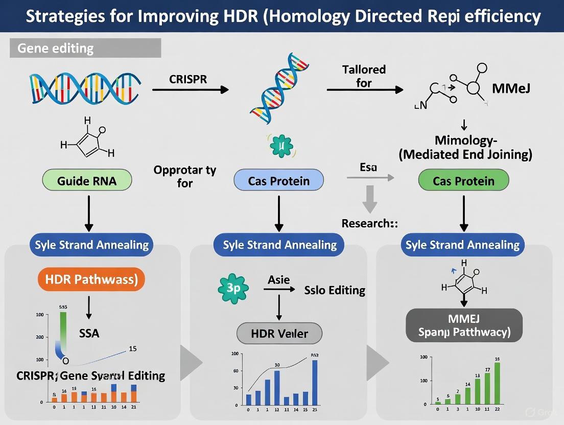
The HDR Challenge: Understanding the Cellular Battlefield of DNA Repair
FAQ: Why does NHEJ happen more often than HDR in my experiments?
In most cells, the Non-Homologous End Joining (NHEJ) pathway is the dominant and more active repair mechanism for double-strand breaks (DSBs) across all phases of the cell cycle. In contrast, the Homology-Directed Repair (HDR) pathway is active only during the S and G2 phases, when a sister chromatid is available to use as a template. Furthermore, NHEJ is a faster process, as it simply ligates the broken DNA ends together without needing a homologous template [1] [2].
The table below summarizes the key competitive advantages of NHEJ.
| Feature | NHEJ | HDR |
|---|---|---|
| Template Requirement | None (error-prone) | Homologous DNA template (precise) |
| Cell Cycle Activity | Active throughout all phases (G1, S, G2) [3] | Primarily restricted to S and G2 phases [1] [3] |
| Speed & Efficiency | Fast, "first-responder" pathway | Slower, more complex process |
| Primary Role in CRISPR | Ideal for gene knockouts (indels) | Ideal for precise knock-ins and specific edits |
Troubleshooting Guide: Strategies to Favor HDR
FAQ: How can I tip the balance in favor of HDR in my experiments?
You can coax cells to favor HDR by using strategic interventions that either suppress the NHEJ pathway or enhance the HDR pathway directly.
Suppressing Competing Repair Pathways
Inhibiting key molecules in the NHEJ and other alternative repair pathways can significantly reduce off-target editing and increase HDR efficiency.
- NHEJ Inhibition: Using small molecule inhibitors like Alt-R HDR Enhancer V2 targets the NHEJ pathway [4].
- SSA & MMEJ Inhibition: Recent research shows that even with NHEJ inhibited, other pathways like Microhomology-Mediated End Joining (MMEJ) and Single-Strand Annealing (SSA) contribute to imprecise repair. Suppressing these by inhibiting their key effectors (POLQ for MMEJ and Rad52 for SSA) can further improve HDR accuracy [4].
The table below lists key reagents for pathway suppression.
| Research Reagent | Target Pathway | Key Effector | Experimental Function |
|---|---|---|---|
| Alt-R HDR Enhancer V2 | NHEJ | DNA Ligase IV complex | Small molecule inhibitor to suppress error-prone NHEJ [4]. |
| ART558 | MMEJ | POLQ (Pol Theta) | Small molecule inhibitor to reduce large deletions and complex indels [4]. |
| D-I03 | SSA | Rad52 | Small molecule inhibitor to reduce asymmetric HDR and other imprecise integration events [4]. |
Enhancing HDR Efficiency Directly
You can also directly stimulate the HDR machinery by modulating protein expression or optimizing the donor template.
- Overproducing HDR-Related Proteins: Studies have shown that overexpression of RAD52, a key protein in homologous recombination, can enhance the integration of single-stranded DNA templates, in some cases increasing HDR efficiency nearly 4-fold [5].
- Optimizing Donor Template Design: The design of your donor DNA is critical.
- 5' Modifications: Adding a 5'-biotin or 5'-C3 spacer to your donor DNA can substantially boost single-copy HDR integration by up to 8-fold and 20-fold, respectively, potentially by protecting the ends and improving recruitment to the break site [5].
- Using Single-Stranded DNA (ssDNA): ssDNA donors are often favored over double-stranded DNA (dsDNA) due to lower cytotoxicity and higher HDR efficiency. For long insertions, methods like Easi-CRISPR that use long single-stranded DNA (ssDNA) donors can achieve efficiencies of 25-50% [6] [3].
- Denaturing dsDNA Templates: Heat-denaturing long double-stranded DNA templates before delivery has been shown to enhance precise editing and reduce the formation of unwanted template concatemers [5].
Diagram 1: The competitive balance between NHEJ and HDR pathways and strategies to shift the outcome.
Quantitative Data on Editing Outcomes
FAQ: How much can these strategies actually improve my HDR rates?
The effectiveness of these strategies is highly dependent on your experimental system, including the target locus, cell type, and nuclease platform [7]. The following table summarizes quantitative findings from recent studies.
| Experimental Strategy | Quantitative Effect on HDR | Effect on Undesired Outcomes | Context / Notes |
|---|---|---|---|
| NHEJ Inhibition (Alt-R HDR Enhancer V2) | Increased knock-in efficiency from ~6% to ~22% (approx. 3-fold) [4] | Significantly reduced small indels [4] | Human RPE1 cells |
| SSA Inhibition (Rad52 suppression) | --- | Reduced asymmetric HDR and other imprecise donor integrations [4] | Improved precision, not necessarily overall efficiency |
| MMEJ Inhibition (POLQ suppression) | Increased perfect HDR frequency [4] | Reduced large deletions (≥50 nt) and complex indels [4] | Human RPE1 cells |
| RAD52 Supplementation | Increased ssDNA integration nearly 4-fold [5] | Accompanied by higher template multiplication [5] | Mouse zygote microinjection |
| 5'-Biotin Donor Modification | Up to 8-fold increase in single-copy integration [5] | --- | Mouse embryo experiment |
| 5'-C3 Spacer Donor Modification | Up to 20-fold rise in correctly edited mice [5] | --- | Mouse embryo experiment |
| Denatured DNA Template | Increased correctly targeted animals from 2% (dsDNA) to 8% (ssDNA) [5] | Reduced template multiplication from 34% to 17% [5] | Mouse Nup93 locus targeting |
Experimental Protocol: A Sample Workflow for Enhancing HDR
This protocol outlines a methodology for improving HDR efficiency in cell culture, based on strategies cited above [4] [5] [3].
Objective: To achieve precise knock-in of a fluorescent tag at an endogenous gene locus in human cells while minimizing NHEJ-derived indels.
Key Reagents:
- RNP Complex: Recombinant Cas9 or Cpf1 protein and synthetic guide RNA (crRNA/tracrRNA).
- Donor Template: Single-stranded DNA (ssDNA) or double-stranded DNA (dsDNA) with homology arms, ideally with 5'-biotin or 5'-C3 modification [5].
- Pathway Inhibitors: Alt-R HDR Enhancer V2 (NHEJi), ART558 (MMEJi), D-I03 (SSAi) [4].
- Cells: hTERT-immortalized RPE1 or other relevant cell line.
Procedure:
- Prepare Donor Template: Design an ssDNA donor with 40+ base homology arms. Synthesize the oligonucleotide with a 5' modification (e.g., biotin) to enhance HDR efficiency [5] [3].
- Form RNP Complex: Pre-complex the Cas protein with the guide RNA to form a ribonucleoprotein (RNP) complex.
- Electroporation: Co-deliver the RNP complex and the donor template into your cells via electroporation.
- Inhibitor Treatment: Immediately after electroporation, treat the cells with a cocktail of small molecule inhibitors (e.g., NHEJi, MMEJi, SSAi) for 24 hours to transiently suppress competing repair pathways during the critical DSB repair window [4].
- Analysis: After 3-4 days, analyze the editing outcomes. Use flow cytometry to assess knock-in efficiency (if using a fluorescent tag) and long-read amplicon sequencing (e.g., PacBio) coupled with computational genotyping (e.g., knock-knock framework) to comprehensively quantify perfect HDR, imprecise integration, and indel frequencies [4].
Diagram 2: A sample experimental workflow for enhancing HDR efficiency.
FAQs and Troubleshooting Guides
FAQ 1: Why is the efficiency of precise genome editing via HDR so low in my experiments?
Answer: The low efficiency of Homology-Directed Repair (HDR) is primarily due to strong cellular competition from other, faster DNA repair pathways.
- Pathway Competition: When CRISPR-Cas9 creates a double-strand break (DSB), the cell's repair machinery is activated. The dominant and most rapid pathway is Non-Homologous End Joining (NHEJ), which operates throughout the cell cycle and simply rejoins the broken DNA ends, often introducing small insertions or deletions (indels). HDR, in contrast, is a more complex process restricted primarily to the S and G2 phases of the cell cycle and requires a homologous DNA template [8] [9].
- Alternative Error-Prone Pathways: Even when NHEJ is suppressed, alternative pathways like Microhomology-Mediated End Joining (MMEJ) and Single-Strand Annealing (SSA) can repair the break. These pathways use short or long homologous sequences, respectively, but are error-prone and often result in deletions, further reducing the yield of perfect HDR events [10] [11].
FAQ 2: What are the key differences between NHEJ, HDR, MMEJ, and SSA?
Answer: These four pathways are distinguished by their mechanisms, template requirements, and fidelity, summarized in the table below.
Table 1: Comparison of Key DNA Double-Strand Break Repair Pathways
| Pathway | Full Name | Mechanism | Template Required? | Fidelity | Key Effector Proteins |
|---|---|---|---|---|---|
| NHEJ | Non-Homologous End Joining | Direct ligation of broken ends [9] | No | Error-Prone (often causes small indels) [9] | Ku70/Ku80, DNA-PKcs, DNA Ligase IV [12] |
| HDR | Homology-Directed Repair | Uses homologous sequence as a template for precise repair [9] | Yes | High-Fidelity (precise) [9] | BRCA1, BRCA2, RAD51, MRN Complex [9] [12] |
| MMEJ | Microhomology-Mediated End Joining | Annealing of short microhomology regions (2-20 nt) flanking the break [11] | No | Error-Prone (causes deletions) [11] | PARP1, POLθ (POLQ), DNA Ligase 3 [10] [11] |
| SSA | Single-Strand Annealing | Annealing of long homologous repeats (>20 nt) flanking the break [10] | No | Error-Prone (causes large deletions) [10] | RAD52, MRN Complex [10] |
FAQ 3: How can I experimentally inhibit competing pathways to improve HDR efficiency?
Answer: You can bias repair toward HDR by using small-molecule inhibitors or genetic knockdown to target key factors of competing pathways.
Table 2: Research Reagent Solutions for Modulating DNA Repair Pathways
| Reagent / Method | Target Pathway | Key Component Inhibited | Effect on Editing Outcomes | Considerations |
|---|---|---|---|---|
| Alt-R HDR Enhancer V2 [10] | NHEJ | DNA-PKcs (implied) | Increases perfect HDR frequency; reduces small indels [10] | A common first-step enhancement strategy. |
| ART558 [10] | MMEJ | POLQ (Polymerase Theta) | Reduces large deletions and complex indels; can increase HDR [10] | Effective in combination with NHEJ inhibition. |
| D-I03 [10] | SSA | RAD52 | Reduces asymmetric HDR and other imprecise donor integrations [10] | Effect may depend on the nature of the DNA cleavage ends. |
| Cell Cycle Synchronization | NHEJ vs. HDR | N/A (chemicals used for sync) | Increases HDR by enriching for S/G2 phase cells [9] | Can be technically challenging and impact cell health. |
Experimental Protocol: Pathway Inhibition for Enhanced HDR
- Design and Complex Formation: Form Ribonucleoprotein (RNP) complexes by pre-incubating your chosen Cas nuclease (e.g., Cas9) with synthesized, chemically modified guide RNAs [13].
- Delivery and Electroporation: Co-deliver the RNP complexes and your donor DNA template into the target cells (e.g., hTERT-immortalized RPE1 cells) via electroporation [10].
- Inhibitor Treatment: Immediately after delivery, treat the cells with pathway-specific inhibitors.
- Use a commercial NHEJ inhibitor like Alt-R HDR Enhancer V2.
- For broader suppression, combine with ART558 (MMEJ inhibitor) and/or D-I03 (SSA inhibitor).
- A typical treatment duration is 24 hours, as HDR often occurs within this window post-Cas9 delivery [10].
- Analysis: After 3-4 days, analyze editing outcomes. Use flow cytometry to assess knock-in efficiency if using a fluorescent tag. For precise repair pattern analysis, perform long-read amplicon sequencing (e.g., PacBio) on the target locus and genotype the results with a computational framework like
knock-knock[10].
FAQ 4: My knock-in results show partial or incorrect integration of the donor template. What could be the cause?
Answer: Imprecise donor integration, such as "asymmetric HDR" (where only one end integrates correctly), is a common issue often linked to the Single-Strand Annealing (SSA) pathway. Even with NHEJ inhibition, the SSA pathway can still use the homology arms on your donor DNA to catalyze erroneous repair, leading to partial integration or duplication of homology arms [10].
Troubleshooting Guide:
- Problem: Observation of various imprecise integration patterns (blunt, asymmetric, imperfect) despite NHEJ inhibition.
- Solution: Suppress the SSA pathway in addition to NHEJ inhibition. Using a Rad52 inhibitor (e.g., D-I03) during the initial repair period has been shown to specifically reduce asymmetric HDR and other faulty integration events, thereby increasing the proportion of perfect HDR [10].
Pathway Diagrams and Experimental Workflows
DNA Repair Pathway Mechanics
HDR Enhancement Experimental Workflow
Frequently Asked Questions
FAQ: Why is Homology-Directed Repair (HDR) restricted to the S and G2 phases of the cell cycle?
HDR is restricted to these phases because it relies on a sister chromatid as a repair template, which is only available after DNA replication has occurred during the S phase. Additionally, key proteins in the HDR pathway are selectively active during these cell cycle stages [9].
FAQ: What are the main DNA repair pathways that compete with HDR after a CRISPR-Cas9 induced double-strand break (DSB)?
The primary competing pathways are:
- Non-Homologous End Joining (NHEJ): An error-prone pathway that is active throughout all cell cycle phases and often results in small insertions or deletions (indels) [9].
- Microhomology-Mediated End Joining (MMEJ): An alternative, highly mutagenic pathway that can generate larger deletions [9].
FAQ: What practical strategies can I use to improve HDR efficiency in my experiments?
Key strategies include:
- Cell Cycle Synchronization: Transiently synchronizing your cell population in the S/G2 phases.
- NHEJ Inhibition: Using chemical inhibitors or genetic knockdown of key NHEJ factors.
- Donor Template Optimization: Employing single-stranded DNA (ssDNA) donors with optimized design.
- HDR Pathway Enhancement: Overexpressing crucial HDR factors.
Experimental Protocols for HDR Enhancement
Chemical Inhibition of NHEJ
Detailed Methodology:
- Transfect cells with your CRISPR-Cas9 components (e.g., Cas9-gRNA RNP complex and donor DNA).
- Treat cells with small-molecule inhibitors targeting key NHEJ proteins. Common inhibitors include:
- The typical treatment window is 24-48 hours post-transfection. Optimize concentration and duration for your specific cell type to minimize cytotoxicity.
Cell Cycle Synchronization
Detailed Methodology:
- Use chemicals to arrest cells at the G1/S boundary before transfection.
- Common Reagents: Aphidicolin, thymidine, or lovastatin.
- Post-arrest, release cells into fresh medium to allow them to progress synchronously into S phase.
- Perform CRISPR transfection/transduction immediately after release to maximize the number of Cas9-induced DSBs occurring in the S/G2 phases, where HDR is active [9].
Using Optimized Single-Stranded DNA (ssDNA) Donors
Detailed Methodology:
- Design ssDNA donors (e.g., ssODNs) with symmetrical homology arms.
- Optimal Arm Length: Typically 40-60 nucleotides on each side of the edit [3].
- Modifications: Consider phosphorothioate (PS) modifications at the 5' and 3' ends to increase donor stability and resistance to exonuclease degradation [3].
- Co-deliver the optimized ssDNA donor with your CRISPR-Cas9 system.
HDR and Competing DNA Repair Pathways
Quantitative Data on HDR Enhancement Strategies
Key Small-Molecule Inhibitors for Modulating DNA Repair
| Inhibitor / Reagent | Target Pathway | Mechanism of Action | Effect on HDR | Typical Working Concentration |
|---|---|---|---|---|
| NU7441 [9] | NHEJ | DNA-PKcs inhibitor | Increases HDR efficiency | 0.5–1 µM |
| M3814 [3] | NHEJ | DNA-PKcs inhibitor | Increases HDR efficiency | Cited in patent application |
| KU0060648 [9] | NHEJ | DNA-PKcs & PI3K-related kinase inhibitor | Increases HDR efficiency | Varies by cell type |
| Roscovitine (Seliciclib) [9] | Cell Cycle | CDK inhibitor | Synchronizes cells at G1/S; enriches S/G2 population | 10–25 µM |
| AZD-7648 [9] | NHEJ | DNA-PKcs inhibitor | Increases HDR efficiency | Varies by cell type |
| RAD51 agonists (e.g., RS-1) [9] | HDR | RAD51 stabilizer | Enhances strand invasion step of HDR | 5–25 µM |
Comparison of DNA Double-Strand Break Repair Pathways
| Feature | NHEJ | MMEJ | HDR |
|---|---|---|---|
| Template Used | None | Microhomology regions | Homologous donor (e.g., sister chromatid) |
| Fidelity | Error-Prone | Highly Error-Prone | Precise |
| Primary Phase | All phases | S/G2 [14] | S and G2 phases [9] |
| Key Initiating Factors | KU70/KU80, 53BP1, DNA-PKcs | PARP1, Pol θ, MRN/CtIP (limited resection) | MRN Complex, CtIP |
| Critical Effector | DNA Ligase IV/XRCC4 | DNA Pol θ | RAD51, BRCA2 |
| Typical Outcome | Small indels | Larger deletions | Precise gene correction/insertion |
The Scientist's Toolkit: Essential Research Reagents
| Reagent / Tool | Function in HDR Editing | Key Considerations |
|---|---|---|
| ssDNA Donors (ssODNs) [3] | Provides template for precise repair; lower cytotoxicity than dsDNA. | Optimize homology arm length (40+ bases); consider phosphorothioate modifications. |
| HDR Booster Modules [3] | Fusion proteins (e.g., nCas9-CtIP) designed to enhance HDR efficiency. | Can bias repair toward HDR by promoting initial resection steps. |
| Virus-Like Particles (VLPs) [14] | Efficient delivery of Cas9 RNP to hard-to-transfect cells (e.g., neurons). | Pseudotype choice (e.g., VSVG, BRL) critically impacts delivery efficiency. |
| Cell Cycle Reporters (e.g., Fucci) | Identifies and/or sorts cells in S/G2 phases for targeted editing. | Allows for tracking cell cycle progression without fixation. |
| NHEJ Chemical Inhibitors [3] [9] | Suppresses competing error-prone pathway to favor HDR. | Requires titration to balance HDR boost with potential cytotoxicity. |
In CRISPR-Cas9-mediated genome editing, achieving precise modifications through Homology-Directed Repair (HDR) is a fundamental goal for researchers. However, HDR competes with the error-prone Non-Homologous End Joining (NHEJ) pathway, which often results in unpredictable insertions or deletions (indels) [15] [16]. This competition significantly limits the efficiency of precise gene knock-ins, point mutations, and gene corrections—a central challenge in therapeutic gene editing and functional genomics. The key to tilting this balance toward HDR lies in understanding and manipulating specific DNA repair proteins, primarily RAD51, RAD52, and the MRN complex (MRE11, RAD50, and NBS1). This technical support center details how these proteins function, how their activity can be enhanced, and how to troubleshoot common experimental hurdles to successfully improve HDR outcomes.
Core Protein Functions & Mechanisms
Q1: What are the specific roles of RAD51, RAD52, and the MRN complex in the HDR pathway?
The HDR pathway is a carefully coordinated process that requires multiple proteins to work in sequence after a CRISPR-Cas9-induced double-strand break (DSB). Below is a summary of their specialized functions.
Table: Core Functions of Key HDR Proteins
| Protein/Complex | Primary Function in HDR | Key Interactions |
|---|---|---|
| MRN Complex | Initial DSB sensing and end resection (5' to 3') to create single-stranded DNA (ssDNA) overhangs [17]. | Recruits and activates ATM; stabilizes the DSB site [17]. |
| RAD51 | Forms a nucleoprotein filament on ssDNA; catalyzes the central step of strand invasion into the homologous donor template [17] [18]. | Loaded with the help of BRCA2 and PALB2; displaces RPA [17]. |
| RAD52 | Mediates the loading of RAD51 onto RPA-coated ssDNA (in yeast) and promotes single-strand annealing (SSA); serves as a backup mediator in human cells, crucial in BRCA-deficient contexts [19] [20]. | Has annealing activity; synthetically lethal with BRCA1/BRCA2 loss [20]. |
The following diagram illustrates the sequential involvement of these proteins in the HDR pathway following a DSB.
Troubleshooting Low HDR Efficiency
Q2: My HDR efficiency is consistently low. What are the primary strategies to enhance it?
Low HDR efficiency is often due to the dominance of the NHEJ pathway. Effective strategies focus on either suppressing NHEJ or directly stimulating the HDR machinery.
- Inhibit the NHEJ Pathway: Transiently inhibiting key NHEJ proteins can shift the repair balance toward HDR. This can be achieved using small molecule inhibitors like SCR7 (targeting DNA Ligase IV) or M3814 (targeting DNA-PKcs) [19] [21]. Alternatively, you can use RNAi to knock down factors like KU70, KU80, or DNA Ligase IV [19].
- Stimulate the HDR Pathway Directly: As detailed in the following sections, you can boost HDR by:
- Optimize Donor Template Design: The design and delivery of your donor template are critical.
- Use single-stranded DNA (ssDNA) donors instead of double-stranded DNA (dsDNA) where possible, as they are generally more efficient and less cytotoxic [21].
- For ssDNA donors, incorporate RAD51-preferred sequence motifs (e.g., containing a "TCCCC" motif) at the 5' end to enhance RAD51 binding and recruitment, a chemical-free method shown to significantly boost HDR [21].
- Chemically modify the 5' ends of donor templates with biotin or a C3 spacer to protect the ends and improve the rate of single-copy integration [5].
Experimental Protocols & Reagents
Q3: What are specific experimental protocols for implementing HDR-enhancing strategies?
Here are detailed methodologies for two key approaches: RAD51/RAD52 overexpression and MRN complex recruitment.
Protocol 1: Enhancing HDR via RAD51 or RAD52 Expression
This protocol involves co-expressing RAD51 or RAD52 with the CRISPR-Cas9 system.
Plasmid Construction:
- For RAD51, clone the human RAD51 coding sequence into an all-in-one CRISPR vector downstream of a T2A or P2A self-cleaving peptide sequence, following Cas9 and a selection marker (e.g., EGFP/Puromycin) [18]. This ensures coordinated expression.
- For RAD52, clone the Saccharomyces cerevisiae Rad52 (ScRad52) gene into an expression vector under a strong promoter (e.g., CBh) [19]. Alternatively, create a fusion construct where ScRad52 is directly fused to the C-terminus of Cas9 via a flexible linker [19].
Cell Transfection and Selection:
- Transfect your target cells (e.g., HEK293T) with the constructed plasmid and your sgRNA and donor template.
- At 48 hours post-transfection, replace the media and begin selection with an appropriate antibiotic (e.g., Puromycin) for 72 hours to enrich for successfully transfected cells [18].
Efficiency Validation:
- Genotypic Analysis: Use T7 Endonuclease I (T7E1) assay or tracking of indels by decomposition (TIDE) to assess mutation rates at the target locus [18]. For precise HDR, perform PCR followed by restriction fragment length polymorphism (RFLP) or Sanger sequencing of cloned amplicons.
- Phenotypic Analysis: Confirm editing success at the protein level via western blotting or fluorescence-activated cell sorting (FACS) if editing introduces or disrupts a fluorescent protein [18].
Protocol 2: Enhancing HDR via MRN Complex Recruitment
This protocol uses a chimeric Cas9 protein fused to an MRN-recruiting domain to localize the repair machinery.
Chimeric Cas9 Construction:
- Fuse the N-terminal 126-amino-acid intrinsically disordered domain from the HSV-1 alkaline nuclease (UL12), known to recruit the MRN complex, to the N- or C-terminus of Streptococcus pyogenes Cas9 (SpCas9) [22]. Ensure the fusion preserves the nuclear localization signals of Cas9.
Delivery into Cells:
- Deliver the chimeric Cas9 as either plasmid DNA or pre-assembled ribonucleoprotein (RNP) complexes.
- For plasmid transfection, use standard methods (e.g., calcium phosphate, PEI, or commercial reagents) to co-deliver the chimeric Cas9 plasmid, sgRNA plasmid, and donor template [22].
- For RNP delivery, complex in vitro transcribed sgRNA with purified chimeric Cas9 protein. Then, transfect this RNP complex along with the donor template using a reagent like Lipofectamine CRISPRMax [22].
Efficiency Assessment:
Research Reagent Solutions
Table: Essential Reagents for HDR Enhancement Experiments
| Reagent | Function/Description | Example Use Case |
|---|---|---|
| HDR Enhancer (RS-1) | Small molecule that stabilizes RAD51-ssDNA filaments. | Add to cell culture media post-transfection to stimulate the strand invasion step of HDR [19]. |
| NHEJ Inhibitor (SCR7) | Small molecule inhibitor of DNA Ligase IV. | Add to media to transiently suppress the competing NHEJ pathway, favoring HDR [19]. |
| Chimeric Cas9-UL12 | Cas9 fused to the MRN-recruiting domain of HSV-1 UL12. | Used in RNP or plasmid formats to locally recruit the MRN complex to the DSB, boosting HDR [22]. |
| 5'-Modified ssDNA Donor | ssDNA donor with 5' biotin or C3 spacer modifications. | Use as a repair template to reduce concatemerization and improve single-copy HDR integration [5]. |
| Modular ssDNA Donor | ssDNA donor with 5' RAD51-preferred binding sequences (e.g., "TCCCC" motif). | A chemical-free donor design that augments affinity for RAD51, enhancing HDR efficiency [21]. |
Quantitative Data & Comparisons
Q4: Is there quantitative data comparing the efficacy of these different HDR-enhancement methods?
Yes, various studies have quantified the improvement in HDR efficiency using these strategies. The following table summarizes key findings for easy comparison.
Table: Quantitative Comparison of HDR Enhancement Strategies
| Enhancement Strategy | Experimental System | Reported HDR Efficiency | Key Findings and Notes |
|---|---|---|---|
| RAD51 Overexpression | Human HEK293T cells (GAPDH KO) | Editing efficiency increased >2.5-fold [18]. | Enhanced both knock-out and knock-in efficiency. |
| RAD52 Supplementation | Mouse zygotes (Nup93 cKO model) | HDR rate increased to 26% (from 2% with dsDNA) [5]. | Accompanied by a higher rate of template multiplication. |
| Cas9-UL12 Fusion (MRN Recruitment) | Human HEK293FT, HCT116, HeLa cells | ~2-fold increase over standard Cas9 [22]. | Effect depended on the MRN-recruiting activity of the UL12 domain. |
| 5'-Biotin Modified Donor | Mouse zygotes | Single-copy integration increased up to 8-fold [5]. | Reduces unwanted template multimerization. |
| 5'-C3 Spacer Modified Donor | Mouse zygotes | Correctly edited mice increased up to 20-fold [5]. | Highly effective in boosting single-copy HDR. |
| RAD51-Preferred ssDNA Module | HEK 293T-BFP reporter cells | HDR efficiency up to 90.03% (median 74.81%) when combined with M3814 [21]. | A chemical-free strategy that synergizes with NHEJ inhibition. |
FAQs on Protein-Specific Issues
Q5: Why would I choose to target RAD52 instead of RAD51, given RAD51's central role?
While RAD51 is the core recombinase, RAD52 offers a unique strategic advantage, particularly in certain genetic contexts. In human cells, the primary mediator for loading RAD51 is actually the BRCA2 protein, not RAD52 [20]. However, in cells with BRCA1, BRCA2, or PALB2 deficiencies (e.g., some cancer cell lines), RAD52 becomes essential as a backup loader. This creates a synthetic lethal interaction, where inhibiting RAD52 is lethal only to the BRCA-deficient cells [20]. Therefore, modulating RAD52 activity can be a more targeted strategy in these contexts, and its strong annealing activity also makes it critical for the SSA sub-pathway of HDR.
Q6: Are there any pitfalls or downsides to overexpressing RAD51 or RAD52?
Yes, potential downsides must be considered. A primary concern with enhancing HDR factors is the potential for increased off-target integration or aberrant recombination events. For instance, one study noted that while RAD52 supplementation significantly boosted precise HDR, it was also accompanied by a near doubling of unwanted template multiplication (concatemer formation) [5]. Furthermore, forced overexpression of these powerful recombinases could, in theory, lead to genomic instability by promoting recombination between non-allelic sequences with low homology. It is crucial to carefully optimize the expression levels and timing of these proteins to maximize on-target HDR while minimizing unintended consequences.
Troubleshooting Guide: FAQs on HDR Efficiency
FAQ: Why is my knock-in efficiency so low, and why do I see random insertions instead of precise edits?
The dominant DNA repair pathway in most cells, particularly postmitotic cells like neurons, is the error-prone non-homologous end joining (NHEJ) pathway. This pathway is active throughout the cell cycle and often outcompetes the precise Homology-Directed Repair (HDR) pathway, leading to a high frequency of insertions and deletions (indels) rather than your desired precise edit [14] [8]. The table below summarizes common issues and solutions.
| Problem | Possible Cause | Recommended Solution |
|---|---|---|
| Low HDR efficiency | NHEJ outcompeting HDR in dividing cells | Use single-stranded DNA (ssDNA) templates; inhibit NHEJ pathways chemically or genetically [5] [8]. |
| Random insertion/deletion (indels) | NHEJ-dominated repair in non-dividing cells | Design gRNAs compatible with MMEJ/SSA; use Cas9 proteins with altered PAM specificities [14]. |
| Donor DNA multimerization/ concatemers | End-joining of linear double-stranded DNA (dsDNA) donor templates | Denature long dsDNA templates into ssDNA; use 5'-end modifications (e.g., C3 spacer, biotin) on donor DNA [5]. |
| Low HDR in iPSCs & HSPCs | Cell-type specific repair pathway dominance | Supplement with HDR-enhancing proteins (e.g., RAD52, commercial Alt-R HDR Enhancer Protein) during editing [5] [23]. |
| Extended editing time in neurons | Slow DSB repair kinetics in postmitotic cells | Allow extended time (up to 2 weeks) for indel accumulation; use virus-like particles (VLPs) for efficient RNP delivery [14]. |
FAQ: My edits work in cell lines but fail in primary or non-dividing cells. Why?
DNA repair is not universal across cell types. Dividing cells, such as iPSCs, frequently use repair pathways like microhomology-mediated end joining (MMEJ), which can create larger deletions. In contrast, non-dividing cells (e.g., neurons, cardiomyocytes) predominantly use classical NHEJ, resulting in smaller indels and a higher proportion of unedited outcomes [14]. Furthermore, homology-directed repair (HDR) is largely restricted to the S and G2 phases of the cell cycle, making it inherently inefficient in non-dividing cells [14] [8].
Quantitative Data on HDR Optimization Strategies
The following table summarizes experimental data from a large-scale study targeting the Nup93 locus in mouse zygotes, comparing the effectiveness of various strategies to improve HDR outcomes [5].
Table: Impact of Different Strategies on HDR Efficiency and Template Multiplication
| crRNAs/Orientation | DNA Type | 5' End Modification | Additional Factor | F0 HDR % | F0 Head-to-Tail % |
|---|---|---|---|---|---|
| crR1-7 (±) | dsDNA | 5'-P | no | 2% | 34% |
| crR1-7 (±) | dsDNA denatured | 5'-P | no | 8% | 17% |
| crR1-7 (±) | dsDNA denatured | 5'-P | RAD52 | 26% | 30% |
| crR1-7 (±) | dsDNA | 5'-C3 Spacer | no | 40% | 9% |
| crR1-7 (±) | dsDNA denatured | 5'-C3 Spacer | no | 42% | 5% |
| crR1-7 (±) | dsDNA | 5'-Biotin | no | 14% | 5% |
Key findings from the data:
- Donor Denaturation: Heat-denaturing a long dsDNA template into ssDNA boosted precise HDR by 4-fold (from 2% to 8%) and reduced template concatemer formation (head-to-tail) by half [5].
- RAD52 Supplementation: Adding RAD52 protein to ssDNA templates increased HDR efficiency dramatically to 26%, a 13-fold increase over standard dsDNA. However, this came with a trade-off of increased template multiplication [5].
- 5' End Modifications: Modifying the 5' end of the donor DNA was highly effective. The 5'-C3 spacer modification yielded the highest HDR efficiency (40-42%) while keeping concatemer formation low (5-9%). 5'-Biotin modification was also effective at reducing multimerization [5].
Experimental Protocols for Enhancing HDR
Protocol 1: Using Denatured DNA Templates and RAD52
This protocol is adapted from methods used to generate conditional knockout mouse models, which resulted in a 4-fold to 13-fold increase in precise editing [5].
- Donor DNA Design: Synthesize a long (e.g., ~600 bp) double-stranded DNA template with homologous arms (60 bp and 58 bp) and monophosphorylated 5' ends.
- Denaturation: Heat-denature the dsDNA template to create a single-stranded donor for microinjection or transfection.
- Complex Formation: Co-inject/co-transfect the denatured DNA template with CRISPR-Cas9 ribonucleoprotein (RNP) complexes. For the test condition, supplement the mix with recombinant human RAD52 protein.
- Analysis: Screen for precise HDR events and use Southern blot analysis to detect single-copy integrations and monitor template multiplication.
Protocol 2: High-Throughput Screening for HDR-Enhancing Chemicals
This protocol outlines steps to identify small molecules that can shift the DNA repair balance toward HDR [24] [25].
- Plate Design: Seed cells expressing CRISPR-Cas9 components and a HDR reporter system into 96-well plates.
- Chemical Library Treatment: Treat cells with a library of small molecules (e.g., HDAC inhibitors, autophagy inducers). Include positive and negative controls.
- Dual Assay Execution: Simultaneously perform a colorimetric assay (e.g., LacZ) to quantify HDR efficiency and a viability assay to control for cytotoxicity.
- Data Analysis: Use a plate reader to collect data. Normalize HDR readouts to cell viability to identify compounds that specifically enhance HDR without detrimental effects.
The Scientist's Toolkit: Key Research Reagent Solutions
Table: Essential Reagents for Optimizing HDR in CRISPR Experiments
| Reagent / Solution | Function in HDR Enhancement |
|---|---|
| Single-Stranded DNA (ssDNA) Donor | Serves as an optimal repair template; reduces concatemer formation compared to dsDNA [5]. |
| RAD52 Protein | A recombination mediator that promotes strand invasion; can significantly boost ssDNA integration [5]. |
| Alt-R HDR Enhancer Protein | A proprietary, protein-based solution that shifts repair pathway balance toward HDR in challenging cells like iPSCs and HSPCs [23]. |
| 5'-C3 Spacer / 5'-Biotin Modifications | Chemical modifications to the 5' end of donor DNA that prevent ligation and multimerization, favoring single-copy HDR integration [5]. |
| Virus-Like Particles (VLPs) | Engineered delivery vehicles for efficient transduction of Cas9 RNP into hard-to-transfect cells, such as human neurons [14]. |
Pathway and Workflow Visualizations
DNA Repair Pathway Competition
HDR Enhancement Experimental Workflow
Practical Strategies for Enhancing HDR in the Lab
FAQs on Donor Template Design
FAQ 1: Should I choose a single-stranded DNA (ssDNA) or double-stranded DNA (dsDNA) donor template for HDR?
The choice between ssDNA and dsDNA donors depends on the size of your intended insertion and the desired balance between efficiency and simplicity.
- ssDNA Donors are typically preferred for introducing shorter edits, such as point mutations or short tags, due to their generally higher HDR efficiency and lower cytotoxicity compared to dsDNA donors [3] [15]. They are effective even with very short homology arms (30-60 nucleotides) [26] [27].
- dsDNA Donors are more suitable for inserting larger DNA fragments (>1-2 kb) [27]. However, they often require significantly longer homology arms (200 bp to over 2,000 bp) to achieve reasonable HDR efficiency and are more prone to random integration or concatemer formation [26] [5].
Table 1: Comparison of ssDNA and dsDNA Donor Templates
| Feature | ssDNA Donors | dsDNA Donors |
|---|---|---|
| Best For | Point mutations, short insertions (a few hundred bases) [27] [3] | Large insertions (1 kb and above) [27] |
| Typical Homology Arm Length | 30-100 nucleotides [26] [27] | 200-2000+ base pairs [26] [27] |
| Efficiency | Generally higher for small edits [3] [15] | Lower, but necessary for large inserts [27] |
| Cytotoxicity | Lower [3] [15] | Higher |
| Common Issues | Limited insert size | Concatemer formation, random integration [5] |
FAQ 2: What is the optimal length for homology arms?
The optimal homology arm length is primarily determined by the type of donor DNA you are using.
- For ssDNA donors, arms as short as 30-40 nucleotides can support efficient HDR [26] [27]. Some studies report that increasing the arm length from 30 nt to ~60-97 nt does not necessarily lead to a significant increase in HDR efficiency, though it may influence the repair pathway choice [26].
- For dsDNA donors, arm length is much more critical. While very short arms (50 bp) can work at low efficiency, arms of 200-300 bp are often sufficient, and for maximum efficiency, arms can be extended to 500-2000 bp or even longer [26] [27].
Table 2: Recommended Homology Arm Lengths
| Donor Type | Recommended Arm Length | Key Evidence |
|---|---|---|
| ssDNA | 30 - 100 nt | HDR achieved with 30 nt arms in potato; 40+ nt recommended for robustness [26] [27] [3]. |
| dsDNA | 200 - 300 bp (sufficient) 500 - 2000+ bp (optimal for large inserts) | 200-300 bp sufficient for HDR in mammalian cells; efficiency increases with arm length up to 2,000 bp [26] [27]. |
FAQ 3: How can I further enhance HDR efficiency with ssDNA donors?
Several advanced strategies can significantly boost the performance of ssDNA donors by promoting their recruitment to the double-strand break site.
Template Modifications:
- 5'-End Modifications: Adding 5'-biotin or a 5'-C3 spacer to the donor DNA can enhance single-copy integration by up to 8-fold and 20-fold, respectively, by preventing multimerization and improving localization to the break site [5].
- HDR-Boosting Modules: Engineering RAD51-preferred binding sequences (e.g., containing a "TCCCC" motif) into the 5' end of the ssDNA donor can augment its affinity for the RAD51 protein, a key player in HDR. This chemical-free method has been shown to increase HDR efficiency dramatically, especially when combined with NHEJ inhibitors [28].
Pharmacological and Protein Interventions:
- Inhibiting Competing Pathways: Transiently inhibiting the NHEJ or MMEJ pathways with small molecules (e.g., DNA-PKcs inhibitors) can shift repair toward HDR [29].
- Supplementing with Repair Proteins: Adding RAD52 protein to the editing mix has been shown to increase ssDNA integration by nearly 4-fold, though it can be accompanied by increased template multiplication [5].
- HDAC Inhibitors: Compounds like tacedinaline and entinostat can upregulate the expression of CRISPR system components (Cas9, sgRNA), thereby enhancing HDR efficiency in some cell types [30].
Experimental Protocols
Protocol 1: Assessing HDR Efficiency Using an ssDNA Donor with Short Homology Arms
This protocol is adapted from a study in potato protoplasts and can be adapted for mammalian cells to rapidly test donor designs [26].
- Design ssDNA Donor: Synthesize a single-stranded donor oligo with your desired edit (e.g., a point mutation or short tag). Flank this edit with homology arms of 30-40 nucleotides on each side, homologous to the sequence surrounding the target site.
- Complex RNP with Donor: Form a ribonucleoprotein (RNP) complex by pre-incubating purified Cas9 protein with your target-specific sgRNA.
- Co-Deliver into Cells: Co-transfect the RNP complex together with the ssDNA donor into your target cells. For mammalian cells, this can be done via electroporation or lipofection.
- Harvest and Extract DNA: Incubate cells for 48-72 hours to allow for repair, then harvest the cells and extract genomic DNA.
- Analyze Editing Efficiency: Amplify the target genomic region by PCR and analyze the editing outcomes using next-generation sequencing (NGS) to quantify the percentage of HDR versus indels.
Protocol 2: Enhancing HDR Using a Modular ssDNA Donor
This protocol details the use of RAD51-preferred sequences to boost HDR, as described in [28].
- Design Modular ssDNA Donor: Synthesize your ssDNA donor with the desired edit and standard homology arms. Incorporate a RAD51-preferred sequence motif (e.g., 5'-TCCCC-3') at the 5' end of the donor molecule, as the 5' end is more tolerant of such additions.
- Prepare Editing Components: Complex high-fidelity Cas9 protein with your sgRNA to form an RNP.
- Transfect and Inhibit NHEJ: Co-deliver the RNP and the modular ssDNA donor into your cells. To maximize HDR, treat the cells with a small-molecule NHEJ inhibitor (e.g., M3814) or employ the HDRobust strategy (combined inhibition of NHEJ and MMEJ) [29].
- Validate HDR Efficiency: After 72 hours, analyze the cells using flow cytometry (if using a fluorescent reporter) or NGS to quantify precise gene editing. This combination has achieved median HDR efficiencies of ~75% in human cells [28].
The Scientist's Toolkit
Table 3: Key Research Reagent Solutions for HDR Optimization
| Reagent / Tool | Function in HDR Optimization |
|---|---|
| CRISPR-Cas9 RNP Complex | Delivers the nuclease activity directly, leading to faster editing and reduced off-target effects compared to plasmid delivery. |
| 5'-Biotin or 5'-C3 Modified Donors | Chemical modifications that tether the donor to Cas9 (biotin) or prevent concatemerization (C3), improving single-copy HDR integration [5]. |
| RAD51-Preferred Sequence Modules | Functional DNA sequences engineered into the donor to recruit endogenous RAD51, enhancing donor recruitment to the break site without chemical modification [28]. |
| NHEJ/MMEJ Inhibitors (e.g., M3814) | Small molecules that transiently inhibit the competing error-prone repair pathways, thereby favoring HDR [29]. |
| HDRobust Substance Mix | A defined mixture that simultaneously inhibits NHEJ and MMEJ, drastically increasing outcome purity by directing most repairs through HDR [29]. |
Visualizing the Strategies and Pathways
The following diagrams summarize the key strategies for optimizing ssDNA donors and the cellular repair pathways involved.
In CRISPR-mediated genome editing, the precise incorporation of desired sequences via Homology-Directed Repair (HDR) competes with the dominant and error-prone Non-Homologous End Joining (NHEJ) pathway. A powerful strategy to shift this balance is the use of small molecule inhibitors that target key proteins in the NHEJ machinery, particularly the DNA-dependent protein kinase catalytic subunit (DNA-PKcs). This guide provides a technical overview and troubleshooting resource for using these chemical boosters to enhance HDR efficiency in your research.
FAQ: Understanding DNA-PKcs Inhibitors
Q1: What is the core mechanism by which DNA-PKcs inhibitors enhance HDR? DNA-PKcs is a critical serine/threonine kinase in the NHEJ pathway. The NHEJ repair process is initiated when the Ku70/Ku80 heterodimer binds to broken DNA ends, subsequently recruiting DNA-PKcs to form the DNA-PK holoenzyme [31] [32]. This enzyme acts as a scaffold, tethering the broken ends together and facilitating their ligation [31]. By inhibiting DNA-PKcs, you pharmacologically suppress the competing NHEJ pathway, thereby creating a cellular environment that is more permissive for the HDR machinery to use your provided donor template [32].
Q2: What are the commonly used DNA-PKcs inhibitors and their reported efficacies? Researchers employ several small molecule inhibitors. The table below summarizes key compounds and their performance in recent studies.
Table 1: Key Small Molecule DNA-PKcs Inhibitors
| Inhibitor | Reported Effect on HDR/NHEJ | Key Findings and Considerations |
|---|---|---|
| AZD7648 | Significantly increases apparent HDR reads in short-read sequencing [33]. | Can cause frequent kilobase- and megabase-scale deletions, chromosome arm loss, and translocations. These large-scale alterations often evade detection by standard short-read NGS assays [33]. |
| NU7026 | Potent and selective inhibitor; suppresses NHEJ [32]. | Shown to prevent CRISPR/Cas9-mediated degradation of HBV cccDNA, leading to frequent on-target deletions. Has ~60-fold higher activity for DNA-PK over PI3K [32]. |
| NU7441 | Drastically reduces NHEJ frequency while increasing HR rates [32]. | Cited as a potent DNA-PKcs inhibitor used in research settings. |
Q3: What are the major pitfalls and how can I detect them? A significant challenge is the potential for on-target genomic instability that is not captured by conventional analysis methods.
- Problem: The inhibitor AZD7648, while boosting HDR reads in short-read sequencing, was found to concurrently increase the frequency of large-scale chromosomal alterations [33].
- Solution: Implement comprehensive genotyping strategies that go beyond short-range PCR and short-read sequencing.
- Long-Range PCR & Long-Read Sequencing (Nanopore): Essential for detecting kilobase-scale deletions [33].
- Droplet Digital PCR (ddPCR): Useful for quantifying copy number variations and gene loss [33].
- Single-Cell RNA Sequencing (scRNA-seq): Can reveal coherent blocks of gene expression loss indicative of large-scale chromosomal aberrations [33].
Q4: Are there alternative or complementary strategies to small molecule inhibitors? Yes, other methods focus on directly enhancing the HDR pathway itself rather than suppressing its competition.
- Protein Engineering: Supplementing with RAD52 protein was shown to increase single-stranded DNA (ssDNA) integration efficiency nearly 4-fold, though it was accompanied by higher template multiplication [5].
- Donor DNA Optimization: Engineering ssDNA donors to include "HDR-boosting modules" with sequences preferred by the HDR protein RAD51 can significantly enhance HDR efficiency without chemical modifications [21]. Modifying the 5' ends of donor DNA with biotin or a C3 spacer can also boost single-copy integration [5].
Experimental Protocols
Protocol: Enhancing HDR using DNA-PKcs Inhibitors in Cell Culture
This protocol outlines the key steps for using inhibitors like AZD7648 or NU7026 in conjunction with CRISPR-Cas9 editing.
Table 2: Key Research Reagent Solutions
| Reagent / Tool | Function / Explanation |
|---|---|
| DNA-PKcs Inhibitor (e.g., AZD7648) | Selective small molecule that inhibits the kinase activity of DNA-PKcs, suppressing the NHEJ pathway. |
| CRISPR-Cas9 RNP Complex | The gene-editing machinery; a ribonucleoprotein complex that creates a specific double-strand break. |
| HDR Donor Template | Single-stranded oligodeoxynucleotide (ssODN) or double-stranded DNA (dsDNA) containing the desired edit flanked by homology arms. |
| Long-Range PCR Kit | For amplifying large fragments around the target site to assess large-scale deletions. |
| ddPCR/scRNA-seq Tools | For in-depth analysis of potential large-scale genomic alterations. |
Workflow:
- Cell Preparation: Seed your target cells (e.g., HEK293T, iPSCs, primary CD34+ HSPCs) at an appropriate density.
- Transfection/Nucleofection: Deliver the CRISPR-Cas9 RNP complex along with your HDR donor template into the cells.
- Inhibitor Treatment: Add the chosen DNA-PKcs inhibitor (e.g., 1 µM AZD7648) to the culture media immediately after or during the delivery step. Include a DMSO-only control.
- Incubation: Incubate the cells for the duration recommended for the specific inhibitor (e.g., 24-72 hours for AZD7648).
- Analysis:
- Primary Screening: Use short-read NGS of a target amplicon to get an initial readout of HDR and indel efficiency.
- Comprehensive Genotyping: To rule out large-scale alterations, perform long-range PCR followed by long-read sequencing (e.g., ONT) and/or ddPCR on edited samples, especially those treated with the inhibitor [33].
Protocol: Optimizing ssDNA Donors with RAD51-Boosting Modules
As an alternative or complementary strategy to chemical inhibition, you can optimize your donor design.
Workflow:
- Design: Synthesize your ssDNA donor with the HDR-boosting sequence module (e.g., the "TCCCC"-containing SSO9 or SSO14 motif [21]) incorporated at its 5' end. The 5' end is generally more tolerant of such additions than the 3' end [21].
- Delivery: Co-deliver the modified ssDNA donor with your CRISPR-Cas9 system.
- Combination Therapy: For maximal HDR, combine the optimized donor with an NHEJ inhibitor (e.g., M3814) or use the HDRobust strategy, which has been shown to achieve HDR efficiencies up to 90% [21].
Pathway and Mechanism Visualization
Frequently Asked Questions (FAQs)
Q1: What is the primary mechanistic role of RAD52 in improving HDR efficiency? RAD52 is a key protein in the homologous recombination (HR) pathway. It directly binds to single-stranded DNA (ssDNA) and facilitates the central step in HDR: the invasion of the ssDNA donor template into the homologous genomic region after a double-strand break. Co-expressing RAD52 with the CRISPR-Cas9 system increases the local concentration of the repair machinery and donor template at the cleavage site, thus promoting precise editing over error-prone repair pathways [19].
Q2: I am using a single-stranded DNA (ssDNA) donor. Should I use a RAD52 fusion protein or co-express it separately? Both strategies are effective, but they have different considerations. Research shows that a Rad52-Cas9 fusion protein can be particularly effective, as it physically recruits the HDR-enhancing factor directly to the site of the DNA break [19]. However, co-expression of non-fused RAD52 also significantly boosts HDR. For ssDNA donors, studies have shown that supplementing with RAD52 protein can increase precise integration by nearly 4-fold, though this can be accompanied by a higher rate of template multiplication (concatemer formation) [5].
Q3: What are the trade-offs of using RAD52 to enhance HDR? The primary trade-off is an increased risk of unwanted template integration events. While RAD52 can dramatically boost precise HDR rates, it can also lead to a higher frequency of head-to-tail multicopy insertions of the donor template into the genome [5]. It is crucial to design your screening strategy to distinguish between single-copy correct integrations and these concatemers.
Q4: Besides RAD52, what other strategies can I combine for a synergistic HDR boost? A multi-faceted approach is often most successful. You can combine RAD52 with:
- Donor DNA Modifications: Using 5′-end modified donors (e.g., 5′-biotin or 5′-C3 spacer) can further enhance single-copy HDR efficiency, with 5′-C3 spacers showing up to a 20-fold increase in some studies [5].
- HDR-Boosting Modules: Engineering ssDNA donors to include specific sequence motifs (like RAD51-preferred binding sequences) can augment affinity for endogenous repair proteins and improve HDR efficiency without chemical modifications [21].
- NHEJ Inhibition: Using small molecules to suppress the non-homologous end joining pathway can help shift the repair balance toward HDR, but caution is advised as some inhibitors can exacerbate large-scale genomic aberrations [34].
Q5: My HDR efficiency is still low after using RAD52. What should I troubleshoot? First, verify the quality and design of your donor template. Using denatured, long double-stranded DNA templates has been shown to boost precision and reduce concatemer formation compared to standard dsDNA [5]. Second, ensure your donor has sufficient homology arm lengths. Finally, confirm the efficiency of your delivery method for both the CRISPR components and the RAD52 protein/mRNA to ensure they are co-localized in the same cells.
Troubleshooting Guides
Problem: Low HDR Efficiency Despite RAD52 Expression
| Possible Cause | Symptoms | Solution |
|---|---|---|
| Inefficient delivery of RAD52 | Low protein expression confirmed by Western blot; no improvement over baseline. | Optimize delivery method (e.g., mRNA co-electroporation, protein delivery); use a fluorescence reporter to confirm co-delivery. |
| Suboptimal donor template design | High rates of indels (NHEJ) but no precise integration. | Use single-stranded DNA (ssDNA) donors; extend homology arms; consider 5′-end modifications (biotin/C3 spacer) [5] [21]. |
| Dominant NHEJ pathway | High indel frequency even with RAD52 present. | Consider low-toxicity, small molecule NHEJ inhibitors (e.g., Scr7), but thoroughly validate genomic integrity afterward [34] [19]. |
| Target site inaccessibility | Low overall editing efficiency (low indels and HDR). | Re-design gRNA to target a more accessible chromatin region; screen multiple gRNAs. |
Problem: High Rates of Unwanted Multi-Copy Donor Insertions
| Possible Cause | Symptoms | Solution |
|---|---|---|
| High concentration of donor template | PCR screening shows larger-than-expected amplicons; Southern blot confirms concatemers. | Titrate the donor DNA to the lowest effective concentration. |
| Use of double-stranded DNA (dsDNA) donors | Frequent head-to-tail template multiplications. | Switch to denatured dsDNA or single-stranded DNA (ssDNA) donors, which reduce this issue [5]. |
| RAD52 activity | Increased HDR is accompanied by a rise in template multiplication. | This is a known trade-off with RAD52 [5]. Combine RAD52 with 5′-C3 spacer or 5′-biotin-modified donors, which are proven to reduce multiplications and boost single-copy integration. |
Experimental Data & Protocols
The table below summarizes key quantitative findings from recent research on RAD52 and related HDR-enhancing strategies in mouse zygotes and cell models.
Table 1: Efficiency of HDR Enhancement Strategies
| Strategy | Test System | HDR Efficiency (Control) | HDR Efficiency (Enhanced) | Fold Increase | Key Observation | Source |
|---|---|---|---|---|---|---|
| RAD52 Supplementation (with denatured DNA) | Mouse zygotes | 8% (ssDNA only) | 26% | ~3.3 fold | Increased template multiplication to 30% | [5] |
| 5′-C3 Spacer (on dsDNA) | Mouse zygotes | Not specified (baseline) | 40% | Up to 20-fold vs. baseline | Significant boost regardless of donor strandness | [5] |
| 5′-Biotin Modification (on dsDNA) | Mouse zygotes | Not specified (baseline) | 14% | Up to 8-fold vs. baseline | Improved single-copy integration | [5] |
| HDR-Boosting Modules (RAD51 sequences in ssDNA) | Human cells (HEK293T) | Varies by locus | 66.62% - 90.03% (when combined with NHEJi) | Significant | Chemical modification-free strategy | [21] |
Detailed Protocol: Enhancing HDR with a RAD52-Cas9 Fusion Strategy
This protocol is adapted from research demonstrating enhanced HDR using a fusion protein strategy in mammalian cells [19].
Objective: To perform precise genome editing by co-delivering a Cas9 nuclease fused with RAD52 and an ssDNA donor template.
Materials:
- Plasmids: Expression vector for the RAD52-Cas9 fusion protein (e.g., pCBh-RAD52-Cas9), and a U6-sgRNA expression vector.
- Donor Template: Single-stranded DNA (ssDNA) oligo with homologous arms (90-100 nt total is common) and the desired edit.
- Cells: Adherent mammalian cells (e.g., HEK293T).
- Transfection Reagent: PEI or a commercial lipofectamine-based reagent.
- Analysis Reagents: Lysis buffer, PCR reagents, T7 Endonuclease I or Surveyor nuclease for indel analysis, sequencing primers.
Workflow:
Step-by-Step Method:
- Construct Cloning: Clone your target-specific sgRNA sequence into a U6-driven expression vector. Obtain or clone a RAD52-Cas9 fusion construct where the RAD52 gene is fused to the Cas9 gene, typically with a flexible linker.
- Donor Design: Design a single-stranded DNA donor oligo with the desired sequence change flanked by homology arms (30-50 nt each). The 5′ end is more tolerant of additional sequence modules if needed [21].
- Cell Transfection: Seed HEK293T cells to reach 70-80% confluency at transfection. Co-transfect the cells with:
- RAD52-Cas9 fusion plasmid (e.g., 500 ng)
- sgRNA plasmid (e.g., 250 ng)
- ssDNA donor oligo (e.g., 100-200 pmol)
- Use an appropriate transfection reagent according to the manufacturer's protocol.
- Incubation: Harvest cells 72-96 hours post-transfection for genomic DNA extraction and downstream analysis.
- Analysis:
- Genomic DNA Extraction: Isolate genomic DNA from transfected cells.
- PCR Amplification: Amplify the targeted genomic region.
- HDR Efficiency Quantification: Use a combination of methods:
- Restriction Fragment Length Polymorphism (RFLP): If the edit introduces or disrupts a restriction site.
- TIDE/TIDER Decomposition Assay: For quantifying HDR and NHEJ frequencies from Sanger sequencing data.
- Next-Generation Sequencing (NGS): For the most accurate and comprehensive analysis of editing outcomes, including precise HDR, indels, and unwanted genomic alterations.
Research Reagent Solutions
Table 2: Essential Reagents for RAD52 HDR Enhancement Experiments
| Reagent | Function | Example & Note |
|---|---|---|
| RAD52 Expression Vector | Provides source of RAD52 protein. | pCBh-RAD52 (for co-expression); pCBh-RAD52-Cas9 (for fusion) [19]. |
| HDR-Enhancing ssDNA Donor | Template for precise repair. | Chemically synthesized ssDNA with 5′ modifications (biotin, C3 spacer) or "HDR-boosting" RAD51-binding sequence modules [5] [21]. |
| NHEJ Inhibitor | Shifts repair balance toward HDR. | Scr7 (DNA Ligase IV inhibitor). Caution: Some DNA-PKcs inhibitors (e.g., AZD7648) can cause severe structural variations [34]. |
| Commercial HDR Enhancer | Optimized, proprietary solutions. | Alt-R HDR Enhancer Protein (IDT); a recombinant protein that boosts HDR with no reported increase in off-target edits [23]. |
| High-Fidelity Cas9 | Reduces off-target cleavage. | SpCas9-HF1 or eSpCas9(1.1) variants. Use when high specificity is critical. |
Safety and Optimization Notes
When implementing HDR-enhancing strategies, it is critical to be aware of broader genomic impacts. Recent studies highlight that some methods, particularly the use of certain DNA-PKcs inhibitors to promote HDR, can lead to unforeseen large-scale structural variations (SVs), including megabase-scale deletions and chromosomal translocations, which may be missed by standard short-read sequencing [34]. Always employ comprehensive genomic integrity assays (e.g., CAST-Seq, LAM-HTGTS) when developing therapeutic approaches.
FAQ: Understanding 5′ Modifications for HDR
What is the primary function of 5′ modifications on dsDNA donor templates? The primary function is to shield the ends of long double-stranded DNA (dsDNA) donors. This shielding prevents the donors from joining together end-to-end (multimerization) and integrating as concatemers, which is a common problem with unmodified linear dsDNA. By promoting a monomeric donor conformation, 5′ modifications favor precise, single-copy integration via Homology-Directed Repair (HDR) [5] [35].
How do 5′-biotin and 5′-C3 spacer modifications directly improve HDR outcomes? These modifications directly enhance the efficiency of precise, single-copy integration. Research shows that 5′-biotin can increase single-copy HDR integration by up to 8-fold, while the 5′-C3 spacer modification can produce a remarkable up to 20-fold rise in correctly edited animal models compared to unmodified donors [5].
What are the practical consequences of using modified versus unmodified donors? The table below summarizes the key differences observed in a study targeting the Nup93 locus in mouse models [5].
| Donor DNA Type | 5′ End Modification | HDR Efficiency (%) | Template Multiplication (Head-to-Tail Integration %) |
|---|---|---|---|
| dsDNA | Unmodified (5'-P) | 2% | 34% |
| dsDNA (denatured) | Unmodified (5'-P) | 8% | 17% |
| dsDNA | 5'-C3 Spacer | 40% | 9% |
| dsDNA | 5'-Biotin | 14% | 5% |
In which experimental scenarios are these modifications most critical? These modifications are particularly beneficial when integrating long dsDNA templates, such as those containing fluorescent reporters or conditional knockout elements like LoxP sites, where single-copy, precise integration is required. They are a practical strategy to enhance HDR without directly interfering with the cellular DNA repair machinery [5] [35].
Troubleshooting Guide: Experimental Challenges and Solutions
Problem: Low HDR Efficiency and High Rates of Unwanted Template Multiplication
Symptoms:
- Genotyping reveals a high proportion of founders with head-to-tail multi-copy insertions of the donor template.
- Very low yield of founders with precise, single-copy HDR events.
- Difficulty in obtaining cleanly edited cell lines or animal models.
Solutions:
- Implement 5′-End Modifications: Switch from unmodified dsDNA donors to those with 5′-biotin or 5′-C3 spacer modifications. These "bulky" moieties sterically hinder the ends of the DNA, preventing concatemer formation and favoring single-copy HDR [5] [35].
- Combine with Denaturation: For unmodified or 5'-monophosphorylated (5'-P) dsDNA, heat denaturation into single-stranded DNA (ssDNA) can also boost precision and reduce template multiplication, though the effect is less potent than chemical modification [5].
- Consider RAD52 Supplementation Cautiously: Adding human RAD52 protein to the injection mix with ssDNA can increase HDR efficiency (e.g., from 8% to 26% in one study). However, this gain may come with a significant trade-off of increased template multiplication (e.g., from 17% to 30%) [5].
Problem: High Embryonic Lethality Post-Microinjection
Symptoms:
- A significant drop in the survival rates of injected embryos, making it impossible to generate founders.
Solutions:
- Avoid Certain Modifications: Some modifications, like 5′-Amino-dT (A-dT), have been associated with high embryonic lethality in model organisms like medaka fish. If encountering toxicity, switch to better-tolerated modifications like 5′-biotin or 5′-C3 spacer [35].
- Optimize Concentration: Titrate the concentration of the CRISPR-Cas9 components (Cas9 mRNA, sgRNA) and the modified donor DNA to find a balance between editing efficiency and cell viability [36].
Experimental Protocol: Utilizing 5′ Modified Donors
This protocol outlines the key steps for using 5' modified long dsDNA donors in CRISPR-Cas9-mediated knock-in experiments, based on methodologies from recent publications [5] [35].
1. Design and Synthesis of the Donor Template
- Homology Arms: Design a donor cassette with homology arms flanking your insert (e.g., a fluorescent protein or LoxP sites). For long dsDNA donors, arms of 400-500 bp are typical.
- PCR Amplification with Modified Primers: Amplify your donor cassette using a high-fidelity PCR system. The critical step is to use primers where the 5′ end is synthesized with a biotin or C3 spacer (propyl) modification. Standard desalting purification of primers is often sufficient.
- Purification: Purify the final PCR product to remove enzymes, salts, and unused nucleotides.
2. Preparation of the CRISPR-Cas9 Injection Mix
- Combine the following components in microinjection buffer:
- Cas9: Cas9 mRNA or protein at an optimized concentration.
- sgRNA: One or more sgRNAs targeting the genomic locus of interest.
- Modified Donor DNA: The purified, 5′ modified dsDNA donor template.
- Optional: RAD52 protein can be added to boost HDR but monitor for increased multimerization.
3. Microinjection and Embryo Transfer
- Perform standard microinjection procedures for your model organism (e.g., into the pronucleus of zygotes).
- Transfer the injected embryos into pseudopregnant foster females to develop to term.
4. Genotyping and Analysis of Founders (F0)
- Screen born founders for the desired edit using a combination of methods:
- Junction PCR: Use primers outside the homology arms and within the inserted sequence to identify precise HDR events.
- Southern Blotting: This is the gold standard for confirming single-copy integration and ruling out concatemers.
- Sequencing: Always sequence the modified locus to verify perfect integration without errors.
Mechanism of Action: How 5' Modifications Facilitate Single-Copy HDR
The following diagram illustrates the mechanism by which 5' modified donor templates prevent multimerization and promote precise single-copy integration.
Research Reagent Solutions
The table below lists key reagents and their functions for implementing this advanced HDR enhancement strategy.
| Reagent | Function in Experiment | Key Consideration |
|---|---|---|
| 5'-Biotin Modified Primers | Chemically synthesizes donor DNA with 5' biotin modification to block end-joining. | Standard desalting purification is often sufficient. [5] [35] |
| 5'-C3 Spacer (Propyl) Modified Primers | Chemically synthesizes donor DNA with an inert carbon spacer to block end-joining. | Can show higher efficiency gains than biotin in some systems. [5] [35] |
| RAD52 Protein | Recombinant protein added to injection mix to enhance HDR efficiency, particularly for ssDNA. | Can increase rates of unwanted template multiplication; use requires optimization. [5] |
| Long dsDNA Donor Template | PCR-amplified DNA cassette containing the insert (e.g., GFP, LoxP) flanked by homology arms. | Homology arm length (e.g., 400-500 bp) and sequence are critical for HDR efficiency. [5] [35] |
In CRISPR-Cas9-mediated homology-directed repair (HDR), the precise integration of donor DNA templates is a cornerstone for generating advanced animal models and therapeutic knock-ins. A significant obstacle in this process is the formation of concatemers—unwanted multi-copy integrations of the donor template arranged in head-to-tail tandem repeats [5] [37]. These concatemers arise when linear double-stranded DNA (dsDNA) donor templates undergo non-homologous integration via the cell's error-prone repair pathways, leading to inaccurate editing outcomes, disrupted transgene expression, and complicating the genotyping of correctly modified cells [5] [38].
Template denaturation, the process of applying heat to separate dsDNA into single-stranded DNA (ssDNA), presents a powerful strategy to mitigate this issue. This article explores the molecular basis of how heat-denatured dsDNA reduces concatemer formation and enhances the precision of HDR, providing a practical troubleshooting guide for researchers aiming to optimize their genome editing experiments.
The Mechanism: Why Denatured Templates Reduce Concatemers
The structure of the DNA donor template directly influences its interaction with the cellular DNA repair machinery. The propensity for linear dsDNA to form concatemers is largely due to its exposed double-stranded ends, which are susceptible to recognition and ligation by the non-homologous end-joining (NHEJ) pathway [38].
The Molecular Basis of Concatemer Formation
- DSB Repair Pathway Competition: When a CRISPR-induced double-strand break (DSB) is introduced, it triggers a competition between the high-fidelity HDR pathway and the error-prone NHEJ pathway [38]. NHEJ is active throughout the cell cycle and often dominates, especially in non-dividing cells.
- Ligation of Exogenous DNA Ends: The free ends of linear dsDNA donors can be recognized by the NHEJ machinery as substrates for direct ligation. This can result in the donor template integrating into the target site—or even off-target sites—in a homology-independent manner [38] [39]. When multiple copies of the donor template ligate together before integration, a concatemer is formed [5].
How Single-Stranded DNA Avoids This Pitfall
Heat denaturation converts dsDNA into ssDNA, which fundamentally alters how the cell processes the donor template.
- Elimination of NHEJ Substrates: Single-stranded DNA lacks double-stranded ends, thereby removing the primary substrate for the NHEJ machinery. This inherently redirects the repair process toward single-strand annealing and HDR-like pathways that utilize homologous sequences [5] [3].
- Enhanced Incorporation Fidelity: Research indicates that ssDNA donors are processed differently, favoring homology-based integration over random end-joining. This leads to a higher proportion of precisely edited alleles and a marked reduction in head-to-tail multiplications [5].
The following diagram illustrates the divergent cellular repair pathways for double-stranded and single-stranded DNA donors, leading to their distinct editing outcomes.
Diagram: Differential repair pathways for dsDNA and ssDNA donors. dsDNA donors with exposed ends are susceptible to NHEJ, leading to concatemer formation. ssDNA donors, lacking these ends, are channeled toward HDR, resulting in precise, single-copy integration.
Key Experimental Evidence & Data
The effectiveness of template denaturation is supported by robust quantitative data. A seminal study targeting the Nup93 locus in mouse zygotes provided a direct comparison of editing outcomes using dsDNA versus heat-denatured ssDNA templates [5].
Quantitative Comparison of Editing Outcomes
The table below summarizes the critical findings from the Nup93 locus study, demonstrating the impact of template denaturation on HDR precision and concatemer formation.
| Template Type | Total Pups Born | Correctly Targeted (HDR%) | Head-to-Tail Multiplication (HtT%) | Locus Modification % |
|---|---|---|---|---|
| dsDNA (5'-P) | 47 | 1 (2%) | 16 (34%) | 40% |
| dsDNA Denatured (5'-P) | 12 | 1 (8%) | 2 (17%) | 50% |
| Denatured + RAD52 | 23 | 6 (26%) | 7 (30%) | 83% |
Table: Impact of template denaturation on HDR efficiency and concatemer formation at the Nup93 locus. Data adapted from [5].
Interpretation of Experimental Data
- Boost in Precision: Denaturing the dsDNA template led to a 4-fold increase in the rate of precise HDR (from 2% to 8%), confirming that ssDNA is a more efficient substrate for precise editing [5].
- Reduction in Concatemers: More strikingly, denaturation caused an almost 2-fold reduction in head-to-tail template multiplication (from 34% to 17%), directly demonstrating its efficacy in suppressing concatemerization [5].
- The Trade-off with RAD52: Supplementing the denatured template with the RAD52 protein, a recombinase enhancer, further increased HDR efficiency to 26% but also increased template multiplication to 30% [5]. This highlights a potential trade-off, where aggressively boosting HDR can re-introduce the risk of concatemers.
Step-by-Step Experimental Protocol
To successfully implement this strategy, follow this detailed protocol for template preparation and microinjection, derived from the methodology that yielded the above data [5].
Donor Template Design and Preparation
- Template Design: Design a dsDNA donor template (e.g., ~600 bp for conditional knockout models) containing your gene of interest flanked by homology arms (60-100 nt is sufficient) and the necessary site-specific recombinase sites (e.g., LoxP) [5].
- 5' End Modification (Optional but Recommended): Synthesize the template with 5'-monophosphates (5'-P) or other modifications like 5'-biotin or 5'-C3 spacer, which have been shown to further boost single-copy HDR integration [5].
- Heat Denaturation:
- Prepare the dsDNA template in nuclease-free buffer. Incubate at 95°C for 5 minutes to ensure complete strand separation.
- Immediately transfer the tube to ice for at least 2 minutes to prevent reannealing. The template is now ready for use as ssDNA.
Microinjection Mix Preparation and Zygote Injection
- Prepare the RNP Complex: Complex the Cas9 protein with crRNAs (designed to target the flanking regions of your genomic locus) to form the ribonucleoprotein (RNP) [5].
- Assemble the Injection Mix: Combine the following components:
- Pre-complexed CRISPR-Cas9 RNP.
- Freshly denatured ssDNA donor template (from Step 1.3).
- (Optional) RAD52 protein (e.g., 100-200 ng/µL). Note: This may increase HDR but also concatemer risk [5].
- Perform Microinjection: Inject the mixture directly into the pronucleus or cytoplasm of mouse zygotes using standard microinjection techniques [5].
- Embryo Transfer and Genotyping: Transfer viable injected embryos into pseudopregnant female mice. Analyze the resulting founder animals (F0) using junction PCR, Southern blotting, or long-read sequencing to accurately determine integration patterns and identify precise HDR events [5] [37].
Troubleshooting FAQs
Q1: My HDR efficiency remains low even after using denatured templates. What can I optimize further?
- A: Investigate the following factors:
- 5' End Modifications: Using 5'-biotin or a 5'-C3 spacer on your donor DNA can dramatically improve single-copy integration. One study showed a 20-fold rise in correctly edited mice with a 5'-C3 spacer modification [5].
- Homology Arm Length: While ssDNA works with short arms, ensure they are long enough (e.g., 90-100 nt) for efficient strand invasion and pairing [40].
- crRNA Design: Targeting the antisense strand with your crRNAs has been shown to improve HDR precision in some contexts [5].
Q2: Are there any cell types or contexts where ssDNA donors are not superior to dsDNA?
- A: Yes, performance can be system-dependent. One study in human diploid RPE1 and HCT116 cells found that for long transgene insertions (like fluorescent reporters), dsDNA donors actually outperformed ssDNA in both efficiency and the ratio of precise insertion [40]. Always validate the optimal donor type for your specific cell line and application.
Q3: I am concerned about large-scale unintended edits. How can I thoroughly screen for them?
- A: This is a critical safety consideration. Conventional short-read sequencing can miss large deletions and rearrangements [33] [34].
- Employ Long-Read Sequencing: Use technologies like Oxford Nanopore (ONT) or PacBio to sequence long-range PCR amplicons spanning the entire integration site. This can reveal kilobase-scale deletions, concatemers, and integration of vector sequences (e.g., AAV ITRs) that are otherwise invisible [33] [37].
- Avoid Aggressive NHEJ Inhibition: While inhibitors like AZD7648 can boost HDR rates, they are also associated with a significant increase in large-scale chromosomal alterations, including megabase-scale deletions and chromosome arm loss [33] [34].
The Scientist's Toolkit: Essential Reagents
| Reagent / Tool | Function / Description | Example Use Case |
|---|---|---|
| 5'-C3 Spacer / 5'-Biotin | Chemical modifications to donor DNA 5' ends that block illegitimate ligation and improve HDR. | Increased single-copy HDR integration by up to 20-fold in mouse models [5]. |
| Alt-R HDR Donor Blocks (IDT) | Chemically modified, sequence-verified dsDNA donors designed for high HDR and low non-homologous integration. | A commercial source of optimized dsDNA donors for knock-in experiments [39]. |
| RAD52 Protein | Recombinase that promotes strand invasion and exchange during homologous recombination. | Enhanced ssDNA integration nearly 4-fold, though with increased template multiplication in zygotes [5]. |
| Long-Range PCR & ONT Sequencing | Critical quality control method to detect concatemers, large deletions, and complex rearrangements. | Identified kilobase-scale deletions and partial AAV vector integrations in founder mice [33] [37]. |
| enGager / TESOGENASE System | Cas9 fused to ssDNA-binding peptides to tether cssDNA donors, increasing local concentration and HDR. | Achieved high-efficiency CAR transgene integration (33%) in primary human T cells [41]. |
Homology-directed repair (HDR) is a precise CRISPR-Cas9-mediated genome editing pathway essential for knock-in (KI) manipulations and gene therapy. However, its efficiency remains a major challenge due to competition with the error-prone non-homologous end joining (NHEJ) pathway. A critical factor governing this competition is the cell cycle: NHEJ operates throughout all phases, whereas HDR is restricted to the S and G2/M phases [42] [43]. This technical guide explores how synchronizing the cell cycle to these phases can strategically enhance HDR efficiency, providing troubleshooting and protocols for researchers.
FAQs and Troubleshooting Guides
FAQ 1: Why is cell cycle synchronization critical for improving HDR efficiency?
Answer: The differential activity of DNA repair pathways across the cell cycle is the fundamental reason. HDR requires a sister chromatid template, which is only available after DNA replication in the S and G2/M phases [42]. In contrast, NHEJ is active throughout the cycle, dominating in G1. Synchronizing a cell population to S and G2/M phases therefore increases the proportion of cells competent for HDR at the time of CRISPR-Cas9 delivery, effectively biasing the repair outcome toward precision editing [42] [43].
FAQ 2: Which small molecules can I use to synchronize the cell cycle for HDR, and what are their optimal concentrations?
Answer: Various cell cycle inhibitors have been demonstrated to enhance HDR. The optimal concentration is cell-type-dependent and must be determined empirically, but the following table summarizes common starting points and mechanisms based on published research [42].
Table 1: Small Molecule Inhibitors for Cell Cycle Synchronization
| Small Molecule | Primary Mechanism | Cell Cycle Phase Arrest | Reported Concentration Range | Key Considerations |
|---|---|---|---|---|
| Nocodazole [42] | Microtubule inhibitor | G2/M | 0.5 - 2.5 µM | Widely used; good efficacy in many cell types. |
| Docetaxel [42] | Microtubule stabilizer | G2/M | 1 - 5 µM | Effective but can show cell-type-specific toxicity. |
| Irinotecan [42] | Topoisomerase I inhibitor (DNA-damaging agent) | S/G2/M | 1 - 10 µM | A DNA-damaging agent that can increase HDR. |
| Mitomycin C [42] | DNA alkylating agent (DNA-damaging agent) | S/G2/M | 1 - 5 µM | A DNA-damaging agent; can be toxic to primary cells at lower doses. |
Troubleshooting Note: If you observe high cell death, titrate the concentration of the small molecule. Primary cells (e.g., Pig Fetal Fibroblasts) are often more vulnerable and require lower doses [42]. Combinatorial use of 3-4 molecules can generate enhanced KI effects but will likely increase cytotoxicity and requires careful optimization [42].
FAQ 3: Besides small molecules, are there genetic strategies to confine Cas9 activity to S/G2 phases?
Answer: Yes, a cell cycle-dependent Cas9 activation system has been developed. This system fuses the anti-CRISPR protein AcrIIA4 to the N-terminal domain of human Cdt1 (amino acids 30-120). The fusion protein, AcrIIA4-Cdt1, is degraded by ubiquitin-mediated proteolysis specifically during the S and G2/M phases [43]. When co-expressed with SpyCas9, this system inhibits Cas9 activity in the G1 phase (when NHEJ dominates) and allows it to become active only in S/G2 phases (when HDR is favored). This method not only increases the HDR frequency but also suppresses off-target effects [43].
FAQ 4: What donor DNA modifications can further improve HDR precision and efficiency?
Answer: The design and modification of the donor template are crucial. Recent studies on generating conditional knockout mice highlight several effective strategies [5]:
- Using Denatured ssDNA Templates: Heat-denaturation of long, 5'-monophosphorylated double-stranded DNA (dsDNA) templates can enhance precise editing and reduce unwanted template concatemerization (head-to-tail multiplications) [5].
- 5' End Modifications: Modifying the 5' ends of donor DNA substantially boosts the efficiency of single-copy HDR integration.
- Supplementation with RAD52: Adding the human RAD52 protein to the injection mix increased precise HDR-mediated targeting by more than 3-fold compared to ssDNA alone. However, this was accompanied by a higher rate of template multiplication, which may be undesirable [5].
Experimental Protocols
Protocol 1: Enhancing HDR in Cultured Cells using Small Molecules
This protocol is adapted from research demonstrating successful HDR enhancement in 293T, BHK-21, and primary cells [42].
Key Research Reagent Solutions:
- Cell Cycle Inhibitors: Prepare stock solutions of chosen inhibitors (e.g., Nocodazole, Docetaxel, Irinotecan, Mitomycin C) in appropriate solvents (e.g., DMSO).
- CRISPR-Cas9 Components: Cas9 (as plasmid, mRNA, or RNP) and target-specific sgRNA.
- HDR Donor Template: ssODN or dsDNA donor with homology arms.
Workflow:
- Cell Seeding: Seed the target cells at an appropriate density and allow them to adhere overnight.
- Synchronization: Treat cells with the selected small molecule(s) at the optimized concentration. Typical treatment durations are 12-24 hours prior to transfection [42].
- Example: For 293T cells, treat with 2.5 µM Nocodazole for 12 hours to arrest a majority of cells at G2/M [42].
- CRISPR Delivery: While the cells are still under chemical treatment, transfert or electroporate with the CRISPR-Cas9 components (Cas9 + sgRNA) and the HDR donor template.
- Post-Transfection: Maintain the cells in the medium containing the small molecule for an additional 12-24 hours after editing to maintain synchronization [42].
- Recovery and Analysis: Replace the medium with standard growth medium. Allow cells to recover for 2-3 days before analyzing editing outcomes via flow cytometry, restriction digest, or sequencing.
Protocol 2: A Cell Cycle-Dependent Cas9 Activation System
This protocol outlines the implementation of a genetic tool for cell cycle-restricted Cas9 activity [43].
Workflow Diagram:
Methodology:
- Vector Construction: Clone a expression cassette for the fusion protein AcrIIA4-Cdt1(30-120) linked to SpyCas9 via a self-cleaving T2A peptide sequence (e.g., in an episomal vector) [43]. This ensures stoichiometric co-expression.
- Cell Transfection: Co-transfect the target cells with this vector and a plasmid expressing the sgRNA of interest.
- Selection: Select for successfully transfected cells (e.g., using hygromycin if the vector contains a resistance marker) [43].
- Analysis: The system operates autonomously. Analyze editing outcomes and HDR efficiency after 2-3 days. This system inherently reduces off-target edits in G1 while promoting HDR in S/G2 [43].
The Scientist's Toolkit: Essential Reagents for HDR Enhancement
Table 2: Key Research Reagent Solutions
| Category | Reagent | Function / Mechanism |
|---|---|---|
| Synchronization Chemicals | Nocodazole, Docetaxel [42] | Microtubule-targeting drugs that induce cell cycle arrest at the G2/M phase. |
| Irinotecan, Mitomycin C [42] | DNA-damaging agents that cause cell cycle arrest in S and G2/M phases. | |
| Genetic Tools | AcrIIA4-Cdt1 fusion protein [43] | A cell cycle-dependent Cas9 inhibitor that is degraded in S/G2, confining editing to HDR-competent phases. |
| Donor Template Modifications | 5'-C3 Spacer / 5'-Biotin [5] | 5' end modifications that significantly improve single-copy HDR integration and reduce concatemer formation. |
| Denatured ssDNA template [5] | Single-stranded donor DNA that can improve precision and reduce random integration compared to dsDNA. | |
| Protein Supplements | Human RAD52 protein [5] | A recombination mediator that can enhance the integration of ssDNA templates, albeit with a risk of increased multi-copy insertions. |
| Delivery Vehicles | AAVs, Lentiviral Vectors, Lipid Nanoparticles (LNPs) [44] [45] | Various methods to deliver CRISPR cargo (DNA, mRNA, RNP) into cells, each with specific advantages for different cargo sizes and cell types. |
Integrating cell cycle synchronization into your CRISPR workflow is a powerful strategy to overcome the innate inefficiency of HDR. This can be achieved through straightforward chemical inhibition or more sophisticated genetic circuits. The methods and reagents detailed in this guide provide a robust foundation for researchers to systematically optimize precise genome editing outcomes. Success hinges on the careful optimization of synchronization conditions, donor template design, and delivery methods tailored to the specific experimental system.
Navigating Pitfalls and Fine-Tuning HDR Workflows
FAQs: Understanding Structural Variations in CRISPR Editing
Q1: What are structural variations (SVs), and why are they a concern in CRISPR-Cas9 experiments? Structural variations (SVs) are large-scale, unintended genomic alterations that can occur during CRISPR-Cas9 editing. Beyond small insertions or deletions (indels), these include kilobase- to megabase-scale deletions, chromosomal translocations, truncations, and even chromothripsis (the shattering and rearranging of chromosomes) [34]. These alterations raise substantial safety concerns because they can delete critical genes or regulatory elements, disrupt genome integrity, and potentially lead to oncogenic transformation [34].
Q2: How do common HDR-enhancing strategies inadvertently increase the risk of SVs? Strategies that inhibit the non-homologous end joining (NHEJ) pathway to promote Homology-Directed Repair (HDR) can severely aggravate genomic instability. For instance, using DNA-PKcs inhibitors (e.g., AZD7648) to suppress NHEJ has been shown to significantly increase the frequency of large deletions and chromosomal arm losses. Alarmingly, such inhibitors can also cause a thousand-fold increase in the frequency of off-target chromosomal translocations [34]. This is because disrupting the natural balance of DNA repair pathways can steer repair toward more error-prone alternatives [34].
Q3: Why do traditional sequencing methods often miss these large deletions, and how can I detect them? Short-read amplicon sequencing, a common method for analyzing editing outcomes, can fail to detect SVs for two key reasons:
- Primer Binding Site Deletion: Large deletions can remove the very sequences where PCR primers bind, making the altered allele impossible to amplify and "invisible" to sequencing [34].
- Fragment Size Limitations: The short DNA fragments generated are often too small to span the entire deleted region, preventing its detection [34]. This limitation leads to an overestimation of HDR rates and a concurrent underestimation of indels and other damaging outcomes [34]. Specialized methods like CAST-Seq and LAM-HTGTS have been developed to detect these genomic aberrations genome-wide [34].
Q4: Does using high-fidelity Cas9 or nickase systems eliminate the risk of structural variations? No. While high-fidelity Cas9 variants and paired nickase (nCas9) strategies are effective at reducing classic off-target activity, they still introduce substantial on-target aberrations, including structural variations [34]. Even base editors and prime editors, which are nick-based, can induce SVs, although potentially at a lower frequency than systems that create double-strand breaks [34] [46].
Troubleshooting Guide: Mitigating Structural Variations
Problem: High incidence of large, on-target deletions after CRISPR editing.
| Potential Cause | Recommended Solution | Principle & Considerations |
|---|---|---|
| Use of DNA-PKcs inhibitors (e.g., AZD7648) [34] | Avoid broad NHEJ inhibition. Consider alternative HDR-enhancing strategies. | DNA-PKcs inhibition disrupts canonical DNA end protection, leading to excessive end resection and large deletions [34]. |
| Persistent Cas9 activity causing re-cleavage of the repaired locus [9] | Use high-fidelity Cas9 variants and transient expression systems (e.g., RNP delivery) to limit nuclease activity duration. | Cas9 can re-cleave the locus if the protospacer adjacent motif (PAM) or target sequence is not disrupted, favoring error-prone repair and indel formation [9]. |
| Active error-prone repair pathways like MMEJ [9] [34] | Co-inhibition of POLQ (Polymerase Theta), a key MMEJ factor, may reduce kilobase-scale deletions [34]. | Note: This approach may not protect against megabase-scale deletions and could increase other risks like loss of heterozygosity [34]. |
| Inadequate detection methods [34] | Employ SV-optimized detection methods (e.g., CAST-Seq, LAM-HTGTS, or long-read sequencing) to accurately quantify outcomes. | Standard amplicon-seq fails to detect deletions that span primer binding sites, leading to misleadingly high HDR efficiency reports [34]. |
Problem: Unacceptable levels of chromosomal translocations in my edited cell population.
| Potential Cause | Recommended Solution | Principle & Considerations |
|---|---|---|
| Simultaneous cutting at multiple genomic loci (on-target and off-target) [34] | Use computational tools to select highly specific gRNAs with minimal off-target potential. Deliver Cas9 as a ribonucleoprotein (RNP) complex for reduced residency time. | Translocations occur when broken ends from different chromosomes are incorrectly joined. Reducing the number of concurrent DSBs lowers this risk [34]. |
| Inhibition of the c-NHEJ pathway [34] | Avoid DNA-PKcs inhibitors. Studies suggest that transient inhibition of 53BP1 may not increase translocation frequency and could be a safer alternative for HDR enhancement [34]. | The c-NHEJ pathway is crucial for the correct ligation of DNA ends. Its broad inhibition leaves broken ends available for aberrant repair [34]. |
| Lack of p53-mediated apoptosis in cells with severe DNA damage [34] | Exercise caution when using p53 inhibitors (e.g., pifithrin-α). While they can improve cell viability and reduce large aberrations, they may also allow the survival of genomically unstable cells [34]. | p53 triggers death in cells with unrepairable damage. Suppressing it can lead to the expansion of clones with hazardous mutations [34]. |
Experimental Protocols for Safer HDR
Protocol 1: Optimizing Donor DNA Design to Reduce Template Multimerization
This protocol is based on a 2025 study that successfully enhanced HDR precision while reducing unwanted plasmid concatemerization in mouse models [5].
Key Materials:
- Donor DNA Template: Can be double-stranded (dsDNA) or single-stranded (ssDNA).
- 5' Modification Reagents: For adding 5'-C3 Spacer (5'-propyl) or 5'-Biotin to the donor DNA.
- RAD52 Protein: For enhancing ssDNA integration (use with caution, as it increases template multiplication).
Workflow:
Detailed Steps:
- Donor Preparation: For dsDNA templates, heat-denature the DNA to create ssDNA. This simple step was shown to boost precise editing and reduce head-to-tail template multiplications [5].
- 5' End Modification: Chemically modify the 5' ends of the donor DNA (either ssDNA or dsDNA). The study found that:
- A 5'-C3 spacer modification produced up to a 20-fold rise in correctly edited mice.
- 5'-biotin modification increased single-copy integration up to 8-fold [5].
- Microinjection/Delivery: Co-inject the modified donor template with CRISPR-Cas9 components (e.g., Cas9 mRNA and sgRNAs) into your target cells (e.g., zygotes). The use of 5'-monophosphorylated DNA is recommended for this process [5].
Protocol 2: A Safer Workflow for HDR Enhancement in Therapeutically Relevant Cells
This protocol synthesizes findings from recent studies to maximize HDR while minimizing genotoxic risks.
Key Materials:
- Cas9 Ribonucleoprotein (RNP) Complex: For transient, highly specific editing.
- Cell Cycle Synchronization Agents: To enrich for S/G2 phase cells where HDR is active.
- Small Molecule Inhibitors: Avoid DNA-PKcs inhibitors. Consider 53BP1 inhibitors as a potentially safer alternative [34].
Workflow:
Detailed Steps:
- Nuclease and Delivery Selection: Choose a high-fidelity nuclease (e.g., eSpOT-ON or hfCas12Max) that creates staggered-end cuts, which can enhance HDR efficiency and reduce translocation risks [46]. Deliver the nuclease as a pre-assembled RNP complex to minimize off-target activity and the duration of DNA break exposure [34] [46].
- Cell Cycle Manipulation: Since HDR is naturally restricted to the S and G2 phases of the cell cycle, synchronizing your cell population to enrich for these phases can improve HDR rates without chemically disrupting the DNA repair machinery [9] [34].
- Strategic Pathway Modulation: Deliberately avoid small-molecule inhibitors of DNA-PKcs. Instead, explore the transient inhibition of 53BP1, which has been shown to promote HDR without increasing translocation frequencies in some studies [34].
- Comprehensive Outcome Analysis: Utilize structural variation-optimized detection methods (e.g., CAST-Seq) to get a true picture of your editing outcomes, ensuring that successful HDR is not accompanied by harmful, large-scale aberrations [34].
The Scientist's Toolkit: Essential Reagents for Managing SVs
| Research Reagent | Function / Relevance to Structural Variations | Key Considerations |
|---|---|---|
| DNA-PKcs Inhibitors(e.g., AZD7648) | Not recommended. Used to suppress NHEJ and favor HDR, but strongly associated with increased large deletions and translocations [34]. | Can cause a thousand-fold increase in translocation frequency. Avoid for safer editing [34]. |
| POLQ (Pol θ) Inhibitors | Suppresses the Microhomology-Mediated End Joining (MMEJ) pathway. | May reduce kilobase-scale deletions but is ineffective against megabase-scale events. Use with caution [34]. |
| RAD52 Protein | Enhances the integration of single-stranded DNA (ssDNA) templates via HDR. | Can increase HDR efficiency ~4-fold, but is accompanied by a higher rate of template multiplication (concatemer formation) [5]. |
| 5'-C3 Spacer / 5'-Biotin | Chemical modifications to the 5' end of donor DNA templates. | Significantly improve single-copy HDR integration (up to 20-fold for 5'-C3) and reduce unwanted template multimerization [5]. |
| p53 Inhibitor(e.g., pifithrin-α) | Improves viability of edited primary cells by suppressing the DNA damage response. | May reduce the frequency of large chromosomal aberrations by preventing damage-induced apoptosis, but carries oncogenic risks by allowing damaged cells to survive [34]. |
| High-Fidelity Cas9 Variants(e.g., eSpOT-ON, HiFi Cas9) | Engineered nucleases with reduced off-target activity. | While they lower classic off-target effects, they do not eliminate on-target structural variations [34] [46]. |
| Staggered-Cut Nucleases(e.g., hfCas12Max, eSpOT-ON) | Create "sticky ends" (overhangs) instead of blunt cuts. | The overhangs can promote more accurate repair, lowering the risk of chromosomal translocations compared to blunt-end cuts from SpCas9 [46]. |
The following table consolidates key quantitative findings from recent research, highlighting the trade-offs between improving HDR efficiency and maintaining genomic stability.
| Experimental Strategy | Effect on HDR Efficiency | Impact on Structural Variations & Other Risks | Key References |
|---|---|---|---|
| DNA-PKcs Inhibition(AZD7648) | Increases HDR (reported rates may be inflated) | ↑↑↑ Large deletions & chromosomal translocations (frequency increased ~1000x); genomic instability [34]. | [34] |
| Donor DNA 5'-Modification(5'-C3 Spacer) | Up to 20-fold increase in single-copy HDR | ↓ Reduces unwanted donor template multimerization (concatemer formation) [5]. | [5] |
| Donor DNA 5'-Modification(5'-Biotin) | Up to 8-fold increase in single-copy HDR | ↓ Reduces unwanted donor template multimerization [5]. | [5] |
| ssDNA + RAD52 Protein | ~4-fold increase in ssDNA integration | ↑ Higher rate of template multiplication (concatemer formation) [5]. | [5] |
| Donor DNA Denaturation(dsDNA to ssDNA) | Increased precise editing (8% vs 2% with dsDNA) | ↓ Almost 2-fold reduction in template multiplication [5]. | [5] |
| 53BP1 Inhibition | Increases HDR | Did not increase translocation frequency in studied contexts; a potentially safer alternative to DNA-PKcs inhibition [34]. | [34] |
| POLQ & DNA-PKcsCo-inhibition | Not primary goal | ↓ Reduced kilobase-scale deletions, but no effect on megabase-scale events [34]. | [34] |
FAQ: DNA-PKcs Inhibition in CRISPR Genome Editing
What is the primary purpose of using DNA-PKcs inhibitors in CRISPR editing? DNA-PKcs inhibitors like AZD7648 are used to enhance Homology-Directed Repair (HDR) efficiency during CRISPR-Cas9 genome editing. They work by inhibiting the non-homologous end joining (NHEJ) pathway, which competes with and typically dominates over the more precise HDR pathway in human cells. This shift in repair mechanism balance increases the likelihood of obtaining precise genetic modifications when a donor DNA template is provided [33] [47] [48].
What are the recently discovered safety concerns associated with DNA-PKcs inhibitors? Recent studies have revealed that DNA-PKcs inhibition during CRISPR editing can cause unanticipated large-scale genomic alterations. These include kilobase-scale and megabase-scale deletions, chromosome arm loss, and translocations across multiple cell types, including primary human hematopoietic stem and progenitor cells (HSPCs) [33] [34]. These structural variations often evade detection by standard short-read sequencing methods, leading to underestimation of their frequency and potential overestimation of HDR efficiency.
Do these safety concerns apply to all HDR-enhancing strategies? Current evidence suggests that not all HDR-enhancing strategies carry the same risks. The concerning genomic alterations have been specifically documented with DNA-PKcs inhibitors like AZD7648. However, transient inhibition of 53BP1, for example, has not been associated with increased translocation frequencies [34]. The key differentiator appears to be how each strategy manipulates the DNA repair machinery and the specific pathways they affect.
How do DNA-PKcs inhibitors affect immune cell function? Beyond genomic instability concerns, DNA-PKcs inhibitors significantly impact immune cell function. Studies demonstrate that inhibitors like AZD7648, M3814, and NU7441 impair activation of both CD4+ and CD8+ T cells, reduce their metabolic activity and proliferation, and diminish cytotoxic function [49]. This immunosuppressive effect raises additional safety considerations for therapeutic applications, particularly in cancer contexts where T cell immunity is valuable for controlling tumor growth.
Are there any strategies to mitigate the risks of DNA-PKcs inhibitors? Emerging research suggests potential mitigation strategies. Co-inhibition of DNA-PKcs and DNA polymerase theta (POLθ) has shown protective effects against kilobase-scale deletions, though not megabase-scale events [34]. Additionally, editing in the presence of pifithrin-α, a p53 inhibitor, was reported to reduce the frequency of large chromosomal aberrations, though this approach carries its own oncogenic concerns due to p53's tumor suppressor role [34].
Troubleshooting Guides
Issue: Unexpected Large-Scale Deletions Detected in Long-Read Sequencing
Problem: After using AZD7648 to enhance HDR, long-range PCR and long-read sequencing reveal kilobase-scale deletions that were not detected by standard short-read sequencing.
Solution:
- Implement Comprehensive Quality Control: Always supplement standard short-read sequencing (e.g., Illumina) with long-read technologies (e.g., Oxford Nanopore) when using DNA-PKcs inhibitors. Amplify large regions (3.5-5.9 kb) around the target site to detect larger deletions [33].
- Apply Orthogonal Validation Methods: Use droplet digital PCR (ddPCR) for copy number quantification to detect megabase-scale events and chromosome arm losses that might escape even long-read sequencing.
- Adjust Inhibitor Concentration: Titrate AZD7648 to the minimum effective concentration. Research indicates that large deletion frequencies are dose-dependent [33].
- Explore Alternative HDR Enhancers: Consider testing HDR-enhancing compounds with different mechanisms of action that may not cause similar genomic instability.
Issue: Discrepancy Between HDR Efficiency Measurements
Problem: Significant differences in reported HDR efficiency between flow cytometry/phenotypic assays and sequencing-based methods when using DNA-PKcs inhibitors.
Solution:
- Investigate Allelic Dropout: The discrepancy often stems from large deletions that remove primer binding sites, preventing PCR amplification of affected alleles. This causes overestimation of HDR rates in sequencing data [33] [34].
- Employ Multi-Method Assessment: Combine sequencing approaches with functional assays. For example, use a fluorescent reporter system (like the FIRE reporter) to track both out-of-frame indels and HDR outcomes through gain of fluorescence [33].
- Calculate Correction Factors: Based on parallel measurements, establish correction factors to adjust sequencing-based HDR estimates when using DNA-PKcs inhibitors.
Issue: Reduced Cell Viability or Function After Editing
Problem: Edited cells, particularly immune cells, show impaired activation, proliferation, or function after editing with DNA-PKcs inhibitors.
Solution:
- Monitor Immune Function: When working with T cells or hematopoietic stem cells, assess activation markers (CD69, CD25), metabolic activity (ECAR measurements), and cytotoxic function after editing [49].
- Optimize Timing: Use transient inhibitor exposure rather than continuous treatment to minimize functional impairment.
- Implement Functional Assays: Include killing assays (for cytotoxic cells) and cytokine production measurements to ensure edited cells retain therapeutic functionality [49].
- Consider Cell-Type Specificity: Be aware that primary cells and stem cells may show different susceptibility to genomic instability than immortalized cell lines.
Quantitative Safety Profile of DNA-PKcs Inhibition
Table 1: Frequency of Large-Scale Genomic Alterations with AZD7648 Treatment
| Cell Type | Kilobase-Scale Deletions | Megabase-Scale Alterations | Chromosome Arm Loss | Translocations |
|---|---|---|---|---|
| RPE-1 p53-null | Up to 43.3% at GAPDH locus (2.0 to 35.7-fold increase) | Detected by ddPCR | Confirmed via scRNA-seq | Significant increase observed |
| K-562 | Increased across multiple loci | Copy number loss up to -0.14±0.018 fractional loss | Arm loss up to +52 Mb | Enhanced translocation frequency |
| Primary HSPCs | 1.2 to 4.3-fold increase at 3 target loci | Not quantified | Up to 22.5% of cells (scRNA-seq) | Not quantified |
| Upper Airway Organoids | Not quantified | Not quantified | Up to 47.8% of cells (scRNA-seq) | Not quantified |
Table 2: Functional Consequences of DNA-PKcs Inhibition on Immune Cells
| Cell Type | Activation Impairment | Metabolic Changes | Cytotoxic Function | Proliferation Effects |
|---|---|---|---|---|
| CD4+ T cells | 57-83% reduction in CD69+/CD25+ cells | Reduced ECAR (glycolysis) | Impaired cytokine production | Decreased proliferation |
| CD8+ T cells | Significant reduction in activation markers | Reduced maximal ECAR levels | Decreased tumor cell killing | Reduced expansion |
| Human PBMCs | Decreased CD69/CD25 expression | Not specifically measured | Reduced cytolytic protein expression | Impaired proliferation |
Experimental Protocols
Protocol 1: Comprehensive On-Target Analysis for detecting AZD7648-Induced Genomic Alterations
Purpose: To fully characterize editing outcomes, including large-scale genomic alterations that evade standard detection methods.
Materials:
- AZD7648 (DNA-PKcs inhibitor)
- CRISPR-Cas9 components (RNP complex recommended)
- DNA template for HDR
- Oxford Nanopore Technologies (ONT) sequencing platform
- Droplet digital PCR (ddPCR) system
- Single-cell RNA sequencing platform
Procedure:
- Cell Editing: Perform CRISPR editing with AZD7648 treatment according to optimized protocols for your cell type.
- Short-Range PCR Amplification: Amplify a 300-500 bp region around the target site for standard Illumina sequencing to assess small indels and apparent HDR efficiency.
- Long-Range PCR Amplification: Design primers to amplify 3.5-6 kb regions surrounding the target site for ONT long-read sequencing to detect kilobase-scale deletions.
- Copy Number Analysis: Use ddPCR to quantify copy number variations at regions megabases away from the cut site to identify very large deletions and chromosome arm losses.
- Single-Cell RNA Sequencing: For primary cells (HSPCs, organoids), perform scRNA-seq to detect coherent blocks of gene expression loss indicative of copy number alterations.
- Data Integration: Correlate findings across all methods to obtain a comprehensive picture of editing outcomes.
Expected Results: This multi-modal approach typically reveals that AZD7648 increases both kilobase-scale deletions (up to 43.3% in some loci) and megabase-scale alterations, while standard short-read sequencing may suggest predominantly HDR outcomes [33].
Protocol 2: Assessment of Immune Function After Editing with DNA-PKcs Inhibitors
Purpose: To evaluate the functional competence of immune cells after genome editing with DNA-PKcs inhibitors.
Materials:
- Primary human T cells (CD4+ and CD8+) or hematopoietic stem cells
- AZD7648, M3814, or other DNA-PKcs inhibitors
- Activation antibodies (αCD3/CD28)
- Flow cytometry with antibodies for CD69, CD25
- Metabolic analyzer for extracellular acidification rate (ECAR)
- Cytotoxicity assay components
Procedure:
- Cell Isolation and Editing: Isolate primary T cells or HSPCs and perform CRISPR editing with DNA-PKcs inhibitors.
- Activation Assay: Stimulate cells with αCD3/CD28 and measure CD69 and CD25 expression at 24h and 48h post-activation using flow cytometry.
- Metabolic Assessment: Measure extracellular acidification rate (ECAR) as an indicator of aerobic glycolysis following activation.
- Cytotoxicity Evaluation: For CD8+ T cells, perform tumor cell killing assays using appropriate target cells (e.g., MC38-SIINFEKL for OTI CD8+ T cells).
- Molecular Analysis: Assess expression of cytolytic genes (granzyme B, perforin) and cytokines (IFN-γ) using qPCR or intracellular staining.
Expected Results: DNA-PKcs inhibitor treatment typically reduces T cell activation markers by 57-83%, impairs glycolytic metabolism, and decreases cytotoxic function, highlighting the importance of functional validation beyond editing efficiency [49].
Pathway Diagrams
Diagram 1: DNA-PKcs Inhibition Mechanisms and Consequences
Research Reagent Solutions
Table 3: Essential Reagents for DNA-PKcs Inhibition Research
| Reagent | Function | Example Applications | Safety Considerations |
|---|---|---|---|
| AZD7648 | Selective DNA-PKcs inhibitor | HDR enhancement in HSPCs, cell lines | Causes large genomic alterations; dose optimization critical |
| M3814 (nedisertib) | DNA-PKcs inhibitor in clinical trials | Cancer therapy combinations, HDR enhancement | Similar genomic risks as AZD7648; immune function impairment |
| NU7441 | Research-grade DNA-PKcs inhibitor | Mechanistic studies, proof-of-concept | Research use only; not for therapeutic applications |
| Oxford Nanopore Technologies | Long-read sequencing platform | Detection of large deletions and structural variations | Essential for comprehensive safety assessment |
| Droplet Digital PCR | Absolute nucleic acid quantification | Copy number variation analysis, large deletion detection | More sensitive than standard qPCR for CNV detection |
| Single-cell RNA-seq | Transcriptome analysis at single-cell level | Detection of chromosome arm losses via expression patterns | Identifies subpopulations with large genomic alterations |
| CAST-seq | Chromosomal translocation detection | Comprehensive off-target and structural variation analysis | Detects homology-mediated translocations and large deletions |
Template multimerization, or concatemerization, is a frequent challenge in CRISPR-Cas9-mediated homology-directed repair (HDR). This process occurs when multiple copies of a donor DNA template integrate into the genome in head-to-tail arrangements, complicating the generation of precisely edited animal models and reducing HDR efficiency. This technical guide addresses specific troubleshooting strategies to promote single-copy integration, a critical aspect of improving HDR efficiency in CRISPR research.
Frequently Asked Questions (FAQs)
Q1: What is template multimerization and why is it problematic for HDR efficiency?
Template multimerization refers to the head-to-tail integration of multiple donor DNA templates into a genomic locus [5]. This is problematic because it reduces the frequency of precise, single-copy integration events, which are essential for generating accurate genetic models. Concatemer formation can complicate phenotypic analysis and necessitates additional screening to identify correctly edited clones, increasing experimental time and resources.
Q2: What are the most effective strategies to reduce template multimerization?
Recent research identifies several effective approaches:
- Donor DNA denaturation: Heat denaturation of double-stranded DNA (dsDNA) templates into single-stranded DNA (ssDNA) reduces concatemer formation [5].
- 5' end modifications: Adding 5'-biotin or 5'-C3 spacer modifications to donor DNA significantly improves single-copy integration [5].
- RAD52 supplementation: While enhancing ssDNA integration efficiency, this approach may increase template multiplication and requires careful optimization [5].
Q3: How do 5' end modifications improve single-copy HDR integration?
5' end modifications such as biotinylation or C3 spacers prevent random multimerization by blocking illegitimate end-joining activities between donor DNA molecules [5]. The modifications enhance proper recruitment to the Cas9 complex and facilitate single-copy integration events. Studies show 5'-C3 spacer modification can produce up to a 20-fold increase in correctly edited mice, while 5'-biotin increases single-copy integration up to 8-fold [5].
Q4: Are single-stranded or double-stranded DNA templates better for avoiding multimerization?
Single-stranded DNA (ssDNA) templates generally produce lower rates of template multimerization compared to double-stranded DNA (dsDNA) templates. Research demonstrates that denaturation of long 5'-monophosphorylated dsDNA templates enhances precise editing and reduces unwanted template multiplications [5]. However, optimal template choice may depend on specific experimental conditions and the size of the intended insertion.
Troubleshooting Guide: Common Problems and Solutions
Table 1: Troubleshooting Template Multimerization Issues
| Problem | Possible Cause | Recommended Solution | Expected Outcome |
|---|---|---|---|
| High rate of head-to-tail template integration | Double-stranded DNA template promoting concatemerization | Denature dsDNA to create ssDNA templates before injection [5] | Nearly 2-fold reduction in template multiplication [5] |
| Low single-copy HDR efficiency | Unmodified donor DNA ends facilitating multimerization | Implement 5'-end modifications (biotin or C3 spacer) [5] | Up to 20-fold increase in correctly edited models [5] |
| Inconsistent HDR efficiency across loci | Variable recombination efficiency | Target antisense strand with two crRNAs [5] | Improved HDR precision in transcriptionally active genes [5] |
| Poor ssDNA integration efficiency | Limited RAD51-mediated strand invasion | Supplement with RAD52 protein [5] | Nearly 4-fold increase in ssDNA integration [5] |
Experimental Protocols for Reducing Multimerization
Protocol 1: DNA Template Denaturation Method
This protocol describes the denaturation of double-stranded DNA templates to create single-stranded DNA for microinjection, reducing template multimerization.
- Design donor DNA template (~600 bp) with homologous arms (60-58 nucleotides) and LoxP sites replacing crRNA-targeted regions [5].
- Prepare 5'-monophosphorylated dsDNA template using standard molecular biology techniques.
- Heat-denature the dsDNA template at 95°C for 5 minutes, then immediately place on ice.
- Use denatured DNA directly for microinjection with CRISPR-Cas9 components.
- Expected Results: 4-fold increase in correctly targeted animals and 2-fold reduction in template multiplication compared to dsDNA templates [5].
Protocol 2: 5' End Modification of Donor DNA
This protocol outlines the use of 5' end modifications to enhance single-copy integration.
- Synthesize donor DNA with 5'-biotin or 5'-C3 spacer modifications commercially.
- Purify modified DNA templates using standard purification methods.
- Prepare injection mix containing CRISPR-Cas9 components and modified donor DNA.
- Microinject into zygotes using standard protocols.
- Screen founders for precise integration events using Southern blot analysis with incorporated restriction sites (EcoRI and BamHI) [5].
- Expected Results: 5'-C3 spacer modification produces up to 40% HDR efficiency, while 5'-biotin modification yields up to 16% HDR efficiency with denatured templates [5].
Table 2: Comparative Efficiency of Strategies to Reduce Multimerization
| Strategy | Template Type | HDR Efficiency | Template Multiplication Rate | Key Advantage |
|---|---|---|---|---|
| Standard dsDNA | dsDNA, 5'-P | 2% | 34% | Baseline approach |
| Denatured DNA | ssDNA, 5'-P | 8% | 17% | 4× increase in precise editing |
| Denatured DNA + RAD52 | ssDNA, 5'-P + RAD52 | 26% | 30% | Nearly 4× boost in ssDNA integration |
| 5'-C3 Modification | dsDNA, 5'-C3 | 40% | 9% | Up to 20× increase in correct editing |
| 5'-Biotin Modification | dsDNA, 5'-biotin | 14% | 5% | Up to 8× increase in single-copy integration |
Research Reagent Solutions
Table 3: Essential Reagents for Combating Template Multimerization
| Reagent | Function | Application Notes |
|---|---|---|
| 5'-C3 Spacer Modified Donor DNA | Prevents multimerization through steric hindrance | Most effective modification, yielding up to 40% HDR efficiency [5] |
| 5'-Biotin Modified Donor DNA | Blocks illegitimate end-joining via biotin-streptavidin interaction | Compatible with Cas9-streptavidin fusion proteins for enhanced recruitment [5] |
| RAD52 Protein | Enhances ssDNA integration efficiency | Improves HDR but may increase template multiplication; requires optimization [5] |
| crRNAs Targeting Antisense Strand | Improves HDR precision in active genes | Use two overlapping crRNAs for each flanking region [5] |
| Denatured DNA Templates | Reduces concatemer formation | Heat-denature 5'-monophosphorylated dsDNA before microinjection [5] |
Visual Guide: Strategic Pathways to Reduce Multimerization
The following diagram illustrates the strategic approaches to combat template multimerization, highlighting the key decision points and their outcomes.
Template multimerization presents a significant barrier to efficient single-copy HDR in CRISPR-Cas9 genome editing. The strategies outlined in this technical support guide—including DNA template denaturation, 5' end modifications, RAD52 supplementation, and antisense strand targeting—provide researchers with practical approaches to enhance precise editing outcomes. By implementing these troubleshooting methods, scientists can significantly improve the efficiency of generating accurate genetic models, advancing both basic research and therapeutic development.
Why is my CRISPR knock-in efficiency low despite NHEJ inhibition?
Even when the non-homologous end joining (NHEJ) pathway is successfully suppressed, your precise Homology-Directed Repair (HDR) efficiency may remain low due to the activity of alternative repair pathways, primarily Microhomology-Mediated End Joining (MMEJ) and Single-Strand Annealing (SSA) [4] [9]. These pathways compete for the same double-strand break (DSB) and often result in imprecise repair outcomes, including large deletions and mis-integration of donor DNA [34] [50].
When NHEJ is inhibited, DSB repair is channeled toward other pathways. If end resection occurs, the break becomes a substrate for MMEJ, SSA, or HDR. MMEJ and SSA are error-prone, typically generating deletions and complex indels, which directly compete with and reduce the frequency of your desired precise HDR event [9] [51].
- MMEJ relies on short microhomology sequences (2-20 base pairs) flanking the break site. Key players include DNA polymerase theta (POLQ) and PARP1. Its activation typically results in deletions of the sequence between the microhomologous regions [9] [50].
- SSA requires longer stretches of homology (typically >20 nucleotides) and is mediated by RAD52. This pathway causes even larger deletions, as the intervening sequence between the homologous repeats is excised [4] [9].
The table below summarizes the core characteristics of these problematic pathways.
| Feature | Microhomology-Mediated End Joining (MMEJ) | Single-Strand Annealing (SSA) |
|---|---|---|
| Key Mediator | DNA Polymerase Theta (POLQ) [50] | RAD52 [4] [9] |
| Homology Required | Short microhomologies (2-20 bp) [9] | Long homologies (>20 bp) [9] |
| Primary Outcome | Deletions flanked by microhomology [9] | Large deletions [4] |
| Impact on HDR | Competes for DSBs, reduces HDR efficiency [29] | Competes for DSBs, causes imprecise integration (e.g., asymmetric HDR) [4] |
Troubleshooting Guide: Strategies for Enhanced Precision
How can I suppress MMEJ to improve editing outcomes?
Inhibiting the MMEJ pathway reduces the frequency of large, on-target deletions and can significantly boost HDR purity.
- Recommended Approach: Target POLQ, the central and largely MMEJ-specific effector.
Experimental Evidence:
- A 2024 study in BMC Biology demonstrated that depletion or small-molecule inhibition of POLQ significantly reduced the frequency of Cas9-induced large deletions in human pluripotent stem cells [50].
- The HDRobust method (published in Nature Methods, 2023), which combines transient inhibition of both NHEJ and MMEJ, achieved HDR in up to 93% of chromosomes with largely abolished indels and rearrangements [29].
Protocol: Pharmacological Inhibition of POLQ
- Reagent: Use a specific POLQ inhibitor such as ART558 [4] [50].
- Treatment: Add the inhibitor to the cell culture medium immediately after the introduction of the CRISPR-Cas9 system (e.g., after electroporation of RNP complexes) [4].
- Duration: A 24-hour treatment window is commonly used and sufficient to influence the initial DNA repair dynamics [4].
How can I suppress SSA to improve knock-in accuracy?
SSA inhibition is particularly effective at reducing imprecise donor integration patterns, such as partial or asymmetric HDR.
- Recommended Approach: Target RAD52, the key annealing factor for SSA.
Experimental Evidence:
- A 2025 study found that suppressing the SSA pathway with the Rad52 inhibitor D-I03 reduced nucleotide deletions around the cut site and decreased various donor mis-integration events, especially asymmetric HDR [4].
- The study concluded that SSA inhibition limited imprecise repair more effectively than targeting MMEJ in their specific knock-in context [4].
Protocol: Pharmacological Inhibition of SSA via RAD52
The following diagram illustrates how suppressing these alternative pathways steers repair toward precise HDR.
What about combining multiple inhibitors?
Combined inhibition of NHEJ and MMEJ has proven to be a highly effective strategy for maximizing HDR.
Evidence for Combined Inhibition:
- The HDRobust protocol achieves superior results by simultaneously inhibiting DNA-PKcs (a key NHEJ factor) and POLQ (for MMEJ), directing repair almost exclusively to HDR [29].
- A critical caveat from a 2025 Nature Communications perspective warns that some HDR-enhancing strategies, particularly the use of DNA-PKcs inhibitors alone, can inadvertently exacerbate genomic aberrations like megabase-scale deletions and chromosomal translocations [34]. However, co-inhibition of DNA-PKcs and POLQ showed a protective effect against kilobase-scale deletions [34].
Integrated Protocol for High-Precision Editing
- Edit: Introduce CRISPR-Cas9 and your donor template into cells.
- Inhibit: Immediately treat cells with a combination of small molecules:
- An NHEJ inhibitor (e.g., Alt-R HDR Enhancer V2 or a DNA-PKcs inhibitor).
- An MMEJ inhibitor (e.g., ART558 against POLQ).
- Incubate: Maintain the inhibitor treatment for 24 hours post-editing.
- Analyze: Use long-read sequencing (e.g., PacBio) to accurately quantify HDR and detect large structural variations that short-read sequencing would miss [34] [4].
The Scientist's Toolkit: Key Research Reagents
| Reagent | Function / Target | Example Use Case |
|---|---|---|
| ART558 | POLQ inhibitor / Suppresses MMEJ [4] [50] | Reduces large deletions and enhances HDR purity [29] [50]. |
| D-I03 | RAD52 inhibitor / Suppresses SSA [4] | Reduces asymmetric HDR and other imprecise donor integrations [4]. |
| Alt-R HDR Enhancer V2 | NHEJ pathway inhibitor [4] | Increases HDR efficiency by suppressing the dominant error-prone pathway. |
| Long-read Amplicon Sequencing (PacBio) | Detects large deletions & complex variants [4] [50] | Essential for accurate quantification of true HDR rates and hidden on-target aberrations [34]. |
| Knock-knock computational framework | Classifies precise and imprecise editing outcomes [4] | Enables detailed genotyping from long-read sequencing data [4]. |
Key Takeaways for Experimental Success
- Look Beyond NHEJ: For high-precision knock-ins, develop a strategy that addresses MMEJ and SSA, not just NHEJ [4].
- Validate with Long-Read Sequencing: Do not rely solely on short-read sequencing or functional assays, as they can dramatically overestimate HDR efficiency by missing large deletions that remove primer binding sites [34] [50].
- Context Matters: The optimal inhibition strategy may vary depending on your cell type, target locus, and desired edit. It is recommended to empirically test combinations of inhibitors [29] [51].
Mitigating Off-Target Effects While Maintaining High On-Target HDR
CRISPR-Cas9-mediated Homology-Directed Repair (HDR) enables precise genome editing for research and therapeutic applications. However, its clinical translation faces a significant challenge: balancing the inherently low efficiency of HDR against the risk of introducing genotoxic off-target mutations and other unintended on-target genomic alterations [52] [34] [48]. While error-prone non-homologous end joining (NHEJ) is the dominant repair pathway, strategies to suppress it and enhance HDR can inadvertently exacerbate structural variations like large deletions and chromosomal translocations [34]. This technical guide outlines integrated strategies to maximize precise editing while safeguarding genomic integrity.
FAQs and Troubleshooting Guides
FAQ 1: What are the primary safety concerns when trying to boost HDR efficiency?
The primary concerns extend beyond simple off-target point mutations. While promoting HDR, certain strategies can introduce severe genomic aberrations [34]:
- Structural Variations (SVs): The use of DNA-PKcs inhibitors (e.g., AZD7648) to suppress NHEJ can lead to a marked increase in kilobase- to megabase-scale deletions and chromosomal arm losses. It can also cause a qualitative and quantitative (up to a thousand-fold) rise in off-target-mediated chromosomal translocations [34].
- On-Target Complexity: Even at the intended target site, CRISPR-Cas9 can generate large deletions, inversions, and complex rearrangements. These are often missed by standard short-read sequencing if primer binding sites are deleted, leading to an overestimation of true HDR efficiency [34].
- Oncogenic Risk: Unintended edits in tumor suppressor genes or proto-oncogenes, driven by selective pressure or large-scale SVs, pose a potential risk of malignant transformation [34] [53].
Troubleshooting Guide: If your HDR-enhancement strategy is causing cell death or genotoxicity, consider these alternatives:
- Avoid DNA-PKcs Inhibitors: Steer clear of small molecules that inhibit DNA-PKcs. Transient inhibition of 53BP1 may be a safer alternative, as it has not been associated with increased translocation frequencies [34].
- Implement Robust Assays: Use genome-wide SV detection methods (e.g., CAST-Seq, LAM-HTGTS, or long-read whole-genome sequencing) to fully characterize editing outcomes, as these can detect large aberrations that amplicon sequencing misses [34].
- Leverage Cellular Selection: In ex vivo settings, instead of aggressively boosting HDR, use post-editing selection methods to enrich for correctly edited cells, as even low editing levels can be sufficient for therapeutic benefit [34].
FAQ 2: How can I design a gRNA and donor template to minimize re-cutting and improve HDR precision?
A core strategy is to prevent the Cas9 nuclease from repeatedly cleaving the genome after successful HDR.
- Silent Mutations: Design your donor template to incorporate silent mutations in the Protospacer Adjacent Motif (PAM) site or within the seed region of the gRNA target sequence. This disrupts the recognition site for the gRNA-Cas9 complex, preventing recutting and allowing the HDR-edited allele to persist [54] [55]. Bioinformatic tools like IDT's Alt‑R HDR Design Tool or GenScript's HDR Design Tool can automate this process [54] [55].
- Optimal Homology Arm Length: The length of the homology arms (HA) in your donor template should be tailored to the size of your insertion [55]:
- Point mutations or small insertions (<100 bp): 40-70 nt arms are sufficient.
- Insertions ≤2 kb: Use single-stranded DNA (ssDNA) with ~250 nt arms or double-stranded DNA (dsDNA) with 150-200 bp arms.
- Insertions >2 kb: Use dsDNA with 300-500 bp homology arms.
FAQ 3: Which Cas enzyme should I choose to reduce off-target effects?
The choice of nuclease is critical for minimizing off-target activity while maintaining effective on-target cleavage for HDR [56] [53].
- High-Fidelity Cas9 Variants: Enzymes like HiFi Cas9 are engineered to have reduced tolerance for gRNA-DNA mismatches, significantly lowering off-target cleavage [34] [53].
- Cas9 Nickases (nCas9): Using a pair of nickases that create adjacent single-strand breaks instead of a double-strand break increases specificity, as two off-target nicks must occur in close proximity to cause a DSB. However, this strategy can still introduce on-target aberrations and may have lower efficiency [34].
- Alternative Editors: For specific point mutations, base editors or prime editors can achieve precise edits without creating DSBs, thereby substantially reducing the risk of off-target indels and structural variations [34] [53].
Table 1: Comparison of CRISPR Nucleases for HDR Experiments
| Nuclease Type | Mechanism | Pros | Cons |
|---|---|---|---|
| Standard Cas9 (SpCas9) | Creates DSBs | High on-target efficiency, widely used | Significant off-target activity, risk of SVs [34] [53] |
| High-Fidelity Cas9 | Creates DSBs with improved specificity | Greatly reduced off-target cleavage | May have reduced on-target efficiency [53] |
| Dual Cas9 Nickase | Creates two adjacent single-strand breaks | High specificity, reduced off-target DSBs | Lower HDR efficiency, can still cause SVs [34] |
| Base Editor | Chemically converts one base to another | No DSB, high precision, low off-target indels | Limited to specific base changes, has a defined editing window [56] [53] |
Advanced Strategies and Protocols
Strategy 1: Optimizing Donor Template Design and Delivery
The physical form and chemical modification of the donor template are powerful levers for improving HDR.
- Template Form and Denaturation: For long dsDNA templates, heat denaturation into single strands before delivery can enhance precise editing and reduce the formation of unwanted template concatemers. One study showed that using denatured dsDNA templates increased correctly targeted animals from 2% to 8% while reducing template multiplication from 34% to 17% [5].
- 5' End Modifications: Chemically modifying the 5' ends of donor DNA can dramatically improve HDR efficiency by protecting the template and potentially enhancing its recruitment to the break site [5].
- 5'-Biotin: Can increase single-copy integration by up to 8-fold.
- 5'-C3 Spacer: Can produce up to a 20-fold rise in correctly edited models [5].
- Co-delivery of HDR Enhancers: Supplementing the CRISPR injection mix with the RAD52 protein, which promotes single-strand annealing and HDR, increased precise HDR-mediated targeting from 8% to 26% in a mouse model. A key consideration is that this was accompanied by a higher rate of template multiplication, indicating a trade-off between efficiency and precision [5].
Table 2: Quantitative Impact of Donor Template Modifications on HDR Efficiency Data derived from mouse zygote injections for generating a conditional knockout model [5]
| Donor Template Type | 5' Modification | Additive | Founders with Precise HDR | Founders with Template Multiplication |
|---|---|---|---|---|
| dsDNA | 5'-Phosphate | None | 2% | 34% |
| Denatured dsDNA (ssDNA) | 5'-Phosphate | None | 8% | 17% |
| Denatured dsDNA (ssDNA) | 5'-Phosphate | RAD52 | 26% | 30% |
| dsDNA | 5'-C3 Spacer | None | 40% | 9% |
| Denatured dsDNA (ssDNA) | 5'-C3 Spacer | None | 42% | 5% |
| dsDNA | 5'-Biotin | None | 14% | 5% |
Strategy 2: Refined gRNA Design and Delivery
- Chemical Modifications: Synthesizing gRNAs with chemical modifications, such as 2'-O-methyl analogs (2'-O-Me) and 3' phosphorothioate bonds (PS), can increase stability and editing efficiency while reducing off-target effects [53].
- Transient Expression: To limit the window for off-target activity, use delivery methods that result in short-term expression of CRISPR components, such as Cas9-gRNA Ribonucleoprotein (RNP) complexes. RNPs are rapidly degraded after delivery, reducing the risk of prolonged off-target cleavage [53].
The following diagram illustrates the core strategic framework for balancing HDR efficiency with safety.
Strategic Framework for HDR Optimization
The Scientist's Toolkit: Essential Reagents and Materials
Table 3: Key Research Reagent Solutions for HDR Experiments
| Reagent / Tool | Function | Example Use Case |
|---|---|---|
| High-Fidelity Cas9 | Nuclease with reduced off-target cleavage | Replacing SpCas9 in sensitive applications to minimize unintended mutations [34] [53]. |
| Chemically Modified gRNA | Synthetic guide RNA with enhanced stability and specificity | Increasing on-target efficiency and reducing off-target effects in therapeutic development [53]. |
| ssODN / lssDNA Donor | Single-stranded oligodeoxynucleotide or long ssDNA donor template | For precise knock-ins of short (<120 nt) or long (up to 2 kb) sequences, respectively [54] [57]. |
| HDR Design Tools | Bioinformatics software for designing gRNA and donor templates | Automating the design process, including adding silent mutations and optimizing homology arms (e.g., IDT Alt‑R, GenScript Tool) [54] [55]. |
| RAD52 Protein | Recombinant protein that enhances HDR-mediated repair | Co-delivery with CRISPR components to boost ssDNA integration rates, with attention to potential template multiplication [5]. |
| RNP Complex | Pre-assembled Ribonucleoprotein of Cas9 and gRNA | Enabling transient editing activity, which reduces off-target effects, and allowing for precise control of concentrations [53]. |
| SV Detection Assays | Analytical methods to find large genomic aberrations (e.g., CAST-Seq) | Comprehensive safety profiling of edited cells to detect chromosomal translocations and large deletions missed by amplicon-seq [34]. |
The following workflow provides a practical protocol for setting up a CRISPR-HDR experiment with safety at its core.
HDR Experiment Workflow
Achieving high-efficiency HDR without compromising genomic integrity requires a multi-faceted approach. No single strategy is sufficient; success lies in combining optimized molecular tools—such as high-fidelity nucleases, strategically designed donor templates, and silent mutations—with careful experimental practices like transient RNP delivery and rigorous, comprehensive genotoxicity screening. By systematically applying these integrated strategies, researchers can advance the safety and efficacy of precise CRISPR genome editing for therapeutic applications.
Frequently Asked Questions
What are the primary causes of donor template toxicity? Donor toxicity can arise from several sources. A significant cause is the degradation of the donor template by cellular nucleases before it can be used for repair. This is especially problematic in challenging cell types like iPSCs and Jurkat cells, which are known for having high nuclease activity [58]. Furthermore, the inherent nuclease activity of the CRISPR system itself can be a factor; for instance, the Cas12a enzyme has been shown to have indiscriminate single-stranded DNA (ssDNA) cleavage activity, which can degrade single-stranded donor oligos if not properly managed [58].
How does the choice between ssDNA and dsDNA donors impact my experiment? The choice is critical and depends on your experimental goals and cell type. Long single-stranded DNA (lssDNA) templates are effective for inserts up to 2 kb, but their efficiency can decline with longer sequences [59]. For larger integrations, double-stranded DNA (dsDNA) donors, such as PCR-amplified or enzyme-linearized fragments, are necessary [59]. A key strategic advantage of dsDNA is its use in methods like TILD-CRISPR, where the linearization of the donor DNA itself has been shown to significantly enhance HDR efficiency [59].
My HDR rates are low in Jurkat/iP cells. What can I do? Difficult-to-transfect cells like Jurkat and iPSCs often have low HDR rates. To overcome this, use chemically modified donor oligos. Incorporating phosphorothioate (PS) modifications at the 5' and 3' ends creates a protective effect, shielding the oligo from exonuclease degradation [58]. Additionally, use a high-activity nuclease like Cas12a Ultra, but carefully optimize its concentration to balance high editing efficiency with minimal donor degradation [58]. Finally, small molecule HDR enhancers can be used to bias the cellular repair machinery towards HDR and away from the error-prone NHEJ pathway [58].
Does the delivery method of CRISPR components affect donor toxicity? Yes, the delivery method is a major factor. Using pre-assembled Ribonucleoprotein (RNP) complexes of Cas9 and guide RNA is highly recommended. RNP delivery is transient, leading to rapid editing and degradation, which minimizes the time window for potential nuclease-related donor degradation and reduces off-target effects compared to plasmid-based methods [59]. Furthermore, advanced nanocarrier systems, such as cationic hyper-branched cyclodextrin-based polymers (Ppoly), have been demonstrated to successfully deliver RNP complexes with minimal cytotoxicity, keeping cell viability above 80% while achieving high knock-in efficiency [59].
Are there specific design rules for donor templates with Cas12a? Absolutely. Cas12a requires distinct donor design strategies compared to Cas9. For single-stranded donor oligos (ssODNs), you should always use the sequence of the non-targeted strand in the homology arms. Using the targeted strand sequence can interfere with Cas12a binding and significantly reduce both total editing and HDR efficiency [58]. Furthermore, because Cas12a creates a staggered cut, you should position your desired insert 2–6 bases away from the cut site and towards the PAM. This "walking" of the insert helps disrupt the protospacer sequence, preventing the Cas12a RNP from repeatedly re-cutting the successfully edited locus and thereby boosting HDR yields [58].
The table below summarizes key donor template formats, their characteristics, and ideal use cases to help you select the right one for your cell type and experimental goal.
| Template Type | Recommended Use Case | Key Advantages | Potential Toxicity & Delivery Considerations |
|---|---|---|---|
| Single-Stranded Oligodeoxynucleotides (ssODNs) | Introducing point mutations, small insertions, or tags [15]. | High efficiency for small edits; minimal sequence handling [15]. | Susceptible to nuclease degradation; use PS-modified oligos for sensitive cells [58]. Vulnerable to cleavage by Cas12a [58]. |
| Long Single-Stranded DNA (lssDNA) | Inserts larger than ssODNs can accommodate, typically up to 2 kb [59]. | More efficient than dsDNA for medium-length inserts in some systems [59]. | Efficiency can decline for inserts longer than 2 kb [59]. |
| Double-Stranded DNA (dsDNA) | Large gene insertions (e.g., fluorescent reporters) [59]. | Necessary for large inserts; TILD-CRISPR method uses linearized dsDNA to boost HDR [59]. | More prone to random integration (RI) than ssODNs [59]. |
| Plasmid DNA | When a circular template is needed for specific engineering tasks. | Flexible for complex designs. | Risk of unintended plasmid integration into the host genome; lower HDR success rates compared to linear donors [59]. |
Troubleshooting Guide: Optimizing Donor Delivery and Viability
Follow this systematic workflow to diagnose and resolve common issues related to donor toxicity and low HDR efficiency.
Diagram Title: HDR Efficiency Troubleshooting Workflow
Experimental Protocol: Achieving High HDR in Low-Efficiency Cell Lines
This protocol is optimized for challenging cells like iPSCs and Jurkat cells, based on proven strategies [58].
1. Reagent Preparation
- CRISPR RNP Complex: Form ribonucleoprotein (RNP) complexes by incubating a moderate concentration (1–2 µM) of Alt-R A.s. Cas12a Ultra nuclease with synthesized sgRNA for 10-20 minutes at room temperature [58].
- Donor Template: Resuspend your single-stranded HDR donor oligo in nuclease-free buffer. For long-term storage, keep at –20°C [60]. Critical: Use a donor with 2 phosphorothioate (2PS) modifications on the 5' and 3' ends to protect against exonuclease degradation [58]. Ensure the homology arms (e.g., 40 nt) contain the sequence of the non-targeted DNA strand [58].
2. Delivery via Electroporation
- Combine the prepared RNP complexes and the modified donor oligo with your target cells (e.g., Jurkat cells) in an electroporation cuvette.
- Electroporate using a pre-optimized program. For many cell types, this method achieves higher transfection efficiency than lipid-based methods [61].
3. Post-Transfection Treatment for Enhanced HDR
- Immediately after delivery, add a small molecule HDR enhancer (e.g., Alt-R HDR Enhancer) to the cell culture media [58].
- Continue incubating the cells with the enhancer for the duration specified by the manufacturer (typically 24-72 hours) to bias the DNA repair pathway towards HDR.
4. Validation and Analysis
- After a suitable recovery period, extract genomic DNA from the edited cells.
- Analyze editing and HDR efficiency using next-generation sequencing (NGS) or restriction fragment length polymorphism (RFLP) if an enzyme site was introduced [58].
The Scientist's Toolkit: Essential Reagents for HDR Optimization
| Reagent / Material | Function | Technical Notes |
|---|---|---|
| Phosphorothioate (PS) Modified Oligos | Protects donor DNA from nuclease degradation, increasing its stability and availability for HDR [58]. | Add two PS bonds at the 5' and 3' termini. Essential for sensitive cells (iPSCs, Jurkat). |
| Cas12a Ultra RNP | A high-activity engineered nuclease that expands targetable sites and improves editing efficiency [58]. | Use at 1–2 µM concentration to balance high editing with low ssDNA donor degradation. |
| HDR Enhancer Molecules | Small molecules that transiently inhibit the NHEJ pathway or promote the HDR machinery, increasing the proportion of precise edits [58]. | Add to culture media post-transfection. Optimize concentration and duration to avoid cytotoxicity. |
| Cationic Cyclodextrin Polymer (Ppoly) | A novel nano-delivery system for RNP complexes, offering high encapsulation efficiency and low cytotoxicity [59]. | Demonstrated >90% RNP encapsulation and >80% cell viability, outperforming some commercial reagents [59]. |
Measuring Success: Validating and Comparing HDR Outcomes
A significant challenge in CRISPR research is that the desired outcome—precise gene modification via Homology-Directed Repair (HDR)—often occurs alongside unintended, complex structural variants (SVs), such as large deletions or insertions [9] [62]. These SVs can disrupt gene function and confound experimental results, but they frequently reside in repetitive or complex genomic regions that are notoriously difficult to characterize with conventional short-read sequencing [63].
Long-read amplicon sequencing technologies, such as those from Pacific Biosciences (PacBio) and Oxford Nanopore Technologies (ONT), overcome these limitations by generating reads that are thousands to tens of thousands of bases long [63]. This allows a single read to span an entire amplicon, capturing large SVs in their complete context and providing a definitive tool for validating HDR outcomes and identifying confounding editing artifacts [64] [62]. This guide provides detailed troubleshooting and best practices for integrating this powerful validation method into your CRISPR workflow.
Technical FAQs: Understanding Long-Read Amplicon Sequencing for SV Detection
Q1: How does long-read amplicon sequencing fundamentally improve the detection of structural variants compared to short-read NGS?
Short-read NGS fragments the target into pieces of 150-300 base pairs for sequencing. When reassembling these fragments, the algorithm cannot resolve repetitive sequences or determine the phase of distant mutations, often missing large deletions, duplications, or complex rearrangements [63]. Long-read sequencing determines the sequence of a single, continuous DNA molecule spanning the entire length of your amplicon. This provides immediate visibility of large SVs and allows for the direct detection of complex haplotypes, which is crucial for verifying that all intended edits are present on a single chromosome [63] [62].
Q2: What are the key differences between PacBio HiFi and ONT for this application?
The choice between platforms involves a trade-off between raw read length and consensus-level accuracy, both of which are critical for SV detection.
Table: Comparison of PacBio HiFi and Oxford Nanopore Technologies for SV Detection
| Feature | PacBio HiFi Sequencing | Oxford Nanopore (ONT) |
|---|---|---|
| Read Length | 10–25 kb (HiFi reads) [63] | Up to >1 Mb (typical reads 20–100 kb) [63] |
| Accuracy | >99.9% (consensus level) [63] | ~98–99.5% (Q20+ chemistry); requires deeper coverage for high consensus accuracy [63] |
| Key Strength for SV Detection | Exceptional base-level precision for confident SV calling in clinical or diagnostic settings [63] | Ultra-long reads to resolve very large or complex SVs; real-time analysis [63] |
| Best Suited For | Applications requiring the highest possible accuracy for variants up to ~25 kb [64] | Detecting massive SVs, complex rearrangements, or sequencing through highly repetitive regions [63] |
Q3: What is a typical workflow for validating CRISPR-HDR outcomes using long-read amplicon sequencing?
The core workflow involves targeted amplification of the edited locus followed by long-read library preparation and sequencing.
Troubleshooting Guide: Resolving Common Experimental Challenges
Problem: Low Library Yield or Failed Sequencing Run
Potential Causes and Solutions:
Cause 1: Poor Input DNA Quality or Contaminants.
- Symptoms: Low starting yield; smear in electropherogram; low library complexity [65].
- Solutions: Re-purify input gDNA using clean columns or beads. Ensure wash buffers are fresh and target high purity (e.g., 260/230 > 1.8). Always use fluorometric quantification (e.g., Qubit) instead of absorbance (e.g., NanoDrop), as the latter can overestimate concentration by counting non-template contaminants [65] [66].
Cause 2: Inefficient Long-Range PCR.
- Symptoms: No or faint amplification product on a gel.
- Solutions:
- Primer Design: Ensure primers are positioned in conserved, unique regions flanking the target site. Verify that the amplicon size is within the practical limits of your polymerase.
- PCR Optimization: Optimize annealing temperature and extension time. Use polymerases specifically designed for long-range amplification. Include positive control templates.
Cause 3: Suboptimal Library Preparation.
- Symptoms: High percentage of adapter dimers (sharp peak at ~70-90 bp in electropherogram); insufficient final library concentration [65].
- Solutions: Titrate the adapter-to-insert molar ratio to avoid excess adapters. Ensure fresh ligase/buffer and maintain optimal reaction temperature. Use bead-based cleanups with precisely calibrated bead-to-sample ratios to remove adapter dimers and retain large fragments [65].
Problem: High Error Rates or Inaccurate Base-Calling
Potential Causes and Solutions:
Cause 1: Insufficient Sequencing Depth.
- Symptoms: Inconsistent variant calls; low confidence in consensus sequence.
- Solutions: For ONT data, using the High Accuracy Calling (HAC) basecalling model in Guppy is essential, as it can achieve >99.8% consensus accuracy with a depth of only 100-300x, whereas the Fast model may require depths of 10,000x for similar accuracy [62]. Aim for a coverage depth that provides a minimum of 20x for a high-quality consensus, though deeper coverage is always beneficial [66].
Cause 2: Systematic Errors in Homopolymer Regions.
- Symptoms: False insertions/deletions in stretches of identical nucleotides (e.g., 'AAAAA').
- Solutions: This is a known limitation of long-read technologies, particularly ONT [66]. Be aware that deletions are the most common error in homopolymers. During analysis, inspect the raw read alignment at these regions instead of relying solely on the automated consensus. PacBio HiFi sequencing, with its circular consensus, is generally more robust in homopolymer regions [63].
Table: Troubleshooting Quick Reference Table
| Problem | Key Symptoms | Recommended Action |
|---|---|---|
| Low Library Yield | Faint/no LR-PCR band; low Qubit concentration | Re-purify gDNA; use fluorometric quantification; optimize LR-PCR [65] |
| Adapter Dimers | Sharp ~70-90 bp peak in Bioanalyzer | Titrate adapter:insert ratio; perform rigorous bead-based cleanup [65] |
| High Error Rate | Inconsistent base calls; poor consensus | Use HAC basecalling (ONT); increase sequencing depth >100x; switch to PacBio HiFi for critical regions [62] |
| Multiple Peaks in Read Histogram | Indicates plasmid mixtures or concatemers in sample | Control sample quality on a gel; use a clonal plasmid population [66] |
The Scientist's Toolkit: Essential Reagents and Materials
A successful experiment relies on high-quality reagents and materials at every step.
Table: Key Research Reagent Solutions for Long-Read Amplicon Sequencing
| Item | Function/Description | Example/Note |
|---|---|---|
| High-Fidelity LR-PCR Kit | Amplifies long target regions (up to 20 kb) with low error rates. | Kits from suppliers like Takara, NEB, or KAPA are commonly used. |
| Fluorometric DNA Quantification Kit | Accurately measures double-stranded DNA concentration. | Qubit dsDNA HS Assay Kit is the gold standard; avoids overestimation from contaminants [64] [66]. |
| SMRTbell Prep Kit / LSK Ligation Kit | Prepares DNA libraries for sequencing on PacBio or ONT platforms, respectively. | These kits attach sequencing adapters to the LR-PCR amplicons. |
| Magnetic Beads for Cleanup | Purifies and size-selects DNA fragments between enzymatic steps. | Agencourt AMPure XP beads are widely used for this purpose [64]. |
| PacBio Sequel II/IIe / ONT PromethION | The sequencing instruments that generate long reads. | Access is often through core facilities or service providers. |
Experimental Protocol: Validating mtDNA Structural Variants Using PacBio HiFi Sequencing
The following detailed protocol is adapted from a study that successfully used PacBio HiFi sequencing to detect a full spectrum of mitochondrial DNA (mtDNA) variants, outperforming NGS, especially for low-frequency SVs and SNVs below 5% heteroplasmy [64].
Step 1: Primer Design and Long-Range PCR
- Design a single pair of primers in the conserved D-loop region of the mitochondrial genome to amplify the entire ~16.5 kb mtDNA molecule.
- Primer Sequences:
- Forward: 5’-CCGCACAAGAGTGCTACTCTCCTC-3’
- Reverse: 5’-GATATTGATTTCACGGAGGATGGTG-3’ [64].
- Perform LR-PCR in a 100 µL reaction. Use cycling conditions: initial denaturation at 94°C for 5 min; followed by 25 cycles of 98°C for 1 min (denaturation), 68°C for 10 min (annealing/extension); and a final extension at 68°C for 20 min [64].
- Purify the resulting PCR product using Agencourt AMPure XP magnetic beads and quantify using a Qubit fluorometer [64].
Step 2: PacBio HiFi SMRTbell Library Preparation and Sequencing
- Use the SMRTbell Express Template Prep Kit 2.0 to create a sequencing library from the purified full-length PCR product.
- The process involves:
- DNA Repair: Treat the DNA to repair damage and create blunt ends.
- Adapter Ligation: Ligate SMRTbell hairpin adapters to the blunt-ended, double-stranded DNA fragments.
- Purification: Remove excess adapters and short fragments using a bead-based cleanup.
- Sequence the final library on a PacBio Sequel II/IIe system. The system's circular consensus sequencing (CCS) mode will generate HiFi reads with >99.9% accuracy [64].
Step 3: Bioinformatic Analysis for Variant Calling
- Process the raw sequencing data using the SMRT Link software suite to generate HiFi reads.
- Map the HiFi reads against the human reference genome (e.g., GRCh37/hg19) using minimap2 [64].
- Process the alignment file (BAM) using samtools and extract read-depth information using mosdepth to identify regions with coverage drops indicative of deletions [64].
- Use specialized SV callers like Sniffles2 or cuteSV to comprehensively detect and characterize SVs from the long-read alignments [63].
The decision-making process for integrating this protocol into a CRISPR validation pipeline can be summarized as follows:
Frequently Asked Questions
What is the knock-knock framework and why is it used? The knock-knock framework is a computational genotyping tool that classifies the diverse outcomes of CRISPR-Cas-mediated knock-in experiments. It uses long-read amplicon sequencing data to categorize each sequencing read into specific repair patterns, such as perfect HDR, imprecise integration, or indels [4]. This is crucial because even with NHEJ inhibition, a significant proportion of integration events can still be imprecise [4]. It allows researchers to accurately quantify the true efficiency and fidelity of their precise gene editing experiments.
What are the common types of imprecise integration events? Imprecise integration occurs when the donor DNA is used, but not perfectly integrated. The knock-knock framework helps identify subtypes of these events, which can include [4]:
- Partial donor integration: Only a part of the donor sequence is incorporated.
- Homology arm duplication: The homologous arms from the donor template are duplicated at the integration site.
- Asymmetric HDR: Only one side (junction) of the donor DNA is precisely integrated, while the other is not.
Why does imprecise integration still occur when I inhibit the NHEJ pathway? Inhibiting NHEJ suppresses the dominant error-prone repair pathway, but other alternative repair pathways remain active. Research shows that even with NHEJ inhibition, imprecise integration can account for nearly half of all integration events [4]. The microhomology-mediated end joining (MMEJ) and single-strand annealing (SSA) pathways contribute significantly to these remaining imprecise outcomes, leading to large deletions and mis-integration of the donor DNA [4].
How can I reduce asymmetric HDR and other imprecise integrations? Recent evidence suggests that suppressing the SSA pathway can be an effective strategy. Inhibition of Rad52, a key protein in the SSA pathway, has been shown to specifically reduce asymmetric HDR and other donor mis-integration events [4]. Combining NHEJ inhibition with SSA suppression may offer a novel strategy to further enhance the proportion of perfect HDR.
Troubleshooting Guides
Problem: Low efficiency of perfect HDR knock-in. Potential Causes and Solutions:
- Cause 1: Dominant NHEJ pathway outcompetes HDR. The NHEJ repair pathway is highly active throughout the cell cycle and often outcompetes the HDR pathway for repairing CRISPR-Cas9-induced double-strand breaks [16] [48].
- Solution: Use an NHEJ inhibitor, such as Alt-R HDR Enhancer V2. This treatment has been shown to increase knock-in efficiency by approximately 3-fold, from around 6-7% to over 20% in human cell lines [4].
- Cause 2: Competition from other non-homologous repair pathways. When NHEJ is inhibited, the MMEJ and SSA pathways can still lead to imprecise repair and reduce perfect HDR yields [4].
- Solution: Consider inhibiting key effectors of alternative pathways. Using an inhibitor for POLQ (the central enzyme in MMEJ) can reduce large deletions and increase perfect HDR frequency. Inhibiting Rad52 (for SSA) can reduce imprecise donor integration, particularly asymmetric HDR [4].
- Cause 3: Suboptimal donor template design. The form and structure of the donor template can significantly impact HDR efficiency.
- Solution: Utilize a double-cut HDR donor. A donor vector that is flanked by sgRNA target sequences (and is linearized in vivo by Cas9) has been shown to increase HDR efficiency by twofold to fivefold compared to a conventional circular plasmid donor [67].
Problem: High levels of imprecise integration despite selection. Potential Causes and Solutions:
- Cause: SSA pathway activity promoting mis-integration. The SSA pathway, which uses Rad52-dependent annealing of homologous sequences, can lead to various faulty repair patterns, even when a donor template is used [4].
- Solution: Suppress the SSA pathway during editing. Treatment with a Rad52 inhibitor, such as D-I03, has been demonstrated to reduce the occurrence of imprecise integration events without significantly affecting the overall knock-in efficiency [4].
- Investigation: Use the knock-knock classification framework on long-read sequencing data from your target locus to confirm the specific patterns of imprecise integration (e.g., asymmetric HDR) that are characteristic of SSA activity [4].
Quantitative Data on DSB Repair Pathways
Table 1: Impact of Inhibiting Specific DNA Repair Pathways on Knock-In Outcomes
| Pathway Targeted | Key Inhibitor | Effect on Perfect HDR | Effect on Imprecise Integration/Indels |
|---|---|---|---|
| NHEJ | Alt-R HDR Enhancer V2 | Increases (~3-fold) [4] | Reduces small deletions and indels [4] |
| MMEJ | ART558 (POLQ inhibitor) | Increases [4] | Reduces large deletions (≥50 nt) and complex indels [4] |
| SSA | D-I03 (Rad52 inhibitor) | No substantial effect on total knock-in efficiency [4] | Reduces asymmetric HDR and other donor mis-integration events [4] |
Table 2: Efficiency of Donor Template Designs for HDR-Mediated Knock-In
| Donor Template Type | Description | Relative HDR Efficiency | Key Considerations |
|---|---|---|---|
| Conventional Circular Plasmid | Circular DNA with homology arms (HAs) flanking the insert. | Baseline | Low efficiency with short HAs; requires longer HAs (e.g., 900 bp) for higher efficiency (~10%) [67]. |
| Double-Cut HDR Donor | Donor flanked by sgRNA target sites, linearized in vivo by Cas9. | 2 to 5-fold increase over circular plasmid [67] | Allows for shorter HAs (300-600 bp) while maintaining high efficiency; up to 30% HDR in iPSCs when combined with cell cycle regulators [67]. |
Experimental Protocols
Protocol 1: Quantifying Knock-In Outcomes with Long-Read Amplicon Sequencing and knock-knock Classification
This protocol outlines how to analyze repair patterns at your knock-in locus [4].
- Knock-In Experiment: Perform your CRISPR-Cas9-mediated knock-in experiment (e.g., via RNP electroporation) in your target cells, with or without pathway inhibitors.
- Genomic DNA Extraction: Harvest cells several days post-editing and extract genomic DNA.
- Target Locus Amplification: Design PCR primers flanking the complete integration site. Amplify the target region from the genomic DNA.
- Library Preparation & Sequencing: Prepare a sequencing library from the PCR amplicons. Perform long-read amplicon sequencing (e.g., using PacBio technology) to generate Hi-Fi reads that cover the entire edited locus.
- Computational Genotyping: Use the knock-knock computational framework to classify each sequencing read. The classifier will categorize outcomes as:
- Wild-type (WT)
- Indels (small or large deletions)
- Perfect HDR
- Subtypes of imprecise integration (e.g., asymmetric HDR, partial integration).
Protocol 2: Enhancing HDR with a Double-Cut Donor and Cell Cycle Regulators
This protocol describes a method to significantly boost HDR efficiency in difficult-to-edit cells like iPSCs [67].
- Vector Construction: Clone your donor cassette (e.g., a fluorescent protein) into a double-cut donor plasmid. Ensure the cassette is flanked by two sgRNA-PAM sequences that are identical to the target site in the genome (or that can be cleaved by the same sgRNA).
- Cell Transfection: Co-transfect your cells with:
- A plasmid expressing Cas9 and the sgRNA.
- The double-cut HDR donor plasmid.
- Cell Cycle Synchronization (Optional): To further enhance HDR, which is favored in the S and G2 phases of the cell cycle, treat cells with small molecules.
- Add CCND1 (a cyclin functioning in G1/S transition) and nocodazole (a G2/M phase synchronizer) after transfection. This combination has been shown to double HDR efficiency in iPSCs.
- Analysis: After a suitable recovery period, analyze knock-in efficiency via flow cytometry (for fluorescent reporters) or sequencing.
Pathway and Workflow Diagrams
Diagram 1: knock-knock Genotyping Workflow
Diagram 2: DSB Repair Pathways and Strategic Inhibition
The Scientist's Toolkit
Table 3: Key Research Reagents for Improving HDR Efficiency
| Reagent / Tool | Function / Description | Example Use Case |
|---|---|---|
| knock-knock Framework | Computational tool for genotyping long-read sequencing data to classify knock-in outcomes [4]. | Precisely quantifying the percentage of perfect HDR vs. various imprecise events in an experiment. |
| NHEJ Inhibitor | Chemical compound that suppresses the classical NHEJ pathway to reduce indel formation [4]. | Alt-R HDR Enhancer V2, used to increase the relative proportion of HDR-mediated repair. |
| SSA Inhibitor | Chemical compound that inhibits Rad52, a key protein in the SSA pathway [4]. | D-I03, used to reduce asymmetric HDR and other donor mis-integration patterns. |
| MMEJ Inhibitor | Chemical compound that inhibits POLQ (DNA polymerase theta), the central effector of MMEJ [4]. | ART558, used to reduce repair outcomes associated with microhomology, such as large deletions. |
| Double-Cut HDR Donor | A donor template designed with sgRNA target sequences flanking the insert, allowing in vivo linearization by Cas9 [67]. | Achieving higher HDR efficiency with shorter homology arms (300-600 bp) in cell lines like iPSCs. |
| Cell Cycle Regulators | Small molecules used to synchronize cells in HDR-favorable phases (S/G2) [67]. | Combining CCND1 and nocodazole treatment post-transfection to boost HDR efficiency. |
| Chemically Modified sgRNA | Synthetic guide RNAs with modifications (e.g., 2'-O-methyl) to improve stability and editing efficiency [13]. | Alt-R CRISPR-Cas9 guide RNAs, used to increase editing efficiency and reduce immune response in cells. |
| Ribonucleoprotein (RNP) | Pre-complexed Cas9 protein and guide RNA, delivered directly into cells [13]. | Achieving high editing efficiency with reduced off-target effects compared to plasmid-based delivery. |
Frequently Asked Questions (FAQs) & Troubleshooting Guides
FAQ 1: What are the primary strategies to enhance HDR efficiency over error-prone NHEJ?
Answer: Enhancing the relatively low efficiency of Homology-Directed Repair (HDR) is a central challenge in precision genome editing. The primary strategies involve manipulating the cellular repair machinery itself, optimizing the design of the donor template, and controlling the timing of editing components.
Inhibiting the NHEJ Pathway: The NHEJ pathway is a dominant and competitive repair mechanism. Using small-molecule inhibitors to suppress key NHEJ factors can significantly shift the balance toward HDR.
Enhancing the HDR Pathway: Directly stimulating factors involved in HDR can improve its success rate.
- RAD52 Supplementation: Adding RAD52 protein to the editing mix has been shown to increase the integration of single-stranded DNA (ssDNA) templates, boosting HDR efficiency. One study reported a nearly 4-fold increase, though it was sometimes accompanied by higher template multiplication [5].
- fusing HDR factors to Cas9: Creating fusion proteins, such as Cas9-CTIP, can recruit HDR-enhancing factors directly to the break site, improving repair outcomes [3].
Cell Cycle Synchronization: HDR is naturally restricted to the S and G2 phases of the cell cycle, while NHEJ is active throughout. Transiently arresting cells at the S/G2 phases using chemicals like nocodazole or thymidine can create a window where HDR is more likely to occur [68] [3].
Optimizing Donor Template Design: The physical form and chemical modification of the donor template are critical.
- Template Strandness: Using single-stranded DNA (ssDNA) donors often results in higher HDR efficiency and lower cytotoxicity compared to double-stranded DNA (dsDNA) in many cell types [3]. For large insertions, however, dsDNA donors with long homology arms may be preferred [69].
- 5' End Modifications: Chemically modifying the 5' ends of donor DNA can dramatically improve HDR. Studies show that adding a 5'-biotin or a 5'-C3 spacer can increase single-copy HDR integration by up to 8-fold and 20-fold, respectively [5].
FAQ 2: My knock-in efficiency is low despite high cutting efficiency. How can I optimize my donor DNA template?
Answer: High Cas9 cutting efficiency does not guarantee successful knock-in, as this depends heavily on the donor template being used correctly by the HDR machinery. Optimization should focus on homology arm structure, strand selection, and blocking re-cutting.
Homology Arm Length: The length of the homologous sequences flanking your insert is crucial.
Strand Selection: The choice of which strand to use for ssDNA donors can impact efficiency, especially for edits far from the cut site.
- For edits within 10 base pairs of the cut site, strand preference may be minimal.
- For PAM-distal edits, the non-targeting strand (the strand not bound by Cas9) often shows higher HDR efficiency [69].
Preventing Re-cleavage: After successful HDR, the target site can be re-cut by Cas9, leading to indels that corrupt the edit. Incorporating "blocking mutations" into the repair template is a highly effective strategy.
- These are silent mutations that alter the PAM sequence or the seed region of the gRNA binding site without changing the amino acid sequence of the protein.
- This prevents Cas9 from recognizing and re-cutting the successfully edited allele, thereby enriching for clean HDR events and increasing accuracy by up to 100-fold for biallelic editing [70].
Cut-to-Mutation Distance: The efficiency with which a mutation is incorporated drops rapidly as its distance from the Cas9 cut site increases.
- For optimal efficiency, design your gRNA to cut within 10-30 base pairs of your intended modification. Efficiency can drop by half at just 10 bp away [70].
The table below summarizes key design parameters for donor templates.
Table 1: Optimizing Donor DNA Template Design
| Design Parameter | Recommendation | Rationale |
|---|---|---|
| Template Type | ssODN for point mutations/short tags; dsDNA for large inserts | ssDNA is less cytotoxic and often more efficient for short edits; dsDNA can accommodate larger payloads [3] [69]. |
| Homology Arm Length | 30-60 nt (ssODN); 200-500 nt (dsDNA) | Provides sufficient homology for the recombination machinery without introducing excessive synthesis errors [3] [69]. |
| Cut-to-Mutation Distance | < 30 bp, ideally within 10 bp | HDR incorporation efficiency decreases sharply with increasing distance from the DSB [70]. |
| Blocking Mutations | Incorporate silent mutations in PAM or gRNA seed sequence | Prevents re-cleavage of the edited allele, dramatically enriching for perfect HDR outcomes [70]. |
FAQ 3: How do HDR-based therapeutic strategies differ between ex vivo and in vivo applications?
Answer: The choice between ex vivo and in vivo delivery of CRISPR-based therapeutics is fundamental and dictates the feasible editing strategies, associated challenges, and clinical considerations.
Ex Vivo Therapy: This involves extracting cells from a patient, editing them in the lab, and then re-infusing them back into the patient.
- HDR Applicability: HDR is highly suitable for ex vivo applications. Researchers can tightly control the editing environment, use cell cycle synchronization, employ small-molecule inhibitors, and select for successfully modified clones before transplantation [71] [72]. This is the basis for advanced therapies like CAR-T cell engineering.
- Key Challenge: The major hurdle is not just editing, but ensuring the functional integration of the transplanted cells into the host tissue. For example, successfully integrating corrected photoreceptors into a patient's retina remains a significant challenge [71].
In Vivo Therapy: This involves delivering the CRISPR editing components directly into the patient's body to modify cells in situ.
- HDR Limitations: HDR is much more challenging in vivo because it is largely restricted to dividing cells. Most target cells for adult genetic diseases (e.g., neurons, muscle cells) are post-mitotic, making HDR-based correction difficult [71].
- Alternative Strategies: Given the hurdles of HDR in vivo, other strategies are often preferred:
- NHEJ-Mediated Ablation: For dominant disorders, CRISPR-Cas9 can be used to disrupt a mutant allele via NHEJ [71].
- Base Editing: This technology can directly convert one base pair to another without inducing a DSB and without requiring a donor template, making it suitable for post-mitotic cells and avoiding the complexities of HDR [71] [72].
- NHEJ-Mediated Excision: For mutations causing frameshifts or aberrant splicing, using two gRNAs to excise a genomic segment can restore the open reading frame, circumventing the need for HDR [71].
The following diagram illustrates the logical decision pathway for choosing between these therapeutic strategies.
FAQ 4: What is the clinical significance of mosaicism in in vivo CRISPR therapies?
Answer: Mosaicism—where a mixture of genetically corrected and uncorrected cells exists within the same tissue—is a critical consideration for in vivo therapies. The stochastic nature of HDR and NHEJ repair means that not all cells will be edited, and not all edited cells will be corrected in the same way.
Therapeutic Potential: Fortunately, studies suggest that even a mosaic distribution of corrected cells can provide significant and long-lasting therapeutic benefit. Research on recessive retinal dystrophy in mice showed that animals with 19-36% mosaicism at the DNA level had 50-76% photoreceptor rescue and improved visual function, indicating a disproportionate protective effect [71]. This suggests a "bystander effect" where corrected cells help support the survival of their neighbors.
Managing Expectations: For NHEJ-based strategies, the probability of disrupting a gene's open reading frame is about two-thirds, meaning one-third of editing events will not be successful. This inherently leads to mosaicism [71]. The clinical goal is therefore to achieve a level of correction that surpasses the therapeutic threshold for a meaningful phenotypic improvement, which may not require 100% correction.
Experimental Protocols & Workflows
Protocol 1: Enhancing HDR in Mouse Zygotes Using Modified Donor Templates
This protocol is adapted from a 2025 study that achieved high-efficiency conditional knockout model generation by optimizing donor DNA design [5].
Key Reagents:
- CRISPR-Cas9 ribonucleoprotein (RNP) complexes (Cas9 protein + crRNAs)
- Long dsDNA or ssDNA donor templates with homology arms
- RAD52 protein (optional, for ssDNA enhancement)
- Microinjection equipment for mouse zygotes
Methodology:
- gRNA Design: Design two crRNAs targeting the flanking intronic regions of the exon to be conditionally knocked out. Targeting the antisense strand can improve HDR precision [5].
- Donor Template Preparation:
- Synthesize a long (~600 bp) dsDNA donor template containing your insert (e.g., LoxP sites) flanked by homology arms (60-nt arms were used in the study).
- Modify the 5' ends of the donor DNA. Use 5'-biotin or a 5'-C3 spacer modification to boost single-copy integration.
- Denature dsDNA templates: Heat-denature 5'-monophosphorylated dsDNA to create ssDNA templates. This was shown to enhance precise editing and reduce unwanted template concatemerization [5].
- Microinjection Mix Preparation:
- Prepare the injection mix containing Cas9 RNP and the donor template.
- Experimental Group: For ssDNA templates, supplement the mix with RAD52 protein to enhance integration.
- Zygote Injection and Transfer: Microinject the mixture into the pronuclei of mouse zygotes. Culture the injected zygotes and transfer viable embryos into pseudopregnant foster mothers.
- Genotyping Founders: Screen the resulting founder animals (F0) via PCR and Southern blot analysis to identify those with precise HDR-mediated knock-in and to check for template multiplication.
Table 2: Quantitative Outcomes of HDR Enhancement Strategies in Mouse Zygotes [5]
| Condition | Donor Type | 5' Modification | Additive | % Correct HDR (F0) | % Template Multiplication (F0) |
|---|---|---|---|---|---|
| 1 | dsDNA | 5'-P | None | 2% | 34% |
| 2 | Denatured DNA (ssDNA) | 5'-P | None | 8% | 17% |
| 3 | Denatured DNA (ssDNA) | 5'-P | RAD52 | 26% | 30% |
| 4 | dsDNA | 5'-C3 Spacer | None | 40% | 9% |
Protocol 2: HDR Knock-in in Primary Human B Cells and Lymphoma Lines
This protocol summarizes state-of-the-art methodology for introducing specific mutations into hard-to-transfect primary human B cells and lymphoma cell lines, a key technique for functional cancer research [69].
Key Reagents:
- Nucleofection System (e.g., Lonza 4D-Nucleofector)
- Cas9 protein or mRNA
- Synthetic sgRNA
- HDR Template: ssODN for point mutations/short tags; dsDNA plasmid for large inserts (e.g., fluorescent proteins)
Methodology:
- Cell Preparation: Isolate primary human B cells or culture lymphoma cell lines. Ensure high cell viability before nucleofection.
- sgRNA and Donor Design:
- Design sgRNAs with high on-target efficiency. Use algorithms to minimize off-target effects.
- For the HDR template, use ssODNs with 30-60 nt homology arms. For the targeting strand preference, follow the rule: use the targeting strand for PAM-proximal edits and the non-targeting strand for PAM-distal edits [69].
- Incorporate silent blocking mutations in the PAM or seed sequence to prevent re-cleavage.
- Nucleofection:
- Combine Cas9 protein (to form RNP) or mRNA with sgRNA and the HDR donor template.
- Use a cell-type-specific nucleofection kit and program. Electroporate the mix into the cells.
- Post-Transfection Culture:
- Immediately transfer cells to pre-warmed culture medium.
- To enhance HDR, consider adding an NHEJ inhibitor (e.g., M3814) to the culture medium for 24-48 hours post-electroporation [3].
- Validation:
- After 48-72 hours, extract genomic DNA and use a combination of PCR, restriction fragment length polymorphism (RFLP), and Sanger sequencing to confirm the precise incorporation of the mutation.
- For bulk cultures, next-generation sequencing (NGS) can be used to quantify the precise HDR efficiency.
The workflow for this protocol is captured in the following diagram.
The Scientist's Toolkit: Essential Reagents for HDR Enhancement
Table 3: Key Research Reagents for Improving HDR Efficiency
| Reagent / Tool | Function / Mechanism | Application Notes |
|---|---|---|
| M3814 (Peposertib) | Selective DNA-PKcs inhibitor that suppresses the classical NHEJ pathway. | Shifts DSB repair balance toward HDR. Particularly effective in primary human cells [3]. |
| RAD52 Protein | Recombinant protein that promotes annealing of complementary DNA strands, facilitating ssDNA integration. | Shown to boost ssDNA HDR efficiency ~4-fold in zygotes; can increase template multiplication [5]. |
| 5'-C3 Spacer / 5'-Biotin | Chemical modifications added to the 5' ends of donor DNA templates. | Protects the donor and enhances recruitment to the break site. 5'-C3 spacer reported to increase correct editing up to 20-fold [5]. |
| ssODN with Blocking Mutations | Single-stranded oligodeoxynucleotide donor with silent mutations in the PAM or seed sequence. | Prevents re-cleavage of successfully edited alleles, dramatically improving the yield of perfect HDR events [70]. |
| CTIP Fusion Protein | Cas9 fused to the HDR-promoting factor CTIP. | Recruits endogenous HDR machinery directly to the DSB, enhancing precise integration [3]. |
| Nocodazole | Reversible cell cycle arresting agent that synchronizes cells in G2/M phase. | Creates a cell population primed for HDR, as the pathway is most active in S/G2 phases [68]. |
Frequently Asked Questions (FAQs)
What are the fundamental mechanistic differences between Cas9 and Cpf1 that affect HDR?
The core differences lie in their guide RNA requirements, protospacer adjacent motif (PAM) recognition, and the nature of the DNA ends they create, all of which influence their suitability for HDR-based applications.
- Guide RNA: Cas9 requires two RNA molecules (a crRNA and a tracrRNA) or a single chimeric guide RNA (sgRNA). In contrast, Cpf1 requires only a single, shorter crRNA, which can be advantageous for delivery [73].
- PAM Sequence: Cas9 recognizes a 3'-NGG PAM, which is GC-rich. Cpf1 recognizes a 5'-TTN or 5'-TTTV PAM, which is T-rich, making it more suitable for targeting AT-rich genomic regions [73] [74].
- DNA Cleavage: Cas9 generates a blunt-end cut located 3 base pairs upstream of the PAM. Cpf1 creates a staggered cut with a 5' overhang, located 18-23 nucleotides away from the PAM. This sticky-end cleavage is thought to facilitate more efficient directional gene transfer during HDR [73] [75].
Which nuclease, Cas9 or Cpf1, generally provides higher HDR efficiency?
Current research indicates that Cpf1 (Cas12a) can provide superior HDR efficiency under optimized conditions. A key study in zebrafish demonstrated that CRISPR-LbCpf1, when used with a single-stranded DNA (ssDNA) donor, significantly increased the efficiency of homology-directed repair compared to CRISPR-Cas9 [76]. Furthermore, the staggered DNA ends created by Cpf1 are considered more favorable for precise gene insertion via HDR compared to the blunt ends generated by Cas9 [73].
Table 1: Comparative Analysis of Cas9 and Cpf1 (Cas12a) Nucleases
| Feature | Cas9 | Cpf1 (Cas12a) |
|---|---|---|
| PAM Site | 3'-NGG (GC-rich) [73] [74] | 5'-TTN or 5'-TTTV (T-rich) [73] [74] |
| Guide RNA | Two RNAs (crRNA & tracrRNA) or a single sgRNA (~100 nt) [73] | Single crRNA (~42-44 nt) [73] [74] |
| Cleavage Type | Blunt ends [73] | Staggered ends with 5' overhangs [73] |
| Cleavage Position | Within target sequence, near PAM [73] | Distal to PAM site [73] [75] |
| HDR Efficiency | Lower in some direct comparisons [76] | Higher in several systems (e.g., zebrafish, plants) [75] [76] |
| Key HDR Advantage | Well-established, wide user base | Staggered cut may improve directional gene transfer; less re-cleavage of edited sites [73] [75] |
My HDR experiment failed. What are the first parameters I should troubleshoot?
If your HDR edit did not work, focus on these core parameters first [77]:
- Guide RNA Activity: Confirm your sgRNA or crRNA has robust on-target editing activity. Use validated design tools and, if possible, test the nuclease's cutting efficiency in your system before attempting HDR.
- Donor Template Design: Optimize the design of your homologous donor template. Key factors include the length of the homology arms, the incorporation of "blocking mutations" to prevent re-cleavage of the edited site, and the strand preference (targeting vs. non-targeting) for single-stranded oligodeoxynucleotide (ssODN) donors [74].
- Cellular Health and State: Ensure your cells are healthy and in an optimal state for HDR. The HDR pathway is most active in the S and G2 phases of the cell cycle, so using actively dividing cells is crucial.
How can I optimize my donor template design to maximize HDR success?
Optimizing the donor template is one of the most critical steps for successful HDR. The following strategies are recommended based on comprehensive design studies [74]:
- Homology Arm Length: For ssODN donors, homology arms of 30-40 nucleotides on each side are often sufficient and highly effective.
- Blocking Mutations: Incorporate silent mutations into the donor template within the protospacer or PAM sequence. This prevents the CRISPR nuclease from re-cleaving the genome after a successful HDR event, thereby increasing the yield of correctly edited cells [74].
- Strand Preference: When using ssODN donors with Cas9, empirical data suggests a preference for the non-targeting strand (the strand that contains the PAM sequence), though this can be context-dependent [74].
Table 2: Key Reagent Solutions for HDR Experiments
| Research Reagent | Function & Explanation |
|---|---|
| LbCpf1 / AsCpf1 Nuclease | The Cas12a effector proteins used for creating staggered-end DSBs. LbCpf1 often shows higher activity in ectothermic systems [76]. |
| Chemically Modified ssODN | Single-stranded oligodeoxynucleotide donor template. Incorporating phosphorothioate (PS) modifications can enhance stability and improve HDR efficiency [74]. |
| Ribonucleoprotein (RNP) Complex | Pre-complexed Cas protein and guide RNA. RNP delivery leads to faster editing, reduced off-target effects, and can be essential for Cpf1 activity in some models [74] [76]. |
| HDR Enhancers (e.g., Alt-R HDR Enhancer) | Chemical additives that can be used to modestly increase the frequency of HDR editing, though their effectiveness can vary [77]. |
| NHEJ Inhibitors | Small molecule compounds (e.g., targeting DNA-PKcs or Ku70/80) that transiently suppress the competing NHEJ repair pathway to bias repair toward HDR [9]. |
Does temperature influence Cpf1 activity and HDR efficiency?
Yes, temperature is a significant modulator of Cpf1 activity, especially for the AsCpf1 variant. Studies in zebrafish and Xenopus have shown that AsCpf1 has markedly lower activity at the lower temperatures these organisms are typically reared at (e.g., 28°C). Raising the temperature to 34°C significantly increased AsCpf1-mediated mutagenesis and HDR efficiency. This temperature effect is less pronounced for LbCpf1, making it the preferred variant for systems that operate at lower temperatures. This property can also be exploited for the temporal control of genome editing [76].
Troubleshooting Guides
Problem: Low HDR Efficiency Despite High Nuclease Cutting Activity
Potential Causes and Solutions:
Cause: Inefficient donor template delivery or design.
Cause: The competing NHEJ pathway is dominating repair.
- Solution: Transiently inhibit key NHEJ factors. This can be achieved using small molecule inhibitors (e.g., for DNA-PKcs) or via RNAi. Synchronizing your cell population to the S/G2 phase can also favor HDR, as it is the only cell cycle phase where this pathway is active [9].
Cause: Suboptimal nuclease selection for the target site or system.
- Solution: Consider switching to Cpf1, particularly if your target is in an AT-rich region or if you are working in a non-mammalian system where Cpf1 has demonstrated higher HDR (e.g., zebrafish, plants) [75] [76]. The staggered ends produced by Cpf1 may be more amenable to HDR in your specific context.
Problem: High Cell Toxicity or Death After CRISPR Delivery
Potential Causes and Solutions:
Cause: Excessive nuclease expression or activity.
Cause: Toxicity from the transfection method or reagents.
Experimental Protocols
Protocol 1: Assessing HDR Efficiency with Cpf1 (Cas12a) RNP Delivery in Cell Culture
This protocol is adapted from optimized methods for highly efficient HDR using RNP complexes [74].
Design and Synthesis:
- Design crRNAs close to your intended edit site using a dedicated online tool.
- Order chemically synthesized crRNAs and purify them (e.g., via HPLC).
- Obtain high-purity recombinant LbCpf1 or AsCpf1 protein.
- Design an ssODN donor template with 30-40 nt homology arms and include blocking mutations in the PAM or seed region to prevent re-cleavage.
RNP Complex Formation:
- Complex the Cpf1 protein with the crRNA at a molar ratio of 1:2 to 1:3 (protein:crRNA) in a suitable buffer.
- Incubate at room temperature for 10-20 minutes to allow RNP formation.
Cell Delivery and Culture:
- Deliver the pre-formed RNP complexes and the ssODN donor template into your target cells using an appropriate method (e.g., electroporation for primary T cells or lipofection for immortalized lines).
- Culture the cells under optimal growth conditions. If using AsCpf1, consider testing the effect of a temporary temperature shift to 33-35°C for 24 hours if your cell type can tolerate it [76].
Analysis of Editing Outcomes:
- Harvest cells 48-72 hours post-delivery.
- Extract genomic DNA and amplify the target region by PCR.
- Quantify HDR efficiency using next-generation sequencing (NGS) or droplet digital PCR (ddPCR) to distinguish precise HDR events from indels caused by NHEJ.
Protocol 2: Temperature-Controlled Genome Editing with Cpf1 in Ectothermic Systems
This protocol leverages temperature to modulate Cpf1 activity, as demonstrated in zebrafish and Xenopus [76].
Microinjection Setup:
- Prepare RNP complexes using LbCpf1 or AsCpf1 protein and a validated crRNA.
- Co-inject the RNP complexes with a single-stranded DNA (ssDNA) HDR donor template into single-cell embryos.
Temperature Modulation:
- Divide the injected embryos into two groups.
- Control Group: Incubate at the standard physiological temperature for the organism (e.g., 28°C for zebrafish).
- Test Group: Incubate at an elevated temperature (e.g., 34°C) for a defined period post-injection (e.g., 8-24 hours). The optimal window and temperature should be determined empirically.
Phenotypic and Genotypic Screening:
- After the temperature shift, return all embryos to the standard temperature to continue development.
- Screen for successful HDR events using phenotypic markers (if available) and confirm by genotyping (PCR and sequencing) at later developmental stages or in the resulting offspring.
Pathways and Workflows
Diagram: HDR Workflow Comparison: Cas9 vs. Cpf1
FAQs and Troubleshooting Guides
FAQ 1: What are the primary strategies for enhancing HDR efficiency in CRISPR editing?
There are three main strategic approaches to improve the efficiency of Homology-Directed Repair (HDR). Chemical inhibition involves using small molecules to suppress the competing Non-Homologous End Joining (NHEJ) pathway or to modulate cellular enzymes like histone deacetylases (HDACs) to create a more favorable environment for HDR [9] [79]. Protein-based enhancement includes supplementing the editing reaction with recombinant proteins, such as RAD52 or proprietary enhancer proteins, that directly facilitate the HDR machinery [5] [23]. Finally, template engineering focuses on optimizing the design and chemical modification of the donor DNA template itself to increase its stability and recruitment to the cut site [5] [80].
FAQ 2: I am getting low HDR efficiency despite optimizing my donor template. What else can I try?
Low HDR efficiency is a common challenge, often due to the dominance of the NHEJ repair pathway. If template optimization hasn't sufficed, consider these approaches:
- Combine Strategies: Integrate multiple enhancement methods. For instance, use a 5'-modified ssDNA template alongside a chemical inhibitor like an HDACi (e.g., entinostat) or a protein enhancer like RAD52 [5] [79].
- Cell Cycle Synchronization: HDR is most efficient in the S and G2 phases. Synchronize your cells to these phases using chemical inhibitors or starvation methods to boost HDR rates [80].
- Evaluate Cas9 Variants: Use high-fidelity or engineered Cas9 variants (e.g., eSpCas9, SpCas9-HF1) that can improve editing precision and potentially reduce re-cleavage of the edited site, giving HDR a better chance [80].
FAQ 3: Are there hidden risks associated with strategies that strongly enhance HDR?
Yes, aggressively pushing the cellular machinery toward HDR can carry unintended consequences. A primary safety concern is the potential increase in structural variations (SVs), such as large chromosomal deletions, translocations, and other genomic rearrangements [34]. These are often underestimated by standard short-read sequencing assays. Strategies that inhibit key NHEJ factors (e.g., using DNA-PKcs inhibitors) have been specifically shown to exacerbate these large-scale on-target and off-target aberrations [34]. It is crucial to balance the pursuit of high efficiency with thorough genomic integrity checks, using assays capable of detecting SVs (e.g., CAST-Seq, LAM-HTGTS) in clinically relevant applications [34].
Quantitative Comparison of HDR Enhancement Strategies
The following table summarizes the performance of various HDR enhancers based on recent experimental data.
Table 1: Quantitative Comparison of HDR Enhancement Strategies
| Strategy Type | Specific Agent/Method | Reported HDR Efficiency Increase | Key Advantages | Key Drawbacks / Risks |
|---|---|---|---|---|
| Chemical Inhibitors | HDAC Inhibitors (e.g., Entinostat, Tacedinaline) | Significant increase (in vivo & in vitro data) [79] | Effective in vivo; can be added directly to cell culture medium [79]. | Potential for widespread epigenetic effects; cytotoxicity requires monitoring [79]. |
| Protein Additives | RAD52 Protein | ~4-fold with ssDNA [5] | Directly acts on DNA repair machinery; strong enhancement for ssDNA templates [5]. | Increased template multiplication (concatemer formation) [5]. |
| Protein Additives | IDT Alt-R HDR Enhancer Protein | Up to 2-fold (in iPSCs, HSPCs) [23] | Preserves cell viability & genomic integrity; no increase in off-target edits reported [23]. | Commercial reagent; specific compatibility with delivery methods should be verified. |
| Template Engineering | Donor DNA Denaturation (dsDNA to ssDNA) | ~4-fold increase in correctly targeted animals [5] | Boosts precision; reduces unwanted template concatemer formation [5]. | Can increase rate of aberrant template integration [5]. |
| Template Engineering | 5'-Biotin Modification | Up to 8-fold increase in single-copy integration [5] | Enhances donor recruitment to Cas9 complex; reduces multimerization [5]. | Requires specialized synthesis of modified templates. |
| Template Engineering | 5'-C3 Spacer Modification | Up to 20-fold rise in correctly edited mice [5] | Large boost in HDR efficiency; works for both ssDNA and dsDNA donors [5]. | Requires specialized synthesis of modified templates. |
Experimental Protocols for Key HDR Enhancement Methods
Protocol 1: Enhancing HDR with Denatured DNA Templates and RAD52
This protocol is adapted from a study targeting the Nup93 locus in mouse zygotes to generate conditional knockout models [5].
Key Research Reagent Solutions:
- CRISPR Components: Cas9 nuclease, crRNAs (designed to target antisense and sense strands flanking the critical exon).
- Donor Template: ~600 bp double-stranded DNA template with 5'-monophosphorylation, containing LoxP sites and short homology arms (60 nt and 58 nt).
- Enhancer: Recombinant human RAD52 protein.
- Microinjection System: For delivery into mouse zygotes.
Methodology:
- Template Preparation: Denature the phosphorylated dsDNA donor template by heating to generate single-stranded DNA (ssDNA) for microinjection [5].
- Injection Mix Preparation: Combine the CRISPR-Cas9 ribonucleoprotein (RNP) complexes with the denatured ssDNA template.
- Enhancer Supplementation: Supplement the injection mix with RAD52 protein. The study indicating a 4-fold HDR increase used this additive specifically with the denatured DNA template [5].
- Microinjection: Inject the final mixture into the pronuclei of over 200 mouse zygotes.
- Analysis: Screen born founders (F0) for precise HDR-mediated integration using Southern blot analysis and PCR to distinguish single-copy integration from template multiplication [5].
Protocol 2: High-Throughput Screening for HDR-Enhancing Compounds
This protocol outlines the process used to identify histone deacetylase inhibitors (HDACis) as HDR enhancers [79].
Key Research Reagent Solutions:
- Cell Line: Engineered with a reporter system capable of detecting HDR events.
- Compound Library: A clinical collection library of 2485 compounds for screening.
- CRISPR Components: Cas9 and sgRNA targeting the reporter locus, plus a donor template for HDR.
- Viability Assay Kit: To perform simultaneous cell viability assessment.
Methodology:
- Cell Preparation: Seed cells expressing Cas9 and the target sgRNA into multi-well plates.
- Transfection & Treatment: Co-transfect with the HDR donor template and treat with individual compounds from the library.
- Dual-Parameter Detection: Use a high-throughput screening system to simultaneously detect HDR events (e.g., via fluorescence) and assess cell viability. This step is critical to exclude severely cytotoxic compounds [79].
- Hit Validation: Identify primary hits (like tacedinaline and entinostat) that significantly boost HDR signal without severe cytotoxicity. Validate these hits in secondary assays and confirm HDR enhancement in vivo [79].
Visualizing HDR Enhancement Strategies and Their Mechanisms
The following diagram illustrates how the different enhancement strategies influence the DNA repair pathway balance toward HDR.
The Scientist's Toolkit: Essential Reagents for HDR Enhancement
Table 2: Key Research Reagent Solutions for HDR Enhancement
| Reagent Category | Specific Examples | Function in HDR Enhancement |
|---|---|---|
| Chemical Enhancers | Tacedinaline, Entinostat (HDAC inhibitors) | Modulates chromatin state; identified via HTS to significantly boost HDR in vivo and in vitro [79]. |
| Protein Additives | RAD52 protein, IDT Alt-R HDR Enhancer Protein | Directly facilitates the strand invasion and exchange steps of homologous recombination [5] [23]. |
| Engineered Donor Templates | 5'-Biotin-modified DNA, 5'-C3 Spacer-modified DNA, Denatured ssDNA | Enhances recruitment to Cas9 complex and reduces template multimerization, dramatically increasing single-copy integration [5]. |
| High-Fidelity Nucleases | eSpCas9(1.1), SpCas9-HF1 | Reduces off-target effects and increases the specificity of the initial DSB, creating a cleaner foundation for HDR [80]. |
| Specialized Delivery Tools | AAV vectors for templates, RNP complexes for Cas9/gRNA | Improves stability and persistence of the donor template or provides transient, highly active editing components to increase HDR likelihood [80]. |
Conclusion
The pursuit of highly efficient HDR is a multi-front effort that requires a nuanced understanding of cellular DNA repair mechanisms and a toolkit of complementary strategies. Success hinges on the synergistic application of optimized donor templates, strategic modulation of DNA repair pathways, and careful timing of nuclease activity. However, as we push the boundaries of efficiency with tools like DNA-PKcs inhibitors and RAD52, we must remain vigilant of unintended consequences, such as increased structural variations, and employ robust validation methods to ensure genomic integrity. The future of HDR optimization lies in developing more sophisticated, context-specific solutions—such as cell-type-specific delivery systems and next-generation Cas fusion proteins—that can achieve perfect editing without compromising safety. The ongoing clinical trials and recent approvals of CRISPR therapies underscore the transformative potential of mastering HDR, paving the way for a new era of precise genetic medicine.
References
- 1. HDR vs NHEJ: DNA Repair Pathway Comparison [https://invivobiosystems.com/crispr/hdr-vs-nhej/]
- 2. HDR vs. NHEJ: CRISPR Gene Editing Techniques [https://www.zeclinics.com/blog/hdr-vs-nhej-in-crispr-gene]
- 3. Review Optimizing homology-directed repair for gene editing [https://www.sciencedirect.com/science/article/abs/pii/S0168952525001064]
- 4. Comparative analysis of multiple DNA double-strand break ... [https://pmc.ncbi.nlm.nih.gov/articles/PMC12075812/]
- 5. CRISPR-Cas9 HDR optimization: RAD52, denatured, and 5 [https://pmc.ncbi.nlm.nih.gov/articles/PMC12616026/]
- 6. CRISPR 101: Homology Directed Repair [https://blog.addgene.org/crispr-101-homology-directed-repair]
- 7. Systematic quantification of HDR and NHEJ reveals effects ... [https://www.nature.com/articles/srep23549]
- 8. DNA repair pathway choices in CRISPR-Cas9 mediated ... [https://pmc.ncbi.nlm.nih.gov/articles/PMC8187289/]
- 9. Fine-Tuning Homology-Directed Repair (HDR) for ... [https://pmc.ncbi.nlm.nih.gov/articles/PMC12071731/]
- 10. Comparative analysis of multiple DNA double-strand break ... [https://www.nature.com/articles/s42003-025-08187-5]
- 11. HDR through MMEJ (alt-NHEJ) [http://reactome.org/content/detail/R-HSA-5685939]
- 12. Damage Response | Cell Signaling Technology DNA [https://www.cellsignal.com/pathways/dna-damage-response-pathway]
- 13. 5 tips when starting a CRISPR-Cas9 experiment | IDT [https://www.idtdna.com/page/support-and-education/decoded-plus/quick-tips-when-starting-a-crispr-experiment/]
- 14. Characterizing and controlling CRISPR repair outcomes in ... [https://www.nature.com/articles/s41467-025-66058-3]
- 15. CRISPR-Cas9-mediated homology-directed repair for ... [https://pmc.ncbi.nlm.nih.gov/articles/PMC11531618/]
- 16. Methodologies for Improving HDR Efficiency [https://www.frontiersin.org/journals/genetics/articles/10.3389/fgene.2018.00691/full]
- 17. strategies to enhance the frequency of homology-directed ... [https://pmc.ncbi.nlm.nih.gov/articles/PMC6177507/]
- 18. Elevated expression of exogenous RAD51 enhances the ... [https://pmc.ncbi.nlm.nih.gov/articles/PMC9978361/]
- 19. Enhancing CRISPR/Cas9-mediated homology-directed ... [https://www.sciencedirect.com/science/article/abs/pii/S1357272517302388]
- 20. Human somatic cells deficient for RAD52 are impaired ... [https://www.sciencedirect.com/science/article/abs/pii/S156878641630461X]
- 21. Enhancing homology-directed repair efficiency with HDR ... [https://www.nature.com/articles/s41467-024-50788-x]
- 22. Recruitment of DNA Repair MRN Complex by Intrinsically ... [https://pmc.ncbi.nlm.nih.gov/articles/PMC6843829/]
- 23. Boost HDR Efficiency with new HDR Enhancer Protein | IDT [https://www.idtdna.com/pages/about/news/2025/09/25/integrated-dna-technologies-launches-new-enhancer-protein-to-accelerate-crispr-based-therapies-from-translational-research-to-clinical-breakthrough]
- 24. Protocol to screen chemicals to enhance homology ... [https://pubmed.ncbi.nlm.nih.gov/40517388/]
- 25. Protocol to screen chemicals to enhance homology ... [https://www.sciencedirect.com/science/article/pii/S2666166725003028]
- 26. Single-stranded DNA (ssDNA) donor repair templates and ... [https://pmc.ncbi.nlm.nih.gov/articles/PMC12491282/]
- 27. Optimal Length for Left & Right Homology Arms for CRISPR [https://www.idtdna.com/pages/support/faqs/when-designing-donor-dna-for-use-in-homology-directed-repair-(hdr)-what-are-the-optimal-lengths-of-the-left-and-right-homology-arms-and-what-is-the-maximum-size-of-sequence-that-can-be-efficiently-inserted-in-mammalian-cells]
- 28. Enhancing homology-directed repair efficiency with HDR- ... [https://pmc.ncbi.nlm.nih.gov/articles/PMC11315919/]
- 29. Efficient high-precision homology-directed repair ... [https://www.nature.com/articles/s41592-023-01949-1]
- 30. 提升体内外HDR 相关CRISPR-Cas9 基因编辑效率 [https://www.ebiotrade.com/newsf/2025-2/20250228115457575.htm]
- 31. Small Molecules, Inhibitors of DNA-PK, Targeting DNA Repair ... [https://pmc.ncbi.nlm.nih.gov/articles/PMC3560216/]
- 32. Suppressing the NHEJ pathway by DNA-PKcs inhibitor ... [https://www.nature.com/articles/s41598-019-38526-6]
- 33. Genome editing with the HDR-enhancing DNA-PKcs ... [https://www.nature.com/articles/s41587-024-02488-6]
- 34. The hidden risks of CRISPR/Cas: structural variations and ... [https://www.nature.com/articles/s41467-025-62606-z]
- 35. Efficient single-copy HDR by 5' modified long dsDNA donors [https://elifesciences.org/articles/39468]
- 36. CRISPR-Based Genome Editing Support—Troubleshooting [https://www.thermofisher.com/us/en/home/technical-resources/technical-reference-library/genome-editing-support-center/crispr-based-genome-editing-support/crispr-based-genome-editing-support-troubleshooting.html]
- 37. Long read sequencing reveals transgene ... [https://www.frontiersin.org/journals/genome-editing/articles/10.3389/fgeed.2025.1582097/full]
- 38. Fine-Tuning Homology-Directed Repair (HDR) for ... [https://www.mdpi.com/1422-0067/26/9/4067]
- 39. Alt-R HDR Donor Blocks for CRISPR Knock-In Experiments | IDT [https://www.idtdna.com/pages/products/crispr-genome-editing/alt-r-hdr-donor-blocks]
- 40. ssDNA is not superior to dsDNA as long HDR donors for CRISPR-mediated endogenous gene tagging in human diploid RPE1 and HCT116 cells | BMC Genomics | Full Text [https://bmcgenomics.biomedcentral.com/articles/10.1186/s12864-023-09377-3]
- 41. Engineering tripartite gene editing machinery for highly ... [https://www.nature.com/articles/s41467-025-59790-3]
- 42. Modulation of cell cycle increases CRISPR-mediated ... [https://pmc.ncbi.nlm.nih.gov/articles/PMC10676593/]
- 43. A cell cycle-dependent CRISPR-Cas9 activation system ... [https://www.nature.com/articles/s42003-020-01340-2]
- 44. Overcoming the Delivery Challenges in CRISPR/Cas9 Gene ... [https://pmc.ncbi.nlm.nih.gov/articles/PMC12434829/]
- 45. CRISPR Delivery Methods: Cargo, Vehicles, and Challenges [https://www.synthego.com/blog/delivery-crispr-cas9/]
- 46. Alternatives to CRISPR-Cas9: Nucleases for Next-Gen ... [https://www.synthego.com/blog/alternatives-to-crispr-cas9-nucleases-for-next-gen-therapy/]
- 47. DNA-PK inhibition enhances gene editing efficiency in ... [https://pmc.ncbi.nlm.nih.gov/articles/PMC11863497/]
- 48. CRISPR-Cas9-mediated homology-directed repair for ... [https://www.sciencedirect.com/science/article/pii/S2162253124002312]
- 49. Chemical inhibition of DNA-PKcs impairs the activation and ... [https://pmc.ncbi.nlm.nih.gov/articles/PMC10527493/]
- 50. Modulation of the microhomology-mediated end joining ... [https://bmcbiol.biomedcentral.com/articles/10.1186/s12915-024-01896-z]
- 51. Small-molecule enhancers of CRISPR-induced homology ... [https://www.sciencedirect.com/science/article/abs/pii/S1359644622002768]
- 52. Off-target effects in CRISPR-Cas genome editing for ... [https://www.sciencedirect.com/science/article/pii/S2162253125001908]
- 53. CRISPR Off-Target Editing: Prediction, Analysis, and More [https://www.synthego.com/blog/crispr-off-target-editing/]
- 54. Tips and tricks for a successful CRISPR knock-in experiment [https://www.idtdna.com/page/support-and-education/decoded-plus/crispr-knock-in/]
- 55. HDR Design Tool FAQ [https://www.genscript.com/hdr-design-tool-faq.html]
- 56. Plan Your CRISPR Experiment [https://www.addgene.org/guides/crispr/plan-your-experiment/]
- 57. CRISPR: Guide to gRNA design [https://www.snapgene.com/guides/design-grna-for-crispr]
- 58. Guidelines for achieving high rates of HDR with CRISPR Ca [https://www.lubio.ch/blog/hdr-crispr-cas12]
- 59. Efficient Delivery of CRISPR-Cas9 RNP Complexes with ... [https://pmc.ncbi.nlm.nih.gov/articles/PMC12608112/]
- 60. Best storage condition for HDR Donor Oligos? | IDT [https://www.idtdna.com/pages/support/faqs/what-is-the-best-storage-condition-for-hdr-donor-oligos]
- 61. CRISPR Gene Editing Workflow: A Step-by-Step Guide [https://www.cd-genomics.com/blog/crispr-gene-editing-workflow-a-step-by-step-guide/]
- 62. Long-read sequencing for fast and robust identification of ... [https://pmc.ncbi.nlm.nih.gov/articles/PMC10954187/]
- 63. Long-Read Sequencing and Structural Variant Detection [https://pmc.ncbi.nlm.nih.gov/articles/PMC12293859/]
- 64. HiFi long-read amplicon sequencing for full-spectrum variants ... [https://bmcgenomics.biomedcentral.com/articles/10.1186/s12864-024-10433-9]
- 65. How to Troubleshoot Sequencing Preparation Errors (NGS ... [https://www.cd-genomics.com/resource-sequencing-preparation-troubleshooting.html]
- 66. Troubleshooting & result interpretation [https://eurofinsgenomics.eu/en/custom-dna-sequencing/eurofins-services/whole-plasmid-sequencing/wps-data-interpretation/]
- 67. Efficient precise knockin with a double cut HDR donor after ... [https://genomebiology.biomedcentral.com/articles/10.1186/s13059-017-1164-8]
- 68. Methodologies for Improving HDR Efficiency - PMC [https://pmc.ncbi.nlm.nih.gov/articles/PMC6338032/]
- 69. Strategies for CRISPR-based knock-ins in primary human ... [https://www.frontiersin.org/journals/immunology/articles/10.3389/fimmu.2025.1589729/full]
- 70. 3 Tips to Improve HDR Efficiency for CRISPR Editing in ... [https://blog.addgene.org/3-tips-for-improving-hdr-efficiency-for-editing-human-cells-with-crispr]
- 71. In vivo versus ex vivo CRISPR therapies for retinal dystrophy [https://pmc.ncbi.nlm.nih.gov/articles/PMC5289756/]
- 72. CRISPR-based therapeutics: current challenges and future ... [https://pmc.ncbi.nlm.nih.gov/articles/PMC9726229/]
- 73. Cpf1 Update: Comparison to Cas9 and NgAgo [https://blog.addgene.org/cpf1-update-comparison-to-cas9-and-ngago]
- 74. Optimized design parameters for CRISPR Cas9 and ... [https://www.nature.com/articles/s41598-021-98965-y]
- 75. Highly efficient homology‐directed repair using CRISPR ... [https://pmc.ncbi.nlm.nih.gov/articles/PMC7540044/]
- 76. CRISPR-Cpf1 mediates efficient homology-directed repair ... [https://www.nature.com/articles/s41467-017-01836-2]
- 77. What should I do if my HDR edit did not work? |IDT [https://www.idtdna.com/pages/support/faqs/what-should-i-do-if-my-hdr-edit-did-not-work]
- 78. Increasing Gene Editing Efficiency via CRISPR/Cas9 [https://pmc.ncbi.nlm.nih.gov/articles/PMC10813735/]
- 79. Optimizing HDAC inhibitors to enhance HDR-associated ... [https://www.sciencedirect.com/science/article/pii/S2405844025013520]
- 80. How to Improve CRISPR HDR Efficiency (Template Design ... [https://synapse.patsnap.com/article/how-to-improve-crispr-hdr-efficiency-template-design-tips]