Boosting CRISPR HDR Efficiency in Hard-to-Edit Cells: A Protocol with Key Enhancer Proteins
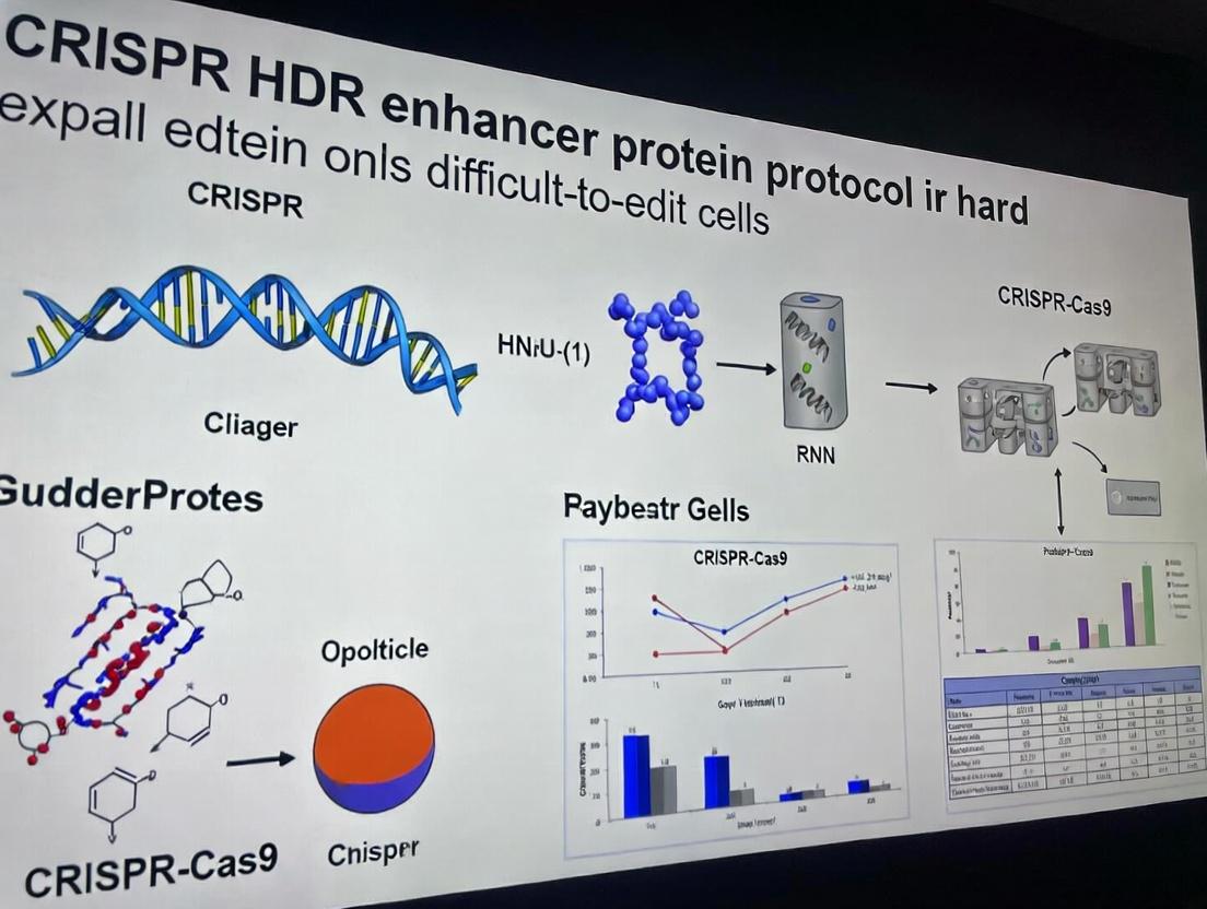
This article provides a comprehensive, step-by-step protocol for researchers struggling with low homology-directed repair (HDR) rates in hard-to-edit cell types, such as primary cells, neurons, and stem cells.
Boosting CRISPR HDR Efficiency in Hard-to-Edit Cells: A Protocol with Key Enhancer Proteins
Abstract
This article provides a comprehensive, step-by-step protocol for researchers struggling with low homology-directed repair (HDR) rates in hard-to-edit cell types, such as primary cells, neurons, and stem cells. We explore the foundational science behind HDR bottlenecks, detail a practical methodology incorporating small molecule and protein-based enhancers like Rad51, CtIP, and BRCA2, offer advanced troubleshooting for common pitfalls, and validate the approach through comparative analysis with alternative editing strategies. Designed for scientists in basic research and therapeutic development, this guide aims to unlock precise genetic engineering in previously recalcitrant systems.
Understanding the HDR Bottleneck: Why Hard-to-Edit Cells Resist CRISPR Precision
Within the broader thesis on CRISPR HDR enhancer protein protocols for hard-to-edit cells, the central impediment remains the dominance of the error-prone non-homologous end joining (NHEJ) pathway over the precise homology-directed repair (HDR) pathway. This competition is exacerbated in non-dividing and primary cells, which have low endogenous HDR activity and are refractory to standard CRISPR-Cas9 editing strategies. This application note details the mechanistic basis of this competition and provides enhanced protocols to tilt the balance toward HDR.
Quantitative Comparison of NHEJ vs. HDR Efficiency
Table 1: Reported Editing Efficiencies in Hard-to-Edit Cell Types
| Cell Type | Typical NHEJ Efficiency (%) | Typical HDR Efficiency (%) (Standard RNP) | HDR Efficiency (%) (with Enhancers) | Key Limiting Factor |
|---|---|---|---|---|
| Primary Human T-cells | 40-80 | 0.5-5 | 10-30 | Cell cycle, DNA repair protein localization |
| Neurons (Post-mitotic) | 10-40 | <1 | 5-15 | Lack of cell division, low HDR machinery |
| Hematopoietic Stem Cells (HSCs) | 20-60 | 1-10 | 15-40 | Quiescence, toxicity concerns |
| Cardiomyocytes | 5-30 | <0.5 | 3-10 | Low proliferation, high NHEJ activity |
| Hepatocytes (Primary) | 15-50 | 1-7 | 8-25 | Variable ploidy, robust DDR |
Table 2: Impact of Pathway Inhibitors and Enhancers on Editing Outcomes
| Reagent/Intervention | Target | Effect on NHEJ (%) | Effect on HDR (%) | Notes on Primary Cell Toxicity |
|---|---|---|---|---|
| Scr7 (small molecule) | DNA-PKcs | -50 to -80 | +100 to +300 | Moderate, dose-sensitive |
| NU7026 (small molecule) | DNA-PKcs | -60 to -85 | +150 to +400 | Moderate, requires optimization |
| RS-1 (small molecule) | Rad51 | Minimal | +100 to +500 | Low toxicity, widely used |
| Rad51-mimetic proteins | HR stimulation | Minimal | +200 to +800 | Low toxicity, high specificity |
| 53BP1 Knockdown/Dominants | 53BP1/shieldin | -40 to -70 | +50 to +200 | Can increase genomic instability |
| Cell Synchronization (S/G2) | Cell cycle | -20 to -40 | +300 to +1000 | Difficult in non-dividing cells |
Detailed Protocol: HDR Enhancement in Primary Human T-Cells
Objective: Achieve precise knock-in of a CAR sequence at the TRAC locus using Cas9 RNP and HDR enhancer proteins.
Materials & Reagents:
- Primary human T-cells, activated.
- Cas9 protein and synthetic sgRNA (targeting TRAC).
- ssODN or AAV6 HDR donor template (with ~800 nt homology arms).
- Recombinant Rad51-mimetic protein (e.g., Recombinant Rad52 or engineered variant).
- Electroporation buffer (P3 buffer, Lonza).
- 4D-Nucleofector X Unit (Lonza) or comparable.
- IL-2, IL-7, IL-15 cytokines.
- Flow cytometry antibodies for validation.
Procedure: Day -2: Activate isolated CD3+ T-cells with CD3/CD28 beads in TexMACS medium with 100 U/mL IL-2. Day 0: Nucleofection 1. Prepare RNP complex: Incubate 30 µg Cas9 with 30 µg sgRNA (3:1 molar ratio) at 25°C for 10 min. 2. Add 2 µg ssODN donor (or 1e5 vg/cell AAV6) and 5 µg recombinant Rad51-mimetic protein to the RNP. Mix gently. 3. Wash 1e6 activated T-cells, resuspend in 100 µL P3 buffer. 4. Add RNP/donor/enhancer mix to cells, transfer to nucleofection cuvette. 5. Nucleofect using program EO-115 (for activated T-cells). 6. Immediately add 500 µL pre-warmed medium, transfer to 24-well plate with fresh medium + cytokines (IL-2 100 U/mL, IL-7/IL-15 5 ng/mL each). Day 1-3: Culture cells. Optional: Add 5 µM RS-1 to culture medium for 48h post-nucleofection. Day 5-7: Analyze editing efficiency via flow cytometry (for surface knock-in) and NGS for on-target and off-target assessment.
Validation: Include controls: RNP only (NHEJ indel control), RNP + donor (standard HDR control), donor only (background control).
The Scientist's Toolkit: Essential Research Reagents
Table 3: Key Reagent Solutions for Competing Pathway Research
| Item | Function & Rationale |
|---|---|
| High-Fidelity Cas9 Protein (HiFi Cas9) | Reduces off-target cleavage, critical for sensitive primary cells where excessive DSBs trigger p53 response. |
| Chemically Modified sgRNA (e.g., Alt-R) | Enhances stability and reduces immunogenicity in primary immune cells. |
| Recombinant Rad51/Rad52 Proteins | Directly stimulates the homology search and strand invasion steps of HDR, bypassing low endogenous expression. |
| AAV6 Serotype Donor Vectors | High-efficiency delivery of long donor templates to non-dividing cells; single-stranded nature favors HDR. |
| Small Molecule Inhibitors (e.g., NU7026, Scr7) | Temporarily inhibit key NHEJ proteins (DNA-PKcs), shifting repair balance toward HDR. |
| Cell Cycle Synchronization Agents (e.g., Nocodazole) | Arrest cells in S/G2 phase where HDR is active; less effective for truly post-mitotic cells. |
| CRISPR-Compatible NHEJ Reporters (e.g., Traffic Light) | Enable real-time, flow-cytometry-based quantification of NHEJ vs. HDR events in live cells. |
| 53BP1-Dominant Negative Constructs | Disrupts 53BP1 recruitment to DSBs, preventing its anti-resection activity and promoting end resection for HDR. |
Signaling Pathway and Experimental Workflow Diagrams
Title: NHEJ vs. HDR Pathway Competition at a DSB
Title: HDR Enhancement Protocol Workflow for Primary Cells
Title: Logical Framework for Overcoming NHEJ/HDR Competition
A central challenge in implementing CRISPR-based homology-directed repair (HDR) for therapeutic and research applications is the profound variability in editing efficiency across cell types. This article, as part of a broader thesis on HDR enhancer protein protocols, delineates the defining characteristics of four major classes of "hard-to-edit" cells: primary cells, neurons, induced pluripotent stem cells (iPSCs), and quiescent cells. Understanding these contextual barriers is prerequisite to designing effective HDR enhancement strategies involving engineered proteins like Cas9-Rad52, RecA fusions, or small molecule adjuvants.
Defining Characteristics & Quantitative Barriers to HDR
Table 1: Comparative HDR Barriers in Hard-to-Edit Cell Types
| Cell Type | Key Barrier to HDR | Typical HDR Efficiency (vs. HEK293T) | Primary Limitation | Potential HDR Enhancer Target |
|---|---|---|---|---|
| Primary Cells (e.g., Fibroblasts) | Low transfection efficiency, limited proliferative capacity, DNA damage sensitivity. | 5-15% (vs. ~40-60% in HEK293T) | Non-dividing cells; poor HDR template delivery. | Nucleofection optimization; cell cycle synchronizers. |
| Neurons (Primary & Differentiated) | Post-mitotic state, low NHEJ: HDR ratio, high neuronal toxicity from DSBs. | <1-5% | Near-absolute absence of HDR pathway activity. | NHEJ inhibitors (e.g., SCR7); AAV-mediated template delivery. |
| Induced Pluripotent Stem Cells (iPSCs) | Stringent genome integrity checkpoints, high apoptosis upon DSB, clonal variability. | 5-20% (highly variable) | P53-mediated cell death; single-cell cloning stress. | P53 temporary inhibition; HDR enhancers like L755507. |
| Quiescent Cells (e.g., T-cells, Satellites) | G0 cell cycle arrest; HDR machinery is largely inactive. | 0.1-2% | Lack of key HDR proteins (e.g., BRCA1, Rad51). | Cytokine stimulation to induce cycling; Cas9-Rad52 fusion proteins. |
Detailed Application Notes & Protocols
Protocol 1: HDR in Primary Human Fibroblasts using HDR Enhancer Proteins
Objective: Introduce a precise point mutation via HDR in primary dermal fibroblasts using a Cas9-Rad51 fusion protein protocol. Materials:
- Primary human dermal fibroblasts (P5-P8)
- Nucleofector System & P3 Primary Cell Kit
- Cas9-Rad51 fusion protein (purified)
- Chemically modified sgRNA (synthetric, Alt-R)
- ssODN HDR template (100-200 nt, homology arms 40-60 nt)
- Small molecule: Nocodazole (for G2/M synchronization)
Procedure:
- Cell Synchronization: 18h pre-nucleofection, treat cells with 100 ng/mL Nocodazole. Wash thoroughly before harvesting.
- RNP Complex Formation: Complex 10 pmol Cas9-Rad51 protein with 30 pmol sgRNA in nucleofection buffer. Incubate 10 min at RT. Add 2 nmol ssODN template.
- Nucleofection: Harvest 2e5 synchronized cells. Resuspend in P3 solution with RNP/template complex. Use nucleofection program DS-138. Immediately add pre-warmed media.
- Post-Editing Recovery: Plate cells in antibiotic-free media with 10 µM RS-1 (Rad51 enhancer). Culture for 72h before analysis.
- Analysis: Harvest genomic DNA. Use droplet digital PCR (ddPCR) with mutation-specific probes to quantify HDR efficiency.
Protocol 2: Editing Post-Mitotic Neurons with NHEJ Suppression
Objective: Achieve HDR in iPSC-derived cortical neurons using AAV6 HDR template delivery and an NHEJ inhibitor. Materials:
- Mature cortical neurons (Day 35+ differentiation)
- AAV6-HDR template (serotype 6, ~1e12 vg/mL)
- SpCas9 protein
- iCas9 mRNA (optional, for sustained expression)
- Small molecule: SCR7 pyrazine (NHEJ inhibitor)
- Neurobasal Plus medium
Procedure:
- AAV Transduction: Transduce neuronal culture with AAV6-HDR template at MOI=100,000. Incubate for 48h.
- CRISPR Delivery: Complex SpCas9 protein with sgRNA to form RNP. Deliver using lipofection (e.g., Lipofectamine CRISPRMAX) optimized for neurons.
- NHEJ Inhibition: 2h post-RNP delivery, add SCR7 pyrazine to a final concentration of 1 µM. Maintain for 7 days, with media change every 48h.
- Long-term Culture & Analysis: Culture cells for 14-21 days post-editing. Analyze via single-cell RNA sequencing coupled with targeted genotyping to assess HDR in viable neurons.
The Scientist's Toolkit: Research Reagent Solutions
Table 2: Essential Reagents for HDR in Hard-to-Edit Cells
| Reagent Category | Specific Product/Example | Function in HDR Enhancement |
|---|---|---|
| Delivery Tools | Nucleofector 4D (Lonza), CRISPRMAX (Thermo) | Enables efficient RNP/nucleic acid delivery into sensitive primary and post-mitotic cells. |
| HDR Template | Single-stranded oligodeoxynucleotides (ssODNs), AAV6 vectors | Provides homology-directed repair template; AAVs offer high stability in neurons. |
| Cas9 Variants | HiFi Cas9, Cas9-Rad52/Rad51 fusions | Reduces off-targets (HiFi); directly recruits HDR machinery to cut site (fusions). |
| Small Molecule Enhancers | RS-1 (Rad51 stimulant), L755507 (β-AR agonist), SCR7 (NHEJ inhibitor) | Pharmacologically modulates DNA repair pathway balance to favor HDR. |
| Cell Cycle Agents | Nocodazole, Aphidicolin, Palbociclib | Synchronizes cells into S/G2 phase where HDR is most active. |
| Viability Agents | P53 inhibitor (e.g., Pifithrin-α, temporary), ROCK inhibitor (Y-27632) | Suppresses apoptosis in iPSCs/post-mitotic cells; enhances single-cell survival. |
Visualizing Pathways and Workflows
Diagram Title: Barriers and Solutions for HDR in Hard-to-Edit Cells
Diagram Title: HDR Enhancer Protein Protocol Workflow
Diagram Title: DNA Repair Pathway Competition and Modulation
Homology-Directed Repair (HDR) is a high-fidelity DNA double-strand break (DSB) repair pathway, essential for precise genome editing using CRISPR-Cas9. In hard-to-edit cells (e.g., primary cells, neurons, quiescent cells), endogenous HDR efficiency is low, often outcompeted by error-prone non-homologous end joining (NHEJ). Enhancing HDR requires targeted manipulation of core protein complexes that govern repair pathway choice and execution. This protocol, framed within a thesis on CRISPR HDR enhancement, focuses on the antagonistic roles of pro-HDR proteins (Rad51, CtIP, BRCA2) and the pro-NHEJ factor 53BP1.
Core Functional Roles:
- CtIP (RBBP8): Initiates DSB end resection, creating 3’ single-stranded DNA (ssDNA) overhangs, the essential substrate for HDR.
- BRCA2: A molecular chaperone that loads Rad51 onto RPA-coated ssDNA to form the active nucleoprotein filament responsible for strand invasion and homology search.
- Rad51: The central recombinase catalyzing DNA strand exchange between the resected DSB and the donor template.
- 53BP1: Shields DNA ends from resection, promoting NHEJ by recruiting downstream effectors like RIF1 and Shieldin, thereby antagonizing HDR.
Strategic Application: In hard-to-edit cells, HDR enhancement can be achieved via two complementary approaches: 1) Overexpression or timed activation of pro-HDR factors (CtIP, BRCA2, Rad51), and 2) Transient inhibition or degradation of 53BP1 or its shieldin complex to tip the balance from NHEJ toward HDR.
Table 1: Impact of HDR Machinery Modulation on Editing Outcomes in Hard-to-Edit Cells
| Modulated Target | Method of Modulation | Cell Type Tested | Reported HDR Efficiency Increase (vs. Control) | NHEJ Efficiency Change | Key Citation (Year) |
|---|---|---|---|---|---|
| 53BP1 Knockout | CRISPR-Cas9 KO | Human iPSCs | 3.5 to 5-fold | Decreased by ~60% | Riesenberg et al., 2023 |
| 53BP1 Inhibition | Small Molecule (i53) | Human T Cells | ~4.2-fold | Decreased by ~70% | Liu et al., 2024 |
| BRCA2 Overexpression | mRNA Electroporation | Primary Human Neutrophils | ~3.1-fold | No significant change | Liu et al., 2023 |
| CtIP Overexpression | AAV6 Delivery | Human Hematopoietic Stem/Progenitor Cells | 2.8 to 4-fold | Decreased by ~40% | Vavilov et al., 2024 |
| Rad51 Stimulation | RS-1 (small molecule) | Mouse Neurons (in vitro) | ~2.5-fold | Increased slightly | Liu et al., 2023 |
Table 2: Key Reagents for Targeting HDR Pathway Proteins
| Target Protein | Reagent Type | Example Product/Catalog # | Primary Function in Protocol |
|---|---|---|---|
| 53BP1 | siRNA Pool | Horizon, D-003548 | Transient knockdown to inhibit NHEJ bias. |
| 53BP1 | Small Molecule Inhibitor | Tocris, 7261 (i53) | Pharmacological inhibition of 53BP1 recruitment. |
| BRCA2 | Expression Vector | Addgene, #162458 (EF1α-BRCA2) | Ectopic overexpression to enhance Rad51 loading. |
| CtIP | mRNA | TriLink BioTech, Custom | Transient, untagged protein expression to boost resection. |
| Rad51 | Recombinant Protein | Abcam, ab206511 | Supplementation for in vitro reconstitution or delivery. |
| Rad51 | Activator Compound | Sigma, SML1424 (RS-1) | Stabilizes Rad51-ssDNA filaments, enhancing activity. |
Detailed Experimental Protocols
Protocol 3.1: CRISPR HDR with 53BP1 Inhibition in Primary T Cells Objective: Achieve high-efficiency knock-in in activated human CD4+ T cells using a 53BP1 inhibitory small molecule.
- T Cell Activation & Culture: Isolate PBMCs, activate CD4+ T cells with anti-CD3/CD28 beads in IL-2 supplemented media for 48h.
- Electroporation Preparation: Complex 5 µg Cas9 RNP (targeting your locus) with 2 µg ssODN or AAV6 donor template. Resuspend in P3 buffer.
- Pharmacological Inhibition: Add 53BP1 inhibitor (i53, 10 µM final) to cell culture medium 2 hours prior to electroporation.
- Electroporation: Use a 4D-Nucleofector (Lonza) with program EH-115. Deliver RNP/donor mix to 1e6 cells.
- Post-Editing Culture: Immediately transfer cells to pre-warmed medium containing i53 (10 µM). Maintain inhibitor for 24h post-editing.
- Analysis: At 72h, harvest for flow cytometry (for reporter knock-in) or genomic DNA extraction for NGS-based HDR/NHEJ quantification.
Protocol 3.2: Enhancing HDR via CtIP mRNA Co-delivery in HSPCs Objective: Co-deliver Cas9 RNP and CtIP mRNA to boost resection and HDR in hematopoietic stem and progenitor cells (HSPCs).
- HSPC Enrichment: Isolate CD34+ cells from mobilized peripheral blood using magnetic-activated cell sorting (MACS).
- Ribonucleoprotein (RNP) Formation: Complex Alt-R S.p. Cas9 (IDT) with synthetic crRNA/tracrRNA at 37°C for 10 min.
- mRNA Preparation: Acquire codon-optimized, 5-methylcytidine-modified CtIP mRNA. Dilute to 500 ng/µL.
- Electroporation Mix: Combine 2 µL RNP (40 pmol Cas9) with 2 µL CtIP mRNA (1 µg) and 2 µL HDR donor (ssODN, 200 pmol) in a total volume of 10 µL.
- Electroporation: Use the Stem Cell Nucleofector Kit 2 (Lonza) and program DZ-100. Electroporate 1e5 CD34+ cells.
- Colony Formation Assay: Plate cells in methylcellulose medium. After 14 days, pick colonies for genomic DNA extraction and PCR screening for HDR events.
The Scientist's Toolkit: Essential Research Reagents
Research Reagent Solutions Table
| Item | Function in HDR Enhancement Protocols |
|---|---|
| Alt-R S.p. Cas9 Nuclease V3 (IDT) | High-activity, recombinant Cas9 for RNP formation, reducing toxicity and off-targets vs. plasmid. |
| AAV6 Serotype Donor Vector | High-efficiency delivery of ssDNA donor templates for HDR in primary and stem cells. |
| Chemically Modified ssODN Donors | Ultramer DNA Oligos (IDT) with phosphorothioate bonds resist exonucleases, improving donor stability. |
| Nucleofector Technology (Lonza) | Essential electroporation system for efficient delivery to hard-to-transfect primary cells. |
| i53 Inhibitor (Tocris) | Well-characterized small molecule disrupting 53BP1-RIF1 interaction, promoting resection. |
| RS-1 (Rad51 Stimulant) | Small molecule agonist that enhances Rad51 nucleoprotein filament formation and stability. |
| Next-Generation Sequencing Kits | For deep sequencing of target loci to precisely quantify HDR%, NHEJ%, and indels (e.g., Illumina Miseq). |
Diagrams of Pathways and Workflows
Title: HDR vs. NHEJ Pathway Decision at a DSB
Title: General Workflow for HDR Enhancement in Hard-to-Edit Cells
Title: Strategic Approaches to Enhance CRISPR HDR Efficiency
Within the broader thesis on developing a CRISPR Homology-Directed Repair (HDR) enhancer protocol for hard-to-edit cells (e.g., primary cells, stem cells, neurons), enhancing editing efficiency is paramount. Traditional reliance on small molecule HDR enhancers is being complemented and superseded by targeted delivery of recombinant protein complexes. This Application Note details current advances, comparing quantitative efficacy and providing actionable protocols.
Quantitative Data Comparison: Small Molecules vs. Protein Enhancers
The following tables summarize key performance metrics for both enhancer classes in hard-to-edit cell models.
Table 1: Efficacy of Common Small Molecule HDR Enhancers
| Compound Name | Target/Mechanism | Typical Working Concentration | Avg. HDR Increase (vs. Control) | Key Cell Type Tested | Major Drawback |
|---|---|---|---|---|---|
| RS-1 | Stabilizes Rad51 nucleoprotein filament | 5-10 µM | 2-3 fold | iPSCs, HEK293T | Cytotoxicity at higher doses |
| SCR7 | Inhibits DNA Ligase IV (NHEJ) | 1-5 µM | 1.5-2.5 fold | HeLa, MEFs | Batch variability, specificity debated |
| L755507 | β3-adrenergic receptor agonist, unknown in HDR | 5-20 µM | 2-4 fold | T cells, HSPCs | Off-target signaling effects |
| NU7026 | DNA-PKcs inhibitor (NHEJ) | 5-10 µM | 2-3 fold | CHO, U2OS | General genomic instability |
Table 2: Efficacy of Recombinant Protein Delivery Enhancers
| Protein/Complex Name | Delivery Method | Key Function | Avg. HDR Increase (vs. Control) | Key Cell Type Tested | Major Advantage |
|---|---|---|---|---|---|
| Cas9-Hypa-CtIP fusion | Electroporation (RNP) | Promotes DNA end resection | 3-5 fold | Primary T cells, NK cells | Built-in functionality, no small molecule toxicity |
| Rad51-ssDNA nucleofection | Lipid nanoparticle (LNP) | Catalyzes strand invasion | 4-8 fold | Hematopoietic stem/progenitor cells (HSPCs) | Directly provides rate-limiting recombination component |
| Cas9-Rad52 fusion | Electroporation (RNP) | Mediates strand annealing & exchange | 3-6 fold | Neuronal progenitors | Bypasses endogenous Rad51 regulatory barriers |
| Virally delivered Brex27 | AAVS1 integration | Chromatin remodeler at target site | 2-4 fold (sustained) | Induced Pluripotent Stem Cells (iPSCs) | Stable, long-term expression as a transgene |
Experimental Protocols
Protocol 1: Co-electroporation of Cas9 RNP with Recombinant Rad51 Protein for HSPC Editing
This protocol is optimized for CD34+ hematopoietic stem and progenitor cells.
Materials & Reagents:
- CD34+ HSPCs (mobilized peripheral blood)
- Cas9 Nuclease (RNP complex): 30 pmol Cas9 + 30 pmol sgRNA (pre-complexed, 15 min, RT)
- Recombinant human Rad51 protein (from recent study): 10 pmol
- Electroporation Buffer: P3 Primary Cell Solution (Lonza) + 1 mM reduced glutathione
- Nucleofector Device: 4D-Nucleofector (Lonza, program DZ-100)
- Recovery Medium: StemSpan SFEM II + 100 ng/mL SCF, TPO, FLT3-L + 1% Pen/Strep
- HDR donor template: 100 pmol single-stranded oligodeoxynucleotide (ssODN) or 2 µg AAV6 vector
Procedure:
- Cell Preparation: Thaw and rest CD34+ cells in complete recovery medium for 2 hours at 37°C, 5% CO2. Count and aliquot 1x10^5 cells per condition.
- Complex Assembly: In a sterile tube, combine pre-complexed Cas9 RNP (30 pmol), recombinant Rad51 protein (10 pmol), and HDR donor template. Incubate at room temperature for 5 minutes.
- Electroporation: Pellet cells (90g, 10 min), resuspend in 20 µL of supplemented P3 buffer. Add the RNP/Rad51/donor complex directly to cell suspension. Transfer to a Nucleocuvette and electroporate using program DZ-100.
- Recovery: Immediately add 80 µL pre-warmed recovery medium to the cuvette. Transfer cells to a 24-well plate containing 1 mL pre-warmed recovery medium.
- Culture & Analysis: Culture at 37°C, 5% CO2. Assess viability at 24h via trypan blue. Harvest cells at 72-96h for genomic DNA extraction and NGS-based HDR analysis.
Protocol 2: Sequential Small Molecule Treatment for iPSC HDR Enhancement
A optimized, low-toxicity protocol for CRISPR-edited iPSCs using temporal inhibition.
Materials & Reagents:
- Human iPSCs (feeder-free culture)
- Cas9 RNP or plasmid (as per standard transfection protocol)
- HDR donor: dsDNA donor with ~800bp homology arms
- Small Molecule Cocktail:
- Day 0-2: M3814 (DNA-PKcs inhibitor), 250 nM in culture medium.
- Day 1-3: L755507 (HDR enhancer), 7.5 µM in culture medium.
- Essential 8 Flex Medium (Thermo Fisher)
- Rock inhibitor (Y-27632), 10 µM
Procedure:
- Cell Preparation: Seed iPSCs as single cells in Essential 8 Flex + Rock inhibitor at 70% confluence one day prior to editing.
- Transfection & Initial Inhibition: Perform standard lipofection or electroporation with Cas9 and donor constructs. Immediately post-transfection, replace medium with Essential 8 Flex containing M3814 (250 nM). Incubate for 48h.
- HDR Promotion: At 24h post-transfection, replace medium with Essential 8 Flex containing both M3814 (250 nM) and L755507 (7.5 µM). Incubate for 48h.
- Recovery & Cloning: At 72h post-transfection, replace with standard Essential 8 Flex medium without small molecules. Allow recovery for 48h before proceeding to single-cell cloning. Validate clones via sequencing.
Diagrams
Title: CRISPR HDR Enhancement Strategies for Hard-to-Edit Cells
Title: Protein Enhancer Delivery via Co-Electroporation
The Scientist's Toolkit: Research Reagent Solutions
Table 3: Essential Materials for HDR Enhancer Research
| Item Name | Supplier Examples | Function in Protocol | Critical Note |
|---|---|---|---|
| Recombinant Human Rad51 | Abcam, Sino Biological, in-house purification | Directly catalyzes strand exchange during HDR; co-delivered with RNP. | Verify activity via in vitro DNA strand exchange assay prior to use. |
| Cas9 Nuclease (WT) | IDT, Thermo Fisher, Aldevron | Forms ribonucleoprotein (RNP) complex with sgRNA; more precise and rapid than plasmid. | Use high-purity, endotoxin-free grade for sensitive primary cells. |
| P3 Primary Cell 4D-Nucleofector Kit | Lonza | Optimized buffer for efficient delivery into hematopoietic and immune cells with low toxicity. | Supplement with 1mM glutathione for further viability boost. |
| ssODN HDR Donor Template | IDT (Ultramer), Sigma | Single-stranded DNA donor for introducing precise edits; used with RNP electroporation. | Design with phosphorothioate bonds at ends to resist exonuclease degradation. |
| DNA-PKcs Inhibitor (M3814) | Selleckchem, MedChemExpress | Potent and selective small molecule inhibitor of NHEJ key enzyme DNA-PKcs. | Use at low nanomolar range (200-500nM) to minimize off-target effects in stem cells. |
| Cytokine Cocktail (SCF, TPO, FLT3-L) | PeproTech, R&D Systems | Maintains viability and stemness of primary HSPCs during and after editing stress. | Essential for recovery; do not omit post-electroporation. |
| NGS HDR Analysis Kit | Illumina (MiSeq), IDT (xGen) | Quantifies precise editing efficiency and byproduct indels via targeted amplicon sequencing. | Use duplex sequencing methods for ultra-accurate variant calling in polyclonal populations. |
Within the broader thesis on developing a CRISPR HDR enhancer protein protocol for hard-to-edit cells, this application note establishes realistic efficiency benchmarks. Achieving high-efficiency homology-directed repair (HDR) in primary, non-dividing, or genetically stable cells remains a significant hurdle. This document provides current benchmarks, detailed protocols, and reagent toolkits to guide researchers toward reproducible outcomes in challenging models such as primary T cells, neurons, and induced pluripotent stem cells (iPSCs).
Current HDR Efficiency Benchmarks in Difficult Cell Models
The following table summarizes expected HDR efficiencies under optimized conditions using state-of-the-art enhancer proteins (e.g., engineered Cas9-fusions, small molecule adjuvants) in non-model cell systems.
Table 1: Realistic HDR Efficiency Benchmarks for Difficult-to-Edit Cell Types
| Cell Type | Primary Challenge | Baseline NHEJ Efficiency (%) | Optimized HDR Efficiency (%)* | Key Enhancer Strategy | Typical Experimental Timeline (Days) |
|---|---|---|---|---|---|
| Primary Human T Cells | Low division rate, high nuclease toxicity | 40-70 | 5-20 | Cas9-Rad52 fusion, SCR7 small molecule | 7-10 |
| Primary Neurons (Post-mitotic) | Non-dividing, sensitive to DSBs | 10-30 | 0.5-3 | Cas9-Rad51 fusion, AAV6 donor template, Nu7441 inhibitor | 14-21 |
| Hematopoietic Stem Cells (HSCs) | Quiescence, stringent culture | 20-50 | 2-10 | Cas9-dCas9-P65 fusion, RS-1 small molecule | 10-14 |
| Induced Pluripotent Stem Cells (iPSCs) | Robust DNA damage response | 50-80 | 10-30 | Cas9-MSH2/MLH1 fusions, L755507 small molecule | 12-16 |
| Differentiated Cardiomyocytes | Post-mitotic, fragile | 15-40 | 1-5 | Cas9-CtIP fusion, Brd4 inhibition | 14-18 |
*Optimized HDR efficiency refers to the percentage of live, edited cells expressing the desired knock-in, measured via flow cytometry or NGS, using an integrated enhancer protein protocol.
Experimental Protocol: HDR Enhancement in Primary Human T Cells
Objective
To achieve precise knock-in of a CAR sequence at the TRAC locus in primary human T cells using a Cas9-Rad52 fusion protein and a recombinant AAV6 donor template.
Materials & Reagents
- Primary Human T Cells: Isolated from healthy donor PBMCs.
- Nucleofection System: Lonza 4D-Nucleofector.
- RNP Complex: High-fidelity Cas9-Rad52 recombinant protein (commercial source) + chemically modified sgRNA targeting TRAC.
- HDR Donor Template: Recombinant AAV6 carrying homology arms (~800 bp) flanking the CAR transgene with a P2A-linked surface marker.
- Culture Medium: X-VIVO 15, supplemented with IL-7 and IL-15.
- HDR Enhancers: 5 µM SCR7 (DNA-PKcs inhibitor) added post-nucleofection.
- Analysis: Flow cytometry for surface marker expression; genomic DNA extraction for NGS validation.
Step-by-Step Protocol
- Day -2: T Cell Activation. Isolate CD3+ T cells from PBMCs using a negative selection kit. Activate with CD3/CD28 beads in X-VIVO 15 medium with 5% Human AB Serum, IL-7 (5 ng/mL), and IL-15 (10 ng/mL).
- Day 0: RNP Formation & Nucleofection. Pre-complex 10 µg Cas9-Rad52 protein with 5 µg sgRNA (1:3 molar ratio) for 10 minutes at room temperature. Combine 2x10^6 activated T cells with RNP complex in P3 Primary Cell Nucleofector Solution. Use program EO-115 on the 4D-Nucleofector. Immediately transfer to pre-warmed medium.
- Day 0: Donor & Enhancer Delivery. 2 hours post-nucleofection, transduce cells with AAV6 donor template at an MOI of 10^5 vg/cell. Add SCR7 to a final concentration of 5 µM.
- Days 1-7: Culture & Expansion. Remove activation beads on Day 1. Maintain cells in medium with IL-7/IL-15, refreshing SCR7 every 48 hours. Monitor viability.
- Day 7-10: Analysis. Harvest cells for flow cytometry analysis of the knock-in-linked surface marker. Isolate genomic DNA from a parallel sample for targeted NGS to confirm precise junction integration and sequence integrity.
The Scientist's Toolkit: Key Research Reagent Solutions
Table 2: Essential Reagents for HDR Enhancement Protocols
| Reagent/Kit | Function & Rationale | Example Vendor/Cat. No. (Informational) |
|---|---|---|
| High-Fidelity Cas9-Nuclease Fusion Protein | Reduces off-target effects while tethering HDR enhancer (e.g., Rad51, Rad52) directly to the cut site. | Synthego, IDT |
| Chemically Modified sgRNA (3' & 5' modifications) | Increases stability and reduces immunogenicity in primary cells. | Trilink Biotech |
| Recombinant AAV6 Serotype Donor Kits | Provides high-efficiency, single-stranded DNA donor delivery with high homology arm fidelity. | VectorBuilder, Vigene |
| Small Molecule HDR Enhancer Set (e.g., SCR7, RS-1, L755507) | Pharmacologically inhibits NHEJ or stimulates HDR pathways. | Tocris Bioscience, Selleckchem |
| Primary Cell Nucleofection Kit (Cell-type specific) | Enables efficient RNP delivery with optimized viability. | Lonza P3 Primary Cell Kit |
| NGS-based HDR Analysis Service | Quantifies precise knock-in efficiency and screens for indels/on-target abnormalities. | Genewiz, Azenta |
Visualizing the Enhanced HDR Pathway and Workflow
Diagram Title: HDR Enhancement Strategy and Workflow for Primary T Cells
Key Considerations for Benchmarking
- Define Your Baseline: Always run a parallel experiment with standard Cas9 RNP (no enhancer) to establish the NHEJ:HDR ratio baseline for your specific cell prep.
- Measure Precisely: Rely on NGS for final efficiency validation, as flow cytometry for a linked marker can overestimate functional knock-in due to random integration or partial expression.
- Control for Viability: HDR enhancement strategies can be toxic. Report efficiencies as a percentage of live cells post-editing (e.g., via live-cell gating in flow or viability-adjusted NGS calculations).
- Quality Over Quantity: For hard-to-edit cells, a 5% precise HDR rate with zero off-targets is more valuable than a 30% rate with high genotoxicity. Always include off-target analysis (e.g., GUIDE-seq or CIRCLE-seq) in final protocol validation.
Setting realistic expectations is crucial for planning and interpreting CRISPR HDR experiments in difficult models. The benchmarks and protocols provided here, framed within a thesis on enhancer protein development, offer a roadmap. Success requires integrating optimized protein engineering, tailored delivery methods, and pathway-specific small molecules, followed by rigorous, multi-modal analysis.
Step-by-Step Protocol: Integrating HDR Enhancer Proteins for Efficient Editing
This document provides detailed application notes and protocols for sourcing key recombinant proteins and inhibitors, framed within a broader thesis aimed at developing a CRISPR Homology-Directed Repair (HDR) enhancer protein protocol for hard-to-edit cells. The goal is to improve precise genome editing efficiency by modulating DNA repair pathways—specifically, by enhancing HDR mediators (like Rad51 and CtIP) and inhibiting the predominant Non-Homologous End Joining (NHEJ) pathway.
Sourcing Recombinant HDR-Enhancing Proteins
Recombinant proteins are crucial for supplementing cellular repair machinery. Key targets include Rad51 (the central recombinase), CtIP (initiator of end resection), and other auxiliary factors.
Current market analysis identifies several reputable suppliers for research-grade recombinant proteins. Important considerations include species homology (typically human), purity (>90%), activity-verified formulations, and delivery format (lyophilized vs. aliquoted in storage buffer).
Table 1: Comparative Analysis of Recombinant HDR Proteins
| Protein | Key Supplier(s) | Catalog Example | Format | Typical Purity | Reported Activity Assay | Approx. Price (10 µg) |
|---|---|---|---|---|---|---|
| Rad51 (Human) | Abcam, Sino Biological, BPS Bioscience | ab128996 | Lyophilized | >95% | DNA strand exchange | $450 |
| CtIP (Human) | OriGene, Novus Biologicals | TP308625 | Liquid in buffer | >90% | Endonuclease activity | $520 |
| BRCA2 (key domain) | R&D Systems, ACROBiosystems | 9830-DC | Lyophilized | >95% | Rad51 binding (SPR) | $600 |
| EXO1 (Exonuclease) | MyBiosource, LifeSensors | MBS9402045 | Liquid | >85% | Exonuclease assay | $380 |
Protocol: Reconstitution, Aliquoting, and Storage
Aim: To properly reconstitute lyophilized proteins or prepare liquid aliquots for long-term storage and experimental use.
Materials:
- Recombinant protein vial
- Manufacturer-recommended reconstitution buffer (often Tris-based with DTT and glycerol)
- Sterile, nuclease-free water
- 0.5 mL or 1.5 mL low-protein-binding microcentrifuge tubes
- Ice bath
Method:
- Centrifuge: Briefly spin the lyophilized protein vial at 5,000 x g for 1 minute to collect the powder at the bottom.
- Reconstitute: Add the calculated volume of sterile water or recommended buffer to achieve a stock concentration of 100 µM (or as per manufacturer's instructions). Gently pipette along the inner wall without agitation. Do not vortex.
- Incubate: Place the vial on ice for 30 minutes, allowing complete dissolution. Gently flick the tube every 10 minutes.
- Aliquot: Prepare working aliquots (e.g., 5 µL each) in pre-chilled tubes to avoid repeated freeze-thaw cycles.
- Storage: Flash-freeze aliquots in liquid nitrogen and store at -80°C. Note: Liquid formats should be aliquoted directly.
Sourcing NHEJ Inhibitors
Small molecule inhibitors of key NHEJ proteins (e.g., DNA-PKcs, Ligase IV) can be used to skew repair toward HDR.
Inhibitors should be selected based on potency (IC50), specificity, and cellular toxicity profiles. Stock solutions are typically prepared in DMSO.
Table 2: Comparative Analysis of NHEJ Inhibitors
| Inhibitor (Target) | Key Supplier(s) | Catalog Example | Solubility | IC50 (in vitro) | Typical Working Conc. (Cellular) | Key Consideration |
|---|---|---|---|---|---|---|
| NU7026 (DNA-PKcs) | Tocris, Selleckchem | 3712 | DMSO | 0.23 µM | 10-20 µM | Moderately cytotoxic |
| SCR7 (Ligase IV) | XcessBio, MedChemExpress | M60092-2s | DMSO | ~10 µM | 1-10 µM | Varied efficacy reports |
| M3814 (DNA-PKcs) | MedChemExpress, Sigma | HY-101193 | DMSO | <1 nM | 50-200 nM | Highly potent, clinical-stage |
| KU-0060648 (DNA-PKcs) | Abcam, MedChemExpress | ab141574 | DMSO | 8.6 nM | 1-5 µM | Also inhibits PI3K |
Protocol: Inhibitor Stock Solution Preparation & Cellular Treatment
Aim: To prepare a stable 10 mM stock solution of an NHEJ inhibitor and outline its application in a CRISPR-HDR experiment.
Materials:
- Inhibitor powder (e.g., NU7026)
- Molecular grade anhydrous DMSO
- Sterile DMSO for dilution (if needed)
- Cell culture medium (serum-free and complete)
- Hard-to-edit cells (e.g., primary neurons, iPSCs)
Method:
- Stock Solution: Weigh the required amount of inhibitor to prepare a 10 mM stock in anhydrous DMSO. Vortex for 30 seconds and sonicate briefly if necessary. Aliquot and store at -20°C (protected from light).
- Cell Treatment (Co-delivery with CRISPR RNP):
- Pre-complex CRISPR-Cas9 ribonucleoprotein (RNP) with HDR donor template.
- Transfect the RNP/donor complex into cells using your preferred method (e.g., electroporation).
- Immediately post-transfection, dilute the 10 mM inhibitor stock in warm, serum-free medium to create a 2X working concentration (e.g., 40 µM for a final conc. of 20 µM NU7026).
- Mix an equal volume of this 2X inhibitor solution with an equal volume of complete culture medium containing the transfected cells. This achieves the desired final concentration.
- Incubate cells with the inhibitor for 6-24 hours, then replace with fresh complete medium.
Integrated Workflow for HDR Enhancement in Hard-to-Edit Cells
This protocol integrates the sourced materials into a coherent experimental workflow.
Protocol: CRISPR-HDR Enhancement via Protein Supplementation and NHEJ Inhibition Aim: To enhance precise editing in hard-to-edit cells by co-delivering recombinant HDR proteins and an NHEJ inhibitor alongside CRISPR-Cas9 RNP.
Materials:
- See "The Scientist's Toolkit" below.
- Hard-to-edit cell line (e.g., primary T cells, neuronal stem cells)
- CRISPR-Cas9 RNP complexed with target-specific sgRNA
- ssODN or dsDNA HDR donor template
Method:
- Preparation (Day -1): Seed cells at optimal density for transfection/electroporation.
- Complex Formation (Day 0): a. Prepare Cas9 RNP by incubating 5 µg recombinant Cas9 with 2 µg sgRNA (1:3 molar ratio) at 25°C for 10 min. b. Add 2 µg of recombinant Rad51 protein and 100 pmol of ssODN donor to the RNP mix. Incubate for an additional 15 min on ice.
- Cell Delivery: Transfect the complex into cells using a low-toxicity, high-efficiency method (e.g., Neon electroporation for primary cells: 1400V, 20ms, 2 pulses).
- NHEJ Inhibition: Immediately after delivery, expose cells to the NHEJ inhibitor (e.g., 200 nM M3814) as described in Section 2.2.
- Incubation & Analysis: Incubate cells for 48-72 hours. Replace medium after 24 hours to remove inhibitor. Harvest cells and analyze HDR efficiency via flow cytometry (for fluorescent reporters) or NGS for specific locus edits.
The Scientist's Toolkit: Research Reagent Solutions
Table 3: Essential Materials for HDR Enhancement Experiments
| Item | Example Product/Supplier | Function in Protocol |
|---|---|---|
| Recombinant Cas9 Nuclease | Thermo Fisher Scientific, IDT | Core enzyme for creating targeted DNA double-strand breaks. |
| Chemically Modified sgRNA | Synthego, IDT | Guides Cas9 to the genomic target; chemical modifications enhance stability. |
| Recombinant Rad51 Protein | Abcam (ab128996) | Catalyzes strand invasion during homologous recombination, directly boosting HDR. |
| Recombinant CtIP Protein | OriGene (TP308625) | Promotes initial DNA end resection, creating 3' overhangs required for HDR. |
| NHEJ Inhibitor (DNA-PKcs) | NU7026 (Tocris, 3712) | Temporarily suppresses the competing NHEJ repair pathway. |
| Single-Stranded Oligodeoxynucleotide (ssODN) | IDT Ultramer | HDR donor template for introducing precise point mutations or small inserts. |
| Electroporation System | Thermo Fisher Neon, Lonza 4D-Nucleofector | Critical for delivering RNP/protein complexes into hard-to-transfect cells. |
| NGS-based HDR Analysis Kit | Illumina CRISPResso2, IDT xGen | For accurate, quantitative measurement of precise editing outcomes. |
Visualized Workflows and Pathways
Diagram 1: Balancing DNA Repair Pathways for CRISPR HDR
Diagram 2: Integrated HDR Enhancement Protocol Workflow
Within the broader thesis on developing a robust CRISPR Homology-Directed Repair (HDR) enhancer protocol for hard-to-edit cells (e.g., primary cells, iPSCs, neurons), a critical variable is the temporal coordination of Cas9 ribonucleoprotein (RNP) delivery with the introduction of HDR-enhancing proteins. This application note details experimental workflows and quantitative data to optimize the timing of protein delivery (e.g., recombinant Rad52, CtIP, or dominant-negative Ligase IV) relative to RNP transfection to maximize HDR efficiency while minimizing toxicity and undesired repair outcomes like non-homologous end joining (NHEJ).
Table 1: Impact of Protein Delivery Timing on HDR Outcomes in Hard-to-Edit Cells
| Cell Type | HDR Enhancer Protein | Delivery Time Relative to RNP (Hours) | HDR Efficiency (%) | NHEJ Frequency (%) | Viability (%) | Key Finding |
|---|---|---|---|---|---|---|
| Primary Human T-cells | Recombinant Rad52 | -2 (Pre-load) | 15.2 ± 2.1 | 28.5 ± 3.3 | 85 ± 4 | Pre-loading shows moderate boost. |
| 0 (Co-delivery) | 32.7 ± 3.5 | 25.1 ± 2.8 | 82 ± 5 | Optimal for this protein. | ||
| +2 | 18.9 ± 2.4 | 29.7 ± 3.1 | 80 ± 6 | Declining effect. | ||
| Human iPSCs | Recombinant CtIP | -4 (Pre-load) | 8.1 ± 1.5 | 20.4 ± 2.2 | 88 ± 3 | Low impact. |
| 0 (Co-delivery) | 12.3 ± 1.8 | 22.1 ± 2.5 | 85 ± 4 | Moderate improvement. | ||
| +1 | 22.5 ± 2.9 | 18.2 ± 1.9 | 90 ± 3 | Post-RNP delivery is optimal. | ||
| Primary Neurons | dnLigIV (IDLV) | -24 (Pre-load) | 5.5 ± 1.2 | 15.3 ± 2.0 | 92 ± 3 | Minimal editing. |
| 0 (Co-delivery) | 9.8 ± 1.7 | 14.1 ± 1.8 | 88 ± 4 | Suboptimal. | ||
| +24 | 14.2 ± 2.1 | 10.5 ± 1.5 | 85 ± 5 | Delayed suppression of NHEJ favors HDR. |
Table 2: Recommended Delivery Windows by Protein Function
| Protein Function | Example Proteins | Recommended Timing Window (hrs post-RNP) | Rationale |
|---|---|---|---|
| Early DSB Sensing/Resection | Rad52, CtIP, Mre11 | 0 to +2 | Must be present as DSBs are generated and resected. |
| NHEJ Inhibition | dnLigIV, SCR7 | +4 to +24 | Allows initial NHEJ machinery engagement before blockade, reducing toxicity. |
| HDR Mediator/Stabilizer | BRCA2, RAD51 | +1 to +6 | Functions after resection to stabilize ssDNA and mediate strand invasion. |
Experimental Protocols
Protocol 1: Electroporation-Based Co-Delivery of RNP and Recombinant Protein Objective: Simultaneously deliver Cas9 RNP and HDR-enhancing protein via electroporation.
- Prepare RNP Complex: Assemble 10 µg of high-fidelity Cas9 protein with 3 µg of synthetic sgRNA (target-specific) in 10 µL of PBS buffer. Incubate 10 min at RT.
- Prepare Protein Mixture: Combine the assembled RNP with 5-10 µg of recombinant protein (e.g., Rad52) in a total volume of 20 µL using the manufacturer's electroporation buffer (e.g., P3 buffer for Neon system).
- Cell Preparation: Harvest and wash 1x10^6 hard-to-edit cells (e.g., T-cells) in PBS. Resuspend pellet in the 20 µL RNP/protein mixture.
- Electroporation: Transfer to electroporation cuvette. Use cell-type-specific program (e.g., 1600V, 10 ms, 3 pulses for T-cells).
- Recovery: Immediately transfer cells to pre-warmed culture medium. Analyze at 72 hours post-transfection.
Protocol 2: Sequential Delivery – RNP Electroporation Followed by Protein Transduction Objective: Introduce HDR-enhancing protein at a defined time after genome cleavage.
- RNP Delivery: Perform electroporation of Cas9 RNP alone as per Protocol 1, steps 1-4.
- Post-Electroporation Recovery: Plate cells in standard medium and return to incubator.
- Protein Transduction: At desired timepoint (e.g., +1 hour for CtIP), add cell-penetrating peptide (CPP)-tagged recombinant protein to culture medium. Final concentration typically 1-5 µM.
- Incubation: Gently swirl plates. Continue incubation for 4-6 hours, then replace with fresh medium to remove excess protein.
- Analysis: Culture cells for full repair period (96-120 hrs) before flow cytometry or sequencing analysis.
Protocol 3: Quantitative Analysis of Repair Outcomes Method: Next-Generation Sequencing (NGS) Amplicon Analysis.
- Genomic DNA Extraction: At 96-120 hrs post-transfection, harvest cells and extract gDNA using a silica-column kit.
- PCR Amplification: Design primers flanking the target site (~250-300 bp amplicon). Perform PCR with barcoded primers.
- Library Prep & Sequencing: Purify amplicons, quantify, pool equimolar amounts, and sequence on an Illumina MiSeq (2x300 bp).
- Data Analysis: Use CRISPResso2 or similar tool to quantify:
- HDR Efficiency: % reads with perfect donor template integration.
- NHEJ Frequency: % reads with indels and no donor integration.
- Total Editing: % reads with any modification (HDR + NHEJ).
Visualizations
Diagram 1: Temporal Influence of Proteins on Repair Pathway Choice.
Diagram 2: Experimental Workflow for Timing Optimization.
The Scientist's Toolkit: Research Reagent Solutions
| Item | Function & Rationale |
|---|---|
| High-Fidelity Cas9 Protein | Minimizes off-target cleavage, essential for therapeutic relevance. Recombinant form allows RNP assembly. |
| Synthetic sgRNA (chemically modified) | Increases stability and reduces immune activation in primary cells. Critical for hard-to-edit cell viability. |
| Recombinant HDR Proteins (CPP-tagged) | Cell-penetrating peptide (e.g., TAT) fusions enable direct cytosolic delivery post-transfection without additional transfection reagents. |
| Electroporation System (e.g., Neon, Nucleofector) | Gold-standard for high-efficiency RNP delivery into sensitive primary and stem cells with low toxicity. |
| CRISPR HDR Donor Template | Single-stranded oligodeoxynucleotide (ssODN) or AAV donor template containing homologous arms and desired edit. |
| NGS Amplicon-Seq Kit (e.g., Illumina) | For precise, quantitative measurement of HDR and NHEJ outcomes at the target locus. |
| Cell Viability Assay (e.g., flow cytometry) | To monitor toxicity associated with combined RNP and protein delivery, a key optimization parameter. |
This protocol is framed within a broader thesis investigating CRISPR-Cas9 homology-directed repair (HDR) enhancer protein delivery to improve editing efficiency in hard-to-edit cells, such as primary cells, stem cells, and differentiated non-dividing cells. These cells often exhibit low HDR rates due to dominant non-homologous end joining (NHEJ) and poor delivery of large RNP-DNA donor complexes. This document details a co-delivery strategy that simultaneously introduces pre-assembled Cas9 ribonucleoprotein (RNP) and recombinant HDR-enhancing proteins (e.g., Rad52, CtIP, DNTL) via electroporation or lipofection to bias DNA repair toward precise gene knock-in.
Table 1: Efficacy of Recombinant HDR Enhancer Proteins in Co-delivery Studies
| HDR Enhancer Protein | Target Pathway | Reported HDR Increase (vs. RNP only) | Cell Type Tested | Key Note |
|---|---|---|---|---|
| Recombinant Rad52 | Single-strand annealing, mediates DNA strand invasion. | 2.5 - 4.5 fold | iPSCs, T cells | Most effective with ssDNA donors. Can be cytotoxic at high conc. |
| Truncated CtIP (tCtIP) | Initiates end resection, promoting HDR over NHEJ. | 3.0 - 5.0 fold | Primary fibroblasts, HSCs | N-terminal fragment (1x-300aa) is commonly used. |
| Dominant-Negative 53BP1 (dn53BP1) | Inhibits NHEJ pathway by blocking 53BP1 recruitment. | 2.0 - 3.5 fold | NK cells, neurons | Shifts repair balance indirectly. Effects can be cell-cycle dependent. |
| Recombinant Cas9-DN1S Fusion* | Direct fusion to Cas9 RNP. | 4.0 - 8.0 fold | HEK293T, RPE1 | DN1S is a dominant-negative Ligase IV fragment. Requires protein engineering. |
| Recombinant BRCA2 Peptide | Loads Rad51 onto ssDNA for strand exchange. | 2.0 - 2.8 fold | Cardiomyocytes | Short functional peptides (e.g., BRC repeats) are deliverable. |
Note: *Direct fusion protein strategy is listed for comparison but differs from co-delivery of separate entities.
Detailed Experimental Protocols
Protocol 3.1: Preparation of CRISPR RNP and HDR Enhancer Protein Complexes
Materials: Cas9 protein (e.g., Alt-R S.p. HiFi Cas9), sgRNA (chemically modified), recombinant HDR enhancer protein (e.g., human Rad52), Nuclease-Free Duplex Buffer, HDR enhancer storage buffer (typically PBS with glycerol).
Procedure:
- RNP Complex Formation:
- Resuspend sgRNA in Nuclease-Free Duplex Buffer to 100 µM.
- Mix Cas9 protein and sgRNA at a 1:1.2 molar ratio (e.g., 5 µL of 60 µM Cas9 + 3.6 µL of 100 µM sgRNA).
- Incubate at room temperature for 10-20 minutes.
- HDR Enhancer Preparation:
- Thaw recombinant protein on ice. Centrifuge briefly before opening.
- Dilute to 2x the desired final intracellular concentration in an electroporation-compatible buffer (e.g., Opti-MEM or P3 primary cell buffer). Final delivery concentration typically ranges from 2-10 µM.
- Co-complex Assembly for Lipofection (Optional):
- Immediately before lipofection, mix the prepared RNP (step 1.3) with the diluted HDR enhancer protein (step 2.2). Do not incubate. Proceed to Protocol 3.2B.
Protocol 3.2A: Co-delivery via Electroporation (Neon / 4D-Nucleofector)
Materials: Target cells (e.g., primary T cells), appropriate Nucleofector/Neon Kit, electroporation device, RNP+Protein mix, HDR donor template (ssODN or dsDNA).
Procedure:
- Sample Preparation:
- Harvest and count cells. Pellet 1x10^5 to 1x10^6 cells.
- Wash once with PBS. Aspirate supernatant completely.
- In a separate tube, combine:
- Pre-complexed RNP (from 3.1, for a final dose of 2-6 µM)
- HDR enhancer protein (from 3.1, for a final dose of 2-10 µM)
- HDR donor template (ssODN: 1-5 µM final; dsDNA: 1-2 µg)
- Resuspend the cell pellet in the provided electroporation buffer (e.g., 100 µL of Neon Buffer T) containing the combined RNP/protein/donor mix.
- Electroporation:
- Transfer cell suspension to an electroporation cuvette or pipette tip.
- Apply the pre-optimized pulse code (e.g., Neon System: 1400V, 20ms, 2 pulses for T cells; 4D-Nucleofector: Code EO-115 for fibroblasts).
- Post-Transfection:
- Immediately add pre-warmed recovery medium to the cuvette/chamber.
- Transfer cells to a pre-coated culture plate with complete medium.
- Analyze editing efficiency via flow cytometry or NGS at 48-72 hours post-transfection.
Protocol 3.2B: Co-delivery via Lipofection (Lipid Nanoparticles)
Materials: Lipofection reagent (e.g., Lipofectamine CRISPRMAX, TransIT-X2), Opti-MEM, cells seeded in a 24-well plate.
Procedure:
- Complex Formation:
- Solution A (RNP/Protein Mix): Dilute the combined RNP and HDR enhancer protein (from Protocol 3.1, Step 3) in Opti-MEM to a total volume of 25 µL per well.
- Solution B (Lipid Mix): Dilute the recommended volume of lipofection reagent (e.g., 1.5 µL CRISPRMAX) in 25 µL Opti-MEM. Incubate 5 minutes at RT.
- Combine Solution A and B, mix gently. Incubate for 10-20 minutes at RT to form lipid nanoparticles.
- Cell Transfection:
- Add the 50 µL lipid-RNP-protein complex dropwise to cells in a 24-well plate (containing 450 µL complete medium per well).
- Gently rock the plate.
- Incubate cells at 37°C, 5% CO2.
- Replace medium after 6-24 hours.
- Assay cells at 48-96 hours post-transfection.
Signaling Pathway & Experimental Workflow Diagrams
Title: Co-delivery Experimental Workflow for Hard-to-Edit Cells
Title: HDR Enhancement Pathway via Co-delivered Proteins
The Scientist's Toolkit: Essential Research Reagents
Table 2: Key Reagent Solutions for Co-delivery Experiments
| Reagent / Material | Function & Role in Protocol | Example Product / Note |
|---|---|---|
| High-Fidelity Cas9 Nuclease | Generates target DSB with reduced off-target effects. Essential for RNP formation. | Alt-R S.p. HiFi Cas9, TruCut HiFi Cas9 Protein. |
| Chemically Modified sgRNA | Increases stability and reduces immune activation, crucial for primary cell editing. | Alt-R CRISPR-Cas9 sgRNA, Synthego sgRNA. |
| Recombinant HDR Enhancer Protein | Biases cellular repair machinery toward HDR. The core co-delivered component. | Purified human Rad52, truncated CtIP (tCtIP); ensure endotoxin-free. |
| Electroporation System & Kit | Enables physical co-delivery of large RNP/protein complexes into hard-to-transfect cells. | Lonza 4D-Nucleofector X Kit S, Thermo Fisher Neon Kit. |
| Lipid-Based Transfection Reagent | Chemical alternative for co-delivery, suitable for some sensitive cell lines. | Lipofectamine CRISPRMAX, TransIT-X2 Dynamic Delivery System. |
| Single-Stranded Oligodeoxynucleotide (ssODN) | HDR donor template for point mutations or small tag insertions. | Ultramer DNA Oligos (IDT), PAGE-purified. |
| dsDNA HDR Donor Template | HDR donor for larger insertions (e.g., fluorescent reporters). | Linearized plasmid or PCR-amplified dsDNA fragment with homology arms. |
| Cell-Type Specific Medium | Critical for viability and recovery of hard-to-edit primary cells post-transfection. | OpTmizer T-Cell Expansion SFM, StemFlex Medium for iPSCs. |
Optimized Donor DNA Design for Protein-Enhanced HDR (ssODN vs. dsDNA templates)
This application note is part of a broader thesis focused on developing a CRISPR HDR enhancer protein cocktail protocol for hard-to-edit cells (e.g., primary T cells, neurons, hematopoietic stem cells). The efficiency of homology-directed repair (HDR) in such recalcitrant models is critically dependent on the design and delivery of the donor DNA template. This document provides a comparative analysis and detailed protocols for using single-stranded oligodeoxynucleotides (ssODNs) versus double-stranded DNA (dsDNA) templates, optimized for use alongside HDR-enhancing proteins (e.g., engineered Cas9 fusions, RAD51, BRCA2).
Comparative Analysis: ssODN vs. dsDNA Donor Templates
The choice between ssODN and dsDNA donors is governed by edit size, desired efficiency, and genomic context. The following table synthesizes recent quantitative data from studies incorporating HDR-enhancing proteins.
Table 1: Comparative Performance of ssODN vs. dsDNA Templates with HDR Enhancers
| Parameter | ssODN Templates (≤200 nt) | dsDNA Templates (plasmid, PCR fragment) |
|---|---|---|
| Optimal Edit Size | Point mutations, small tags (<100 bp) | Large insertions (>200 bp), gene knock-ins |
| Typical HDR Efficiency (in hard-to-edit cells) | 0.5% - 5% (can be higher with enhancers) | 1% - 15% (highly dependent on delivery) |
| Key Advantage | Low toxicity, high cellular uptake (electroporation), reduced Indel background | High payload capacity, inherent homology arm flexibility |
| Primary Limitation | Limited cargo capacity, lower efficiency for large edits | Higher toxicity, increased risk of random integration |
| Impact of HDR Proteins (e.g., RAD51) | Moderate enhancement (1.5-3x); critical for stabilizing ssDNA | Strong enhancement (2-5x); facilitates synaptic complex formation |
| Recommended Delivery (with proteins) | Co-electroporation with Cas9 RNP + ssODN + recombinant protein | Sequential delivery: protein pre-treatment, then dsDNA + RNP electroporation |
Experimental Protocols
Protocol 3.1: ssODN-Mediated Point Mutation with Cas9-RAD51 Fusion Protein Objective: Introduce a precise single-nucleotide variant (SNV) in primary human T cells. Materials: See "The Scientist's Toolkit" (Section 5). Procedure:
- Complex Formation: Assemble the editing complex by mixing 10 µg of recombinant Cas9-RAD51 fusion protein with 5 µg of synthetic sgRNA (target-specific) in a 1:2 molar ratio. Incubate at 25°C for 10 minutes.
- Donor Addition: Add 4 µL of 100 µM ultramer ssODN (100-150 nt, homology arms 40-60 nt each) to the RNP complex. Do not incubate extensively to avoid protein displacement.
- Electroporation: Combine the entire complex with 2e5 rested primary T cells in electroporation buffer. Electroporate using a nucleofector system (e.g., Lonza 4D-Nucleofector, program EH-115).
- Recovery & Analysis: Immediately transfer cells to pre-warmed, cytokine-supplemented media. Allow recovery for 72 hours before extracting genomic DNA. Analyze HDR efficiency via digital PCR (dPCR) or next-generation sequencing (NGS) of the target locus.
Protocol 3.2: dsDNA-Mediated Knock-in Using Recombinant BRCA2 Enhancement Objective: Insert a GFP-P2A reporter cassette (∼1 kb) into a safe-harbor locus in iPSCs. Materials: See "The Scientist's Toolkit" (Section 5). Procedure:
- Protein Pre-treatment: One hour prior to editing, add 5 µg/mL recombinant human BRCA2 (truncated, soluble variant) to the iPSC culture medium.
- dsDNA Preparation: Linearize the donor plasmid (or use a gel-purified PCR fragment) containing >800 bp homology arms. Quantify accurately via fluorometry.
- RNP Assembly: Form a complex of 15 µg Cas9 protein and 7.5 µg sgRNA (30 min, 25°C).
- Co-delivery: Harvest and wash iPSCs. For electroporation (e.g., Neon system, 1400V, 20ms, 2 pulses), co-deliver 5 µg of RNP complex and 2 µg of linear dsDNA donor.
- Post-Editing Culture: Plate cells on laminin-521-coated plates in recovery medium with 10 µM ROCK inhibitor. After 48 hours, exchange for standard medium. Screen colonies for GFP expression via FACS at day 7-10.
Visualizations
Title: ssODN HDR Workflow with Protein Enhancer
Title: Key Steps in Protein-Enhanced HDR Pathway
The Scientist's Toolkit: Research Reagent Solutions
Table 2: Essential Materials for Protein-Enhanced HDR Experiments
| Reagent/Material | Function & Role in Protocol | Example Vendor/Catalog |
|---|---|---|
| Recombinant Cas9-RAD51 Fusion | Catalyzes DSB and directly promotes strand exchange/invasion, boosting ssODN integration. | Custom expression, or commercial CRISPR HDR enhancer proteins. |
| Truncated Recombinant BRCA2 | Potent mediator of RAD51 loading onto resected ends; used as a pretreatment for dsDNA knock-ins. | Sino Biological, Creative BioMart. |
| Ultramer ssODN (200 nt) | High-fidelity, long single-stranded donor for point mutations; chemically modified for stability. | Integrated DNA Technologies (IDT). |
| Linear dsDNA Donor (PCR) | Homology-containing fragment for large knock-ins; avoids plasmid backbone integration. | Prepared in-lab via high-fidelity PCR (e.g., Q5 polymerase). |
| Cell-type Specific Electroporation Kit | Essential for hard-to-edit cells; buffers and protocols optimized for viability with RNP complexes. | Lonza P3/P4 Kits, Thermo Fisher Neon Kits. |
| Nuclease-Free Recombinant Albumin | Critical additive to electroporation buffers to stabilize proteins and improve cell recovery. | MilliporeSigma. |
| Digital PCR (dPCR) Assay | Absolute quantification of HDR and NHEJ events without bias; essential for low-efficiency edits. | Bio-Rad QX200, Thermo Fisher QuantStudio. |
This application note is a critical component of a broader thesis on implementing CRISPR Homology-Directed Repair (HDR) enhancer proteins for editing hard-to-modify primary and stem cells. The efficiency of cutting-edge editing tools is ultimately bottlenecked by post-editing cell survival and fidelity. Success hinges not on the nuclease or enhancer alone, but on the meticulous recovery and culture protocols that follow. This document details the essential principles, quantitative benchmarks, and step-by-step methodologies for nurturing sensitive, edited cells back to robust health, enabling accurate phenotyping and expansion.
Core Principles of Post-Editing Recovery
Successful recovery addresses three simultaneous insults: 1) Physical and metabolic trauma from transfection/nucleofection, 2) DNA damage response (DDR) activation from CRISPR-induced double-strand breaks, and 3) Potential mismatch repair (MMR) activation from HDR template presence. The goal is to mitigate apoptosis, suppress aberrant differentiation (in stem cells), and promote precise repair.
Key Signaling Pathways Influencing Post-Editing Survival:
- p53-Mediated Apoptosis Pathway: A primary cause of cell death post-CRISPR cutting, especially in sensitive cells.
- Cell Cycle Arrest & DNA Damage Response (DDR): Critical for understanding recovery timelines and editing outcomes.
Title: p53 Pathway Post-CRISPR Cutting
Quantitative Benchmarks for Recovery
The table below summarizes target metrics for successful post-editing recovery across common sensitive cell types, based on current literature and best practices.
Table 1: Post-Editing Recovery Benchmarks for Sensitive Cell Types
| Cell Type | Recommended Viability at 24h (Post-Transfection) | Target Confluency for Re-plating | Critical Medium Supplement(s) | Optimal Assay Timing (Post-Editing) |
|---|---|---|---|---|
| Human iPSCs | >60% | 40-60% as clumps | 10µM ROCK inhibitor (Y-27632), bFGF | Genotyping: 72-96h; Clonal Expansion: 7-10 days |
| Primary T Cells | >70% | N/A (culture in suspension) | 100 IU/mL IL-2, 5ng/mL IL-7/IL-15 | Flow Analysis: 5-7 days; Functional Assay: 10-14 days |
| Hematopoietic Stem Cells (HSCs) | >50% | N/A | 100ng/mL SCF, TPO, FLT3-Ligand | Colony Forming Unit Assay: 14 days |
| Neuronal Progenitors | >55% | 60-70% as single cells | 20ng/mL BDNF, GDNF, 10µM ROCK inhibitor | Immunostaining: 10-14 days |
| Primary Keratinocytes | >65% | 50-60% as single cells | 1.5mM Calcium, EGF | Clonal Analysis: 7 days |
Detailed Experimental Protocols
Protocol 1: Enhanced Recovery of Edited Human iPSCs
I. Materials: The Scientist's Toolkit
| Research Reagent Solution | Function & Rationale |
|---|---|
| ROCK Inhibitor (Y-27632) | Reduces anoikis (detachment-induced apoptosis) in single cells/clumps; critical for post-editing survival. |
| RevitaCell Supplement | Defined antioxidant cocktail; reduces cellular stress and improves cloning efficiency. |
| CloneR Supplement | Chemically defined supplement designed to enhance single-cell stem cell survival. |
| mTeSR Plus / Essential 8 Medium | Feeder-free, defined maintenance medium for stable pluripotency. |
| Gentle Cell Dissociation Reagent | Enzyme-free dissociation to preserve surface proteins and minimize damage. |
| Geltrex / Matrigel | Defined extracellular matrix for consistent attachment and signaling. |
| Small Molecule p53 Inhibitor (e.g., Pifithrin-µ) | Optional, for p53-sensitive lines. Temporarily dampens p53-mediated apoptosis post-cutting. |
II. Step-by-Step Workflow
Title: iPSC Post-Editing Recovery Workflow
III. Procedure:
- Pre-Editing Culture: Maintain cells in log-phase growth in optimal conditions for ≥2 passages.
- Day 0 – Editing & Recovery Setup:
- Perform CRISPR RNP + HDR enhancer protein delivery via nucleofection.
- Critical Step: Pre-warm recovery medium (mTeSR Plus supplemented with 2x CloneR or 1x RevitaCell and 10µM Y-27632).
- Immediately post-nucleofection, resuspend cells in pre-warmed recovery medium and plate onto a pre-coated 96-well plate at 2-3x higher density than standard.
- Place plate in incubator with minimal disturbance for 24 hours.
- Day 1 – Medium Transition:
- Gently perform a full medium change to standard mTeSR Plus + 10µM Y-27632 (no recovery supplements).
- Day 2-3 – Monitoring & Expansion:
- Monitor daily. When colonies reach ~40-60% confluency, passage as small clumps using Gentle Dissociation Reagent.
- A portion can be harvested for initial genotyping (PCR, Sanger sequencing).
- Day 5+ – Clonal Isolation:
- For clonal lines, dissociate to single cells and re-plate with CloneR supplement in a FACS-sorted or limited dilution format.
Protocol 2: Recovery of Edited Primary Human T Cells
I. Materials: The Scientist's Toolkit
| Research Reagent Solution | Function & Rationale |
|---|---|
| IL-2 (Proleukin) | Promotes survival and proliferation of activated T cells. Essential for outgrowth post-editing. |
| IL-7 & IL-15 Cytokines | Homeostatic cytokines that promote memory T cell survival and sustained proliferation with less exhaustion than IL-2 alone. |
| Immunocult-XF T Cell Medium | Serum-free, optimized medium for primary human T cell culture. |
| DNase I | Prevents cell clumping due to DNA release from dead cells post-activation/editing. |
| RPMI-1640 + 10% FBS | Standard medium for reference; may contain variable factors. |
II. Procedure:
- Activation & Editing: Activate CD3/CD28 beads 24-48h prior to nucleofection with CRISPR RNP.
- Day 0 – Post-Editing Recovery:
- Post-nucleofection, transfer cells to pre-warmed recovery medium: Immunocult-XF T Cell Medium, 100 IU/mL IL-2, 5ng/mL IL-7, 5ng/mL IL-15, and 10-20 U/mL DNase I.
- Plate in a non-tissue culture treated 24-well plate at 0.5-1x10⁶ cells/mL.
- Critical: Remove activation beads 24h post-editing to prevent over-stimulation.
- Day 1-14 – Maintenance & Expansion:
- Add fresh cytokines (IL-2/7/15) every 2-3 days.
- Maintain density between 0.5-2x10⁶ cells/mL by splitting with fresh medium.
- Assess editing efficiency via flow cytometry on day 5-7 post-editing.
Troubleshooting: Key Parameters
Table 2: Troubleshooting Post-Editing Recovery Problems
| Problem | Possible Cause | Suggested Solution |
|---|---|---|
| Massive Cell Death (<30% viability at 24h) | Toxicity from transfection reagent, excessive DNA damage, improper recovery medium. | Titrate delivery reagent/RNP dose. Implement recovery medium immediately. Use a p53 inhibitor (transiently, 24-48h). |
| Poor Cell Attachment (Adherent Cells) | ECM degradation, over-dissociation, lack of pro-survival signals. | Freshly coat plates. Use enzyme-free dissociation post-editing. Ensure ROCK inhibitor is present. |
| Failure to Proliferate Post-Recovery | Persistent cell cycle arrest, senescence, or off-target effects. | Use a defined, nutrient-rich medium. FACS-sort viable cells to remove debris. Verify cell type-specific growth factors. |
| Differentiation of Stem Cells | Editing stress, inappropriate colony density, suboptimal matrix. | Plate at higher clump density to promote self-renewal signaling. Use a defined ECM. Screen colonies early for pluripotency markers. |
Integrating these tailored recovery protocols is non-negotiable for advancing HDR enhancer protein research in hard-to-edit cells. The difference between a failed experiment and a clonal, precisely edited line often resides in the 72 hours following CRISPR delivery. By systematically addressing the unique vulnerabilities of each cell type through defined media, timing, and pathway modulation, researchers can unlock the true potential of advanced genome editing tools for therapeutic development.
Solving Common Problems: Expert Troubleshooting for Low HDR and High Toxicity
Within CRISPR HDR enhancer protein protocols for hard-to-edit cells (e.g., primary cells, neurons, stem cells), achieving efficient and precise genome editing is fraught with potential points of failure. This Application Note provides a structured diagnostic framework and associated protocols to dissect whether low HDR efficiency stems from inadequate delivery, reagent toxicity, or the inherent dominance of competing DNA repair pathways.
Diagnostic Framework & Quantitative Benchmarks
The following table summarizes key metrics and their acceptable ranges for successful HDR in hard-to-edit cell types. Deviations indicate a specific failure mode.
Table 1: Diagnostic Metrics for CRISPR HDR Failure Analysis
| Diagnostic Category | Key Metric | Target Range (Hard-to-Edit Cells) | Indication if Out of Range |
|---|---|---|---|
| Delivery | Nucleofection Viability (24h) | >70% | Poor cellular health post-delivery. |
| RNP Complexation & Delivery Efficiency (Flow Cytometry for Fluorescent Cas9) | >80% of viable cells | Inefficient RNP entry. | |
| Target Site Cleavage Efficiency (T7E1 or ICE assay) | >60% indels | gRNA/Cas9 is not functional or not delivered. | |
| Toxicity | 72h Post-Editing Viability | >50% relative to control | Reagent or HDR enhancer toxicity. |
| Apoptosis Marker Activation (Caspase 3/7 assay) | <2-fold increase over control | Cellular stress response. | |
| Cell Proliferation Arrest (EdU assay) | Minimal arrest | DNA damage response or toxicity. | |
| Pathway Choice | HDR Efficiency (Flow for reporter, PCR+Seq for endogenous) | 5-40% (context-dependent) | NHEJ outcompetes HDR. |
| NHEJ:Indel Ratio (NGS) | Target HDR > NHEJ | Pathway imbalance. | |
| S/G2 Cell Cycle Phase Population | >30% (for HDR) | Insufficient HDR-competent cells. |
Experimental Protocols for Diagnosis
Protocol 2.1: Multiparametric Flow Cytometry for Concurrent Delivery & Viability Assessment
Purpose: To simultaneously measure RNP delivery efficiency and early-stage toxicity. Materials:
- Fluorescently labeled Cas9 protein (e.g., Cas9-EGFP)
- Target hard-to-edit cells
- Nucleofection system & kit optimized for cell type
- Propidium Iodide (PI) or LIVE/DEAD Fixable Near-IR stain
- Flow cytometer.
Procedure:
- Complex Formation: Form RNP using fluorescent Cas9 and target-specific gRNA (20 µg Cas9: 60 pmol gRNA, 15 min, RT).
- Nucleofection: Use 2e5 cells per condition. Resuspend cell pellet in nucleofection solution with RNP complex + 1µM HDR enhancer protein (e.g., engineered Rad52 variant). Electroporate using cell-type-specific program.
- Incubation & Staining: Immediately transfer to pre-warmed medium. At 24 hours, harvest cells, wash with PBS, and stain with viability dye (e.g., 1:1000 dilution of Near-IR dead stain, 30 min on ice).
- Flow Analysis: Analyze using a 488 nm laser for EGFP-Cas9 and a 640 nm laser for the viability dye. Gate for single, live cells.
- Calculation: Delivery Efficiency = (% GFP+ cells in live gate). Viability = (% live cells in total population).
Protocol 2.2: Cell Cycle Profiling to Assess Pathway Competence
Purpose: To determine the percentage of cells in HDR-permissive cell cycle phases (S/G2). Materials:
- EdU (5-ethynyl-2’-deoxyuridine)
- Click-iT Plus EdU Flow Cytometry Assay Kit (with Alexa Fluor 647)
- RNase A
- Propidium Iodide (PI) solution
- Flow cytometer.
Procedure:
- EdU Pulse: At 48h post-nucleofection (after recovery), pulse cells with 10 µM EdU for 1 hour.
- Harvest & Fix: Harvest cells, wash with PBS, and fix with 4% paraformaldehyde (15 min, RT).
- Permeabilization & Click Reaction: Permeabilize with saponin-based buffer. Perform Click-iT reaction per kit instructions to label EdU with Alexa Fluor 647.
- DNA Staining: Resuspend cells in PBS containing RNase A (100 µg/mL) and PI (50 µg/mL). Incubate 30 min at RT, protected from light.
- Flow Analysis: Use 640 nm laser for Alexa Fluor 647 (EdU) and 488 nm laser for PI. Gate for single cells. Plot EdU signal vs. PI signal to identify G1, S, and G2/M populations.
- Calculation: % HDR-Competent Cells = % Cells in (S phase + G2/M phase).
Protocol 2.3: NGS-Based Quantification of HDR vs. NHEJ Outcomes
Purpose: To definitively quantify precise HDR and error-prone NHEJ events at the target locus. Materials:
- Genomic DNA extraction kit
- High-fidelity PCR master mix
- Primers flanking the edit site (amplicon ~300bp)
- NGS library prep kit (e.g., for Illumina)
- Bioinformatics tools (CRISPResso2, custom pipelines).
Procedure:
- gDNA Extraction: Extract gDNA from edited and control cells at 72-96h post-editing.
- Amplification: Perform PCR to amplify the target locus. Clean up amplicons.
- Library Preparation & Sequencing: Prepare sequencing libraries using a dual-indexing strategy. Pool and sequence on a MiSeq (2x250 bp) to achieve >10,000x coverage.
- Bioinformatic Analysis:
- Demultiplex reads.
- Align reads to the reference amplicon sequence using CRISPResso2.
- Provide the HDR donor template as the "expected sequence" for the --expected-hdr-allele parameter.
- Run:
CRISPResso2 -r1 read1.fq -r2 read2.fq -a amplicon_seq.txt -g gRNA_seq.txt -e hdr_donor_seq.txt.
- Quantification: Extract key outputs: % reads aligned, % HDR, % indels (NHEJ), % unmodified.
Research Reagent Solutions Toolkit
Table 2: Essential Reagents for HDR Enhancement in Hard-to-Edit Cells
| Reagent | Function | Example Product/Note |
|---|---|---|
| High-Activity Cas9 Protein | Ensures high cleavage efficiency; fluorescent tags enable delivery tracking. | Alt-R S.p. HiFi Cas9 Nuclease V3, TagGFP2-Cas9. |
| Chemically Modified sgRNA | Increases stability and reduces immune activation in sensitive cells. | Alt-R CRISPR-Cas9 sgRNA with 2'-O-methyl 3' phosphorothioate modifications. |
| HDR Enhancer Protein | Suppresses NHEJ and/or stimulates HDR pathway. Critical component of thesis. | Recombinant Rad52 mutants, Cas9-DN1S fusion proteins, or small molecule (e.g., RS-1). |
| Cell-Type-Specific Nucleofection Kit | Optimized buffers/electroporation programs for maximum viability and delivery. | Lonza P3 Primary Cell 96-well Kit, Amaxa Mouse Neuron Kit. |
| Single-Stranded DNA Donor (ssODN) | HDR template; chemical modifications (e.g., phosphorothioate) enhance stability. | Ultramer DNA Oligos, 100-200 nt, homologous arms ~60 nt each. |
| Cell Cycle Synchronization Agents | Enrich for S/G2 populations to boost HDR competence. | Nocodazole (G2/M arrest), Thymidine (S-phase block). Use with caution due to toxicity. |
| Viability/Proliferation Assays | Quantify toxicity and growth arrest. | RealTime-Glo MT Cell Viability Assay, Incucyte Caspase-3/7 reagent. |
| NGS-Based Outcome Analysis Kit | Gold-standard for quantifying editing outcomes. | Illumina DNA Prep Kit, IDT for Illumina UD Indexes. |
Diagnostic Pathway & Workflow Diagrams
Title: CRISPR HDR Failure Diagnostic Decision Tree
Title: DNA Repair Pathway Competition and Modulation Points
Title: Integrated Experimental Workflow for HDR Diagnosis
Optimizing Protein and Doner Concentrations to Balance Efficiency and Cell Viability
Application Notes
CRISPR-based Homology-Directed Repair (HDR) in hard-to-edit cells (e.g., primary cells, stem cells, neurons) presents a significant challenge due to low HDR rates and high cytotoxicity from the CRISPR machinery and transfection reagents. The central thesis of this research is that a synergistic "enhancer" protein, combined with optimized concentrations of ribonucleoprotein (RNP) and donor template, can shift this balance toward high-efficiency editing while preserving cell health. This protocol focuses on the titration of two critical components: Cas9/sgRNA RNP and single-stranded oligodeoxynucleotide (ssODN) donor.
Key Quantitative Findings Summary
Table 1: Titration of Cas9 RNP Complex in a Hard-to-Edit Cell Line (e.g., iPSC-derived Cardiomyocytes)
| RNP Concentration (nM) | HDR Efficiency (%) | Indel Formation (%) | Cell Viability (72h post-transfection, %) | Recommended Use Case |
|---|---|---|---|---|
| 50 | 1.2 | 5.1 | 95 | Sensitive cell types, multiplexing |
| 100 | 3.8 | 12.4 | 88 | Standard balance point |
| 200 | 5.5 | 25.7 | 72 | For robust, easy-to-edit cells |
| 400 | 6.1 | 41.2 | 54 | Not recommended for hard-to-edit cells |
Table 2: Titration of ssODN Donor Template with Fixed RNP (100nM)
| ssODN:RNP Molar Ratio | HDR Efficiency (%) | Cell Viability (%) | Notes |
|---|---|---|---|
| 10:1 | 2.1 | 90 | Suboptimal donor saturation |
| 30:1 | 3.9 | 87 | Recommended starting point |
| 60:1 | 4.8 | 85 | Peak efficiency, minor viability cost |
| 100:1 | 4.9 | 78 | Diminishing returns, increased toxicity |
Table 3: Impact of HDR Enhancer Protein (e.g., engineered Rad52 variant) Addition
| Condition (100nM RNP, 30:1 Donor) | HDR Efficiency (%) | Viability (%) | HDR:Indel Ratio |
|---|---|---|---|
| No Enhancer | 3.9 | 87 | 0.31 |
| With Enhancer Protein (5μM) | 9.7 | 84 | 0.89 |
Experimental Protocols
Protocol 1: Titration of Cas9 RNP and ssODN Donor for Hard-to-Edit Cells
RNP Complex Formation:
- For each titration point, dilute purified SpCas9 protein to 2x the final desired concentration (e.g., 200 nM for a final 100 nM dose) in sterile nuclease-free buffer (e.g., 20 mM HEPES, 150 mM KCl, pH 7.5).
- Dilute chemically modified sgRNA to the same 2x concentration in the same buffer.
- Combine equal volumes of Cas9 and sgRNA solutions. Mix gently and incubate at room temperature for 10-20 minutes to form the RNP complex.
Donor Template Preparation:
- Resuspend HPLC-purified ssODN in nuclease-free water. For a 30:1 molar ratio with 100nM final RNP, prepare a 6 µM ssODN stock (2x final concentration of 3 µM).
Cell Preparation and Transfection:
- Harvest and count target hard-to-edit cells. Seed at optimal density for your transfection system (e.g., nucleofection) in a complete growth medium 24 hours prior if needed.
- For electroporation (e.g., Neon, Lonza 4D): Prepare a master mix containing the 2x RNP complex and 2x ssODN donor. Combine this with 1e5 - 2e5 cells resuspended in the appropriate electroporation buffer. Transfer to a cuvette or tip and electroporate using a pre-optimized pulse code (e.g., 1400V, 10ms, 3 pulses for primary T cells).
- Immediately transfer cells to pre-warmed, antibiotic-free medium supplemented with recovery factors (e.g., Rho kinase inhibitor for stem cells).
Analysis (72 hours post-transfection):
- Viability: Analyze using flow cytometry with a LIVE/DEAD fixable dye or an automated cell counter with trypan blue exclusion.
- Efficiency: Harvest genomic DNA. Use a hybrid assay of PCR/restriction digest (for HDR) and T7 Endonuclease I or ICE analysis (for indels). Quantify via capillary electrophoresis (e.g., Fragment Analyzer) or next-generation sequencing (NGS) for the highest accuracy.
Protocol 2: Co-delivery of HDR Enhancer Protein
Enhancer Protein Preparation:
- Dialyze or dilute the recombinant enhancer protein (e.g., Rad52 variant, CtIP fusion) into a compatible, low-endotoxin buffer.
- Prepare a 10x stock solution (e.g., 50 µM for a final 5 µM dose).
Co-Transfection Complex Assembly:
- Form the RNP complex as in Protocol 1, step 1.
- In the following order, mix in a sterile tube: nuclease-free water, electroporation buffer, 2x ssODN donor, 10x enhancer protein, and 2x RNP complex. Mix gently after each addition.
- Combine the entire mixture with the prepared cell suspension and proceed with electroporation immediately.
Post-Transfection Culture:
- Plate cells in enhancer-supplemented medium if required for stability.
- Allow recovery for 48-72 hours before analysis, following the steps in Protocol 1, step 4.
Visualizations
Title: HDR Optimization Experimental Workflow
Title: Key Pathway & Optimization Points for HDR
The Scientist's Toolkit: Research Reagent Solutions
Table 4: Essential Materials for HDR Optimization in Hard-to-Edit Cells
| Item | Function & Rationale |
|---|---|
| Recombinant Cas9 Protein (HiFi variant) | High-specificity nuclease; reduces off-target indels and associated toxicity, improving viability. |
| Chemically Modified sgRNA (e.g., 2'-O-methyl, phosphorothioate) | Increases RNP stability, reduces immune activation in primary cells, and improves editing efficiency. |
| HPLC-purified ssODN Donor | Removes truncated oligonucleotides that can act as toxic DNA damage agents; ensures maximum HDR template availability. |
| Recombinant HDR Enhancer Protein (e.g., Rad52/RecA fusion) | Directly binds resected DNA and ssODN donor, chaperoning strand invasion to boost HDR rates competitively against NHEJ. |
| Cell-Type Specific Electroporation Kit | Optimized buffer/pulse conditions are critical for hard-to-edit cell viability and RNP/donor delivery efficiency. |
| Rho Kinase (ROCK) Inhibitor (Y-27632) | Enhances survival of sensitive cells (e.g., stem cells, primary cells) post-transfection by inhibiting apoptosis. |
| NGS-based Editing Analysis Service/Kit | Provides unambiguous, quantitative data on HDR efficiency, indel spectrum, and allele frequency for precise optimization. |
Mimating Off-Target Effects and Large Deletions with Protein Enhancers
Within the broader thesis on developing a robust CRISPR HDR enhancer protein protocol for hard-to-edit cells (e.g., primary T-cells, neurons, iPSCs), a critical, often overlooked, validation step is the comprehensive profiling of unintended editing outcomes. The use of protein-based HDR enhancers (e.g., CtIP, RAD51, 53BP1 inhibition, engineered viral proteins) can significantly improve precise editing efficiency. However, their mechanistic action—promoting resection, stabilizing ssDNA, or altering the DNA repair milieu—may inadvertently increase the risk of specific off-target effects and large, on-target genomic deletions. This application note details protocols to deliberately mimic and quantify these risks, establishing essential safety benchmarks for therapeutic development.
Table 1: Reported Incidences of Unintended Edits with Common HDR Enhancers
| HDR Enhancer (Example) | Target Cell Type | Avg. HDR Increase (%) | Reported Large Deletion (>100 bp) Frequency | Off-Target Mutation Rate (vs. Control) | Citation (Year) |
|---|---|---|---|---|---|
| rAAV6 ssODN template | Primary Human T-cells | 30-50% | 1.5 - 4.2% | 1.8x | Roth et al. (2018) |
| Cas9-Gemini fusion | HEK293T | 2.5-fold | Not assessed | 2.1x (by GUIDE-seq) | Charpentier et al. (2018) |
| 53BP1 dominant-negative (dn53BP1) | Mouse Embryonic Stem Cells | 5.3-fold | 3.7% | No significant change | Jayavaradhan et al. (2019) |
| CtIP overexpression | Human iPSCs | 3.1-fold | Up to 8.5% | Variable by locus | Liang et al. (2022) |
| RAD51 stimulator (RS-1) | Primary Neurons | ~2-fold | Elevated in microarray | 2.5x (by CIRCLE-seq) | Wilde et al. (2021) |
| CRISPRprime (PE2 + donor) | U2OS | High | <2% (indels) | Context-dependent | Anzalone et al. (2019) |
Table 2: Assay Comparison for Detecting Large Deletions & Off-Targets
| Assay Method | Detection Principle | Sensitivity | Throughput | Cost | Best for Mimicking... |
|---|---|---|---|---|---|
| Long-range PCR & Sanger Seq | Amplification across target locus | Low (≥5% allele freq.) | Low | $ | Initial screening of large deletions. |
| ddPCR for Junction Loss | Quantitative partition-based PCR | High (0.1-0.01%) | Medium | $$ | Quantifying common deletion isoforms. |
| NGS Amplicon-Seq | Deep sequencing of target locus | Very High (≤0.1%) | High | $$$ | Comprehensive on-target indel spectrum, large deletions. |
| GUIDE-seq / CIRCLE-seq | Genome-wide capture of DSBs | High for in vitro | Medium-High | $$$$ | Unbiased off-target site identification. |
| CAST-seq | Long-read sequencing of structural variations | High for large rearrangements | Medium | $$$$ | Chromosomal translocations, extreme deletions. |
Experimental Protocols
Protocol 3.1: Inducing and Detecting Large On-Target Deletions
Objective: Mimic and quantify large (>100 bp) deletions at the intended target locus following HDR enhancement. Materials: Hard-to-edit cells (e.g., resting T-cells), Nucleofection system, Cas9 RNP, HDR enhancer protein (e.g., recombinant CtIP), ssODN donor, QIAamp DNA Mini Kit, LongAmp Taq PCR Kit, Agilent TapeStation.
Procedure:
- Cell Preparation & Transfection:
- Split cells into three conditions: i) Cas9 RNP only (control), ii) Cas9 RNP + ssODN donor, iii) Cas9 RNP + ssODN donor + HDR enhancer protein.
- For primary T-cells, activate for 48h if needed, then nucleofect using program EO-115 (Lonza) with 2µg Cas9 protein, 2µg sgRNA, 2µg ssODN, and 200ng enhancer protein.
Culture & Harvest:
- Culture cells for 72-96 hours to allow repair. Harvest genomic DNA using the QIAamp kit.
Long-Range PCR:
- Design primers 1-2 kb upstream and downstream of the cut site.
- Perform PCR: 94°C 30s; 35 cycles of (94°C 30s, 60°C 30s, 68°C 3 min); 68°C 5 min.
- Run products on a 1% agarose gel or TapeStation. A shorter product indicates a large deletion.
Quantification by ddPCR:
- Design two TaqMan probe assays: one spanning the cut site (detects wild-type/intact allele) and one for a reference locus.
- Perform ddPCR according to manufacturer protocols (Bio-Rad). Calculate the frequency of cut-site loss normalized to reference.
Validation by NGS Amplicon-Seq:
- Perform a second, shorter PCR (300-400 bp) around the cut site with overhang adapters.
- Purify, index, and sequence on a MiSeq (2x300 bp). Analyze with CRISPResso2 or similar for deletion spectra.
Protocol 3.2: Profiling Protein Enhancer-Associated Off-Target Effects
Objective: Map genome-wide off-target DSBs potentiated by HDR enhancer activity. Materials: CIRCLE-seq kit (or GUIDE-seq tag oligos), NEB Next Ultra II DNA Library Prep Kit, Illumina sequencer, BLAT/Bowtie2 software.
Procedure (CIRCLE-seq Focus):
- In Vitro Cleavage & Circularization:
- Form Cas9 RNP with sgRNA. Incubate with 1µg of genomic DNA (from untreated cells) and the HDR enhancer protein (test condition) or buffer (control) for 4h at 37°C.
- Repair ends with NEBNext End Repair Module and ligate with Circligase to form single-strand circles.
Exonuclease Digestion & PCR:
- Digest linear DNA with Plasmid-Safe ATP-Dependent DNase.
- Amplify circularized DNA containing Cas9-induced breaks by rolling circle amplification.
Library Prep & Sequencing:
- Fragment amplified DNA, prepare sequencing library with NEB Ultra II kit.
- Sequence on Illumina NextSeq. Map reads to reference genome (hg38). Sites enriched in test vs. control indicate enhancer-potentiated off-targets.
Validation in Cells:
- For top-ranked off-target sites, design PCR primers and perform targeted NGS on edited cell DNA from Protocol 3.1 to confirm in vivo activity.
Visualizations
Diagram 1: Enhancer-Mediated Repair Pathway Divergence
Diagram 2: Workflow for Mimating & Quantifying Unintended Edits
The Scientist's Toolkit: Research Reagent Solutions
Table 3: Essential Materials for Off-Target & Deletion Mimicry Studies
| Item | Example Product/Catalog # | Function in Protocol |
|---|---|---|
| Recombinant HDR Enhancer Protein | ActiveMotif, Recombinant human CtIP (31113) | Mechanistically promotes resection to mimic potential pathological overexpression in hard-to-edit cells. |
| Cas9 Nuclease (RNP grade) | IDT, Alt-R S.p. Cas9 Nuclease V3 (1081058) | Ensures consistent, high-activity DSB induction for baseline comparison. |
| Chemically Modified sgRNA | Synthego, CRISPR 3-part modified sgRNA | Increases stability and reduces innate immune response in primary cells. |
| Long-Range PCR Enzyme | NEB, LongAmp Taq DNA Polymerase (M0323) | Robust amplification of large genomic regions to detect major deletions. |
| ddPCR Supermix for Probes | Bio-Rad, ddPCR Supermix for Probes (No dUTP) (1863024) | Enables absolute, sensitive quantification of specific allele loss without standards. |
| CIRCLE-seq Kit | Originally described by Tsai et al. (2017); core enzymes available from NEB/Lucigen. | Provides a controlled in vitro system to identify enhancer-potentiated off-target sites genome-wide. |
| NGS Amplicon Library Prep | Illumina, Nextera XT DNA Library Prep Kit (FC-131-1096) | Rapid preparation of multiplexed, target-enriched libraries for sequencing. |
| Nucleofector Kit for Primary Cells | Lonza, P3 Primary Cell 4D-Nucleofector X Kit (V4XP-3024) | High-efficiency delivery of RNP and proteins into difficult-to-transfect cells. |
This document provides detailed application notes for adapting a core CRISPR-HDR (Homology-Directed Repair) protocol, enhanced with recombinant proteins, for use in hard-to-edit primary cells. The overarching thesis posits that a universal "HDR enhancer protein cocktail" (e.g., RecQ, Rad51, LigIV, or i53) requires systematic, cell-type-specific optimization of delivery, timing, and culture conditions to achieve clinically relevant editing efficiencies in neurons, hematopoietic stem cells (HSCs), and T-cells.
Table 1: Cell-Type-Specific Barriers to HDR and Common Optimization Targets
| Cell Type | Primary HDR Barrier(s) | Typical Baseline HDR Efficiency (RNP) | Key Optimization Levers | Reported Post-Optimization HDR Efficiency |
|---|---|---|---|---|
| Primary Neurons | Low NHEJ/HDR activity; post-mitotic state; toxicity from electroporation. | <0.5% | AAV6 donor delivery; i53 protein co-delivery; non-electroporation methods (e.g., magnetojection). | 5-15% (AAV6 + i53) |
| Hematopoietic Stem Cells (HSCs) | Quiescence; high DNA repair fidelity; donor delivery inefficiency. | 1-5% | Cytokine prestimulation (SCF, TPO, FLT3L); ssODN vs. AAV6 donors; small molecule (e.g., Alt-R HDR Enhancer, L755507) timing. | 20-40% (ssODN + Enhancer) |
| Primary T-cells | Robust NHEJ; activation state dependency; donor size limitations. | 5-15% | Activation reagent (CD3/CD28) timing; Cas9 RNP:donor ratio; HDR enhancer protein (i53/Rad51) co-delivery via electroporation. | 25-60% (Optimized RNP + ssODN) |
Table 2: Comparison of Primary Delivery Methods for HDR Components
| Method | Cell Type Suitability | Key Advantage for HDR | Major Limitation | Typical Viability Impact (24h post) |
|---|---|---|---|---|
| Electroporation (4D-Nucleofector) | HSCs, T-cells, iPSC-derived Neurons | High RNP/protein delivery efficiency. | High toxicity/ stress. | 40-70% recovery |
| Viral Delivery (AAV6) | Neurons, HSCs | Highly efficient donor delivery; low immunogenicity. | Size limit (~4.7kb), pre-existing immunity, cost. | >90% |
| Magnetofection | Sensitive neurons (primary) | Low cellular stress; compatible with complex media. | Lower absolute efficiency; reagent optimization needed. | >85% |
| Lentiviral Transduction | T-cells (for large donors) | Stable genomic integration of large donors. | Random integration risks; not for ssODNs. | >80% |
Detailed Experimental Protocols
Protocol 3.1: Enhanced HDR in Primary Human CD4+ T-cells
Objective: Knock-in a CAR sequence into the TRAC locus using Cas9 RNP and an AAV6 donor template. Key Adaptation: Synchronizing cell cycle via precise activation and using an NHEJ inhibitor (i53 protein).
- T-cell Activation: Isolate PBMCs, enrich CD4+ T-cells. Culture in ImmunoCult-XF T-cell Expansion Medium with 1:100 dilution of Human CD3/CD28 T Cell Activator. Incubate for 48 hours.
- RNP/Donor Complex Formation: For 1e6 cells, combine 6 µg Alt-R S.p. Cas9 Nuclease V3 with 6 µg TRAC-targeting crRNA:tracrRNA duplex in 100 µL P3 Nucleofector Solution. Incubate 10 min at RT. Add 2 µg recombinant i53 protein and 5e8 vg of AAV6-HDR-donor (kept on ice).
- Electroporation: Transfer mixture to a 16-well Nucleocuvette. Use the EH-115 program on a 4D-Nucleofector X Unit. Immediately add 80 µL pre-warmed medium post-pulse.
- Recovery & Culture: Transfer cells to a 24-well plate with 1 mL pre-warmed medium. Add 5 µM Alt-R HDR Enhancer V2 at 2 hours post-nucleofection. Culture for 72 hours before flow cytometry analysis.
Protocol 3.2: Enhanced HDR in Human Hematopoietic Stem Cells (CD34+)
Objective: Correct a point mutation in the HBB gene using Cas9 RNP and an ssODN donor. Key Adaptation: Pre-stimulation to gently prime cells for repair and use of a small molecule enhancer.
- HSC Pre-stimulation: Thaw mobilized human CD34+ cells. Culture in StemSpan SFEM II with 100 ng/mL each of SCF, TPO, FLT3L, and IL-6. Incubate for 24-36 hours.
- Nucleofection Preparation: For 2e5 cells, combine 4 µg Cas9 RNP (complexed as above with HBB-targeting guide) and 2.5 nmol (high-purity, HPLC-grade) ssODN donor in 20 µL P3 Primary Cell Solution. Add 1 µL of 5 mM L755507 stock (final 250 µM in cuvette).
- Electroporation & Recovery: Use the DZ-100 program on a 4D-Nucleofector. Recover cells in pre-warmed expansion medium with cytokines. Do not add small molecules post-pulse to avoid toxicity.
- Analysis: Culture for 5-7 days before harvesting for targeted NGS to quantify HDR and indels.
Protocol 3.3: Enhanced HDR in Post-Mitotic Cortical Neurons
Objective: Introduce a disease-relevant SNP in the MAPT gene in iPSC-derived neurons using a magnetofection-based "all-in-one" delivery. Key Adaptation: Avoiding electroporation toxicity and using AAV for donor delivery.
- Neuron Preparation: Plate mature iPSC-derived cortical neurons (DIV 21+) in a 24-well plate at 1.5e5 cells/well in neuron maintenance medium.
- Magnetojection Complex Formation:
- a. Dilute 2 µg Cas9 protein, 2 µg sgRNA, and 0.5 µg recombinant Rad51 protein in 50 µL neuron medium.
- b. Add 2e9 vg of AAV6-HDR-donor.
- c. Add 2 µL CombiMag magnetic nanoparticles, vortex briefly.
- d. Incubate at RT for 15 min.
- Transduction: Add the complex dropwise to neurons. Place the plate on a magnetic plate for 15 minutes. Return to standard incubator.
- Culture & Analysis: Change medium after 24 hours. Maintain cultures for 14 days to allow for slow HDR in post-mitotic cells before fixation and single-cell sequencing analysis.
Visualization of Protocols and Pathways
Title: Neuron HDR via Magnetojection Workflow
Title: HSC Priming Pathway for HDR
Title: T-cell Cycle Sync for HDR Knock-in
The Scientist's Toolkit: Key Research Reagent Solutions
Table 3: Essential Reagents for Cell-Type-Specific CRISPR-HDR Enhancement
| Reagent/Category | Example Product(s) | Function in Protocol | Cell-Type Specificity Note |
|---|---|---|---|
| CRISPR Nuclease System | Alt-R S.p. Cas9 Nuclease V3 (IDT), TrueCut Cas9 Protein v2 (Thermo) | Creates target DNA double-strand break (DSB). | High-purity, endotoxin-free protein critical for sensitive primary cells. |
| HDR Enhancer Proteins | Recombinant i53, Rad51, Cas9-Rad51 fusions (Custom/Addgene) | Inhibits NHEJ (i53) or promotes homologous recombination (Rad51). | i53 broadly useful; Rad51 may benefit post-mitotic cells. |
| Donor Template | Ultramer ssODN (IDT), AAV6-HDR Donor (Vigene), dsDNA Donor | Provides homology-directed repair template. | ssODN for point edits in cycling cells; AAV6 for large knock-ins in neurons/HSCs. |
| Cell Activation/Priming Media | ImmunoCult CD3/CD28 T Cell Activator (STEMCELL), StemSpan SFEM II with Cytokines (STEMCELL) | Drives cells into cell cycle phases more permissive for HDR. | Essential for HSCs and T-cells; detrimental for post-mitotic neurons. |
| Small Molecule Enhancers | Alt-R HDR Enhancer V2 (IDT), L755507, RS-1 (Tocris) | Modulates DNA repair pathways to favor HDR. | Concentration and timing are cell-type and donor-dependent. |
| Specialized Delivery Reagents | P3 Primary Cell 4D-Nucleofector Kit (Lonza), CombiMag (OZ Biosciences), Lipofectamine CRISPRMAX (Thermo) | Enables efficient, low-toxicity delivery of RNP/donor complexes. | P3 for HSCs/T-cells; Magnetofection/lipofection for sensitive neurons. |
| Cell Culture Media | BrainPhys Neuronal Medium (STEMCELL), TexMACS Medium (Miltenyi) | Supports viability and function post-editing stress. | Tailored media significantly improves recovery and editing outcomes. |
Application Notes: Enhancing CRISPR HDR in Hard-to-Edit Cells
CRISPR-Cas9 homology-directed repair (HDR) is inefficient in many therapeutically relevant primary and stem cells, often termed "hard-to-edit" cells. This limitation stems from dominant non-homologous end joining (NHEJ) pathways, cell cycle dependencies, and poor delivery. This document, framed within a broader thesis on CRISPR HDR enhancer protocols, details two alternative strategies when standard Cas9 ribonucleoprotein (RNP) delivery fails: Cas9 fusion proteins and novel small molecule enhancers.
Cas9 Fusion Proteins: These are engineered proteins where Cas9 is fused to functional domains that directly recruit HDR components or suppress NHEJ. Common fusions include CtIP, RAD52, or dominant-negative versions of 53BP1 (dn53BP1). They are delivered as purified protein complexes or encoded via viral vectors.
Novel Small Molecules: These are chemically defined compounds that transiently modulate DNA repair pathways or cell cycle checkpoints to favor HDR. They offer advantages in dose and temporal control compared to genetic fusions.
Key Comparative Data: The following table summarizes quantitative performance metrics for selected enhancers in hard-to-edit human cells (e.g., induced Pluripotent Stem Cells (iPSCs), primary T cells).
Table 1: Performance of HDR Enhancement Strategies in Hard-to-Edit Cell Types
| Enhancer Strategy | Target/Mechanism | HDR Efficiency Increase (vs. Cas9 RNP alone) | Reported Cell Viability | Key Cell Type Tested | Delivery Method |
|---|---|---|---|---|---|
| Cas9-dn53BP1 Fusion | Inhibits 53BP1 recruitment, shifting balance to HDR | 3- to 7-fold (up to ~40% absolute) | >80% | Primary Human T cells, iPSCs | mRNA or RNP |
| Cas9-CtIP Fusion | Promotes end resection for HDR | 2- to 5-fold (up to ~30% absolute) | ~70-80% | Human iPSCs | RNP |
| RS-1 (Small Molecule) | RAD51 stabilizer, enhances strand invasion | 2- to 4-fold | Can reduce at high dose | Mouse embryonic stem cells | Culture media additive |
| SCR7 (Small Molecule) | Ligase IV inhibitor, suppresses NHEJ | 2- to 6-fold (high variability) | ~60-70% | Various cell lines | Culture media additive |
| NU7441 (Small Molecule) | DNA-PKcs inhibitor, suppresses NHEJ | 3- to 5-fold | Can reduce with extended treatment | Human hematopoietic stem cells | Culture media additive |
| L755507 (Small Molecule) | β3-adrenergic receptor agonist, novel HDR enhancer | Up to 8-fold (in specific contexts) | >90% | Human iPSCs, cardiomyocytes | Culture media additive |
Protocols
Protocol 1: HDR Editing in iPSCs Using Cas9-dn53BP1 Fusion RNP
This protocol uses purified Cas9-dn53BP1 fusion protein complexed with sgRNA as an RNP.
Materials:
- Hard-to-edit cells (e.g., human iPSCs)
- Purified Cas9-dn53BP1 fusion protein
- Target-specific sgRNA (chemically modified, alt-R grade)
- Single-stranded DNA oligo donor (ssODN, 100-200 nt, homology arms ~60 nt each)
- Electroporation system (e.g., Neon, Nucleofector)
- Appropriate electroporation kit (e.g., P3 Primary Cell Kit)
- Small molecule enhancer (optional, e.g., 1 µM NU7441)
- Cell culture reagents for maintenance
Procedure:
- Prepare RNP Complex: Anneal sgRNA to Cas9-dn53BP1 protein at a 1.2:1 molar ratio in duplex buffer. Incubate at room temperature for 10-20 minutes.
- Prepare Cells: Culture and passage iPSCs to ~80% confluency. Harvest cells using gentle dissociation reagent. Count and resuspend 1e5 cells in 20 µL of electroporation buffer.
- Prepare Electroporation Mixture: Combine 20 µL cell suspension, 2 µL of RNP complex (at 6 µM final), and 2 µL of ssODN donor (at 3 µM final). Mix gently.
- Electroporation: Load mixture into a electroporation cuvette or tip. Use manufacturer-optimized program for iPSCs (e.g., Neon: 1400V, 10ms, 3 pulses).
- Recovery and Culture: Immediately transfer electroporated cells to pre-warmed culture medium. Optionally, add small molecule enhancer (e.g., NU7441) for 24 hours post-electroporation.
- Analysis: Culture for 48-72 hours, then harvest for genomic DNA extraction. Assess HDR efficiency via next-generation sequencing (NGS) of the target locus or droplet digital PCR (ddPCR).
Protocol 2: Small Molecule-Enhanced HDR in Primary Human T Cells
This protocol uses standard Cas9 RNP co-delivered with a small molecule cocktail.
Materials:
- Isolated primary human T cells
- Wild-type Cas9 protein
- Target-specific sgRNA
- ssODN or AAV6 donor template
- Immunocult or similar T cell expansion medium
- Recombinant human IL-2
- Small molecules: L755507 (final 5 µM), NU7441 (final 1 µM)
- Nucleofection system (Amaxa)
Procedure:
- T Cell Activation: Activate isolated CD3+ T cells with CD3/CD28 beads for 24-48 hours prior to editing.
- RNP Formation: Complex Cas9 protein and sgRNA as in Protocol 1.
- Nucleofection: Resuspend 1e6 activated T cells in 100 µL of primary cell nucleofection solution. Add RNP complex and donor template. Transfer to nucleofection cuvette and run the appropriate program (e.g., EO-115 on 4D-Nucleofector).
- Small Molecule Treatment: Immediately post-nucleofection, resuspend cells in pre-warmed medium containing IL-2 and the small molecule cocktail (L755507 + NU7441). Culture for 48 hours.
- Wash and Expand: After 48h, wash cells twice with fresh medium to remove small molecules. Continue expansion with IL-2.
- Validation: Extract genomic DNA from an aliquot at day 5-7. Analyze editing efficiency by NGS or flow cytometry if the edit introduces a surface marker.
Diagrams
Title: Two Alternative Strategies to Overcome Low HDR Efficiency
Title: General Workflow for Fusion Protein/Small Molecule HDR Enhancement
Title: Molecular Pathways Targeted by Enhancer Strategies
The Scientist's Toolkit: Research Reagent Solutions
Table 2: Essential Materials for HDR Enhancement Experiments
| Item | Function & Rationale | Example Product/Type |
|---|---|---|
| Purified Cas9 Fusion Protein | Catalyzes DSB at target locus while directly tethering an HDR-enhancing or NHEJ-inhibiting domain. Bypasses need for co-expression. | Cas9-dn53BP1, Cas9-CtIP (custom purified or from specialty vendors). |
| Chemically Modified sgRNA | Increases stability and reduces immune activation in primary cells, crucial for hard-to-edit systems. | alt-R CRISPR-Cas9 sgRNA (IDT) with 2'-O-methyl 3' phosphorothioate ends. |
| Single-Stranded Oligo Donor (ssODN) | Template for HDR. Symmetric homology arms (60+ nt) show high efficiency. Ultramer DNA Oligos are standard. | IDT Ultramer DNA Oligos, HPLC purified. |
| AAV6 Donor Vector | For larger insertions (>1kb). Highly efficient delivery into dividing and non-dividing cells like T cells and stem cells. | Recombinant AAV6 with homology-flanked cargo. |
| Cell-Specific Electroporation Kit | Optimized buffer/nucleofector solutions are critical for viability of sensitive cells. | Lonza P3 Primary Cell Kit (iPSCs), Human T Cell Kit (T cells). |
| HDR-Enhancing Small Molecules | Transiently modulate cellular pathways to favor HDR. Often used as cocktails. | L755507 (Sigma), NU7441 (Tocris), Resveratrol (RS-1). |
| NGS-based HDR Assay | Gold standard for quantifying precise editing efficiency and identifying unwanted indels. | Illumina MiSeq amplicon sequencing, CRISPResso2 analysis. |
| Droplet Digital PCR (ddPCR) | Absolute quantification of HDR and NHEJ events without NGS. Useful for rapid screening. | Bio-Rad QX200 system with allele-specific probes. |
Benchmarking Success: How Protein Enhancement Compares to Other Editing Strategies
Within the broader thesis investigating a novel CRISPR HDR enhancer protein protocol for recalcitrant, hard-to-edit cells (e.g., primary T-cells, neurons, iPSCs), robust validation is paramount. This document outlines integrated Application Notes and Protocols for quantifying Homology-Directed Repair (HDR) efficiency using three orthogonal methods: Flow Cytometry for rapid, bulk population assessment; Next-Generation Sequencing (NGS) for precise, unbiased quantification at the target locus; and Functional Assays to confirm phenotypic correction. This multi-tiered approach ensures comprehensive validation of enhancer protein efficacy.
Table 1: Comparative Overview of HDR Validation Methods
| Method | Primary Readout | Throughput | Sensitivity | Key Advantage | Key Limitation | Approximate Cost per Sample (USD) |
|---|---|---|---|---|---|---|
| Flow Cytometry | Fluorescence (e.g., % GFP+ cells) | High (10⁴-10⁵ cells) | Moderate (≥0.5%) | Live-cell analysis, sorting capability | Requires reporter construct; indirect measure | $50 - $200 |
| Next-Generation Sequencing (NGS) | DNA sequence variant frequency | Medium (10²-10⁶ amplicons) | High (≥0.1%) | Base-pair resolution, detects indels & complex events | Bioinformatics required; not single-cell live | $100 - $500 |
| Functional Assay (e.g., ELISA) | Protein expression/activity | Medium to Low | High (confirms function) | Validates phenotypic correction | Assay-specific; may be low-throughput | $100 - $300 |
Table 2: Example HDR Efficiency Data from Hard-to-Edit T-Cells (with/without Enhancer Protein)
| Condition | Flow Cytometry (% GFP+) | NGS (% HDR Alleles) | NGS (% Indel Alleles) | Functional Correction (% WT Protein) |
|---|---|---|---|---|
| RNP Only (Control) | 5.2% ± 0.8 | 4.1% ± 0.5 | 41.3% ± 3.2 | 4.5% ± 1.1 |
| RNP + HDR Enhancer Protein | 18.7% ± 2.1 | 15.6% ± 1.8 | 35.2% ± 2.7 | 16.8% ± 2.3 |
| Donor Only | 0.1% ± 0.05 | 0.08% ± 0.02 | 0.5% ± 0.1 | 0.1% ± 0.1 |
Experimental Protocols
Protocol 1: HDR Efficiency Analysis by Flow Cytometry (Fluorescent Reporter) Application: Rapid quantification of HDR in bulk transfected cells using a fluorescent reporter system (e.g., GFP reconstitution). Materials: Edited cell population, flow cytometer, appropriate buffer (PBS + 2% FBS), viability dye (e.g., DAPI). Procedure:
- Harvest Cells: 72-96 hours post-transfection/nucleofection, harvest cells, wash with PBS.
- Viability Staining: Resuspend cell pellet in buffer containing a viability dye (1 µg/mL DAPI) and incubate for 5 min on ice.
- Acquisition: Analyze samples on flow cytometer. Collect ≥10,000 viable (DAPI-negative) single-cell events.
- Gating & Analysis: Gate on live, single cells. Quantify the percentage of cells positive for the HDR-dependent fluorescent signal (e.g., GFP). Compare to untransfected and negative control (RNP only, no donor) samples.
Protocol 2: HDR Efficiency Analysis by Targeted NGS (Amp-Seq) Application: Precise quantification of HDR and indel frequencies at the genomic target locus. Materials: Genomic DNA extraction kit, PCR primers flanking target site, high-fidelity PCR master mix, NGS library prep kit, sequencer. Procedure:
- gDNA Isolation: Isolate genomic DNA from edited cell population (≥48h post-edit) using a column-based kit. Quantify.
- Primary PCR: Amplify target locus (∼300-500bp amplicon) using high-fidelity polymerase. Include sample-specific barcodes in primers for multiplexing.
- Library Purification: Clean amplicons with magnetic beads. Quantify using fluorometry.
- Library Pooling & Sequencing: Pool barcoded libraries in equimolar ratios. Sequence on an Illumina MiSeq (2x300bp) to achieve high coverage (>10,000x).
- Bioinformatics Analysis:
- Trim primers and low-quality bases.
- Align reads to reference sequence using tools like CRISPResso2 or BWA.
- Quantify the percentage of reads with perfect HDR sequence, indels, and wild-type sequence.
Protocol 3: HDR Validation by Functional ELISA Assay Application: Confirming correction of a disease-relevant protein deficiency (e.g., cytokine secretion). Materials: ELISA kit for protein of interest, cell culture stimulants (e.g., PMA/Ionomycin for T-cells), microplate reader. Procedure:
- Stimulate Edited Cells: 5-7 days post-edit, stimulate cells under conditions that induce expression/ secretion of the corrected protein (e.g., 24h stimulation for cytokine release).
- Collect Supernatant: Centrifuge culture, collect cell-free supernatant.
- Perform ELISA: Follow manufacturer's protocol for the specific protein. Include a standard curve, wild-type cell control, and uncorrected mutant cell control.
- Analysis: Calculate protein concentration from standard curve. Express functional correction as a percentage of protein level secreted by wild-type/isogenic control cells.
Visualizations
Title: Multi-Method Validation Workflow for CRISPR HDR
Title: HDR vs NHEJ Pathways with Enhancer Action
The Scientist's Toolkit: Research Reagent Solutions
Table 3: Essential Materials for HDR Efficiency Quantification
| Item | Function & Application | Example/Notes |
|---|---|---|
| HDR Enhancer Protein | Recombinant protein (e.g., Rad52, CtIP variants) to bias repair toward HDR in hard-to-edit cells. Key thesis component. | Purified protein, tag-free or His-tagged for tracking. |
| Cas9 Nuclease (HiFi) | High-fidelity Cas9 protein for RNP formation. Reduces off-target effects, critical for therapeutic research. | Alt-R S.p. HiFi Cas9, TruCut Cas9. |
| Chemically Modified ssODN | Single-stranded oligodeoxynucleotide HDR donor template. Phosphorothioate modifications enhance stability. | Ultramer DNA Oligos, with homology arms (∼90nt total). |
| Cell-Type Specific Nucleofector Kit | Electroporation reagent for efficient RNP/donor delivery into sensitive primary cells. | Lonza P3 Kit (T-cells), Amaxa NSC Kit (neurons). |
| Flow Cytometry Viability Dye | Distinguishes live from dead cells during analysis, ensuring accuracy of HDR% calculations. | DAPI, Zombie dyes, 7-AAD. |
| High-Fidelity PCR Master Mix | For accurate amplification of target locus prior to NGS, minimizing PCR-introduced errors. | Q5 Hot Start, KAPA HiFi. |
| NGS Multiplexing Barcodes | Unique dual-index primers allow pooling of many samples in a single sequencing run, reducing cost. | Illumina Nextera indexes, IDT for Illumina. |
| CRISPResso2 Software | Open-source bioinformatics tool for precise quantification of HDR and indel frequencies from NGS data. | Run locally or via web platform. |
| Protein-Specific ELISA Kit | Validates functional protein restoration post-HDR, linking genetic edit to phenotypic correction. | Quantikine ELISA kits, LEGEND MAX. |
Application Notes: Enhancing CRISPR/Cas9 HDR in Hard-to-Edit Cells
Within the pursuit of a robust CRISPR HDR enhancer protein protocol for recalcitrant cell types (e.g., primary cells, iPSCs, certain cancer lines), researchers must strategically select an approach to favor precise gene editing over error-prone non-homologous end joining (NHEJ). This document provides a comparative analysis and practical protocols for three leading strategies.
Table 1: Head-to-Head Comparison of HDR Enhancement Strategies
| Feature | Protein Enhancers (e.g., Cas9-HF1, eCas9, Alt-R HDR Enhancer) | Small Molecule Inhibitors (e.g., SCR7, RS-1, NU7026) | NHEJ-KO Cell Lines (e.g., Lig4-/-, Ku70-/-, 53BP1-/-) |
|---|---|---|---|
| Primary Mechanism | Engineered Cas9 variants with reduced off-target activity or proprietary protein formulations that may stabilize HDR intermediates. | Pharmacological inhibition of NHEJ core components (e.g., DNA Ligase IV, DNA-PK) or stimulation of HDR factors (e.g., Rad51). | Genetic ablation of key NHEJ pathway genes, constitutively disabling the dominant repair pathway. |
| Typical HDR Boost | 1.5x to 3x over wild-type Cas9. | 2x to 5x, but highly variable by cell type & molecule. | 3x to 10x increase, depending on the gene knocked out. |
| Key Advantages | Often compatible with standard delivery; some reduce off-targets. Simple to use. | Reversible, tunable via concentration/duration. Can be added transiently. | Sustained, maximal HDR bias. Consistent background for screening. |
| Key Limitations | Modest enhancement. Protein-specific optimization needed. | Can be cytotoxic. Off-target cellular effects. Optimal concentration is empirical. | Cell line engineering is resource-intensive. Potential genomic instability and fitness defects. |
| Best Application Context | Initial protocol where simplicity and safety are priorities. | Rapid testing in multiple wild-type cell lines where genetic engineering is not feasible. | Long-term, high-throughput precision editing projects in a standardized cell model. |
| Major Practical Consideration | Requires sourcing or engineering of variant proteins. | Requires dose-response and toxicity validation for each new cell type. | Requires creation and validation of clonal KO lines, which may have altered physiology. |
Table 2: Example Quantitative Outcomes in Hard-to-Edit iPSCs (Hypothetical data based on recent literature trends)
| Treatment/Condition | HDR Efficiency (%) | NHEJ Indel Frequency (%) | Cell Viability (%) Post-Editing |
|---|---|---|---|
| Wild-type Cas9 + Donor (Baseline) | 5.2 | 32.1 | 85 |
| + Alt-R HDR Enhancer v3 (Protein) | 12.8 | 28.5 | 82 |
| + 5 µM SCR7 (48 hr) | 18.4 | 15.7 | 70 |
| + 10 µM RS-1 (24 hr) | 22.1 | 30.2 | 75 |
| Lig4-/- iPSC Line + Cas9 | 41.5 | 3.2 | 88* |
Viability may be high post-transfection, but *Lig4-/- lines may have long-term proliferation defects.
Detailed Experimental Protocols
Protocol A: Co-delivery of CRISPR RNP with HDR Enhancer Protein Objective: To improve HDR in primary T cells using a commercially available HDR enhancer protein.
- Complex Formation: Prepare two separate complexes in a 20µL total volume of Opti-MEM:
- Complex 1 (RNP): 5µg (60pmol) recombinant SpCas9 protein, 3µg synthetic sgRNA (crRNA:tracrRNA duplex). Incubate 10 min at RT.
- Complex 2 (Enhancer): 5µg proprietary HDR Enhancer protein.
- Combination & Transfection: Mix Complex 1 and 2. Add 100pmol single-stranded oligodeoxynucleotide (ssODN) HDR donor template. Combine with 20µL P3 Primary Cell 4D-Nucleofector X Kit solution. Transfer to a cuvette with 1x10^6 primary human T cells. Nucleofect using program EO-115.
- Recovery & Analysis: Immediately add pre-warmed IL-2 supplemented media. Plate cells. Refresh media after 24 hours. Analyze editing efficiency by NGS or droplet digital PCR at 72-96 hours post-transfection.
Protocol B: Small Molecule (RS-1) Treatment for HDR Enhancement Objective: To transiently boost HDR in adherent mammalian cell lines (e.g., HEK293).
- Transfection: Deliver CRISPR/Cas9 plasmid or RNP and donor DNA via your standard method (lipofection, electroporation).
- Small Molecule Administration: 2 hours post-transfection, supplement culture media with RS-1 dissolved in DMSO to a final concentration of 7.5 µM. Include a vehicle control (DMSO only).
- Treatment Duration: Incubate cells with RS-1 for 16-24 hours.
- Washout & Recovery: Aspirate media containing RS-1. Wash cells once with PBS and replenish with standard growth media. Allow cells to recover for 48-72 hours before analysis.
Protocol C: Generating an NHEJ-KO Cell Line for HDR Optimization Objective: Create a Lig4-/- HeLa cell line using CRISPR/Cas9.
- Dual sgRNA Design: Design two sgRNAs flanking critical exons of the LIG4 gene to excise a portion of the coding sequence.
- Co-transfection: Co-transfect a plasmid expressing Cas9 and the two sgRNAs (or deliver as RNPs) into wild-type HeLa cells.
- Clonal Isolation: 48 hours post-transfection, begin single-cell sorting by FACS into 96-well plates.
- Genotype Screening: Expand clones for 2-3 weeks. Screen by PCR across the targeted locus. Clones showing a smaller PCR product indicate successful deletion.
- Functional Validation: Validate NHEJ deficiency by challenging clones with a Cas9-induced DSB in the absence of an HDR donor and quantifying a pronounced reduction in indel formation via T7E1 assay or NGS compared to parental cells.
Visualizations
Title: Strategic Approaches to Bias CRISPR Repair Toward HDR
Title: Decision Workflow for Selecting an HDR Enhancement Method
The Scientist's Toolkit: Essential Reagent Solutions
| Reagent / Material | Function & Rationale |
|---|---|
| Recombinant Cas9 Protein (NLS-tagged) | Enables rapid formation of ribonucleoprotein (RNP) complexes for delivery, reducing off-target exposure time compared to plasmid DNA. Essential for primary and hard-to-transfect cells. |
| Chemically Modified sgRNA (e.g., Alt-R CRISPR-Cas9 sgRNA) | Incorporation of 2'-O-methyl and phosphorothioate modifications increases stability and reduces innate immune responses, improving editing efficiency. |
| Single-Stranded Oligodeoxynucleotide (ssODN) | The preferred donor template for introducing point mutations or short tags via HDR. Can be chemically modified to resist exonuclease degradation. |
| HDR Enhancer v3 (Protein) | A proprietary protein additive hypothesized to bind and protect resected DNA ends, favoring Rad51 binding and recombination. Used in Protocol A. |
| RS-1 (Rad51 stimulator compound 1) | Small molecule agonist of human Rad51 that stabilizes its presynaptic filament, promoting strand invasion during homologous recombination. Used in Protocol B. |
| SCR7 (pyrazine derivative) | Inhibits DNA Ligase IV, the final enzyme in the NHEJ pathway, blocking ligation and theoretically shifting repair toward HDR. Specificity and efficacy can vary. |
| 4D-Nucleofector System & Kits | Electroporation platform with optimized protocols and buffers for challenging cell types (e.g., primary cells, neurons, iPSCs). Critical for consistent RNP delivery. |
| NHEJ-KO Validated Cell Line | Commercially available or lab-generated cell line (e.g., Lig4-/-) with a characterized NHEJ deficiency, providing a controlled system for HDR optimization. |
This application note details a case study within a broader thesis focused on optimizing CRISPR-Cas9 homology-directed repair (HDR) using enhancer proteins in hard-to-edit cell types. The study corrects a defined pathogenic point mutation (e.g., in the HTT or MYBPC3 gene) in patient-derived induced pluripotent stem cells (iPSCs). The primary objective is to compare the efficiency and fidelity of HDR-mediated correction using a standard CRISPR-Cas9 ribonucleoprotein (RNP) complex versus an RNP complex co-delivered with a recombinant, cell-penetrating Rad51 protein, a known enhancer of homologous recombination.
Table 1: Summary of Correction Efficiency and Outcomes
| Parameter | Condition: CRISPR-Cas9 RNP + ssODN only | Condition: CRISPR-Cas9 RNP + ssODN + Rad51 |
|---|---|---|
| HDR Efficiency (%) | 12.3% ± 2.1 | 28.7% ± 3.4 |
| Indel Frequency (%) | 41.5% ± 5.2 | 25.8% ± 4.1 |
| Absolute Clone Survival | 65% | 58% |
| Clonal Correction Rate | 1 in 12 | 1 in 5 |
| Perfect Correction (No Silent Mutations) | 60% of corrected clones | 85% of corrected clones |
| Average Protocol Duration (Days) | 21 | 21 |
Detailed Experimental Protocols
Protocol 3.1: Culture and Preparation of Patient-Derived iPSCs
- Materials: Feeder-free iPSC culture medium, vitronectin-coated plates, Y-27632 ROCK inhibitor, PBS without Ca²⁺/Mg²⁺, Gentle Cell Dissociation Reagent.
- Procedure: Maintain iPSCs in a feeder-free system. For passaging, aspirate medium, wash with PBS, add Gentle Dissociation Reagent for 5-7 minutes at 37°C. Aspirate reagent, add fresh medium containing 10 µM Y-27632, and dissociate into small clumps. Seed at appropriate density on coated plates in medium with Y-27632. Change to standard medium after 24h. For editing, harvest a confluent well as a single-cell suspension using Accutase, count, and resuspend in RNP electroporation buffer.
Protocol 3.2: RNP Complex Assembly and Electroporation
- Materials: Alt-R S.p. HiFi Cas9 nuclease, Alt-R CRISPR-Cas9 sgRNA (designed for target locus), Ultramer ssODN HDR template (with synonymous PAM-disrupting mutations and 100-nt homology arms), Recombinant cell-penetrating Rad51 protein, Electroporation buffer, Electroporation cuvettes & system.
- Procedure:
- RNP Formation: Complex 60 pmol of HiFi Cas9 with 120 pmol of sgRNA in duplex buffer. Incubate 10-20 min at room temperature.
- HDR Template Addition: Add 200 pmol of ssODN HDR template to the RNP complex.
- (Conditional) Rad51 Addition: For the test condition, add 2 µg of recombinant Rad51 protein to the mixture.
- Electroporation: Mix 2x10^5 iPSCs in 20µL electroporation buffer with the complete RNP/ssODN(±Rad51) complex. Transfer to a 1mm cuvette and electroporate using manufacturer-recommended program (e.g., 1700V, 20ms, 1 pulse). Immediately add pre-warmed recovery medium with Y-27632.
- Recovery: Plate cells onto a vitronectin-coated plate. Change medium after 24h to remove Y-27632.
Protocol 3.3: Clonal Isolation, Screening, and Validation
- Materials: CloneR supplement, 96-well plates, DNA lysis buffer, PCR reagents, Sanger sequencing primers, T7 Endonuclease I or ICE analysis software.
- Procedure:
- Clonal Expansion: At 5-7 days post-electroporation, dissociate and re-plate cells at clonal density in medium supplemented with CloneR. After 10-14 days, manually pick ~100 individual colonies.
- Primary Screening (PCR & T7E1): Split each colony. Lyse one portion for genomic DNA. Perform PCR amplification of the target locus. Run T7E1 assay on PCR products to identify clones with modified sequences (both HDR and indels).
- Secondary Screening (Sanger Sequencing): Sequence PCR products from T7E1-positive clones. Identify clones with the precise nucleotide correction.
- Validation: Expand candidate corrected clones. Perform off-target analysis at predicted sites and confirm pluripotency marker expression (via flow cytometry for OCT4, SOX2, TRA-1-60).
Visualizations
Title: iPSC Gene Correction and Screening Workflow
Title: Mechanistic Comparison of Standard vs Rad51-Enhanced HDR
The Scientist's Toolkit: Research Reagent Solutions
Table 2: Essential Materials for Rad51-Enhanced Gene Correction in iPSCs
| Item | Function & Rationale |
|---|---|
| HiFi Cas9 Nuclease | High-fidelity variant of SpCas9 reduces off-target editing, critical for therapeutic modeling. |
| Chemically Modified sgRNA | Enhances stability and reduces immunogenicity in cells. |
| Ultramer ssODN Template | Long, single-stranded DNA donors (up to 200nt) with phosphorothioate modifications provide high homology arm length and nuclease resistance for improved HDR. |
| Recombinant Cell-Penetrating Rad51 | Bypasses the need for viral delivery; directly facilitates strand invasion and exchange during HDR, boosting efficiency. |
| CloneR Supplement | A defined supplement that significantly improves survival of single-cell plated iPSCs, enabling clonal recovery. |
| Vitronectin XF | Defined, xeno-free recombinant attachment matrix for feeder-free iPSC culture. |
| Y-27632 (ROCK Inhibitor) | Improves viability of dissociated iPSCs by inhibiting apoptosis. |
| T7 Endonuclease I | Mismatch-specific nuclease for initial, rapid detection of indel mutations at the target locus. |
Introduction Within a broader thesis investigating CRISPR Homology-Directed Repair (HDR) enhancer proteins for hard-to-edit cell research, the isolation of clonal populations with precise edits is paramount. While HDR enhancers boost the frequency of desired edits, they do not eliminate the concurrent generation of unwanted genetic outcomes such as stochastic indels, partial integrations, or random transgene integrations. This application note details integrated strategies to assess the purity and clonality of edited pools and to subsequently isolate and validate precisely edited monoclonal cell lines.
Criticality of Purity Assessment in Edited Pools Before initiating clonal isolation, quantifying the proportion of precisely edited cells in the bulk population informs the screening strategy. A low-purity pool (<5%) necessitates high-throughput screening, while a higher-purity pool may enable limited dilution with confidence. Key analytical methods for bulk population assessment are summarized below.
Table 1: Quantitative Methods for Bulk Edit Purity Assessment
| Method | Primary Target | Throughput | Key Metric | Typical Purity Range in HDR+Enhancer Studies |
|---|---|---|---|---|
| Digital PCR (dPCR) | Allele-specific sequence (SNV, small tag) | Medium-High | Ratio of precise HDR allele to reference allele | 1-25% |
| Next-Gen Sequencing (NGS) Amplicon | Full edit junction & surrounding locus | High | % reads with perfect HDR sequence vs. all alleles | 0.5-30% |
| Flow Cytometry | Fluorescent protein knock-in or surface tag | Very High | % fluorescent-positive cells | 5-60% (tag-dependent) |
| T7 Endonuclease I / Surveyor | Gross indels at cut site | Medium | % indel-containing alleles | 20-80% (does not quantify HDR) |
Experimental Protocol 1: Bulk Population Purity Analysis via NGS Amplicon Sequencing Objective: To quantitatively determine the percentage of alleles with perfect HDR integration in a transfected cell pool. Materials: Genomic DNA extraction kit, PCR primers flanking edit site, high-fidelity PCR master mix, NGS library prep kit, sequencer. Procedure:
- Harvest Cells: 72-96 hours post-transfection/RNP delivery, collect ~1e6 cells from the bulk edited population.
- Extract gDNA: Use a column- or bead-based kit. Elute in 50 µL.
- Primary PCR: Design primers ~150-250 bp upstream/downstream of the edit. Perform PCR with high-fidelity polymerase.
- Indexing PCR: Add Illumina-compatible indices and adapters via a second, limited-cycle PCR.
- Purify & Quantify: Clean amplicons with SPRI beads and quantify via fluorometry.
- Sequencing: Pool libraries and sequence on a MiSeq or comparable platform (2x300 bp recommended).
- Analysis: Align reads to reference and HDR template sequences. Precisely edited allele % = (reads with perfect HDR junction / total aligned reads) * 100.
The Scientist's Toolkit: Key Research Reagent Solutions Table 2: Essential Materials for Clone Isolation & Validation
| Reagent/Material | Function | Example/Notes |
|---|---|---|
| CRISPR HDR Enhancer Protein | Boosts precise edit rates in hard-to-edit cells (e.g., primary, non-dividing). | Recombinant Rad51, BRCA2, or small molecule (e.g., RS-1). |
| Electroporation System | High-efficiency delivery of RNP and HDR template into sensitive cells. | Neon (Thermo), 4D-Nucleofector (Lonza). |
| CloneSelect Imager / Limited Dilution Plate | Confirms single-cell deposition for clonality assurance. | Sartorius CloneSelect Single-Cell Printer, FACS into 96/384-well plates. |
| Genomic DNA Lysis Buffer (Direct) | Rapid gDNA prep for PCR from small cell numbers. | 20-50 µL of alkaline lysis buffer (e.g., 25 mM NaOH, 0.2 mM EDTA). |
| Multiplex PCR Kit | Simultaneously amplifies target locus and a control locus from minimal gDNA. | Qiagen Multiplex PCR Plus, Takara LA Taq. |
| HRM-Compatible PCR Master Mix | Enables high-resolution melt analysis for rapid indel screening. | Idylla, Bio-Rad Precision Melt Supermix. |
| Long-Range Sequencing Kit | Validates entire knock-in cassette integration and integrity. | Oxford Nanopore Cas9-assisted targeting kit, PacBio HiFi. |
Strategies for Monoclonal Cell Isolation & Validation Following bulk assessment, single cells must be isolated and expanded. The workflow logic is as follows.
Diagram Title: Workflow for Clone Isolation Based on Edit Purity
Experimental Protocol 2: Multi-Tiered Validation of Clonal Lines Objective: To systematically confirm clonality, genotype, and edit integrity of expanded clones. Materials: 96-well clone plates, direct lysis buffer, PCR reagents, gel electrophoresis system, Sanger sequencing reagents, off-target prediction software. Procedure:
- Clonality Check: Image plates weekly. Discard wells with evidence of multiple colonies.
- Initial Genomic Screen: a. At ~50% confluence, transfer 20% of cells to a PCR plate for lysis (add 25 µL direct lysis buffer, incubate at 75°C for 15 min, neutralize). b. Perform multiplex PCR targeting the edited locus (product size A) and a control locus (product size B). Analyze on agarose gel. Clones lacking the edited locus band are negative.
- Primary Sequence Validation: Sanger sequence the PCR product from step 2b. Use trace decomposition software (e.g., TIDE, ICE) or alignment to check for biallelic vs. monoallelic perfect edits and indels.
- Off-Target Screening: Based on bioinformatic prediction (e.g., Cas-OFFinder), amplify top 3-5 potential off-target sites from gDNA of positive clones and subject to deep sequencing or T7E1 assay.
- Final Comprehensive Validation: a. Southern Blot or Long-Read Sequencing: Confirm single-copy, correct integration of large knock-in cassettes. b. Functional Assay: Perform a phenotype-specific test (e.g., ELISA, western blot, electrophysiology) to confirm intended functional outcome.
The validation pathway integrates these sequential assays.
Diagram Title: Multi-Tiered Clonal Validation Pathway
Conclusion The integration of HDR enhancer proteins into editing workflows for recalcitrant cell types must be paired with rigorous, quantitative assessment of editing outcomes at both the bulk and clonal levels. The protocols outlined here provide a framework to navigate from a transfected pool to a certified monoclonal line with a precise edit. This systematic approach is critical for downstream research and development applications, including cellular modeling and therapeutic drug discovery, where genetic homogeneity and sequence precision are non-negotiable.
Within the broader thesis on optimizing CRISPR Homology-Directed Repair (HDR) enhancer protein protocols for hard-to-edits cells, ensuring the long-term stability of edited genotypes in culture is a critical downstream validation step. This Application Note details protocols and analytical methods for monitoring and verifying the persistence of intended edits over serial passages, a prerequisite for reliable experimental and therapeutic applications.
Key Analytical Methods for Stability Assessment
Long-term stability is assessed through a combination of quantitative genomic, phenotypic, and functional assays performed at defined passage intervals.
Table 1: Core Assays for Long-Term Stability Monitoring
| Assay Type | Target Metric | Measurement Interval | Key Performance Indicator |
|---|---|---|---|
| Droplet Digital PCR (ddPCR) | Allelic Fraction (%) | Every 5-10 passages | <5% deviation from baseline edit frequency. |
| Next-Gen Sequencing (NGS) | Indel Spectrum, Off-Target Checks | Baseline, Mid-point (e.g., P15), Endpoint (e.g., P30) | >95% intended sequence; no expansion of unintended variants. |
| Flow Cytometry | Fluorescent Reporter Signal (%) | Every 3-5 passages | Stable median fluorescence intensity (MFI) & >90% reporter-positive cells. |
| Functional Assay | Protein Activity/Expression | Baseline and final passage | Consistent activity (e.g., enzymatic, binding) relative to control. |
| Karyotyping/G-Banding | Genomic Integrity | Baseline and final passage | No major chromosomal aberrations introduced. |
Detailed Experimental Protocols
Protocol 3.1: Longitudinal Tracking of Edit Frequency by ddPCR
Objective: Quantify the percentage of alleles carrying the intended edit across serial cell passages.
- Cell Culture & Sampling: Passage edited polyclonal or monoclonal cell lines per standard protocol. Harvest 1x10^5 cells at predetermined passages (e.g., P5, P10, P15, P20, P25, P30). Extract genomic DNA.
- Assay Design: Design two TaqMan probe/primers sets: one specific for the HDR-edited allele (FAM), one for a reference sequence (HEX) in unedited genomic DNA.
- Reaction Setup: Prepare 20µL ddPCR reactions using a QX200 system (Bio-Rad): 10µL 2x ddPCR Supermix, 1µL each primer/probe assay (900nM/250nM final), 50-100ng gDNA, nuclease-free water.
- Droplet Generation & PCR: Generate droplets; run PCR: 95°C (10 min), 40 cycles of 94°C (30s) / 60°C (60s), 98°C (10 min; hold).
- Quantification: Read droplets on QX200 Droplet Reader. Calculate allelic fraction: (FAM-positive droplets / HEX-positive droplets) * 100 * (1/ploidy factor). Plot fraction vs. passage number.
Protocol 3.2: Deep Sequencing for Genotypic Stability & Off-Target Monitoring
Objective: Comprehensively assess sequence integrity at on- and off-target loci over time.
- Amplicon Library Prep: Design primers to generate 300-400bp amplicons covering the on-target edit site and top predicted off-target sites (from GUIDE-seq or in silico prediction). Use high-fidelity polymerase.
- Indexing & Pooling: Attach dual indices and sequencing adapters via a limited-cycle PCR. Clean up libraries and quantify by qPCR.
- Sequencing: Pool libraries and sequence on an Illumina MiSeq (2x300bp) to achieve >100,000x read depth per amplicon.
- Bioinformatic Analysis: Align reads to reference genome. Use CRISPResso2 or similar to quantify HDR efficiency, indel percentages, and nucleotide variants. Compare variant profiles between early and late passages.
Protocol 3.3: Phenotypic Stability via Flow Cytometry
Objective: Monitor stability of a reporter gene (e.g., GFP) knocked-in via HDR.
- Cell Harvesting: At each time point, harvest and wash cells in PBS + 2% FBS.
- Staining (if required): For surface markers, incubate with antibody for 30 min on ice.
- Analysis: Analyze on flow cytometer. Gate on live, single cells. Record percentage of positive cells and Median Fluorescence Intensity (MFI) for the reporter channel. A stable line will show consistent values.
Visualization of Workflows and Pathways
Diagram Title: Long-Term Stability Analysis Workflow
Diagram Title: Factors Influencing Edit Stability
The Scientist's Toolkit: Research Reagent Solutions
Table 2: Essential Materials for Long-Term Stability Studies
| Item | Function & Rationale | Example Product/Catalog |
|---|---|---|
| ddPCR Supermix for Probes | Enables absolute quantification of edit allele frequency without standard curves. High partition count increases sensitivity for detecting small frequency shifts. | Bio-Rad ddPCR Supermix for Probes (No dUTP) #1863024 |
| TaqMan Genotyping Assays | Sequence-specific probes (FAM/HEX) designed to discriminate between edited and wild-type alleles with high specificity in ddPCR/qPCR. | Thermo Fisher Scientific Custom TaqMan SNP Genotyping Assay |
| High-Fidelity PCR Master Mix | Critical for error-free amplification of NGS amplicons covering target sites to avoid introducing artificial sequence variants. | NEB Q5 Hot Start High-Fidelity 2X Master Mix #M0494S |
| Illumina-Compatible Indexing Kit | Allows multiplexed sequencing of multiple samples and time points in a single run for cost-effective NGS monitoring. | IDT for Illumina DNA/RNA UD Indexes #20027213 |
| Genomic DNA Extraction Kit | Provides high-quality, high-molecular-weight gDNA from mammalian cells for both PCR and NGS applications across many passages. | QIAGEN DNeasy Blood & Tissue Kit #69504 |
| Flow Cytometry Viability Dye | Distinguishes live from dead cells during phenotypic analysis, ensuring data reflects health of edited population. | Thermo Fisher Scientific LIVE/DEAD Fixable Aqua Dead Cell Stain #L34957 |
| Karyotyping Reagents | For metaphase spread preparation and G-banding to assess gross chromosomal stability after editing and extended culture. | Gibray KaryoMAX Colcemid #15212012 |
Conclusion
Incorporating HDR-enhancing proteins into CRISPR workflows represents a transformative strategy for achieving precise genome editing in historically challenging cell models. By understanding the foundational biology, implementing a robust co-delivery protocol, proactively troubleshooting, and rigorously validating outcomes, researchers can significantly improve success rates. This approach not only advances basic science in neurobiology, immunology, and developmental studies but also accelerates the development of ex vivo cell therapies and disease models. Future directions will likely involve engineered fusion proteins, cell-cycle-specific delivery, and in vivo applications, further pushing the boundaries of therapeutic genome engineering.