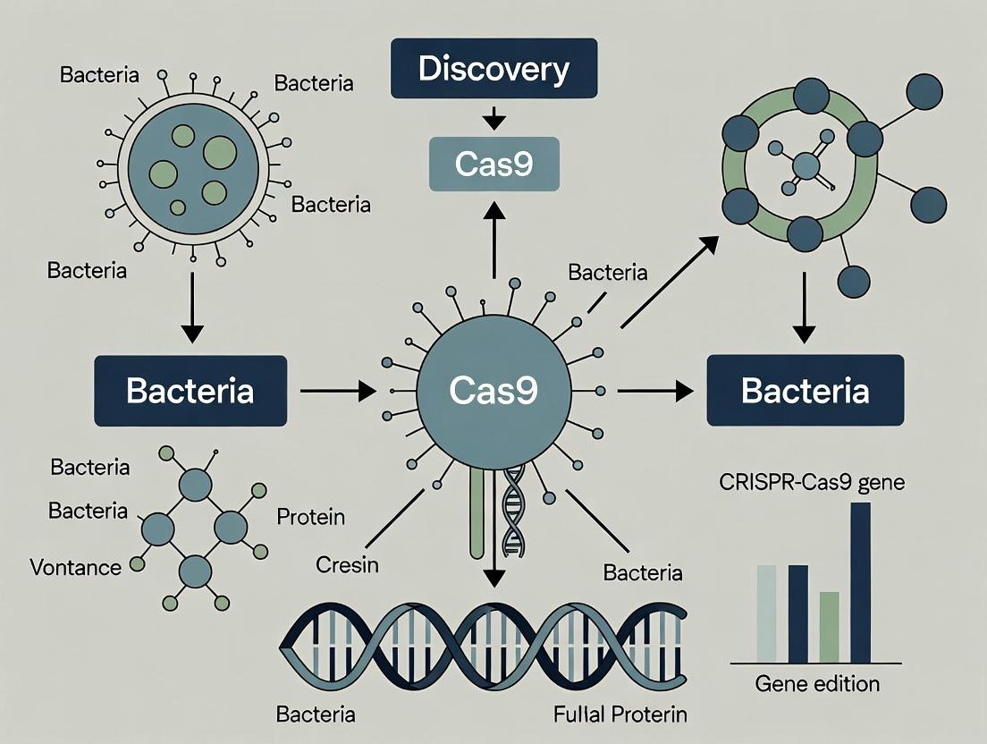
Cas9 Protein in Bacteria: From Natural Immune Function to CRISPR Biotech Revolution
This article provides a comprehensive analysis of the Cas9 protein's discovery and its native function within bacterial adaptive immunity.
Cas9 Protein in Bacteria: From Natural Immune Function to CRISPR Biotech Revolution
Abstract
This article provides a comprehensive analysis of the Cas9 protein's discovery and its native function within bacterial adaptive immunity. Aimed at researchers, scientists, and drug development professionals, it explores the foundational biology of CRISPR-Cas9 systems, details methodological applications in genetic engineering, addresses common experimental challenges and optimization strategies, and validates findings through comparative analysis with other nucleases. The synthesis offers critical insights for harnessing Cas9's potential in therapeutic development and advanced research applications.
The CRISPR-Cas9 Origin Story: Unraveling Bacterial Adaptive Immunity
The discovery of the CRISPR-Cas9 system represents a paradigm shift in molecular biology, originating from fundamental bacterial research. This whitepaper frames the elucidation of this adaptive immune system within the broader thesis of Cas9 protein discovery and function. Initially observed as mysterious, regularly spaced repeats in prokaryotic genomes, these loci were later defined as the cornerstone of a sophisticated defense mechanism against mobile genetic elements. The journey from curiosity-driven observation to a defined molecular machinery underscores the critical role of basic bacterial research in revealing universal biological principles with transformative applications.
Historical Timeline and Key Quantitative Data
Table 1: Historical Milestones in CRISPR-Cas Discovery
| Year | Discovery | Key Researchers/Team | Significance |
|---|---|---|---|
| 1987 | Identification of unusual repetitive DNA in E. coli | Ishino et al. | Initial observation of "clustered regularly interspaced short palindromic repeats" (CRISPR). |
| 2002 | Coining of "CRISPR" and identification of associated (cas) genes | Jansen et al. | Defined the genetic locus and predicted a functional role. |
| 2005 | Spacers derived from phage/plasmid DNA | Three independent groups (Mojica, Pourcel, Bolotin) | Proposed an adaptive immune function based on sequence homology. |
| 2007 | Experimental proof of adaptive immunity in Streptococcus thermophilus | Barrangou et al. | Demonstrated that CRISPR confers resistance to bacteriophages. |
| 2008 | CRISPR targets DNA; Cas9 is the nuclease | Marraffini & Sontheimer; Brouns et al. | Defined DNA as the target and identified Cas9's role in cleavage. |
| 2010 | In vitro reconstitution of Cas9 activity | Deltcheva et al. | Showed tracrRNA is essential for processing pre-crRNA and guiding Cas9. |
| 2012 | Engineering of single-guide RNA (sgRNA) and programmable DNA cleavage | Jinek et al. | Simplified the system to a two-component tool (Cas9 + sgRNA), enabling genome engineering. |
Table 2: Core Quantitative Metrics of the Type II-A CRISPR-Cas9 System from Streptococcus pyogenes (SF370)
| Component | Metric | Value/Description | Functional Implication |
|---|---|---|---|
| Cas9 Protein | Molecular Weight | ~160 kDa | A large, multi-domain endonuclease. |
| Domain Structure | RuvC, HNH, REC, PAM-Interacting | RuvC and HNH cleave target/non-target strands; REC binds RNA; PI domain reads PAM. | |
| CRISPR Array | Repeat Length | 36 bp | Forms hairpin structures critical for processing. |
| Spacer Length | 30 bp (variable) | Provides the sequence-specific memory of past invasions. | |
| PAM (Protospacer Adjacent Motif) | Sequence | 5'-NGG-3' (canonical) | Essential for self vs. non-self discrimination; target site selection. |
| Guide Complex | crRNA:tracrRNA Duplex | ~20 nt + ~42 nt (native) | Directs Cas9 to complementary DNA sequences. |
| sgRNA (engineered) | ~100 nt chimeric RNA | Combines essential portions of crRNA and tracrRNA for simplified application. |
Detailed Experimental Protocols
Protocol 1: Demonstration of CRISPR Adaptive Immunity in Bacteria (Barrangou et al., 2007) Objective: To prove that CRISPR spacers acquired from phage DNA confer resistance to subsequent phage infection.
- Phage Challenge & Survivor Isolation: Infect a culture of S. thermophilus with bacteriophage. Plate on agar to isolate surviving bacterial colonies.
- CRISPR Locus Analysis: Extract genomic DNA from survivors and naive controls. Amplify the CRISPR locus via PCR using primers flanking the array.
- Spacer Sequencing: Clone and sequence PCR products. Compare spacer sequences to the phage genome using BLAST.
- Spacer Acquisition Verification: Identify new spacers in survivor CRISPR arrays that are 100% identical to protospacers in the phage genome used for challenge.
- Gain-of-Function Test: Clone the modified CRISPR locus from a survivor into a naive, phage-sensitive strain via electroporation. Challenge the transformed strain with the same phage. Resistance confirms the CRISPR array alone is sufficient for immunity.
Protocol 2: In Vitro Reconstitution of Cas9 Cleavage Activity (Jinek et al., 2012) Objective: To define the minimal components required for programmable DNA cleavage.
- Component Purification: Express and purify recombinant S. pyogenes Cas9 protein in E. coli. Chemically synthesize mature crRNA and tracrRNA (or a chimeric sgRNA).
- Target DNA Preparation: Generate a linear, double-stranded DNA substrate containing a target sequence (complementary to crRNA) and a correct PAM (5'-NGG-3').
- Ribonucleoprotein (RNP) Complex Formation: Incubate Cas9 protein with crRNA and tracrRNA (or sgRNA) in reaction buffer (20 mM HEPES pH 7.5, 150 mM KCl, 10 mM MgCl2, 5% glycerol) at 37°C for 10 mins.
- Cleavage Reaction: Add the target DNA substrate to the pre-formed RNP complex. Incubate at 37°C for 1 hour.
- Analysis: Run products on an agarose gel. Successful cleavage yields two smaller DNA fragments compared to the uncut control. Include controls lacking Cas9, RNA, or Mg2+ (essential cofactor).
Visualizations
Diagram 1: CRISPR-Cas9 Adaptive Immune Pathway
Diagram 2: Cas9 DNA Cleavage Mechanism
The Scientist's Toolkit: Key Research Reagent Solutions
Table 3: Essential Reagents for CRISPR-Cas9 Research
| Item | Function & Application |
|---|---|
| Recombinant Cas9 Nuclease (wild-type) | Purified protein for in vitro cleavage assays, structural studies, and RNP delivery in genome editing. |
| Cas9 Expression Plasmids | For stable or transient expression of Cas9 in mammalian, plant, or bacterial cells (e.g., pSpCas9, pX系列). |
| sgRNA Cloning Kits & Backbone Vectors | Streamlined systems (e.g., BsaI digestion, Golden Gate assembly) for expressing custom guide RNAs in cells. |
| Synthetic sgRNA (chemically modified) | For direct RNP formation; enhanced stability and reduced immunogenicity in therapeutic contexts. |
| In Vitro Transcription Kits | For high-yield synthesis of sgRNA or tracrRNA/crRNA for biochemical experiments. |
| PAM Discovery Libraries (e.g., SPAMALOT, PAMDA) | Plasmid-based libraries to characterize Cas9 variant PAM specificity. |
| Target DNA Substrates (linearized plasmids/PCR amplicons) | Defined targets for in vitro cleavage efficiency and specificity assays. |
| Next-Generation Sequencing (NGS) Kits for GUIDE-seq, CIRCLE-seq | For genome-wide profiling of off-target effects. |
| Cell Lines with Reporter Assays (e.g., GFP disruption, SURVEYOR) | To quantify editing efficiency and specificity in living cells. |
| Anti-Cas9 Monoclonal Antibodies | For detection, immunoprecipitation (ChIP), and inhibition studies. |
The discovery of the CRISPR-Cas9 system represents a paradigm shift in molecular biology. Within the broader thesis of bacterial adaptive immunity, the identification and functional characterization of the Cas9 protein from Streptococcus pyogenes provided the foundational insight that a single, RNA-guided endonuclease could be programmed for precise DNA cleavage. This whitepaper details the structural and mechanistic principles of Cas9, framing it as the central effector protein that converted a prokaryotic defense mechanism into a universal programmable genetic tool.
Cas9 Protein Architecture
Cas9 is a multidomain protein with distinct functional lobes. The latest structural data (PDB IDs: 4OO8, 5F9R) confirm a bilobed architecture: the Recognition (REC) lobe and the Nuclease (NUC) lobe.
Domain Organization
- REC Lobe: Comprises the REC1, REC2, and REC3 domains. Primarily responsible for sgRNA binding and recognition of the DNA-RNA heteroduplex.
- NUC Lobe: Contains the two nuclease domains (HNH and RuvC-like) and the C-terminal domain (CTD). The CTD includes the Protospacer Adjacent Motif (PAM) interaction site.
Table 1: Key Structural Domains of S. pyogenes Cas9 (SpCas9)
| Domain/Lobe | Primary Function | Key Structural Features |
|---|---|---|
| REC Lobe | sgRNA & DNA-RNA heteroduplex binding, conformational activation | Helical bundle; arginine-rich bridge helix (REC3) |
| HNH Domain | Cleaves the target DNA strand (complementary to crRNA) | ββα-metal fold; requires Mg²⁺ |
| RuvC Domain | Cleaves the non-target DNA strand | RNase H-like fold; requires Mg²⁺ |
| PAM-Interacting (PI) Domain | Binds to the 5'-NGG-3' PAM sequence in target DNA | Contains a PAM-interacting β-sheet and loop motifs |
| Linker Regions | Enable large conformational changes | Flexible hinges between lobes |
Diagram 1: Cas9 protein domain architecture.
Catalytic Mechanism of DNA Cleavage
Cas9 functions as a monomeric endonuclease that introduces a blunt-ended, double-strand break (DSB) 3 bp upstream of the PAM site. The mechanism is a sequential, conformationally driven process.
Table 2: Key Quantitative Parameters of SpCas9 Catalysis
| Parameter | Value | Experimental Basis (Typical Assay) |
|---|---|---|
| PAM Sequence | 5'-NGG-3' (canonical) | In vitro SELEX or plasmid cleavage assays |
| Cleavage Position | 3 bp upstream of PAM | DNA sequencing of cleavage products |
| Kₘ (DNA substrate) | ~0.5 - 5 nM | Steady-state kinetics (FRET-based cleavage) |
| kₐₜ (turnover) | ~0.01 - 0.1 s⁻¹ | Pre-steady-state kinetic analysis |
| Mg²⁺ Requirement | Essential (0.5-10 mM) | EDTA inhibition; restoration by Mg²⁺ |
| Optimal Temperature | 37°C | In vitro activity assays |
Step-by-Step Mechanism
- PAM Recognition & DNA Binding: The PI domain scans dsDNA for the correct PAM (NGG). PAM binding initiates local DNA melting.
- R-Loop Formation: The crRNA guide sequence invades the DNA duplex, base-pairing with the target strand (complementary strand). This displaces the non-target strand, forming an R-loop structure.
- Conformational Activation: Successful R-loop formation triggers a large conformational change in the REC lobe, repositioning the HNH domain.
- Strand-Specific Cleavage:
- The HNH domain rotates into position and cleaves the target DNA strand.
- The RuvC domain, now positioned adjacent to the displaced non-target strand, cleaves it.
Diagram 2: Cas9 catalytic mechanism steps.
Experimental Protocols for Key Assays
1In VitroCleavage Assay (Gel-Based)
Purpose: To validate Cas9-sgRNA ribonucleoprotein (RNP) activity and specificity. Protocol:
- RNP Formation: Incubate purified Cas9 protein (100 nM) with synthetic sgRNA (120 nM) in cleavage buffer (20 mM HEPES pH 7.5, 150 mM KCl, 10 mM MgCl₂, 5% glycerol) at 25°C for 10 min.
- Reaction Initiation: Add linearized plasmid or PCR-amplified DNA substrate (10 nM) containing the target sequence and PAM.
- Incubation: Incubate at 37°C for 30-60 minutes.
- Reaction Stop: Add Proteinase K (0.2 mg/mL) and SDS (0.1%) and incubate at 56°C for 15 min.
- Analysis: Run products on a 1% agarose gel stained with ethidium bromide. Cleavage is indicated by the conversion of a supercoiled/linear substrate band into two smaller fragments.
Kinetic Analysis Using Single-Turnover FRET
Purpose: To determine the catalytic rate constant (kₐₜ) under pre-steady-state conditions. Protocol:
- Substrate: Use a dsDNA oligonucleotide labeled with a fluorophore (e.g., Cy3) on one end and a quencher (e.g., Iowa Black) on the other, spanning the cleavage site.
- RNP Pre-formation: Prepare Cas9-sgRNA RNP at 500 nM in cleavage buffer without Mg²⁺.
- Rapid Kinetics: Use a stopped-flow apparatus. Load one syringe with RNP complex (final 100 nM after mixing). Load the second with DNA substrate (final 10 nM) and MgCl₂ (final 10 mM) to initiate the reaction.
- Data Acquisition: Monitor fluorescence increase (due to cleavage and fluorophore separation from quencher) over time (0.1-100 s). Fit the time-course data to a single-exponential equation to obtain the observed rate constant (kₒbₛ).
The Scientist's Toolkit: Research Reagent Solutions
Table 3: Essential Reagents for Cas9 Structural & Mechanistic Studies
| Reagent/Kit | Provider Examples | Function in Research |
|---|---|---|
| Recombinant Cas9 Nuclease (Wild-type & mutants) | Thermo Fisher, NEB, Origene | Purified protein for in vitro assays, structural studies, and RNP formation. |
| Custom sgRNA Synthesis Kit | IDT, Synthego, Trilink | High-quality, chemically modified sgRNAs for enhanced stability and specificity in RNP experiments. |
| Fluorescent dNTPs/Quencher Probes | Jena Bioscience, Lumiprobe | For constructing FRET-based DNA substrates to monitor cleavage kinetics in real-time. |
| Surface Plasmon Resonance (SPR) Chips (e.g., SA) | Cytiva, Bruker | Immobilize biotinylated DNA or RNA to measure binding kinetics and affinity (K_D) of Cas9 RNP. |
| Cryo-EM Grids & Vitrification System | Quantifoil, Thermo Fisher | Prepare frozen-hydrated samples of Cas9-DNA-RNA complexes for high-resolution structural determination. |
| Mg²⁺/Mn²⁺ Chelator Resins | Chelex, Sigma-Aldrich | To create metal-free buffers for controlled restoration experiments, proving metal ion cofactor requirements. |
The systematic elucidation of CRISPR-Cas9 function in bacterial adaptive immunity stands as a cornerstone of modern molecular biology. Framed within the broader thesis of Cas9 protein discovery and function in bacteria, this guide details the seminal studies that deconstructed this prokaryotic defense system, paving the way for its revolutionary application as a genome engineering tool.
Landmark Papers and Experimental Protocols
1. Jansen et al. (2002) – Naming the System
- Thesis Context: The foundational paper that identified and named the CRISPR loci and associated cas genes, providing the genomic scaffold for all subsequent functional studies on Cas9.
- Protocol: Comparative genomic analysis.
- Method: Performed BLAST analysis of then-available microbial genomes to identify conserved repeated sequences (direct repeats).
- Key Step: Clustered these repeats and searched for conserved open reading frames (ORFs) in their vicinity.
- Analysis: Coined the acronym "CRISPR" and identified the first four cas gene families (cas1 to cas4).
2. Barrangou et al. (2007) – Demonstrating Adaptive Immunity
- Thesis Context: Provided the first experimental proof that the CRISPR-Cas system confers adaptive immunity against phages in bacteria.
- Protocol: Phage resistance assay in Streptococcus thermophilus.
- Method: Challenged bacterial strains with virulent phages and isolated resistant mutants.
- Key Step: Sequenced the CRISPR loci of parental and phage-resistant strains.
- Analysis: Showed that new spacers derived from phage genomic sequences were added to the CRISPR array, correlating with resistance.
3. Garneau et al. (2010) – Defining the Interference Mechanism
- Thesis Context: Elucidated the precise mechanism of CRISPR-mediated DNA cleavage, showing it targets and degrades invasive DNA.
- Protocol: In vitro DNA cleavage assay.
- Method: Purified the Cas9 (formerly Csn1) protein from S. thermophilus.
- Key Step: Incubated Cas9 with CRISPR RNA (crRNA) transcripts and target plasmid DNA containing a protospacer sequence.
- Analysis: Used gel electrophoresis to demonstrate sequence-specific, double-stranded DNA cleavage, dependent on the crRNA and a protospacer adjacent motif (PAM).
4. Jinek et al. (2012) – Re-engineering for Programmability
- Thesis Context: A pivotal study in applied thesis, demonstrating the reconstitution of a minimal two-component system (Cas9 + single guide RNA) and its programmability for in vitro DNA targeting.
- Protocol: In vitro reconstitution and cleavage.
- Method: Expressed and purified recombinant Streptococcus pyogenes Cas9 protein. Chemically synthesized tracrRNA and crRNA (later fused into a single-guide RNA, sgRNA).
- Key Step: Assembled ribonucleoprotein (RNP) complexes and tested cleavage on linear DNA substrates.
- Analysis: Mapped cleavage sites, confirmed PAM requirement (5'-NGG), and proved the fusion of tracrRNA:crRNA into sgRNA retained function.
Table 1: Key Parameters from Foundational Cas9 Studies
| Study (First Author, Year) | System / Organism | Key Quantitative Finding | Measured Outcome |
|---|---|---|---|
| Barrangou, 2007 | S. thermophilus DGCC7710 | Spacer acquisition frequency: ~10^-6 to 10^-7 per cell per generation. | Phage resistance efficiency |
| Garneau, 2010 | S. thermophilus Cas9 | Cleavage occurred 3 bp upstream of the PAM. | DNA cleavage site position |
| Jinek, 2012 | S. pyogenes Cas9 | Optimal in vitro cleavage temperature: 37°C; Time: 1 hour. | Reaction efficiency |
| Deltcheva, 2011 | S. pyogenes | Identified a 75-nucleotide tracrRNA and 39-42 nt crRNA intermediates. | RNA processing product sizes |
Visualizing the Discovery Pathway
Title: Cas9 Discovery Timeline
Title: Bacterial CRISPR-Cas9 Immune Pathway
The Scientist's Toolkit: Key Research Reagent Solutions
Table 2: Essential Reagents for Cas9 Function Studies
| Reagent / Material | Function in Research | Example from Landmark Studies |
|---|---|---|
| High-Efficiency Competent Cells | For generating phage-resistant mutants or cloning CRISPR constructs. | S. thermophilus strains used in Barrangou et al. (2007). |
| Phage Lysate / Genomic DNA | Source of protospacers for spacer acquisition assays and target DNA for cleavage assays. | Virulent phages 858, 2972, etc., used as selective pressure. |
| Cas9 Expression Vectors | Recombinant production of His-tagged or other affinity-tagged Cas9 protein for purification. | pET-based plasmids expressing S. pyogenes Cas9 in Jinek et al. (2012). |
| T7 RNA Polymerase Kit | For in vitro transcription of crRNA, tracrRNA, and sgRNA molecules. | Used to generate guide RNAs for in vitro cleavage assays. |
| Nuclease-Free Buffers & ATP | Essential for maintaining RNA integrity and providing energy for Cas enzyme activities. | Used in Garneau et al. (2010) in vitro cleavage reactions. |
| Ni-NTA Agarose Resin | Immobilized metal affinity chromatography for purifying polyhistidine-tagged Cas9 protein. | Standard for purifying recombinant Cas9 from E. coli lysates. |
| Synth. Oligos & Cloning Kits | For constructing plasmid targets with specific protospacers and PAMs. | Key for creating tailored DNA substrates for cleavage assays. |
| Agarose Gel Electrophoresis System | Standard method for analyzing DNA cleavage products and verifying spacer acquisition. | Used in all cited studies to visualize DNA fragmentation or PCR products. |
The discovery of the Cas9 protein and its function represents a paradigm shift in molecular biology, extending far beyond its origins as a bacterial adaptive immune system. This whitepaper positions the discovery of Cas9 within the broader thesis of bacterial research, wherein understanding the natural function and diversity of CRISPR-Cas systems is fundamental to repurposing them as programmable genomic tools. Cas9, the hallmark nuclease of Type II systems, is just one component in a vast array of CRISPR-Cas architectures. Appreciating its evolutionary context—where it fits within the classification of systems and how its mechanism compares to others—is critical for researchers and drug development professionals aiming to exploit, engineer, or inhibit these systems for therapeutic and biotechnological applications.
Classification and Diversity of CRISPR-Cas Systems
CRISPR-Cas systems are broadly divided into two classes based on the architecture of their effector complexes.
- Class 1 (Types I, III, and IV): Utilize multi-subunit effector complexes for target interference. These systems are more common in bacteria and archaea but are less frequently harnessed for biotechnology due to their complexity.
- Class 2 (Types II, V, and VI): Employ a single, large effector protein for interference. This simplicity has made them the foundation for genome-editing tools. Type II systems use the Cas9 protein, Type V systems use Cas12-family proteins (e.g., Cas12a/Cpf1), and Type VI systems use Cas13-family proteins which target RNA.
Table 1: Core Characteristics of Major CRISPR-Cas Types
| Feature | Type II (Class 2) | Type V (Class 2) | Type I (Class 1) | Type III (Class 1) |
|---|---|---|---|---|
| Signature Protein | Cas9 | Cas12 (e.g., Cas12a) | Cascade complex (Cas3) | Csm/Cmr complex |
| Target Molecule | DNA | DNA | DNA | DNA/RNA |
| Pre-crRNA Processing | Requires tracrRNA & RNase III | Self-processes pre-crRNA | Requires Cas6 | Requires Cas6 |
| Cleavage Mechanism | Blunt ends, dual HNH & RuvC nickases | Staggered ends, single RuvC-like nuclease | Unwinds DNA, recruits Cas3 helicase/nuclease | Cleaves DNA/RNA via Cas7 subunits |
| PAM Requirement | Yes (3′-NGG for SpCas9) | Yes (5′-TTTV for AsCas12a) | Yes (specific to subtype) | Not strictly required |
| Key Biotech Application | Genome editing (HDR/NHEJ) | Genome editing, DNA detection | Large deletions, antimicrobials | RNA targeting, antiviral |
Type II System Mechanism: The Cas9 Paradigm
The function of the Type II system in bacterial immunity involves three key stages, with Cas9 central to the interference stage.
Stage 1: Adaptation The Cas1-Cas2 integrase complex captures short fragments of invading DNA (protospacers) and inserts them as new spacers into the CRISPR array. This immunizes the host against future infection.
Stage 2: Expression & Processing The CRISPR array is transcribed into a long pre-crRNA. In Type II systems, a second small RNA, the trans-activating CRISPR RNA (tracrRNA), is essential. The tracrRNA base-pairs with the repeat regions of the pre-crRNA, and the duplex is cleaved by RNase III in the presence of Cas9 to generate mature crRNA:tracrRNA duplexes.
Stage 3: Interference (Cas9 Function) The mature crRNA:tracrRNA duplex (or a engineered single-guide RNA, sgRNA) assembles with Cas9. This ribonucleoprotein (RNP) complex surveils the cell for DNA sequences complementary to the crRNA spacer. Binding requires the presence of a short Protospacer Adjacent Motif (PAM) adjacent to the target sequence. Upon recognition, Cas9 undergoes a conformational change, activating its two nuclease domains (HNH and RuvC-like). The HNH domain cleaves the DNA strand complementary to the crRNA (target strand), while the RuvC-like domain cleaves the opposite strand (non-target strand), generating a double-strand break (DSB).
Diagram 1: Type II CRISPR-Cas Adaptive Immunity Pathway
Key Experimental Protocol: Validating Cas9In VitroCleavage
To study and validate the function of a newly discovered or engineered Cas9 ortholog, an in vitro cleavage assay is fundamental.
Protocol: In Vitro DNA Cleavage Assay
Objective: To confirm the nuclease activity, guide RNA specificity, and PAM requirement of a purified Cas9 protein.
Reagents & Materials:
- Purified Cas9 Protein: Recombinantly expressed and purified.
- Target DNA Plasmid: A circular plasmid containing a target sequence with a suspected PAM.
- Control DNA Plasmid: A plasmid lacking the target sequence or PAM.
- Guide RNA: Synthesized crRNA:tracrRNA duplex or sgRNA complementary to the target.
- Reaction Buffer: Typically containing Tris-HCl, NaCl, MgCl₂ (essential for nuclease activity), and DTT.
- Proteinase K: To stop the reaction.
- Agarose Gel Electrophoresis System: For analyzing cleavage products.
Procedure:
- Prepare Reaction Mixes: Set up 20 µL reactions containing 1x reaction buffer, 100 ng of target or control plasmid, 100 nM Cas9 protein, and 120 nM guide RNA. Include controls without protein, without guide RNA, and with a non-targeting guide RNA.
- Incubation: Incubate reactions at 37°C (or the optimal temperature for the specific Cas9) for 1 hour.
- Reaction Termination: Add Proteinase K and SDS to final concentrations of 0.6 µg/µL and 0.1% respectively. Incubate at 56°C for 10 minutes to digest Cas9.
- Analysis: Load the products onto a 1% agarose gel stained with ethidium bromide. Run the gel and visualize under UV light.
- Interpretation: Successful cleavage converts supercoiled plasmid (Form I) into a linearized product (Form III). Non-specific cleavage or the presence of nicks will produce open circular DNA (Form II). Specificity is confirmed by cleavage only in the presence of the correct guide RNA and target+PAM plasmid.
Diagram 2: In Vitro Cas9 Cleavage Assay Workflow
The Scientist's Toolkit: Key Research Reagent Solutions
Table 2: Essential Reagents for CRISPR-Cas9 Research
| Research Reagent | Function & Application | Key Considerations |
|---|---|---|
| Recombinant Cas9 Nuclease (Wild-type) | In vitro cleavage assays, biochemical characterization, RNP delivery for genome editing. | High purity and nuclease activity are critical. Source (bacterial, human cells) affects modification state. |
| Cas9-D10A or H840A Nickase Mutants | Generate single-strand breaks (nicks) for base editing or to reduce off-target effects in paired-nickase strategies. | Must be validated for loss of one nuclease activity while retaining DNA binding. |
| Catalytically Dead Cas9 (dCas9) | Binds DNA without cleavage. Used for transcriptional repression/aactivation (CRISPRi/a), epigenetic editing, and live imaging. | Fusion to effector domains (e.g., VP64, KRAB) is common. PAM specificity remains. |
| Chemically Modified Synthetic sgRNAs | Enhanced stability and reduced immunogenicity for therapeutic applications (e.g., in vivo editing). | Common modifications: 2′-O-methyl, phosphorothioate backbones. Must maintain Cas9 binding affinity. |
| PAM Library Plasmids | High-throughput determination of Cas9 ortholog PAM specificity using in vivo selection or in vitro display. | Essential for characterizing novel or engineered Cas9 variants. |
| Off-Target Prediction Software & Validation Kits | Predict potential off-target sites (e.g., using GUIDE-seq or CIRCLE-seq algorithms) and validate editing fidelity. | Crucial for therapeutic development. Kits often include optimized PCR and NGS protocols. |
| Cas9-Specific Monoclonal Antibodies | Detection of Cas9 expression (Western blot, ELISA, immunofluorescence), immunoprecipitation of Cas9 complexes. | Important for quality control and mechanistic studies. Should recognize denatured and native protein. |
Harnessing Cas9: Experimental Protocols and Research Applications
The discovery of the Cas9 endonuclease within bacterial adaptive immune systems (CRISPR) has revolutionized genetic engineering. The core thesis underpinning this guide is that the native function of Cas9 in bacteria—a programmable DNA-targeting complex guided by RNA for precise phage defense—directly informs its modern applications. This whitepaper details the three essential technical components derived from this biological principle: the design of guide RNA (gRNA), the delivery of the Cas9 machinery, and the provision of repair templates for desired edits.
gRNA Design: Principles and Parameters
Effective gRNA design is critical for maximizing on-target cleavage efficiency and minimizing off-target effects. Key quantitative parameters are summarized below.
Table 1: Key Quantitative Parameters for gRNA Design
| Parameter | Optimal Value/Range | Impact & Rationale |
|---|---|---|
| GC Content | 40-60% | Influences stability and binding efficiency; low GC reduces specificity, high GC may increase off-targeting. |
| On-Target Efficiency Score | >60 (tool-dependent) | Predictive score from algorithms (e.g., Doench et al. 2016 rules) for likely cleavage activity. |
| Off-Target Mismatch Tolerance | ≤3 mismatches in seed region (PAM-proximal 8-12 bases) | Mismatches in the seed region dramatically reduce cleavage; distal mismatches are more tolerated. |
| Specificity (Number of Genomic Off-Target Sites) | Aim for 0-5 sites with ≤3 mismatches | Minimizing predicted off-target sites is essential for precise editing. |
| Poly-T Sequences | Avoid | Four consecutive T's can act as a termination signal for RNA Pol III promoters (e.g., U6). |
Experimental Protocol: In Silico gRNA Design and Validation
- Target Selection: Identify the genomic locus of interest. For gene knockouts, target early exons; for precise editing, target within 10 bp of the desired edit site.
- gRNA Candidate Generation: Use design tools (e.g., CRISPick, CHOPCHOP) with the appropriate reference genome. Input the sequence flanking the target. The tool will output all possible gRNAs with their associated PAM (e.g., 5'-NGG-3' for SpCas9).
- Filtering by Efficiency and Specificity: Filter candidates using the parameters in Table 1. Prioritize gRNAs with high on-target scores and the fewest predicted off-target sites with ≤3 mismatches.
- Experimental Validation (Essential): a. Cloning: Clone the top 3-4 gRNA sequences into a plasmid containing the Cas9 nuclease and a selection marker. b. Delivery: Transfect the constructs into a relevant cell line. c. Assessment: After 48-72 hours, harvest genomic DNA. Assess editing efficiency via T7 Endonuclease I (T7EI) assay or tracking of indels by decomposition (TIDE) analysis on PCR products spanning the target site.
Cas9 Delivery: Methods and Efficiencies
Delivery modality profoundly impacts editing outcomes, toxicity, and applicability. Quantitative data on common methods are tabled below.
Table 2: Comparison of Cas9 Delivery Methods
| Method | Typical Delivery Efficiency (in Vitro) | Cargo Capacity | Key Advantages | Key Limitations |
|---|---|---|---|---|
| Plasmid DNA Transfection | 20-80% (cell-type dependent) | High (Cas9 + gRNA + template) | Low cost, stable expression, easy to produce. | Risk of random integration, prolonged Cas9 expression increases off-targets. |
| RNP (Ribonucleoprotein) Electroporation | 70-95% in immune cells, stem cells | Low (pre-complexed Cas9 protein + gRNA) | Rapid action, reduced off-targets, no DNA integration. | Technically demanding, transient activity, high cost for protein. |
| Lentiviral Vector | >90% (dividing cells) | Moderate (Cas9 + gRNA) | High efficiency in hard-to-transfect cells, stable expression. | Smaller cargo limit vs. plasmid, risk of insertional mutagenesis, biosafety level 2. |
| AAV (Adeno-Associated Virus) | Variable by serotype | Very Low (<4.7 kb) | Low immunogenicity, high in vivo delivery efficiency to specific tissues. | Extremely limited cargo size (requires split Cas9 systems), potential pre-existing immunity. |
Experimental Protocol: RNP Delivery via Electroporation for Primary T Cells
This protocol exemplifies a high-efficiency, low-off-target delivery method critical for therapeutic applications like CAR-T engineering.
- RNP Complex Formation: Recombinant Cas9 protein (e.g., SpyFi Cas9) is complexed with synthetic, chemically modified crRNA:tracrRNA duplex at a molar ratio of 1:2 (Cas9:gRNA). Incubate at 25°C for 10-20 minutes to form active RNP complexes.
- Cell Preparation: Isolate and activate primary human T cells. Wash and resuspend cells in electroporation buffer (e.g., P3 buffer) at a concentration of 1-2 x 10^8 cells/mL.
- Electroporation: Mix 10 µL of cell suspension with 2-5 µL of RNP complex (at 60 µM) in a 16-well electroporation cuvette. Electroporate using a 4D-Nucleofector (pulse code EH-115 for T cells). Immediately add pre-warmed medium.
- Post-Transfection Culture: Transfer cells to culture plates. Assess viability and editing efficiency after 3-5 days via flow cytometry (if a reporter is disrupted) or NGS of the target locus.
Repair Template Design for HDR
Precise editing requires a donor DNA template to direct homology-directed repair (HDR). Design is critical for efficiency.
Table 3: Design Parameters for HDR Repair Templates
| Parameter | Recommendation | Rationale |
|---|---|---|
| Template Form | Single-stranded oligodeoxynucleotide (ssODN) for point edits; double-stranded DNA (dsDNA) for large inserts. | ssODNs are efficient for <100 bp edits; dsDNA donors (plasmid, PCR product) are needed for larger inserts. |
| Homology Arm Length | ssODN: 50-90 bp total (25-45 bp each arm). dsDNA: 500-1000 bp each arm. | Longer arms increase HDR efficiency but are harder to synthesize. Optimal ssODN arms balance efficiency and cost. |
| Symmetry | Place desired edit asymmetrically relative to the Cas9 cut site. | Prevents re-cutting of the successfully edited allele, enriching for HDR-modified cells. |
| Modifications | Incorporate silent mutations in the PAM or seed sequence of the template. | Prevents Cas9 from binding and cleaving the newly integrated template DNA. |
Experimental Protocol: HDR for Introducing a Point Mutation via ssODN
- Design ssODN: For a point mutation, design a single-stranded DNA oligo with the desired mutation flanked by homology arms (e.g., 40 bp each). Phosphorothioate modifications on the 5' and 3' ends enhance stability.
- Co-Delivery: Co-deliver the Cas9/gRNA (as plasmid, mRNA, or RNP) with the ssODN repair template. For RNP delivery, include the ssODN in the electroporation mixture at a final concentration 5-10x higher than the RNP (e.g., 2 µM RNP, 10-20 µM ssODN).
- Enrichment and Screening: If no selection marker is used, culture cells for 5-7 days to allow fixation of edits. Screen populations via targeted PCR followed by Sanger sequencing and decomposition analysis (TIDE) or deep sequencing to quantify HDR efficiency.
The Scientist's Toolkit: Research Reagent Solutions
Table 4: Essential Materials for CRISPR-Cas9 Genome Editing
| Item | Function & Key Feature |
|---|---|
| SpyFi Cas9 Nuclease (High Fidelity) | Engineered version of S. pyogenes Cas9 with reduced off-target effects while maintaining high on-target activity. Essential for sensitive applications. |
| Chemically Modified Synthetic gRNA (2-part crRNA:tracrRNA) | Provides increased nuclease stability and reduced immunogenicity compared to in vitro transcribed gRNA, especially for RNP delivery. |
| T7 Endonuclease I (T7EI) | Enzyme used in the mismatch detection assay to quickly estimate indel formation efficiency at a target locus without sequencing. |
| Lipofectamine CRISPRMAX Transfection Reagent | A lipid-based formulation optimized for the delivery of CRISPR-Cas9 plasmids, RNPs, or ribonucleoprotein complexes into a wide range of mammalian cell lines. |
| NEBuilder HiFi DNA Assembly Master Mix | For seamless cloning of gRNA sequences into expression vectors or assembly of large dsDNA repair templates from PCR fragments. |
| KAPA HiFi HotStart ReadyMix | High-fidelity PCR enzyme for amplifying genomic regions around target sites for sequencing analysis and for generating dsDNA donor templates. |
| Nucleofector Kit for Primary Cells | Cell-type specific kits containing optimized buffers and protocols for high-efficiency RNP electroporation into primary and hard-to-transfect cells. |
The seminal discovery of the Cas9 protein as an adaptive immune effector in Streptococcus pyogenes revolutionized our understanding of bacterial defense. This foundational research, which detailed how CRISPR-Cas9 systems cleave invasive nucleic acids in vivo within the bacterial cell, provided the mechanistic blueprint for repurposing this molecular machinery. The core thesis of Cas9 function—a programmable endonuclease guided by RNA—bridges directly to its bifurcated application today: as a purified tool for precise in vitro reactions and as an engineered vector for complex in vivo cellular editing. This guide delineates the technical paradigms, from controlled bench-top cleavage to the dynamic challenges of intracellular genome engineering.
Core Comparative Analysis: In Vitro vs. In Vivo Applications
The utility of CRISPR-Cas9 diverges fundamentally based on the environment of use, impacting design, delivery, outcome, and analysis.
Table 1: Fundamental Comparison of CRISPR-Cas9 Applications
| Parameter | In Vitro Applications | In Vivo (Cellular) Applications |
|---|---|---|
| Primary Environment | Cell-free, controlled buffer system. | Within living cells (cultured cells, tissues, organisms). |
| Key Components | Purified Cas9 protein, synthetic sgRNA, target DNA substrate. | Delivery vehicle (e.g., plasmid, RNP), cellular machinery, genomic DNA. |
| Main Objective | High-specificity DNA cleavage, genotyping, cloning, NGS library prep. | Heritable genomic modification (KO, KI, correction), transcriptional regulation. |
| Delivery Challenge | None (components mixed directly). | Major hurdle (viral, physical, or chemical methods required). |
| Off-Target Assessment | Direct sequencing of reaction products (precise). | Complex (requires whole-genome sequencing methods like GUIDE-seq). |
| Throughput | Very high for target validation. | Lower, limited by delivery and cell viability. |
| Key Advantage | Precision, control, lack of cellular confounding factors. | Physiological relevance, study of functional genomics and therapeutic potential. |
Table 2: Quantitative Performance Metrics
| Metric | Typical In Vitro Efficiency | Typical In Vivo (Mammalian Cell) Efficiency | Measurement Method |
|---|---|---|---|
| Cleavage/Knockout Efficiency | >90% (of input substrate) | 20-80% (varies by cell type, locus, delivery) | Gel electrophoresis / T7E1 assay; NGS, flow cytometry. |
| Off-Target Cleavage Rate | Very low with high-fidelity Cas9 variants. | Can be significant; requires rigorous profiling. | NGS of predicted sites or unbiased methods (GUIDE-seq, CIRCLE-seq). |
| Turnaround Time (Core Reaction) | 1-3 hours. | Days to weeks (including delivery, expansion, analysis). | - |
| Optimal sgRNA Length | 17-20 nt (tolerant of truncation). | Strictly 20 nt (for SpCas9). | - |
Experimental Protocols
Protocol 1: In Vitro Cleavage Assay for sgRNA Validation
Purpose: To verify the activity and specificity of synthesized sgRNAs before costly cellular experiments.
- Reagent Assembly: In a nuclease-free microtube, combine:
- 1 µg of purified, linear target DNA (200-1500 bp).
- 100-200 nM purified recombinant Cas9 protein (e.g., SpCas9).
- 200-400 nM synthetic sgRNA (full-length, chemically modified).
- 1X Cas9 reaction buffer (typically: 20 mM HEPES, 100 mM KCl, 5 mM MgCl2, 1 mM DTT, pH 7.5).
- Nuclease-free water to 20 µL.
- Incubation: Mix gently and incubate at 37°C for 1 hour.
- Reaction Termination: Add 2 µL of Proteinase K (10 mg/mL) and 1 µL of 10% SDS. Incubate at 56°C for 10 minutes.
- Analysis: Run the entire product on a 1.5-2% agarose/TAE gel stained with ethidium bromide. Successful cleavage yields two distinct bands smaller than the uncut control.
Protocol 2: Ribonucleoprotein (RNP) Delivery for In Vivo Knockout in Mammalian Cells
Purpose: High-efficiency, transient delivery of CRISPR-Cas9 for gene knockout via non-homologous end joining (NHEJ).
- RNP Complex Formation:
- Resuspend synthetic crRNA and tracrRNA to 100 µM in duplex buffer. Anneal equimolar amounts (95°C for 5 min, ramp down to 25°C) to form sgRNA.
- Mix 6 µL of 10 µM sgRNA with 4 µL of 10 µM purified Cas9 protein (final 3 µL of 20 µM RNP).
- Incubate at room temperature for 10-20 minutes.
- Cell Preparation: Harvest and count HEK293T or other adherent cells. Resuspend in electroporation buffer (e.g., Neon or SE Cell Line 4D-Nucleofector buffer) at 1x10⁶ cells/20 µL.
- Electroporation: Combine 20 µL cell suspension with 3 µL RNP complex. Transfer to a certified cuvette. Electroporate using a cell-type-optimized protocol (e.g., 1350V, 30ms, 1 pulse for HEK293T with Neon).
- Recovery & Analysis: Immediately transfer cells to pre-warmed culture medium. After 72 hours, harvest cells for genomic DNA extraction. Assess editing efficiency via T7 Endonuclease I (T7E1) assay or targeted deep sequencing.
Visualizations
Title: In Vitro Cleavage Assay Workflow
Title: Key Pathways in Cellular CRISPR Editing
The Scientist's Toolkit: Essential Research Reagent Solutions
Table 3: Core Reagents for CRISPR-Cas9 Experiments
| Reagent | Function & Key Characteristics | Typical Application |
|---|---|---|
| Recombinant HiFi Cas9 | High-fidelity mutant (e.g., SpCas9-HF1) with reduced off-target activity. | Both in vitro and in vivo where specificity is critical. |
| Chemically Modified sgRNA | Synthetic sgRNA with 2'-O-methyl and phosphorothioate modifications for stability. | In vivo RNP delivery; enhances resistance to nucleases. |
| Electroporation/Transfection Reagents | Specialized buffers and devices for physical delivery (e.g., Neon System, Lipofectamine CRISPRMAX). | In vivo delivery of RNP or plasmid to hard-to-transfect cells. |
| T7 Endonuclease I (T7E1) | Enzyme that cleaves mismatched heteroduplex DNA. | Initial, low-cost validation of in vivo editing efficiency. |
| Donor DNA Template | Single-stranded oligodeoxynucleotide (ssODN) or double-stranded DNA vector for HDR. | In vivo precise knock-in or point mutation correction. |
| GUIDE-seq or CIRCLE-seq Kit | Comprehensive kits for unbiased genome-wide off-target profiling. | Critical safety assessment for therapeutic in vivo applications. |
| Next-Generation Sequencing (NGS) Library Prep Kit for Amplicons | Enables deep sequencing of PCR-amplified target loci. | Gold-standard quantitative measurement of editing efficiency and outcome analysis. |
The discovery of the Cas9 endonuclease in bacterial adaptive immunity (CRISPR-Cas) systems revolutionized genome engineering. The foundational thesis of Cas9 function—a programmable RNA-guided DNA cleaver—provided the conceptual framework for a suite of transformative derivatives. By moving beyond the creation of double-strand breaks (DSBs) and their error-prone repair, these tools—dCas9, Base Editors, and Prime Editors—offer precise, efficient, and versatile manipulation of genetic information, directly addressing limitations inherent in the wild-type protein's activity.
Catalytically Dead Cas9 (dCas9): The Programmable Scaffold
Core Principle: Mutation of the two catalytic residues (D10A in RuvC and H840A in HNH domains) in Streptococcus pyogenes Cas9 abolishes its endonuclease activity while preserving its ability to bind DNA in an RNA-programmed manner. This creates a versatile DNA-targeting platform.
Key Applications & Research Reagent Solutions:
| Reagent/Material | Function in Research |
|---|---|
| dCas9 Expression Vector | Delivery vehicle for the catalytically inactive protein. |
| sgRNA Scaffold | Guides dCas9 to the specific genomic locus. |
| dCas9-Effector Fusion Constructs | dCas9 linked to transcriptional activators (e.g., VP64, p65AD), repressors (e.g., KRAB), or epigenetic modifiers (e.g., DNMT3A, TET1). |
| Fluorescent Protein-dCas9 Fusions | For live imaging of genomic loci (e.g., dCas9-EGFP). |
Experimental Protocol: dCas9-Mediated Transcriptional Repression (CRISPRi)
- Design: Design sgRNAs to target the promoter or early exon of the gene of interest.
- Cloning: Clone sgRNA sequences into an appropriate expression plasmid.
- Delivery: Co-transfect a mammalian cell line with plasmids expressing dCas9-KRAB (a potent repressor domain) and the sgRNA(s).
- Analysis: After 48-72 hours, harvest cells. Quantify gene knockdown using RT-qPCR (mRNA level) or western blot (protein level). Compare to cells expressing dCas9-KRAB with a non-targeting control sgRNA.
Base Editors (BEs): Precise Chemical Conversion
Core Principle: Base Editors are fusion proteins of dCas9 (or a nickase variant, nCas9) with a nucleobase deaminase enzyme. They mediate direct, irreversible chemical conversion of one base pair to another without requiring a DSB or a donor DNA template.
Types and Quantitative Performance Data:
| Editor Type | Deaminase | Catalytic Core | Conversion | Typical Efficiency* | Primary Byproducts & Limitations |
|---|---|---|---|---|---|
| Cytosine Base Editor (CBE) | APOBEC1 | nCas9 (D10A) | C•G to T•A | 15-50% | Indels, unwanted C edits within window. |
| Adenine Base Editor (ABE) | TadA* | nCas9 (D10A) | A•T to G•C | 20-50% | Lower efficiency for some A positions. |
| Dual Base Editor | e.g., CGBE, A&C-BEmax | nCas9 | C•G to G•C, A•T to G•C | 10-40% | Broader edit profiles require careful characterization. |
*Efficiencies are highly context-dependent and vary by cell type and delivery method.
Experimental Protocol: Base Editing in Cultured Mammalian Cells
- Target Analysis: Identify the target base within the protospacer. Optimal positioning is typically within positions 4-8 (counting from the PAM-distal end).
- Plasmid Preparation: Select an appropriate BE plasmid (e.g., BE4max for CBE, ABE8e for ABE). Clone the desired sgRNA sequence.
- Delivery: Transfect the BE plasmid into cells (e.g., HEK293T) using a method suitable for your cell line.
- Harvest & Analysis: Extract genomic DNA 3-5 days post-transfection. Amplify the target region by PCR and submit for Sanger sequencing. Quantify editing efficiency by chromatogram decomposition (e.g., using EditR or BE-Analyzer) or next-generation sequencing.
Prime Editors (PEs): Search-and-Replace Genomics
Core Principle: Prime Editors are fusion proteins of nCas9 (H840A) with a reverse transcriptase (RT). They are programmed with a Prime Editing Guide RNA (pegRNA), which both specifies the target site and encodes the desired edit. The system nickases the non-edited strand and uses the pegRNA's 3' extension as a primer for reverse transcription of the new sequence, which is then incorporated into the genome.
Workflow and Efficiency Data:
| Component | Description | Key Parameter |
|---|---|---|
| PE2 | Core editor: nCas9-RT fusion + pegRNA. | Baseline efficiency (1-20%). |
| PE3 | PE2 + a second sgRNA to nick the non-edited strand, enhancing integration. | Higher efficiency (5-50%), but increased indel rates. |
| PE3b | PE2 + a second sgRNA designed to nick the original sequence strand. | Reduced indel rates vs. PE3. |
| pegRNA | Extended sgRNA with RT template (contains edit) and primer binding site (PBS). | Critical optimization of PBS length (8-18 nt) and RT template length. |
Experimental Protocol: Prime Editing Setup
- pegRNA Design: For a given edit, design multiple pegRNAs varying in PBS length (e.g., 10-13 nt) and RT template length. Use design tools (e.g., PrimeDesign).
- Cloning: Clone pegRNA sequences into a suitable expression backbone. The PE protein (e.g., PE2) is expressed from a separate plasmid or mRNA.
- Co-delivery: Co-transfect or co-electroporate the PE and pegRNA constructs into target cells.
- Validation: Harvest genomic DNA after 5-7 days. Analyze via next-generation sequencing (amplicon sequencing) to quantify precise editing efficiency, indel rates, and byproduct formation.
Visualizations
Title: dCas9 Fusion Applications Map
Title: Cytosine Base Editor Mechanism
Title: Prime Editor Step-by-Step Workflow
The discovery of the Cas9 endonuclease within bacterial adaptive immune systems (CRISPR-Cas) represents a foundational breakthrough in molecular biology. This whitepaper details the application of CRISPR-Cas9 for high-throughput functional genomics screening, a direct technological evolution from understanding its native function in cleaving foreign bacteriophage DNA. The transition from a bacterial defense mechanism to a programmable genomic scalpel enables systematic interrogation of gene function at scale, revolutionizing target discovery in biomedical research.
Core Principles of CRISPR-Cas9 Screening
CRISPR-Cas9 screening employs vast libraries of single guide RNAs (sgRNAs) to direct the Cas9 nuclease to specific genomic loci, creating targeted gene knockouts. In pooled screening formats, cells are transduced with a lentiviral sgRNA library at low multiplicity of infection (MOI) to ensure one modification per cell. Following selection and application of a selective pressure (e.g., drug treatment, growth factor withdrawal), next-generation sequencing (NGS) quantifies sgRNA abundance to identify genes essential for survival or response.
Table 1: Common Genome-Scale CRISPR Knockout (GeCKO) Library Parameters
| Library Name | Target Organism | Total sgRNAs | Genes Covered | sgRNAs per Gene | Control sgRNAs | Primary Vector |
|---|---|---|---|---|---|---|
| GeCKO v2 Human | Homo sapiens | 123,411 | 19,050 protein-coding | 6 | 1,000 non-targeting | lentiCRISPR v2 |
| Mouse Brunello | Mus musculus | 77,441 | 19,674 protein-coding | 4 | 1,000 non-targeting | lentiGuide-Puro |
| Human CRISPRa v2 (SAM) | Homo sapiens | 70,290 | 23,430 transcripts | 3 | 1,000 non-targeting | lentiSAMv2 |
| Human CRISPRi v2 | Homo sapiens | 58,009 | 18,543 protein-coding | 3-5 | 1,000 non-targeting | lentiGuide-Puro |
Detailed Experimental Protocol: A Pooled Knockout Screen
Protocol: Genome-wide CRISPR-KO Screen for Drug Resistance Genes
Objective: Identify genes whose knockout confers resistance to a chemotherapeutic agent.
Part 1: Library Preparation & Virus Production
- Obtain Library: Acquire lyophilized GeCKO v2 library (Addgene #1000000049). Resuspend in TE buffer, transform into stable E. coli, and amplify to obtain >200x library representation. Ispute plasmid DNA via Maxiprep.
- Generate Lentivirus: Co-transfect 293T cells (in 15-cm dish) using:
- 10 µg library plasmid
- 7.5 µg psPAX2 packaging plasmid
- 2.5 µg pMD2.G envelope plasmid
- 60 µL PEI transfection reagent.
- Change medium after 6-8 hours. Harvest virus-containing supernatant at 48 and 72 hours post-transfection. Concentrate via ultracentrifugation (80,000g, 2h). Titer virus on target cells.
Part 2: Cell Transduction & Screening
- Cell Line: Use a diploid, rapidly dividing human cancer cell line (e.g., A549, MCF-7).
- Transduction: Plate 2e7 cells. Transduce at an MOI of ~0.3 with 1 µg/mL polybrene to ensure >90% of cells receive ≤1 sgRNA. Achieve >500x library representation.
- Selection: Begin puromycin selection (dose predetermined by kill curve, e.g., 2 µg/mL) 48h post-transduction. Maintain for 5-7 days until all non-transduced control cells are dead.
- Experimental Arms: Split cells into two populations:
- Treated: Culture in medium containing the chemotherapeutic agent (e.g., 100 nM Dabrafenib).
- Control: Culture in standard medium.
- Maintain cultures for 14-21 days, passaging every 3-4 days, keeping >500x representation.
Part 3: Sequencing & Analysis
- Genomic DNA Harvest: Pellet 1e7 cells per arm. Extract gDNA using a Maxi-prep kit (e.g., Qiagen Blood & Cell Culture DNA Maxi Kit).
- sgRNA Amplification & Sequencing: Amplify integrated sgRNA cassettes from 20 µg gDNA per sample via two-step PCR (PCR1: add Illumina adapters; PCR2: add barcodes and flow cell sequences). Use Phusion U Green Master Mix.
- Quantification: Sequence on an Illumina HiSeq 4000 (minimum 100 reads/sgRNA). Align reads to the reference library using a tool like MAGeCK. Identify significantly enriched or depleted sgRNAs/genes by comparing treated vs. control arms (using MAGeCK's robust rank aggregation algorithm; FDR < 0.05).
Visualization of Workflows and Pathways
Diagram Title: Pooled CRISPR-Cas9 Screening Experimental Workflow
Diagram Title: CRISPR-Cas9 Mechanism Leading to Gene Knockout
The Scientist's Toolkit: Essential Research Reagents
Table 2: Key Reagents for CRISPR-Cas9 High-Throughput Screening
| Reagent / Material | Function / Role | Example Product / Vendor |
|---|---|---|
| Genome-Scale sgRNA Library | Delivers pooled guide RNAs targeting all genes; the core screening reagent. | Human Brunello CRISPR Knockout Pooled Library (Sigma-Aldrich), GeCKO v2 (Addgene) |
| Lentiviral Packaging Plasmids | Required for producing replication-incompetent lentiviral particles to deliver sgRNA/Cas9. | psPAX2 (packaging), pMD2.G (envelope) (Addgene) |
| Cas9-Expressing Cell Line | Stable cell line expressing Cas9 endonuclease, simplifying screening to single-vector (sgRNA only) delivery. | HEK293T-Cas9, A549-Cas9 (commercially available or generated in-house) |
| Lentiviral Transduction Reagent | Enhances viral infection efficiency, especially in difficult-to-transduce cells. | Polybrene (Hexadimethrine bromide), Protamine Sulfate |
| Puromycin / Selection Antibiotic | Selects for cells successfully transduced with the lentiviral sgRNA vector carrying the resistance marker. | Puromycin Dihydrochloride (Thermo Fisher) |
| Next-Generation Sequencing Kit | For preparing sgRNA amplicon libraries from genomic DNA for deep sequencing. | NEBNext Ultra II DNA Library Prep Kit (Illumina) |
| Bioinformatics Analysis Software | Computationally identifies enriched/depleted sgRNAs and statistically significant hit genes from NGS data. | MAGeCK, CRISPResso2, BAGEL2 |
| High-Grade Genomic DNA Extraction Kit | For reliable, high-yield gDNA extraction from millions of screened cells, critical for accurate representation. | QIAamp DNA Blood Maxi Kit (Qiagen) |
The discovery of the Cas9 protein within the bacterial adaptive immune system (CRISPR) has catalyzed a revolution in genetic engineering. This whitepaper frames the therapeutic application of Cas9 within the broader thesis of its native biological function. In bacteria, Cas9 serves as an RNA-guided DNA endonuclease, providing sequence-specific defense against bacteriophages and plasmids. This fundamental mechanism—programmable DNA recognition and cleavage—has been repurposed to create a versatile platform for therapeutic genome editing. The transition from a prokaryotic immune factor to a clinical drug candidate represents a paradigm shift in drug development, moving from modulating protein function to directly correcting genetic errors.
The following tables summarize the current landscape of Cas9-based therapies in development.
Table 1: Key Cas9-Based Therapies in Clinical Trials (as of 2023-2024)
| Therapeutic Name (Company/Sponsor) | Target Disease & Gene | Delivery Method | Phase | Key Clinical Trial Identifier |
|---|---|---|---|---|
| exa-cel (CTX001) (Vertex/CRISPR Tx) | Transfusion-Dependent β-Thalassemia (BCL11A), Sickle Cell Disease (BCL11A) | Ex vivo (CD34+ HSPCs) | Approved (US/UK/EU) | NCT03655678, NCT03745287 |
| CASGEVY (exa-cel) | Sickle Cell Disease, β-Thalassemia | Ex vivo (CD34+ HSPCs) | Approved (US/UK/EU) | As above |
| EDIT-101 (Editas Medicine) | Leber Congenital Amaurosis 10 (CEP290) | In vivo (Subretinal AAV5) | Phase 1/2 (Completed) | NCT03872479 |
| NTLA-2001 (Intellia/Regeneron) | Transthyretin Amyloidosis (TTR) | In vivo (Systemic LNP) | Phase 3 | NCT04601051 |
| CTX110 (CRISPR Tx) | B-cell Malignancies (CD19-specific CAR-T) | Ex vivo (Allogeneic T Cells) | Phase 1 | NCT04035434 |
| VCTX210 (ViaCyte/CRISPR Tx) | Type 1 Diabetes (Immune Evasion & Function in Stem Cell-Derived Islets) | Ex vivo (Encapsulated Pancreatic Progenitor Cells) | Phase 1/2 | NCT05210530 |
Table 2: Major Preclinical Research Areas for Cas9 Therapeutics
| Disease Area | Target Genes/Pathways | Primary Delivery Challenge | Key Development Stage |
|---|---|---|---|
| Neurological (e.g., Huntington's, ALS) | mHTT, SOD1, C9orf72 | Blood-brain barrier penetration, neuronal transduction | Lead optimization, IND-enabling studies |
| Metabolic (e.g., PCSK9 hypercholesterolemia) | PCSK9, ANGPTL3 | Hepatocyte-specific, durable editing | Preclinical proof-of-concept |
| Genetic Liver Diseases (e.g., Alpha-1 Antitrypsin Deficiency) | SERPINA1 (PiZ mutation) | Hepatocyte targeting, minimizing off-target effects | Late preclinical |
| Muscular Dystrophies (e.g., Duchenne) | DMD exon skipping | Muscle-wide delivery, efficiency in mature myofibers | Early preclinical/lead identification |
| Infectious Diseases (e.g., HIV-1) | Proviral DNA integration | Targeting latent reservoir cells | Proof-of-concept in models |
Experimental Protocols for Key Preclinical & Clinical Assessments
Protocol 1: Assessment of On-Target Editing Efficiency and Specificity (Guide RNA Validation)
- Objective: Quantify indel formation at the target locus and identify potential off-target sites.
- Materials: Designed sgRNA, Cas9 nuclease (or mRNA), target cell line, transfection reagent.
- Method:
- Transfection: Deliver ribonucleoprotein (RNP) complex or plasmid encoding Cas9 and sgRNA into target cells.
- Genomic DNA Extraction: Harvest cells 72-96 hours post-transfection. Extract gDNA using a silica-column method.
- On-Target Analysis: Amplify the target region by PCR using flanking primers. Quantify indel percentage via T7 Endonuclease I (T7E1) assay or next-generation sequencing (NGS).
- Off-Target Prediction & Analysis: Use computational tools (e.g., CIRCLE-Seq, GUIDE-seq) to identify potential off-target sites. Design PCR primers for top predicted sites and analyze by NGS. Calculate the variant allele frequency for each site.
- Data Interpretation: A therapeutic candidate should demonstrate >70% on-target modification in relevant cell types with off-target events at or near background sequencing error rates.
Protocol 2: In Vivo Efficacy and Biodistribution Study (LNP-delivered mRNA)
- Objective: Evaluate therapeutic editing in an animal model and determine organ distribution of the editor.
- Materials: Cas9 mRNA, sgRNA, target-specific LNP formulation, disease animal model (e.g., transgenic mouse).
- Method:
- Formulation & Dosing: Formulate Cas9 mRNA and sgRNA into LNPs. Administer via tail-vein injection at a dose of 1-3 mg/kg mRNA.
- Tissue Collection: At predefined endpoints (e.g., 1 week for biodistribution, 4-12 weeks for efficacy), collect blood, liver, spleen, and other relevant organs.
- Biodistribution: Quantify Cas9 mRNA or protein levels in tissues using qRT-PCR or immunoassay.
- Efficacy Analysis: Extract gDNA from target tissue (e.g., liver). Assess editing efficiency at the genomic level by NGS. Measure downstream phenotypic effects (e.g., serum protein reduction, functional recovery).
- Data Interpretation: Successful candidates show high, target-organ-specific editing with corresponding durable phenotypic correction and minimal editing in non-target tissues.
Essential Signaling and Workflow Visualizations
Title: Therapeutic Pipeline from Discovery to Approval
Title: From Bacterial Defense to Therapeutic Genome Editing
The Scientist's Toolkit: Key Research Reagent Solutions
Table 3: Essential Reagents for Cas9 Therapeutic Development
| Reagent / Material | Primary Function in Development | Example/Catalog Consideration |
|---|---|---|
| High-Fidelity Cas9 Variants (e.g., HiFi Cas9, eSpCas9) | Reduces off-target editing while maintaining on-target activity; critical for safety profiling. | Recombinant protein or mRNA from commercial vendors (IDT, Thermo Fisher). |
| Chemically Modified sgRNAs | Enhances stability in vivo, reduces immunogenicity, improves RNP formation efficiency. | Chemically synthesized sgRNAs with 2'-O-methyl, phosphorothioate backbone modifications. |
| In Vivo Delivery Vehicles | Enables transport of editor to target cells/tissues (LNPs, AAVs, viral-like particles). | Custom LNP formulations for mRNA/sgRNA; specific AAV serotypes (AAV9 for CNS, AAV8/LNP for liver). |
| Relevant Cell Models | Provides physiologically relevant context for on- and off-target assessment (primary cells, iPSCs). | Disease-specific patient-derived iPSCs; primary hepatocytes or T-cells. |
| NGS-Based Assay Kits | Comprehensive analysis of on-target editing efficiency, purity, and genome-wide off-target effects. | Illumina-based amplicon sequencing kits for targeted loci; CIRCLE-Seq or GUIDE-seq kits for off-target discovery. |
| Validated Antibodies | Detects Cas9 protein expression, assesses biodistribution, and monitors immune responses in animal models. | Anti-Cas9 antibodies for ELISA, Western Blot, and immunohistochemistry. |
| Reference Control gDNA | Essential standardized controls for NGS assay development and validation. | Cell line-derived or synthetic reference standards with known, validated edits. |
Optimizing Cas9 Experiments: Solving Off-Target Effects and Efficiency Challenges
The discovery and functional elucidation of the Cas9 protein within bacterial adaptive immune systems (CRISPR-Cas) stands as a landmark in molecular biology. Derived from Streptococcus pyogenes and other bacteria, Cas9’s programmable RNA-guided DNA endonuclease activity has been repurposed for precise genome editing. However, a core challenge undermining its specificity is off-target cleavage—the unintended modification of DNA sequences with partial complementarity to the single guide RNA (sgRNA). This whitepaper, framed within the broader thesis of Cas9's native function in bacterial immunity and its subsequent technological adaptation, provides an in-depth technical guide on predictive computational algorithms and empirical methods for identifying and minimizing these off-target events, a critical concern for therapeutic development.
Part 1: Predictive Algorithms for Off-Target Site Identification
Computational prediction is the first line of defense in assessing sgRNA specificity. Algorithms score and rank potential off-target sites based on sequence similarity to the on-target.
Core Algorithmic Principles
Most predictive tools evaluate:
- Seed Sequence Match: The 8-12 base pairs proximal to the Protospacer Adjacent Motif (PAM) are critical.
- Mismatch Tolerance: Position-dependent penalty scores for mismatches and bulges.
- PAM Variants: Recognition of non-canonical PAM sequences.
- Genomic Context: Chromatin accessibility and DNA methylation data may be incorporated.
Quantitative Comparison of Major Predictive Tools
Table 1: Comparison of Off-Target Prediction Algorithms
| Algorithm | Key Features | Input Requirements | Output | Limitations |
|---|---|---|---|---|
| CRISPOR | Integrates multiple scoring algorithms (Doench '16, Moreno-Mateos), in silico off-target search. | Target sequence, reference genome. | List of potential off-targets with scores, primer design. | Relies on pre-defined mismatch limits; may miss distal sites. |
| CCTop | User-defined mismatch/indel parameters, integrates guide efficiency prediction. | Target sequence, reference genome. | Ranked list with efficiency and specificity scores. | Computational time increases with permissible mismatches. |
| Cas-OFFinder | Searches for off-targets with bulges (RNA/DNA), supports various PAMs. | sgRNA sequence, PAM, mismatch/bulge numbers. | List of genomic loci matching search criteria. | Purely sequence-based; does not provide cleavage likelihood scores. |
| GuideSeq | Empirical, uses data from the GUIDE-seq method to predict genome-wide off-targets. | Experimental GUIDE-seq dataset. | High-confidence list of in cellulo off-target sites. | Requires prior experimental data from the same or similar cell type. |
Part 2: Empirical Methods for Genome-Wide Off-Target Detection
Predictive algorithms have false negatives. Empirical methods are essential for unbiased, genome-wide profiling.
Detailed Experimental Protocols
Method 1: GUIDE-seq (Genome-wide, Unbiased Identification of DSBs Enabled by Sequencing)
Principle: Captures double-strand breaks (DSBs) by integrating a short, double-stranded oligonucleotide (dsODN) tag. Protocol:
- Transfection: Co-deliver Cas9 RNP (or expression plasmid) and the dsODN tag into target cells.
- Tag Integration: Cellular repair of Cas9-induced DSBs incorporates the dsODN via non-homologous end joining (NHEJ).
- Genomic DNA Preparation: Harvest cells 48-72h post-transfection. Extract and shear genomic DNA.
- Library Preparation: a. Enrichment: Perform PCR using one primer specific to the integrated dsODN tag and one primer binding to a common adapter ligated to sheared DNA ends. b. Sequencing: Prepare Illumina-compatible libraries from enriched products for deep sequencing.
- Bioinformatics: Map sequencing reads to the reference genome. Off-target sites are identified as genomic loci flanked by dsODN sequences.
Method 2: CIRCLE-seq (Circularization forIn VitroReporting of Cleavage Effects by Sequencing)
Principle: An in vitro, highly sensitive method using circularized genomic DNA as a substrate for Cas9 cleavage. Protocol:
- Genomic DNA Circularization: Extract genomic DNA from target cell type. Shear, end-repair, and ligate to form circular molecules.
- Cas9 Cleavage In Vitro: Incubate circularized genomic DNA with pre-assembled Cas9-sgRNA RNP. Cleaved circles become linearized.
- Selective Linear DNA Amplification: Treat with exonuclease to degrade remaining uncut circular DNA. Amplify linearized fragments via PCR with primers containing Illumina adapters.
- Sequencing & Analysis: Sequence and map reads to the reference genome. Breakpoints indicate Cas9 cleavage sites.
Table 2: Comparison of Key Empirical Detection Methods
| Method | Sensitivity | Throughput | Key Advantage | Key Limitation |
|---|---|---|---|---|
| GUIDE-seq | High (detects sites at ~0.1% frequency) | Moderate | Captures cleavage in living cells with native chromatin context. | Requires dsODN delivery; efficiency depends on NHEJ activity. |
| CIRCLE-seq | Very High (detects rare sites) | High | Ultra-sensitive in vitro profile; no cellular delivery constraints. | Lacks cellular context (chromatin, repair factors). |
| Digenome-seq | High | High | Uses in vitro digested whole genome for sequencing; no amplification bias. | Requires high sequencing depth; in vitro context only. |
| BLISS | Moderate | Low to Moderate | Direct labeling of DSBs in fixed cells and tissues. | Lower throughput; requires precise imaging or sequencing. |
Part 3: Strategies for Minimizing Off-Target Cleavage
Leveraging insights from predictive and empirical profiling, several strategies have been developed.
Protein Engineering:
- High-Fidelity Cas9 Variants (e.g., SpCas9-HF1, eSpCas9): Engineered to weaken non-specific DNA contacts, reducing off-target activity while retaining on-target potency.
- HypaCas9: Incorporates mutations that stabilize the Cas9 active conformation only upon correct target strand hybridization.
sgRNA Modification:
- Truncated sgRNAs (tru-gRNAs): Shortening the sgRNA spacer (17-18nt) can increase specificity by reducing stability of off-target interactions.
- Chemical Modifications: 2'-O-methyl-3'-phosphonoacetate (MP) modifications at sgRNA termini can improve stability and slightly alter cleavage kinetics, favoring specific sites.
Reaction Condition Modulations:
- Cas9 Delivery as RNP: Delivery of pre-formed ribonucleoprotein (RNP) complexes reduces persistence of Cas9 in cells, limiting the time window for off-target cleavage.
- Dose Optimization: Using the minimal effective concentration of Cas9/sgRNA favors on-target over off-target editing.
Table 3: Performance of Engineered High-Fidelity Cas9 Variants
| Variant | Key Mutations | On-Target Efficiency (% of WT) | Off-Target Reduction (Fold vs WT) | Primary Mechanism |
|---|---|---|---|---|
| SpCas9-HF1 | N497A/R661A/Q695A/Q926A | ~40-70% | 10-100x | Reduced non-specific DNA backbone contacts. |
| eSpCas9(1.1) | K848A/K1003A/R1060A | ~50-80% | 10-100x | Weakened non-target strand binding. |
| HypaCas9 | N692A/M694A/Q695A/H698A | ~40-60% | 50-1000x | Allosteric control of nuclease activation. |
| evoCas9 | Phage-assisted continuous evolution derived | ~50-100% | 10-100x | Broadly optimized for specificity. |
The Scientist's Toolkit: Research Reagent Solutions
Table 4: Essential Reagents for Off-Target Assessment
| Item | Function & Application | Example/Notes |
|---|---|---|
| Recombinant High-Fidelity Cas9 Nuclease | Purified protein for RNP assembly in specificity-optimized experiments. | SpCas9-HF1 (NEB), Alt-R S.p. HiFi Cas9 (IDT). |
| Chemically Modified Synthetic sgRNAs | Enhanced nuclease resistance and reduced immunogenicity for in vivo studies. | Alt-R CRISPR-Cas9 sgRNA (IDT) with 2'-O-methyl modifications. |
| GUIDE-seq dsODN Tag | Double-stranded oligodeoxynucleotide for DSB tag integration in GUIDE-seq protocol. | Designed as per Tsai et al., Nat Biotechnol, 2015. Available as custom synthesis. |
| Genomic DNA Isolation Kit (Column-Free) | High-quality, high-molecular-weight DNA for methods like CIRCLE-seq. | Phenol-chloroform or magnetic bead-based clean-up. |
| Cas9 Electroporation Enhancer | Improves delivery efficiency of RNP complexes into hard-to-transfect cells. | Alt-R Cas9 Electroporation Enhancer (IDT). |
| In Vitro Transcription Kit | For generating sgRNA when chemical synthesis is not feasible. | MEGAshortscript T7 Kit (Thermo Fisher). |
| NGS Library Prep Kit for Amplicon Sequencing | To sequence PCR-amplified regions surrounding predicted off-target sites. | Illumina DNA Prep, or locus-specific custom amplicon kits. |
| Positive Control Plasmid with Known Off-Target | Contains an on-target and a validated off-target site for assay calibration. | Commercial or internally cloned controls. |
The journey from understanding the native function of Cas9 in bacterial immunity to harnessing it for precise genome editing is marred by the challenge of off-target cleavage. A robust strategy integrates in silico prediction with empirical, genome-wide verification, followed by the deployment of engineered high-fidelity nucleases and optimized sgRNAs. As the field advances within drug development, this multi-faceted approach is paramount to ensuring the safety and efficacy of CRISPR-Cas9-based therapeutics, solidifying the transition from a bacterial defense mechanism to a reliable human therapeutic tool.
The discovery of the Cas9 protein and its function in bacterial adaptive immunity (CRISPR-Cas) represents a paradigm shift in genetic engineering. The core thesis underpinning this field posits that understanding the native biological context of Cas9—as a precise DNA-targeting complex guided by RNA in bacteria—provides the fundamental blueprint for its repurposing as a programmable genome editor. This whitepaper translates that foundational thesis into practical application, focusing on the two most critical determinants of editing success in mammalian cells: the strategic design of the single guide RNA (gRNA) and the optimization of its delivery alongside the Cas9 machinery.
Strategic gRNA Design: Principles and Data
The gRNA is the target-seeking component of the CRISPR-Cas9 system. Its design dictates specificity, efficiency, and off-target potential.
2.1 Core Design Parameters:
- Target Sequence (20 nt): Must be adjacent to a Protospacer Adjacent Motif (PAM, 5'-NGG-3' for SpCas9). The sequence itself is critical.
- GC Content: Optimal between 40-60%. Affects gRNA stability and binding energy.
- Specificity: Requires BLAST search against the target genome to minimize off-target sites with sequence homology, especially in the "seed region" (positions 1-12 proximal to PAM).
2.2 Quantitative Predictors of Efficiency: Multiple algorithms score gRNA efficacy. Data from recent benchmarking studies (2023-2024) are summarized below.
Table 1: Comparison of Major gRNA On-Target Efficacy Prediction Tools
| Tool Name | Core Algorithm/Feature | Input Required | Reported Predictive Accuracy (R²/Pearson) | Key Advantage |
|---|---|---|---|---|
| DeepCRISPR | Convolutional Neural Network (CNN) | Target sequence + chromatin context (if available) | 0.60 - 0.75 (varies by cell type) | Incorporates epigenetic features from public data. |
| Rule Set 2 | Linear Regression Model | Target 30mer (20nt spacer + PAM + flanking) | ~0.50 - 0.60 | Robust, experimentally derived, widely validated. |
| CRISPOR | Meta-scorer (e.g., Doench '16, Moreno-Mateos) | Target 30mer | Varies by underlying model | Integrates multiple scoring models and off-target prediction. |
| CRISPRscan | Gradient Boosting Machine | Target sequence + genomic context | ~0.55 - 0.65 | Optimized for in vivo applications (zebrafish/mouse). |
2.3 Experimental Protocol: In Silico gRNA Design and Selection
- Identify Target Region: Define the genomic locus (e.g., exon for knockout, regulatory region for modulation).
- Scan for PAMs: Use tools like CRISPRseek or UCSC Genome Browser to find all 5'-NGG-3' sites in the region.
- Extract Candidate Spacers: Compile the 20 nucleotides immediately 5' to each PAM.
- Score for On-Target Efficiency: Input each candidate 30mer (4bp 5' context + 20mer + PAM + 3bp 3' context) into at least two tools from Table 1.
- Assess Specificity: Perform genome-wide off-target search using Cas-OFFinder or the CRISPOR tool. Filter out gRNAs with perfect or near-perfect matches (≤3 mismatches) elsewhere in the genome.
- Final Selection: Rank candidates by high on-target score, high specificity, and optimal GC content. Select 3-4 top gRNAs for empirical validation.
Delivery Optimization: Methods and Metrics
Efficient co-delivery of Cas9 and gRNA is essential. The choice of delivery vector impacts cargo size, immunogenicity, tropism, and editing outcome (e.g., HDR vs. NHEJ).
3.1 Delivery Modalities Comparison
Table 2: Key Delivery Modalities for CRISPR-Cas9 Components
| Modality | Typical Cargo Format | Max Capacity | Key Advantages | Key Limitations | Primary Use Case |
|---|---|---|---|---|---|
| Lentiviral Vector (LV) | Plasmid, gRNA cassette | ~8 kb | Stable genomic integration, high titer, broad tropism, long-term expression | Insertional mutagenesis risk, immunogenic, size-limited for Cas9 variants. | Engineering stable cell lines, in vitro pooled screens. |
| Adeno-Associated Virus (AAV) | ssDNA genome | ~4.7 kb | Low immunogenicity, high in vivo transduction efficiency, long-term episomal expression. | Very small cargo capacity (requires split-Cas9 systems), pre-existing immunity in population. | In vivo gene therapy, targeted organ editing. |
| Lipid Nanoparticles (LNP) | mRNA + synthetic gRNA | N/A (co-encapsulation) | High efficiency in vitro/vivo, transient expression (reduces off-targets), no viral components. | Potential cytotoxicity, mainly targets liver after systemic delivery, complex formulation. | Therapeutic in vivo editing (e.g., liver targets), primary cell editing. |
| Electroporation (Nucleofection) | RNP (Cas9 protein + gRNA) or mRNA/gRNA plasmids | N/A | Most efficient for hard-to-transfect cells (e.g., T-cells, iPSCs), rapid action, minimal off-target persistence. | High cell mortality, requires specialized equipment, not suitable for in vivo systemic delivery. | Ex vivo therapeutic editing (CAR-T, stem cells). |
3.2 Experimental Protocol: LNP-Mediated Delivery of Cas9 mRNA and gRNA This protocol details a standard method for editing hepatocytes in vitro or in vivo.
- Component Preparation: Synthesize Cas9 mRNA (5-methylcytidine, pseudouridine-modified) and chemically modified sgRNA (2'-O-methyl, phosphorothioate bonds). Resuspend in nuclease-free buffer.
- Lipid Formulation: Prepare an ethanol solution of ionizable cationic lipid (e.g., DLin-MC3-DMA), phospholipid, cholesterol, and PEG-lipid at a defined molar ratio (e.g., 50:10:38.5:1.5).
- Microfluidics Mixing: Using a microfluidic device, rapidly mix the aqueous phase (mRNA + gRNA in citrate buffer, pH 4.0) with the ethanol lipid phase at a 3:1 flow rate ratio. This induces spontaneous nanoparticle formation.
- Buffer Exchange & Purification: Dialyze or use tangential flow filtration against PBS (pH 7.4) to remove ethanol and raise pH, stabilizing the LNP. Concentrate to desired formulation volume.
- Characterization: Measure particle size and polydispersity index (PDI) via Dynamic Light Scattering (target: 70-100 nm, PDI <0.2). Measure encapsulation efficiency using Ribogreen assay.
- Delivery: For in vitro delivery, incubate LNPs with cells at a specific mRNA dose (e.g., 50 ng/well in a 24-well plate). For in vivo delivery, administer via tail-vein injection in mice (e.g., 1-3 mg mRNA/kg body weight).
- Efficiency Assessment: Harvest cells/tissue 72-96 hours post-delivery. Assess editing efficiency via T7E1 assay, next-generation sequencing (NGS) of the target locus, and/or tracking of indels by decomposition (TIDE) analysis.
Visualizing the Workflow and Pathways
Diagram 1: Strategic gRNA design and delivery workflow (43 chars)
Diagram 2: From bacterial discovery to repurposed tool (47 chars)
The Scientist's Toolkit: Research Reagent Solutions
Table 3: Essential Reagents for gRNA Design and Delivery Experiments
| Reagent / Material | Function / Purpose | Example Vendor/Product (Illustrative) |
|---|---|---|
| CRISPR Design Software | Identifies high-efficiency, specific gRNA targets with off-target analysis. | Benchling CRISPR, Synthego CRISPR Design Tool, IDT CRISPR-Cas9 guide RNA design checker. |
| Chemically Modified sgRNA | Enhances stability, reduces immune response, increases editing efficiency in vivo. | Synthego Synthetic GuideRNA, TriLink CleanCap Cas9 mRNA + modified sgRNA. |
| Ionizable Cationic Lipid | Key component of LNPs; protonates in endosome to facilitate mRNA release. | Precision NanoSystems GenVoy-ionizable lipids, Avanti (research lipids like DLin-MC3-DMA). |
| Cas9 mRNA (modified) | Template for transient Cas9 protein expression; nucleoside modifications enhance stability/translation. | TriLink CleanCap Cas9 mRNA, Thermo Fisher TrueCut Cas9 Protein v2 (for RNP). |
| Nucleofection Kit | Specialized reagent/device for high-efficiency RNP or plasmid delivery via electroporation. | Lonza Nucleofector Kits (cell type-specific). |
| T7 Endonuclease I (T7E1) | Enzyme for quick, low-cost detection of insertion/deletion mutations post-editing. | NEB T7 Endonuclease I. |
| NGS Library Prep Kit for CRISPR | Enables deep sequencing of target amplicons for precise quantification of editing and off-targets. | IDT xGen CRISPR NGS Library Prep Kit, Takara SeqWell CRISPR NGS kit. |
The discovery of the CRISPR-Cas9 system in bacterial adaptive immunity revolutionized genome engineering. Foundational research into bacterial defense mechanisms revealed that the Cas9 endonuclease, guided by RNA, creates precise double-strand breaks (DSBs) in invading DNA. In eukaryotic cells, these programmable DSBs are repaired primarily by two competing pathways: the error-prone non-homologous end joining (NHEJ) and the high-fidelity homology-directed repair (HDR). The inherent bias towards NHEJ over HDR in most mammalian cells, particularly in non-dividing cells, presents a significant bottleneck for precise therapeutic knock-in applications. This whitepaper examines strategies to modulate this repair bias, framed within the broader understanding of Cas9 function derived from foundational bacterial research.
Understanding the Repair Pathway Bias
Quantitative analysis reveals a strong cellular preference for NHEJ over HDR. The bias varies by cell type, cell cycle phase, and genomic context.
Table 1: Comparative Efficiency of NHEJ vs. HDR in Mammalian Cells
| Cell Type | Typical NHEJ Efficiency (%) | Typical HDR Efficiency (%) | Primary Experimental Readout |
|---|---|---|---|
| HEK293T (Dividing) | 20-40% | 5-20% | Fluorescence reporter or NGS |
| iPSCs (Dividing) | 10-30% | 1-10% | PCR + sequencing |
| Primary T Cells (Non-dividing) | 10-25% | <0.5-2% | Flow cytometry for surface marker |
| Neurons (Post-mitotic) | 5-15% | <0.1% | Digital PCR |
Core Strategies to Modulate the HDR/NHEJ Balance
Cell Cycle Synchronization
HDR is active primarily during the S and G2 phases when sister chromatids are available as templates. Experimental protocols often involve chemical synchronization.
Protocol: Cell Cycle Synchronization for HDR Enhancement
- Seed HEK293T or other target cells at 50% confluence.
- Treat with 2 mM thymidine (in complete medium) for 18 hours to arrest cells at the G1/S boundary.
- Release by washing 3x with pre-warmed PBS and adding fresh complete medium.
- Incubate for 5-6 hours to allow cells to progress into S phase.
- Transfert/Transduce with CRISPR-Cas9 components (RNP + donor template) during this S-phase window.
- Analyze editing outcomes 48-72 hours post-delivery via flow cytometry or NGS.
Pharmacological Inhibition of NHEJ Key Factors
Small molecule inhibitors of core NHEJ proteins can shunt repair toward HDR.
Table 2: Pharmacological Modulators of DNA Repair Pathways
| Compound | Target | Effect on HDR | Typical Working Concentration | Key Consideration |
|---|---|---|---|---|
| Scr7 | DNA Ligase IV | Increases | 1-10 µM | May be cytotoxic over long exposure |
| NU7026 | DNA-PKcs | Increases | 10 µM | Potent but can induce p53 response |
| KU-0060648 | DNA-PKcs | Increases | 1 µM | High specificity |
| M3814 (Peposertib) | DNA-PKcs | Increases | 50-100 nM | Clinical-stage inhibitor |
| RS-1 | Rad51 stabilizer | Increases | 7.5 µM | Can increase off-target integration |
| AZD-7648 | DNA-PKcs | Increases | 30-100 nM | High potency and selectivity |
Protocol: NHEJ Inhibition with SCR7 or DNA-PKcs Inhibitors
- Pre-treat cells with the chosen inhibitor (e.g., 10 µM NU7026) 1 hour before CRISPR delivery.
- Perform CRISPR delivery (e.g., electroporation of RNP and ssODN donor).
- Maintain inhibitor in the culture medium for 24-48 hours post-editing.
- Wash cells and change to standard growth medium.
- Assay for knock-in efficiency after 72-96 hours.
Engineering of Cas9 Fusion Proteins
Fusing Cas9 to proteins that promote HDR or inhibit NHEJ can bias repair outcomes.
Protocol: Testing Cas9 Fusion Constructs
- Clone a fusion construct (e.g., Cas9-Geminin, Cas9-53BP1*-sh, Cas9-Rad52) into your preferred delivery vector (lentivirus, AAV, plasmid).
- Co-transfect/transduce target cells with the fusion construct, sgRNA, and a fluorescent reporter donor template.
- Harvest cells 72-96 hours post-delivery.
- Analyze via flow cytometry for reporter expression (HDR) and perform T7E1 or ICE analysis on bulk PCR product to measure total indels (NHEJ).
- Calculate the HDR/NHEJ ratio relative to wild-type Cas9 controls.
Diagram: Key Pathways and Intervention Points
Title: DNA Repair Pathway Competition and Intervention Points
Optimized Donor Template Design and Delivery
The form and delivery method of the donor template are critical.
Table 3: Donor Template Design Comparison
| Template Type | Optimal Length | Delivery Method | Relative HDR Efficiency | Key Advantage |
|---|---|---|---|---|
| Single-Stranded Oligodeoxynucleotide (ssODN) | 50-200 nt | Co-electroporation with RNP | Low-Medium | Simplicity, low cost |
| Double-Stranded DNA (dsDNA) Plasmid | 1-3 kb homology arms | Nucleofection or transfection | Medium | Carries larger payloads |
| Viral Vector (e.g., AAV) | ~1 kb total homology | Viral transduction | High in some cell types | High delivery efficiency |
| Linear Double-Stranded DNA (PCR amplicon) | 0.5-2 kb homology arms | Electroporation | Medium-High | No bacterial sequence |
Advanced Integrated Workflow for High-Efficiency Knock-in
Diagram: Integrated Workflow for Precision Knock-in
Title: Optimized Knock-in Experimental Workflow
The Scientist's Toolkit: Key Research Reagent Solutions
Table 4: Essential Reagents for Modulating HDR/NHEJ Bias
| Reagent / Kit | Supplier Examples | Function in Experiment | Critical Application Note |
|---|---|---|---|
| Cas9 Nuclease (WT), HiFi | IDT, Thermo Fisher, Synthego | Creates targeted DSB. HiFi variant reduces off-targets. | Use electroporation-enhanced delivery (e.g., Neon, Lonza) for primary cells. |
| Synthetic sgRNA (chemically modified) | IDT, Synthego, Horizon | Guides Cas9 to target locus. Chemical modifications increase stability. | Co-complex with Cas9 to form RNP for highest efficiency and fastest kinetics. |
| ssODN Ultramer Donor | IDT, Genewiz | Template for HDR with homology arms. | Phosphorothioate linkages on ends increase resistance to exonucleases. |
| NHEJ Inhibitors (e.g., M3814, SCR7) | Selleckchem, Sigma, Tocris | Shifts repair balance from NHEJ toward HDR. | Titrate for each cell type; monitor cytotoxicity with viability dyes. |
| HDR Enhancers (e.g., RS-1) | Tocris, MedChemExpress | Stabilizes Rad51 filament, promoting strand invasion. | Can increase off-target donor integration; use appropriate controls. |
| Cell Cycle Synchronization Reagents (Thymidine, Nocodazole) | Sigma, Thermo Fisher | Enriches for S/G2 phase cells where HDR is active. | Can induce stress responses; optimize release time for your cell line. |
| Electroporation Kit (e.g., Neon, Nucleofector) | Thermo Fisher, Lonza | High-efficiency delivery of RNP and donor into difficult cells. | Program optimization is essential; use cell-type specific solutions. |
| NGS-based HDR/NHEJ Analysis Kit (e.g., Illumina MiSeq) | Illumina, Amplicon-EZ (Genewiz) | Quantifies precise HDR and diverse NHEJ outcomes at scale. | Include UMIs (Unique Molecular Identifiers) to correct for PCR bias. |
| Flow Cytometry Reporter Cell Line | Custom or commercial (e.g., TaqMan) | Enables rapid, live-cell enrichment of HDR-successful cells. | Fluorescent protein knocked into a safe harbor locus (e.g., AAVS1). |
The strategies outlined here, from pharmacological inhibition to Cas9 engineering, provide a multifaceted toolkit to address the HDR/NHEJ bias—a challenge rooted in the fundamental cellular interpretation of the bacterial Cas9-induced DSB. Future directions include the development of next-generation base editors and prime editors that bypass DSB formation entirely, as well as engineered Cas9 variants fused to more potent HDR-promoting domains discovered through continued study of DNA repair in bacterial and eukaryotic systems. The integration of multiple synergistic approaches, informed by quantitative data and robust protocols, is key to achieving the high-efficiency precision knock-ins required for research and therapeutic applications.
Bypassing Cellular Toxicity and Immune Responses to Cas9
The discovery of the Cas9 endonuclease within the bacterial CRISPR-Cas adaptive immune system represented a paradigm shift in genetic engineering. In its native context, Cas9 functions as a precise molecular scalpel, cleaving invasive phage DNA to protect the bacterial cell. This prokaryotic immune function, however, presents a fundamental translational challenge: mammalian cells interpret the introduction of the bacterial Cas9 protein and its associated nucleic acids as a threat, triggering cytotoxic and immunogenic responses that can derail therapeutic applications. This whitepaper provides an in-depth technical guide to the mechanisms of these responses and the experimental strategies developed to bypass them, thereby enabling safer and more effective Cas9-based technologies.
Mechanisms of Cellular Toxicity and Immune Recognition
Toxicity arises from both on-target and off-target activities of Cas9.
- On-Target Toxicity: P53-mediated DNA damage response is activated following double-strand breaks (DSBs). Persistent DSB signaling can lead to cell cycle arrest or apoptosis.
- Off-Target Toxicity: Mismatch-tolerant guide RNA (gRNA) binding can lead to cleavage at unintended genomic loci, disrupting essential genes or causing genomic instability.
- Cellular Stress: High levels of Cas9 expression, particularly from strong viral promoters like CMV, can overwhelm cellular transcription/translation machinery and induce a stress response.
Adaptive and Innate Immune Responses
- Pre-Existing Adaptive Immunity: A significant proportion of the human population possesses circulating antibodies and memory T-cells against Streptococcus pyogenes Cas9 (SpCas9) and other orthologs, due to common bacterial exposures.
- Innate Immune Sensing: Cytosolic delivery of Cas9-encoding DNA/RNA or the protein itself activates pattern recognition receptors (PRRs).
- cGAS-STING Pathway: Cytosolic DNA (e.g., from plasmid delivery) is sensed by cGAS, producing cGAMP that activates STING, leading to a potent type I interferon (IFN) response.
- RIG-I/MDA5 Pathway: In vitro transcribed (IVT) mRNA or self-complementary AAV genomes can be detected by RIG-I/MDA5, also triggering IFN and inflammatory cytokine production.
Table 1: Summary of Key Immune Pathways Activated by Cas9 Delivery Components
| Delivery Component | Immune Sensor | Signaling Pathway | Primary Output |
|---|---|---|---|
| Plasmid DNA (cytosolic) | cGAS | cGAS → STING → TBK1 → IRF3 | Type I IFN (IFN-β) |
| IVT mRNA (5' triphosphate) | RIG-I | RIG-I → MAVS → TBK1/IKKε → IRF3/NF-κB | Type I IFN & Pro-inflammatory cytokines |
| AAV Vector (ssDNA) | cGAS (if reverse transcribed) / TLR9 (endosomal) | cGAS-STING / TLR9-MyD88 | IFN / Inflammatory cytokines |
| Bacterial Cas9 Protein | Antibodies (extracellular) / TLRs (endosomal) | Fc Receptor / TLR2/4-MyD88 | Phagocytosis / Inflammatory cytokines |
Diagram 1: Immune Sensing Pathways for Cas9 Delivery (87 chars)
Experimental Protocols for Assessing Toxicity and Immunity
Protocol: Measuring cGAS-STING Pathway Activation
Objective: Quantify IFN-β production following plasmid DNA transfection.
- Cell Seeding: Seed HEK293T cells (or relevant primary cells) in a 24-well plate.
- Transfection: Co-transfect cells with a SpCas9 expression plasmid (e.g., pSpCas9(BB)) and a firefly luciferase reporter plasmid under the control of the IFN-β promoter (pIFNβ-Luc). Include a Renilla luciferase control (pRL-TK) for normalization.
- Inhibition Control: Treat a parallel sample with a STING inhibitor (e.g., H-151, 1 µM) 1 hour prior to transfection.
- Luciferase Assay: At 24-48 hours post-transfection, lyse cells and measure firefly and Renilla luciferase activity using a dual-luciferase reporter assay system.
- Data Analysis: Normalize firefly luminescence to Renilla. Fold induction is calculated relative to mock-transfected controls.
Protocol: Detecting Pre-Existing Anti-Cas9 Humoral Immunity
Objective: Determine serum antibody titers against SpCas9 in human donors.
- ELISA Plate Coating: Coat a 96-well ELISA plate with 100 µL of purified recombinant SpCas9 protein (1 µg/mL in PBS) overnight at 4°C.
- Blocking: Block with 200 µL of 5% BSA in PBS-Tween for 2 hours at room temperature (RT).
- Serum Incubation: Add serial dilutions (e.g., 1:50 to 1:10,000) of human serum samples in duplicate. Incubate for 2 hours at RT.
- Detection: Wash and incubate with HRP-conjugated anti-human IgG secondary antibody for 1 hour at RT.
- Development: Add TMB substrate, stop reaction with sulfuric acid, and read absorbance at 450 nm. Titers are defined as the highest dilution giving an absorbance >2x the background.
Strategies to Bypass Toxicity and Immune Responses
Engineering Improved Cas9 Variants
- High-Fidelity Variants: SpCas9-HF1 and eSpCas9(1.1) incorporate point mutations that reduce non-specific interactions with the DNA phosphate backbone, lowering off-target cleavage and associated toxicity.
- Reduced Size Variants: Staphylococcus aureus Cas9 (SaCas9) is ~1kb smaller than SpCas9, enabling more efficient AAV packaging, which reduces the need for high viral doses.
- PAM-Altering Variants: xCas9 and SpCas9-NG broaden targeting range, reducing the need for suboptimal gRNAs with higher off-target potential.
Modulating Delivery Methods and Forms
- Switch to RNP Delivery: Direct delivery of pre-complexed Cas9 protein and guide RNA (ribonucleoprotein, RNP) eliminates DNA/RNA sensing, reduces exposure time, and minimizes off-targets. It is less immunogenic than viral delivery.
- Use of Modified Nucleic Acids: Incorporating modified nucleotides (e.g., pseudouridine, 5-methylcytidine) into IVT mRNA abrogates RIG-I/MDA5 sensing. Using plasmid minicircles devoid of bacterial backbone sequences reduces TLR9 activation.
- Optimized AAV Capsids: Engineering AAV capsids with enhanced tropism for specific tissues (e.g., liver-tropic AAV-LK03) allows for lower, less immunogenic doses. Self-complementary AAVs (scAAV) further reduce intracellular DNA sensing.
Pharmacological and Genetic Immune Suppression
- Transient Immunomodulation: Co-administration of Cas9 with short-course, low-dose corticosteroids (e.g., dexamethasone) or STING inhibitors (e.g., C-176) can blunt acute inflammatory responses.
- CRISPR-Based Knockout of Sensors: Pre-treatment of cells ex vivo with a CRISPR knockout of cGAS or RIG-I can create immune-privileged cellular products for therapies like CAR-T.
Table 2: Quantitative Comparison of Strategies to Mitigate Cas9 Toxicity & Immunity
| Strategy | Targeted Issue | Key Metric Improvement | Reported Efficacy (Representative Study) |
|---|---|---|---|
| SpCas9-HF1 | Off-target toxicity | Off-target indel rate reduction | >85% reduction vs. wild-type SpCas9 |
| AAV vs. RNP Delivery | Adaptive immunity & DNA sensing | Anti-Cas9 antibody neutralization | RNP: No neutralization; AAV: >90% loss of activity in seropositive mice |
| Pseudouridine-mRNA | RIG-I sensing (Innate) | IFN-β secretion reduction | >80% reduction vs. unmodified mRNA in dendritic cells |
| STING Inhibitor (C-176) | cGAS-STING signaling | Luciferase reporter signal reduction | ~70% inhibition of IFN-β promoter activity |
Diagram 2: Strategic Framework for Bypassing Cas9 Toxicity (84 chars)
The Scientist's Toolkit: Research Reagent Solutions
Table 3: Essential Reagents for Studying Cas9 Immune Responses
| Reagent / Material | Supplier Examples | Primary Function in Research |
|---|---|---|
| Recombinant SpCas9 Protein | Thermo Fisher, Sigma-Aldrich, IDT | Antigen for ELISA; component for in vitro cleavage assays and RNP formation. |
| IFN-β Reporter Plasmid (pIFNβ-Luc) | Addgene (#102597) | Luciferase-based reporter to quantitatively measure cGAS-STING/RIG-I pathway activation. |
| STING Inhibitors (H-151, C-176) | Cayman Chemical, Sigma-Aldrich | Small molecule tools to pharmacologically inhibit the STING pathway in control experiments. |
| Pseudouridine-5'-Triphosphate | Trilink BioTechnologies | Modified nucleotide for generating IVT mRNA with reduced immunogenicity. |
| Anti-human IFN-β ELISA Kit | PBL Assay Science, R&D Systems | Quantify secreted IFN-β protein from cultured cells post-Cas9 delivery. |
| HD-Adeno Helper Virus | Vector Biolabs | Essential component for generating high-titer, recombinant AAV vectors for in vivo delivery studies. |
| Lipofectamine CRISPRMAX | Thermo Fisher | A lipid nanoparticle formulation optimized for high-efficiency, low-toxicity delivery of CRISPR RNPs. |
| cGAS Monoclonal Antibody (D1D3G) | Cell Signaling Technology | Detect cGAS expression and localization via Western blot or immunofluorescence. |
The discovery of the Cas9 protein and its function within bacterial adaptive immune systems (CRISPR-Cas) has revolutionized molecular biology. However, translating this bacterial defense mechanism into a reliable research and therapeutic tool has been fraught with challenges. This guide, framed within the broader thesis of Cas9’s native biological role and its engineered applications, addresses the core experimental pitfalls of low efficiency, variable outcomes, and inadequate controls that persistently plague CRISPR-Cas9 workflows. Mastery of these issues is fundamental for both basic research into bacterial immunity and applied drug development.
Quantitative Analysis of Common Pitfalls
The following table summarizes key quantitative factors contributing to experimental variability, derived from recent literature and empirical data.
Table 1: Primary Contributors to Low Efficiency and Variable Outcomes in CRISPR-Cas9 Experiments
| Factor | Typical Impact Range | Description & Mechanism |
|---|---|---|
| gRNA Design & Specificity | On-target efficiency: 10-80%Off-target rate: Up to 50%+ of total edits | Determinant of Cas9 binding. Dependent on sequence composition (e.g., GC content, ~40-60% optimal), poly-T terminators, and specific nucleotides at PAM-proximal positions. |
| Cellular Delivery Efficiency | Transfection: 20-90%Electroporation: 40-80%Viral Transduction: 30-95% | Limits the proportion of cells receiving editing components. Method-dependent and highly cell-type specific. |
| Cas9 Expression & Stability | Protein half-life: ~24h (mammalian cells) | Overly sustained expression increases off-target effects. Insufficient expression reduces on-target editing. |
| Cell Division State | Editing efficiency in non-dividing cells: <5% of dividing cells | NHEJ is active in most cells, but HDR requires active cell cycle (S/G2 phases). |
| Target Chromatin State | Efficiency variance: Up to 10-fold | Heterochromatin (closed) is less accessible than euchromatin (open), impeding Cas9 binding. |
Detailed Experimental Protocols for Robust Results
Protocol 1: Validating gRNA EfficacyIn VitroBefore Cellular Delivery
This pre-validation step mitigates the major variable of gRNA failure.
- Materials: Purified Cas9 protein, in vitro transcribed or synthesized target gRNA, PCR-amplified target DNA template (300-500 bp).
- Assembly: Combine 100 nM Cas9, 120 nM gRNA, and 10 nM DNA template in 1x Cas9 reaction buffer. Incubate at 37°C for 1 hour.
- Analysis: Run products on a 2% agarose gel. An effective gRNA will result in ~50-100% cleavage of the DNA template, visualized as two smaller bands. gRNAs with <20% in vitro cleavage should be redesigned.
Protocol 2: Quantitative Measurement of Editing Outcomes via NGS
A controlled, high-resolution method to assess both on- and off-target editing.
- Genomic DNA Extraction: Harvest cells 72-96h post-editing. Use a column-based kit for high-quality gDNA.
- Amplicon Library Preparation: Design primers (with overhangs) to generate 200-300 bp amplicons spanning the on-target and predicted top 5-10 off-target sites (predicted by tools like CRISPRseek or CHOPCHOP). Perform PCR.
- Indexing & Sequencing: Attach dual-index barcodes via a second limited-cycle PCR. Pool libraries and sequence on an Illumina MiSeq (2x250 bp).
- Data Analysis: Align reads to the reference genome. Use CRISPResso2 or similar software to quantify insertion/deletion (indel) percentages at each locus.
Protocol 3: Implementing Internal Control Reporters
To normalize for delivery and cellular health variables.
- Co-delivery of Fluorescent Reporter: Co-transfect/transduce the editing constructs (Cas9 + gRNA) with a plasmid expressing a fluorescent protein (e.g., GFP) from a constitutive promoter at a fixed molar ratio (e.g., 1:5 editor:reporter).
- Flow Cytometry Gating: 72 hours post-delivery, analyze cells by flow cytometry. Gate specifically on the GFP-positive (successfully transfected) population before harvesting for genomic analysis or functional assays. This controls for variable transfection efficiency.
The Scientist's Toolkit: Essential Research Reagents
Table 2: Key Research Reagent Solutions for Controlled CRISPR-Cas9 Experiments
| Reagent / Material | Function & Importance in Troubleshooting |
|---|---|
| Recombinant HiFi Cas9 Protein | High-fidelity variant of Cas9 with reduced off-target activity. Critical for improving specificity and experimental reproducibility. |
| Chemically Modified Synthetic gRNA | Incorporation of 2'-O-methyl and phosphorothioate backbone modifications increases stability, reduces innate immune response, and improves editing efficiency. |
| Validated Positive Control gRNA & Target Plasmid | A well-characterized gRNA-target pair (e.g., targeting the AAVS1 safe harbor locus) serves as an essential internal benchmark for experimental setup. |
| Next-Generation Sequencing (NGS) Kit for Amplicon Analysis | Enables precise, quantitative measurement of on- and off-target editing frequencies, moving beyond qualitative T7E1 or surveyor assays. |
| Cell Line-Specific Transfection Reagent or Electroporation Kit | Optimized for specific cell types (primary, stem, adherent, suspension). Using the wrong reagent is a primary cause of low efficiency. |
| pDNA and gRNA Purification Kits (Anion-Exchange & HPLC) | High-purity nucleic acids are essential. Endotoxin-free plasmid prep and HPLC-purified gRNA remove contaminants that cause cytotoxicity and variability. |
Visualizing Workflows and Pathways
Native CRISPR-Cas9 Bacterial Immune Pathway
Systematic CRISPR-Cas9 Troubleshooting Workflow
Cas9 Validation and Benchmarking: Comparison to Alternative Genome Editors
1. Introduction: Validation in the Age of Cas9 Discovery
The discovery and functional characterization of bacterial defense systems, such as the CRISPR-Cas9 system, rely on a multi-layered validation strategy. Initial genomic identification via sequencing must be rigorously confirmed and followed by assays proving predicted function. This guide details the core validation techniques—Sanger sequencing, Next-Generation Sequencing (NGS), and functional phenotyping—within the context of authenticating and studying Cas9 and related systems in bacterial research. Robust validation is critical for downstream applications in genome editing and therapeutic development.
2. Sanger Sequencing: The Gold Standard for Targeted Confirmation
Sanger sequencing remains the definitive method for validating specific genetic constructs, mutations, or amplicons identified through other means. In Cas9 research, it is indispensable for confirming guide RNA (gRNA) target site sequences, verifying cloned CRISPR arrays, and checking for off-target edits in small-scale experiments.
2.1. Detailed Protocol: Sanger Verification of a gRNA Target Locus
- PCR Amplification: Design primers flanking the putative target genomic region (150-500 bp). Perform PCR using a high-fidelity polymerase on purified bacterial genomic DNA.
- PCR Clean-up: Purify the amplicon using a spin column or enzymatic clean-up kit to remove primers and dNTPs.
- Sequencing Reaction: Set up a cycle sequencing reaction with:
- Purified PCR product (1-10 ng)
- 3.2 pmol of a single sequencing primer (forward or reverse)
- BigDye Terminator v3.1 mix
- Sequencing buffer
- Thermal Cycling: Run: 96°C for 1 min, then 25 cycles of (96°C for 10s, 50°C for 5s, 60°C for 4 min).
- Purification: Remove unincorporated dye terminators using a ethanol/EDTA precipitation or a column-based clean-up.
- Capillary Electrophoresis: Load samples onto a sequencer. The instrument detects fluorescently labeled fragments.
- Analysis: Use software (e.g., Sequencing Analysis Software, FinchTV) to visualize chromatograms. Manually inspect for clean peaks, double peaks (indicating heterogeneity), and exact sequence match to the expected target.
2.2. Research Reagent Solutions for Sanger Sequencing
| Reagent/Material | Function in Cas9 Research Validation |
|---|---|
| High-Fidelity DNA Polymerase (e.g., Phusion, Q5) | Amplifies target locus from bacterial genomic DNA with minimal error for accurate sequencing. |
| BigDye Terminator v3.1 Cycle Sequencing Kit | Contains fluorescently labeled ddNTPs for the chain-termination sequencing reaction. |
| Spin Columns (PCR Purification) | Removes primers, salts, and enzymes to purify template DNA for sequencing reactions. |
| POP-7 Polymer (Capillary Electrophoresis) | Matrix used in the sequencer's capillary for high-resolution fragment separation. |
3. Next-Generation Sequencing (NGS): Comprehensive Genomic Interrogation
NGS provides a hypothesis-agnostic, genome-wide view essential for discovering novel CRISPR-Cas loci, profiling spacer acquisition, and comprehensively assessing off-target effects of Cas9 activity.
3.1. Detailed Protocol: Whole-Genome Sequencing for Novel Cas Locus Discovery
- Library Preparation: Fragment genomic DNA via sonication or enzymatic digestion. Repair ends, add adenosine overhangs, and ligate platform-specific adapters (e.g., Illumina). Optional: Perform size selection.
- Library Quantification & Pooling: Quantify libraries precisely using qPCR. Pool multiple libraries (multiplexing) using unique dual-index barcodes.
- Cluster Generation & Sequencing: Denature libraries and load onto a flow cell. Fragments hybridize to primers on the cell surface and undergo bridge amplification to form clusters. Sequencing-by-synthesis is performed using fluorescently labeled nucleotides.
- Data Analysis (Bioinformatic Pipeline):
- Demultiplexing: Assign reads to samples based on barcodes.
- Quality Control & Trimming: Use FastQC and Trimmomatic.
- De Novo Assembly: For novel bacteria, assemble reads into contigs using SPAdes or Unicycler.
- Annotation & Search: Annotate contigs with Prokka or RAST. Use BLASTP and HMMER to search for Cas protein homologs (e.g., RuvC, HNH nuclease domains) and identify CRISPR array repeats.
3.2. Quantitative Data: NGS Platform Comparison (2023-2024)*
| Platform (Example) | Read Length | Output per Run | Primary Use in Cas9/Bacterial Research | Approx. Cost per Gb* |
|---|---|---|---|---|
| Illumina NovaSeq X Plus | 2x150 bp | 8-16 Tb | Population studies, extensive off-target screening, metagenomics | $5 - $8 |
| Illumina NextSeq 1000/2000 | 2x150 bp | 120-360 Gb | Bacterial WGS, transcriptomics (RNA-seq), targeted sequencing | $15 - $25 |
| PacBio Revio (HiFi) | 15-20 kb | 120-360 Gb | De novo assembly of bacterial genomes, resolving complex CRISPR arrays | $12 - $20 |
| Oxford Nanopore PromethION 2 | >10 kb (variable) | 100-200 Gb+ | Real-time sequencing, direct detection of base modifications (e.g., methylation), large structural variants | $10 - $18 |
*Cost is highly variable based on throughput, vendor, and service model.
4. Functional Phenotyping: Establishing Causal Biological Activity
Sequencing identifies genetic potential; functional phenotyping demonstrates it. For Cas9, this involves assays proving its role in adaptive immunity.
4.1. Detailed Protocol: Plasmid Interference Assay for Cas9 Function This assay tests whether a putative CRISPR-Cas system can cleave invading plasmid DNA.
- Construct Target Plasmid: Clone a protospacer sequence (matching a spacer in the bacterial CRISPR array) into a conjugative or electroporatable plasmid. Include an antibiotic resistance marker.
- Prepare Competent Cells: Make electrocompetent cells of the Cas9-positive bacterial strain and an isogenic Cas9-negative mutant.
- Transformation: Electroporate the target plasmid and a non-target control plasmid (lacking the protospacer) into both strains.
- Recovery & Plating: Recover cells in non-selective media, then plate on agar containing the plasmid's antibiotic.
- Quantification: Count Colony Forming Units (CFUs). Functional Cas9 immunity is shown by a reduction in transformation efficiency (CFUs) only in the Cas9-positive strain with the target plasmid.
4.2. Research Reagent Solutions for Functional Phenotyping
| Reagent/Material | Function in Cas9 Research Validation |
|---|---|
| Electrocompetent Cell Preparation Kit | Standardizes production of cells with high transformation efficiency for plasmid interference assays. |
| Conjugative or Shuttle Plasmid Vectors | Delivers target protospacer sequences into the bacterial host to challenge the CRISPR-Cas system. |
| Isogenic Mutant Strains (Δcas9, Δcas3) | Critical controls to directly link observed phenotypes (e.g., loss of immunity) to the specific Cas gene. |
| Selective Growth Media & Antibiotics | Allows for quantitative measurement of plasmid transformation/conjugation efficiency via CFU counts. |
5. Integrated Validation Workflow
The definitive characterization of a Cas9 system requires sequential and complementary application of these techniques, as depicted in the following workflow.
Title: Integrated Workflow for Cas9 System Validation
6. Signaling Pathway of CRISPR-Cas9 Adaptive Immunity
Functional validation requires understanding the underlying biological pathway. The following diagram outlines the core steps of CRISPR-Cas9 adaptive immunity in bacteria, which functional assays aim to reconstitute.
Title: CRISPR-Cas9 Adaptive Immunity Pathway in Bacteria
7. Conclusion
The convergence of precise Sanger sequencing, expansive NGS, and hypothesis-driven functional phenotyping forms an irrefutable validation framework. This triad is fundamental not only for elucidating the native biology of Cas9 in bacteria but also for ensuring the specificity and efficacy of engineered Cas9 systems in therapeutic development. As the field advances, continued refinement of these techniques—particularly in long-read sequencing and high-throughput functional screens—will further accelerate discovery and translation.
Within the broader thesis on the discovery and function of Cas proteins in bacterial adaptive immunity, the comparison between the pioneering Cas9 and the subsequently discovered Cas12a is critical. Both are RNA-guided endonucleases revolutionizing genetic engineering, but they derive from distinct bacterial immune pathways (Class 2, Types II and V, respectively). This whitepaper provides a technical, comparative analysis for research and therapeutic development professionals.
Core Molecular Mechanisms & Specificity
Cas9 Mechanism
Cas9, derived from Streptococcus pyogenes (SpCas9), requires two RNA components: a CRISPR RNA (crRNA) and a trans-activating crRNA (tracrRNA), often fused into a single guide RNA (sgRNA). It recognizes a 5’-NGG-3’ Protospacer Adjacent Motif (PAM) and creates blunt-ended double-strand breaks (DSBs) 3 bp upstream of the PAM via its HNH and RuvC nuclease domains.
Cas12a Mechanism
Cas12a (e.g., from Acidaminococcus or Lachnospiraceae), requires only a single crRNA. It recognizes a T-rich PAM (5’-TTTV-3’) and creates staggered, sticky-ended DSBs with a 5’ overhang, cutting distal to the PAM site. Notably, upon binding and cleaving its target DNA, Cas12a exhibits trans- or collateral cleavage activity, indiscriminately degrading single-stranded DNA.
Quantitative Comparison of Core Properties
Table 1: Core Molecular Properties of Cas9 and Cas12a
| Property | Cas9 (SpCas9) | Cas12a (AsCas12a) |
|---|---|---|
| Class/Type | Class 2, Type II | Class 2, Type V |
| Guide RNA | crRNA + tracrRNA (or fused sgRNA) | Single crRNA (shorter, no tracrRNA needed) |
| PAM Sequence | 5’-NGG-3’ (3’ proximal, downstream) | 5’-TTTV-3’ (5’ proximal, upstream) |
| Cleavage Pattern | Blunt-ended DSB | Staggered DSB (5’ overhangs) |
| Cleavage Site | 3 bp upstream of PAM | Distal to PAM, 18-23 bp downstream |
| Collateral Activity | No | Yes (ssDNA trans-cleavage post-activation) |
| Typical Size | ~1368 aa | ~1300 aa |
Specificity Analysis: Off-Target Effects
Specificity is paramount for therapeutic applications. Cas9’s tolerance to mismatches, especially in the 5’ end of the guide RNA, can lead to off-target editing. High-fidelity engineered variants (e.g., SpCas9-HF1, eSpCas9) mitigate this. Cas12a demonstrates higher intrinsic specificity in some genomic contexts, with decreased tolerance for mismatches in the seed region proximal to the PAM. However, its trans-cleavage activity is a source of nonspecific activity in diagnostic, but not typically genome-editing, contexts.
Table 2: Specificity and Fidelity Metrics
| Metric | Cas9 (Wild-type) | Cas12a (Wild-type) | High-Fidelity Cas9 Variants |
|---|---|---|---|
| Reported Off-Target Rate (varies by locus) | Moderate to High | Generally Lower | Significantly Reduced |
| Mismatch Tolerance | Higher (esp. 5’ end) | Lower (esp. PAM-distal seed) | Severely Reduced |
| Key Specificity Enhancement | Modified sgRNA structures, engineered protein variants | crRNA engineering, temperature optimization | Protein engineering (e.g., HF1) |
| Collateral Nuclease Activity | None | Present (ssDNA) | None |
Ease-of-Use in Experimental Workflows
Cloning and Multiplexing
Cas12a offers streamlined multiplexing. Its native processing of a single CRISPR array transcript into individual crRNAs allows simultaneous targeting of multiple genomic loci from a single construct. Cas9 multiplexing typically requires expression of multiple sgRNAs or complex polycistronic systems.
Experimental Protocol: Assessing On- and Off-Target Activity (Guide-seq)
Method: GUIDE-seq (Genome-wide, Unbiased Identification of DSBs Enabled by sequencing) is a robust method to profile off-target effects.
Reagents:
- Oligonucleotide Duplex (OD): A blunt, double-stranded oligodeoxynucleotide tag with phosphorothioate modifications. Serves as a marker for DSB integration.
- Transfection Reagent: For delivering RNP complexes and ODs into cells.
- PCR Reagents: For amplification of tag-integrated genomic loci.
- NGS Library Prep Kit: For preparation of sequencing libraries.
- Cas9/Cas12a RNP: Purified nuclease complexed with in vitro-transcribed guide RNA(s).
Procedure:
- Co-deliver the Cas protein:guide RNA ribonucleoprotein (RNP) complex and the OD into cultured human cells.
- Allow 48-72 hours for DSB repair and integration of the OD tag via non-homologous end joining (NHEJ).
- Harvest genomic DNA and shear by sonication.
- Perform PCR to enrich for genomic fragments containing the integrated OD tag.
- Prepare an NGS library from the PCR products.
- Sequence and align reads to the reference genome. Clusters of tag integrations indicate potential on- and off-target DSB sites.
- Validate putative off-target sites by targeted deep sequencing.
The Scientist's Toolkit: Key Reagent Solutions
Table 3: Essential Research Reagents for CRISPR-Cas Experiments
| Reagent / Solution | Function / Explanation |
|---|---|
| High-Fidelity Cas9 Protein (RNP) | Engineered for reduced off-target cleavage; used for precise RNP transfection/electroporation. |
| Wild-type & Engineered Cas12a Protein | For comparison studies and applications benefiting from staggered cuts or multiplexing. |
| Chemically Modified Synthetic sgRNA/crRNA | Enhanced stability, reduced immunogenicity, and potentially improved specificity. |
| GUIDE-seq Oligoduplex (OD) | Double-stranded tag for unbiased, genome-wide detection of nuclease-induced DSBs. |
| HDR Donor Template (ssODN/dsDNA) | For precision genome editing via homology-directed repair. Cas12a's overhangs can facilitate specific donor designs. |
| T7 Endonuclease I / Cel-I | Enzyme for mismatch detection in initial, low-throughput off-target assessment. |
| Next-Generation Sequencing (NGS) Kits | For deep sequencing of target loci and GUIDE-seq libraries to quantify editing efficiency and specificity. |
| Cell Line-Specific Transfection Reagents | Optimized for delivery of RNP complexes into difficult-to-transfect primary or stem cells. |
Visualization of Mechanisms and Workflows
The choice between Cas9 and Cas12a hinges on the experimental or therapeutic goal. Cas9, with its extensive history and array of high-fidelity variants, remains the versatile workhorse for many knockout and editing applications. Cas12a offers distinct advantages in intrinsic specificity for certain targets, simplified multiplexing, and sticky-end generation for directional cloning. Its collateral activity, while a caution for cellular delivery, is harnessed in diagnostic tools like DETECTR. Both systems, products of fundamental bacterial immunity research, provide a powerful, complementary toolkit for precision genetic manipulation. Future engineering will continue to blur their functional lines, enhancing specificity and expanding their utility.
The discovery of the CRISPR-Cas9 system as an adaptive immune mechanism in bacteria revolutionized genome engineering. This whitepaper benchmarks Cas9 against its two primary precursor technologies: Zinc Finger Nucleases (ZFNs) and Transcription Activator-Like Effector Nucleases (TALENs). The core thesis is that understanding the native function and evolution of the Cas9 protein in bacterial defense provides a critical framework for appreciating the comparative advantages and limitations of all three platforms. The simplicity of Cas9, derived from its role in targeting foreign nucleic acids via RNA-guided DNA recognition, fundamentally distinguishes it from the protein-DNA recognition paradigms of ZFNs and TALENs.
Zinc Finger Nucleases (ZFNs): Engineered proteins comprising a DNA-binding domain (an array of Cys2-His2 zinc finger motifs, each recognizing ~3 bp) fused to the non-specific cleavage domain of the FokI restriction endonuclease. FokI requires dimerization to cut, necessitating the design of a pair of ZFNs binding opposite DNA strands.
Transcription Activator-Like Effector Nucleases (TALENs): Engineered proteins comprising a DNA-binding domain (an array of TALE repeats, each recognizing a single base pair via two hypervariable residues) fused to the FokI cleavage domain. Like ZFNs, TALENs function as obligate dimers.
CRISPR-Cas9: A two-component system: a single guide RNA (sgRNA) containing a ~20 nucleotide spacer sequence for DNA target recognition via Watson-Crick base pairing, and the Cas9 endonuclease which introduces a double-strand break. Recognition requires a Protospacer Adjacent Motif (PAM) sequence adjacent to the target, a signature of its bacterial origin in distinguishing self from non-self.
Quantitative Benchmarking Data
Table 1: Core Characteristics Comparison
| Parameter | ZFNs | TALENs | CRISPR-Cas9 (Streptococcus pyogenes) |
|---|---|---|---|
| DNA Recognition Mechanism | Protein-DNA (Zinc finger motifs) | Protein-DNA (TALE repeats) | RNA-DNA (sgRNA spacer) |
| Targeting Specificity | 3 bp per zinc finger | 1 bp per TALE repeat | ~20 bp per sgRNA |
| Nuclease Domain | FokI (requires dimerization) | FokI (requires dimerization) | Cas9 (single protein, two nuclease lobes) |
| Target Design Complexity | High (context-dependent effects) | Moderate (modular repeat assembly) | Low (base pairing rules) |
| Typical Development Timeline | Months | Weeks | Days |
| Typical Multiplexing Capacity | Low | Low | High (multiple sgRNAs) |
| Primary Off-Target Risk | Moderate (due to finger context effects & FokI homodimers) | Low (high specificity per repeat) | Variable (tolerates mismatches, especially distal from PAM) |
| Key Constraint | Requires precise dimerization spacing; difficult to design & validate | Large repeat array size can hinder delivery | Requires PAM sequence (NGG for SpCas9) |
Table 2: Performance Metrics in Human Cells (Representative Data)
| Metric | ZFNs | TALENs | CRISPR-Cas9 |
|---|---|---|---|
| Editing Efficiency (at model locus, %)* | 1-50% | 1-60% | 20-80% |
| Cell Viability / Toxicity | Can be high (FokI toxicity) | Generally lower | Generally low (high Cas9 levels can be toxic) |
| Ease of Vector Delivery | Moderate (size ~1 kb per ZFN) | Challenging (large repeat array, ~3 kb per TALEN) | Easy (Cas9 ~4.2 kb, sgRNA ~0.3 kb) |
| Relative Cost of Constructs | Very High (commercial) | High | Low |
| Primary Advantage | Small protein size | High design flexibility & specificity | Unparalleled ease of design & multiplexing |
*Efficiencies are highly dependent on locus, delivery method, and cell type. Data synthesized from recent literature.
Detailed Experimental Protocol: A Comparative Editing Workflow
This protocol outlines a side-by-side assessment of nuclease activity for ZFNs, TALENs, and CRISPR-Cas9 at a single genomic locus in HEK293T cells.
Objective: To quantify and compare targeted double-strand break (DSB) induction and homology-directed repair (HDR) efficiency.
Materials:
- Cells: HEK293T (or other relevant cell line).
- Target Locus: A well-characterized, permissive genomic site (e.g., AAVS1, HPRT1).
- Nuclease Constructs:
- ZFN Pair: Plasmids encoding left and right ZFNs under a CMV promoter.
- TALEN Pair: Plasmids encoding left and right TALENs under a CMV promoter.
- CRISPR-Cas9: Plasmid encoding SpCas9 and sgRNA (targeting the same locus) under U6 and CBh promoters, respectively.
- Reporter/Delivery: A donor template for HDR (if measuring precise editing) and a transfection reagent (e.g., Lipofectamine 3000).
Procedure:
- Design & Cloning: Design ZFN pairs, TALEN pairs, and sgRNA sequences for the identical target sequence within the chosen locus. Clone each nuclease system into appropriate mammalian expression vectors. Note: ZFN and TALEN design requires specialized expertise or commercial services.
- Cell Seeding: Seed HEK293T cells in 24-well plates at 70-80% confluency one day prior to transfection.
- Transfection: For each nuclease system, transfect cells in triplicate using 500 ng of total nuclease plasmid DNA (250 ng of each ZFN/TALEN plasmid, or 500 ng of the Cas9-sgRNA plasmid). Include a GFP-only plasmid transfection control.
- Harvest: Harvest cells 72 hours post-transfection.
- Analysis of Editing:
- A. Surveyor/Cel-1 Nuclease Assay (Indel Detection): Extract genomic DNA. PCR-amplify the target region (amplicon ~500-800 bp). Hybridize, re-anneal PCR products to form heteroduplexes if indels are present. Digest with Surveyor nuclease, which cleaves mismatched DNA. Analyze fragments by agarose gel electrophoresis. Calculate indel percentage from band intensities.
- B. Next-Generation Sequencing (NGS) Analysis (Comprehensive): Perform PCR amplification of the target region from genomic DNA using barcoded primers for each sample. Pool amplicons and perform deep sequencing (e.g., MiSeq). Analyze reads for the presence of indels or precise HDR events using bioinformatics tools (e.g., CRISPResso2). This provides the most quantitative and detailed profile, including off-target analysis if the sequencing panel is expanded.
- Data Interpretation: Compare indel frequencies and HDR efficiencies across the three platforms. Assess cell viability via confluence/metabolic assays relative to the GFP control.
Visualization of Nuclease Mechanisms and Workflow
Diagram 1: DNA Recognition & Cleavage Mechanisms of ZFNs, TALENs, and Cas9
Diagram 2: Comparative Nuclease Validation Workflow
The Scientist's Toolkit: Key Research Reagent Solutions
Table 3: Essential Reagents for Genome Editing Experiments
| Reagent / Solution | Function / Description | Key Considerations |
|---|---|---|
| Mammalian Expression Vectors | Plasmids for delivering nuclease genes (e.g., CMV promoter for ZFNs/TALENs, U6/CBh for CRISPR). | Choose systems with validated high expression and low toxicity. |
| Chemically Competent E. coli (High-Efficiency) | For cloning and amplifying large plasmid DNA (especially critical for large TALEN arrays). | Use strains like NEB Stable or Stbl3 to maintain repeat stability. |
| Lipofectamine 3000 or Similar | Lipid-based transfection reagent for delivering plasmid DNA or RNP into mammalian cells. | Optimize ratio for cell type; consider electroporation for hard-to-transfect cells. |
| Surveyor Nuclease Assay Kit | Enzyme mix for detecting small indels at target sites via mismatch cleavage. | Cost-effective for initial screening; less sensitive than NGS for low-frequency edits. |
| PCR Reagents (High-Fidelity Polymerase) | For amplifying genomic target regions from extracted DNA with minimal errors. | Essential for both Surveyor and NGS library preparation steps. |
| NGS Library Prep Kit (Amplicon) | Kit for attaching sequencing adapters and barcodes to PCR amplicons. | Allows multiplexing of many samples. Critical for quantitative, unbiased analysis. |
| Cas9 Nuclease (Recombinant) | Purified Cas9 protein for forming Ribonucleoprotein (RNP) complexes with synthetic sgRNA. | Reduces off-targets and toxicity vs. plasmid delivery; enables precise dosing. |
| Synthetic sgRNA (chemically modified) | In vitro transcribed or chemically synthesized guide RNA for RNP complex formation. | Chemical modifications (e.g., 2'-O-methyl) enhance stability and reduce immune response. |
| Donor DNA Template (ssODN or dsDNA) | Single-stranded oligodeoxynucleotide or double-stranded DNA donor for HDR-mediated precise editing. | ssODNs are standard for short edits; dsDNA vectors for larger insertions. |
| Cell Viability/Cytotoxicity Assay (e.g., MTS) | Colorimetric assay to measure metabolic activity as a proxy for nuclease-associated toxicity. | Important control to distinguish editing efficiency from general cell death. |
The discovery of the Cas9 protein from Streptococcus pyogenes marked a watershed moment in molecular biology, transitioning CRISPR from a curious bacterial adaptive immune system into a programmable genome engineering toolkit. The core thesis framing this field posits that understanding the native function and evolution of Cas9 in prokaryotes is not merely historical but is fundamental for rationally evaluating, engineering, and deploying both classic and newly discovered CRISPR systems. This whitepaper evaluates the established paradigm of Cas9 against emerging CRISPR systems like Cas13 (RNA-targeting) and CasΦ (ultra-compact, phage-derived), analyzing their mechanisms, capabilities, and experimental applications within this foundational thesis framework.
Comparative Analysis of CRISPR Systems: Cas9 vs. Emerging Contenders
The following table synthesizes key quantitative and functional data for these systems, highlighting their distinct niches.
Table 1: Comparative Summary of Cas9, Cas13, and CasΦ Systems
| Feature | Cas9 (SpCas9 Model) | Cas13 (e.g., LwaCas13a, RfxCas13d) | CasΦ (Cas12j, e.g., CasΦ-2) |
|---|---|---|---|
| Primary Target | DNA (dsDNA) | RNA (ssRNA) | DNA (ssDNA & dsDNA) |
| Nuclease Activity | Creates DSBs via RuvC & HNH domains | Collateral cleavage of ssRNA via 2 HEPN domains | Creates staggered DSBs via single RuvC domain |
| PAM/PFS Requirement | Yes (e.g., 5'-NGG-3') | Yes, Protospacer Flanking Site (PFS, often 5'-H-3') | Yes (minimal, e.g., 5'-TN-3' or 5'-TBN-3') |
| crRNA Structure | ~100 nt; tracrRNA:crRNA duplex | ~60-70 nt; single guide, no tracrRNA | ~60 nt; single guide, no tracrRNA |
| Protein Size (aa) | ~1,368 (SpCas9) | ~1,150 - 1,300 | ~700 - 800 |
| Collateral Activity | No | Yes (trans-RNAse upon target binding) | Limited/Contested; some reports of trans-ssDNA cleavage |
| Primary Applications | Gene knockout, knock-in, activation/repression | RNA knockdown, RNA editing, diagnostics (e.g., SHERLOCK) | Genome editing in compact AAV delivery, AT-rich targets |
Detailed Experimental Protocols
Protocol: AssessingIn VitroDNA Cleavage by CasΦ
Objective: To characterize the cleavage efficiency and product formation of a novel CasΦ protein on plasmid DNA. Materials: Purified CasΦ protein, in vitro-transcribed crRNA targeting a plasmid amplicon, target plasmid, NEBuffer r3.1, MgCl₂. Procedure:
- Reaction Setup: In a 20 µL reaction, combine 100 ng of plasmid DNA, 200 nM CasΦ:crRNA RNP (pre-complexed 15 min at 25°C), 1X NEBuffer r3.1, and 5 mM MgCl₂.
- Incubation: Incubate at 37°C for 60 minutes.
- Reaction Stop: Add Proteinase K (0.5 mg/mL final) and EDTA (10 mM final), incubate at 55°C for 15 min.
- Analysis: Run products on a 1% agarose gel stained with SYBR Safe. Expect a shift from supercoiled/linear plasmid to cleaved fragments. Include Cas9 (with appropriate PAM) as a positive control and CasΦ without crRNA as a negative control.
Protocol: RNA Knockdown and Collateral Effect Detection using Cas13
Objective: To measure targeted RNA knockdown and collateral trans-cleavage activity in mammalian cells. Materials: HEK293T cells, plasmid expressing LwaCas13a and crRNA, synthetic RNA reporter for collateral cleavage (e.g., quenched fluorescent RNA probe from commercial kits). Procedure:
- Transfection: Seed HEK293T cells in 24-well plates. Co-transfect with 500 ng Cas13 expression plasmid and 100 ng of a plasmid expressing a crRNA targeting a specific endogenous mRNA (e.g., PPIB).
- Collateral Assay (Supernatant): 48h post-transfection, lyse a subset of cells. Incubate clarified lysate with a commercial Cas13 collateral detection reporter mix (e.g., containing quenched RNase Alert substrate) for 1h at 37°C. Measure fluorescence (Ex/Em ~485/535 nm).
- Knockdown Validation: Isolate total RNA from parallel wells using TRIzol. Perform RT-qPCR for the target gene (PPIB) and a housekeeping control (GAPDH). Calculate fold-change vs. non-targeting crRNA control.
Visualizing Key Mechanisms and Workflows
The Scientist's Toolkit: Research Reagent Solutions
Table 2: Essential Reagents for CRISPR System Evaluation
| Item | Function/Application | Example Vendor/Product |
|---|---|---|
| Recombinant Cas Protein (Cas9, Cas13, CasΦ) | Purified protein for in vitro biochemistry, RNP formation, and cleavage assays. | GenScript, NEB, Sino Biological, in-house purification. |
| Custom crRNA & tracrRNA (DNA/RNA oligos) | For guiding CRISPR complexes to specific targets. Chemically modified for stability. | IDT, Synthego, Horizon Discovery. |
| Fluorescent Reporters for Collateral Cleavage | Quenched RNA or DNA probes that fluoresce upon Cas13/Cas12 collateral cleavage; used in diagnostics. | NEB (EnGen LbaCas12a, LwaCas13a kits), IDT (DetectX). |
| PAM Screening Kit (e.g., Saturated Target Library) | High-throughput identification of permissive PAM sequences for novel Cas enzymes. | Custom library prep followed by NGS. |
| In Vivo Editing Reporter Cell Lines | Stable cell lines with integrated GFP-to-BFP conversion or luciferase reporters to quantify HDR/NHEJ efficiency. | Takara Bio, System Biosciences, or custom generation. |
| AAV Delivery Vectors (esp. for CasΦ) | Serotyped AAV plasmids/capsids for testing ultra-compact CRISPR systems in gene therapy models. | Addgene (vector backbones), Vigene Biosciences. |
| Next-Gen Sequencing Kit for Editing Analysis | Amplicon-seq library prep for unbiased quantification of indel spectra and efficiency (ICE, CRISPResso2). | Illumina (Nextera XT), Paragon Genomics. |
The discovery of the CRISPR-Cas9 system fundamentally revolutionized genome engineering. This in-depth guide provides a decision framework for selecting nucleases, framed within the broader thesis of Cas9's discovery and function in bacterial adaptive immunity. The elucidation of the Cas9 protein’s mechanism—a dual-RNA guided DNA endonuclease—from Streptococcus pyogenes was a pivotal moment, demonstrating how bacteria utilize a programmable system to cleave invasive genetic material. This foundational research unlocked a new class of programmable nucleases, extending beyond CRISPR-Cas9 to include other Cas variants, engineered nucleases, and natural enzymes, each with distinct properties suited for specific research and therapeutic goals.
Nuclease Classification and Core Characteristics
The selection process begins with a clear understanding of the major nuclease classes, their origins, and their defining molecular features.
Table 1: Core Classes of Nucleases for Genome Engineering
| Nuclease Class | Prototype Example | Programmable Guide Component | Cleavage Pattern (Blunt/Sticky) | PAM/PAM-like Requirement? | Primary Repair Pathway Engaged |
|---|---|---|---|---|---|
| CRISPR-Cas9 | SpCas9 | sgRNA (single guide RNA) | Blunt ends (predominantly) | Yes (e.g., NGG for SpCas9) | NHEJ, HDR |
| CRISPR-Cas12a | AsCas12a | crRNA | Sticky ends (5' overhang) | Yes (e.g., TTTV) | NHEJ, HDR |
| Base Editors | BE4max | sgRNA | Nickase or no DSB; direct base conversion | Yes (derived from Cas9/Cas12 nickase) | Base Excision Repair |
| Prime Editors | PE2 | Prime Editing Guide RNA (pegRNA) | Nickase only; reverse transcription templated | Yes (derived from Cas9 nickase) | DNA mismatch repair |
| TALENs | - | TALE protein array (DNA-binding) | Sticky ends (customizable, often 5' overhang) | Defined by TALE binding sites | NHEJ, HDR |
| Zinc Finger Nucleases | - | ZF protein array (DNA-binding) | Sticky ends (often 5' overhang) | Defined by ZF binding sites | NHEJ, HDR |
| Restriction Enzymes | EcoRI | None (inherent sequence recognition) | Sticky or blunt (enzyme-specific) | Fixed recognition site | NHEJ, HDR if DSB induced |
Decision Framework: Aligning Nuclease with Research Goal
The optimal choice depends on the specific experimental outcome desired. The following framework is based on current best practices and literature.
Table 2: Nuclease Selection Framework Based on Primary Research Goal
| Primary Research Goal | Recommended Nuclease Class(es) | Key Rationale & Considerations | Typical Efficiency Range* | Key Limitations |
|---|---|---|---|---|
| Knockout Gene via INDELs | CRISPR-Cas9, CRISPR-Cas12a, TALENs, ZFNs | Efficient induction of DSBs repaired by error-prone NHEJ. CRISPR systems offer easiest multiplexing. | 20-80% INDELs (varies by cell type & locus) | Off-target effects; PAM constraint. |
| Precise Knock-in (HDR) | CRISPR-Cas9 (nickase or nuclease), TALENs | Requires co-delivery of donor template. Cas9 nickases can reduce indels while enabling HDR. | 1-20% HDR (often <10% in mammalian cells) | Low efficiency in non-dividing cells; requires donor design. |
| Single Base Substitution (no DSB) | Base Editors (CBE or ABE) | Direct chemical conversion of C•G to T•A or A•T to G•C without a DSB or donor template. | 10-50% editing (can be >80% in clones) | Restricted to certain transitions; bystander edits; size limits. |
| Flexible Small Edits (insertions, deletions, all base changes) | Prime Editors | pegRNA programs targeted incorporation of edits via a reverse transcriptase template; highly versatile, low off-targets. | 5-30% editing (varies widely) | Lower efficiency than Cas9 nuclease; complex pegRNA design. |
| Multiplexed Gene Regulation | dCas9 (catalytically dead Cas9) fused to effector domains | Enables simultaneous activation/repression (CRISPRa/i) without DNA cleavage. | N/A (measured by transcript/protein change) | Potential for off-target transcriptional effects. |
| Large DNA Fragment Deletion | Dual CRISPR-Cas9 guides, Cas12a | Two distal guides induce simultaneous DSBs to excise intervening sequence. | Efficiency decreases with fragment size >1kb | Translocations risk from mis-repair. |
| In Vivo Therapeutic Delivery | Compact Cas variants (e.g., SaCas9, Cas12f), Base Editors | Smaller payload size is critical for AAV vector packaging (<4.7kb). | Therapeutic levels are goal-dependent (e.g., >20% in liver) | Immune response; delivery efficiency to target tissue. |
*Efficiencies are highly variable and depend on delivery method, cell type, and target locus.
Detailed Experimental Protocols
Protocol 1: Mammalian Cell Gene Knockout Using CRISPR-Cas9 Nuclease
Objective: Generate frameshift mutations via NHEJ to disrupt a protein-coding gene. Key Reagents:
- Expression Plasmid: px458 (Addgene #48138) encoding SpCas9-2A-GFP and a BbsI cloning site for sgRNA insertion.
- sgRNA Oligonucleotides: Designed using tools like CRISPick or CHOPCHOP. Include 4bp overhangs complementary to BbsI-digested vector.
- Target Cells: HEK293T or other relevant mammalian cell line.
- Transfection Reagent: Polyethylenimine (PEI) or Lipofectamine 3000. Methodology:
- Cloning: Anneal and phosphorylate sgRNA oligos. Ligate into BbsI-digested px458 vector. Transform into competent E. coli, sequence-verify clones.
- Cell Transfection: Seed cells in a 24-well plate. At 70-80% confluency, transfect with 500ng of purified plasmid using appropriate reagent.
- Enrichment: 48h post-transfection, harvest cells and use Fluorescence-Activated Cell Sorting (FACS) to isolate GFP-positive cells.
- Analysis: Extract genomic DNA from pooled sorted cells or single-cell derived clones. Perform T7 Endonuclease I assay or TIDE analysis on PCR-amplified target region to assess INDEL frequency. Confirm knockout by western blot.
Protocol 2: Prime Editing in Cultured Cells
Objective: Introduce a specific point mutation without generating a double-strand break. Key Reagents:
- Plasmids: PE2 editor (e.g., pCMV-PE2, Addgene #132775) and pegRNA expression vector (e.g., pU6-pegRNA-GG-acceptor, Addgene #132777).
- pegRNA Design: Design pegRNA with a 13-nt primer binding site (PBS) and an RT template containing the desired edit using design tools like PE-Designer.
- Target Cells: Adherent cell line amenable to transfection. Methodology:
- Cloning: Clone annealed oligos encoding the pegRNA scaffold and target-specific spacer into the BsaI site of the pegRNA acceptor vector.
- Co-transfection: Seed cells in a 96-well plate. Co-transfect with PE2 editor plasmid (100ng) and pegRNA plasmid (100ng) per well.
- Harvest and Screen: 72-96h post-transfection, harvest cells. Extract genomic DNA and amplify the target locus by PCR.
- Detection: Sequence PCR products via Sanger sequencing and analyze using chromatogram decomposition tools (e.g., ICE Synthego) or next-generation sequencing to quantify prime editing efficiency.
The Scientist's Toolkit: Essential Research Reagent Solutions
Table 3: Key Research Reagents for Nuclease-Based Experiments
| Reagent / Solution | Function & Application | Example Product / Vendor |
|---|---|---|
| CRISPR-Cas9 Expression Vector | All-in-one plasmid for mammalian expression of Cas9 and sgRNA. Enables rapid screening. | lentiCRISPR v2 (Addgene #52961) |
| In Vitro Transcription Kit | Generates high-purity Cas9 mRNA and sgRNA for microinjection or ribonucleoprotein (RNP) delivery. | MEGAshortscript T7 Kit (Thermo Fisher) |
| Synthetic crRNA & tracrRNA | For flexible RNP complex formation with recombinant Cas9 protein; reduces off-target effects and enables rapid delivery. | Alt-R CRISPR-Cas9 crRNA & tracrRNA (IDT) |
| Recombinant Cas9 Protein | For direct delivery of pre-formed RNP complexes via electroporation or lipofection. | TrueCut Cas9 Protein v2 (Thermo Fisher) |
| HDR Enhancer Molecules | Small molecules that transiently inhibit NHEJ or promote HDR to increase knock-in efficiency. | Alt-R HDR Enhancer (IDT), SCR7 |
| Next-Generation Sequencing Library Prep Kit | For unbiased, genome-wide assessment of on-target editing and off-target effects. | Illumina Nextera DNA Flex Library Prep |
| T7 Endonuclease I | Enzyme that cleaves heteroduplex DNA formed by annealing wild-type and edited strands; measures INDEL frequency. | New England Biolabs (#M0302) |
| AAV Serotype Vectors | For efficient in vivo delivery of nuclease payloads to specific tissues (e.g., liver, CNS, muscle). | AAV-DJ, AAV9, AAVrh.10 (Vector Biolabs) |
Visualizing the Decision Framework and Pathways
Title: Nuclease Selection Decision Tree Based on Research Goal
Title: Native CRISPR-Cas9 Adaptive Immunity Pathway in Bacteria
Conclusion
The discovery of the Cas9 protein's function in bacterial immunity has catalyzed a paradigm shift in genetic research and therapeutic development. From its foundational role as a prokaryotic defense mechanism, Cas9 has been methodologically refined into a precise, programmable tool, though not without challenges requiring diligent optimization and validation. When compared to alternative nucleases, Cas9 remains a versatile cornerstone of the CRISPR toolbox. Future directions hinge on overcoming delivery hurdles, enhancing fidelity, and expanding the clinical translation of Cas9-based therapies, promising profound implications for treating genetic disorders, cancers, and infectious diseases. Continued research into natural Cas9 diversity will further fuel this biotechnological revolution.