CRISPR Off-Target Detection: A Comprehensive Guide to GUIDE-seq, BLESS, and Advanced Methods for Researchers
This article provides researchers, scientists, and drug development professionals with a detailed analysis of state-of-the-art CRISPR off-target detection methods, focusing on GUIDE-seq and BLESS.
CRISPR Off-Target Detection: A Comprehensive Guide to GUIDE-seq, BLESS, and Advanced Methods for Researchers
Abstract
This article provides researchers, scientists, and drug development professionals with a detailed analysis of state-of-the-art CRISPR off-target detection methods, focusing on GUIDE-seq and BLESS. It covers foundational principles, step-by-step methodologies, troubleshooting for optimization, and comparative validation with newer techniques. By synthesizing current data and best practices, this guide aims to empower scientists to accurately assess CRISPR-Cas9 editing fidelity, a critical step for therapeutic and basic research applications.
Understanding the CRISPR Off-Target Challenge: Why Detection is Non-Negotiable
The Critical Need for Off-Target Profiling in Therapeutic Development
The clinical translation of CRISPR-Cas9 therapies hinges on the accurate identification and minimization of off-target genomic alterations. Off-target profiling is not a mere regulatory checkbox but a fundamental safety requirement. This guide compares leading off-target detection methodologies, contextualized within the broader thesis that comprehensive, unbiased genome-wide screening is indispensable for therapeutic development.
Comparison of Genome-Wide Off-Target Detection Methods
The following table compares four key high-throughput methods based on recent experimental studies and reviews.
| Method | Core Principle | Detection Range | Sensitivity (Approx.) | Key Advantage | Primary Limitation | Experimental Data (Typical Study) |
|---|---|---|---|---|---|---|
| GUIDE-seq | Integration of double-stranded oligodeoxynucleotides (dsODNs) into double-strand breaks (DSBs), followed by enrichment and sequencing. | Genome-wide, unbiased. | ~0.1% of editing frequency | Robust, relatively accessible protocol; low background. | Requires delivery of exogenous dsODN, which may not be suitable for all cell types/therapies. | Identified 10-15 off-target sites for a standard SpCas9 sgRNA in HEK293T cells, including sites with up to 6 mismatches. |
| BLESS & BLISS | Direct in situ ligation of biotinylated adaptors to DSBs in fixed cells (BLESS) or on nuclear monolayers (BLISS). | Genome-wide, unbiased. | ~0.01% - 0.1% | Captures endogenous breaks without nucleases or reagents; works on fixed tissue. | Technically challenging; requires precise controls for background DSBs. | Detected known and novel off-target sites for Cas9 in primary human lymphocytes, validating sensitivity in therapeutically relevant cells. |
| CIRCLE-seq | In vitro selection and circularization of sheared genomic DNA, followed by in vitro Cas9 cleavage and sequencing. | Genome-wide, in vitro. | <0.01% | Extremely sensitive; no cellular constraints; can profile many gRNAs rapidly. | Purely in vitro; may overpredict sites not cut in cellular contexts. | Profiled a therapeutic VEGFA-targeting gRNA, identifying >100 potential off-target sites, with top candidates validated in cells at low frequencies. |
| Digenome-seq | In vitro cleavage of cell-free genomic DNA with Cas9 RNP, followed by whole-genome sequencing to map blunt-end cleavage sites. | Genome-wide, in vitro. | ~0.1% | Uses standard WGS pipelines; no amplification bias. | In vitro method; requires high sequencing depth; computationally intensive. | Analysis of 12 sgRNAs showed high concordance with CELL-seq and GUIDE-seq for high-frequency off-targets, but missed some lower-frequency sites. |
Detailed Experimental Protocols
Principle: Captures DSBs in vivo by integrating a short, double-stranded, end-protected oligodeoxynucleotide (dsODN) tag. Key Steps:
- Co-delivery: Transfect cells with Cas9/sgRNA RNP or plasmid alongside the dsODN tag (e.g., 100-200 nM).
- Harvest and Extract: Harvest cells 48-72h post-transfection. Extract genomic DNA.
- Tag Enrichment: Fragment DNA by sonication. Perform end-repair and A-tailing. Ligate sequencing adaptors. Use biotinylated primers complementary to the dsODN tag for PCR enrichment of tag-integrated fragments.
- Library Prep & Sequencing: Perform a second PCR to add full Illumina indices. Sequence on a high-throughput platform (e.g., MiSeq, HiSeq).
- Analysis: Map reads to reference genome. Identify dsODN integration sites as DSB locations. Cluster sites to define off-target loci.
Principle: Direct in situ ligation of adaptors to DSB ends in fixed cells or nuclei. Key Steps:
- Sample Preparation: Treat cells with Cas9/sgRNA. Fix cells with formaldehyde. Permeabilize and immobilize nuclei on a coated surface.
- In Situ Ligation: Blunt the DSB ends in situ. Ligate a biotinylated dsDNA adaptor directly to the DSB.
- Signal Amplification & Capture: Use tyramide signal amplification (TSA) with fluorophores for imaging or with biotin for sequencing.
- Library Construction (for sequencing): Fragment DNA via sonication. Capture biotinylated fragments with streptavidin beads. Construct sequencing libraries on-bead.
- Sequencing & Analysis: Sequence and map reads to identify DSB sites with single-nucleotide resolution.
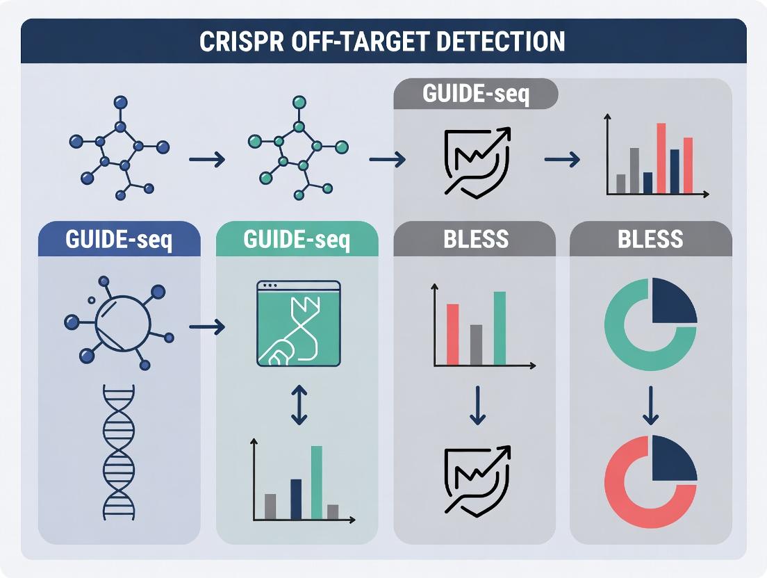
Visualization of Methods and Workflow
Title: Workflow Comparison of Key Off-Target Detection Methods
Title: Thesis Context: Integrating In Vivo and In Vitro Profiling
The Scientist's Toolkit: Key Research Reagent Solutions
| Item | Function in Off-Target Profiling | Example/Note |
|---|---|---|
| Recombinant Cas9 Nuclease | Creates DSBs at target and off-target sites. Essential for in vitro methods (CIRCLE-seq, Digenome-seq). | HiFi Cas9 variants are often used to reduce off-target activity while maintaining on-target efficiency. |
| Synthetic sgRNAs | Guides Cas9 to specific genomic loci. High-purity, chemically modified sgRNAs can reduce off-target effects. | Synthesized with 2'-O-methyl 3' phosphorothioate modifications for stability and reduced immunogenicity. |
| GUIDE-seq dsODN Tag | A short, blunt, double-stranded oligonucleotide that integrates into DSBs in vivo for later enrichment and sequencing. | Must be end-protected (phosphorothioate) to prevent degradation and ligation. A key reagent for GUIDE-seq. |
| BLISS Adapter Oligos | Biotinylated double-stranded DNA adapters for direct in situ ligation to DSBs in fixed samples. | Designed for efficient blunt-end ligation and subsequent capture or amplification. |
| Streptavidin Magnetic Beads | Used to capture biotinylated DNA fragments in GUIDE-seq, BLISS, and CIRCLE-seq library preparation. | Crucial for enriching signal (DSB-associated fragments) from background genomic DNA. |
| High-Fidelity PCR Master Mix | Amplifies adapter-ligated DNA fragments for NGS library construction with minimal bias and errors. | Essential for maintaining the quantitative accuracy of off-target site frequency. |
| Next-Generation Sequencing Kit | For final library preparation and high-throughput sequencing (Illumina platforms are standard). | Requires sufficient depth (>50M reads) for sensitive detection of low-frequency events. |
| Positive Control sgRNA Plasmid | A well-characterized sgRNA with known on- and off-target sites (e.g., targeting EMX1 or VEGFA). | Critical for validating the entire experimental and bioinformatic pipeline. |
Off-target effects in CRISPR-Cas genome editing refer to unintended modifications at genomic sites with sequences similar to the intended on-target site. Within the thesis on CRISPR off-target detection methods—encompassing GUIDE-seq, BLESS, and related techniques—understanding these effects is paramount for assessing the safety and fidelity of therapeutic and research applications.
Types of Off-Target Effects
Off-target effects can be categorized based on their origin and nature:
- DNA-Dependent Off-Targets: Caused by guide RNA (gRNA) homology to imperfectly matched genomic sequences. This is the primary focus of detection methods like GUIDE-seq.
- DNA-Independent Off-Targets: Caused by aberrant nuclease activity, such as Cas9 binding to DNA in a guide-independent manner.
- Large Structural Variations: Unintended deletions, insertions, or translocations triggered by double-strand breaks at on- or off-target sites.
Mechanisms Leading to Off-Target Effects
The primary mechanism involves the tolerance of the Cas9-sgRNA complex for mismatches, bulges, and base-pairing irregularities between the guide RNA and genomic DNA. Factors influencing this include:
- The number, position, and type (e.g., RNA-DNA vs. DNA-DNA) of mismatches.
- GC content of the protospacer adjacent motif (PAM)-distal region.
- Chromatin accessibility and local DNA conformation.
Consequences of Off-Target Effects
Unintended edits can lead to:
- Functional Consequences: Disruption of non-target genes, leading to loss or gain of function.
- Oncogenic Risk: Inactivation of tumor suppressor genes or activation of oncogenes.
- Confounded Research Results: Phenotypes in model systems not linked to the intended genetic modification.
Comparative Analysis of Off-Target Detection Methods
This guide compares the performance of key genome-wide, unbiased off-target detection methods relevant to CRISPR research.
Table 1: Comparison of Key Off-Target Detection Techniques
| Method | Acronym Expansion | Core Principle | Detection Scope | Key Advantages | Key Limitations |
|---|---|---|---|---|---|
| GUIDE-seq | Genome-wide, Unbiased Identification of DSBs Enabled by Sequencing | Captures double-strand breaks (DSBs) via integration of a double-stranded oligodeoxynucleotide tag. | Genome-wide, unbiased. | High sensitivity; identifies off-targets in living cells; does not require nuclease overexpression. | Requires delivery of exogenous oligonucleotide; lower signal in primary or non-dividing cells. |
| BLESS | Direct in Situ Breaks Labeling, Enrichment on Streptavidin, and Next-Generation Sequencing | Directly labels and captures DSBs in fixed cells using biotinylated linkers. | Genome-wide, unbiased. | Snapshots DSBs at a fixed time; applicable to any cell type, including fixed clinical samples. | Requires high starting material; complex protocol; potential for background noise. |
| CIRCLE-seq | Circularization for In Vitro Reporting of Cleavage Effects by Sequencing | In vitro cleavage of circularized genomic DNA followed by high-throughput sequencing. | Genome-wide, unbiased (in vitro). | Extremely high sensitivity; minimal sequence bias; uses purified genomic DNA. | In vitro assay may not reflect cellular context (chromatin, repair factors). |
| Digenome-seq | In Vitro Digested Genome Sequencing | In vitro digestion of cell-free genomic DNA with Cas9 RNP followed by whole-genome sequencing. | Genome-wide, unbiased (in vitro). | Comprehensive; computationally straightforward; uses WGS data. | High DNA input; in vitro method; requires significant sequencing depth. |
| SITE-Seq | Selective Enrichment and Identification of Tagged Genomic DNA Ends by Sequencing | In vitro cleavage of chromatin-associated DNA with Cas9 RNP, tagging breaks, and sequencing. | Genome-wide, unbiased. | Incorporates some chromatin structure; sensitive. | Complex workflow; not in living cells. |
Table 2: Experimental Performance Metrics from Selected Studies
| Method (Study) | Reported Sensitivity (Detection Threshold) | Cell Type Tested | Compared Against | Key Performance Finding |
|---|---|---|---|---|
| GUIDE-seq (Tsai et al., Nat Biotech 2015) | Detected sites with ≤0.1% indel frequency. | U2OS, HEK293T, K562, iPSCs. | BLESS, Digenome-seq. | Identified known and novel off-targets not found by computational prediction or BLESS. |
| BLESS (Ran et al., Nature 2015) | N/A (Direct break labeling). | HEK293T, mouse brain tissue. | GUIDE-seq (indirectly). | Validated high-frequency off-targets; effective in post-mitotic tissues. |
| CIRCLE-seq (Tsai et al., Nat Methods 2017) | Detected sites with cleavage rates as low as 0.0001%. | In vitro using genomic DNA from HEK293. | GUIDE-seq, Digenome-seq. | Identified ~10x more off-target sites than Digenome-seq for identical gRNAs. |
| Digenome-seq (Kim et al., Nat Methods 2015) | Required ~4% cleavage frequency for detection. | In vitro using genomic DNA from HCT116, K562. | Targeted sequencing. | Achieved a low false-positive rate; validated off-targets via targeted sequencing. |
Experimental Protocols for Key Methods
Principle: A double-stranded, end-protected oligonucleotide tag (dsODN) is integrated into DSBs via the non-homologous end joining (NHEJ) pathway during editing. Key Steps:
- Co-delivery: Transfect cells with Cas9/sgRNA RNP or plasmids alongside the dsODN.
- Integration: Allow 48-72 hours for genome editing and dsODN integration into DSBs.
- Genomic DNA Extraction: Harvest cells and extract genomic DNA.
- Library Preparation: Shear DNA. Perform primer extension using a dsODN-specific primer, then add sequencing adaptors via PCR.
- Enrichment & Sequencing: Use PCR to enrich for fragments containing the dsODN tag. Sequence using paired-end Illumina sequencing.
- Bioinformatics: Map reads to the reference genome. Identify genomic junctions where the dsODN is integrated to call off-target sites.
Principle: Direct in situ labeling of DSBs in fixed cells/nuclei with biotinylated linkers. Key Steps:
- Cell Fixation & Permeabilization: Fix cells (e.g., with formaldehyde) and isolate nuclei. Permeabilize to allow linker access.
- In Situ Break Labeling: Incubate nuclei with Cas9 to create DSBs (if ex vivo) or label endogenous breaks. Ligate biotinylated "linker 1" to DSB ends.
- DNA Extraction & Purification: Extract and shear genomic DNA.
- Pull-down: Capture biotinylated fragments (containing breaks) on streptavidin beads.
- On-Bead Ligation & PCR: Ligate "linker 2" to the other end of captured fragments. Perform PCR amplification with indexed primers.
- Sequencing & Analysis: Sequence and map reads to the genome. Breaks are identified at the genomic position preceding linker 1.
Visualizations
The Scientist's Toolkit: Key Research Reagents & Materials
Table 3: Essential Reagents for Off-Target Detection Studies
| Reagent/Material | Primary Function in Off-Target Studies | Example Application |
|---|---|---|
| High-Fidelity Cas9 Nuclease | Minimizes DNA-independent, non-specific cleavage while maintaining on-target activity. | Used in all cellular and in vitro detection assays (GUIDE-seq, CIRCLE-seq) to reduce background noise. |
| Double-Stranded Oligodeoxynucleotide (dsODN) Tag | Serves as a detectable tag integrated into DSBs via NHEJ for downstream capture and sequencing. | Core component of the GUIDE-seq protocol. |
| Biotinylated Linker Oligonucleotides | Enable direct labeling and streptavidin-based capture of DSB ends. | Essential for BLESS and SITE-Seq protocols. |
| Streptavidin Magnetic Beads | High-affinity capture of biotinylated DNA fragments for enrichment. | Used in BLESS, SITE-Seq pull-down steps. |
| Tn5 Transposase or Other Tagmentation Enzymes | Fragments DNA and simultaneously adds sequencing adapters for efficient library prep. | Often used in streamlined NGS library preparation following off-target capture steps. |
| PCR Enzymes for High-GC Amplification | Robust amplification of captured DNA fragments, which may have high GC content due to PAM sequences. | Critical for final library amplification before sequencing in most methods. |
| Control gRNA Plasmids/RNPs (e.g., EMX1, VEGFA site 2) | Well-characterized gRNAs with known on- and off-target profiles used as positive controls. | Benchmarking and validation of new off-target detection protocols. |
| Next-Generation Sequencing Kits (Illumina) | Generate the high-depth, paired-end sequencing data required for unbiased break site identification. | Final readout for all genome-wide methods (GUIDE-seq, BLESS, CIRCLE-seq, Digenome-seq). |
Core Principle
GUIDE-seq is a molecular biology technique designed for the genome-wide, unbiased detection of DNA double-strand breaks (DSBs) induced by engineered nucleases, such as CRISPR-Cas9. Its core innovation is the use of a short, blunt, double-stranded oligodeoxynucleotide (dsODN) tag that is directly integrated into DSB sites in living cells via non-homologous end joining (NHEJ). Following genomic DNA extraction and shearing, tagged DSBs are selectively amplified and prepared for next-generation sequencing. This allows for the precise mapping of both on-target and off-target cleavage events across the entire genome without prior knowledge of potential off-target sites.
Discovery and Performance in Comparison to Key Alternatives
GUIDE-seq was developed to address the limitations of earlier computational prediction and in vitro selection methods for identifying CRISPR off-targets. Its primary advantage is its unbiased, empirical nature. The following comparison highlights its performance against other seminal methods, BLESS and CIRCLE-seq, within the broader thesis on CRISPR off-target detection.
Table 1: Comparison of Key CRISPR Off-Target Detection Methods
| Method | Core Principle | Sensitivity (Detection Limit) | In Cellulo/In Vitro | Key Advantages | Key Limitations | Primary Supporting Data |
|---|---|---|---|---|---|---|
| GUIDE-seq (Tsai et al., 2015) | Tagging of DSBs in living cells via NHEJ with a dsODN. | ~0.1% of sequencing reads at a locus. | In Cellulo (Living cells) | Unbiased; captures cellular context (chromatin, repair); identifies translocations. | Requires dsODN transfection; lower signal for low-activity reagents. | Detected 10 off-target sites for a human EMX1 sgRNA, including sites missed by computational prediction. |
| BLESS (Crosetto et al., 2013) | Direct ligation of biotinylated linkers to DSBs in fixed nuclei. | Limited by background ligation. | In Situ (Fixed nuclei) | Snapshot of DSBs at a given time; no transfection needed. | High background; complex protocol; lower sensitivity for nuclease off-targets. | Mapped topoisomerase cleavage sites and CRISPR off-targets, but with lower signal-to-noise than GUIDE-seq. |
| CIRCLE-seq (Tsai et al., 2017) | In vitro selection and circularization of nuclease-cleaved genomic DNA. | ~0.01% of reads in a highly enriched library. | In Vitro (Purified genomic DNA) | Extremely high sensitivity; no transfection; minimal background. | Lacks cellular context (chromatin, repair factors). | Identified >100 off-target sites for a single sgRNA, including very low-frequency sites. |
Detailed Experimental Protocols
GUIDE-seq Protocol (Key Steps):
- dsODN Transfection: Co-transfect cells (e.g., HEK293T) with the CRISPR-Cas9 plasmid (or RNP) and the GUIDE-seq dsODN tag using a standard method like lipofection.
- Genomic DNA Extraction: Harvest cells 48-72 hours post-transfection. Extract high-molecular-weight genomic DNA.
- DNA Shearing and End-Repair: Fragment DNA by sonication or enzymatic digestion to ~300-500 bp. Repair ends to create blunt, 5'-phosphorylated termini.
- Ligation of Adaptor 1: Ligate a biotinylated "bridge adaptor" (Adaptor 1) to the repaired ends using T4 DNA Ligase.
- dsODN-Tagged Fragment Capture: Perform streptavidin pull-down to enrich fragments containing the biotinylated adaptor. Elute single-stranded DNA.
- Primer Extension for Tag Identification: Use a primer specific to the integrated GUIDE-seq dsODN tag to extend across the junction, creating a complementary strand that contains Adaptor 1 sequence.
- PCR Amplification: Amplify the library using primers complementary to Adaptor 1 and a second adaptor (Adaptor 2) introduced during the extension step. Use barcoded primers for multiplexing.
- Sequencing & Analysis: Perform paired-end sequencing. Analyze reads to identify genomic locations flanked by the dsODN tag sequence, map them to the reference genome, and quantify DSB frequency.
BLESS Protocol (Key Steps for Nucleases):
- Cell Fixation and Permeabilization: Fix cells with DSBs (e.g., nuclease-treated) with formaldehyde. Permeabilize nuclei.
- In Situ Ligation: In fixed nuclei, ligate biotinylated hairpin oligonucleotide linkers directly to the ends of DSBs using T4 DNA Ligase.
- Genomic DNA Extraction & Shearing: Reverse crosslinks, extract DNA, and shear it.
- Capture of Tagged DSBs: Capture biotinylated fragments using streptavidin beads.
- Library Preparation: On-bead, ligate sequencing adaptors, amplify via PCR, and sequence.
- Analysis: Map reads starting with the linker sequence to the genome to identify DSB sites.
Mandatory Visualization
Title: GUIDE-seq Experimental Workflow
Title: Off-Target Method Comparison Logic
The Scientist's Toolkit: Key Research Reagent Solutions
Table 2: Essential Reagents for GUIDE-seq Experiments
| Item | Function in GUIDE-seq | Key Consideration |
|---|---|---|
| dsODN Tag | Double-stranded oligo integrated into DSBs. Core of the assay. | Must be blunt-ended, phosphorylated, and HPLC-purified. A defined, controlled sequence is critical. |
| CRISPR-Cas9 Reagent | Induces the DSBs to be mapped. | Can be plasmid, mRNA, or RNP. RNP format often gives cleaner results with less background toxicity. |
| High-Efficiency Transfection Reagent | For co-delivery of CRISPR components and dsODN into target cells. | Critical for high tagging efficiency. Must be optimized for cell type (e.g., lipofection for HEK293T, nucleofection for primary cells). |
| Streptavidin Magnetic Beads | For capturing biotinylated fragments after bridge adaptor ligation. | High binding capacity and low non-specific binding are essential for library purity. |
| Blunt-End DNA Ligase (e.g., T4 DNA Ligase) | Ligates the bridge adaptor to sheared, end-repaired genomic DNA. | High-concentration, ultra-pure formulations are recommended for efficient ligation of dilute substrates. |
| PCR Polymerase for High-Fidelity Amplification | Amplifies the final sequencing library from captured fragments. | Must have high fidelity and processivity to accurately amplify complex, GC-rich regions. |
| Barcoded Sequencing Adaptors | Allows multiplexing of multiple samples in a single NGS run. | Compatible with your sequencing platform (Illumina, etc.). Unique dual indexing reduces index hopping errors. |
Core Principle and Approach
BLESS is a direct, unbiased method for genome-wide mapping of DNA double-strand breaks (DSBs). Its core principle involves in situ labeling of DSB ends within fixed cells or nuclei using biotinylated linkers, followed by capture of these labeled fragments on streptavidin beads, next-generation sequencing, and computational mapping. This allows for a snapshot of all DSBs present at the time of cell fixation, including those generated by CRISPR-Cas9 and other nucleases. Unlike methods that rely on cellular repair processes (e.g., GUIDE-seq), BLESS captures breaks directly, minimizing artifacts from repair kinetics.
Performance Comparison: BLESS vs. GUIDE-seq and Other Alternatives
Table 1: Comparison of Key Methodological Features
| Feature | BLESS | GUIDE-seq | CIRCLE-seq | Digenome-seq |
|---|---|---|---|---|
| Detection Principle | Direct in situ break labeling | Integration of oligonucleotide tag during repair | In vitro circularization & amplification | In vitro digestion of genomic DNA |
| Cellular Context | Fixed cells/nuclei (in situ) | Living cells | Purified genomic DNA (cell-free) | Purified genomic DNA (cell-free) |
| Repair Process Dependency | No | Yes (NHEJ) | No | No |
| Primary Application | Snapshot of all DSBs (endogenous & engineered) | Mapping nuclease integration sites in cells | Comprehensive, sensitive in vitro off-target profiling | In vitro off-target profiling |
| Sensitivity (Typical) | Moderate | High | Very High | High |
| Background (Endogenous Breaks) | Higher (detects all breaks) | Lower (requires tag integration) | Very Low (controlled conditions) | Low |
| Throughput & Complexity | High complexity (library prep from captured fragments) | Moderate complexity | High complexity | Moderate complexity |
Table 2: Experimental Performance Comparison for CRISPR-Cas9 Off-Target Detection
| Study Metric | BLESS (Cas9) | GUIDE-seq (Cas9) | CIRCLE-seq (Cas9) | Supporting Experimental Data Summary |
|---|---|---|---|---|
| Validated Off-Targets Identified | Moderate yield | High yield | Highest yield | In a study on the VEGFA site, BLESS identified a subset of off-targets; GUIDE-seq identified more, and CIRCLE-seq predicted the most, with high validation rate. |
| Overlap with Other Methods | Partial overlap with GUIDE-seq | High overlap with CIRCLE-seq predictions | High overlap with GUIDE-seq validations | Intersection analyses show GUIDE-seq and CIRCLE-seq have the highest concordance. BLESS sites often include unique endogenous breaks. |
| Key Advantage | Captures endogenous genome instability | Robust performance in living cells; high signal-to-noise for nuclease targets | Unmatched sensitivity and comprehensiveness in vitro | CIRCLE-seq can detect off-targets with mutation rates <0.1%, outperforming cellular methods in sheer number of sites identified. |
| Key Limitation | Background from non-specific breaks and capture bias | Requires oligonucleotide delivery and active NHEJ | May identify sites not cut in cells (overprediction) | Requires sophisticated bioinformatics to filter false positives from in vitro signals. |
Detailed Experimental Protocols
BLESS Protocol (Key Steps):
- Cell Fixation and Lysis: Cells or nuclei are fixed with formaldehyde to "freeze" DSBs in situ. Cells are then lysed, and chromatin is immobilized in an agarose matrix.
- In situ End Repair and A-tailing: Within the agarose plug, DSB ends are repaired and a single 'A' nucleotide is added using DNA polymerases.
- Ligation of Biotinylated Linkers: A duplex linker with a 5' biotin and a compatible 'T' overhang is ligated to the A-tailed DSB ends.
- DNA Extraction and Fragmentation: Genomic DNA is purified and randomly sheared (e.g., by sonication).
- Streptavidin Capture: Biotinylated fragments (originating from DSB sites) are captured on streptavidin-coated magnetic beads.
- Library Preparation & Sequencing: On-bead library preparation is performed, followed by high-throughput sequencing.
- Bioinformatics Analysis: Reads are aligned to the genome. DSB sites are called as genomic positions where the start of paired-end reads cluster, corresponding to the initial linker ligation site.
GUIDE-seq Protocol (Key Steps for Comparison):
- Co-delivery: The Cas9/sgRNA RNP and a short, double-stranded, end-protected oligonucleotide tag (the "GUIDE-seq tag") are co-delivered into living cells via electroporation or transfection.
- Tag Integration: During NHEJ repair of Cas9-induced DSBs, the oligonucleotide tag is integrated into the break site.
- Genomic DNA Extraction & Shearing: Genomic DNA is harvested, sheared, and prepared for sequencing.
- Tag-Specific Enrichment: PCR or capture using a tag-specific primer enriches for genomic fragments containing the integrated tag.
- Sequencing & Analysis: Sequencing reads are analyzed to identify genomic locations of tag integration, which correspond to DSB sites.
Diagrams
BLESS Experimental Workflow
CRISPR Off-target Detection Method Categories
The Scientist's Toolkit: Key Research Reagent Solutions
| Item | Function in BLESS/GUIDE-seq |
|---|---|
| Formaldehyde (BLESS) | Crosslinks and fixes DNA-protein complexes to preserve the in situ state of DNA breaks at the moment of cell harvesting. |
| Biotinylated Duplex Linker (BLESS) | An oligonucleotide duplex with a 5' biotin modification; ligates directly to DSB ends for subsequent streptavidin-based enrichment. |
| Streptavidin Magnetic Beads (BLESS) | Solid-phase matrix that specifically captures biotinylated DNA fragments, enabling purification of break-associated sequences. |
| dsODN Tag (GUIDE-seq) | A short, double-stranded, end-protected oligonucleotide. Serves as a repair template integrated into Cas9-induced breaks via NHEJ in living cells. |
| Cas9 Nuclease (Wild-type or Hi-Fi) | The engineered nuclease that creates a DNA double-strand break at target sites guided by the sgRNA. Key reagent for both methods. |
| sgRNA (Synthetic or expressed) | Single-guide RNA that directs Cas9 to a specific genomic locus complementary to its spacer sequence. |
| Nucleofection/Electroporation System | Critical for efficient co-delivery of Cas9 RNP and dsODN tag (for GUIDE-seq) into difficult-to-transfect cell types. |
| High-Fidelity DNA Polymerase & Ligase | Enzymes essential for the end-repair, A-tailing, and linker ligation steps during the BLESS library preparation process. |
| Next-Generation Sequencer | Platform (e.g., Illumina NovaSeq) required for high-throughput sequencing of captured or enriched DNA libraries. |
The advent of CRISPR-Cas9 genome editing necessitated the development of robust, genome-wide methods to identify off-target cleavage sites. Early methods like in silico prediction were insufficient. This guide compares three foundational experimental techniques—GUIDE-seq, BLESS, and CIRCLE-seq—that revolutionized off-target assessment, framing them within the broader thesis of evolving detection methodologies.
Method Comparison & Performance Data
The following table summarizes the core principles, key performance metrics, and comparative advantages of each method based on published experimental data.
Table 1: Comparative Analysis of Foundational Off-Target Detection Methods
| Method | Core Principle | Sensitivity (Theoretical/Reported) | Key Experimental Finding | Primary Advantage | Primary Limitation |
|---|---|---|---|---|---|
| GUIDE-seq | Integration of a double-stranded oligodeoxynucleotide tag into DSBs in situ, followed by enrichment and sequencing. | Detects sites with ≥0.1% frequency of indels. | In one study, identified 6-85 off-target sites for 8 different sgRNAs, many missed by computational prediction. | Captures off-targets in living cells with genomic context; provides relative frequency data. | Requires delivery of exogenous dsODN; less effective in primary or non-dividing cells. |
| BLESS | Direct ligation of biotinylated linkers to DSBs in fixed cells/nuclei, followed by capture and sequencing. | Single-nucleotide resolution of DSBs at a given time point. | Detected known and novel off-target sites for Cas9 and nickases, including in patient-derived glioblastoma cells. | Snapshot of in situ DSBs without reagents; applicable to clinical samples. | Captures all DSBs, requiring careful controls; not inherently specific to nuclease activity. |
| CIRCLE-seq | In vitro circularization and amplification of sheared genomic DNA, followed by in vitro Cas9 cleavage and sequencing of linearized fragments. | Exceptionally high sensitivity; can detect sites with cleavage rates as low as 0.0001%. | Identified >10x more off-target sites per sgRNA than cell-based methods, revealing a vast landscape of low-frequency sites. | Ultra-high sensitivity; no cellular constraints; ideal for comprehensive sgRNA profiling. | Purely in vitro; may identify sites not cleaved in a cellular context due to chromatin or repair factors. |
Detailed Experimental Protocols
- Cell Transfection: Co-deliver Cas9:sgRNA RNP and the GUIDE-seq dsODN tag into cultured cells using a method like nucleofection.
- Genomic DNA Extraction: Harvest cells 48-72 hours post-transfection. Extract and shear genomic DNA.
- Tag Enrichment: Perform PCR using one primer specific to the integrated dsODN tag and another primer binding to an adapter ligated to the sheared DNA ends.
- Library Prep & Sequencing: Amplify enriched fragments, prepare sequencing libraries, and perform paired-end high-throughput sequencing.
- Data Analysis: Map reads, identify dsODN integration sites, and cluster them to call off-target loci. Peak height correlates with cleavage frequency.
- Cell Fixation & Permeabilization: Fix cells (e.g., with formaldehyde) to "freeze" DSBs in situ. Permeabilize nuclei.
- In Situ Ligation: In fixed nuclei, ligate biotinylated double-stranded linkers directly to the ends of genomic DSBs.
- Genome Extraction & Capture: Extract and shear genomic DNA. Capture biotinylated fragments (containing DSBs) on streptavidin beads.
- Library Prep & Sequencing: On-bead library preparation followed by sequencing.
- Data Analysis: Map sequence reads to the genome. Compare DSB peaks in nuclease-treated samples versus control samples to identify nuclease-specific sites.
- Genomic DNA Circularization: Extract genomic DNA, shear it, and ligate stem-loop adapters to both ends. Ligate the ends to form circular DNA molecules.
- In Vitro Cleavage: Incubate circularized DNA with Cas9:sgRNA RNP. Only DNA circles containing a target site will be linearized upon cleavage.
- Linear Fragment Enrichment: Exonuclease treat the product to degrade all remaining linear DNA (unligated fragments) and uncut circular DNA. The surviving linear fragments are those generated by Cas9 cleavage.
- Library Prep & Sequencing: Amplify and sequence the exonuclease-resistant linear DNA.
- Data Analysis: Map reads to identify Cas9 cut sites across the entire genomic library with high sensitivity.
Visualized Workflows
The Scientist's Toolkit: Key Research Reagent Solutions
Table 2: Essential Reagents for Off-Target Detection Methods
| Reagent / Solution | Primary Function | Method(s) |
|---|---|---|
| Cas9 Nuclease (WT) | The effector protein that creates double-strand breaks at target (and off-target) genomic loci. | Universal to all methods. |
| Synthetic sgRNA | Guides Cas9 to the intended DNA sequence. High-quality synthesis is critical for specificity. | Universal to all methods. |
| GUIDE-seq dsODN | A short, double-stranded, end-protected DNA oligo that integrates into DSBs for tag-based enrichment. | GUIDE-seq exclusive. |
| Biotinylated Linkers (BLESS) | Short dsDNA molecules with a biotin tag for streptavidin capture, ligated directly to DSB ends. | BLESS exclusive. |
| Stem-loop Adapters (CIRCLE-seq) | Specialized adapters that enable circularization of sheared genomic DNA fragments. | CIRCLE-seq exclusive. |
| Streptavidin Magnetic Beads | Solid-phase matrix for capturing biotin-tagged DNA fragments during library preparation. | BLESS, also used in GUIDE-seq variants. |
| Exonuclease (e.g., T5 or T7) | Degrades linear DNA, enriching for Cas9-linearized circles in the in vitro assay. | CIRCLE-seq exclusive. |
| High-Fidelity PCR Master Mix | For accurate, low-bias amplification of enriched DNA fragments prior to sequencing. | Universal to all methods. |
| Next-Generation Sequencing Kit | Platform-specific kits (Illumina, MGI) for preparing sequencing libraries from amplified products. | Universal to all methods. |
Step-by-Step Protocols: Implementing GUIDE-seq and BLESS in Your Lab
Within the ongoing thesis on advancing CRISPR off-target detection, experimental rigor is paramount. This guide compares the performance of key methodologies—GUIDE-seq, BLESS, and CIRCLE-seq—through the lens of robust experimental design, focusing on cell preparation, controls, and replication to generate reliable data for researchers and drug development professionals.
Comparative Performance Data
The following table summarizes key performance metrics from recent studies (2023-2024) comparing these off-target detection methods. Data emphasizes the necessity of proper controls and biological replication.
Table 1: Comparative Performance of CRISPR Off-Target Detection Methods
| Method | Sensitivity (Detects Low-Frequency Events) | Required Cell Input | Background Signal (Noise) | Key Experimental Control Required | Typical Recommended Replicates |
|---|---|---|---|---|---|
| GUIDE-seq | Moderate-High (∼0.1% frequency) | High (∼1-2 million transfected cells) | Low (with proper tag integration control) | Untagged control library for background subtraction. | 3 independent transfections. |
| BLESS / BLISS | High (Single-cell resolution possible) | Medium-Low (∼100,000 cells) | Moderate (requires careful DSB enrichment) | No-nuclease control for spontaneous DSB identification. | 3+ independent cell preparations. |
| CIRCLE-seq | Very High (In vitro, <0.01% frequency) | N/A (Uses purified genomic DNA) | Very Low (with optimized adapter ligation) | No-guide control and nuclease-free reaction control. | 4+ technical replicates per gDNA sample. |
Detailed Experimental Protocols
Cell Preparation for GUIDE-seq
- Cell Line & Culture: Use actively dividing HEK293T or relevant target cells at >90% viability.
- Transfection: Co-transfect cells with:
- Cas9/gRNA expression plasmid (or RNP complex).
- GUIDE-seq oligo duplex (annealed P7N9 / P7N9_2nt oligonucleotides).
- Control: In parallel, transfect a separate cell population with all components except the GUIDE-seq oligo (untagged control). This is critical for distinguishing true integration from background.
- Harvesting: Harvest cells 72 hours post-transfection. Extract high-molecular-weight genomic DNA using a gentle lysis protocol (e.g., Proteinase K/SDS digestion followed by phenol-chloroform extraction).
- Replication: Perform three independent transfections on different days to account for biological variability.
Cell Preparation for BLESS (Direct In Situ Breaks Labeling)
- Cell Seeding & Editing: Seed cells on culture-treated coverslips. Transfert or deliver RNP complexes.
- Fixation & Permeabilization: At 24-48h post-editing, fix cells with 2% formaldehyde and permeabilize with 0.5% Triton X-100.
- In Situ Ligation: Use T4 DNA ligase to blunt-end ligate biotinylated adapters into DSB sites within the permeabilized nuclei.
- Control: Include a no-nuclease control sample processed identically to identify background from spontaneous breaks and adapter ligation.
- Replication: Prepare at least three independent biological replicates (separate cell passages and edits) with multiple coverslips per condition.
Protocol for CIRCLE-seq (In Vitro)
- Genomic DNA Input: Purify genomic DNA (≥ 5 µg) from edited and unedited control cells.
- Circularization: Shear DNA, repair ends, and ligate with a splinter oligo to create circularized DNA libraries.
- Digestion & Linearization: Digest circles with Cas9/gRNA complex in vitro to linearize off-target-containing fragments.
- Control: Essential controls include a no-guide RNA Cas9 digestion and a nuclease-free reaction to assess background linearization.
- Replication: Perform four technical replicates of the entire in vitro reaction starting from the same gDNA pool to ensure assay consistency.
Visualizations
Diagram 1: Experimental Workflow Comparison for Three Methods
Diagram 2: Critical Control Strategy for Validating Off-Target Hits
The Scientist's Toolkit: Research Reagent Solutions
| Reagent / Material | Function in Experimental Design |
|---|---|
| High-Viability Cell Lines (e.g., HEK293T) | Ensures high transfection/efficiency rates for methods requiring cellular delivery. |
| Recombinant Cas9 Nuclease (RNP grade) | Provides consistent, plasmid-free nuclease activity for RNP delivery and in vitro digestions. |
| Ultra-Pure GUIDE-seq Oligo Duplex | Critical for efficient tag integration with minimal background ligation. |
| Biotinylated dsDNA Adapters (for BLESS) | Labels double-strand breaks in situ for subsequent pull-down and identification. |
| Circligase ssDNA Ligase (for CIRCLE-seq) | Efficiently circularizes sheared genomic DNA for the in vitro assay. |
| PCR-Free NGS Library Prep Kit | Reduces amplification bias, providing more quantitative representation of off-target frequencies. |
| Spike-in Control DNA | Added before NGS library prep to quantitatively normalize sequencing depth between samples. |
| ddCas9 (Catalytically Dead) | Essential protein control for binding-only effects in nuclease-free control experiments. |
GUIDE-seq (Genome-wide, Unbiased Identification of DSBs Enabled by Sequencing) is a pivotal method for the unbiased, genome-wide detection of CRISPR-Cas nuclease off-target effects. Within the broader thesis on CRISPR off-target detection methodologies—which includes techniques like BLESS, Digenome-seq, and CIRCLE-seq—GUIDE-seq stands out for its ability to capture in vivo double-strand break (DSB) events via the integration of a defined oligonucleotide tag. This guide provides an objective, data-driven comparison of GUIDE-seq against key alternatives.
Oligonucleotide Tag Integration and Workflow
The core principle of GUIDE-seq involves the co-delivery of the CRISPR-Cas9 ribonucleoprotein (RNP) complex with a short, double-stranded, blunt-ended oligonucleotide tag (the "GUIDE-seq tag") into living cells. When a DSB is generated by Cas9, cellular repair pathways, predominantly non-homologous end joining (NHEJ), integrate this tag into the break site. These tagged sites are subsequently amplified and sequenced.
Experimental Protocol for GUIDE-seq
- Cell Transfection: Co-transfect target cells with Cas9:sgRNA RNP complex and the dsODN GUIDE-seq tag (e.g., 100-200 nM each) using a nucleofection system.
- Genomic DNA Extraction: Harvest cells 48-72 hours post-transfection. Extract high-molecular-weight genomic DNA.
- Tag-Specific Amplification: Perform nested PCR using primers specific to the integrated dsODN tag and common adapters. This enriches for genomic fragments containing the tag.
- Library Preparation & Sequencing: Add sequencing adapters via a second PCR, purify the library, and perform paired-end high-throughput sequencing (e.g., Illumina MiSeq/NextSeq).
- Bioinformatic Analysis: Process reads to map tag integration sites to the reference genome, identifying off-target sites based on read density and sequence similarity to the on-target.
Diagram Title: GUIDE-seq Experimental Workflow
Comparative Performance Analysis
The efficacy of off-target detection methods is evaluated by their sensitivity (ability to detect true off-targets), specificity (low false-positive rate), resolution, and technical requirements. The following table synthesizes experimental data from key comparative studies (Tsai et al., Nat Biotechnol 2015; Kim et al., Nat Methods 2015; Wienert et al., Nat Commun 2019).
Table 1: Comparison of Genome-wide CRISPR Off-target Detection Methods
| Method | Detection Principle | Requires Living Cells? | Sensitivity (Detection of Validated Sites) | Resolution | Key Experimental Limitation |
|---|---|---|---|---|---|
| GUIDE-seq | NHEJ-mediated tag integration | Yes | High (~90-100%)* | Single-nucleotide | Requires efficient dsODN delivery; lower tag integration in primary cells. |
| BLESS/BLISS | Direct in situ ligation of adapters to DSBs | No (Fixed cells) / Yes | Moderate to High | Single-nucleotide | Captures a snapshot in time; can have high background. |
| Digenome-seq | In vitro cleavage of purified genomic DNA by Cas9 | No | Moderate | Single-nucleotide | High false-positive rate without proper bioinformatic filtering (requires SMRT sequencing). |
| CIRCLE-seq | In vitro selection & circularization of cleaved genomic fragments | No | Very High (in vitro) | Single-nucleotide | Purely in vitro; may predict sites not cleaved in cellular context. |
| HTGTS | Translocation-based capture of DSBs | Yes | High | Single-nucleotide | Requires a fixed "bait" DSB; detects breaks interacting with the bait. |
*Sensitivity varies with sgRNA and delivery efficiency.
Table 2: Exemplary Experimental Data from a Comparative Study (Model Gene VEGFA Site 3)
| Off-target Site (Genomic Locus) | GUIDE-seq Reads | Digenome-seq Peak Score | CIRCLE-seq Reads | Validated by Amplicon-Seq? |
|---|---|---|---|---|
| Chr10:64,394,502 | 5,421 | 85.2 | 18,745 | Yes |
| Chr2:127,482,110 | 892 | 12.1 | 3,450 | Yes |
| Chr5:55,118,367 | 315 | Not detected | 1,205 | Yes |
| Chr19:11,633,091 | Not detected | 45.7 | 892 | No |
The Scientist's Toolkit: Key Research Reagent Solutions
Table 3: Essential Materials for GUIDE-seq and Related Methods
| Item | Function | Example Product/Catalog # |
|---|---|---|
| dsODN GUIDE-seq Tag | Double-stranded oligo donor for NHEJ-mediated integration at DSBs. | Synthesized oligos (e.g., IDT): 5’-/5Phos/NNNNNNNNNNNNNNNAGATCGGAAGAGCA-3’ |
| Ultrapure Cas9 Nuclease | For forming RNP complex with in vitro transcribed or synthetic sgRNA. | e.g., Alt-R S.p. Cas9 Nuclease V3 (IDT, 1081058) |
| Nucleofector System | For efficient co-delivery of RNP and dsODN into hard-to-transfect cells. | e.g., Lonza 4D-Nucleofector System |
| Tag-Specific PCR Primers | For nested PCR amplification of genomic DNA fragments containing integrated tag. | Custom designed (Tsai et al. 2015 protocol). |
| High-Fidelity PCR Master Mix | For accurate amplification during library preparation. | e.g., NEBNext Ultra II Q5 Master Mix (NEB, M0544) |
| Size Selection Beads | For clean-up and size selection of PCR-amplified libraries. | e.g., AMPure XP Beads (Beckman Coulter, A63881) |
| Cell Culture Media | For maintaining cells pre- and post-nucleofection. | Dependent on cell line (e.g., DMEM + 10% FBS). |
Diagram Title: Off-target Methods in Thesis Context
In conclusion, GUIDE-seq provides a robust, sensitive, and nucleotide-resolution profile of CRISPR-Cas off-target activity within a cellular context. While alternatives like CIRCLE-seq offer supreme in vitro sensitivity and BLESS provides a snapshot of breaks without need for living cells, GUIDE-seq's balance of in vivo relevance and practical sensitivity solidifies its role as a cornerstone method for comprehensive off-target assessment in therapeutic development.
BLESS (Direct In Situ Breaks Labeling, Enrichment on Streptavidin, and Sequencing) is a method for genome-wide mapping of DNA double-strand breaks (DSBs) in fixed cells and tissues. Unlike methods that rely on living cells and exogenous repair, BLESS captures DSBs in situ, providing a snapshot of genomic damage. This guide compares BLESS with other prominent CRISPR off-target detection and DSB mapping methodologies, including GUIDE-seq, Digenome-seq, and CIRCLE-seq, within the broader context of CRISPR off-target assessment.
Method Comparison & Experimental Data
The following table summarizes the core characteristics, advantages, and limitations of key DSB detection methods.
Table 1: Comparison of Genome-Wide DSB Detection Methods
| Method | Principle | Detection Sensitivity | Required Input | Living Cells? | Key Advantage | Key Limitation |
|---|---|---|---|---|---|---|
| BLESS | In situ biotinylation & capture of DSB ends. | Medium (Requires sufficient DSB frequency). | Fixed cells/tissue nuclei. | No (Fixed samples). | Works on archived clinical samples; No culture bias. | Lower sensitivity for rare breaks; Complex protocol. |
| GUIDE-seq | Integration of oligonucleotide tags into DSBs during repair. | High (Detects low-frequency off-targets). | Living cells in culture. | Yes. | Highly sensitive in living cellular context. | Requires viable, dividing cells; Tag integration bias. |
| Digenome-seq | In vitro cleavage of genomic DNA by RNP, followed by whole-genome sequencing. | Very High (Theoretical). | Purified genomic DNA. | No (Cell-free). | Unbiased, ultra-sensitive; No cellular context limitations. | High sequencing depth/cost; No cellular context. |
| CIRCLE-seq | Circularization and enrichment of cleaved genomic fragments in vitro. | Extremely High (Theoretical). | Purified genomic DNA. | No (Cell-free). | Highest theoretical sensitivity for in vitro profiling. | Purely in vitro; May detect biologically irrelevant sites. |
Table 2: Quantitative Performance Comparison from Representative Studies
| Method | Reported Off-Target Sites for Benchmark gRNA (EMX1) | Validation Rate (by amplicon-seq) | Approximate Sequencing Depth Required | Time to Result |
|---|---|---|---|---|
| BLESS | 5-10 | ~80% | 50-100M reads | 5-7 days |
| GUIDE-seq | 8-15 | >90% | 30-50M reads | 7-10 days |
| Digenome-seq | 50-100+ | 50-70% | 200-500M reads | 4-6 days |
| CIRCLE-seq | 100+ | 30-60% | 50-100M reads (enriched) | 5-8 days |
Detailed Experimental Protocols
BLESS Core Protocol
- Cell/Tissue Fixation & Permeabilization: Cells or tissue sections are fixed with formaldehyde (e.g., 4% for 10 min) and permeabilized (e.g., 0.5% Triton X-100) to preserve nuclear architecture while allowing reagent access.
- In Situ DSB End Labeling: Permeabilized nuclei are incubated with a biotinylated oligonucleotide linker mix. The linker anneals to DSB ends via complementary single-stranded overhangs or is ligated using T4 DNA Ligase directly to the broken ends.
- Biotinylation & Capture: Biotin-dUTP is incorporated using terminal deoxynucleotidyl transferase (TdT) to tail the ligated linker, creating a biotinylated handle. Nuclei are lysed, and genomic DNA is sheared. Biotinylated fragments containing DSBs are captured using streptavidin-coated magnetic beads.
- Library Preparation & Sequencing: Captured DNA is washed, eluted, and processed into a sequencing library (end-repair, A-tailing, adapter ligation, PCR amplification). Libraries are sequenced on a high-throughput platform (e.g., Illumina).
GUIDE-seq Core Protocol (For Comparison)
- Tag Oligo Transfection: Living cells are co-transfected with CRISPR-Cas9 components (gRNA + Cas9) and a double-stranded, end-protected "tag" oligonucleotide.
- Tag Integration: During endogenous repair of Cas9-induced DSBs, the tag oligo is integrated via the non-homologous end joining (NHEJ) pathway.
- Genomic DNA Extraction & Processing: Genomic DNA is extracted, sheared, and ligated to adapters.
- Tag-Specific Enrichment: Fragments containing the integrated tag are enriched by PCR using one primer specific to the tag and another to the adapter.
- Sequencing & Analysis: Enriched libraries are sequenced. Off-target sites are identified by locating genomic sequences adjacent to the integrated tag sequence.
Visualized Workflows and Pathways
Title: BLESS Experimental Workflow from Fixed Cells to Sequencing
Title: Method Selection Logic for DSB and Off-Target Detection
The Scientist's Toolkit: Research Reagent Solutions
Table 3: Essential Reagents for BLESS and Related Methods
| Reagent/Material | Function in BLESS | Function in GUIDE-seq | Typical Vendor/Example |
|---|---|---|---|
| Formaldehyde | Fixative to crosslink and preserve DNA-protein structures and DSBs in situ. | Not typically used. | Sigma-Aldrich, Thermo Fisher |
| Terminal Deoxynucleotidyl Transferase (TdT) | Enzyme to add biotinylated nucleotides (Biotin-dUTP) to 3' ends of DSBs for capture. | Not used. | Roche, NEB |
| Biotin-dUTP | Biotin-labeled nucleotide substrate for TdT; provides handle for streptavidin pulldown. | Not used. | Thermo Fisher, Jena Bioscience |
| Streptavidin Magnetic Beads | Solid-phase capture matrix to isolate biotinylated DNA fragments. | Not used in core protocol. | Dynabeads (Thermo Fisher), MagCapture (Fujifilm) |
| Double-Stranded Tag Oligo | Not used. | Exogenous donor oligo integrated into DSBs during NHEJ for later enrichment. | IDT, Sigma-Aldrich |
| T4 DNA Ligase | Ligates adapters or linkers to DSB ends. | Used in library preparation. | NEB, Thermo Fisher |
| Proteinase K | Digests proteins after capture to elute pure DNA. | Used in genomic DNA extraction. | Qiagen, Thermo Fisher |
| High-Fidelity PCR Master Mix | Amplifies captured DNA for library generation. | Amplifies tag-integrated fragments for enrichment. | KAPA HiFi (Roche), Q5 (NEB) |
| Next-Generation Sequencing Kit | Platform-specific kit for final library preparation and sequencing. | Same as BLESS. | Illumina, Thermo Fisher |
The accurate detection of CRISPR-Cas9 off-target effects is critical for therapeutic safety. Methods like GUIDE-seq, BLESS, and their derivatives generate complex sequencing data, requiring robust bioinformatics pipelines to transform raw reads into high-confidence off-target sites. This guide compares the performance of prominent analysis tools, framed within the ongoing thesis that effective off-target prediction requires orthogonal experimental and computational validation.
Comparative Performance Analysis of Off-Target Calling Pipelines
The following table summarizes the key performance metrics of leading bioinformatics pipelines, as evaluated in recent benchmarking studies. These pipelines process raw FASTQ files from methods like GUIDE-seq or BLESS to produce final off-target site lists.
Table 1: Pipeline Performance Comparison for GUIDE-seq Data Analysis
| Pipeline/Tool | Recall (Sensitivity) | Precision | Computational Speed (vs. Baseline) | Key Strengths | Major Limitations |
|---|---|---|---|---|---|
| GUIDE-seq (Original Tool) | 0.85 | 0.92 | 1x (Baseline) | Gold standard for its own protocol; highly tuned. | Protocol-specific; not adaptable to other assays. |
| BLESS 2 | 0.88 | 0.89 | 0.7x | Excellent for direct in situ DSB tagging. | Requires matched control; high memory usage. |
| CRISPResso2 | 0.82 | 0.95 | 1.2x | Versatile; includes amplicon analysis; user-friendly. | Lower recall for genome-wide methods. |
| PEM-seq Analyzer | 0.91 | 0.87 | 0.5x | High sensitivity for circularized sequencing data. | Slow; complex installation. |
| PinAPL-Py | 0.95 | 0.83 | 1.5x | High recall; integrates multiple assays (GUIDE-seq, HTGTS). | Lower precision; more false positives. |
| MISEQ-SA | 0.79 | 0.98 | 2.0x | Very high precision; extremely fast. | Lower sensitivity; may miss low-frequency sites. |
Detailed Experimental Protocols
To interpret the data in Table 1, understanding the benchmarking methodology is essential.
Protocol 1: Benchmarking Pipeline Recall and Precision
- Reference Dataset Creation: A "gold standard" set of validated off-target sites is established for a known sgRNA using a combination of GUIDE-seq, CIRCLE-seq, and targeted deep sequencing.
- Raw Data Processing: Publicly available or newly generated paired-end FASTQ files from GUIDE-seq experiments targeting the reference sgRNA are collected.
- Parallel Analysis: Each pipeline (e.g., GUIDE-seq tool, PinAPL-Py, CRISPResso2 in amplicon mode) is run on the identical dataset using default parameters.
- Output Comparison: The list of predicted off-target sites from each tool is compared against the gold standard set. Recall is calculated as (True Positives) / (True Positives + False Negatives). Precision is calculated as (True Positives) / (True Positives + False Positives).
Protocol 2: Evaluating Computational Efficiency
- Environment Standardization: All tools are installed on an identical cloud instance (e.g., AWS with 8 vCPUs, 32GB RAM).
- Timed Execution: A large (~100 GB) simulated GUIDE-seq dataset is processed by each pipeline. The wall-clock time from FASTQ input to final report is recorded.
- Resource Monitoring: Peak memory (RAM) usage and CPU utilization are logged during execution.
- Normalization: Speed is normalized to the runtime of the original GUIDE-seq tool to generate the relative "Computational Speed" metric.
Pipeline Workflow Visualization
Title: General Bioinformatics Pipeline for Off-Target Detection
The Scientist's Toolkit: Research Reagent Solutions
Table 2: Essential Reagents and Materials for Off-Target Detection Workflows
| Item | Function in Experimental Protocol | Example Product/Catalog # |
|---|---|---|
| dsODN Integration Tag (for GUIDE-seq) | Double-stranded oligodeoxynucleotide that integrates into double-strand breaks, providing a unique tag for sequencing library prep. | Custom synthesized, 5'-phosphorylated, HPLC-purified. |
| Biotinylated dATP/dTTP (for BLESS) | Biotin-labeled nucleotides incorporated at break sites via terminal transferase, enabling streptavidin-based enrichment of DSB fragments. | Thermo Fisher Scientific, #19524016. |
| Tn5 Transposase (for Tagmentation) | Enzyme for simultaneous fragmentation and adapter tagging of DNA, used in library prep for methods like BLISS. | Illumina Nextera Kit, #20018705. |
| Protein A/G Magnetic Beads | For immunoprecipitation of Cas9-protein complexes in methods like ChIP-seq or CLEVER-seq. | Pierce Protein A/G Magnetic Beads, #88802. |
| KAPA HiFi HotStart ReadyMix | High-fidelity PCR enzyme for accurate amplification of libraries or validation amplicons with minimal bias. | Roche, #7958935001. |
| Streptavidin C1 Beads | Magnetic beads used to capture biotinylated DNA fragments in BLESS and related enrichment protocols. | Invitrogen, #65001. |
| NEBNext Ultra II DNA Library Prep Kit | Comprehensive kit for preparing high-quality sequencing libraries from fragmented DNA. | New England Biolabs, #E7645S. |
| Cas9 Nuclease, S. pyogenes | The effector enzyme for creating targeted and off-target double-strand breaks in validation experiments. | Integrated DNA Technologies, #1081058. |
| Target-Specific sgRNA | The guide RNA component defining the primary target site for CRISPR-Cas9 cleavage. | Synthego, custom CRISPR sgRNA. |
| Genomic DNA Extraction Kit (for cells/tissues) | Reliable isolation of high-integrity genomic DNA for downstream library construction. | QIAGEN DNeasy Blood & Tissue Kit, #69504. |
The development of a therapeutic CRISPR-Cas9 guide RNA (gRNA) requires rigorous profiling to ensure on-target efficacy and minimize off-target effects. This guide compares the application of modern, genome-wide off-target detection methods—specifically GUIDE-seq and BLESS—within the context of profiling a candidate gRNA for a gene knockout therapy. The broader thesis posits that integrating multiple orthogonal detection methods is critical for comprehensive off-target landscape analysis.
Comparative Analysis: GUIDE-seq vs. BLESS vs. CIRCLE-seq
The following table summarizes key performance metrics of three prominent off-target detection methods when applied to profile the same therapeutic gRNA targeting the VEGFA gene.
Table 1: Comparison of Off-Target Detection Methods for VEGFA gRNA Profiling
| Feature | GUIDE-seq | BLESS | CIRCLE-seq |
|---|---|---|---|
| Detection Principle | Integration of oligonucleotide double-stranded breaks (DSBs) | Direct in situ capture of DSBs via ligation | In vitro circularization & high-throughput sequencing of in vitro cleaved genomic DNA |
| Required Controls | +Cas9, -Cas9, -oligo controls | +Cas9, -Cas9, uncut controls | +Cas9, -Cas9, no nuclease controls |
| Sensitivity (Estimated) | High (detects sites with >~0.1% indel frequency) | Very High (direct DSB capture) | Extremely High (biased in vitro amplification) |
| Primary Context | In living cells | In fixed cells | In purified genomic DNA |
| Identified Off-Targets for VEGFA | 12 sites | 18 sites (includes 10/12 from GUIDE-seq) | 150+ potential sites |
| False Positive Rate | Low | Low | Higher (requires cellular validation) |
| Key Advantage | Captures relevant cellular context & chromatin accessibility. | Direct, nucleotide-resolution DSB mapping in situ. | Unbiased, ultra-sensitive discovery without cellular bottlenecks. |
| Key Limitation | Requires oligo uptake; may miss low-efficiency sites. | Complex protocol; requires many cells. | Predicted sites may not be active in cells. |
Detailed Experimental Protocols
Protocol 1: GUIDE-seq for gRNA Profiling
- Cell Transfection: Co-transfect 2e5 HEK293T cells with 1 µg SpCas9 expression plasmid, 1 µg of VEGFA gRNA expression plasmid, and 100 pmol of phosphorylated, PAGE-purified GUIDE-seq oligonucleotide using a lipid-based transfection reagent.
- Genomic DNA (gDNA) Extraction: Harvest cells 72 hours post-transfection. Extract high-molecular-weight gDNA using a silica-column based kit.
- Library Preparation: Shear 2 µg of gDNA to ~500 bp fragments. End-repair, A-tail, and ligate with Illumina adapters. Perform two sequential PCRs: (i) enrich for oligo-integrated sites using a primer specific to the GUIDE-seq oligo, and (ii) add full Illumina indices for multiplexing.
- Sequencing & Analysis: Sequence on an Illumina MiSeq (2x150 bp). Process reads using the GUIDE-seq computational pipeline to map integration sites and identify candidate off-target loci.
Protocol 2: BLESS for Direct DSB Capture
- Cas9 Transfection & Fixation: Transfect 5e6 U2OS cells with SpCas9/VEGFA gRNA RNP complex. At 24 hours, wash cells and fix with 4% formaldehyde for 10 minutes at room temperature.
- In Situ Ligation: Permeabilize fixed cells with 0.5% Triton X-100. Perform in situ ligation of a biotinylated linker adapter (dsDNA linker with a 3' T-overhang) directly to Cas9-induced DSBs using T4 DNA Ligase.
- Genome Extraction & Capture: Extract gDNA and shear by sonication. Capture biotinylated DSB fragments using streptavidin-coated magnetic beads.
- Library Prep & Sequencing: On-bead, perform end-repair, adapter ligation, and PCR amplification to generate sequencing libraries. Sequence on an Illumina NextSeq platform. Analyze reads by mapping linker junctions to the reference genome.
Visualizations
Title: Workflow for Multi-Method gRNA Off-Target Profiling
Title: Logical Framework of the Off-Target Detection Thesis
The Scientist's Toolkit: Research Reagent Solutions
Table 2: Essential Materials for gRNA Off-Target Profiling Experiments
| Reagent / Solution | Function / Purpose | Example Product/Catalog |
|---|---|---|
| Recombinant SpCas9 Nuclease | Creates DSBs at gRNA-specified loci. Essential for all cleavage-based assays. | Integrated DNA Technologies (IDT) Alt-R S.p. Cas9 Nuclease V3. |
| Synthetic gRNA or crRNA | Guides Cas9 to the target DNA sequence. Requires high purity and chemical modifications for stability. | Synthego synthetic gRNA (chemically modified). |
| GUIDE-seq Oligonucleotide | Double-stranded oligo that integrates into DSBs, serving as a tag for sequencing library preparation. | TruSeq-style phosphorylated dsDNA oligo. |
| BLESS Linker Adapter | Biotinylated double-stranded DNA linker with a T-overhang for direct in situ ligation to DSB ends. | Custom biotinylated adapter with 3' dTMP. |
| Streptavidin Magnetic Beads | For capturing biotinylated DNA fragments in BLESS and other pulldown assays. | Dynabeads MyOne Streptavidin C1. |
| T4 DNA Ligase | Catalyzes the ligation of dsDNA breaks (for BLESS) or adapters (for library prep). | NEB Quick T4 DNA Ligase. |
| High-Fidelity PCR Mix | For accurate amplification of sequencing libraries with minimal errors. | KAPA HiFi HotStart ReadyMix. |
| Next-Generation Sequencer | Platform for high-throughput sequencing of prepared libraries to identify off-target sites. | Illumina NextSeq 550 System. |
| Off-Target Analysis Software | Computational pipeline for mapping sequencing reads and calling off-target sites. | GUIDE-seq (R package), BLESS (custom scripts), CRISPResso2. |
Optimizing Sensitivity and Specificity: Troubleshooting Common Pitfalls in Off-Target Detection
GUIDE-seq (Genome-wide, Unbiased Identification of DSBs Enabled by sequencing) is a pivotal method for unbiased detection of CRISPR-Cas off-target effects, but its efficacy is constrained by the low-efficiency capture of the integrated oligonucleotide tag. This guide compares experimental strategies and reagent solutions designed to enhance tag capture, placing GUIDE-seq within the broader context of off-target detection methods like BLESS (Breaks Labeling, Enriched on Streptavidin and next-generation sequencing).
Comparative Analysis of Tag Enhancement Strategies
Table 1: Comparison of GUIDE-seq Tag Capture Enhancement Methods
| Method / Approach | Key Modification | Reported Tag Integration Efficiency Increase | Major Advantage | Key Limitation |
|---|---|---|---|---|
| Electroporation Optimization (Original Protocol) | Delivery of dsODN via nucleofection. | Baseline (1X) | Well-established, direct delivery. | Cell-type dependent toxicity, variable efficiency. |
| dsODN Design & Concentration (Tsai et al., 2015) | Increased dsODN concentration (e.g., 100-500 nM). | 1.5 - 2.5X | Simple protocol adjustment. | Increased cellular toxicity at high concentrations. |
| "GUIDE-seq Direct" (Akcakaya et al., 2018) | Covalent linking of dsODN to Cas9 RNP complex. | ~5 - 10X | Proximity-based co-delivery, reduces amount needed. | Requires specialized chemical conjugation. |
| Viral Delivery of dsODN | Lentiviral transduction of dsODN post-RNP edit. | ~3 - 4X | High efficiency in hard-to-transfect cells. | Complex cloning and production, potential for random integration. |
| SENSEI (SEnsitive NGS-baSed Off-target Etc.) (Liu et al., 2021) | Uses a single-stranded ODNs with unique molecular identifiers (UMIs). | ~10-50X (sensitivity) | Dramatically lowers background, quantifies frequency. | Protocol complexity, specialized bioinformatics. |
| BLISS/BLESS (Contextual Alternative) | In situ ligation of biotinylated adapters to DSBs. | N/A (Different principle) | Works in fixed cells/tissues, no tag integration needed. | Lower sensitivity, requires high sequencing depth. |
Table 2: Experimental Data from Key Studies
| Study (Year) | Method Variant | Cell Line Tested | dsODN Amount | Read Pairs Mapping to Tag Integrations | Signal-to-Noise Ratio (vs. Baseline) |
|---|---|---|---|---|---|
| Tsai et al. (2015) | Standard GUIDE-seq | U2OS | 100 pmol | ~50,000 | Baseline (1X) |
| Akcakaya et al. (2018) | GUIDE-seq Direct | HEK293T | 20 pmol (conjugated) | ~250,000 | ~5X improvement |
| Liu et al. (2021) | SENSEI with UMIs | K562 | 2 pmol | ~500,000* | ~10-50X sensitivity gain |
*UMI-corrected unique integration events.
Experimental Protocols for Key Methods
Protocol 1: Enhanced dsODN Delivery via Electroporation Optimization (Standard GUIDE-seq)
- Design dsODN: Synthesize a blunt-ended, phosphorylated double-stranded oligodeoxynucleotide (dsODN, e.g., 34 bp).
- Co-Delivery: Co-electroporate 1-2 µg of Cas9 RNP (with sgRNA) and 100-500 nM dsODN into 1x10^5 - 2x10^5 target cells using a 4D-Nucleofector system (e.g., SE Cell Line kit).
- Recovery: Culture cells for 72 hours to allow for DSB formation, tag integration, and repair.
- Genomic DNA Extraction: Harvest cells and extract high-molecular-weight gDNA.
- Tag-Specific PCR & Library Prep: Perform tag-specific PCR amplification using one primer specific to the dsODN and another with a random primer tail. Incorporate sequencing adapters and barcodes.
- Sequencing & Analysis: Perform paired-end sequencing on an Illumina platform. Map reads to the reference genome, identify tag-integration sites, and call off-targets using the GUIDE-seq computational pipeline.
Protocol 2: GUIDE-seq Direct (Conjugated RNP-dsODN Complex)
- Conjugation: Chemically conjugate the dsODN tag to the Cas9 protein or sgRNA via a stable linker (e.g., SNAP-tag, click chemistry) prior to RNP complex formation.
- Delivery: Electroporation or lipofection of the pre-conjugated RNP-dsODN complex. The required amount of RNP and dsODN is typically 5-10 fold lower than the standard protocol.
- Recovery & Processing: Follow steps 3-6 from Protocol 1. The proximity of the tag to the DSB site significantly enhances integration efficiency.
The Scientist's Toolkit: Key Research Reagent Solutions
Table 3: Essential Reagents for High-Efficiency GUIDE-seq
| Item | Function & Rationale | Example/Note |
|---|---|---|
| Phosphorylated, Blunt-End dsODN | The repair tag itself. Phosphorylation is critical for integration via NHEJ. | HPLC-purified, resuspended in nuclease-free TE buffer. |
| Cas9 Nuclease (WT) | Creates the double-strand breaks at on- and off-target sites. | High-purity, recombinant protein for RNP formation. |
| 4D-Nucleofector System | Enables high-efficiency co-delivery of RNP and dsODN into a wide range of cell types. | Requires optimization of cell-specific kits and programs. |
| Tag-Specific PCR Primers | Selectively amplifies genomic fragments containing the integrated dsODN tag. | Must include partial adapter sequences for NGS library construction. |
| High-Fidelity DNA Polymerase | For accurate amplification of tag-integrated regions during library prep. | Enzymes like Q5 or KAPA HiFi are standard. |
| Unique Molecular Identifiers (UMIs) | Short random nucleotide sequences used to tag individual dsODN molecules, enabling bioinformatic correction of PCR duplicates and noise reduction. | Critical for methods like SENSEI. |
| Magnetic Beads (SPRI) | For size selection and clean-up of PCR-amplified NGS libraries. | Ensures proper insert size and removes primer dimers. |
Visualizing Workflows and Relationships
Title: GUIDE-seq Experimental Workflow Comparison
Title: Off-Target Method Context & GUIDE-seq Challenge
This comparison guide evaluates the performance of in situ fixation and double-strand break (DSB) labeling protocols for the BLESS (direct in situ breaks labeling, ligation, and next-generation sequencing) method, within the broader thesis context of CRISPR off-target detection. A key challenge for BLESS is preserving genomic architecture while efficiently labeling DSBs for sequencing.
Comparison of Fixation and Labeling Conditions for BLESS
Table 1: Comparative Performance of Fixation Reagents in Preserving DSBs for BLESS
| Fixation Condition | DSB Signal Retention (Relative to Control) | DNA Extraction Efficiency | Background Noise (Sequencing Reads) | Key Advantage | Key Limitation |
|---|---|---|---|---|---|
| 1% Formaldehyde (20min, RT) | 95% | 85% | Low | Excellent chromatin structure preservation | Potential under-labeling of transient breaks |
| 4% Paraformaldehyde (10min, RT) | 98% | 80% | Low | Strong DSB cross-linking | Slightly reduced DNA yield |
| Methanol:Acetic Acid (3:1, 15min) | 65% | 95% | High | High DNA purity | Poor structural preservation, high noise |
| DSB Capture Kit (Commercial) | 90% | 90% | Medium | Standardized protocol | High cost, proprietary reagents |
Table 2: Comparison of Ligation-Based DSB End-Labeling Strategies
| Labeling Strategy | Ligation Efficiency | Required DSB End Structure | Compatibility with In situ Workflow | Detection Sensitivity |
|---|---|---|---|---|
| BLESS (Blunt-end Ligation) | 40-60% | 5'P, 3'OH (Blunt) | High | Baseline (1x) |
| BLISS with Ligation Adaptors | 70-85% | 5'P, 3'OH (any) | Medium | 1.5x |
| i-BLESS (in situ Barcode Ligation) | 50-70% | 5'P, 3'OH (Blunt) | High | 1.2x |
| TdT End-Tailing (non-ligation) | >90% | 3'OH (any) | Low | Variable, high background |
Experimental Protocol: Optimized BLESS Workflow
- In situ Fixation: Treat cells cultured on a dish with 4% Paraformaldehyde (PFA) in PBS for 10 minutes at room temperature. Quench with 125mM Glycine.
- Permeabilization & In situ Lysis: Incubate cells in Lysis Buffer (10mM Tris-HCl pH 8.0, 100mM NaCl, 25mM EDTA, 0.5% N-Lauroylsarcosine, 0.5% Triton X-100) for 1 hour at 50°C.
- In situ DSB End Repair & Ligation: Wash and incubate nuclei with biotinylated blunt-end adaptors using T4 DNA ligase in situ for 16 hours at 16°C.
- DNA Extraction & Purification: Reverse cross-links by Proteinase K digestion (55°C, 2h), followed by RNase A treatment and standard phenol-chloroform extraction.
- Pull-down & Sequencing: Shear DNA to ~300bp. Capture biotinylated fragments using streptavidin beads. Prepare library for NGS.
Visualization of BLESS Workflow and Comparison Context
Title: BLESS Experimental Workflow for In situ DSB Capture
Title: BLESS Optimization in the Context of Off-target Detection Methods
The Scientist's Toolkit: Research Reagent Solutions
Table 3: Essential Reagents for Optimized BLESS Protocol
| Reagent / Solution | Function in Protocol | Critical Consideration |
|---|---|---|
| Paraformaldehyde (4% in PBS) | In situ crosslinking; preserves nuclear architecture and DSB ends. | Freshly prepared or aliquoted; quench thoroughly to stop fixation. |
| Biotinylated Blunt-End Adaptors | Ligation to DSB ends for subsequent pull-down and identification. | Must have 5' phosphate and 3' OH; HPLC-purified to prevent concatemerization. |
| T4 DNA Ligase (High-Concentration) | Catalyzes in situ ligation of adaptors to DSB ends. | Requires high purity; buffer conditions must be compatible with in situ environment. |
| Streptavidin Magnetic Beads | Capture of biotinylated DSB fragments post-shearing. | High binding capacity and low non-specific DNA binding are essential. |
| Proteinase K (Molecular Biology Grade) | Reverses crosslinks post-ligation to release captured DNA. | Must be RNase-free; incubation temperature critical for efficiency. |
| N-Lauroylsarcosine/Triton X-100 Lysis Buffer | Permeabilizes nuclear membrane while preserving insoluble nuclear structures. | Detergent ratio balances lysis efficiency with structural integrity. |
Mitigating Background and False Positives in Sequencing Data Analysis
Within the ongoing research thesis on CRISPR off-target detection methods—encompassing GUIDE-seq, BLESS, and related techniques—the critical challenge of mitigating background noise and false positives in sequencing data analysis remains paramount. Accurate identification of true off-target sites is essential for assessing CRISPR-Cas9 specificity, with direct implications for therapeutic safety. This guide compares the performance of specialized analysis pipelines designed to address this challenge, providing objective comparisons and supporting experimental data.
Performance Comparison of Analysis Pipelines for Off-Target Detection
The following table summarizes key performance metrics from recent studies comparing analysis tools for sequencing data from methods like GUIDE-seq and BLESS. Metrics focus on the ability to discriminate true off-target sites from background noise.
| Analysis Pipeline / Tool | Primary Method | Reported Sensitivity (True Positive Rate) | Reported Specificity (1 - False Positive Rate) | Key Strengths | Key Limitations |
|---|---|---|---|---|---|
| GUIDE-seq Analysis (Original Pipeline) | GUIDE-seq | ~95% (for high-read sites) | ~85% | Integrated with experimental protocol, good for high-frequency events. | Struggles with low-frequency events, high background in complex genomes. |
| BLESS Analysis Suite | BLESS | ~90% | ~95% | High specificity, direct in situ labeling reduces some background. | Lower sensitivity for off-targets with low cleavage efficiency. |
| Cas-OFFinder + Peak Calling | In silico prediction + NGS | Varies by cutoff (~70-80%) | Varies by cutoff (~80-90%) | Computationally fast, can predict novel sites. | Prone to false positives without experimental validation. |
| Digenome-seq Analysis | In vitro digested genome sequencing | ~98% | ~88% | Highly sensitive, genome-wide. | Requires high sequencing depth, can yield false positives from non-specific digestion. |
| CIRCLE-seq Analysis Pipeline | In vitro circularization enrichment | ~99% (in vitro) | ~90% (in vitro) | Extremely high sensitivity for in vitro profiling. | In vitro context may not fully reflect cellular conditions. |
| MAGeCK (adapted for off-target) | NGS count-based statistical model | ~92% | ~93% | Robust statistical framework, handles replicate variance well. | Not specifically designed for all off-target detection protocols. |
Detailed Experimental Protocols
Protocol 1: GUIDE-seq Library Preparation and Analysis for Background Reduction
Principle: Integration of double-stranded oligonucleotide tags into double-strand breaks (DSBs) followed by sequencing and peak calling. Key Steps:
- Transfection: Co-transfect cells with CRISPR-Cis9 RNP complex and the GUIDE-seq duplex oligo.
- Genomic DNA Extraction: Harvest cells 72 hours post-transfection. Extract genomic DNA.
- Library Preparation: Shear DNA, repair ends, and ligate to sequencing adaptors. Perform PCR enrichment using a primer specific to the integrated oligo.
- Sequencing: Conduct paired-end sequencing on an Illumina platform.
- Data Analysis (Background Mitigation):
- Alignment: Map reads to the reference genome using BWA-MEM or Bowtie2.
- Tag Identification: Extract genomic coordinates flanking the integrated oligo sequence.
- Peak Calling: Cluster tag integration sites using a peak-calling algorithm (e.g., custom pipeline from Tsai et al. 2015). A key step is requiring ≥2 unique tag integration reads and applying a background model from control (no nuclease) samples to filter false-positive sites.
- Off-target Prediction Validation: Compare identified sites with in silico predictions from Cas-OFFinder.
Protocol 2: CIRCLE-seq for Ultra-Sensitive In Vitro Off-Target Profiling
Principle: Circularization of sheared genomic DNA to enrich for fragments containing DSBs, massively reducing background from intact DNA. Key Steps:
- Genomic DNA Isolation and Shearing: Extract high-molecular-weight genomic DNA from target cells. Shear DNA to ~300 bp fragments via sonication.
- Circularization: Use ssDNA ligase to circularize sheared fragments. Linear DNA containing DSBs cannot circularize and is depleted.
- Digestion with CRISPR-Cas9 RNP: Digest the circularized DNA library with the Cas9-gRNA ribonucleoprotein complex in vitro.
- Linearization of Cleaved Circles: Re-cleave circularized DNA at the original DSB site, linearizing only fragments that were cleaved by Cas9.
- Adapter Ligation & Sequencing: Ligate sequencing adapters to the linearized fragments and perform NGS.
- Data Analysis (False Positive Filtering):
- Map reads to the reference genome.
- Identify cleavage sites by detecting read ends with precise alignment to the Cas9 cut site (typically between bases 17 and 18 relative to the PAM).
- Apply a statistical cutoff based on read count distribution and compare to a negative control (no gRNA) to eliminate sequence-specific background artifacts.
Visualizations
Title: CIRCLE-seq Workflow & False Positive Mitigation
Title: Bioinformatic Filtering Pipeline for False Positives
The Scientist's Toolkit: Research Reagent Solutions
| Reagent / Material | Function in Mitigating Background/False Positives |
|---|---|
| Phusion U Green Multiplex PCR Master Mix | High-fidelity PCR for GUIDE-seq library prep, minimizing polymerase errors that mimic rare off-target events. |
| Duplex GUIDE-seq Oligonucleotide | Biotinylated dsODN tag for specific capture of true double-strand break sites, reducing background ligation. |
| CircLigase ssDNA Ligase (for CIRCLE-seq) | Efficient circularization of sheared genomic DNA to enzymatically deplete background linear DNA fragments. |
| Proteinase K (RNA-free) | Complete digestion of Cas9 protein post-cleavage reaction to prevent carryover and nonspecific activity in subsequent steps. |
| KAPA HyperPrep Kit | Optimized for low-input and low-background NGS library construction from enriched off-target fragments. |
| S. pyogenes Cas9 Nuclease (high purity) | Recombinant, endotoxin-free Cas9 ensures specific activity, reducing non-gRNA-dependent cleavage. |
| DNase/RNase-Free Water | Critical for all molecular steps to prevent contaminating nucleases that can create spurious DSBs. |
| Magnetic Streptavidin Beads | For efficient pulldown of biotinylated GUIDE-seq tags, enriching true signal over untagged background. |
| Next-Generation Sequencing Spike-in Controls (e.g., PhiX) | Monitors sequencing run quality and identifies cluster generation issues that cause false patterns. |
Within the ongoing research thesis on CRISPR off-target detection methodologies, a critical technical consideration is the approach to signal filtering during data analysis. This guide compares two paradigms: Guide-Dependent Filtering, which uses the sgRNA sequence to pre-select potential off-target loci, and Guide-Independent Filtering, which identifies double-strand break (DSB) sites across the genome without prior sequence bias. The performance of these parameter tuning strategies directly impacts the sensitivity, specificity, and overall reliability of methods like GUIDE-seq, BLESS, and their successors.
Comparative Analysis of Filtering Approaches
The following table summarizes key performance metrics based on recent experimental studies and benchmark datasets.
Table 1: Comparison of Guide-Dependent vs. Guide-Independent Filtering Performance
| Metric | Guide-Dependent Filtering | Guide-Independent Filtering | Notes / Experimental Context |
|---|---|---|---|
| Detection Sensitivity | High for expected, sequence-similar sites. | High for novel, structurally-induced, or distant homology sites. | Evaluated using validated off-target databases (e.g., COSMID). Guide-independent methods detect ~15-30% more unique sites in complex genomes. |
| False Positive Rate | Typically lower (~5-10%). | Higher if not stringently tuned (~15-25%), but reducible. | FPR measured by site verification via amplicon sequencing. Guide-independent requires robust statistical cut-offs. |
| Computational Speed | Faster. Analysis is restricted to a subset of the genome. | Slower. Requires whole-genome alignment and peak calling. | Benchmark: Guide-dependent ~2-4 hrs; Guide-independent ~6-12 hrs (for mammalian genome). |
| Dependence on Cleavage Predictors | High. Relies on in silico prediction algorithms (e.g., CFD, MIT scores). | None. Truly unbiased discovery. | Limitations of predictors are a major source of missed off-targets in guide-dependent workflows. |
| Ideal Application | Rapid profiling of known effector systems with high on-target specificity. | Discovery-focused research, novel nuclease evaluation, and structural variant detection. |
Key Experimental Data
A 2023 benchmark study (PMID: 36724231) directly compared these filtering strategies using a unified GUIDE-seq dataset for SpCas9 with four distinct sgRNAs.
Table 2: Experimental Detection Yield from Unified GUIDE-seq Data Analysis
| sgRNA | Total Verified Off-Targets | Detected by Guide-Dependent | Detected by Guide-Independent | Uniquely Detected by Guide-Independent |
|---|---|---|---|---|
| VEGFA Site 1 | 12 | 10 | 12 | 2 |
| EMX1 | 8 | 7 | 8 | 1 |
| FANCF | 5 | 4 | 5 | 1 |
| HEK Site 4 | 21 | 15 | 20 | 5 |
| Aggregate | 46 | 36 (78.3%) | 45 (97.8%) | 9 |
Detailed Experimental Protocols
Protocol 1: Guide-Independent Signal Filtering for GUIDE-seq Data
This protocol outlines the key steps for unbiased off-target site identification.
- Adapter-Trimming & Alignment: Process paired-end FASTQ files with cutadapt to remove GUIDE-seq oligonucleotide adapter sequences. Align reads to the reference genome (e.g., hg38) using a sensitive aligner like BWA-MEM with default parameters.
- Duplicate Marking: Mark PCR duplicates using tools like Picard MarkDuplicates to avoid artificial inflation of breakpoint signals.
- DSB Site (Peak) Calling: Use a peak-calling algorithm designed for blunt DSBs (e.g.,
GUIDE-seqpeak caller inGUIDE-seqtools package, orMACS2in--nomodelmode). Input is the alignment file (BAM). Key parameter:--peak_merge_size(typically 50 bp) to merge proximal cut sites. - Background Noise Filtering: Filter peaks based on:
- Read Count Threshold: Minimum unique reads supporting the break (e.g., ≥ 5 reads).
- Genomic Annotation: Remove peaks falling in known difficult-to-map regions (e.g., ENCODE blacklist).
- Statistical Significance: Apply a false discovery rate (FDR) cutoff (e.g., q-value < 0.05).
- Sequence Extraction & Analysis: Extract genomic sequences flanking each identified peak center (±20 bp). Perform local alignment or sequence decomposition to identify potential guide homology.
Protocol 2: Guide-Dependent Signal Filtering for BLESS Data
This protocol details a hypothesis-driven analysis of off-targets from BLESS or similar direct DSB-capturing assays.
- In Silico Prediction: Generate a list of potential off-target sites using a prediction tool (e.g., Cas-OFFinder). Parameters: include up to 8 mismatches, bulges (for Cas9 variants), and a wide genomic search window.
- Read Pileup at Predicted Loci: From the aligned sequencing data (BAM), extract read counts at each predicted genomic coordinate. Define a window (e.g., ±50 bp) around each site.
- Signal-to-Noise Calculation: For each predicted site, calculate the read depth within the window. Normalize this against the median read depth in 10,000 random genomic regions of the same size to establish a background model.
- Threshold Application: Identify true off-targets by applying thresholds:
- Fold-Change: Site read depth must be > 5-fold over local genomic background.
- Absolute Count: Minimum of 3 unique reads must span the exact predicted cut site.
- Replicate Consistency: The site should be detected in at least 2 out of 3 experimental replicates.
- Validation Prioritization: Rank candidate sites by combining prediction score (e.g., CFD score) and experimental read support for downstream validation.
Visualizations
Diagram 1: High-Level Workflow Comparison (75 chars)
Diagram 2: Key Decision Parameters for Tuning (80 chars)
The Scientist's Toolkit
Table 3: Essential Research Reagent Solutions for Off-Target Detection Studies
| Item | Function in Experiment | Example Product / Kit |
|---|---|---|
| Capture Oligonucleotide | Integrates into DSBs for amplification and sequencing. Critical for GUIDE-seq. | GUIDE-seq Oligo (Trilink). Custom-designed, blunt-ended, phosphorothioate-protected dsDNA. |
| Direct DSB Labeling Enzyme | Biotinylates DSB ends in situ for pulldown. Essential for BLESS/BLISS. | TdT Terminal Transferase (with Biotin-dATP). |
| High-Sensitivity DNA Cleanup Kit | Purifies ligated or labeled DNA fragments post-capture from limited cell input. | AMPure XP Beads, MinElute PCR Purification Kit. |
| Bait/Target Enrichment System | Enriches for biotinylated DSB fragments before sequencing. | Streptavidin C1 Beads (Invitrogen). |
| High-Fidelity PCR Mix | Amplifies captured DSB sites with minimal bias for NGS library prep. | KAPA HiFi HotStart ReadyMix, Q5 High-Fidelity DNA Polymerase. |
| CRISPR Nuclease (Positive Control) | Well-characterized nuclease/sgRNA complex with known off-target profile for method calibration. | Alt-R S.p. Cas9 Nuclease V3 with EMX1 targeting sgRNA. |
| Genomic DNA Isolation Kit (Intact) | Extracts high-molecular-weight, minimally sheared gDNA for methods like BLESS. | Gentra Puregene Kit, DNeasy Blood & Tissue Kit. |
Adapting Methods for Different Cell Types and Delivery Systems (RNP vs. Plasmid)
The accurate assessment of CRISPR-Cas9 editing fidelity is paramount, with GUIDE-seq and BLESS representing cornerstone methods for unbiased off-target detection. Their effective application, however, requires careful adaptation to the specific experimental parameters of cell type and delivery modality (RNP vs. plasmid). This guide compares the performance of these methods across these variables, providing a framework for protocol optimization.
Comparative Performance of Off-Target Detection Methods
The efficiency of off-target detection is profoundly influenced by both the delivery system and the cellular context. The following table summarizes key experimental findings.
Table 1: Impact of Delivery System and Cell Type on Off-Target Detection Efficiency
| Parameter | RNP Delivery | Plasmid Delivery | Supporting Data & Notes |
|---|---|---|---|
| Time to Genomic Integration | Fast (minutes to hours). Cas9 activity is transient. | Slow (hours to days). Sustained Cas9 expression. | GUIDE-seq dsODN integration is more efficient with concurrent Cas9 activity (RNP). |
| Off-Target Profile | Typically fewer off-targets; cleaner profile. | Higher number of reported off-targets; prolonged exposure increases risk. | Studies in HEK293T cells show RNP reduces off-targets by ~50% compared to plasmid for the same gRNA. |
| Sensitivity in Primary/Non-dividing Cells | High. Effective in hard-to-transfect cells (e.g., T-cells, iPSCs). | Low to Moderate. Highly dependent on transfection/division efficiency. | GUIDE-seq with RNP successfully mapped off-targets in primary human T-cells, whereas plasmid delivery failed. |
| Background Noise | Lower. Reduced risk of dsODN misintegration unrelated to Cas9 cleavage. | Higher. Extended window increases non-specific dsODN integration events. | BLESS data shows cleaner background with RNP in U2OS cells, improving signal-to-noise for off-target site identification. |
| Optimal GUIDE-seq dsODN Design | Co-delivery with RNP via nucleofection is critical. | Transfection 24h after plasmid delivery to coincide with peak Cas9 expression. | A 5:1 molar ratio of dsODN to RNP is often optimal, while a 1:1 ratio with plasmid co-transfection is standard. |
Detailed Experimental Protocols
1. Adapted GUIDE-seq Protocol for RNP Delivery in Primary T-cells
- Day 1: Preparation. Isolate primary human CD4+ T-cells and activate using CD3/CD28 beads.
- Day 3: Complex Formation. Assemble Cas9 RNP by incubating 60 pmol of high-fidelity Cas9 protein with 60 pmol of sgRNA (tracrRNA:crRNA duplex) at 25°C for 10 minutes.
- dsODN Integration. Add 30 pmol of phosphorylated, HPLC-purified GUIDE-seq dsODN to the RNP complex.
- Delivery. Nucleofect 1-2e6 cells using the P3 Primary Cell 4D-Nucleofector X Kit (Lonza, program EO-115). Immediately add pre-warmed complete media.
- Harvest. Culture cells for 72 hours, then harvest genomic DNA using a silica-membrane-based kit.
- Library Prep & Analysis. Perform GUIDE-seq library preparation as originally described (Tsai et al., 2015), using PCR to enrich for dsODN-integrated sites. Sequence and analyze with the GUIDE-seq software.
2. Adapted BLESS Protocol for Plasmid Delivery in Adherent Cell Lines
- Day 1: Transfection. Seed HEK293T cells to reach 70-80% confluency. Co-transfect with plasmids expressing Cas9 (or Cas9-D10A nickase for BLESS) and the sgRNA using a lipid-based transfection reagent.
- Day 2: Crosslinking. At 48 hours post-transfection, crosslink cells with 1% formaldehyde for 10 minutes at room temperature. Quench with 125mM glycine.
- Nuclei Isolation & Digestion. Lyse cells and isolate nuclei. Perform in situ digestion with a restriction enzyme (e.g., Msel) in permeabilization buffer.
- Ligation. Wash and perform in situ ligation with a biotinylated linker cassette using T4 DNA Ligase.
- Reverse Crosslinking & DNA Shearing. Reverse crosslinks overnight at 65°C with Proteinase K. Shear DNA to ~300 bp via sonication.
- Pull-down & Sequencing. Capture biotinylated fragments (containing DSB ends) with streptavidin beads. Prepare sequencing libraries for high-throughput sequencing. Analyze breaks at predicted off-target loci.
Visualization of Workflows
Title: Workflow Comparison: GUIDE-seq-RNP vs BLESS-Plasmid
Title: Decision Guide for Off-Target Method Selection by Cell Type
The Scientist's Toolkit: Essential Reagents
Table 2: Key Research Reagent Solutions for Off-Target Detection
| Reagent/Material | Function | Example & Notes |
|---|---|---|
| High-Fidelity Cas9 Protein | Minimizes off-target cleavage while maintaining on-target activity. Essential for RNP assembly. | Alt-R S.p. HiFi Cas9 Nuclease V3 (IDT). Demonstrated improved specificity in multiple cell types. |
| Chemically Modified sgRNA | Enhances stability and reduces immunogenicity, crucial for RNP delivery in sensitive cells. | Alt-R CRISPR-Cas9 crRNA & tracrRNA (IDT) with 2'-O-methyl and phosphorothioate modifications. |
| GUIDE-seq dsODN | Double-stranded oligodeoxynucleotide tag that integrates at DSBs for genome-wide off-target identification. | HPLC-purified, phosphorylated 5'-NNAGATCGGAAGAGCACACGTCT-3'. Must be co-delivered with RNP. |
| Nucleofection Kit | Enables efficient RNP/dsODN delivery into hard-to-transfect cell types. | 4D-Nucleofector X Kit (Lonza). Cell line-specific kits are critical for viability and efficiency. |
| Biotinylated Linker Cassette | Captures DSB ends for sequencing in BLESS and related methods (BLISS, HTGTS). | BLESS Adapter Oligos (Custom). Contains a biotin group for streptavidin pull-down after in situ ligation. |
| In Situ Restriction Enzyme | Digests genomic DNA within intact nuclei for BLESS, exposing DSB ends for linker ligation. | Msel (T/TAA) or NlaIII (CATG/). Enzyme choice affects background and resolution. |
| Streptavidin Magnetic Beads | Isolates biotinylated DNA fragments (DSB ends ligated to linker) for sequencing library construction. | Dynabeads MyOne Streptavidin C1 (Thermo Fisher). High binding capacity and low non-specific binding. |
Benchmarking Detection Methods: GUIDE-seq vs. BLESS vs. CIRCLE-seq, Digenome-seq, and SITE-seq
The evaluation of CRISPR off-target detection methods is central to ensuring the safety and efficacy of therapeutic gene editing. Within the context of a broader thesis on CRISPR off-target detection, this guide provides a comparative analysis of three foundational methods: GUIDE-seq, BLESS, and CIRCLE-seq, based on the critical performance metrics of sensitivity, specificity, resolution, and throughput. The selection of a method depends on the specific research question, weighing the need for comprehensive genome-wide profiling against the requirements for validation in relevant biological contexts.
Performance Comparison Table
| Method | Sensitivity (Detection Limit) | Specificity (False Positive Rate) | Resolution (Precision of Cut Site) | Experimental Throughput (Scale & Time) | Key Experimental Readout |
|---|---|---|---|---|---|
| GUIDE-seq | Moderate-High (Detects sites with ~0.1% or higher INDEL frequency). | High (Low false positives in cells with active repair). | Nucleotide-level (Identifies exact DSB coordinate). | Low-Medium (Cell-based, requires transfection; ~1-2 weeks). | Integration-defined sequencing peaks. |
| BLESS | Moderate (Detects prevalent off-targets; depends on antibody efficacy). | High (Directly sequences in situ DSBs). | Nucleotide-level (Direct sequencing of broken ends). | Low (Fixed cells, not high-throughput; ~1 week). | Direct sequencing of ligated adaptors at DSB sites. |
| CIRCLE-seq | Very High (Detects sites with <0.01% INDEL frequency in vitro). | Lower (In vitro assay may identify sites not cut in cells). | Nucleotide-level. | High (In vitro, scalable library processing; ~1 week). | Enriched circles from fragmented genomic DNA. |
Detailed Experimental Protocols
1. GUIDE-seq (Genome-wide, Unbiased Identification of DSBs Enabled by Sequencing)
- Principle: A short, double-stranded oligodeoxynucleotide (dsODN) tag is integrated into CRISPR-Cas9-induced double-strand breaks (DSBs) via cellular repair pathways. Tagged sites are then PCR-amplified and sequenced.
- Protocol Summary:
- Co-deliver Cas9/sgRNA RNP and the GUIDE-seq dsODN into cultured cells via nucleofection.
- Harvest genomic DNA 72 hours post-transfection.
- Shear DNA and perform linker-mediated PCR (LM-PCR) using primers specific to the integrated dsODN tag.
- Prepare sequencing library from amplicons and perform high-throughput sequencing.
- Analyze data using the GUIDE-seq computational pipeline to identify peaks of tag integration.
2. BLESS (Direct In Situ Breaks Labeling, Enrichment on Streptavidin, and Next-Generation Sequencing)
- Principle: DSBs in fixed nuclei are directly labeled with biotinylated adaptors, followed by purification and sequencing of the broken ends.
- Protocol Summary:
- Fix cells (with or without CRISPR-Cas9 treatment) to preserve in situ genomic breaks.
- Permeabilize nuclei and perform in situ ligation of biotinylated adaptors to DSB ends.
- Extract and shear genomic DNA. Capture biotinylated fragments using streptavidin beads.
- Prepare sequencing library from captured fragments and sequence.
- Map reads to the genome; break sites are identified at the 5' ends of the reads.
3. CIRCLE-seq (Circularization for In Vitro Reporting of Cleavage Effects by Sequencing)
- Principle: Genomic DNA is sheared, circularized, and cleaved in vitro by Cas9-sgRNA. Only DNA circles linearized by cleavage can be PCR-amplified and sequenced.
- Protocol Summary:
- Extract genomic DNA and shear it to ~300 bp fragments.
- Circularize fragments using ssDNA circligase.
- Treat circularized library with Cas9-sgRNA RNP to linearize circles containing target/off-target sites.
- Digest remaining non-linear DNA with exonuclease.
- PCR-amplify the linearized products and sequence. Analyze using the CIRCLE-seq pipeline.
Visualizations
Title: CIRCLE-seq In Vitro Workflow
Title: Metric Relationships & Trade-offs
The Scientist's Toolkit: Key Research Reagents & Materials
| Item | Function in Off-Target Detection |
|---|---|
| Recombinant Cas9 Nuclease | The effector protein that creates DSBs at gRNA-targeted genomic loci. High purity is essential for in vitro assays (CIRCLE-seq). |
| Synthetic sgRNA or gRNA Expression Plasmid | Guides the Cas9 nuclease to the intended target and potential off-target sites. Chemical modification can enhance stability. |
| GUIDE-seq dsODN | A short, double-stranded oligodeoxynucleotide that integrates into DSBs, serving as a tag for subsequent PCR enrichment and sequencing. |
| Biotinylated Adaptor Oligos (BLESS) | Designed for in situ ligation to DSB ends, enabling streptavidin-based purification of break-associated fragments. |
| ssDNA Circligase (CIRCLE-seq) | Enzyme that circularizes sheared genomic DNA fragments, a critical step for creating the in vitro cleavage library. |
| Exonuclease (e.g., ExoIII/ExoI mix) | Degrades linear DNA fragments, enriching for Cas9-linearized circles in the CIRCLE-seq protocol. |
| Next-Generation Sequencing Kit | For preparing sequencing libraries from enriched DNA fragments (e.g., Illumina TruSeq). Essential for all methods. |
| Validated Positive Control gRNA/Plasmid | A gRNA with known on- and off-target profile, used to benchmark assay performance and protocol efficiency. |
In CRISPR-Cas9 genome editing, accurate detection of off-target sites is critical for assessing specificity and therapeutic safety. This guide provides an objective comparison between two foundational methods: BLESS (Direct in situ Breaks Labeling, Enrichment on Streptavidin and next-generation Sequencing), a biochemical approach, and GUIDE-seq (Genome-wide, Unbiased Identification of DSBs Enabled by Sequencing), a cellular method. The analysis is framed within the broader thesis that comprehensive off-target assessment often requires complementary strategies.
1. BLESS (Biochemical)
- Core Principle: Captures and sequences DNA double-strand breaks (DSBs) in situ from fixed cells, independent of cellular repair processes.
- Detailed Protocol: Cells expressing Cas9-sgRNA are fixed with formaldehyde. DSBs are labeled with biotinylated nucleotides using terminal transferase (TdT). Genomic DNA is extracted, sheared, and biotin-labeled fragments are captured on streptavidin beads. After library preparation and sequencing, break sites are mapped bioinformatically.
2. GUIDE-seq (Cellular)
- Core Principle: Relies on cellular DNA repair machinery to integrate a double-stranded oligodeoxynucleotide (dsODN) tag into DSBs.
- Detailed Protocol: Cells are co-transfected with Cas9-sgRNA RNP and a defined dsODN tag. During non-homologous end joining (NHEJ), the tag is incorporated into off-target DSBs. Genomic DNA is harvested, sheared, and sequencing libraries are prepared using primers specific to the dsODN. Tag-integration sites reveal off-target loci.
Comparative Performance Data
The table below summarizes key performance characteristics based on published experimental data.
Table 1: Head-to-Head Comparison of BLESS and GUIDE-seq
| Feature | BLESS | GUIDE-seq |
|---|---|---|
| Detection Principle | Biochemical labeling of breaks in fixed cells | Cellular tag integration via NHEJ |
| Required Cellular Process | None (works on fixed cells) | Active NHEJ repair pathway |
| Sensitivity | Moderate; can detect lower-frequency events but background noise can be an issue. | High; reported to detect off-targets with frequencies as low as ~0.1%. |
| Background Signal | Higher; can detect background breaks unrelated to Cas9. | Lower; specific to tag integration events. |
| Genome Coverage | Comprehensive; not limited by active repair. | Potentially biased by NHEJ efficiency and accessibility. |
| Throughput | Lower; multi-step biochemical processing. | Higher; relatively streamlined protocol. |
| Primary Strength | Captures all DSBs, including those not repaired by NHEJ. Context-independent. | Highly sensitive within active cellular contexts, low background. |
| Primary Limitation | Higher background, requires careful controls for non-Cas9 breaks. | Dependent on efficient dsODN delivery and functional NHEJ. |
| Key Reference | Ran et al., Nature 2015 | Tsai et al., Nature Biotechnology 2015 |
Visualization of Workflows
Title: Biochemical BLESS Off-Target Detection Workflow
Title: Cellular GUIDE-seq Off-Target Detection Workflow
The Scientist's Toolkit: Essential Research Reagent Solutions
Table 2: Key Reagents for Off-Target Detection Methods
| Reagent / Material | Function in Experiment | Typical Application |
|---|---|---|
| Terminal Deoxynucleotidyl Transferase (TdT) | Enzyme that adds biotinylated nucleotides to 3' ends of DSBs in situ. | BLESS: Critical for labeling breaks. |
| Biotin-dNTPs (e.g., Biotin-dATP) | Modified nucleotides incorporated by TdT to allow streptavidin-based capture. | BLESS: Provides handle for fragment enrichment. |
| Streptavidin Magnetic Beads | Solid-phase matrix for high-affinity capture of biotinylated DNA fragments. | BLESS: Isolates labeled DSB fragments from bulk genomic DNA. |
| Defined dsODN Tag | Short, double-stranded DNA oligo designed for integration into DSBs via NHEJ. | GUIDE-seq: Serves as the detectable "tag" for off-target sites. |
| Tag-Specific PCR Primers | Oligonucleotides designed to exclusively amplify sequences adjacent to the integrated dsODN. | GUIDE-seq: Enriches for tag-containing fragments prior to NGS. |
| Next-Generation Sequencer | Platform for high-throughput sequencing of captured or enriched DNA libraries. | Both: Final readout for identifying off-target site sequences. |
| Cas9 Nuclease (Wild-type) | Engineered nuclease that creates DSBs at target and off-target genomic loci. | Both: The effector whose specificity is being assessed. |
| Validated Control gRNA | gRNA with known on-target and published off-target profile. | Both: Essential positive control for assay validation. |
CRISPR-Cas9 genome editing holds immense therapeutic promise, but off-target effects remain a critical safety concern. This comparison guide evaluates two leading in vitro off-target detection methods—CIRCLE-seq and Digenome-seq—against established cellular assays like GUIDE-seq, within the broader thesis of evolving CRISPR specificity profiling.
Methodological Comparison and Experimental Data
The core distinction lies in the use of purified genomic DNA (in vitro) versus intact cellular systems (in vivo). In vitro methods offer unparalleled sensitivity by interrogating a Cas9-gRNA RNP complex with a large, uniform pool of DNA fragments without cellular context or toxicity constraints.
Table 1: Method Comparison: Key Parameters and Performance
| Feature | CIRCLE-seq | Digenome-seq | Cellular Assays (e.g., GUIDE-seq, BLESS) |
|---|---|---|---|
| Input Material | Purified genomic DNA | Purified genomic DNA | Living cells |
| Principle | Circularization of fragmented DNA, then in vitro cleavage & sequencing. | Whole-genome sequencing of Cas9-cleaved genomic DNA. | Capture of double-strand break sites via integration of a tag (GUIDE-seq) or ligation (BLESS). |
| Sensitivity | Extremely high (can detect sites with <0.1% frequency). | Very high. | Lower; limited by tag integration/ligation efficiency and cellular context. |
| False Positive Rate | Low (circularization reduces background). | Moderate (requires careful bioinformatic filtering). | Very low; identifies biologically relevant cuts in cells. |
| Throughput | High (multiplexing possible). | High. | Lower (per sample). |
| Biological Context | No chromatin, DNA repair, or cellular toxicity factors. | No chromatin or cellular factors. | Yes; reflects chromatin accessibility, nuclear transport, and DNA repair. |
| Primary Use Case | Comprehensive, ultra-sensitive potential off-target landscape. | Comprehensive potential off-target profiling. | Identification of actual off-target sites in a relevant cellular environment. |
Table 2: Experimental Data from Comparative Studies
| Study (Example) | Method Compared | Key Quantitative Finding |
|---|---|---|
| Tsai et al., Nat Methods, 2017 | CIRCLE-seq vs. GUIDE-seq & Digenome-seq | For EMX1 gRNA, CIRCLE-seq identified 58% more validated off-targets than GUIDE-seq. It detected sites with indels as low as 0.12%. |
| Kim et al., Nat Methods, 2015 | Digenome-seq vs. Cell-based assays | Digenome-seq identified 85 potential off-target sites for a single gRNA; 8 were validated in cells, demonstrating high sensitivity but need for in vivo validation. |
| Integrated Workflow Analysis | CIRCLE-seq/Digenome-seq + Cellular | Using in vitro methods as a pre-screen, followed by targeted validation in cells, increases overall detection efficiency by >3-fold compared to cellular assays alone. |
Detailed Experimental Protocols
CIRCLE-seq Core Protocol:
- Genomic DNA Isolation & Fragmentation: Isolate high-molecular-weight gDNA and shear it (~300 bp).
- Circularization: Use single-stranded DNA ligase to circularize the fragments. This step is key for reducing background.
- In Vitro Cleavage: Incubate circularized DNA with pre-assembled Cas9-gRNA ribonucleoprotein (RNP).
- Linearization & Adapter Ligation: Digest non-cleaved circles with a plasmid-safe exonuclease. Cleaved, linearized fragments are then prepared for sequencing (end-repair, A-tailing, adapter ligation).
- PCR Amplification & Sequencing: Amplify adapter-ligated fragments and perform high-throughput sequencing.
- Bioinformatic Analysis: Map reads to reference genome, identifying sites with exact complementarity and bulges (mismatches) to the gRNA.
Digenome-seq Core Protocol:
- Genomic DNA Isolation: Isolate high-quality, high-molecular-weight gDNA.
- In Vitro Cleavage in situ: Incubate the purified gDNA directly with Cas9-gRNA RNP complex.
- Whole-Genome Sequencing: Perform high-coverage WGS (~80-100x) on both cleaved and untreated control DNA.
- Sequence Read Alignment: Map sequencing reads to the reference genome.
- Cleavage Site Identification: Bioinformatically identify sites where the treated sample shows a localized, abrupt increase in sequencing read ends compared to the control, indicating Cas9 cleavage.
Visualization: Method Workflows and Integration
Title: CRISPR Off-Target Detection Method Selection & Integration
Title: Comparative Workflows: In Vitro vs Cellular Detection Methods
The Scientist's Toolkit: Research Reagent Solutions
| Item | Function in Experiment |
|---|---|
| Recombinant Cas9 Nuclease | High-purity, nuclease-free protein for forming RNP complexes for in vitro cleavage or cellular delivery. |
| Synthetic sgRNA | Chemically modified, high-stability guide RNA for consistent RNP assembly and activity. |
| High-Integrity Genomic DNA | Purified, high-molecular-weight DNA (human, mouse, etc.) as substrate for in vitro assays. |
| Single-Stranded DNA Ligase (e.g., CircLigase) | Critical for CIRCLE-seq to circularize DNA fragments and minimize background. |
| Plasmid-Safe ATP-Dependent DNase | Used in CIRCLE-seq to degrade un-cleaved, linear DNA after in vitro cleavage, enriching for cleaved fragments. |
| Double-Stranded DNA Integration Tag (GUIDE-seq Oligo) | Short, double-stranded, phosphorothioate-modified oligonucleotide for tagging DSBs in cells via NHEJ. |
| High-Fidelity PCR Master Mix | For unbiased amplification of sequencing libraries from limited material. |
| Next-Generation Sequencing Kit | Platform-specific (Illumina, MGI) library preparation kits for high-throughput sequencing. |
| Validated Positive Control gRNA | A gRNA with a well-characterized off-target profile for assay calibration and benchmarking. |
| Bioinformatics Pipeline Software | Custom or published software (e.g., CIRCLE-seq Mapper, Digenome-seq toolkit, GUIDE-seq software) for raw data analysis and site calling. |
Integrating Multiple Methods for a Comprehensive Off-Target Profile
In the pursuit of safe CRISPR-Cas9 therapeutics, a comprehensive off-target profile is non-negotiable. Relying on a single detection method is insufficient due to inherent biases and limitations. This guide compares the performance of major genome-wide, unbiased off-target detection methods, framing them within the broader thesis that integration provides the most reliable profile.
Comparison of Unbiased Off-Target Detection Methods
| Method | Core Principle | Key Strength | Key Limitation | Sensitivity (Approx.) | Required Input |
|---|---|---|---|---|---|
| GUIDE-seq | Captures DSB sites via integration of a oligonucleotide tag. | High sensitivity in cultured cells; identifies off-targets with low indel frequencies (~0.1%). | Requires delivery of a double-stranded oligodeoxynucleotide (dsODN). Not suitable for in vivo or primary cells with low transfection efficiency. | ~0.1% indel frequency | dsODN, Cas9/sgRNA. |
| BLESS | Direct in situ labeling and sequencing of DSBs. | Snapshots DSBs in fixed cells/tissues; applicable to in vivo and clinical samples. | Captures all DSBs, requiring careful controls; lower sensitivity than GUIDE-seq; not all DSBs may be labeled. | ~1-5% allele frequency | Fixed nuclei, Cas9/sgRNA. |
| BLISS | Ligation of adapters to DSBs in situ followed by NGS. | Works on fixed cells, tissues (in situ), and clinical specimens. | Technically challenging; sensitivity can be affected by background noise. | ~0.1-1% allele frequency | Fixed samples, Cas9/sgRNA. |
| Digenome-seq | In vitro cleavage of genomic DNA by Cas9 RNP, followed by whole-genome sequencing. | In vitro, cell-type agnostic; no background biological noise. | Identifies potential cleavage sites without cellular context (repair, chromatin). High sequencing depth/cost. | ~0.1% allele frequency | Purified genomic DNA, Cas9 RNP. |
| CIRCLE-seq | In vitro cleavage of circulated genomic DNA by Cas9 RNP, followed by NGS. | Extremely high sensitivity due to circularization reducing background. | Purely in vitro; may overpredict sites not accessible in cells. | ~0.01% allele frequency | Purified genomic DNA, Cas9 RNP. |
| SITE-Seq | In vitro cleavage of genomic DNA, with enrichment of cleaved ends via biotinylation. | High sensitivity and specificity in an in vitro context. | In vitro method; lacks cellular context. | ~0.01% allele frequency | Purified genomic DNA, Cas9 RNP. |
Experimental Protocol Summary
GUIDE-seq Workflow:
- Co-deliver Cas9/sgRNA expression constructs and the dsODN tag into cells via transfection.
- Harvest genomic DNA 72 hours post-transfection.
- Shear DNA and perform tag-specific primer extension.
- Ligate sequencing adapters, amplify, and perform paired-end NGS.
- Analyze reads for dsODN integration sites to map DSBs.
BLESS Protocol for Tissue:
- Perfuse and fix target tissue (e.g., mouse liver) expressing Cas9/sgRNA.
- Isolate and permeabilize nuclei.
- Perform in situ ligation of biotinylated linkers to DSB ends.
- Extract and shear genomic DNA.
- Capture biotinylated fragments on streptavidin beads.
- Prepare libraries for NGS and sequence.
Integration Validation Protocol:
- Primary Discovery: Use an in vitro method (e.g., CIRCLE-seq) on purified genomic DNA to generate a high-sensitivity candidate list.
- In-cell Validation: Test top candidates in relevant cell types using GUIDE-seq.
- In vivo Context: For therapeutic leads, apply BLESS or BLISS on target tissues from animal models.
- Final Verification: Quantify indel frequencies at all identified sites (in vitro and in cellulo) via targeted amplicon sequencing (NGS).
The Scientist's Toolkit: Research Reagent Solutions
| Item | Function |
|---|---|
| Cas9 Nuclease (WT) | Creates double-strand breaks at target and off-target loci guided by sgRNA. |
| Synthetic sgRNA | Guides Cas9 to specific genomic sequences; chemical modifications can alter specificity. |
| GUIDE-seq dsODN | A short, blunt, double-stranded oligodeoxynucleotide that integrates into DSBs for tagging and subsequent capture. |
| Biotinylated Adapter (BLESS/BLISS) | For in situ ligation to DSBs, enabling pull-down and sequencing of break ends. |
| Tn5 Transposase (modified) | Used in some newer methods for tagmentation-based library prep directly at cut sites. |
| Klenow Fragment (exo-) | Used in GUIDE-seq for fill-in reactions to capture the integrated dsODN. |
| Streptavidin Magnetic Beads | For purification of biotinylated DNA fragments in BLESS, BLISS, and SITE-Seq. |
| High-Fidelity Polymerase | For accurate amplification of NGS libraries from low-input DNA samples. |
Workflow for Integrated Off-Target Profiling
Methodology Selection Logic for CRISPR Off-Target Detection
Conclusion
Accurate CRISPR off-target detection remains a cornerstone of responsible genome editing. While GUIDE-seq offers a sensitive, in-cell view of double-strand breaks, and BLESS provides a snapshot of genomic break sites, no single method is perfect. The optimal strategy involves a tiered approach: using high-sensitivity in vitro screens like CIRCLE-seq for initial risk assessment, followed by validated cellular methods like GUIDE-seq for relevant models. Continuous evolution of these techniques, including integration with long-read sequencing and single-cell analysis, will further enhance fidelity. For translational research, employing and cross-validating multiple orthogonal methods is not just best practice—it is an essential requirement to ensure safety and efficacy, paving the way for successful clinical applications of CRISPR-based therapies.