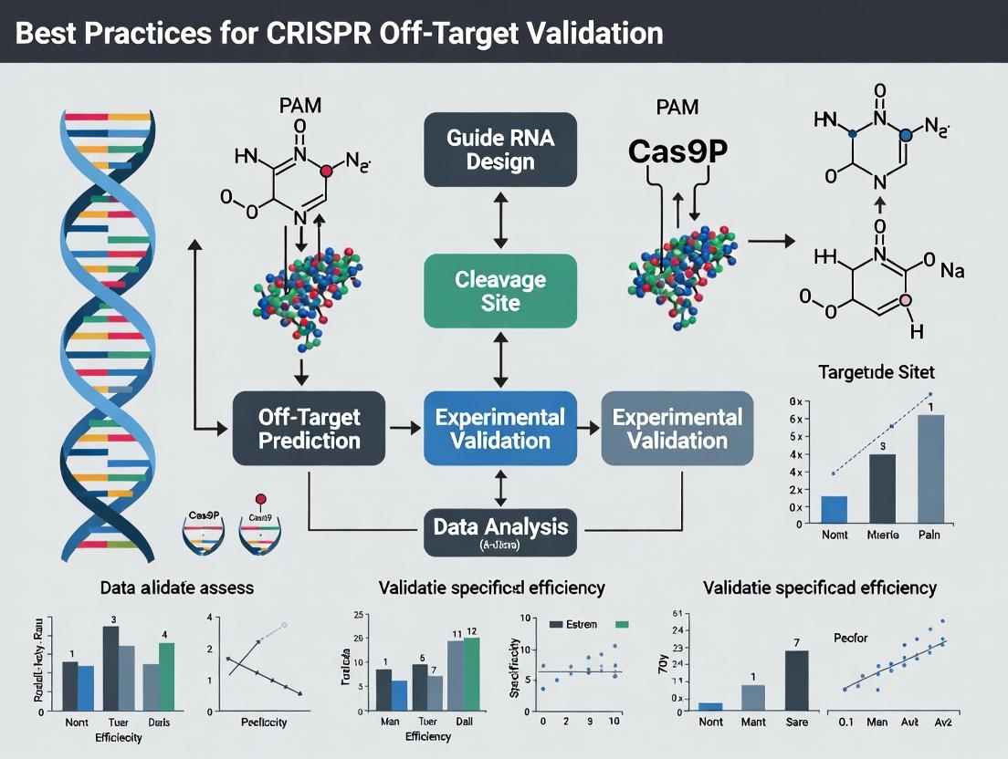
CRISPR Off-Target Validation: A Comprehensive Guide to Methods, Best Practices, and Clinical Standards
This article provides a detailed guide for researchers and drug development professionals on validating CRISPR off-target effects.
CRISPR Off-Target Validation: A Comprehensive Guide to Methods, Best Practices, and Clinical Standards
Abstract
This article provides a detailed guide for researchers and drug development professionals on validating CRISPR off-target effects. It covers foundational concepts of off-target cleavage, explores the latest in silico prediction tools and experimental validation methods (including GUIDE-seq, CIRCLE-seq, and Digenome-seq), offers troubleshooting strategies for common pitfalls, and compares the strengths and limitations of different validation approaches. The goal is to equip scientists with the knowledge to design rigorous, reproducible validation workflows essential for preclinical research and therapeutic development.
Understanding CRISPR Off-Target Effects: Mechanisms, Risks, and Foundational Concepts
In CRISPR-based therapeutic development, the primary goal is precise genomic editing at the intended locus (on-target). Off-target effects refer to unintended edits at sites with similar sequences, which can compromise efficacy and safety. Rigorous off-target validation is therefore a critical component of the development pathway.
Technical Support Center: Troubleshooting Off-Target Analysis
FAQs & Troubleshooting Guides
Q1: Our GUIDE-seq experiment yields an overwhelming number of potential off-target sites. How do we prioritize them for validation? A: This is common. Prioritize sites based on:
- Mismatch Count & Location: Sites with ≤4 mismatches, especially in the PAM-distal "seed" region (nucleotides 1-12), are higher risk.
- Read Count: Sites with higher GUIDE-seq read counts indicate higher cleavage efficiency.
- Genomic Context: Prioritize sites within exons, regulatory elements, or known oncogenes/tumor suppressors.
Q2: We suspect our cell line has genetic variants that affect gRNA binding and off-target profiles. How should we proceed? A: Always sequence the target genomic region in your specific cell line or model before designing gRNAs. Use the exact reference sequence for off-target prediction. For validation, use an endogenously tagged assay (like targeted NGS) rather than relying solely on synthetic reporter assays.
Q3: Our targeted next-generation sequencing (NGS) for off-target validation shows high background noise. How can we improve the signal-to-noise ratio? A:
- Increase Sequencing Depth: Aim for a minimum of 100,000x read depth per amplicon.
- Optimize PCR: Use high-fidelity polymerase and minimize PCR cycles to reduce errors.
- Use Duplex Sequencing: Implement a method that sequences both strands of DNA, requiring mutations on both to call a true edit, drastically reducing false positives.
- Include Biological Replicates: Noise is inconsistent; true off-targets are reproducible.
Q4: What are the key advantages and limitations of computational prediction vs. unbiased biochemical assays for off-target identification? A: See the comparison table below.
Table 1: Comparison of Off-Target Identification Methods
| Method | Principle | Key Advantage | Key Limitation | Best For |
|---|---|---|---|---|
| In Silico Prediction(e.g., Cas-OFFinder) | Algorithms scan genome for sequences similar to the gRNA. | Fast, inexpensive, guides initial design. | Misses sites with structural variants or >4-5 mismatches; high false-negative rate. | Initial gRNA screening and risk assessment. |
| Biochemical Assays(e.g., CIRCLE-seq) | Purified Cas9-gRNA complex cleaves sheared genomic DNA in vitro; sites are sequenced. | Unbiased, genome-wide, sensitive, no cellular context needed. | May overpredict sites not accessible in chromatin in vivo. | Comprehensive, cell-type-agnostic risk profile. |
| Cellular Assays(e.g., GUIDE-seq) | Integration of a double-stranded oligodeoxynucleotide tag into double-strand breaks in living cells. | Captures off-targets in the native chromatin context of the specific cell type. | Requires efficient delivery of the dsODN tag; may miss low-frequency events. | Gold standard for validating off-targets in the relevant therapeutic cell model. |
Experimental Protocol: GUIDE-seq for Unbiased Off-Target Detection in Cells
Objective: To identify Cas9 off-target cleavage sites in a relevant cell line.
Materials: See "The Scientist's Toolkit" below. Workflow:
- Co-delivery: Transfect cells with three components: a) plasmid expressing SpCas9 and gRNA, b) GUIDE-seq dsODN tag (50-200 nM), and c) a fluorescent reporter plasmid to assess transfection efficiency.
- Harvest & Genomic DNA Extraction: Harvest cells 72 hours post-transfection. Extract high-molecular-weight genomic DNA.
- Library Preparation:
- Shear genomic DNA to ~500 bp fragments.
- Repair ends and ligate sequencing adaptors.
- Perform nested PCR using one primer specific to the dsODN tag and another specific to the adaptor to enrich for tag-integrated fragments.
- Next-Generation Sequencing: Sequence the amplified library on a high-output platform (e.g., Illumina MiSeq).
- Bioinformatics Analysis:
- Map sequences to the reference genome.
- Identify genomic junctions containing the dsODN tag sequence.
- Cluster junction sites and rank them by read count.
- Compare sites to the on-target sequence to identify mismatches.
GUIDE-seq Experimental Workflow for Off-Target Detection.
The Scientist's Toolkit: Essential Reagents for Off-Target Validation
| Reagent / Solution | Function in Experiment |
|---|---|
| High-Fidelity PCR Enzyme(e.g., Q5, KAPA HiFi) | Amplifies target loci for NGS with ultra-low error rates, crucial for detecting low-frequency edits. |
| GUIDE-seq dsODN Tag | A blunt, double-stranded oligodeoxynucleotide that integrates into Cas9-induced DSBs, serving as a molecular barcode for later enrichment and sequencing. |
| Validated SpCas9 Nuclease | Consistent, high-activity nuclease is essential for reproducible on- and off-target cleavage profiles. Use a well-characterized commercial source. |
| Targeted Locus Amplification Primers | Primer pairs designed to amplify the on-target site and predicted/computed off-target loci (~300-400 bp amplicons) for deep sequencing. |
| Duplex Sequencing Adapters | Specialized NGS adapters that allow for bioinformatic pairing of reads from complementary DNA strands, enabling error correction and ultra-sensitive variant detection. |
| Positive Control gRNA/Plasmid | A gRNA with a well-published off-target profile (e.g., for VEGFA site 3) to serve as a system control for assay performance. |
Conceptual Framework: The Role of Specificity in Therapeutic Development
Impact of Editing Tool Specificity on Therapeutic Outcomes.
Best Practices Thesis Context: A robust off-target validation strategy, as part of the broader thesis on best practices, must employ a tiered approach. This begins with in silico prediction for gRNA selection, proceeds to an unbiased biochemical method (like CIRCLE-seq) for a comprehensive risk assessment, and culminates in a cellular context-specific assay (like GUIDE-seq or targeted NGS) in the most therapeutically relevant model. Quantitative data from these orthogonal methods must be summarized and compared to build a credible safety profile before clinical translation.
Troubleshooting Guide & FAQs
Q1: Why does my CRISPR-Cas9 experiment show unexpected bands on a gel, even with a highly specific gRNA? A: This is likely due to off-target cleavage. Cas9 tolerates mismatches between the gRNA and genomic DNA, particularly in the 5' "seed" region (nucleotides 1-12 proximal to the PAM) and if mismatches are non-consecutive. Additionally, non-canonical PAM sequences (e.g., NAG for SpCas9) can be recognized at lower efficiency, leading to cleavage at unintended genomic loci.
Q2: How many mismatches can a gRNA typically tolerate before off-target effects are eliminated? A: There is no absolute number; tolerance depends on mismatch position, type, and distribution. Central mismatches (positions 4-12) are generally less tolerated than distal ones. However, up to 5 or more mismatches have been reported to cause cleavage in some contexts, especially if accompanied by a permissive PAM.
Q3: What is the most reliable method to identify potential off-target sites for my gRNA? A: A combination of in silico prediction and unbiased genome-wide validation is considered best practice. Relying solely on prediction algorithms (which use rules based on mismatch tolerance and PAM flexibility) can miss true off-targets. Experimental methods like CIRCLE-seq or GUIDE-seq are recommended for comprehensive profiling.
Q4: Our drug development project requires minimal off-target risk. Which Cas enzyme variant should we consider? A: High-fidelity variants (e.g., SpCas9-HF1, eSpCas9(1.1)) are engineered to reduce mismatch tolerance. For broader PAM targeting with potentially different off-target profiles, consider Cas12a or evolved SpCas9 variants like SpRY. The choice must be validated empirically for your specific target sequence.
Q5: How does chromatin accessibility influence off-target cleavage? A: Open chromatin regions are more susceptible to both on- and off-target cleavage. A predicted off-target site in heterochromatin may not be cleaved in vivo, while a site in euchromatin with multiple mismatches might be. This underscores the need for validation in relevant cellular contexts.
Key Experimental Protocols for Off-Target Validation
Protocol 1: GUIDE-seq (Genome-wide, Unbiased Identification of DSBs Enabled by Sequencing)
- Transfection: Co-deliver your CRISPR-Cas9 ribonucleoprotein (RNP) complex with a double-stranded oligonucleotide tag (GUIDE-seq tag) into your target cells.
- Integration: Upon Cas9-mediated double-strand break (DSB), the tag integrates into the break site via non-homologous end joining (NHEJ).
- Genomic DNA Extraction & Shearing: Harvest cells after 48-72 hours, extract gDNA, and shear it to ~500 bp fragments.
- Library Prep & Enrichment: Perform end-repair, A-tailing, and adapter ligation. Use PCR to enrich for tag-containing fragments.
- Sequencing & Analysis: Sequence using paired-end Illumina kits. Align reads to the reference genome and identify tag integration sites as potential DSB loci. Compare to negative control samples.
Protocol 2: CIRCLE-seq (Circularization for In Vitro Reporting of Cleavage Effects by Sequencing)
- Genomic DNA Isolation & Shearing: Isolate genomic DNA from your cell type of interest and shear it.
- Circularization: Ligate sheared DNA into circular molecules using ssDNA ligase.
- In Vitro Cleavage: Incubate the circularized genomic DNA with your Cas9 RNP complex. Circular DNA is resistant to non-specific digestion but linearizes upon Cas9 cleavage at a target site.
- Exonuclease Digestion: Treat with exonuclease to degrade all non-cleaved, linear DNA, leaving only linearized fragments originating from Cas9 cut sites.
- Library Prep & Sequencing: Add sequencing adapters via PCR or ligation, then sequence. Map breakpoints to identify off-target sites with single-nucleotide resolution.
Data Presentation
Table 1: Comparison of Key Off-Target Detection Methods
| Method | Principle | Sensitivity | Throughput | Required Control | Key Limitation |
|---|---|---|---|---|---|
| GUIDE-seq | Tag integration into DSBs in vivo | High (detects sites at ~0.1% frequency) | Genome-wide | Untransfected cells | Requires efficient delivery of oligonucleotide tag. |
| CIRCLE-seq | In vitro cleavage of circularized genomic DNA | Very High (detects low-affinity sites) | Genome-wide | No-RNP or mock digest | Purely in vitro; may miss chromatin effects. |
| Digenome-seq | In vitro cleavage of cell-free genomic DNA, then whole-genome sequencing | High | Genome-wide | Mock-digested genomic DNA | In vitro method; high sequencing depth/cost. |
| BLISS | Direct labeling and capture of DSB ends | Moderate-High | Targeted or Genome-wide | Background DSB controls | Technically challenging; lower signal-to-noise. |
| In Silico Prediction | Algorithmic scanning for similar sequences | Low-Moderate (prone to false +/-) | High | N/A | Misses sites with atypical mismatch patterns or PAMs. |
Table 2: gRNA Mismatch Tolerance Profile (Representative SpCas9 Data)
| Mismatch Position(s) | Relative Cleavage Efficiency* | Notes |
|---|---|---|
| Distal (PAM-distal 1-5) | 60-100% | Often well-tolerated, especially single mismatches. |
| Seed Region (PAM-proximal 1-12) | <1-20% | Dramatically reduces cleavage; central mismatches (8-12) are most disruptive. |
| ≥3 Non-consecutive, distributed | 1-50% | Highly variable; depends on context and PAM. |
| ≥2 Consecutive in seed region | <1% | Typically abolishes cleavage. |
| PAM (NGG → NAG) | ~25% of NGG efficiency | Common non-canonical PAM for SpCas9. |
| PAM (NGG → NGA) | <1% of NGG efficiency | Very poor recognition by wild-type SpCas9. |
*Efficiency is highly sequence-dependent. Data aggregated from multiple studies (Tsai et al., 2015; Hsu et al., 2013; Zhang et al., 2015).
Visualization: Mechanisms and Workflows
Title: Off-Target Cleavage Determinants & Consequences
Title: GUIDE-seq Experimental Workflow
The Scientist's Toolkit: Research Reagent Solutions
Table 3: Essential Reagents for Off-Target Validation Research
| Item | Function/Description | Example/Note |
|---|---|---|
| High-Fidelity Cas9 Variants | Engineered Cas9 proteins with reduced mismatch tolerance to minimize off-target cleavage. | SpCas9-HF1, eSpCas9(1.1). Critical for therapeutic applications. |
| Alt-R S.p. HiFi Cas9 Nuclease | A commercial, high-fidelity Cas9 nuclease optimized for RNP delivery with reduced off-target activity. | Integrated DNA Technologies (IDT). |
| GUIDE-seq Oligonucleotide Tag | A defined, double-stranded oligonucleotide that integrates into DSBs for genome-wide off-target identification. | Required for the GUIDE-seq protocol. |
| CIRCLE-seq Kit | Commercial kit providing optimized reagents for circularization and in vitro cleavage assays. | Available from vendors like ToolGen. |
| Next-Generation Sequencing (NGS) Kit | For preparing libraries from validation assays (GUIDE-seq, CIRCLE-seq, Digenome-seq). | Illumina TruSeq, NEBNext Ultra II. |
| Prediction Algorithm Software | In silico tools to predict potential off-target sites based on sequence similarity. | CRISPOR, Cas-OFFinder, ChopChop. Use as a first pass, not definitive. |
| Positive Control gRNA | A gRNA with known, validated off-target sites for assay calibration and troubleshooting. | e.g., VEGFA site 2 or EMX1 gRNAs with published off-target profiles. |
| Negative Control RNP | RNP with a non-targeting gRNA or catalytically dead Cas9 (dCas9) to establish background signal. | Essential for distinguishing specific cleavage events. |
Troubleshooting Guides & FAQs
Q1: Our targeted deep sequencing results show high background noise, making it difficult to call potential off-target sites. What could be the cause? A: This is often due to insufficient specificity in the PCR amplification step. Ensure you are using validated, high-fidelity polymerase and optimized cycling conditions. Primer-dimer formation or non-specific amplification can be mitigated by using touchdown PCR or incorporating blocking oligonucleotides. Re-validate your primer sets in silico and with a no-template control.
Q2: We are using CIRCLE-seq and finding an overwhelming number of potential off-target sites, many in genomic regions that seem irrelevant. How do we prioritize for validation? A: It is normal for sensitive in vitro methods like CIRCLE-seq to generate large candidate lists. Prioritize sites based on: 1) The number of mismatches from the on-target sequence (sites with ≤5 mismatches are higher priority), 2) The presence of mismatches in the seed region (PAM-proximal 8-12 bases), which is more disruptive, and 3) Genomic context (prioritize sites within exons or regulatory regions of active genes). Use predictive algorithms (e.g., CRISPRseek, Cas-OFFinder) to rank by likelihood.
Q3: During GUIDE-seq, we are not detecting integration events, leading to no off-target data. What are the key troubleshooting steps? A: First, confirm the double-stranded oligonucleotide tag is properly purified and at a sufficient concentration (typically 100-250 nM final). Second, optimize the transfection/nucleofection efficiency for your cell line; low delivery efficiency is the most common failure point. Third, ensure you are using a high-activity Cas9/gRNA RNP complex. Include a positive control with a well-characterized gRNA.
Q4: Discrepancies exist between off-target sites identified by biochemical methods (e.g., Digenome-seq) and cellular methods (e.g., GUIDE-seq). Which results are more reliable? A: Each method has strengths. Biochemical methods (Digenome-seq, CIRCLE-seq) are highly sensitive and unbiased by cellular context but may identify sites not cut in cells. Cellular methods (GUIDE-seq, SITE-seq) reflect the chromatin environment and repair activity of your specific cell type but may miss low-frequency events. The most rigorous validation uses a complementary approach, prioritizing sites identified by multiple, orthogonal methods. For clinical applications, a union of the most sensitive methods is recommended.
Q5: How do we functionally validate a predicted off-target edit, especially when it's in a non-coding region? A: For coding regions: sequence the locus and assess protein changes/expression. For non-coding regions: 1) Assess chromatin accessibility (ATAC-seq) and histone marks (ChIP-seq) to see if the site is regulatory, 2) Perform RNA-seq or qPCR on nearby genes to check for expression dysregulation, 3) Use a reporter assay (e.g., luciferase) by cloning the wild-type and edited genomic region upstream of a minimal promoter. Phenotypic assays relevant to your study (proliferation, differentiation) are also crucial.
The following table summarizes the core characteristics, detection limits, and requirements of major off-target profiling methods.
| Method | Principle | Detection Limit | Throughput | Key Requirement | Primary Advantage | Key Limitation |
|---|---|---|---|---|---|---|
| GUIDE-seq | Integration of double-stranded oligo tags at DSBs via NHEJ. | ~0.1% indel frequency | Medium | High transfection efficiency; living cells. | Unbiased detection in relevant cellular context. | Requires non-homologous end joining (NHEJ). |
| CIRCLE-seq | In vitro circularization and sequencing of Cas9-cleaved genomic DNA. | ≤0.01% in vitro | High | High-quality genomic DNA; biotinylated gRNA. | Extremely sensitive, genome-wide, and cell-context independent. | Purely in vitro; may overpredict sites. |
| Digenome-seq | In vitro Cas9 cleavage of naked genomic DNA followed by whole-genome sequencing. | ≤0.1% in vitro | High | High-coverage WGS (~80x); high-fidelity Cas9. | Single-nucleotide resolution, genome-wide. | Uses naked DNA; computationally intensive. |
| SITE-seq | Selective capture and sequencing of Cas9-cleaved genomic DNA ends. | ~0.1% in vitro | Medium | Biotinylated Cas9 protein; streptavidin capture. | Sensitive; uses cellular chromatin as substrate. | Requires optimized chromatin extraction. |
| BLISS | Direct labeling and sequencing of DSB ends in situ. | Single-molecule | Low-Medium | Fixed cells or nuclei; requires in situ ligation. | Can be applied to fixed samples and low-input. | Technically challenging; lower coverage. |
Experimental Protocols
Protocol 1: Targeted Deep Sequencing for Off-Target Validation This protocol is used to quantify indel frequencies at predicted off-target loci.
- Design Primers: For each candidate off-target locus and the on-target locus, design ~200-300 bp amplicons using tools like Primer-BLAST. Add universal adapter sequences for next-generation sequencing (NGS).
- Extract Genomic DNA: Harvest edited cells 72+ hours post-transfection. Use a column-based kit for high-purity gDNA. Quantify via fluorometry.
- PCR Amplification: Perform first-round PCR with locus-specific primers (10-15 cycles) using a high-fidelity polymerase. Use a pooling strategy if validating >10 sites.
- Indexing PCR: Add sample-specific barcodes and full NGS adapters in a second, limited-cycle (8-12 cycles) PCR.
- Purify & Pool: Purify PCR products with magnetic beads, quantify, and pool equimolarly.
- Sequencing: Run on an Illumina MiSeq or similar platform (2x250 bp recommended).
- Analysis: Use pipelines like CRISPResso2, TIDE, or BAT to align reads and calculate indel percentages from the amplicon data.
Protocol 2: CIRCLE-seq Workflow This protocol outlines the key steps for sensitive, in vitro off-target discovery.
- Genomic DNA Isolation & Shearing: Extract high-molecular-weight gDNA (>50 kb) from your target cell type. Shear to an average fragment size of 500 bp using a focused-ultrasonicator.
- End Repair & Circularization: Repair sheared DNA ends and 5'-phosphorylate. Ligate the DNA into circles using T4 DNA ligase under dilute conditions to favor intramolecular circularization.
- Cas9 Cleavage In Vitro: Incubate the circularized DNA with purified, active Cas9 protein complexed with the sgRNA of interest. Cas9 will linearize circles containing a target sequence.
- Removal of Uncut Circles & Library Prep: Treat with Plasmid-Safe ATP-dependent exonuclease to degrade all remaining circular and linear chromosomal DNA, enriching for Cas9-linearized fragments. Purify the resistant DNA.
- Sequencing Library Construction: Perform end repair, A-tailing, and adapter ligation on the enriched linear DNA. PCR amplify with indexed primers.
- Bioinformatic Analysis: Map sequencing reads to the reference genome. Identify sites of cleavage as genomic positions where multiple reads start or end abruptly (junction sites). Use peak-calling algorithms specific to CIRCLE-seq data.
Visualization
Title: Off-Target Validation Strategy Workflow
Title: DNA Repair Pathways After CRISPR Cleavage
The Scientist's Toolkit: Research Reagent Solutions
| Reagent / Material | Function / Application | Key Considerations |
|---|---|---|
| High-Fidelity Cas9 Nuclease | Catalyzes the DNA double-strand break. Reduces inherent off-target activity compared to wild-type SpCas9. | Use HiFi Cas9 or eSpCas9(1.1) for improved specificity in validation experiments. |
| Synthetic sgRNA (chemically modified) | Guides Cas9 to the target DNA sequence. Chemical modifications (e.g., 2'-O-methyl, phosphorothioate) enhance stability and can reduce off-target effects. | Preferred over plasmid-based expression for RNP delivery; increases precision and reduces persistence. |
| Next-Generation Sequencing (NGS) Kit | For high-throughput sequencing of amplicons or whole genomes in off-target discovery/validation. | Select kits based on read length (amplicon) or coverage depth (WGS). Ensure low error rate. |
| Genomic DNA Extraction Kit (Magnetic Beads) | For clean, high-molecular-weight gDNA preparation from cells post-editing. Essential for CIRCLE-seq, Digenome-seq, and targeted sequencing. | Avoid shearing; kits designed for long-read sequencing often provide superior quality. |
| dsODN Tag (for GUIDE-seq) | A short, double-stranded oligonucleotide that integrates into DSBs via NHEJ, serving as a tag for sequencing-based capture and identification. | Must be PAGE-purified and used at an optimized concentration to balance efficiency and toxicity. |
| CRISPR Off-Target Analysis Software (e.g., CRISPResso2) | Bioinformatics tool for quantifying indels from targeted amplicon sequencing data. Aligns reads, identifies mutations, and calculates editing efficiency. | Choose tools that account for complex indels and provide clear visualizations. Essential for validation. |
Troubleshooting Guides & FAQs
Q1: Our GUIDE-seq experiment detected unexpected off-target sites with multiple mismatches. How do we determine if these are biologically relevant? A: This is a common challenge. The biological relevance depends on the chromatin context of the off-target site. Use the following workflow to prioritize and validate.
- Prioritize: Cross-reference GUIDE-seq hits with in silico prediction tools (e.g., Cas-OFFinder). Filter for sites with ≤5 mismatches and/or bulges.
- Assess Chromatin State: Perform an ATAC-seq or public ChIP-seq data (e.g., ENCODE) check on the off-target loci. Open chromatin (high accessibility) increases the risk of cleavage.
- Validate Functionally: Perform targeted deep sequencing (amplicon-seq) on the top 10-15 prioritized off-target loci from edited and control samples to quantify actual indel frequencies.
Q2: We suspect bulge off-targets are being missed by our CIRCLE-seq assay. What is the best practice for comprehensive bulge detection? A: CIRCLE-seq is highly sensitive but its library preparation can under-represent certain structural variants. Implement a complementary strategy:
- Optimize CIRCLE-seq: Ensure your protocol uses a polymerase with high processivity (e.g., Phi29) to fully circularize and amplify gDNA fragments containing bulges.
- Supplement with In Silico Screening: Use tools like Cas-OFFinder with settings that allow for both DNA and RNA bulge searches (specify bulge types and sizes).
- Key Reagent: For the polymerase amplification step, use Phi29 DNA Polymerase for its strong strand displacement activity, crucial for amplifying complex secondary structures.
Q3: How does chromatin accessibility quantitatively impact off-target cleavage efficiency? A: Data consistently shows a strong correlation. Off-target sites in open chromatin are cleaved orders of magnitude more efficiently than those in closed, heterochromatic regions.
Table 1: Impact of Chromatin Context on Off-Target Cleavage Efficiency
| Off-Target Site Characteristic | Relative Cleavage Efficiency (vs. On-Target) | Experimental Method Used for Validation |
|---|---|---|
| ≤3 mismatches in open chromatin (DNase I hypersensitive site) | 0.1% - 10% | GUIDE-seq + ATAC-seq correlation |
| ≤3 mismatches in closed chromatin (H3K9me3 marked) | <0.01% | ChIP-seq + targeted amplicon-seq |
| 1-nt RNA bulge in open chromatin | Up to 1% | CIRCLE-seq + in vitro cleavage assay |
| 1-nt DNA bulge in closed chromatin | Often undetectable | Biochemical Cas9 cleavage assay |
Q4: What is a definitive step-by-step protocol for validating a specific suspected off-target site identified in silico? A: Protocol for Targeted Locus Amplification & Deep Sequencing (TLA-amplicon-seq)
- Design Primers: Design PCR primers (amplicon size 250-350 bp) flanking the suspected off-target locus.
- PCR Amplification: Perform first-round PCR on purified genomic DNA from edited and control cell pools (n≥3). Use a high-fidelity polymerase.
- Indexing & Clean-up: Add Illumina sequencing adapters and barcodes via a second limited-cycle PCR. Purify amplicons with SPRI beads.
- Sequencing & Analysis: Pool and sequence on a MiSeq (≥50,000x read depth per amplicon). Analyze files with a pipeline like CRISPResso2 to quantify indel percentages.
Q5: Our drug development program requires the highest confidence in off-target profiling. What is the current gold-standard combinatorial approach? A: The most rigorous practice combines in vitro, in silico, and cellular methods.
- Primary Screen: Perform CIRCLE-seq or SITE-seq on the purified RNP complex to identify in vitro cleavage sites comprehensively, including bulges.
- Cellular Relevance Filter: Perform GUIDE-seq or DISCOVER-seq in your relevant cell type to identify sites cut in the native chromatin context.
- Final Validation: Use targeted amplicon-seq (as in Q4) on the union of top hits from steps 1 & 2 to provide quantitative, cell-based indel rates for a final, manageable list of loci (<20).
The Scientist's Toolkit: Research Reagent Solutions
Table 2: Essential Reagents for Comprehensive Off-Target Analysis
| Reagent / Kit | Primary Function in Off-Target Validation |
|---|---|
| CIRCLE-seq Kit | Provides optimized reagents for the circularization, digestion, and amplification steps of the CIRCLE-seq protocol, enhancing sensitivity for bulge detection. |
| GUIDE-seq Oligoduplex | A double-stranded, end-protected oligo that integrates into double-strand breaks during GUIDE-seq to tag and identify off-target sites in cells. |
| High-Fidelity DNA Polymerase (e.g., Q5, KAPA HiFi) | Critical for error-free amplification of genomic loci during amplicon-seq for validation, preventing false-positive indel calls. |
| Recombinant SpCas9 Nuclease | For in vitro cleavage assays and RNP complex formation in CIRCLE-seq. Ensures the protein component is consistent and pure. |
| Phi29 DNA Polymerase | Used in CIRCLE-seq for rolling circle amplification; its high processivity is key to amplifying complex or damaged DNA templates. |
| CRISPResso2 Software | The standard analysis suite for deep sequencing data from amplicon-seq. Precisely quantifies indel frequencies and types from NGS reads. |
Experimental Workflow Visualization
Diagram Title: Integrated Off-Target Analysis Workflow
Diagram Title: Chromatin Context Modifies Off-Target Risk
Technical Support Center: Troubleshooting Guides and FAQs for CRISPR Off-Target Validation
Thesis Context: This support content is framed within a thesis on "Best Practices for CRISPR Off-Target Validation Research," a critical prerequisite for robust preclinical data packages supporting Investigational New Drug (IND) or Clinical Trial Application (CTA) submissions.
Frequently Asked Questions (FAQs)
Q1: What are the key off-target validation expectations from health authorities (e.g., FDA, EMA) for an IND/CTA involving a CRISPR-based therapy? A: Regulators expect a risk-based, multi-faceted approach. The core expectation is the use of complementary, orthogonal methods to profile off-target activity. Key requirements include:
- In Silico Prediction: Use of established tools (e.g., CFD score, MIT specificity score) to identify potential off-target sites.
- Biochemical/Cell-Free Assays: Performing methods like CIRCLE-seq or SITE-seq to identify cleavage-prone sites in a genome-wide, unbiased manner.
- Cellular-Based Assays: Validating top candidate off-target sites identified above using targeted deep sequencing in relevant cell types.
- Rationale: A clear justification for the chosen methods, gRNA design, and the cellular context of the testing.
Q2: Our targeted deep sequencing data shows low-frequency indels (<0.1%) at predicted off-target sites. Are these biologically relevant, and must they be reported? A: Yes, they must be reported and contextualized. The threshold for reporting is often not absolute but risk-based. Considerations include:
- Location: Is the off-target site in an oncogene, tumor suppressor, or functional genomic region?
- Frequency: How does the frequency compare to the natural mutation rate or negative control?
- Dose-Response: Does the indel frequency correlate with editing efficiency or dose?
- Best Practice: Include all data, provide a biological risk assessment, and discuss follow-up plans (e.g., in vivo safety studies).
Q3: We are getting high background noise in our GUIDE-seq or Digenome-seq experiments. What are the common troubleshooting steps? A: High background is often due to non-specific tag integration or DNA damage/sequencing artifacts.
- For GUIDE-seq: Ensure optimal electroporation conditions for tag oligo delivery. Titrate the amount of tag oligo (typical range 0.5-5 µM). Include a nuclease-negative control to identify background integration sites. Optimize PCR amplification cycles to prevent over-amplification.
- For Digenome-seq: Use high-quality, high-molecular-weight genomic DNA. Precisely optimize the concentration of purified Cas9-gRNA RNP for in vitro digestion. Include a no-RNP control to identify baseline DNA break sites. Ensure complete digestion and careful purification before sequencing library prep.
Q4: How do we justify which cell type to use for off-target validation if our therapy targets a specific tissue? A: The strongest justification uses therapeutically relevant cells. If using primary cells is not feasible, justify your model:
- Primary cells or target tissue explants (Gold standard).
- Differentiated iPSCs into the relevant lineage.
- Cell lines that endogenously express the target gene at similar levels.
- Justification must be provided if using an alternative model (e.g., HEK293T for accessibility), acknowledging the limitation and outlining any plans for supplementary testing.
Troubleshooting Guides
Issue: Inconsistent or Low Signal in Targeted Amplicon Sequencing for Off-Target Validation.
| Symptom | Potential Cause | Solution |
|---|---|---|
| Low read depth for specific amplicons | Primer bias or inefficient amplification | Redesign primers using amplicon melting temperature (Tm) balancing tools. Use a high-fidelity, GC-rich polymerase. |
| High false-positive indel calls in controls | Sequencing errors or PCR artifacts | Apply a minimum variant frequency threshold (e.g., 0.01%–0.1%). Use duplex sequencing or unique molecular identifiers (UMIs). Include multiple negative controls (untransfected, nuclease-dead). |
| Inability to amplify large amplicons (>500bp) | Complex genomic regions or gDNA quality | Optimize PCR extension time. Check gDNA integrity on agarose gel. Consider designing multiple, smaller overlapping amplicons. |
Issue: Poor Efficiency in Biochemical Off-Target Profiling (e.g., CIRCLE-seq).
| Symptom | Potential Cause | Solution |
|---|---|---|
| Low library complexity after circularization | Incomplete ssDNA ligation or RNase H step failure | Precisely quantify ssDNA before circularization. Freshly prepare RNase H buffer and confirm enzyme activity. |
| High reads mapping to mitochondrial DNA | Contamination with mitochondrial DNA or non-specific cleavage | Use a nuclear DNA isolation kit. Include a Cas9-only (no gRNA) control to identify sequence-independent cleavage. |
| Failure to detect known positive control sites | Over-digestion or suboptimal RNP concentration | Titrate RNP concentration (start with 50-200 nM). Reduce digestion time (start with 1 hour at 37°C). |
Experimental Protocol: Integrated Off-Target Analysis Workflow
Protocol: Orthogonal Validation for IND/CTA-Enabling Studies
Objective: To identify and validate CRISPR-Cas9 off-target sites using a two-step, orthogonal methodology.
Part A: Unbiased Genome-Wide Screening (CIRCLE-seq)
- Genomic DNA Isolation: Isolate high-molecular-weight gDNA (>50 kb) from relevant cell type.
- DNA Shearing and End-Repair: Fragment gDNA (Covaris) to ~300 bp. Repair ends, add dA-tail.
- Adapter Ligation & Circularization: Ligate Y-shaped adapters. Treat with RNase H and APE1 to create ssDNA nicks. Circularize using ssDNA ligase.
- In Vitro Digestion: Incubate circularized DNA with purified Cas9-gRNA RNP (100 nM) for 1 hour at 37°C.
- Library Preparation: Linearize cleaved circles. PCR amplify with indexed primers. Sequence on a high-output platform (NovaSeq).
- Bioinformatics: Map reads, identify cleavage-enriched peaks. Rank sites by read count and CFD score.
Part B: Targeted Deep Sequencing Validation
- Primer Design: Design 200-350 bp amplicons for top 10-20 off-target sites from Part A plus on-target.
- Cell Transfection: Edit relevant cells using your clinical-grade method (e.g., electroporation of RNP).
- gDNA Harvest & PCR: Harvest gDNA at peak editing time (e.g., 72 hrs). Perform PCR with barcoded primers.
- Sequencing & Analysis: Pool amplicons for Illumina MiSeq (2x300 bp). Analyze with CRISPResso2 or similar. Report indel frequencies and statistical significance vs. controls.
The Scientist's Toolkit: Research Reagent Solutions for CRISPR Off-Target Validation
| Item | Function & Rationale |
|---|---|
| High-Fidelity Cas9 Nuclease | Ensures precise on-target activity and reduces spurious cleavage. Critical for generating reliable RNP for biochemical assays. |
| Chemically Modified sgRNA | Increases stability and reduces immune activation in cellular assays. Use with RNP delivery for more therapeutically relevant models. |
| CIRCLE-seq Kit | Commercial kit (e.g., from IDT or NEB) standardizes the complex workflow, improving reproducibility for IND-enabling studies. |
| Duplex Sequencing Adapters | Enables error-corrected, ultra-sensitive sequencing by using UMIs, crucial for detecting very low-frequency (<0.1%) off-target events. |
| Relevant Genomic DNA | gDNA from primary cells or the target tissue provides the most physiologically relevant substrate for in vitro and cellular assays. |
| Positive Control gRNA | A gRNA with well-characterized off-target profile (e.g., VEGFA site 3) is essential for validating the performance of your entire workflow. |
| CRISPResso2 Software | Standardized, peer-reviewed bioinformatics tool for consistent analysis of targeted deep sequencing data, ensuring regulatory compliance. |
The Validation Toolkit: In Silico Prediction and Cutting-Edge Experimental Assays
Technical Support Center
Troubleshooting Guides & FAQs
Q1: I entered a target sequence into Cas-OFFinder but got zero predicted off-targets. Is my guide RNA perfect? A: Not necessarily. A null result often stems from overly strict default parameters.
- Check Mismatch Tolerance: The default is often 3 or fewer mismatches. Increase this to 4-6, especially for initial screening.
- Check the PAM Sequence: Ensure you selected the correct PAM (e.g., NGG for SpCas9) for your specific Cas nuclease variant.
- Verify Input Sequence: Ensure the input is the 20-nt spacer sequence only, without the PAM.
Q2: CHOPCHOP provides many different scores (e.g., Efficiency, Specificity). Which ones are most critical for minimizing off-targets? A: For off-target validation research, prioritize:
- Specificity Score: Directly correlates with predicted off-target potential. A lower score indicates higher risk.
- CFD (Cutting Frequency Determination) Score: Predicts the likelihood of cleavage at off-target sites with mismatches. Integrate this with specificity.
- Do-It-Yourself (DIY) Guide Score: A composite score weighing both efficiency and specificity. Use it for a balanced view.
Q3: CRISPOR's output lists hundreds of potential off-target sites. How do I prioritize which ones to validate experimentally? A: Use a tiered prioritization strategy based on CRISPOR’s combined output:
- Tier 1 (Highest Priority): Sites with ≤3 mismatches, high CFD off-target score (>0.1), and located in exonic or regulatory regions.
- Tier 2 (Medium Priority): Sites with 4 mismatches but a high CFD score, or sites with ≤3 mismatches in intronic/non-genic regions.
- Tier 3 (Lower Priority): Sites with ≥5 mismatches or very low CFD scores (<0.01).
Q4: The predicted top off-target sites from these three tools do not fully overlap. Which tool should I trust? A: This is expected due to different algorithms and databases. Best practice is to use a consensus approach.
- Action: Combine the top 10-20 predicted off-targets from each tool into a master list. Remove duplicates and validate sites that appear in predictions from at least two tools.
Q5: My guide RNA has a high predicted on-target efficiency but also many high-risk off-targets. What are my options? A: Consider the following before proceeding:
- Select an Alternate Guide: Use the tools to screen adjacent guides targeting the same genomic region.
- Switch Cas Nuclease: Consider high-fidelity variants (e.g., SpCas9-HF1, eSpCas9) which are often pre-modeled in these tools.
- Use Truncated Guides (tru-gRNAs): Shortening the guide to 17-18nt can increase specificity, though it may reduce on-target efficiency.
Comparative Tool Analysis & Best Practices for Off-Target Validation
Table 1: Core Algorithm & Database Comparison
| Feature | Cas-OFFinder | CHOPCHOP | CRISPOR |
|---|---|---|---|
| Core Algorithm | Exact string search with Hamming distance. | Bowtie for alignment, integrates MIT/CFD specificity. | BWA for alignment, integrates MIT and CFD scores. |
| Primary Scoring | Mismatch count & pattern. | MIT specificity score, DIY score, efficiency predictions. | MIT specificity score, CFD on/off-target scores, Doench '16 efficiency. |
| Genome Database | User-provided or pre-built FASTA. | Direct links to Ensembl, UCSC, RefSeq. | Direct links to Ensembl, UCSC, with more frequent updates. |
| Key Strength | Unrestricted PAM definition, ultra-fast for bulk screening. | Integrated design for knock-ins/primers, user-friendly. | Most comprehensive score integration and detailed output. |
Table 2: Quantitative Output Metrics for a Representative gRNA (SpCas9, Human GRCh38)
| Metric | Cas-OFFinder | CHOPCHOP | CRISPOR |
|---|---|---|---|
| Predicted On-Target Efficiency | Not Provided | 68 (DIY Score) | 70 (Doench '16 Score) |
| No. of Off-Targets (≤3 mismatches) | 15 | 12 | 18 |
| No. of Off-Targets (≤4 mismatches) | 142 | 127 | 155 |
| Top Off-Target CFD Score | Not Provided | 0.95 | 0.89 |
Protocol 1: Consensus Off-Target Site Identification for Validation
- Input: Define your 20-nt spacer sequence and precise Cas nuclease PAM requirement.
- Parallel Analysis: Run the same gRNA sequence through Cas-OFFinder (set mismatch=4, DNA bulge=0, RNA bulge=0), CHOPCHOP (select specificity scoring), and CRISPOR.
- Data Extraction: Compile the top 20 predicted off-target sites from each tool, including genomic location, mismatch count/pattern, and CFD score (if available).
- Generate Master List: Merge lists into a single spreadsheet. Rank by: a) Appearance in multiple tools, b) Mismatch count (fewer first), c) CFD score (higher first).
- Final Selection: Select the top 10-15 unique genomic loci from the ranked master list for experimental validation (e.g., GUIDE-seq, targeted deep sequencing).
The Scientist's Toolkit: Research Reagent Solutions
| Item | Function in Off-Target Validation |
|---|---|
| High-Fidelity Cas9 Variant (e.g., Alt-R S.p. HiFi Cas9) | Recombinant nuclease engineered for reduced off-target cleavage while maintaining high on-target activity. |
| Synthetic Modified gRNA (e.g., Alt-R CRISPR-Cas9 crRNA+tracrRNA) | Chemically modified synthetic RNAs enhance stability and can reduce immune responses in cells, improving assay reliability. |
| GUIDE-seq Kit | All-in-one solution for unbiased, genome-wide off-target profiling. Includes annealed tag oligo and PCR primers for library prep. |
| Off-Target PCR Primers | Primer sets designed to amplify genomic loci identified by in silico tools for targeted deep sequencing validation. |
| NGS Library Prep Kit for Amplicons | Kit to prepare barcoded sequencing libraries from PCR-amplified potential off-target sites for multiplexed analysis. |
Visualization: Off-Target Validation Workflow
Title: CRISPR Off-Target Prediction & Validation Workflow
Technical Support Center: Troubleshooting & FAQs
Context: This support content is provided within the framework of a thesis on Best Practices for CRISPR Off-Target Validation Research. GUIDE-seq is a critical, genome-wide method for unbiased detection of CRISPR-Cas9 off-target cleavage sites.
Frequently Asked Questions (FAQs)
Q1: During GUIDE-seq library preparation, my PCR amplification after adapter ligation yields no product. What could be wrong? A: This is often due to inefficient integration of the GUIDE-seq oligonucleotide tag into double-strand breaks (DSBs). Ensure the following:
- The electroporation or transfection efficiency for delivering the Cas9/sgRNA RNP complex and the blunt, double-stranded oligodeoxynucleotide (dsODN) tag is sufficiently high (>70% for cell lines).
- The dsODN tag is purified (e.g., HPLC-grade) and resuspended in nuclease-free buffer. Verify its concentration and integrity on a gel.
- The ratio of dsODN to Cas9 RNP is optimized. A typical starting molar ratio is 100:1 to 500:1 (dsODN:Cas9 RNP). Titrate this ratio in your system.
- Sufficient genomic DNA is used as input for the sonication and library prep steps (≥ 2 µg recommended).
Q2: My sequencing data shows a very high background of dsODN tag reads, obscuring real off-target sites. How can I reduce this? A: High background is commonly caused by incomplete purification of tag-integrated DNA fragments.
- Critical Step: Strictly follow the size selection protocol after the A-tailing and adapter ligation steps. Use SPRI beads with a stringent double size selection (e.g., 0.55x and 1.2x bead-to-sample ratios) to exclude unintegrated, free dsODN tag and very small fragments.
- Include the recommended "no-RNP" negative control in every experiment. This identifies background tag integration events independent of Cas9 cleavage. Genuine off-target sites should be enriched in the +RNP sample compared to this control.
- Verify that your dsODN tag does not contain sequences homologous to your target genome.
Q3: The identified off-target sites from GUIDE-seq do not validate using amplicon sequencing or T7E1 assays. Why the discrepancy? A: Discrepancies can arise from several factors:
- Sensitivity Differences: GUIDE-seq is highly sensitive and can detect very low-frequency events (<0.1%). Secondary validation methods like amplicon-seq may have higher detection limits. Use deep sequencing (>100,000x read depth per site) for validation.
- Experimental Timing: GUIDE-seq captures DSBs at the time of tag integration (typically 48-72h post-delivery). Validation assays performed at a different time point may miss transient editing events.
- Sequence Context: Some identified sites may be in repressed chromatin states in your validation cell type but were accessible during the initial GUIDE-seq experiment. Ensure cell type consistency.
Q4: What are the key negative and positive controls for a robust GUIDE-seq experiment? A: A well-controlled experiment includes:
- No-RNP Control: Cells treated only with the dsODN tag. Essential for identifying background tag integration.
- Transfection/Elec troporation Control: Cells treated with delivery reagent only.
- Positive Control sgRNA: A well-characterized sgRNA with known off-target profile (e.g., VEGFA site 2).
- No-Tag Control (optional): Cells transfected with RNP only, to check for non-specific amplification.
Experimental Protocol: Standard GUIDE-seq Workflow
1. Delivery of RNP and dsODN Tag.
- Materials: Cas9 nuclease, sgRNA (syntheti c or in vitro transcribed), HPLC-purified dsODN tag (e.g., 5'-Phos/[C6 spacer]/GTGACTGGAGTTCAGACGTGTGCTCTTCCGATC*T-[Barcode]-/3BioTEG/-3').
- Method: Form RNP complex by incubating Cas9 and sgRNA at 37°C for 10 min. Co-deliver the RNP complex and dsODN tag into your target cells (e.g., U2OS, HEK293T) via nucleofection. Use 2pmol of Cas9 RNP and 200pmol of dsODN per 100,000 cells as a starting point. Harvest cells 72 hours post-delivery.
2. Genomic DNA Extraction & Shearing.
- Extract gDNA using a phenol-chloroform method or large-fragment compatible kit. Shear 2-4 µg of gDNA to ~500 bp using a focused ultrasonicator (e.g., Covaris).
3. Library Preparation for Sequencing.
- End Repair & A-Tailing: Use standard NGS library prep enzymes.
- Adapter Ligation: Ligate methylated sequencing adapters.
- Critical Size Selection: Perform double-sided SPRI bead cleanup (e.g., 0.55x to retain fragments >~300 bp, then 1.2x to exclude fragments >~700 bp) to deplete unintegrated dsODN.
- PCR Enrichment: Perform 12-14 cycles of PCR using primers containing unique dual indexes. Use a polymerase optimized for GC-rich regions.
- Final Purification: Clean up PCR product with a 0.8x SPRI bead ratio. Quantity by Qubit and analyze fragment size on a Bioanalyzer.
4. Sequencing & Data Analysis.
- Sequencing: Pool libraries and sequence on an Illumina platform (2x150 bp paired-end recommended).
- Analysis: Use the published GUIDE-seq analysis software (available on GitHub) or similar pipelines. Key steps include:
- Align reads to the reference genome + dsODN tag sequence.
- Identify tag integration sites (breakpoints).
- Cluster breakpoints to define candidate off-target loci.
- Annotate loci with mismatch information relative to the sgRNA.
- Compare to the no-RNP control to filter background.
Table 1: Typical GUIDE-seq Experimental Parameters and Outcomes
| Parameter | Typical Value/Range | Notes |
|---|---|---|
| Cells per reaction | 1x10^5 - 2x10^5 | For nucleofection in a 20µL cuvette. |
| Cas9 RNP amount | 2 pmol | For SpCas9 (160 kDa). |
| dsODN:RNP Molar Ratio | 100:1 to 500:1 | Must be titrated for each cell type. |
| Post-delivery incubation | 48 - 72 hours | Allows for tag integration and repair. |
| gDNA Input | ≥ 2 µg | For optimal library complexity. |
| Sequencing Depth | 30 - 50 million PE reads | For human genome (~3 Gb). |
| Detection Sensitivity | < 0.1% indel frequency | More sensitive than NGS amplicon-seq. |
Table 2: Comparison of Common Off-Target Detection Methods
| Method | Principle | Sensitivity | Bias | Throughput | Cost |
|---|---|---|---|---|---|
| GUIDE-seq | Capture of dsODN tag at DSB sites | Very High (<0.1%) | Unbiased (Genome-wide) | High | Medium-High |
| CIRCLE-seq | In vitro circularization & sequencing of Cas9-cleaved genomic DNA | Extremely High (in vitro) | Unbiased (in vitro) | High | Medium |
| Digenome-seq | In vitro Cas9 cleavage, whole-genome sequencing | High (in vitro) | Unbiased (in vitro) | High | High |
| BLISS | Direct ligation of adapters to DSB ends in situ | High | Unbiased | Medium | Medium-High |
| Targeted Amplicon-Seq | Deep sequencing of predicted off-target sites | Medium (≈0.5-1%) | Biased (Predicted sites only) | Low-Medium | Low |
Diagrams
Diagram 1: GUIDE-seq Experimental Workflow
Title: Step-by-step GUIDE-seq Experimental Workflow
Diagram 2: Molecular Mechanism of dsODN Tag Integration
Title: dsODN Tag Integration into Cas9-Induced DSB via NHEJ
The Scientist's Toolkit: Research Reagent Solutions
Table: Essential Reagents for GUIDE-seq Experiments
| Reagent / Material | Function & Specification | Critical Notes |
|---|---|---|
| Recombinant SpCas9 Nuclease | Creates the targeted double-strand break. High purity (>90%) and activity are essential. | Use validated, endotoxin-free protein. Titrate amount for each cell line. |
| Synthetic sgRNA or Transcription Kit | Guides Cas9 to the target genomic locus. | HPLC-purified synthetic sgRNA recommended for reproducibility. |
| GUIDE-seq dsODN Tag | Blunt, double-stranded oligo integrated at DSB sites for later capture. Must have 5' phosphorylation and 3' biotin modification. | HPLC purification is mandatory to remove failure sequences that cause high background. |
| Nucleofector System / Electroporator | For efficient co-delivery of RNP (large protein) and dsODN into cells. | Optimization of cell-specific program and solution is crucial for viability and efficiency. |
| Methylated Adapters & Index Primers | For preparing sequencing-compatible libraries. Methylation protects from digestion by subsequent enzymes. | Ensure compatibility with your sequencing platform (e.g., Illumina). |
| SPRI Beads (e.g., AMPure XP) | For precise size selection during library prep to remove free dsODN tag. | Accurate bead:sample ratios are the most critical step to reduce background. |
| Cas9 Positive Control sgRNA | e.g., sgRNA targeting the human VEGFA site 2. Provides a benchmark for experimental success. | Compare your results to published profiles for this control. |
| GUIDE-seq Analysis Software | Open-source computational pipeline to identify tag integration sites from sequencing data. | Available on GitHub. Requires basic bioinformatics skills or server access. |
Technical Support Center
Troubleshooting Guides & FAQs
Q1: My CIRCLE-seq library has very low yield after amplification. What could be the cause? A: Low library yield is often due to inefficient circularization or nicking. Ensure the genomic DNA is thoroughly sheared to the optimal 300-500 bp range. Precipitate the DNA after blunt-end repair and A-tailing steps to increase concentration and purity. Titrate the nicking enzyme concentration, as excess can over-digest the template. Use a high-fidelity polymerase with limited PCR cycles (12-14) to prevent bias and drop-out.
Q2: I observe high background reads mapping to non-genomic sequences in my data analysis. How can I resolve this? A: This typically indicates adapter dimer contamination. Increase the stringency of your SPRI bead-based size selection after library amplification to remove fragments <200 bp. You can also perform a double-sided SPRI cleanup. Re-quantify adapter concentration before ligation and use a 10:1 molar ratio of adapter to insert to minimize dimer formation.
Q3: The off-target sites identified by my CIRCLE-seq experiment are not validated by orthogonal methods (e.g., targeted deep sequencing). What should I check? A: Focus on your experimental controls. Run a no-guide RNA control experiment in parallel. Any sites identified in this control are assay artifacts and should be subtracted from your sample results. Ensure the RNP complex is freshly assembled. Re-analyze sequencing data with the latest version of the CIRCLE-seq analysis pipeline, using the recommended significance cutoff (FDR < 0.05). Consider that validation methods may have lower sensitivity.
Q4: The cleavage pattern from my in vitro CIRCLE-seq reaction does not match my cell-based data. Is this expected? A: Yes, to some degree. CIRCLE-seq detects potential off-target sites with high sensitivity in an open, chromatin-free DNA context. Cellular chromatin accessibility, repair mechanisms, and nuclear delivery influence which sites are actually cleaved in vivo. CIRCLE-seq provides a comprehensive list for prioritization. Best practice is to use CIRCLE-seq for risk assessment and then validate top-ranked sites in your specific cellular model.
Key Research Reagent Solutions
| Reagent / Material | Function in CIRCLE-seq |
|---|---|
| Purified Genomic DNA | Substrate for in vitro cleavage. High molecular weight, quality DNA from your target cell type is ideal. |
| CIRCLE-seq Adapters | Double-stranded DNA adapters containing a nicking enzyme recognition site. Essential for circularization and subsequent linearization/amplification. |
| Nicking Endonuclease (e.g., Nt.BspQI) | Cuts one DNA strand at the adapter site, allowing polymerase to "roll" around the circle and amplify off-target cleavage sites. |
| Cas9 Nuclease (or other CRISPR nuclease) | Forms an RNP complex with the gRNA to perform in vitro cleavage on the genomic DNA library. |
| High-Fidelity DNA Polymerase | Used for limited-cycle PCR amplification of the nicked, linearized fragments. Critical for maintaining sequence fidelity. |
| Magnetic SPRI Beads | For size selection and cleanup after DNA shearing, end repair, adapter ligation, and PCR. Ensures proper fragment size and removes contaminants. |
| Next-Generation Sequencing Kit | For final library preparation and high-depth sequencing (e.g., Illumina platforms). |
Table 1: Comparison of Off-Target Detection Methods
| Method | Sensitivity (Theoretical) | Requires Living Cells? | Throughput | Key Limitation |
|---|---|---|---|---|
| CIRCLE-seq | Very High (can detect <0.1% frequency) | No (in vitro) | High | May identify sites not cleaved in cells |
| Guide-seq | High | Yes | Medium | Requires nucleofection and dsODN integration |
| Digenome-seq | High | No | High | Requires high sequencing depth; computationally intensive |
| BLISS | Medium-High | Yes | Low-Medium | Captures in situ breaks; lower genome coverage |
| CHANGE-seq | Very High | No | High | Similar to CIRCLE-seq; different library prep |
Table 2: Typical CIRCLE-seq Experimental Parameters
| Parameter | Recommended Specification | Notes |
|---|---|---|
| Input Genomic DNA | 1-5 µg | Should be high-quality (e.g., A260/A280 ~1.8) |
| DNA Shear Size | 300-500 bp | Use focused ultrasonication or enzymatic shearing |
| Cas9:gRNA Ratio | 1:2 (molar ratio) | Pre-complex as RNP for 10-15 min at 25°C |
| In Vitro Cleavage Time | 1-2 hours at 37°C | |
| Nicking Enzyme Incubation | 1 hour at 50°C | Titrate for each new batch |
| Final PCR Cycles | 12-14 cycles | Use high-fidelity polymerase to minimize errors |
| Sequencing Depth | >50M paired-end reads | Depth correlates with sensitivity |
Detailed Experimental Protocol: CIRCLE-seq
1. Genomic DNA Library Preparation.
- Isolate genomic DNA from target cells using a method that minimizes shear (e.g., phenol-chloroform).
- Fragment 1-5 µg of DNA to an average size of 300-500 bp using a focused ultrasonicator.
- Repair DNA ends using a blunt-end repair kit, followed by A-tailing using a dA-tailing enzyme.
- Ligate CIRCLE-seq adapters to the A-tailed fragments using T4 DNA Ligase. Clean up with SPRI beads.
2. In Vitro Cleavage with RNP Complex.
- Design and synthesize the target gRNA.
- Assemble the RNP complex by incubating purified Cas9 nuclease with a 2x molar excess of gRNA in NEBuffer 3.1 at 25°C for 10 minutes.
- Incubate the adapter-ligated genomic DNA library with the RNP complex (e.g., 100 nM RNP) in 1x CutSmart buffer at 37°C for 1 hour.
- Run a no-guide RNA control reaction in parallel.
3. Circularization, Nicking, and Amplification.
- Purify the DNA post-cleavage and circularize fragments using Circligase II ssDNA Ligase.
- Treat the circularized DNA with the nicking endonuclease Nt.BspQI to linearize molecules at the adapter site.
- Amplify the library using primers complementary to the adapter arms and a high-fidelity polymerase for 12-14 PCR cycles.
- Perform a double-sided SPRI bead cleanup (e.g., 0.5X and 1.5X ratios) to select fragments of 200-700 bp.
4. Sequencing and Data Analysis.
- Quantify the final library by qPCR and sequence on an Illumina platform (2x150 bp recommended).
- Process raw reads using the CIRCLE-seq analysis pipeline: Trim adapters, map to the reference genome, identify junction reads indicative of cleavage, and cluster reads to call off-target sites with statistical significance (FDR calculation).
Experimental Workflow Diagram
CIRCLE-seq Experimental Workflow
Off-Target Validation Decision Pathway
Off Target Validation Decision Pathway
This support center addresses common challenges in Digenome-seq experiments, framed within the thesis context of establishing robust, high-sensitivity best practices for CRISPR off-target validation research.
Troubleshooting Guides & FAQs
Q1: My sequencing data shows an extremely low signal-to-noise ratio for cleavage sites. What are the primary causes and solutions? A: This is often due to insufficient in vitro cleavage or suboptimal sequencing library preparation.
- Cause 1: Inefficient Cas9 RNP cleavage.
- Solution: Verify the molar ratio of sgRNA to Cas9 nuclease (typically 1.2:1 to 1.5:1). Ensure the reaction buffer contains 5-10 mM MgCl₂, which is critical for Cas9 activity. Perform a time-course experiment (e.g., 1, 4, 16 hours) to optimize digestion.
- Cause 2: Incomplete genomic DNA purification or fragmentation.
- Solution: Post-digestion, purify genomic DNA using silica-column based kits designed for high-molecular-weight DNA. Avoid excessive shearing. Analyze DNA integrity on a pulsed-field or standard agarose gel.
Q2: How do I distinguish true off-target sites from background sequencing/alignment noise? A: Apply stringent bioinformatic filtering. True sites must satisfy multiple criteria.
- Solution: Use a validated pipeline (e.g., Digenome 2.0, Cas-Analyzer) and apply the following filters:
- Cleavage Score Threshold: Set a minimum Digenome-seq cleavage score (e.g., ≥ 2.0).
- Read Count Threshold: Require a minimum number of supporting reads with 5'-ends aligning to the site.
- Guide Homology: Confirm the presence of a protospacer adjacent motif (PAM) and sequence homology to the sgRNA (allowing for mismatches/bulges).
- Replicate Concordance: Require the site to be identified in at least two independent experimental replicates.
Q3: What are the critical controls for a valid Digenome-seq experiment? A: Proper controls are essential for benchmarking sensitivity and specificity.
- Essential Controls:
- No-Cas9 Control: Genomic DNA processed identically but without Cas9 RNP. This identifies background fragmentation and sequencing bias.
- On-target Positive Control: Confirms the experimental system is functional. A strong cleavage peak must be observed at the intended target locus.
- In Silico Prediction Comparison: Compare your results with sites predicted by algorithms like Cas-OFFinder to assess concordance.
Table 1: Optimized In Vitro Digestion Reaction Conditions
| Component | Recommended Concentration/Amount | Purpose & Notes |
|---|---|---|
| Cas9 Nuclease | 100-200 nM | High-purity, recombinant. Titrate for each lot. |
| sgRNA | 120-300 nM (1.2-1.5x Cas9) | Chemically synthesized or in vitro transcribed, purified. |
| Genomic DNA | 1-2 µg | High molecular weight (>20 kb). Isolate with minimal shear. |
| Reaction Buffer | 1X provided with Cas9 | Must be supplemented with MgCl₂ to 5-10 mM final. |
| Incubation | 37°C for 4-16 hours | Longer incubation increases cleavage efficiency for some targets. |
| Reaction Volume | 50-100 µL | Minimizes dilution of reagents. |
Table 2: Bioinformatic Filtering Parameters for High-Confidence Off-Targets
| Parameter | Typical Threshold | Rationale |
|---|---|---|
| Cleavage Score | ≥ 2.0 | Metric quantifying peak sharpness and significance. |
| Minimum Read Count | 5-10 reads | Filters low-frequency, stochastic events. |
| PAM Presence | NGG (SpCas9) mandatory | Absolute requirement for SpCas9 binding. |
| Maximum Mismatches | Variable (often ≤7) | Defined by your study's sensitivity goal. |
Experimental Protocol: Key Methodology
Protocol: Digenome-seq In Vitro Cleavage & Library Preparation
- Complex Formation: Assemble Cas9 ribonucleoprotein (RNP) by incubating 100 nM Cas9 with 120 nM sgRNA in 1X reaction buffer + 6 mM MgCl₂ at 25°C for 10 minutes.
- Genomic DNA Digestion: Add 1 µg of genomic DNA to the RNP complex. Adjust MgCl₂ to a final concentration of 8 mM. Incubate at 37°C for 16 hours.
- Reaction Termination & Purification: Add Proteinase K (0.2 mg/mL) and incubate at 56°C for 30 minutes. Purify DNA using a magnetic bead-based clean-up system. Elute in 30 µL nuclease-free water.
- Sequencing Library Construction: Use a Th5-transposase based kit (e.g., Nextera) for simultaneous fragmentation and adapter tagging. Perform limited-cycle PCR amplification (12-15 cycles).
- Sequencing: Sequence on a high-throughput platform (Illumina HiSeq/X series) to achieve >50x genome coverage.
- Data Analysis: Map sequence reads to the reference genome. Identify cleavage sites by detecting genomic positions with a significant cluster of read 5'-ends. Analyze using dedicated Digenome-seq analysis software.
The Scientist's Toolkit: Research Reagent Solutions
Table 3: Essential Materials for Digenome-seq
| Item | Function | Example/Notes |
|---|---|---|
| High-Fidelity Cas9 Nuclease | Catalyzes targeted DNA double-strand breaks. | Recombinant SpCas9 (≥95% purity), aliquot to avoid freeze-thaw cycles. |
| Chemically Modified sgRNA | Guides Cas9 to specific genomic loci. | Chemically synthesized with 2'-O-methyl and phosphorothioate modifications at 3-5 terminal nucleotides for enhanced stability. |
| High Molecular Weight gDNA Kit | Provides intact substrate for in vitro cleavage. | Qiagen Genomic-tip or MagAttract HMW DNA Kit. |
| Magnetic Bead Clean-up Kit | Purifies DNA post-digestion without shearing. | SPRIselect or AMPure XP beads. |
| Th5-Based Library Prep Kit | Efficiently fragments and tags cleaved DNA for NGS. | Illumina Nextera XT or DNA Library Prep Kit. |
| Bioinformatics Pipeline | Identifies and scores cleavage sites from sequencing data. | Digenome 2.0 (https://github.com/chizksh/digenome-toolkit2) or Cas-Analyzer. |
Visualizations
Diagram 1: Digenome-seq Experimental Workflow
Diagram 2: Data Analysis Logic for Off-Target Identification
Troubleshooting Guides and FAQs
Q1: We are getting very low sequencing library yield from our SITE-seq experiment. What could be the primary causes and solutions?
A: Low yield in SITE-seq typically originates from inefficient steps in the multi-stage adapter ligation and capture process.
- Cause: Incomplete or inefficient fragmentation and end-repair after the initial in vitro cleavage. This prevents successful adapter ligation.
- Solution: Precisely quantify input gDNA after fragmentation using a fluorometric method (e.g., Qubit). Ensure the fragmentation and end-repair enzyme mix is fresh and not subjected to freeze-thaw cycles. Increase the amount of input genomic DNA (aim for >1µg) if possible.
- Cause: Suboptimal performance of the streptavidin bead capture for biotinylated dsDNA adapters.
- Solution: Ensure beads are thoroughly washed and equilibrated according to protocol. Verify the pH of the binding and wash buffers. Increase the incubation time with rotation for the capture step.
Q2: In DISCOVER-Seq, we observe a high background of reads not localized to expected Cas9 cutting sites. How can we improve specificity?
A: High background noise is often due to non-specific binding of the MRE11 antibody or insufficient washing.
- Cause: The ChIP-grade anti-MRE11 antibody may have non-specific binding.
- Solution: Titrate the antibody concentration. Use an isotype control IP to establish a baseline. Ensure the sonication/shearing conditions are optimized to produce chromatin fragments between 200-600 bp.
- Cause: Insufficient stringency in wash buffers after immunoprecipitation.
- Solution: Increase the number of high-salt wash buffer steps. Consider using a more stringent wash buffer (e.g., with LiCl) as an additional wash. Pre-clear the chromatin lysate with protein A/G beads before adding the antibody.
Q3: For both methods, how do we determine an appropriate sequencing depth?
A: Sequencing depth is critical for detecting rare off-target events.
- Guideline: For genome-wide unbiased detection, a minimum of 50-100 million paired-end reads per sample is recommended for mammalian genomes. Deeper sequencing (>100M reads) increases sensitivity for low-frequency events. Always include a negative control (e.g., no nuclease) to filter out background sequencing artifacts.
Q4: Our DISCOVER-Seq data shows poor peak resolution at on-target sites. What experimental parameter should we check?
A: Poor peak resolution is frequently a timing issue.
- Cause: The timepoint for cell harvesting after Cas9 induction/transfection is suboptimal. MRE11 recruitment is transient.
- Solution: Perform a time-course experiment. Harvest cells at earlier timepoints (e.g., 2, 4, 6, 8 hours post-transfection or induction) to capture the peak of MRE11 recruitment before DNA repair processes obscure the signal.
Key Experimental Protocols
SITE-seq Core Workflow:
- In Vitro Cleavage: Incubate purified genomic DNA (1-5 µg) with pre-assembled Cas9-gRNA ribonucleoprotein (RNP) complex in NEBuffer r3.1 at 37°C for 4-16 hours.
- Fragmentation & End Repair: Fragment the DNA using a controlled mechanical or enzymatic shearing method (e.g., Covaris sonication to ~200 bp). Perform end-repair and A-tailing using a standard library prep kit.
- Adapter Ligation: Ligate a specially designed, biotinylated double-stranded adapter to the repaired ends. This adapter contains a 5' overhang complementary to the Cas9-cleaved end structure.
- Capture & Purification: Bind the ligated products to streptavidin magnetic beads. Perform stringent washes to remove non-specifically bound DNA.
- Library Amplification & Sequencing: Elute the captured DNA and amplify it with indexed PCR primers. Purify the library and sequence on an Illumina platform.
DISCOVER-Seq Core Workflow:
- In Vivo Cleavage & Fixation: Transfert or induce Cas9-gRNA in target cells. At an optimal timepoint (e.g., 6h post-transfection), harvest cells and crosslink with 1% formaldehyde for 10 min at room temperature. Quench with glycine.
- Chromatin Prep & Shearing: Lyse cells and isolate nuclei. Sonicate chromatin to an average size of 200-500 bp using a sonicator (e.g., Covaris or Bioruptor).
- Immunoprecipitation: Incubate chromatin with a ChIP-validated antibody against MRE11. Use protein A/G magnetic beads for capture. Perform sequential washes with low-salt, high-salt, and LiCl buffers, followed by TE buffer.
- Decrosslinking & Purification: Elute chromatin from beads, reverse crosslinks by incubating at 65°C overnight, and treat with Proteinase K and RNase A. Purify DNA using a column-based method.
- Library Prep & Sequencing: Prepare a sequencing library from the purified DNA using a standard kit (e.g., NEBNext Ultra II). Sequence on an Illumina platform.
Table 1: Comparison of SITE-seq and DISCOVER-Seq Method Characteristics
| Feature | SITE-seq | DISCOVER-Seq |
|---|---|---|
| Cellular Context | In vitro (purified genomic DNA) | In vivo (living cells) |
| Basis of Capture | Biotinylated adapter ligation to Cas9-cleaved ends | Antibody IP of MRE11 repair protein |
| Key Reagent | Biotinylated dsDNA adapter | Anti-MRE11 antibody |
| Identifies | All biochemical cleavage sites | Cleavage sites within active repair loci |
| Throughput | High (post-experiment processing) | Moderate (depends on ChIP) |
| Requires Crosslinking | No | Yes (formaldehyde) |
Table 2: Recommended Sequencing Parameters for Off-Target Detection
| Parameter | SITE-seq | DISCOVER-Seq |
|---|---|---|
| Recommended Depth | 50-100M paired-end reads | 50-100M paired-end reads |
| Read Length | 2x150 bp | 2x75 bp or 2x150 bp |
| Control Sample | Genomic DNA + RNP (No guide) | Cells expressing Cas9 only (no guide) |
| Primary Analysis Tool | Custom pipeline (e.g., SITE-Seq Mapper) | Standard ChIP-seq pipeline (e.g., MACS2) |
Visualizations
The Scientist's Toolkit: Essential Research Reagents
Table 3: Key Reagent Solutions for SITE-seq and DISCOVER-Seq
| Reagent | Function | Critical Note |
|---|---|---|
| High-Fidelity Cas9 Nuclease | Creates consistent double-strand breaks at target sites. | Use a high-purity, endotoxin-free preparation for both in vitro and cellular work. |
| SITE-seq Biotinylated Adapter | Contains a specific overhang designed to ligate preferentially to Cas9-cleaved DNA ends. | Aliquot to avoid freeze-thaw cycles. Verify solubility before use. |
| Streptavidin Magnetic Beads (C1) | Captures biotinylated adapter-ligated fragments with high specificity. | Use beads with low non-specific DNA binding. Always prepare fresh wash buffers. |
| ChIP-Grade α-MRE11 Antibody | Specifically immunoprecipitates the MRE11-DNA repair complex in DISCOVER-Seq. | Validate for ChIP-seq. Titration is essential to minimize background. |
| Protein A/G Magnetic Beads | Binds the antibody-MRE11-chromatin complex for isolation. | Pre-block with BSA and sheared salmon sperm DNA to reduce non-specific binding. |
| Next-Generation Sequencing Library Prep Kit | Prepares captured DNA fragments for Illumina sequencing. | Select a kit optimized for low-input or ChIP DNA. Minimize PCR cycles to retain complexity. |
Troubleshooting Guides & FAQs
Q1: Our targeted deep sequencing run shows very low or zero reads for our predicted off-target loci. What are the primary causes? A: This is typically due to poor primer design or inefficient hybridization capture. Ensure your custom hybridization probes have sufficient overlap (≥10-15 bp flanking the cut site) and are designed against the correct genomic reference. High GC content in the target region can also hinder capture. Redesign probes using updated bioinformatics tools and include positive control loci known to capture efficiently.
Q2: We observe high background noise and inconsistent variant allele frequency (VAF) measurements across replicates. How can we improve reproducibility? A: This often stems from inadequate PCR duplicate removal or library preparation artifacts. Use unique molecular identifiers (UMIs) in your library prep to tag original molecules, allowing for accurate PCR duplicate collapsing. Standardize input DNA quality and quantity across samples. The table below summarizes common causes and solutions:
| Issue | Potential Cause | Recommended Solution |
|---|---|---|
| Low/No Reads | Poor probe design, Low probe titer | Redesign probes with better coverage, QC probe concentration |
| High Background | Incomplete blocking of repetitive elements, PCR artifacts | Use optimized blocking agents (e.g., Cot-1 DNA), implement UMIs |
| Inconsistent VAF | Variable hybridization, PCR bias | Use robotic hybridization stations, increase PCR cycles carefully |
| Low Sequencing Depth | Poor library complexity, insufficient sequencing | Increase input DNA, pool fewer samples per sequencing lane |
Q3: How do we definitively distinguish a true CRISPR-induced variant from a sequencing error or a pre-existing single nucleotide polymorphism (SNP)? A: You must compare your treated sample to an isogenic untreated control sample (e.g., from the same cell line before editing). Analyze both samples in parallel using the same targeted sequencing panel. True off-target edits will show a significant increase in variant frequency in the treated sample versus the control. Establish a statistical threshold (e.g., VAF > 0.1% with p-value < 0.01) and confirm indels are centered at the expected cut site (typically within 5 bp of the PAM).
Q4: What is the recommended sequencing depth for reliable off-target detection? A: Required depth depends on the detection sensitivity required for your application. The table below provides general guidelines:
| Application Context | Recommended Minimum Depth | Goal Detection Limit (VAF) |
|---|---|---|
| Basic research, cell lines | 5,000 - 10,000x | ~0.1% - 0.5% |
| Therapeutic development (lead selection) | > 20,000x | ~0.05% - 0.1% |
| Clinical trial material characterization | > 50,000x | < 0.05% |
Q5: Our bioinformatics pipeline is failing to align reads near predicted off-target sites with bulges or mismatches. What should we check? A: Standard aligners like BWA-MEM may not optimally handle gapped alignments from bulge edits. Use specialized CRISPR-aware alignment tools (e.g., CRISPResso2, CrispRVariants) that account for potential complex indels. Ensure your reference sequence includes the specific off-target locus with sufficient flanking region and double-check that the alignment parameters allow for a higher gap/open penalty.
Experimental Protocol: Targeted Deep Sequencing for Off-Target Validation
1. Probe Design & Panel Preparation:
- Input: List of predicted off-target loci (from GUIDE-seq, CIRCLE-seq, or in silico prediction) plus on-target locus.
- Action: Design 80-120 bp biotinylated DNA or RNA probes targeting each locus, with the cut site centrally located. Include probes for positive control (on-target) and negative control (genomic region with no expected editing) loci. Synthesize probes and pool.
2. Genomic DNA Preparation & Shearing:
- Input: Genomic DNA (≥ 1 µg) from CRISPR-treated and isogenic control cells.
- Action: Fragment DNA to 150-300 bp via sonication. Repair ends, add A-tails, and ligate with dual-indexed sequencing adapters containing UMIs. Size-select libraries (e.g., using SPRI beads).
3. Hybridization Capture:
- Action: Denature library DNA (95°C, 10 min) and hybridize with the probe pool in a heated thermocycler (65°C, 16-24 hrs) with appropriate blocking agents. Capture probe-bound DNA using streptavidin magnetic beads. Perform stringent post-capture washes.
4. PCR Amplification & Sequencing:
- Action: Amplify captured DNA with 10-14 PCR cycles using primers complementary to the adapters. Purify the final library. Quantify by qPCR and pool at equimolar ratios. Sequence on an Illumina platform (Paired-end 2x150 bp recommended) to achieve desired depth.
5. Data Analysis:
- Action: Process raw FASTQ files: 1) Demultiplex, 2) Trim adapters, 3) Collapse reads based on UMIs, 4) Align to reference genome (using CRISPR-aware aligner), 5) Call variants at target loci, 6) Compare variant frequencies between treated and control samples.
Visualizations
Targeted Sequencing Off-Target Workflow
True vs False Off-Target Decision Logic
The Scientist's Toolkit: Research Reagent Solutions
| Item | Function in Experiment |
|---|---|
| Biotinylated DNA/RNA Capture Probes | Designed against specific loci to enrich target sequences from a fragmented genomic library via streptavidin-bead pull-down. |
| Streptavidin-Coated Magnetic Beads | Solid support for capturing and washing biotin-probe:DNA hybrids to isolate targeted sequences. |
| Unique Molecular Identifiers (UMIs) | Short random nucleotide sequences ligated to each DNA fragment before PCR to enable accurate deduplication and quantitative analysis. |
| Hybridization Buffer & Blockers | Contains agents (like Cot-1 DNA, Salmon Sperm DNA) to block repetitive genomic sequences, reducing non-specific probe binding. |
| High-Fidelity PCR Master Mix | For limited-cycle amplification of captured libraries, minimizing PCR-introduced errors and bias. |
| CRISPR-Aware Bioinformatics Software | Tools (e.g., CRISPResso2, MAGeCK) specifically designed to align reads and call indels at CRISPR cut sites, accounting for complex mutations. |
| Isogenic Unedited Control gDNA | Critical negative control from the same genetic background as edited cells to establish baseline variant frequencies and distinguish true edits. |
Technical Support Center: Troubleshooting CRISPR Off-Target Validation
Frequently Asked Questions (FAQs)
Q1: Our CRISPR-Cas9 editing experiment shows high on-target efficiency, but our primary off-target prediction tool lists no potential sites. What could be wrong? A: This is often due to overly stringent default parameters in prediction algorithms. First, verify you are using the correct reference genome assembly (e.g., GRCh38 vs. GRCh37). Manually check the PAM sequence requirements for your specific Cas nuclease variant (e.g., SpCas9 uses NGG, but SaCas9 uses NNGRRT). Broaden the search parameters to allow for more mismatches (e.g., up to 5-6) and consider using a second, complementary prediction algorithm (e.g., if you started with Cas-OFFinder, try CCTop or CHOPCHOP) to cross-reference results. Ensure your gRNA sequence input does not contain any 5' or 3' non-genomic extensions.
Q2: We detect unexpected bands or smearing in T7 Endonuclease I (T7EI) or Surveyor nuclease assays. How should we interpret this? A: Non-clean banding patterns typically indicate suboptimal assay conditions or complex indel mixtures. First, confirm the integrity of your PCR amplicon by Sanger sequencing of the untreated control sample to ensure it is specific and mutation-free. Excessive smearing can be caused by PCR over-amplification or impurities; re-optimize PCR cycles and perform thorough PCR product purification. Faint or multiple unexpected bands may suggest the assay is detecting true, low-frequency indels or, alternatively, non-specific cleavage due to enzyme concentration being too high. Titrate the T7EI enzyme (common range: 0.1-0.5 µL per 20 µL reaction) and ensure precise heteroduplex formation via the recommended annealing protocol.
Q3: GUIDE-seq or CIRCLE-seq background is too high, obscuring real off-target signals. What are the key steps to reduce noise? A: High background in these sequencing-based assays is critical to control.
- For GUIDE-seq: The most common issue is suboptimal concentration of the oligonucleotide tag. Perform a titration of the GUIDE-seq oligo (e.g., from 0.1 to 10 pmol per 100,000 cells). Excess oligo leads to random integration. Ensure efficient electroporation/delivery and use the recommended controls (no-oligo, no-Cas9).
- For CIRCLE-seq: The primary source of noise is incomplete circularization or non-specific fragmentation. Rigorously purify genomic DNA to remove RNAs and proteins that can inhibit circularization. Precisely calibrate the fragmentation conditions (e.g., sonication time) to achieve the desired fragment size. Include a "no Cas9" control library to identify sequence background.
Q4: Discrepancy between predicted off-targets and those validated by NGS. How do we resolve this? A: This is expected due to algorithmic limitations. Prediction tools primarily rely on sequence homology but cannot account for chromatin accessibility, local DNA secondary structure, or epigenetic modifications. Follow this protocol:
- Expand the Search: Use multiple prediction tools and aggregate results.
- Wet-Lab Validation: Perform targeted deep sequencing (amplicon-seq) on all top bioinformatically predicted sites (e.g., top 20-50), even those with low scores.
- Hypothesis-Free Methods: Integrate an unbiased method like GUIDE-seq or Digenome-seq for that specific gRNA/Cas9 combination in your cell type. The union of these lists represents your empirical off-target profile.
Q5: Our cell line is difficult to transfect, making GUIDE-seq or other delivery-dependent assays challenging. What are the alternatives? A: For hard-to-transfect primary or suspension cells, consider in vitro assays that do not require delivery.
- Digenome-seq: Uses purified genomic DNA digested with Cas9 in vitro, bypassing delivery challenges. It is highly sensitive but requires significant sequencing depth and careful control for in vitro cleavage artifacts.
- SITE-Seq: Another in vitro method using Cas9 RNP complex on purified genomic DNA, with a different biophysical separation of cleaved ends.
- Change Delivery Method: For cells resistant to lipofection, test nucleofection/electroporation with optimized kits for your cell type.
Key Experimental Protocols Cited
Protocol 1: T7 Endonuclease I (T7EI) Mismatch Cleavage Assay
- Genomic DNA Extraction: Harvest cells 72h post-transfection/delivery. Extract gDNA using a silica-column-based kit. Elute in nuclease-free water or TE buffer. Quantify by spectrophotometry.
- PCR Amplification: Design primers ~200-400bp flanking the on-target or predicted off-target site. Perform PCR using a high-fidelity polymerase (e.g., Q5, KAPA HiFi) to minimize PCR-generated errors. Use 30-35 cycles. Verify amplicon size and purity on an agarose gel.
- Heteroduplex Formation: Purify PCR product. Use the following program in a thermal cycler: 95°C for 5 min, ramp down to 85°C at -2°C/sec, then ramp to 25°C at -0.1°C/sec. Hold at 4°C.
- T7EI Digestion: Prepare 20 µL reaction: 200 ng of re-annealed PCR product, 1 µL T7 Endonuclease I (NEB, #M0302L), 2 µL NEBuffer 2. Incubate at 37°C for 30-60 minutes.
- Analysis: Run products on a 2-2.5% agarose gel or Agilent Bioanalyzer/TapeStation. Calculate indel frequency using the formula: % Indel = 100 * (1 - sqrt(1 - (b+c)/(a+b+c))), where
ais integrated intensity of undigested bands, andb&care cleavage products.
Protocol 2: Targeted Amplicon Deep Sequencing for Off-Target Validation
- Primer Design: Design two-step PCR primers. Step 1: Locus-specific primers with partial Illumina adapter overhangs. Ensure amplicon length <300bp for optimal sequencing. Step 2: Full-length indexed Illumina primers for multiplexing.
- Library Preparation: Perform first-step PCR (8-12 cycles) on 10-50ng gDNA. Purify amplicons using SPRi beads (0.8x ratio). Perform a second, indexing PCR (6-10 cycles). Purify final library (0.8x ratio).
- Quantification & Pooling: Quantify libraries by qPCR (e.g., Kapa Library Quant Kit). Pool libraries equimolarly.
- Sequencing: Run on an Illumina MiSeq or HiSeq with a 2x150bp or 2x250bp paired-end run to ensure sufficient overlap for consensus building.
- Bioinformatic Analysis:
- Align reads to reference genome (BWA-MEM).
- Merge paired-end reads (PEAR).
- Quantify indels using tools like CRISPResso2, running with parameters
--quantification_window_center -3 --quantification_window_size 21 --ignore_substitutions.
Data Presentation: Comparison of Primary Off-Target Detection Methods
Table 1: Key Characteristics of Major Off-Target Detection Methods
| Method | Principle | Detection Scope | Sensitivity (Approx.) | Throughput | Key Advantage | Key Limitation |
|---|---|---|---|---|---|---|
| In Silico Prediction | Sequence homology search | Predicted sites only | N/A | Very High | Fast, inexpensive, guides design | High false-negative rate, misses structural/context effects |
| T7EI / Surveyor | Mismatch cleavage of heteroduplexes | Targeted sites | ~1-5% | Medium | Low cost, simple workflow | Low sensitivity, qualitative/semi-quantitative |
| Targeted Amplicon Seq | Deep sequencing of PCR amplicons | Targeted sites | ~0.1-0.5% | Medium-High | Quantitative, precise indel characterization | Requires prior site selection; bias from PCR/amplification |
| GUIDE-seq | Integration of double-stranded oligo tags | Genome-wide in cells | ~0.1% | High | Unbiased, works in living cells | Requires efficient dsODN delivery; can miss low-activity sites |
| CIRCLE-seq | In vitro circularization & cleavage | Genome-wide in vitro | ~0.01% | High | Extremely sensitive, no delivery needed | Purely in vitro; may overestimate cleavage potential in cells |
| Digenome-seq | In vitro whole-genome Cas9 cleavage | Genome-wide in vitro | ~0.1% | Very High | Unbiased, minimal sequence bias | High sequencing cost/cell; in vitro context only |
Mandatory Visualizations
Title: Layered CRISPR Off-Target Validation Workflow
Title: DNA Repair Pathways After CRISPR Cleavage
The Scientist's Toolkit: Research Reagent Solutions
Table 2: Essential Reagents for a Layered Off-Target Workflow
| Item | Example Product (Supplier) | Function in Validation Workflow |
|---|---|---|
| High-Fidelity PCR Polymerase | Q5 Hot Start (NEB), KAPA HiFi (Roche) | Generates clean, error-free amplicons for T7EI and sequencing libraries from precious genomic DNA. |
| T7 Endonuclease I | T7EI (NEB, #M0302L) | Detects heteroduplex mismatches in PCR products, enabling quick, low-cost screening for indels at targeted loci. |
| Next-Generation Sequencing Library Prep Kit | NEBNext Ultra II FS (NEB), Illumina DNA Prep | Facilitates the construction of high-complexity, indexed sequencing libraries from amplicons or genomic DNA. |
| GUIDE-seq Double-Stranded Oligo (dsODN) | Custom 5'-phosphorylated, 3'-protected dsODN (IDT) | Tags double-strand break sites in cells for subsequent amplification and genome-wide, unbiased off-target discovery. |
| Recombinant Cas9 Nuclease (WT) | HiFi SpCas9 (IDT), Alt-R S.p. Cas9 Nuclease V3 (IDT) | Provides a consistent, delivery-ready nuclease for in vitro validation assays like CIRCLE-seq or Digenome-seq. |
| Genomic DNA Purification Kit (Column-Based) | DNeasy Blood & Tissue (Qiagen), Quick-DNA Miniprep (Zymo) | Yields high-purity, high-molecular-weight gDNA essential for all downstream PCR and sequencing applications. |
| Cell Transfection/Nucleofection Reagent | Lipofectamine CRISPRMAX (Thermo), Lonza Nucleofector Kits | Enables efficient delivery of RNP complexes and dsODN into hard-to-transfect cell lines for GUIDE-seq. |
| CRISPResso2 Software | CRISPResso2 (GitHub) | A standardized, critical bioinformatics pipeline for precise quantification and characterization of indels from NGS data. |
Optimizing Your Validation Strategy: Troubleshooting Common Pitfalls and Enhancing Specificity
Troubleshooting Guide & FAQs
FAQ 1: My GUIDE-seq experiment detected no off-target sites. Does this mean my guide RNA is perfectly specific? Answer: Not necessarily. A null result in GUIDE-seq can stem from low assay sensitivity rather than perfect specificity. Common pitfalls include:
- Low Tagmentation Efficiency: The tagmentation step may fail to integrate the oligo tag into double-strand break (DSB) sites.
- Suboptimal PCR Amplification: Biased or inefficient amplification of tag-integrated sites can lead to missed detections.
- Sequencing Depth Insufficiency: Inadequate sequencing depth fails to capture rare off-target events.
Troubleshooting Protocol:
- Positive Control: Always run a validated, known active gRNA as a positive control to confirm the entire workflow is functional.
- Spike-in Control: Spike a known, synthetic off-target sequence into the sample before library prep to track recovery through PCR and sequencing.
- Tagmentation QC: Verify oligo tag concentration and activity. Test different input DNA amounts (e.g., 1 µg vs. 5 µg).
- PCR Optimization: Perform qPCR on a known on-target locus before the final enrichment PCR to gauge relative amplification efficiency. Adjust cycle numbers to prevent over-amplification.
FAQ 2: My CIRCLE-seq data shows hundreds of potential off-target sites, but my cell-based NGS validation confirms very few. Why the discrepancy? Answer: This is a classic sensitivity vs. specificity issue. CIRCLE-seq, being an in vitro, genomic DNA-based assay, has extremely high sensitivity but lower biological specificity. It identifies sites the Cas9 enzyme can cut, not necessarily sites it does cut in a specific cellular context.
Troubleshooting Protocol for Validation:
- Prioritization: Rank CIRCLE-seq hits by alignment score and read count. Focus validation on top 20-50 sites plus sites in protein-coding or regulatory regions.
- Cellular Validation via NGS: Design primers to amplify top candidate loci from treated and control cell DNA.
- Protocol: Isolate genomic DNA. Amplify loci (~250-300 bp flanking the predicted cut site) using high-fidelity polymerase. Attach Illumina sequencing adapters via a second PCR. Sequence to depth >100,000X per amplicon. Analyze for insertion/deletion (indel) frequencies using tools like CRISPResso2.
- Threshold Setting: Set a biologically relevant indel frequency cutoff (e.g., 0.1% or 0.5%) to filter out background noise.
FAQ 3: For my drug development project, which off-target detection method balances sensitivity and specificity best for lead candidate validation? Answer: For final therapeutic candidate validation, a method with high specificity that reflects the in vivo context is paramount. CHANGE-seq or SITE-seq (in vitro) followed by targeted NGS in relevant primary cells or tissues offers a robust tiered approach.
Comparative Data of Common Assays
Table 1: Key Off-Target Detection Assays: Sensitivity vs. Specificity Trade-offs
| Assay Name | Principle | Typical Sensitivity (Lower Limit of Detection) | Specificity (Cellular Context) | Key Limitation |
|---|---|---|---|---|
| GUIDE-seq | Oligo tag integration into DSBs in cells. | ~0.1% indel frequency | High (in situ, cellular) | Low editing efficiency can hinder tag integration. |
| CIRCLE-seq | In vitro circularized genomic DNA digested by Cas9-gRNA. | ~0.01% (in vitro) | Low (biochemical, no cellular context) | High false-positive rate; requires extensive validation. |
| Digenome-seq | In vitro genomic DNA digested, whole-genome sequenced. | ~0.1% (in vitro) | Low (biochemical) | Computationally intensive; high false positives. |
| CHANGE-seq | In vitro adapter-tagging of Cas9 DSBs. | ~0.01% (in vitro) | Medium (controlled biochemical) | Lower false positives than CIRCLE-seq; still requires cellular validation. |
| Targeted NGS | Deep sequencing of candidate loci from cell/ tissue DNA. | ~0.1-0.5% indel frequency | High (direct measurement) | Only assays pre-determined sites; discovery-agnostic. |
Table 2: Research Reagent Solutions for Off-Target Analysis
| Reagent / Material | Function & Importance in CRISPR Off-Target Validation |
|---|---|
| High-Fidelity PCR Enzyme (e.g., Q5, KAPA HiFi) | Essential for unbiased, error-free amplification of target loci for NGS library prep, preventing false indel calls. |
| Integrated DNA Oligos (GUIDE-seq) | Double-stranded, end-protected oligos that integrate into DSBs; purity and design are critical for success. |
| T7 Endonuclease I / Surveyor Nuclease | For initial, low-cost cleavage assays to check predicted off-targets. Low sensitivity (~5% indel). |
| CRISPResso2 Software | Standardized, quantitative bioinformatics pipeline for precise indel quantification from NGS data. |
| Synthetic gRNA + Cas9 RNP | Using pre-complexed ribonucleoprotein (RNP) rather than plasmid transfection reduces noise and false positives from persistent Cas9 expression. |
| Positive Control gRNA Plasmid | A gRNA with well-characterized off-target profile (e.g., VEGFA site 3) is mandatory for assay calibration. |
Experimental Protocols
Protocol 1: Tiered Off-Target Analysis for Therapeutic Development Aim: Comprehensively validate off-target effects for a lead therapeutic gRNA. Methodology:
- Discovery Phase (High Sensitivity): Perform CHANGE-seq on genomic DNA extracted from a relevant cell type using the RNP complex. This identifies the biochemical cutting landscape in vitro.
- Prioritization: Rank all identified sites. Filter out sites with no sequence similarity to the target (likely artifacts) and sites in genomic "deserts". Prioritize sites in genes, enhancers, or oncogenic loci.
- Validation Phase (High Specificity): Transfer the gRNA via RNP into primary human cells relevant to the therapy (e.g., T-cells, hepatocytes). After 72 hours, extract genomic DNA.
- Targeted Deep Sequencing: Design amplicons for the top 50-100 candidate sites plus the on-target. Use a multiplex PCR approach to generate NGS libraries. Sequence to ultra-deep coverage (>200,000X). Quantify indels using CRISPResso2 with rigorous statistical thresholds (e.g., significance over control + indel frequency > 0.2%).
Protocol 2: Optimizing GUIDE-seq for Sensitive Detection Aim: Maximize the chance of detecting rare off-target events in cells. Methodology:
- Cell Transfection: Co-transfect 1x10^6 HEK293T cells (or target cell line) with 2 µg of Cas9 expression plasmid (or 5 µg of RNP), 2 µg of gRNA expression plasmid, and 100 pmol of GUIDE-seq oligo using a high-efficiency transfection reagent (e.g., Lipofectamine 3000).
- Genomic DNA Extraction: At 72 hours post-transfection, extract genomic DNA using a silica-column method to ensure high purity and size.
- Tagmented DNA Enrichment: Shear 1.5 µg of genomic DNA to ~500 bp. Perform a non-PCR-based enrichment using biotinylated probes complementary to the GUIDE-seq oligo and streptavidin beads. This reduces PCR bias.
- Library Prep & Sequencing: Prepare sequencing libraries from the enriched DNA. Use a minimum of 50 million paired-end 150 bp reads on an Illumina platform.
- Analysis: Use the GUIDE-seq analysis software with default parameters, but manually inspect aligned reads for low-abundance sites not automatically reported.
Pathway and Workflow Visualizations
Decision Workflow for Off-Target Method Selection
GUIDE-seq Core Mechanism
Troubleshooting Guides & FAQs
FAQ 1: Why does my GUIDE-seq experiment show no integrated oligos (a false negative), despite successful Cas9/sgRNA transfection?
Answer: This is commonly due to suboptimal experimental conditions preventing efficient integration and recovery of the oligo tag.
- Primary Cause & Solution: The double-stranded oligodeoxynucleotide (dsODN) concentration is critical. If too low, integration events are rare. Best Practice: Titrate dsODN concentration. A starting point is 50-100 nM final concentration. For difficult cell lines, increase up to 250 nM.
- Control to Implement: Always run a positive control sgRNA with a known high on-target activity and previously validated off-target sites. This confirms the entire GUIDE-seq workflow (transfection, integration, PCR, sequencing) is functional.
- Other Checks: Ensure the dsODN has the correct structure (phosphorothioate modifications, blunt ends) and is purified by HPLC. Verify PCR amplification efficiency for the integrated tag using gel electrophoresis before sequencing library prep.
FAQ 2: My CIRCLE-seq results show an overwhelming number of putative off-target sites, many likely false positives. How do I filter the data?
Answer: CIRCLE-seq is highly sensitive in vitro and requires stringent bioinformatic filtering.
- Primary Cause: Background from non-specific fragmentation or sequencing errors.
- Best Practice & Control: Apply the following sequential filters, comparing to essential control experiments:
| Filter Step | Purpose | Typical Threshold |
|---|---|---|
| Control (No-Protein) Subtraction | Remove sequence-independent background noise. | Subtract sites found in the "No Cas9/sgRNA" control library. |
| Read Count & Peak Analysis | Identify significant cleavage events. | Require ≥ 5-10 split reads per site and a defined peak center. |
| Mismatch Tolerance | Limit biologically relevant sites. | Typically, allow up to 6-7 total mismatches, with stricter limits in the seed region (PAM-proximal 8-12 bases). |
- Protocol Note: The "No-Protein" control is mandatory. Process genomic DNA through the entire CIRCLE-seq protocol (circularization, digestion, amplification) in the absence of Cas9 protein. This identifies background cleavage sites for subtraction.
FAQ 3: For SITE-seq, how do I reduce background signal in the "No Guide" control, which can lead to false positive calls?
Answer: High background is often due to non-specific DNA damage or RNase contamination.
- Primary Cause: Non-targeted DNA nicks or double-strand breaks during the in vitro incubation step.
- Best Practice:
- Handle DNA gently: Use wide-bore pipette tips and avoid vortexing to prevent shearing.
- Quality Control Proteins: Ensure recombinant Cas9 nuclease is highly purified and free of contaminating nucleases. Aliquot and store correctly.
- Optimize Incubation Time: Over-incubation can increase background. Titrate time (e.g., 1 hr vs 4 hrs) to maximize signal-to-noise.
- Control: The "No Guide RNA" control is non-negotiable. It must use the same Cas9 protein and genomic DNA batch as the test sample. Any sites found in this control should be considered background and excluded from the final off-target list.
FAQ 4: When using targeted NGS for validation, what controls are needed to avoid false negative validation?
Answer: False negatives in validation occur when PCR amplification fails for certain loci.
- Primary Cause: Primer design issues due to local sequence complexity (e.g., high GC content, secondary structure).
- Best Practice & Control:
- Design Multiple Primers: Design 2-3 independent primer pairs per candidate off-target locus.
- Implement an Amplification Control: Spike a small amount of a synthetic DNA template containing the primer binding sites into a parallel PCR reaction. This confirms the primers themselves are functional.
- Use a Positive Amplification Control Locus: Include primers for a known, easy-to-amplify genomic region (e.g., a housekeeping gene) in every PCR run to confirm reagent and thermal cycler function.
FAQ 5: How do I choose a primary detection method and orthogonal validation strategy to balance false positives and negatives?
Answer: Use a tiered approach combining an unbiased discovery method with targeted validation.
Diagram Title: Tiered Off-Target Identification & Validation Workflow
Best Practice Protocol for Orthogonal Validation:
- Discover: Perform one primary unbiased biochemical method (e.g., CIRCLE-seq) on purified genomic DNA.
- Prioritize: Merge results with in silico predictions (e.g., using Cas-OFFinder). Rank sites by read count/mismatch number.
- Validate: Test top-ranked sites (e.g., top 10-50) in your actual cellular context using:
- Targeted NGS: The gold standard. Amplify loci from treated cell genomic DNA and sequence deeply (>100,000X coverage) to detect low-frequency indels.
- T7 Endonuclease I (T7E1) or Surveyor Assay: Quick, cost-effective methods to confirm cleavage at specific sites. Less sensitive than NGS.
- Report: Only sites confirmed in the validation step should be reported as bona fide off-targets.
The Scientist's Toolkit: Research Reagent Solutions
| Item | Function in Off-Target Validation |
|---|---|
| HPLC-Purified dsODN (for GUIDE-seq) | Double-stranded oligo with phosphorothioate modifications; integrates at double-strand breaks. High purity is essential for efficient tag integration and low background. |
| Recombinant High-Fidelity Cas9 Protein | Purified Cas9 nuclease (wild-type or high-fidelity variant) for in vitro assays (CIRCLE-seq, SITE-seq). Must be free of non-specific nuclease contaminants. |
| T7 Endonuclease I | Enzyme that cleaves mismatched heteroduplex DNA. Used in validation assays to detect indel mutations at predicted off-target sites. |
| High-Sensitivity DNA Kit (e.g., Qubit/Agilent TapeStation) | Accurate quantification and quality assessment of genomic DNA and sequencing libraries. Critical for input normalization in NGS-based methods. |
| Multiplex PCR Kit (for Targeted NGS) | Enables simultaneous amplification of dozens to hundreds of candidate off-target loci from a single DNA sample for efficient validation sequencing. |
| Positive Control sgRNA Plasmid | A sgRNA with well-characterized on- and off-target profiles. Essential as a process control to troubleshoot failed experiments and benchmark new methods. |
Technical Support Center
Troubleshooting Guides & FAQs
FAQ 1: Why do I still observe high off-target effects even when using an online gRNA design tool that provides a specificity score?
- Answer: Most online tools (e.g., CRISPOR, ChopChop) predict off-targets based on sequence similarity to the reference genome. A high score does not guarantee biological fidelity. Common issues include:
- Incomplete Reference Genome: The tool may not account for genetic variations (SNPs, indels) in your specific cell line or model organism.
- Epigenetic Factors: The tool does not consider chromatin accessibility (e.g., closed heterochromatin vs. open euchromatin), which drastically affects gRNA binding.
- Algorithm Limitations: Scoring algorithms (e.g., CFD, MIT) are predictive models trained on historical data and may not generalize to all genomic contexts.
- Solution: Always validate in silico predictions with experimental off-target assessment methods like GUIDE-seq or CIRCLE-seq. Use cell-type-specific epigenetic data (ATAC-seq, ChIP-seq) to inform gRNA selection for hard-to-edit regions.
FAQ 2: My high-fidelity Cas9 nuclease (e.g., SpCas9-HF1, eSpCas9) still shows unexpected editing at a predicted low-risk site. What could be the cause?
- Answer: "High-fidelity" Cas9 variants reduce, but do not eliminate, off-target activity. Unexpected editing can arise from:
- gRNA Overexpression: Very high gRNA expression from strong promoters (e.g., U6) can saturate the high-fidelity Cas9, forcing it to tolerate mismatches.
- PAM Flexibility: The canonical NGG PAM has some flexibility (e.g., NAG, NGA), which is not completely abolished in all engineered variants.
- Long Exposure: Extended duration of nuclease expression in cells increases the probability of rare off-target events.
- Solution: Titrate gRNA expression levels using weaker promoters or adjust delivery amounts. Use a truncated gRNA scaffold (tru-gRNA) which can further enhance specificity. Perform a time-course experiment and harvest cells as soon as efficient on-target editing is achieved.
FAQ 3: How do I choose between different off-target prediction algorithms (CFD, MIT, etc.) when they give conflicting scores for my candidate gRNA?
- Answer: Different algorithms weigh mismatch position, type, and distribution differently. Conflicting scores indicate a gRNA with ambiguous risk.
- CFD (Cutting Frequency Determination): More sensitive to mismatch type (e.g., rG:dT vs. rG:dA).
- MIT Score: Primarily considers mismatch position and the presence of seed region mismatches.
- Solution: Refer to the comparative data table below. Prioritize gRNAs that are consistently ranked as high-specificity across multiple algorithms. When scores conflict, default to the most conservative prediction (i.e., the worse score) and plan for empirical validation.
FAQ 4: What is the most critical step in the gRNA design workflow to minimize off-targets for therapeutic development?
- Answer: The integration of comprehensive in silico screening with empirical, genome-wide off-target validation is non-negotiable for therapeutics. The critical step is to not rely solely on prediction. Use a two-stage screening approach:
- Stage 1 (In Silico): Filter for gRNAs with high on-target efficiency scores and zero or minimal predicted off-targets with ≤3 mismatches.
- Stage 2 (Empirical): Subject the top 3-5 candidate gRNAs from Stage 1 to an unbiased, genome-wide verification method like CIRCLE-seq or SITE-seq in vitro, followed by amplicon-based NGS of the top predicted and empirical off-target sites in your relevant cellular model.
Table 1: Comparison of gRNA Off-Target Prediction Algorithms
| Algorithm | Key Parameters Considered | Strengths | Limitations | Best Use Case |
|---|---|---|---|---|
| MIT Score | Mismatch position (seed vs. distal), count | Simple, interpretable; strong on seed mismatches. | Does not differentiate mismatch types. | Initial gRNA screening and prioritization. |
| CFD Score | Mismatch type & position, empirical data | Accounts for nucleotide substitution effects; often more accurate. | Model based on SpCas9; may not generalize to all variants. | Secondary, detailed risk assessment of top candidates. |
| CCTop | Combination of rules & alignments | Considers bulges; user-friendly web interface. | Less transparent weighting of factors. | Quick, integrated design for standard applications. |
| Elevation | Machine learning on large datasets | Holistic score; integrates multiple features. | Computationally intensive; "black box" model. | Advanced screening for critical applications (e.g., therapeutics). |
Table 2: Performance of High-Fidelity Cas9 Variants
| Cas9 Variant | On-Target Efficiency (Relative to WT SpCas9) | Off-Target Reduction (Fold vs. WT) | Key Mechanism | Recommended Paired gRNA Design |
|---|---|---|---|---|
| SpCas9-HF1 | ~60-80% | 10-100x | Weakened non-catalytic DNA interactions. | Standard or truncated gRNA (tru-gRNA). |
| eSpCas9(1.1) | ~70-90% | 10-100x | Weakened non-target strand binding. | Standard gRNA. |
| HypaCas9 | ~50-70% | 100-1000x | Enhanced proofreading via allosteric control. | Standard gRNA. |
| Sniper-Cas9 | ~80-100% | 10-100x | Directed evolution for specificity. | Standard gRNA. |
| evoCas9 | ~60-80% | 100-1000x | Directed evolution for fidelity. | Standard gRNA. |
Experimental Protocols
Protocol 1: Integrated gRNA Design & Pre-Validation Workflow
- Target Identification: Define genomic target region (e.g., exon for knockout, regulatory region for modulation).
- In Silico Design:
- Use CRISPOR (http://crispor.tefor.net) to generate all possible gRNAs in your target window.
- Extract data for: On-target efficiency scores (Doench '16, Moreno-Mateos), CFD off-target score, MIT specificity score, and predicted off-target sites.
- Filter: Exclude gRNAs with any predicted off-target site with ≤2 mismatches or a CFD score >0.1.
- Epigenetic Filtering (If Data Available):
- Overlay candidate gRNA target sites with cell-type-specific ATAC-seq or H3K27ac ChIP-seq peaks.
- Prioritize gRNAs targeting regions of high chromatin accessibility for robust on-target activity.
- Final Selection: Select 3-5 gRNAs with the best composite profile (high on-target, low off-target, open chromatin).
- Empirical Pre-Validation (Cell-Free):
- Perform CIRCLE-seq on the final candidate gRNAs. This protocol involves: a. Genomic DNA isolation and fragmentation. b. In vitro cleavage of the genomic DNA library with the Cas9/gRNA RNP complex. c. Circularization of cleaved fragments and sequencing library preparation. d. NGS and bioinformatic analysis to identify all potential cleavage sites genome-wide.
- Use CIRCLE-seq results to generate a final, empirical off-target list for downstream cellular validation.
Protocol 2: Cellular Off-Target Validation via Amplicon Sequencing
- Target List Compilation: Combine top 10-20 off-target sites from in silico prediction (Step 2 above) and in vitro CIRCLE-seq (Step 5 above).
- Primer Design: Design ~200-250bp PCR amplicons around each potential off-target site.
- Genomic DNA Harvest: Extract genomic DNA from edited cells and control cells at the relevant time point post-editing.
- Amplicon Library Preparation:
- Perform PCR to amplify each target locus from sample gDNA.
- Attach unique dual indices (UDIs) and sequencing adapters in a second PCR.
- Pool and purify all amplicons.
- Sequencing & Analysis:
- Sequence on an NGS platform (MiSeq, NextSeq) to achieve high read depth (>100,000x per amplicon).
- Analyze reads using a tool like CRISPResso2 to quantify insertion/deletion (indel) frequencies at each locus.
- An off-target site is considered validated if its indel frequency is statistically significant (e.g., >0.1%) above the background noise in the control sample.
Mandatory Visualization
Title: High-Fidelity gRNA Selection and Validation Workflow
Title: Engineering Strategies for High-Fidelity Cas9 Variants
The Scientist's Toolkit: Research Reagent Solutions
Table 3: Essential Reagents for High-Fidelity gRNA Validation
| Item | Function | Example/Supplier (Illustrative) |
|---|---|---|
| High-Fidelity Cas9 Nuclease | Engineered protein with reduced off-target cleavage. | SpCas9-HF1 (IDT), HiFi Cas9 (Integrated DNA Technologies). |
| Truncated gRNA (tru-gRNA) Scaffold | 17-18nt guide sequence reduces off-targets by weakening non-perfect binding. | Synthesized as chemically modified RNA (Alt-R CRISPR-Cas9 crRNA, IDT). |
| CIRCLE-seq Kit | All-in-one kit for unbiased, genome-wide off-target profiling in vitro. | CIRCLE-seq Kit (ToolGen) or published protocol components (T7 Endonuclease I, Circligase). |
| Next-Generation Sequencing Kit | For preparing deep sequencing libraries from amplicons or CIRCLE-seq libraries. | Illumina DNA Prep kits, NEBNext Ultra II FS DNA Library Prep Kit. |
| Chromatin Accessibility Data | Public or custom ATAC-seq/ChIP-seq data to inform gRNA selection in relevant cell type. | ENCODE Project, Cistrome DB, or custom sequencing service. |
| Off-Target Analysis Software | Bioinformatics tools to quantify indels from NGS data. | CRISPResso2, Cas-Analyzer, Integrative Genomics Viewer (IGV). |
| Synthetic dsODN Donor Template | For homology-directed repair (HDR) experiments; should be designed with blocking mutations to prevent re-cutting. | Ultramer DNA Oligos (IDT), gBlocks Gene Fragments (IDT). |
Troubleshooting Guides and FAQs
Q1: Our off-target validation experiment shows high off-target activity even when using SpCas9-HF1. What could be the cause? A: This can result from several factors. First, verify the sgRNA design using an updated tool like CHOPCHOP or CRISPick, as high-fidelity variants remain sensitive to problematic sgRNA designs with high off-target potential. Second, confirm the expression levels of the Cas9 variant; overexpression can saturate fidelity mechanisms. Third, ensure your validation method (e.g., GUIDE-seq, CIRCLE-seq) is appropriately sensitive. For cell-based assays, check the cell line's DNA repair machinery, as imbalances in repair pathways can influence outcomes.
Q2: We observe significantly reduced on-target editing efficiency with eSpCas9(1.1) compared to wild-type SpCas9. How can we mitigate this? A: Reduced on-target efficiency is a known trade-off for enhanced specificity. To mitigate: 1) Use a high-fidelity variant with a higher RNP concentration (e.g., 200-400 nM for eSpCas9 versus 100 nM for WT in electroporation). 2) Design and test multiple sgRNAs for your target; some guides are less affected by the fidelity-enhancing mutations. 3) Consider using HypaCas9, which in some contexts maintains higher on-target activity relative to its fidelity. 4) Optimize delivery method—nucleofection often yields better results than lipofection for RNPs.
Q3: Which high-fidelity variant is best for screening applications where complete knockout is critical? A: HypaCas9 is often recommended for pooled screens where maintaining on-target potency is paramount, as it demonstrates a superior fidelity/activity profile in many comparative studies. However, the "best" choice requires validation. Implement a pilot experiment: test your top 3-5 sgRNAs with HypaCas9, SpCas9-HF1, and eSpCas9(1.1) using a T7E1 or next-generation sequencing (NGS) assay on your target locus. Select the variant that achieves >80% indels at the on-target site with minimal predicted off-target editing in your system.
Q4: How do I choose the right off-target validation method for my high-fidelity Cas9 experiment? A: The choice depends on your required throughput and detection limit. For unbiased genome-wide profiling, use in vitro methods like CIRCLE-seq or SITE-seq for maximum sensitivity. For cell-based, unbiased identification, GUIDE-seq or DISCOVER-Seq are recommended. For targeted validation of predicted sites, amplicon-based NGS is the standard. Always include a wild-type SpCas9 control in the same experiment to directly quantify the fidelity improvement.
Q5: Do high-fidelity variants have altered PAM preferences or compatibility with base/prime editors? A: SpCas9-HF1, eSpCas9(1.1), and HypaCas9 all retain the standard NGG PAM. They are primarily used for standard CRISPR knockout. For base editing (BE), the BE4max editor often uses a high-fidelity Cas9 domain (e.g., eSpCas9(1.1) or HypaCas9) to reduce off-targets. For prime editing (PE), the PE2 system typically utilizes a wild-type SpCas9 nickase; high-fidelity variants in the PE context are an active area of research and not yet standard.
Quantitative Comparison of High-Fidelity Cas9 Variants
Table 1: Characteristics and Performance Metrics of Common High-Fidelity Cas9 Variants
| Variant | Key Mutations | On-Target Efficiency (Relative to WT) | Off-Target Reduction (Fold vs. WT) | Primary Validation Study | Best For |
|---|---|---|---|---|---|
| SpCas9-HF1 | N497A, R661A, Q695A, Q926A | ~50-70% | 10-100x | Kleinstiver et al., Nature, 2016 | Experiments where maximal specificity is the top priority, accepting moderate efficiency loss. |
| eSpCas9(1.1) | K848A, K1003A, R1060A | ~60-80% | 10-100x | Slaymaker et al., Science, 2016 | General high-specificity applications; often shows a good balance in many cell types. |
| HypaCas9 | N692A, M694A, Q695A, H698A | ~70-90% | 10-200x | Chen et al., Nature, 2017 | Applications requiring high on-target activity with enhanced fidelity, like genetic screens. |
Experimental Protocols
Protocol 1: Off-Target Assessment Using Targeted Amplicon Sequencing
- sgRNA Design: Design sgRNA using a web tool (e.g., CRISPick). Generate a list of top 10-20 predicted off-target sites.
- Genomic DNA Extraction: Co-transfect cells with your chosen high-fidelity Cas9 variant and sgRNA expression plasmids or RNPs. After 72 hours, extract genomic DNA.
- PCR Amplification: Design primers flanking the on-target and each predicted off-target site (amplicon size: 250-400 bp). Perform PCR using a high-fidelity polymerase.
- NGS Library Prep: Barcode the amplicons in a second PCR round using indexing primers. Purify libraries.
- Sequencing & Analysis: Pool libraries and sequence on an Illumina MiSeq. Analyze reads using CRISPResso2 or similar to calculate indel frequencies at each site.
Protocol 2: Unbiased Off-Target Discovery Using GUIDE-seq
- Oligonucleotide Design: Synthesize the GUIDE-seq oligonucleotide (dsODN) as per Tsai et al., Nat. Biotechnol., 2015.
- Co-delivery: Co-deliver the Cas9 variant RNP (100-200 nM), sgRNA, and dsODN (50-250 nM) into cells via nucleofection.
- Genomic DNA Extraction & Shearing: Extract genomic DNA after 72 hours. Shear DNA to ~500 bp fragments.
- Library Preparation & Enrichment: Perform end-repair, A-tailing, and adapter ligation. Enrich for dsODN-integrated fragments via PCR.
- Sequencing & Analysis: Sequence on an Illumina platform. Analyze using the GUIDE-seq software pipeline to identify off-target integration sites.
Diagrams
Workflow for High-Fidelity Cas9 Experimental Design and Validation
Decision Tree for Selecting an Off-Target Validation Method
The Scientist's Toolkit: Research Reagent Solutions
Table 2: Essential Materials for High-Fidelity CRISPR Off-Target Validation
| Item | Function | Example/Supplier |
|---|---|---|
| High-Fidelity Cas9 Expression Plasmids | Source of SpCas9-HF1, eSpCas9, HypaCas9 proteins. | Addgene: #72247 (SpCas9-HF1), #71814 (eSpCas9(1.1)), #101007 (HypaCas9). |
| In Vitro-Transcribed (IVT) or Synthetic sgRNA | For RNP complex formation; synthetic sgRNA offers higher purity and consistency. | Synthego, IDT. |
| Nucleofection/K2 Transfection System | Efficient delivery of RNP complexes, especially in hard-to-transfect cells. | Lonza 4D-Nucleofector, Biorad Gene Pulser. |
| High-Sensitivity DNA Polymerase | For accurate amplification of on/off-target loci prior to NGS. | Q5 Hot-Start (NEB), KAPA HiFi. |
| NGS Library Prep Kit for Amplicons | Streamlined barcoding and preparation of amplicon sequencing libraries. | Illumina DNA Prep, Swift Biosciences Accel-NGS 2S. |
| Unbiased Detection Kit (e.g., GUIDE-seq) | All-in-one reagent kits for genome-wide off-target discovery. | Integrated DNA Technologies GUIDE-seq Kit. |
| CRISPR Analysis Software | For NGS data analysis, indel quantification, and off-target site identification. | CRISPResso2, Cas-OFFinder, GUIDE-seq software. |
Technical Support & Troubleshooting Center
FAQs & Troubleshooting Guides
Q1: What are the critical quality metrics for input gDNA for NGS-based off-target analysis, and how do deviations impact results? A: Poor input DNA quality is a primary cause of failed libraries or high noise. Key metrics are:
- Concentration: ≥ 15 ng/µL (Qubit dsDNA HS Assay). Low yield can lead to insufficient library complexity.
- Purity: A260/A280 ratio of ~1.8, A260/A230 ratio of ~2.0. Deviations indicate contaminants (phenol, salts) that inhibit enzymatic steps.
- Integrity: DNA Integrity Number (DIN) ≥ 7.0 (Agilent TapeStation/Fragment Analyzer). Fragmented DNA causes biased amplification and uneven coverage.
Table 1: Impact of Suboptimal gDNA Quality on Off-Target Sequencing
| Metric | Optimal Range | Suboptimal Value | Potential Experimental Consequence |
|---|---|---|---|
| Concentration | ≥ 15 ng/µL | < 5 ng/µL | Low library diversity, high duplicate reads, failure to detect rare variants. |
| A260/A280 | 1.8 - 2.0 | < 1.7 | Inhibition of shearing/ligation enzymes, reduced library yield. |
| DIN | ≥ 7.0 | < 4.0 | Over-representation of fragmented genomic regions, false-positive indels in broken regions. |
Protocol: gDNA Quality Assessment for Off-Target Studies
- Quantify using a fluorescence-based method (e.g., Qubit dsDNA HS Assay).
- Assess Purity using a microvolume spectrophotometer (e.g., NanoDrop). If ratios are off, perform an ethanol precipitation cleanup.
- Evaluate Integrity via automated electrophoresis (e.g., Agilent 4200 TapeStation with Genomic DNA ScreenTape). Do not proceed if DIN < 7.0.
Q2: How do I determine the appropriate sequencing depth for my specific CRISPR off-target validation assay (e.g., whole-genome sequencing vs. targeted amplicon sequencing)? A: Required depth balances sensitivity with cost and is assay-dependent.
Table 2: Recommended Sequencing Depth by Off-Target Analysis Method
| Analysis Method | Recommended Minimum Depth | Rationale | Typical Read Configuration |
|---|---|---|---|
| Whole-Genome Sequencing (WGS) | 40-50x | Provides uniform genome coverage to detect off-targets in unanticipated loci. Higher depth (80x+) improves sensitivity for low-VAF edits. | Paired-end 150 bp |
| Targeted Amplicon Sequencing | 5,000 - 100,000x per amplicon | Enables detection of very rare editing events (<0.1% variant allele frequency) at known or suspected off-target sites. | Paired-end 150-300 bp |
| CIRCLE-seq / GUIDE-seq | 20-50x (WGS-like) for enriched libraries | Depth focuses on computationally or experimentally enriched regions. Must account for background noise from in vitro steps. | Paired-end 150 bp |
Protocol: Calculating Required Sequencing Depth For targeted amplicon sequencing of predicted off-targets:
- Define the Limit of Detection (LOD) required (e.g., 0.1% VAF).
- Use the formula: Required Depth = (1 / LOD) * Coverage Factor. A coverage factor of 100-1000 provides statistical confidence.
- Example: For 0.1% LOD (0.001), Depth = (1/0.001) * 100 = 100,000x. Adjust based on the number of sites multiplexed per run.
Q3: What are the key data analysis thresholds (VAF, read quality, statistical significance) for confidently calling true off-target events versus background noise? A: Stringent bioinformatic filters are mandatory to separate signal from pervasive noise.
Table 3: Critical Data Analysis Thresholds for Off-Target Calling
| Parameter | Recommended Threshold | Purpose |
|---|---|---|
| Variant Allele Frequency (VAF) Cutoff | ≥ 0.1% for targeted assays; ≥ 5% for WGS (context-dependent) | Filters sequencing errors and ultra-low-frequency background. |
| Minimum Supporting Reads | ≥ 3-5 reads per strand | Reduces false positives from PCR stutter or optical duplicates. |
| Mapping Quality (MAPQ) | ≥ 20 | Ensures reads are uniquely mapped to the correct genomic locus. |
| Base Quality (Phred Score) | ≥ 30 at the variant site | Ensures high-confidence base calling. |
| p-value (Fisher's Exact Test) | < 0.01 (corrected for multiple testing) | Determines statistical significance vs. control sample (e.g., untreated). |
| Required in Control? | Must be absent or significantly lower in the control sample (e.g., <10% of treated VAF). | Excludes germline variants and systematic artifacts. |
Protocol: Basic Off-Target Variant Calling Workflow
- Alignment: Map trimmed FASTQ files to reference genome (e.g., hg38) using BWA-MEM or Bowtie2.
- Duplicate Marking: Use GATK MarkDuplicates to tag PCR duplicates.
- Variant Calling: Use specialized tools (e.g., CRISPResso2, Pinpoint) for amplicon data, or GATK Mutect2 for WGS, comparing treated vs. control samples.
- Filtering: Apply thresholds from Table 3 using tools like bcftools.
- Annotation: Annotate remaining variants with tools like SnpEff/SnpSift to predict functional impact.
Visualizations
Title: Off-Target Validation Experimental Workflow
Title: Off-Target Variant Filtering Decision Tree
The Scientist's Toolkit: Research Reagent Solutions
Table 4: Essential Reagents and Kits for CRISPR Off-Target Validation
| Item | Function | Example Product/Catalog |
|---|---|---|
| High-Fidelity DNA Polymerase | Accurate amplification of target loci for amplicon sequencing. Reduces PCR errors. | Q5 High-Fidelity DNA Polymerase (NEB M0491) |
| dsDNA Quantitation Kit | Accurate quantification of low-concentration gDNA and libraries. Essential for pooling. | Qubit dsDNA HS Assay Kit (Thermo Fisher Q32851) |
| gDNA Integrity Assay | Assesses fragmentation level of input genomic DNA prior to library prep. | Genomic DNA ScreenTape (Agilent 5067-5365) |
| PCR Cleanup/Size Selection Beads | Purifies and size-selects amplicons or libraries; removes primers, adapters, and fragments. | AMPure XP Beads (Beckman Coulter A63881) |
| Library Prep Kit for NGS | Facilitates end-repair, adapter ligation, and library amplification in a streamlined workflow. | KAPA HyperPrep Kit (Roche 07962363001) |
| Multiplexing Oligos (Indexes) | Allows pooling of multiple samples in one sequencing run by adding unique barcodes. | IDT for Illumina UD Indexes |
| Positive Control gDNA | Contains known engineered variants; used as a spike-in control for assay sensitivity and VAF calibration. | Genome in a Bottle Reference Materials (e.g., HG002) |
| CRISPR-specific Analysis Software | Aligns reads, calls variants, and calculates editing efficiency specifically for CRISPR edits. | CRISPResso2 (open source) |
Troubleshooting Guides & FAQs
FAQ: General Principles & Selection
Q1: Why does my choice of delivery method (viral vs. non-viral) critically impact off-target detection in different cell types? A1: The delivery method dictates the kinetics, persistence, and level of Cas9/gRNA expression, which are primary determinants of off-target activity. Lentiviral vectors lead to sustained, high-level expression, increasing the risk of cumulative off-target effects but are essential for hard-to-transfect cells like primary neurons. Electroporation or lipofection results in transient expression, potentially reducing off-targets but may be inefficient in sensitive primary cells. Accurate detection (e.g., by GUIDE-seq or CIRCLE-seq) must account for these variables, as a method yielding low editing efficiency might produce undetectable off-targets not because they aren't present, but because the assay's sensitivity is insufficient for that specific cell-delivery combination.
Q2: How do I choose a validation assay based on my delivery method? A2: Match the assay to the delivery's efficiency and genomic input requirements.
- High-efficiency delivery (e.g., Lentivirus in HEK293T): Use cell-based, sequencing-heavy assays like GUIDE-seq or BLISS.
- Low-efficiency or transient delivery (e.g., Lipofection in primary T-cells): Opt for in vitro, amplified assays like CIRCLE-seq or Digenome-seq that use purified genomic DNA, as they do not rely on high editing rates within the cell population.
Q3: My off-target validation results from an in vitro assay (CIRCLE-seq) don't match my cell-based NGS data. What's the cause? A3: This is a common discrepancy rooted in cell-type specificity. In vitro assays identify potential off-target sites with relaxed chromatin conditions. In actual cells, chromatin accessibility, cell cycle status, and DNA repair machinery—all of which vary by cell type—dictate which sites are actually cut. A site identified in vitro may be silent in one cell type (e.g., fibroblasts with compact chromatin) but active in another (e.g., activated lymphocytes with more open chromatin). Your delivery method must also be efficient enough in the target cell type to generate detectable signal.
Troubleshooting: Specific Experimental Issues
Q4: Issue: After lentiviral delivery in iPSC-derived neurons, I cannot detect any off-target sites using GUIDE-seq, despite high on-target editing.
- Potential Cause 1: The GUIDE-seq oligo failed to integrate into double-strand breaks (DSBs). Neuronal cells have distinct DNA repair pathway biases (often favoring non-homologous end joining, NHEJ, but with unique kinetics).
- Solution: Optimize the concentration and timing of the GUIDE-seq oligo delivery. For lentiviral CRISPR, co-deliver the electroporated oligo after stable expression is achieved. Increase oligo concentration by 1.5-2x for post-mitotic cells.
- Potential Cause 2: Genomic DNA input is insufficient due to low cell yield from neuronal cultures.
- Solution: Scale up the neuronal culture or use an assay requiring less input, like CIRCLE-seq, which involves pre-amplification of potential off-target sites.
Q5: Issue: When using electroporation for primary human T cells, my off-target detection by targeted NGS shows high variability between donors.
- Potential Cause: Donor-specific differences in cell viability, transfection efficiency, and innate immune responses to CRISPR components affect the effective dose.
- Solution:
- Normalize: Always measure and report editing efficiency (via T7E1 or NGS) for the same batch of cells used for off-target analysis.
- Titrate: Perform a Cas9/gRNA RNP dose curve for each new donor to find the minimum dose yielding consistent on-target editing.
- Pool Donors: For discovery-phase validation, pool genomic DNA from ≥3 donors to average out individual variability before running assays like GUIDE-seq or Digenome-seq.
Table 1: Comparison of Off-Target Detection Assays by Suitability for Delivery Method
| Assay Name | Required Genomic Input | Detection Principle | Best Paired With Delivery Method | Limitations for Cell-Type Specificity |
|---|---|---|---|---|
| GUIDE-seq | 1-5 µg (≥200k edited cells) | Oligo tag integration into DSBs, followed by NGS | Electroporation (RNP/mRNA), Lentivirus (in highly editable cells) | Low-editing-efficiency systems (<20%) yield poor tag integration. Inefficient in non-dividing cells. |
| CIRCLE-seq | 50-500 ng (any source) | In vitro cleavage, circularization, & amplification | Any (uses purified gDNA); ideal for low-efficiency or hard-to-transfect cells (e.g., neurons, macrophages) | Identifies biochemical potential, not cellular context. May yield false positives. |
| Digenome-seq | 5-20 µg | In vitro Cas9 digestion, whole-genome sequencing | Any (uses purified gDNA); good for comprehensive profiling | High cost, computational burden. Like CIRCLE-seq, lacks cellular context. |
| BLISS | Single-cell to 1 µg | Ligation of adaptors to DSBs in fixed chromatin | CRISPR RNP Electroporation, Lipofection | Preserves chromatin context. Sensitive but can have high background; requires careful optimization per cell type. |
| Targeted NGS | Varies by panel size | PCR amplicon sequencing of predicted sites | All methods, as a follow-up validation | Relies on accurate prediction algorithms; misses novel sites. |
Table 2: Impact of Common Delivery Methods on Key Detection Parameters
| Delivery Method | Typical Editing Efficiency Range | Expression Kinetics | Key Cell-Type Suitability Notes | Primary Impact on Detection Strategy |
|---|---|---|---|---|
| Lentivirus (LV) | High (70-95%) | Stable, long-term | Excellent for dividing cells; poor for non-dividing. High risk for immune cells (IFN response). | High efficiency supports tag-based assays (GUIDE-seq). Risk of cumulative off-targets mandates thorough validation. |
| Adeno-Associated Virus (AAV) | Moderate to High (30-80%) | Sustained, but often non-integrating | Broad tropism; good for in vivo and non-dividing cells (e.g., neurons, hepatocytes). | Package size limit requires split systems. Validation should mirror the final therapeutic delivery format. |
| Electroporation (RNP) | Very High (80-95%) in amenable cells | Transient (<24-48 hrs) | Gold standard for immune cells, iPSCs. Cytotoxic for sensitive cells (e.g., some primary neurons). | Transient window reduces off-target risk. Enables high-efficiency GUIDE-seq. Ideal for BLISS due to clean DSB generation. |
| Lipofection/Chemical | Low to Moderate (20-70%) | Transient (24-72 hrs) | Works for many immortalized lines (HEK293, HeLa). Very low efficiency in most primary and stem cells. | Low editing rates may preclude direct detection. Often requires in vitro assays (CIRCLE-seq) followed by targeted NGS validation in cells. |
Experimental Protocols
Protocol 1: Performing GUIDE-seq in Electroporated Primary T Cells
Objective: To genome-wide profile off-target sites in primary human T cells edited via Cas9 RNP electroporation. Materials: Neon Transfection System (Thermo Fisher), Cas9 Nuclease, synthetic sgRNA, GUIDE-seq Oligo (Tsai et al., 2015), P3 Primary Cell 96-well Kit (Lonza), PCR reagents, NGS library prep kit. Method:
- Complex Formation: Assemble Cas9 RNP by incubating 60 pmol Cas9 with 120 pmol sgRNA (2:1 molar ratio) for 10 min at 25°C.
- Oligo Addition: Add 100 pmol of phosphorylated, annealed GUIDE-seq oligo to the RNP complex.
- Electroporation: Mix with 2e5 primary T cells (activated) in 20µL resuspension buffer. Electroporate using program: 1600V, 10ms, 3 pulses. Immediately add pre-warmed media.
- Culture & Harvest: Culture cells for 72 hours. Extract genomic DNA using a silica-column based kit.
- Library Preparation: Perform nested PCR (PCR1 with GUIDE-seq-specific primers, PCR2 with Illumina adaptors) using 500 ng gDNA as input. Purify and quantify the library.
- Sequencing & Analysis: Run on Illumina MiSeq (2x150 bp). Analyze using the open-source GUIDE-seq analysis software (available on GitHub) with default parameters against the human reference genome (hg38).
Protocol 2: CIRCLE-seq for Validating Hard-to-Transfect Cell Types
Objective: To identify potential Cas9 off-target sites independent of cellular delivery efficiency, suitable for neurons or macrophages. Materials: Purified genomic DNA (from edited or unedited cells), Cas9 Nuclease, sgRNA, T5 Exonuclease, Circligase ssDNA Ligase (Lucigen), Phi29 Polymerase, NGS library prep kit. Method:
- Genomic DNA Preparation: Extract high-molecular-weight gDNA (>50 kb) from your target cell type. For a negative control, use gDNA from unedited cells.
- In Vitro Digestion: Incubate 500 ng gDNA with 100 nM Cas9:sgRNA RNP in CutSmart buffer for 6 hours at 37°C.
- DNA End Repair & Circularization: Treat reaction with T5 Exonuclease to degrade linear DNA fragments. Purify remaining DNA. Perform ssDNA ligation with Circligase to circularize the Cas9-cleaved fragments.
- Rolling Circle Amplification: Use Phi29 polymerase to amplify circularized DNA for 12-16 hours at 30°C.
- Fragmentation & Library Prep: Fragment the amplified product by sonication to ~300 bp. Prepare an NGS library using a standard kit (e.g., Illumina TruSeq).
- Analysis: Map sequences to the reference genome. Identify sites of junctional breaks (mis-mapping at guide-adjacent sequences). Use the CIRCLE-seq analysis pipeline to call off-target sites with ≥2 read support and a significant increase over background (negative control).
Visualizations
Flowchart: Selecting an Off-Target Detection Assay
Diagram: How Delivery Method Parameters Influence Detection
The Scientist's Toolkit: Research Reagent Solutions
Table 3: Essential Reagents for Cell-Type Specific Off-Target Validation
| Item | Function | Example Product/Catalog # | Application Note |
|---|---|---|---|
| Recombinant HiFi Cas9 | High-fidelity nuclease variant with reduced off-target activity while maintaining on-target efficiency. | Integrated DNA Technologies (IDT) Alt-R HiFi S.p. Cas9 | Critical baseline for any therapeutic development; use as the nuclease in validation studies to establish a minimal off-target profile. |
| Chemically Modified sgRNA | Synthetic guide with phosphorothioate bonds and 2'-O-methyl analogs; increases stability and reduces immune response. | Synthego sgRNA, IDT Alt-R CRISPR-Cas9 sgRNA | Essential for sensitive primary cells (e.g., T cells, hepatocytes) to achieve high efficiency with lower RNP doses, simplifying detection. |
| GUIDE-seq Oligo Duplex | Defined double-stranded oligo that integrates into Cas9-induced DSBs for genome-wide tagging. | Custom synthesized from IDT or Thermo Fisher (Per Tsai et al., 2015 sequence). | Must be HPLC-purified. Optimization of concentration is required for each new cell type. |
| Cell-Type Specific Electroporation Kit | Optimized buffers and protocols for delivering RNP into sensitive cells. | Lonza P3 Primary Cell 96-well Kit (T cells), Neon Kit (iPSCs) | Maximizes viability and editing efficiency, which is the foundation for reliable cell-based off-target detection. |
| CIRCLE-seq Kit | Reagents optimized for the circularization and amplification steps of the CIRCLE-seq protocol. | MyONE Circligase Kit (Thermo Fisher) | Standardizes the in vitro assay, reducing variability and improving reproducibility between labs and cell type gDNA sources. |
| Multiplexed PCR Kit for Targeted NGS | Polymerase and buffer system for highly specific, parallel amplification of dozens of on- and off-target loci. | Takara Bio PrimeSTAR GXL, IDT xGen Amplicon Panels | Enables cost-effective, deep-sequencing validation of candidate off-target sites across many samples and conditions. |
Benchmarking Validation Methods: A Comparative Analysis for Rigorous Assessment
This technical support center is framed within the best practices for CRISPR off-target validation research. The following guides and FAQs address common experimental issues, ensuring researchers can accurately interpret data from key assays, including GUIDE-seq, CIRCLE-seq, Digenome-seq, and SITE-seq, based on 2024 performance metrics and costs.
Troubleshooting Guides & FAQs
Q1: Our GUIDE-seq experiment shows high background noise and low signal-to-noise ratio. What are the primary causes and solutions? A: High background in GUIDE-seq is often due to inefficient tag integration or excessive PCR amplification.
- Solution 1: Optimize the concentration of the dsODN tag. Perform a titration (e.g., 0.5 µM to 5 µM) to find the optimal level for your cell type.
- Solution 2: Ensure Tn5 transposase is fresh and active. Include a positive control (a known gDNA sample with integrated tags) to verify enzyme activity.
- Solution 3: Use a higher-fidelity polymerase during the nested PCR step and limit PCR cycles to 20-25 to reduce amplification artifacts.
Q2: When performing CIRCLE-seq, we get low library complexity. What steps can we take to improve it? A: Low library complexity stems from insufficient genomic DNA circularization or biased amplification.
- Solution 1: Precisely quantify genomic DNA after fragmentation and before circularization using a fluorometric assay. Use 1-2 µg as starting material.
- Solution 2: Extend the circularization reaction time to 60 minutes at 30°C and heat-inactivate the enzyme at 80°C for 10 minutes.
- Solution 3: Purify circularized DNA twice with solid-phase reversible immobilization (SPRI) beads at a 1.8x ratio to remove all linear DNA fragments thoroughly.
Q3: Digenome-seq analysis reveals an overwhelming number of potential off-target sites, many of which are likely false positives. How can we filter the data more stringently? A: This is common due to background DNA cleavage or sequencing errors.
- Solution 1: Adjust the read count threshold. Instead of using default parameters, require a minimum of 5-10 split reads supporting the cleavage site.
- Solution 2: Apply a binomial test to compare read depths at cut sites versus flanking regions. Use a p-value threshold of < 0.001.
- Solution 3: Process the control sample (untreated genomic DNA) identically and subtract any sites also found in the control from your experimental list.
Q4: SITE-seq is proving to be cost-prohibitive for a genome-wide screen. Are there ways to reduce expenses without significantly compromising data quality? A: Yes, costs can be managed through pooling and sequencing adjustments.
- Solution 1: Pool multiple gRNA conditions in a single sequencing run by using unique dual-index barcodes. This requires careful experimental design but drastically reduces per-sample cost.
- Solution 2: Sequence to a moderate depth (~50 million paired-end reads per pooled sample) initially. Focus deeper sequencing only on samples showing positive on-target activity.
- Solution 3: Use in-house tagmentation and library preparation kits instead of premium-priced commercial suites, provided you validate the protocol first.
Comparative Assay Data (2024)
Table 1: Performance and Cost Comparison of Major Off-Target Detection Assays
| Assay | Sensitivity (Theoretical) | Practical Specificity | Approximate Cost per Sample (USD) | Key Limitation |
|---|---|---|---|---|
| GUIDE-seq | High (in cells) | High | $1,200 - $1,800 | Requires dsODN delivery; cell-type dependent efficiency. |
| CIRCLE-seq | Very High (in vitro) | Medium-High | $800 - $1,400 | In vitro; may predict sites not cut in cellular context. |
| Digenome-seq | High (in vitro) | Medium | $700 - $1,200 | High sequencing depth required; computationally intensive. |
| SITE-seq | High (in vitro) | High | $1,500 - $2,200 | Higher reagent cost; complex workflow. |
| Targeted NGS | Variable | Very High | $300 - $600 | Requires prior knowledge of suspected off-target loci. |
Note: Costs include reagents and sequencing for a standard human cell line experiment, excluding labor. Sensitivity and specificity are relative comparisons based on current literature.
Experimental Protocols
Protocol 1: Optimized GUIDE-seq Workflow
- Cell Transfection: Co-transfect 500,000 HEK293T cells with 2 µg SpCas9-gRNA RNP and 100 pmol of dsODN tag using your preferred method (e.g., electroporation).
- Genomic DNA Extraction: Harvest cells 72 hours post-transfection. Extract gDNA using a silica-column-based kit, eluting in 50 µL of nuclease-free water.
- Tagmentation: Assemble a 50 µL reaction with 500 ng gDNA and 1 µL of loaded Tn5 transposase (pre-loaded with mosaic ends compatible with your sequencing platform). Incubate at 55°C for 10 minutes.
- Nested PCR: Perform two rounds of PCR (15 cycles each) using Phusion U Green Multiplex PCR Master Mix. Use primers complementary to the dsODN tag and add platform-specific adapters.
- Sequencing & Analysis: Purify the library and sequence on a NextSeq 2000 (2x150 bp). Analyze using the GUIDE-seq analysis software (v2.3) with default parameters, aligning to the relevant reference genome.
Protocol 2: High-Complexity CIRCLE-seq Library Preparation
- Genomic DNA Fragmentation: Sonicate 2 µg of purified genomic DNA to an average size of 300 bp.
- End-Repair & A-tailing: Use a commercial end-prep module. Incubate fragmented DNA at 20°C for 30 minutes, then 65°C for 30 minutes.
- Splinkerette Adapter Ligation: Ligate Y-shaped splinkerette adapters to the A-tailed DNA using T4 DNA Ligase at 20°C for 1 hour.
- Circularization: Treat DNA with Plasmid-Safe ATP-Dependent DNase at 37°C for 60 minutes to degrade linear DNA, leaving circularized molecules intact.
- Digestion & Library Amplification: Digest circularized DNA with SpCas9-gRNA RNP at 37°C for 1 hour. Use the splinkerette adapter sequences as priming sites for PCR amplification (18 cycles). Sequence and analyze with the CIRCLE-seq analysis pipeline.
Diagrams
The Scientist's Toolkit
Table 2: Essential Research Reagent Solutions for CRISPR Off-Target Validation
| Item | Function & Rationale |
|---|---|
| High-Fidelity Cas9 Nuclease | Ensures clean, specific on-target cutting, reducing false positives from nuclease variability. |
| Chemically Modified dsODN Tag (for GUIDE-seq) | Enhances stability and cellular uptake during co-delivery, improving tag integration efficiency. |
| Loaded Tn5 Transposase (for GUIDE-seq) | Pre-complexed with sequencing adapters, enabling direct tagmentation and streamlined library prep. |
| Splinkerette Adapters (for CIRCLE-seq) | Y-shaped adapters prevent amplification of non-circularized DNA, enriching for true off-target events. |
| Plasmid-Safe ATP-Dependent DNase | Specifically degrades linear DNA, critical for isolating circularized molecules in CIRCLE-seq. |
| High-Sensitivity DNA Assay Kit | Accurate quantification of low-concentration DNA libraries is essential for balanced sequencing. |
| Dual-Indexed UMI Adapter Kit | Allows multiplexing of many samples and corrects for PCR duplicates via Unique Molecular Identifiers. |
| Validated Positive Control gRNA/Plasmid | Essential for troubleshooting and verifying that the entire experimental workflow is functional. |
Technical Support Center: CRISPR Off-Target Validation
This support center provides troubleshooting guidance for common issues encountered during CRISPR off-target validation across experimental stages.
FAQs & Troubleshooting Guides
Q1: Our in vitro cleavage assay (e.g., GUIDE-seq, Digenome-seq) shows many potential off-target sites, but we cannot validate them in cells. What could be wrong? A: This is a common discrepancy. In vitro assays use purified genomic DNA and Cas9/gRNA complex, often under ideal conditions that maximize cleavage. This can reveal sites with low cellular accessibility. Troubleshooting Steps:
- Check Chromatin State: The potential site may be in heterochromatin. Cross-reference with public histone modification ChIP-seq data (e.g., H3K9me3 marks).
- Assess Guide Mismatch Profile: Use predictive algorithms like CFD (Cutting Frequency Determination) or MIT scores. Sites with mismatches in the PAM-distal "seed" region (nucleotides 1-12) are less likely to cut in cells.
- Optimize Cellular Delivery: Ensure your ribonucleoprotein (RNP) or plasmid is efficiently delivered into the relevant cell type.
Q2: When performing in cellulo validation (e.g., targeted amplicon sequencing), we get no amplification from predicted off-target loci. What are the primary causes? A: PCR failure is a key hurdle.
- Primer Design: Off-target loci often contain mismatches. Design primers with degenerate bases or universal tags. Use tools like Primer-BLAST with relaxed specificity settings.
- Low Abundance: The off-target event frequency may be below your PCR's detection limit. Optimize PCR cycle number and use high-fidelity polymerases. Consider nested PCR for ultra-low frequency detection.
- Genomic Context: High GC content or secondary structure can impede amplification. Include PCR enhancers like DMSO or betaine in your mix.
Q3: Our in vivo mouse model shows no phenotype despite clear on-target editing and in cellulo off-target activity. Could off-targets still be an issue? A: Yes, but context is critical. Troubleshooting:
- Tissue-Specific Expression: The gRNA or Cas9 may not be expressed in the affected tissue. Validate expression via RT-qPCR or IHC across tissues.
- Developmental Compensation: The organism may compensate for off-target effects. Consider inducible/conditional knockout models.
- Limit of Detection: Off-target frequency in a subset of cells may be diluted below phenotypic threshold. Perform deep sequencing on bulk tissue or analyze single-cell clones.
Q4: How do we choose between BLISS, CIRCLE-seq, and GUIDE-seq for initial in vitro screening? A: Selection depends on sensitivity, input, and workflow needs.
| Method | Key Principle | Recommended DNA Input | Reported Sensitivity | Primary Advantage |
|---|---|---|---|---|
| BLISS | Direct ligation of adapters to Cas9-induced DSBs in fixed cells or nuclei. | ~10,000 cells | ~0.1% frequency | Can be applied to fixed clinical samples; works in cellulo. |
| CIRCLE-seq | Circularization and amplification of genomic DNA, then in vitro cleavage. | 30-150 ng genomic DNA | ≤0.01% frequency | Extremely high sensitivity; minimal background. |
| GUIDE-seq | Integration of a dsODN tag into DSBs in living cells. | ~1 million cells | ~0.1% frequency | Captures cellular context (chromatin, repair). |
Q5: We observe high variance in off-target rates between biological replicates in our cell-based assay. How can we improve consistency? A: This often points to delivery or cell state variability.
- Standardize Delivery: Use electroporation (nucleofection) over lipid transfection for RNP delivery. Precisely titrate RNP concentration.
- Synchronize Cell State: Use cells in consistent passage number and growth phase. Consider cell cycle synchronization if relevant.
- Increase N: Include a minimum of 3-4 independent biological replicates (separate transfections/passages).
- Use Positive Controls: Include a known, well-characterized gRNA as a process control.
Experimental Protocols
Protocol 1: Two-Step Verification Workflow (In Vitro to In Cellulo)
- Step 1 (In Vitro Screening): Perform CIRCLE-seq.
- Extract genomic DNA from target cell line.
- Fragment and circularize DNA.
- Incubate circularized DNA with pre-complexed Cas9 RNP (guide of interest) for 4-6 hours.
- Linearize cleaved circles, prepare sequencing library, and sequence on a high-throughput platform.
- Align reads to reference genome; identify off-target sites with >0.01% read frequency.
- Step 2 (In Cellulo Validation): Perform Targeted Amplicon Sequencing.
- Design primers for top 10-20 off-target loci from Step 1 plus the on-target site.
- Transfert/electroporate cells with the same CRISPR components.
- Harvest genomic DNA 72-96 hours post-editing.
- Amplify each locus in a separate, barcoded PCR reaction. Pool amplicons for sequencing.
- Analyze sequences with tools like CRISPResso2 to calculate indel percentages.
Protocol 2: In Vivo Off-Target Assessment in Murine Tissues
- Step 1: Generate or obtain treated animal (e.g., AAV-delivered CRISPR).
- Step 2: Harvest target and potential off-target tissues (e.g., liver, brain, gonads).
- Step 3: Extract high-molecular-weight genomic DNA from each tissue.
- Step 4: Use a hybrid capture approach:
- Design biotinylated RNA probes complementary to all predicted and in vitro identified off-target regions (~200 bp flanking the cut site).
- Shear genomic DNA, prepare library, and hybridize with probes.
- Capture, amplify, and deep sequence the enriched DNA.
- Step 5: Perform comparative analysis of indel frequencies across tissues and against control (untreated) animal DNA.
Visualizations
Title: CRISPR Off-Target Validation Staged Workflow
Title: Method Comparison Matrix
The Scientist's Toolkit: Research Reagent Solutions
| Reagent / Material | Function in Off-Target Validation |
|---|---|
| High-Fidelity Cas9 Nuclease | Ensures precise cutting; reduces spurious cleavage events during in vitro assays. |
| Synthetic crRNA & tracrRNA (vs. plasmid) | Allows rapid RNP formation; reduces off-targets from prolonged gRNA expression. |
| dsODN Tag (for GUIDE-seq) | Double-stranded oligodeoxynucleotide that integrates into DSBs, enabling tag-specific sequencing. |
| Hybrid Capture Probes (e.g., xGen) | Biotinylated RNA probes for enriching specific off-target loci from complex in vivo genomic DNA. |
| PCR Additives (DMSO, Betaine) | Improve amplification efficiency from GC-rich or structured genomic regions at off-target loci. |
| NGS Spike-in Controls (e.g., phiX) | Provides a base-calling accuracy control during sequencing of low-frequency off-target events. |
| CRISPResso2 Software | Quantifies indel frequencies from NGS data, distinguishing true editing from sequencing noise. |
Technical Support Center: Troubleshooting Guides & FAQs
This technical support section addresses common challenges encountered during CRISPR off-target validation experiments, framed within industry best practices.
FAQ: Off-Target Prediction & Analysis
Q1: My GUIDE-seq experiment yields very few or no integration events. What could be wrong?
- A: Low efficiency can stem from several factors. First, ensure the tag oligo is present at a high concentration (e.g., 100-250 nM final) during nucleofection/transfection. Second, verify the tag oligo design: it must be double-stranded, phosphorylated, and have 3'-dideoxy or phosphorothioate modifications to prevent ligation. Third, low editing efficiency at the on-target site will directly reduce tag integration; confirm cutting efficiency via T7E1 or NGS.
Q2: For CIRCLE-seq, I am getting high background noise in my control (uncut) sample. How can I mitigate this?
- A: High background often indicates non-specific fragmentation or adapter ligation. Strictly use a negative control with non-targeting gRNA. Ensure thorough purification of genomic DNA and the circularized library to remove all linear DNA fragments. Optimize the digestion conditions with the nicking enzyme to minimize over-digestion, which can create spurious cleavage sites.
Q3: When comparing off-target sites from different prediction algorithms (e.g., CFD vs. MIT scores), which should I prioritize for validation?
- A: Prioritize sites that appear across multiple algorithms and have high aggregate risk scores. Leading programs use a composite ranking. Validate all predicted sites with a CFD score > 0.1 or MIT score > 50, as well as any sites in coding exons of known oncogenes or tumor suppressors, regardless of score.
Q4: My NGS validation of predicted off-targets shows indels, but also high variability in read depth. Is this a concern?
- A: Variable depth can skew indel frequency calculations. Use a spike-in control, such as a synthetic DNA sequence with known variants, to normalize for amplification and sequencing bias. Implement stringent PCR conditions with limited cycles and use unique molecular identifiers (UMIs) to correct for PCR duplicates in the final analysis.
Experimental Protocols for Cited Methods
Protocol 1: GUIDE-seq
- Design: Co-deliver RNP complex (Cas9 + gRNA) and a double-stranded, modified "tag" oligo into target cells via nucleofection.
- Integration: Upon DSB, the tag oligo integrates via NHEJ.
- Library Prep: Harvest genomic DNA after 48-72 hours. Shear DNA, enrich tag-containing fragments via PCR, and prepare for NGS.
- Analysis: Map sequence reads to the reference genome to identify tag integration sites, which correspond to DSB locations.
Protocol 2: CIRCLE-seq
- Circularization: Isolate genomic DNA and shear. Repair ends and ligate into circular molecules.
- Digestion: Digest circularized DNA with a cocktail of Cas9 protein complexed with the gRNA of interest. This linearizes circles only at sites complementary to the gRNA.
- Adapter Ligation: Ligate NGS adapters to the newly created ends of the linearized fragments.
- Amplification & Sequencing: PCR amplify and sequence. Map breaks to the reference genome to identify off-target cleavage sites in vitro.
Quantitative Data Summary: Comparison of Off-Target Detection Methods
| Method | Principle | Detection Scope | Sensitivity (Approx.) | Throughput | Key Limitation |
|---|---|---|---|---|---|
| GUIDE-seq | Tag integration via NHEJ in live cells | Genome-wide, in cells | ~0.1% | High | Requires efficient delivery of tag oligo. |
| CIRCLE-seq | In vitro cleavage of circularized DNA | Genome-wide, in vitro | ~0.01% | Very High | Purely biochemical; lacks cellular context. |
| Digenome-seq | In vitro digestion of genomic DNA, sequenced | Genome-wide, in vitro | ~0.1% | High | Requires high sequencing depth; can have background. |
| Targeted NGS | PCR amplicon sequencing of predicted sites | Targeted, in cells | ~0.1-0.5% | Medium | Relies on prediction algorithms; not unbiased. |
| BLISS | Direct labeling of DSBs in fixed cells | Genome-wide, in cells/single-cell | N/A | High | Complex workflow; lower mapping efficiency. |
Visualizations
Title: Off-Target Validation Decision Workflow
Title: CRISPR Repair Pathways & Detection
The Scientist's Toolkit: Research Reagent Solutions
| Item | Function in Off-Target Validation |
|---|---|
| Recombinant HiFi Cas9 or Cas9-VQR | Engineered Cas9 variants with increased specificity to reduce off-target cleavage. |
| Modified Tag Oligo (for GUIDE-seq) | Double-stranded, phosphorothioate-modified oligonucleotide that integrates into DSBs for genome-wide tagging. |
| Circularized Genomic DNA Library (for CIRCLE-seq) | Purified, circularized DNA substrate used for highly sensitive in vitro Cas9 digestion assays. |
| UMNIs & High-Fidelity PCR Mix | Unique Molecular Identifiers and low-error PCR enzymes for accurate, duplicate-corrected NGS quantification of indels. |
| Predesigned Targeted Amplicon Panels | Multiplex PCR panels targeting predicted off-sites for scalable validation sequencing. |
| Positive Control gRNA with Known Off-Targets | Validated control gRNA (e.g., targeting VEGFA site 3) to benchmark assay sensitivity and performance. |
FAQs & Troubleshooting for CRISPR Off-Target Validation
Q1: My GUIDE-seq experiment shows no detectable off-target sites. Is this sufficient evidence to declare my guide RNA as highly specific for my IND-enabling studies? A: A single negative result from GUIDE-seq is not definitive. GUIDE-seq can miss off-target sites with low indel frequencies (<~0.1%) or in chromatin-inaccessible regions. Best practice is to employ a complementary orthogonal method (e.g., Digenome-seq or CIRCLE-seq) to increase confidence. For an IND application, the FDA expects a tiered approach using both in silico prediction and at least two empirical, cell-based or biochemical assays.
Q2: How many cell types or replicates are needed to validate the absence of off-target effects? A: Validation should be performed in therapeutically relevant cell types. A minimum of two biological replicates is standard. For IND, if your therapy targets multiple tissues (e.g., ex vivo edited hematopoietic stem cells that differentiate), you may need to test off-targets in the edited progenitor cells and key differentiated lineages (e.g., myeloid, lymphoid).
Q3: What sequencing depth is required for NGS-based off-target validation assays (like targeted amplicon sequencing)? A: Sequencing depth must be sufficient to achieve statistical power for detecting rare events. See the table below for general guidelines.
Q4: How do I handle computationally predicted potential off-target sites that are not validated empirically? A: All predicted sites (typically with up to 4-5 mismatches) must be empirically tested. For publication or IND, you must provide a clear table listing each predicted site, its genomic location, mismatch count, and the experimental result (indel %). Sites with negative results must be validated with deep sequencing (see depth requirements above).
Q5: What are the common pitfalls in off-target validation that lead to false negatives? A: Key pitfalls include:
- Insufficient sequencing depth.
- Using an irrelevant cell type that doesn't express necessary factors for cleavage.
- Poor primer design for targeted amplicon sequencing, failing to capture genomic variants or difficult-to-amplify regions.
- Inadequate positive controls (e.g., not confirming nuclease activity and assay sensitivity).
Table 1: Recommended Sequencing Depth for Off-Target Validation
| Assay Type | Minimum Recommended Depth | Limit of Detection (Theoretical) | Key Consideration |
|---|---|---|---|
| Targeted Amplicon Seq | 100,000x per site | ~0.01% | Depth must be increased if background error rate of assay is high. |
| GUIDE-seq | 50-100 million total reads | ~0.1% | Sensitivity depends on tag integration efficiency. |
| Digenome-seq | 300-500 million total reads | ~0.1% | Requires high depth of whole-genome sequencing. |
| CIRCLE-seq | 50-100 million total reads | ~0.01% in vitro | Biochemical assay; may overpredict cleavage-competent sites in cells. |
Table 2: Tiered Validation Strategy for IND Submission
| Tier | Method | Purpose | Required for IND? |
|---|---|---|---|
| 1 | In silico Prediction (e.g., Cas-OFFinder) | Identify potential off-target sites for empirical testing. | Yes |
| 2 | Biochemical, Genome-wide (e.g., CIRCLE-seq) | Unbiased, sensitive identification of cleavage sites in vitro. | Highly Recommended |
| 3 | Cell-based, Genome-wide (e.g., GUIDE-seq, SITE-seq) | Identify off-targets in the actual cellular context. | At least one method required. |
| 4 | Targeted NGS Validation | Confirm and quantify indel frequencies at predicted & identified sites. | Yes, for all sites from Tiers 1-3. |
Experimental Protocols
Protocol 1: Targeted Amplicon Sequencing for Off-Target Validation
Purpose: Quantify indel frequencies at specific genomic loci predicted or identified as potential off-target sites. Steps:
- Design Primers: Design ~200-300 bp amplicons flanking each target and potential off-target site. Include Illumina adapter overhangs.
- PCR Amplification: Perform first-round PCR from genomic DNA (isolated from edited and control cells) using high-fidelity polymerase.
- Indexing PCR: Add unique dual indices (UDIs) and full adapter sequences via a second, limited-cycle PCR.
- Pool & Clean: Pool purified amplicons equimolarly.
- Sequencing: Sequence on an Illumina MiSeq or HiSeq platform (2x250 bp or 2x150 bp) to achieve >100,000x depth per amplicon.
- Analysis: Use pipelines like CRISPResso2, CRISPResso2Batch, or custom alignments to quantify indel percentages relative to control samples.
Protocol 2: GUIDE-seq (Genome-wide, Unbiased Identification of Double-Strand Breaks)
Purpose: Identify off-target sites in living cells without prior sequence bias. Steps:
- Co-delivery: Co-transfect cells with the RNP (Cas9+gRNA) and the double-stranded GUIDE-seq oligonucleotide tag using an appropriate method (e.g., nucleofection).
- Genomic DNA Extraction: Harvest cells 72 hours post-transfection.
- Tag Enrichment: Shear DNA and perform tag-specific enrichment via PCR or capture.
- Library Prep & Sequencing: Prepare an NGS library and sequence on an Illumina platform (50-100M reads).
- Analysis: Use the GUIDE-seq software (or updated alternatives) to identify tag integration sites, which correspond to double-strand break locations.
Visualizations
Diagram 1: CRISPR Off-Target Validation Decision Pathway
Diagram 2: Key NGS-Based Off-Target Assay Workflow
The Scientist's Toolkit: Research Reagent Solutions
| Item | Function in Off-Target Validation | Example/Notes |
|---|---|---|
| High-Fidelity DNA Polymerase | Accurate amplification of target loci for amplicon sequencing to prevent PCR errors from being mis-assigned as indels. | Q5 High-Fidelity, KAPA HiFi. |
| Validated Control gRNA | Positive control for nuclease activity and assay sensitivity. A gRNA with known, measurable off-targets is crucial. | Often a well-characterized gRNA targeting a safe-harbor locus like AAVS1. |
| Genomic DNA Isolation Kit | Yield high-quality, high-molecular-weight DNA for unbiased library preparation (critical for GUIDE-seq, Digenome-seq). | DNeasy Blood & Tissue Kit, phenol-chloroform extraction. |
| NGS Library Prep Kit | Prepare sequencing libraries from amplicons or enriched genomic fragments. | Illumina DNA Prep, NEBNext Ultra II. |
| CRISPR-Cas9 RNP Complex | The active editing complex. Using recombinant, purified Cas9 protein and synthetic gRNA (RNP) increases consistency and reduces variability. | Synthego TrueCut Cas9 Protein, Alt-R S.p. Cas9 Nuclease. |
| Genome-Wide Assay Kits | Commercial kits that standardize complex assays like GUIDE-seq or CIRCLE-seq. | Integrated DNA Technologies GUIDE-seq Kit, CIRCLE-seq Kit. |
| Bioinformatics Software | Essential for analyzing NGS data to call and quantify indels or identify break sites. | CRISPResso2, GUIDE-seq software package, Cas-OFFinder. |
Technical Support Center: Troubleshooting Off-Target Analysis
FAQs & Troubleshooting Guides
Q1: Why does my NGS-based off-target analysis show high background noise, making it difficult to distinguish true off-target sites from sequencing errors? A: High background noise is often due to PCR artifacts during library preparation or inadequate read depth.
- Troubleshooting Steps:
- Use Duplex Sequencing: Implement a method that sequences both strands of DNA, requiring mutations to be present on both to be called, drastically reducing false positives from polymerase errors.
- Increase Replicate Number: Perform experimental replicates (n≥3) to distinguish consistent sites from stochastic noise.
- Apply Robust Bioinformatics Filters: Set appropriate thresholds for read count and variant allele frequency (VAF). A common starting point is a minimum of 10-20 reads supporting the variant and a VAF > 0.1% for cell pools.
- Protocol: Duplex Sequencing Adapter Ligation (Simplified Overview)
- Genomic DNA is sheared and end-repaired.
- Special duplex adapters containing random molecular barcodes are ligated to both ends of each DNA fragment. Each double-stranded molecule receives a unique tag.
- Samples are amplified and prepared for sequencing.
- Post-sequencing, bioinformatics tools group reads originating from the same original DNA molecule using the barcodes. A mutation is only called if it is found in both strands of the original duplex.
Q2: How should I handle and present discrepant results between in silico prediction tools (e.g., GUIDE-seq vs. CIRCLE-seq) for the same gRNA? A: Discrepancies are common as each method has different biases and detection limits. Transparent reporting is key.
- Actionable Guide:
- Present Data Comparatively: Use a unified table (see Table 1) to list all predicted/identified sites from all methods used.
- Report Tool Parameters: Clearly state the version of the prediction tool and all input parameters (e.g., mismatch/bulge allowances, CFD score cut-off).
- Validate Experimentally: Prioritize sites identified by multiple methods or those with high predicted scores for orthogonal validation (e.g., amplicon sequencing).
- Discuss Limitations: In the manuscript, briefly discuss the known limitations of each method that may explain major discrepancies.
Q3: What are the minimum sequencing depth and coverage requirements for reliable off-target detection via amplicon sequencing? A: Requirements depend on expected editing frequency and detection sensitivity.
- Recommendations:
- For high-sensitivity detection (e.g., for therapeutic applications): Aim for a depth of >100,000x per amplicon to reliably detect variants at frequencies as low as 0.01% with statistical confidence.
- For routine research validation: A depth of 10,000-50,000x is often sufficient to detect events above ~0.1-0.5%.
- Always include controls: Sequence matched, untreated or mock-treated samples at the same depth to establish the baseline error rate for each target locus.
Data Presentation Tables
Table 1: Comparative Off-Target Site Summary for gRNA-X
| Locus Name | Prediction Tool (Score) | GUIDE-seq Reads | CIRCLE-seq Reads | Validation Amplicon Seq VAF (%) | Notes |
|---|---|---|---|---|---|
| On-Target | N/A | 45,210 | 38,550 | 72.5 | Positive control |
| OT-1 | Cas-OFFinder (CFD: 0.15) | 1,045 | 890 | 5.2 | Validated site |
| OT-2 | CHOPCHOP (Score: 85) | 0 | 1,150 | 0.08 | Detected by CIRCLE-seq only |
| OT-3 | Cas-OFFinder (CFD: 0.08) | 87 | 0 | Not Detected | Potential false positive from GUIDE-seq |
Table 2: Recommended Sequencing Parameters for Off-Target Methods
| Method | Recommended Minimum Depth | Key Quality Control Metric | Typical Detection Limit |
|---|---|---|---|
| Genome-wide (CIRCLE-seq, SITE-seq) | 20-50 million total reads | Percentage of reads mapped in peaks | <0.01% in vitro |
| Targeted Amplicon Seq | 10,000 - 100,000x per amplicon | Mean base quality score (Q30) | 0.01% - 0.1% |
| WGS | 30-50x genome coverage | PCR duplicate rate | ~5% (cost-prohibitive for rare events) |
Experimental Protocols
Protocol: GUIDE-seq Workflow for In-Cell Off-Target Profiling
- Cell Transfection/Nucleofection: Co-deliver the CRISPR RNP (Cas9 + gRNA) with the double-stranded GUIDE-seq oligonucleotide (dsODN) into target cells.
- Genomic DNA Extraction: Harvest cells 72 hours post-delivery. Extract high-molecular-weight gDNA.
- Library Preparation:
- Fragment gDNA by sonication or enzymatic digestion.
- End-repair, A-tail, and ligate sequencing adapters.
- Perform a first PCR enrichment with one primer specific to the adapter and one specific to the integrated GUIDE-seq dsODN.
- Run a second, indexed PCR to add sample-specific barcodes.
- Sequencing & Analysis: Sequence on an Illumina platform. Use the GUIDE-seq computational pipeline (or similar) to identify genomic junctions containing the dsODN tag, which mark double-strand break sites.
Protocol: CIRCLE-seq for In Vitro Comprehensive Off-Target Identification
- Genomic DNA Preparation: Extract gDNA and shear it to ~300 bp fragments. Ligate adapters to form circular DNA libraries.
- In Vitro Cleavage: Incubate the circularized genomic library with the CRISPR RNP (Cas9 + gRNA of interest). Circular DNA is resistant to non-specific digestion but will be linearized if cleaved by Cas9.
- Selective Amplification: Treat the reaction with an exonuclease to degrade all linear DNA (including non-cleaved linear fragments and the original RNP). Only DNA linearized by Cas9 cleavage will possess adapter sequences on both ends and can be amplified by PCR.
- Sequencing & Analysis: Sequence the amplified products. Map reads to the reference genome; breakpoints indicate Cas9 cleavage sites. Use peak-calling algorithms to identify significant off-target loci.
Diagrams
Title: CIRCLE-seq Experimental Workflow
Title: Off-Target Data Integration & Reporting Pathway
The Scientist's Toolkit: Research Reagent Solutions
| Reagent / Material | Function in Off-Target Analysis |
|---|---|
| High-Fidelity DNA Polymerase (e.g., Q5, KAPA HiFi) | Critical for accurate, low-error PCR amplification during NGS library prep, minimizing false positive variants. |
| Duplex Sequencing Adapters | Special adapters containing random molecular barcodes that enable error suppression and ultra-sensitive variant detection. |
| GUIDE-seq dsODN Tag | A defined double-stranded oligonucleotide that integrates into double-strand breaks in cells, serving as a molecular tag for genome-wide break site identification. |
| CRISPR-Cas9 Ribonucleoprotein (RNP) | The pre-assembled complex of Cas9 protein and gRNA. Preferred over plasmid delivery for reduced off-target effects and more reproducible cleavage profiles. |
| Exonuclease V (RecBCD) | Used in CIRCLE-seq to digest linear DNA, selectively enriching for circular DNA that was linearized by Cas9 cleavage. |
| Target-Specific PCR Primers with Overhang | Used for amplicon sequencing validation. The overhangs add universal adapter sequences for efficient NGS library construction of multiple targets. |
| Negative Control gRNA | A non-targeting or targeting a safe genomic locus. Essential for establishing background noise and sequencing error rates. |
Troubleshooting Guides & FAQs for CRISPR Off-Target Validation
FAQ 1: What are the current FDA/EMA expectations for off-target analysis in a Clinical Trial Application (CTA) for a CRISPR-based therapy?
- Answer: Regulatory agencies expect a comprehensive, orthogonal strategy for off-target assessment. The FDA's "Human Gene Therapy Products Incorporating Human Genome Editing" (Jan 2024) and EMA's reflection paper (Dec 2023) emphasize risk-based approaches. Key expectations include:
- In Silico Prediction: Using multiple algorithms (e.g., CFD score, MIT specificity score) to identify potential off-target sites.
- In Vitro Biochemical Assays: Performing unbiased, genome-wide methods like CIRCLE-seq or SITE-seq to empirically identify cleavage sites.
- Cell-Based NGS: Validating top candidate off-targets in therapeutically relevant cell types using targeted amplicon sequencing.
- Reporting: Providing detailed bioinformatics parameters, assay sensitivity limits (e.g., limit of detection), and quantitative indel percentages at all investigated loci.
FAQ 2: My CIRCLE-seq experiment shows high background noise. How can I improve the signal-to-noise ratio?
- Answer: High background often stems from non-specific fragmentation or adapter ligation. Troubleshoot using this guide:
| Issue | Possible Cause | Solution |
|---|---|---|
| High background reads | Incomplete digestion of non-circularized DNA | Optimize ATP concentration for ligation; include a digestion control with linear DNA. |
| Low on-target read count | Inefficient circularization or amplification | QC the size-selected genomic DNA library; titrate PCR cycle number to avoid over-amplification. |
| Inconsistent replicates | Variability in Cas9 RNP activity | Use freshly prepared, high-specificity Cas9 nuclease and perform activity checks via gel cleavage assay. |
FAQ 3: How do I determine the limit of detection (LOD) for my targeted amplicon sequencing validation assay, and what LOD is acceptable to regulators?
- Answer: The LOD must be established experimentally for each assay. Prepare a dilution series of synthetic indel-containing DNA fragments in wild-type genomic DNA (e.g., from 10% to 0.1% variant allele frequency). Sequence and analyze to determine the lowest frequency reliably detected. Current best practice suggests an LOD of 0.1% or lower is expected for high-sensitivity validation of predicted off-target sites. Data should be presented in a table:
| Off-Target Locus | Indel Frequency (%) | Sequencing Depth | LOD Achieved (%) | Pass/Fail (≤0.1%) |
|---|---|---|---|---|
| OTS_01 | 0.05 | 100,000x | 0.02 | Pass |
| OTS_02 | 0.00 | 150,000x | 0.01 | Pass |
| OTS_03 | 0.25 | 80,000x | 0.05 | Fail |
FAQ 4: Which orthogonal in vitro methods are recommended by regulators?
- Answer: The FDA and EMA recommend combining a biochemical, genome-wide method with a cell-based validation method. A standard workflow is:
- Initial Screening: CIRCLE-seq or SITE-seq (in vitro, unbiased).
- Prioritization: Rank sites by in vitro cleavage frequency and sequence homology.
- Validation: Targeted NGS in relevant human primary cells or cell lines.
- Risk Assessment: Assess functional impact of any validated off-target edits.
Experimental Protocol: CIRCLE-seq for Unbiased Off-Target Discovery
Detailed Methodology:
- Genomic DNA Isolation: Extract high-molecular-weight gDNA (>40 kb) from relevant cell types.
- In Vitro Cleavage: Incubate 5 µg of sheared, size-selected gDNA (300-400 bp) with pre-complexed Cas9 RNP (100 nM) for 16h at 37°C.
- Circularization: Repair ends, add A-overhangs, and use T4 DNA Ligase to circularize cleaved fragments. Critical Step: Digest any remaining linear DNA with Plasmid-Safe ATP-dependent exonuclease.
- Linearization & Amplification: Re-cleave circularized DNA with Cas9 RNP at the same on-target site to linearize only circles containing the target site. Amplify resulting fragments with indexed primers.
- Sequencing & Analysis: Perform paired-end NGS (Illumina). Map reads to the reference genome, identify junctions, and use specialized software (e.g., CIRCLE-seq analysis pipeline) to identify and rank off-target sites by read count.
Diagram: CRISPR Off-Target Assessment Workflow
Diagram: Key CRISPR Off-Target Analysis Methods
(Note: GUIDE-seq is a cell-based, genome-wide method but requires delivery of a double-stranded oligo template and may not be suitable for all cell types.)
The Scientist's Toolkit: Research Reagent Solutions for CRISPR Off-Target Validation
| Item | Function & Relevance |
|---|---|
| High-Fidelity Cas9 Nuclease | Minimizes non-specific DNA binding, crucial for reducing false positives in biochemical assays. |
| Recombinant Human Genomic DNA | Positive control substrate for in vitro assays (e.g., CIRCLE-seq) ensuring consistency. |
| CIRCLE-seq Kit | Commercial kit providing optimized buffers, enzymes, and controls for standardized genome-wide screening. |
| Synthetic Indel Standards | Pre-mixed DNA fragments with known mutations for establishing NGS assay LOD and quantitative accuracy. |
| Targeted Amplicon NGS Panel | A custom panel (e.g., Illumina TruSeq) designed to sequence all predicted and in vitro-identified off-target loci. |
| CFD Score Algorithm Script | Essential bioinformatics tool for initial gRNA specificity scoring and off-target site prediction. |
| Positive Control gRNA/Cas9 | A well-characterized gRNA with known off-target profile to calibrate assay sensitivity and performance. |
Conclusion
Robust off-target validation is non-negotiable for credible CRISPR research and safe clinical translation. A successful strategy moves beyond reliance on a single in silico tool, embracing a tiered experimental approach that combines genome-wide screening methods like CIRCLE-seq or GUIDE-seq with targeted deep sequencing. Optimization through high-fidelity gRNA design and engineered Cas variants further mitigates risk. As the field advances, standardization of validation protocols and acceptance criteria will be crucial. Future directions point toward single-cell off-target detection, long-read sequencing integration, and AI-enhanced prediction models, all aiming to achieve the ultimate goal: safe, precise, and effective CRISPR-based therapies.