CRISPRa vs CRISPRi with dCas9 High-Fidelity: A Complete Guide for Precision Gene Regulation in 2024
This comprehensive guide explores the cutting-edge applications and methodologies of CRISPR activation (CRISPRa) and CRISPR interference (CRISPRi) utilizing high-fidelity dead Cas9 (dCas9-HF) variants.
CRISPRa vs CRISPRi with dCas9 High-Fidelity: A Complete Guide for Precision Gene Regulation in 2024
Abstract
This comprehensive guide explores the cutting-edge applications and methodologies of CRISPR activation (CRISPRa) and CRISPR interference (CRISPRi) utilizing high-fidelity dead Cas9 (dCas9-HF) variants. Tailored for researchers, scientists, and drug development professionals, it provides a foundational understanding of these orthogonal gene regulation tools, details optimized protocols for robust transcriptional control, addresses common troubleshooting challenges, and offers a comparative analysis of their specificity, efficacy, and suitability for functional genomics screens and therapeutic development. The article synthesizes key insights to empower precise and reliable genetic perturbation studies.
CRISPRa and CRISPRi Fundamentals: Understanding dCas9-HF for Precise Transcriptional Control
The discovery of the CRISPR-Cas9 system revolutionized genetic engineering by enabling precise DNA cleavage. The core innovation for regulation lies in the catalytically dead Cas9 (dCas9), generated by point mutations (D10A and H840A in Streptococcus pyogenes Cas9) that abolish nuclease activity while retaining DNA-binding capability. When fused to effector domains, dCas9 becomes a programmable DNA-targeting platform for transcriptional modulation (CRISPRa/i), epigenetic editing, and imaging, central to high-fidelity therapeutic and research applications.
dCas9 Mechanism and Comparison to Wild-Type Cas9
Table 1: Comparison of Cas9 and dCas9 Properties
| Property | Wild-Type Cas9 (spCas9) | Catalytically Dead Cas9 (dCas9) |
|---|---|---|
| Catalytic Activity | Double-strand DNA break (cleaves both strands) | No nuclease activity |
| Key Mutations | None | D10A, H840A (for spCas9) |
| Primary Function | Genome editing (knockout, knock-in) | DNA targeting for regulation, imaging, or base editing |
| Fusion Partners | Limited; often used alone | Transcriptional activators (VP64, p65), repressors (KRAB), epigenetic modifiers, fluorescent proteins |
| Outcome | Indels via NHEJ/HR | Precise transcriptional upregulation (CRISPRa) or downregulation (CRISPRi) without altering DNA sequence |
| Common Delivery | Plasmid, mRNA, RNP | Plasmid, lentivirus, RNP |
| Typical Off-Target Concerns | DNA cleavage at mismatched sites | Lower off-target effects, but dCas9 binding can still be promiscuous; high-fidelity variants reduce this. |
Key Research Reagent Solutions
Table 2: Essential Toolkit for dCas9-based CRISPRa/i Experiments
| Reagent / Material | Function & Explanation |
|---|---|
| dCas9 Expression Vector | Plasmid or viral vector encoding the catalytically dead Cas9. Serves as the DNA-binding scaffold. |
| Guide RNA (sgRNA) Expression System | Delivers the sequence-specific 20-nt guide RNA. Often cloned into a separate or all-in-one vector. |
| Effector Domain Fusion Constructs | For CRISPRa: dCas9-VP64 (minimal activator), dCas9-p65-HSF1, or SunTag systems. For CRISPRi: dCas9-KRAB (Krüppel-associated box) domain for repression. |
| High-Fidelity dCas9 Variants (e.g., dCas9-HF1) | Engineered dCas9 with reduced off-target binding, crucial for high-specificity regulation studies. |
| Delivery Vehicle (Lipofectamine, Lentivirus, AAV) | Transfection or transduction reagents to introduce constructs into target cells (mammalian, bacterial, etc.). |
| Reporter Cell Line | Cell line with a luciferase or fluorescent protein reporter under control of a targetable promoter to quantify regulation efficiency. |
| qRT-PCR Assay Kits | For quantifying changes in endogenous mRNA expression levels of target genes post-CRISPRa/i. |
| Next-Generation Sequencing (NGS) Library Prep Kits | For genome-wide profiling of transcriptional changes (RNA-seq) or off-target binding assessment (ChIP-seq). |
Experimental Protocols
Protocol 1: Design and Cloning of sgRNA for dCas9-Mediated Regulation
Objective: To clone a single guide RNA (sgRNA) targeting the promoter or transcriptional start site (TSS) of a gene of interest into an appropriate expression vector.
- sgRNA Design: Identify a 20-nt target sequence within -50 to +300 bp relative to the TSS. For CRISPRi, target near the TSS; for CRISPRa, target upstream of the TSS. Use tools like CHOPCHOP or Benchling. Avoid sequences with high homology to other genomic loci.
- Oligonucleotide Annealing: Synthesize forward and reverse oligonucleotides (5'-CACCG[20-nt guide]-3' and 5'-AAAC[reverse complement of 20-nt guide]C-3'). Resuspend to 100 µM. Mix 1 µL of each oligo with 1 µL of 10x T4 Ligation Buffer and 7 µL H₂O. Anneal in a thermocycler (95°C for 5 min, ramp down to 25°C at 5°C/min).
- Vector Digestion & Ligation: Digest the sgRNA expression plasmid (e.g., pLKO.5-sgRNA, Addgene #57822) with BsmBI or BsaI. Gel-purify the linearized backbone. Ligate the annealed oligo duplex into the digested vector using T4 DNA ligase (1:3 vector:insert ratio) at room temperature for 10 min.
- Transformation & Verification: Transform ligation into competent E. coli. Isolate plasmid DNA from colonies. Verify insertion by Sanger sequencing using a U6 promoter primer.
Protocol 2: Lentiviral Production for Stable dCas9-Effector Cell Line Generation
Objective: To produce lentivirus encoding dCas9-KRAB (for CRISPRi) or dCas9-VP64 (for CRISPRa) and establish stable mammalian cell lines.
- Day 1 - Plate Cells: Seed HEK293T cells in a 6-well plate in DMEM + 10% FBS (no antibiotics) to reach 70-80% confluency the next day.
- Day 2 - Transfection: For one well, prepare transfection mix in Opti-MEM: 1 µg dCas9-effector lentivector (e.g., pLV-dCas9-KRAB), 0.9 µg psPAX2 (packaging plasmid), 0.1 µg pMD2.G (VSV-G envelope plasmid), and 6 µL of PEI transfection reagent (1 mg/mL). Vortex, incubate 15 min, add dropwise to cells.
- Day 3/4 - Media Change & Harvest: 6-8h post-transfection, replace media with fresh complete DMEM. At 48h and 72h post-transfection, collect viral supernatant, filter through a 0.45 µm PVDF filter, and store at 4°C (short-term) or -80°C.
- Day 5 - Transduction: In the presence of 8 µg/mL polybrene, transduce target cells (e.g., HEK293) with filtered virus. After 24h, replace with fresh media.
- Day 6+ - Selection: Begin selection with appropriate antibiotic (e.g., 2 µg/mL puromycin) 48h post-transduction. Maintain selection for 5-7 days to establish a stable polyclonal pool. Validate dCas9 expression via western blot.
Protocol 3: Quantitative Assessment of Transcriptional Modulation by qRT-PCR
Objective: To measure changes in endogenous mRNA levels following CRISPRa or CRISPRi.
- Experimental Setup: Co-transfect stable dCas9-effector cells (from Protocol 2) with the validated sgRNA plasmid (Protocol 1). Include non-targeting sgRNA and no sgRNA controls. Perform triplicate transfections.
- RNA Extraction: 48-72h post-transfection, lyse cells and extract total RNA using a silica-membrane column kit. Treat with DNase I. Measure RNA concentration.
- cDNA Synthesis: Using 1 µg total RNA, perform reverse transcription with random hexamers and a reverse transcriptase enzyme.
- qPCR: Prepare reactions with cDNA, SYBR Green master mix, and gene-specific primers. Use a housekeeping gene (e.g., GAPDH, ACTB) for normalization. Run on a real-time PCR instrument.
- Data Analysis: Calculate ∆∆Ct values. Fold-change in gene expression = 2^(-∆∆Ct). Report as mean ± SD from biological replicates. Statistical significance is typically assessed via Student's t-test (p < 0.05).
Visualizations
Diagram 1: From DNA Cleavage to dCas9 Regulation
Diagram 2: Core Workflow for CRISPRa/i Experiments
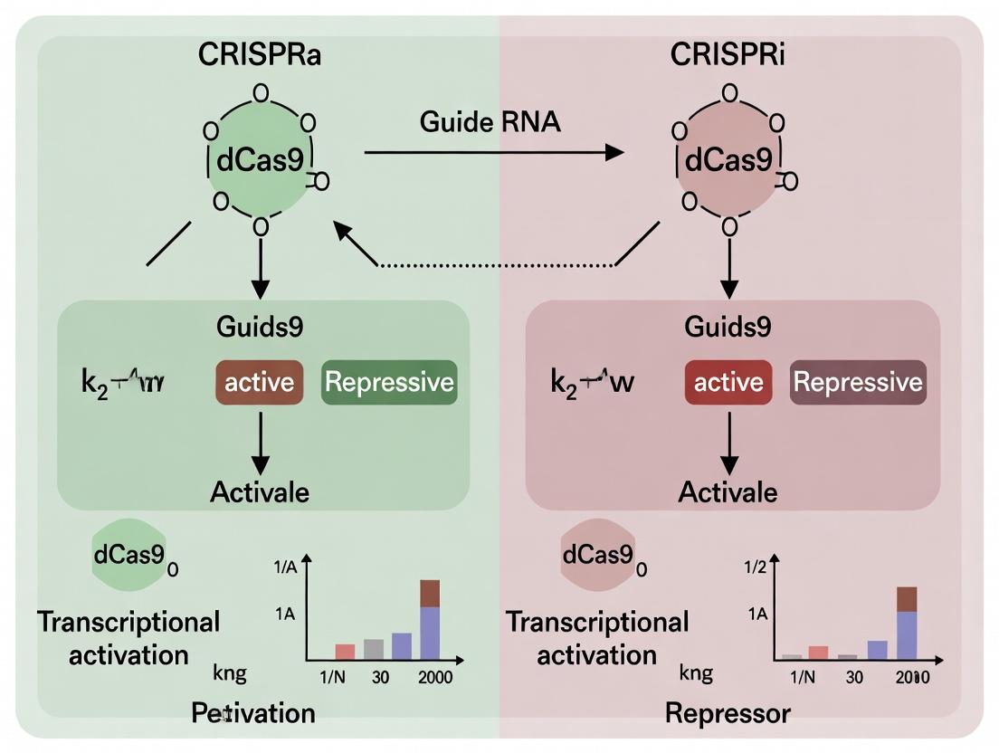
Diagram 3: Mechanisms of dCas9-based CRISPRi and CRISPRa
This application note details CRISPR activation (CRISPRa), a method for precise upregulation of endogenous gene expression. It forms a core component of a broader thesis investigating high-fidelity CRISPR/dCas9 systems for transcriptional modulation (CRISPRa and CRISPRi). While CRISPRi (interference) silences genes, CRISPRa recruits transcriptional activators to gene promoters, offering a powerful tool for functional genomics, disease modeling, and potential therapeutic development.
Core CRISPRa Systems: Mechanism and Comparison
CRISPRa systems utilize a catalytically dead Cas9 (dCas9) protein, guided by a single guide RNA (sgRNA) to a target DNA sequence near a gene promoter. dCas9 serves as a docking platform to recruit transcriptional activation domains. The two most prominent engineered systems are VPR and SAM.
Diagram 1: Core CRISPRa Mechanism
Table 1: Comparison of Major CRISPRa Systems
| Feature | dCas9-VPR | dCas9-SAM (Synergistic Activation Mediator) |
|---|---|---|
| Activation Domains | VP64, p65, Rta (VPR) fused directly to dCas9. | MS2-p65-HSF1 fusion proteins recruited via sgRNA scaffolds. |
| Architecture | Single fusion protein. | Two-component system: dCas9-VP64 + engineered sgRNA with MS2 aptamers. |
| Typical Fold Activation | ~50-300x (varies by gene/cell type). | ~100-1000x (varies by gene/cell type). |
| Key Advantage | Simpler delivery (single construct). | Higher activation potency for many targets. |
| Key Limitation | Larger fusion protein, potentially lower potency on some targets. | Requires engineered sgRNA, more complex delivery. |
| Primary Citation | Chavez et al., Nat Methods, 2015. | Konermann et al., Nature, 2015. |
Detailed Experimental Protocols
Protocol 3.1: CRISPRa Knock-in via Lentiviral Delivery for Stable Cell Line Generation
Objective: Stably integrate the dCas9-activator and sgRNA expression cassettes into a mammalian cell line (e.g., HEK293T) for long-term gene activation studies.
Materials (Research Reagent Solutions):
- dCas9-Activator Plasmid: lenti dCas9-VPR or lenti dCas9-VP64_Blast (Addgene #114199, #61425).
- SAM Component Plasmids: lenti MS2-P65-HSF1_Hygro (Addgene #61426).
- sgRNA Expression Plasmid: lenti sgRNA(MS2)_zeo backbone (for SAM) or lenti sgRNA backbone (for VPR) (Addgene #61427).
- Lentiviral Packaging Plasmids: psPAX2 and pMD2.G (Addgene #12260, #12259).
- HEK293T Cells: For virus production and transduction.
- Transfection Reagent: Polyethylenimine (PEI) or commercial equivalent (e.g., Lipofectamine 3000).
- Selection Antibiotics: Blasticidin, Hygromycin, Zeocin (concentration must be titrated for each cell line).
Procedure:
- sgRNA Design & Cloning: Design sgRNAs targeting regions -200 to -50 bp upstream of the transcription start site (TSS). Clone annealed oligonucleotides into the appropriate BsmBI-digested lentiviral sgRNA vector.
- Lentivirus Production: a. Seed HEK293T cells in a 6-well plate. b. Co-transfect with 1 µg transfer plasmid (dCas9-activator or sgRNA), 0.75 µg psPAX2, and 0.25 µg pMD2.G using PEI. c. Replace media after 6-8 hours. Harvest viral supernatant at 48 and 72 hours post-transfection. Filter through a 0.45 µm filter.
- Cell Line Generation: a. For VPR: Transduce target cells with dCas9-VPR virus first. Select with appropriate antibiotic (e.g., Blasticidin, 5-10 µg/mL) for 7+ days. b. For SAM: Co-transduce target cells with dCas9-VP64 and MS2-P65-HSF1 viruses. Select with Blasticidin and Hygromycin. c. Transduce the polyclonal dCas9-expressing cells with the lentiviral sgRNA. Select with Zeocin.
- Validation: After selection (≥7 days), harvest cells for RNA extraction. Measure target gene expression via RT-qPCR.
Diagram 2: Stable CRISPRa Cell Line Generation
Protocol 3.2: Transient Transfection for Rapid Gene Activation Assay
Objective: Quickly assess activation efficiency of multiple sgRNAs by transiently delivering all CRISPRa components.
Materials:
- All-in-One Plasmid: Expressing dCas9-VPR and sgRNA from a single vector (e.g., Addgene #63798).
- SAM Plasmids: dCas9-VP64, MS2-P65-HSF1, and sgRNA(MS2) expression plasmids.
- Transfection Reagent: Optimized for your cell type (e.g., Lipofectamine 3000 for HEK293T).
- Reporter Cells (Optional): Cell line with luciferase or GFP under control of a minimal promoter.
Procedure:
- Plate cells in 24-well or 96-well format 24 hours prior.
- Prepare DNA Mixtures:
- For VPR: 500 ng all-in-one plasmid per well (24-well).
- For SAM: 250 ng dCas9-VP64, 250 ng MS2-P65-HSF1, 250 ng sgRNA(MS2) plasmid per well.
- Transfect using manufacturer's protocol.
- Harvest Cells 48-72 hours post-transfection.
- Analyze via RT-qPCR (endogenous genes) or fluorescence/luminescence (reporters).
The Scientist's Toolkit: Key Research Reagent Solutions
Table 2: Essential Reagents for CRISPRa Experiments
| Item | Function | Example Source/ID |
|---|---|---|
| dCas9-VPR Lentiviral Plasmid | Expresses the direct fusion activator. Stable integration. | Addgene #114199 |
| dCas9-VP64 Lentiviral Plasmid | Core component of the SAM system. | Addgene #61425 |
| MS2-P65-HSF1 Lentiviral Plasmid | Second component of SAM; recruited via MS2. | Addgene #61426 |
| lenti sgRNA(MS2) Cloning Vector | Backbone for expressing sgRNAs with MS2 aptamers for SAM. | Addgene #61427 |
| All-in-One dCas9-VPR Plasmid | For transient transfection assays. | Addgene #63798 |
| Lentiviral Packaging Plasmids | Required for producing lentiviral particles. | Addgene #12260 (psPAX2), #12259 (pMD2.G) |
| BsmBI Restriction Enzyme | For cloning sgRNA sequences into lentiviral backbones. | NEB #E0582S |
| Polybrene (Hexadimethrine bromide) | Enhances retroviral transduction efficiency. | Sigma-Aldrich #H9268 |
| Validated Positive Control sgRNA | Targets a known highly activatable locus (e.g., CXCR4 promoter). | From literature or commercial suppliers |
Within the broader thesis on CRISPR activation (CRISPRa) and CRISPR interference (CRISPRi) with high-fidelity dCas9, this document focuses on the application of CRISPRi. CRISPRi is a robust, programmable method for gene silencing that utilizes a catalytically dead Cas9 (dCas9) fused to transcriptional repressor domains. This approach allows for precise, reversible, and multiplexed gene knockdown without altering the underlying DNA sequence, making it invaluable for functional genomics, pathway analysis, and drug target validation.
Core Mechanism & Repressor Domains
The dCas9 protein, devoid of endonuclease activity, is guided by a single guide RNA (sgRNA) to a specific genomic locus complementary to its spacer sequence. Once bound, it sterically blocks RNA polymerase elongation. Enhanced repression is achieved by fusing dCas9 to effector domains that recruit endogenous chromatin-modifying complexes.
The two most prominent repressor domains are:
- KRAB (Krüppel-Associated Box): Derived from mammalian zinc-finger proteins, KRAB recruits heterochromatin-forming complexes via KAP1, leading to histone H3 lysine 9 trimethylation (H3K9me3) and spread of facultative heterochromatin, resulting in stable, long-term silencing.
- SID4x (SRF Interaction Domain 4x): A concatenated quartet of the SRF repression domain, SID4x is a potent synthetic repressor that recruits co-repressor complexes, often leading to more immediate and stronger repression than KRAB in certain contexts, though with potentially less spread.
Quantitative Comparison of Key Repressor Domains
Table 1: Comparison of Major Transcriptional Repressor Domains for CRISPRi
| Repressor Domain | Origin | Primary Mechanism | Typical Repression Efficiency* | Key Characteristics |
|---|---|---|---|---|
| KRAB | Mammalian ZFP | Recruits KAP1, induces H3K9me3 & heterochromatin | 70-95% (High) | Stable, long-term silencing; some epigenetic memory; can spread ~1-2 kb. |
| SID4x | Synthetic (SRF) | Recruits co-repressor complexes (e.g., NuRD) | 80-98% (Very High) | Potent, immediate repression; minimal spread; may be more sensitive to sgRNA position. |
| MeCP2 | Mammalian | Binds methylated DNA & recruits repressors | 60-90% (Moderate-High) | Context-dependent; effective in methylated regions. |
*Efficiency ranges are generalized from literature and can vary significantly by gene, cell type, and sgRNA design.
Application Notes
Functional Genomics & Genetic Screens
CRISPRi pooled libraries enable genome-wide or focused loss-of-function screens. The reversibility and specificity of CRISPRi reduce confounding off-target effects compared to RNAi, providing higher confidence hits for drug target identification.
Pathway Analysis and Synthetic Lethality
Precise, multiplexed silencing of multiple genes allows for the dissection of genetic interactions and identification of synthetic lethal pairs, which are prime targets for combination therapies in oncology.
Drug Development
CRISPRi facilitates target validation by mimicking the effect of a therapeutic inhibitor. It is also used in pharmacokinetic studies to modulate drug-metabolizing enzyme expression.
Detailed Protocols
Protocol 1: CRISPRi Knockdown in Mammalian Cells Using dCas9-KRAB
Objective: Stable, inducible silencing of a target gene in HEK293T cells.
Materials: See "The Scientist's Toolkit" below.
Method:
- sgRNA Design & Cloning:
- Design sgRNAs targeting the transcriptional start site (TSS) -50 to +300 bp. Use established algorithms (e.g., CRISPick).
- Clone annealed oligonucleotides into the lentiviral sgRNA expression plasmid (e.g., pLV hU6-sgRNA hUbC-dCas9-KRAB-T2a-Puro) via BsmBI sites.
- Sequence-verify the construct.
Lentivirus Production (Day 1-3):
- Plate HEK293T cells in a 6-well plate.
- Co-transfect with:
- 1 µg sgRNA expression plasmid
- 0.9 µg psPAX2 (packaging plasmid)
- 0.1 µg pMD2.G (VSV-G envelope plasmid) using a transfection reagent.
- Replace medium after 6-8 hours.
- Harvest viral supernatant at 48 and 72 hours post-transfection. Pool, filter (0.45 µm), and store at -80°C.
Cell Transduction & Selection (Day 4-10):
- Seed target HEK293T cells. Add viral supernatant + polybrene (8 µg/mL).
- Spinfect at 800 x g for 30 min at 32°C.
- After 48 hours, begin selection with 2 µg/mL puromycin for 5-7 days.
Validation (Day 11-14):
- Harvest cells and extract RNA.
- Perform RT-qPCR to assess mRNA knockdown relative to a non-targeting sgRNA control.
- Optional: Assess protein level by western blot.
Protocol 2: Acute Repression Using SID4x-delivery via Protein Transduction
Objective: Rapid, dose-dependent gene silencing without genetic modification.
Materials: Purified dCas9-SID4x protein, synthetic sgRNA, cell-penetrating peptide (CPP).
Method:
- Ribonucleoprotein (RNP) Complex Formation:
- Reconstitute synthetic sgRNA in nuclease-free buffer.
- Incubate dCas9-SID4x protein (100 pmol) with sgRNA (120 pmol) in a 1:1.2 molar ratio in PBS for 20 min at 25°C.
Complex Delivery:
- Mix the RNP complex with a CPP (e.g., based on TAT peptide) at a specified weight ratio.
- Add the RNP-CPP complex directly to the cell culture medium of adherent cells.
Analysis:
- Monitor gene expression by RT-qPCR at 24, 48, and 72 hours post-delivery.
- Repression peaks typically between 48-72 hours and decays as the RNP is diluted/depleted.
Diagrams
Title: CRISPRi Experimental Workflow Using Lentiviral Delivery
Title: CRISPRi Mechanism: dCas9-Repressor Silences Transcription
The Scientist's Toolkit
Table 2: Essential Research Reagents for CRISPRi Experiments
| Reagent / Material | Function | Example Product/Catalog |
|---|---|---|
| dCas9-Repressor Expression Plasmid | Expresses the core dCas9 protein fused to a repressor domain (KRAB, SID4x). | pLV hU6-sgRNA hUbC-dCas9-KRAB-Puro (Addgene #71236) |
| Lentiviral sgRNA Backbone Plasmid | Vector for cloning and expressing sgRNA; often part of a dual-vector system. | lentiGuide-Puro (Addgene #52963) |
| Lentiviral Packaging Plasmids | Required for production of replication-incompetent lentivirus. | psPAX2 (packaging), pMD2.G (envelope) |
| Puromycin Dihydrochloride | Selective antibiotic for cells transduced with puromycin resistance-containing vectors. | Common laboratory supplier |
| Polybrene (Hexadimethrine Bromide) | A cationic polymer that enhances viral transduction efficiency. | Common laboratory supplier |
| BsmBI Restriction Enzyme | Type IIS enzyme used for cloning sgRNA sequences into backbone vectors. | Common laboratory supplier |
| Validated sgRNA Controls | Non-targeting/scrambled sgRNA (negative control) and sgRNA targeting essential gene (positive control). | Commercially available from Horizon, Sigma, etc. |
| RT-qPCR Kit | For quantitative validation of target gene mRNA knockdown. | One-step or two-step kits from Thermo, Bio-Rad, etc. |
| Purified dCas9-Repressor Protein | For RNP-based, transient delivery protocols. | Recombinant dCas9-KRAB protein (e.g., from Aldevron, Thermo) |
| Synthetic sgRNA (chemically modified) | For use with RNP delivery; modifications enhance stability. | Synthesized by IDT, Synthego, etc. |
Within the framework of CRISPRa (activation) and CRISPRi (interference) research, the specificity of the dCas9-effector complex is paramount. High-Fidelity (HF) variants of dCas9 have been engineered to minimize off-target binding, a critical source of experimental noise and phenotypic ambiguity. Off-target effects can lead to misinterpretation of gene function, reduced efficacy in therapeutic contexts, and increased risk of adverse events in drug development. This application note details the quantitative advantages of HF-dCas9 systems and provides protocols for assessing and achieving cleaner genetic perturbations.
Quantitative Comparison: Wild-Type vs. HF dCas9 Systems
Recent studies utilizing genome-wide binding assays (e.g., ChIP-seq) and transcriptomic profiling (RNA-seq) have quantified the improved specificity of HF variants. The data below summarize key performance metrics.
Table 1: Specificity and Efficacy Metrics of dCas9 Variants in CRISPRa/i Applications
| dCas9 Variant | Primary Mutation(s) | Reported On-Target Efficacy (% of WT) | Reduction in Off-Target Sites (vs. WT) | Key Assessment Method | Reference |
|---|---|---|---|---|---|
| WT dCas9 | None | 100% (baseline) | 1x (baseline) | ChIP-seq, GUIDE-seq | (1) |
| dCas9-HF1 | N497A, R661A, Q695A, Q926A | 85-95% | 10-20 fold reduction | BLISS, RNA-seq | (2, 3) |
| HypaCas9 (for CRISPRa/i) | N692A, M694A, Q695A, H698A | ~70-80% | >50 fold reduction (binding) | ChIP-seq, Phenotypic Screens | (4) |
| eSpCas9(1.1) (as dCas9) | K848A, K1003A, R1060A | 75-90% | 5-15 fold reduction | DIG-seq, RNA-seq | (5) |
| Sniper-Cas9 (HF) | F539S, M763I, K890N | >90% | Significant reduction (quantified by ChIP) | ChIP-exo, Transcriptomics | (6) |
Note: Efficacy can vary based on gRNA design, target locus, and cell type. Off-target reduction is relative to WT dCas9 binding or transcriptional changes.
Protocol: Assessing Off-Target Binding and Transcriptional Effects
This protocol outlines a combined method using ChIP-seq and RNA-seq to evaluate the specificity of a dCas9-effector (e.g., dCas9-VPR for activation, dCas9-KRAB for interference) system.
Chromatin Immunoprecipitation Sequencing (ChIP-seq) for dCas9 Binding
Objective: Genome-wide mapping of dCas9 on-target and off-target binding sites.
Materials:
- Cells expressing HF-dCas9 or WT-dCas9 fused to an effector (e.g., VPR) and a specific single-guide RNA (sgRNA).
- Crosslinking solution (1% formaldehyde).
- Cell lysis buffers (with protease inhibitors).
- Antibody for immunoprecipitation (anti-FLAG for tagged dCas9, or anti-dCas9 specific).
- Protein A/G magnetic beads.
- DNA purification kit.
- Library preparation kit for Illumina sequencing.
Procedure:
- Crosslink: Treat ~10^7 cells with 1% formaldehyde for 10 min at room temperature. Quench with 125mM glycine.
- Cell Lysis: Lyse cells sequentially with buffers to isolate nuclei and then shear chromatin via sonication (target fragment size: 200-500 bp).
- Immunoprecipitation: Incubate sheared chromatin with 2-5 µg of specific antibody overnight at 4°C. Add beads and incubate for 2 hours.
- Wash & Elute: Wash beads with low-salt, high-salt, LiCl, and TE buffers. Elute complexes with elution buffer (1% SDS, 0.1M NaHCO3).
- Reverse Crosslinks: Incubate eluates at 65°C overnight with NaCl. Treat with RNase A and Proteinase K.
- DNA Purification & Library Prep: Purify DNA using a spin column. Prepare sequencing library following kit instructions.
- Bioinformatics Analysis: Align sequences to the reference genome. Call peaks (MACS2). Compare peak locations between HF and WT samples, focusing on sites with perfect vs. mismatched gRNA complementarity.
RNA Sequencing for Phenotypic Specificity
Objective: Determine transcriptional changes induced by CRISPRa/i, identifying on-target and off-target gene expression changes.
Procedure:
- RNA Extraction: Isolate total RNA from experimental and control cells (e.g., non-targeting sgRNA) using a TRIzol-based method.
- Library Preparation: Deplete ribosomal RNA. Synthesize cDNA and prepare libraries using a stranded mRNA-seq kit.
- Sequencing & Analysis: Sequence on an Illumina platform. Align reads (STAR aligner). Quantify gene expression (featureCounts, DESeq2). Identify differentially expressed genes (DEGs). Off-target transcriptional events are defined as DEGs without a predicted dCas9 binding site within a defined window (e.g., ±5 kb from TSS).
Visualization of Concepts and Workflows
Diagram Title: Comparison of WT vs. HF dCas9 Specificity Workflow
Diagram Title: Mechanism of HF-dCas9 Minimizing Off-Target Effects
The Scientist's Toolkit: Research Reagent Solutions
Table 2: Essential Reagents for High-Fidelity CRISPRa/i Research
| Reagent / Material | Function / Purpose | Example Supplier/Catalog Consideration |
|---|---|---|
| HF-dCas9 Expression Plasmid | Delivers high-fidelity, nuclease-dead Cas9 variant fused to transcriptional effector (e.g., VPR, KRAB). | Addgene: dCas9-HF1-VPR (plasmid #), HypaCas9-KRAB. |
| sgRNA Cloning Vector | Backbone for expressing single-guide RNA targeting gene of interest; often includes selection marker. | Addgene: Lentiguide-puro, MS2-based scaffolds for recruitment. |
| Chromatin IP-Grade Antibody | For ChIP-seq; specific to epitope tag (FLAG, HA) on dCas9 or to dCas9 protein itself. | Cell Signaling Tech, Abcam: anti-FLAG M2, anti-dCas9. |
| High-Sensitivity DNA/RNA Kits | Purify fragmented chromatin (ChIP) or intact total RNA (RNA-seq) with minimal loss. | Qiagen, Zymo Research, NEB. |
| Next-Gen Sequencing Library Prep Kit | Prepare barcoded, sequencing-ready libraries from ChIP-DNA or RNA. | Illumina, NEB Next, KAPA Biosystems. |
| Cell Line with Reporter | Validated cell line with sensitive, quantifiable reporter (e.g., GFP under target promoter) for phenotype screening. | ATCC, or engineer using lentiviral transduction. |
| Genome-Wide Off-Target Prediction Tool | In silico guide design to predict and minimize potential off-target sites. | IDT's guide design tool, CHOPCHOP, CRISPick. |
| Validated Positive Control gRNA | gRNA with known high on-target activity for the chosen HF-dCas9 system, used as a benchmark. | Published resources, e.g., for housekeeping gene promoters. |
Application Notes
CRISPR activation (CRISPRa) and interference (CRISPRi) systems, utilizing a nuclease-dead Cas9 (dCas9), represent a transformative approach for precise gene regulation without inducing DNA double-strand breaks. This eliminates the risks associated with on- and off-target DNA damage, such as genomic instability, p53 activation, and unintended translocations. The system's core advantages are its reversibility, tunability, and multiplexability, making it indispensable for functional genomics, synthetic biology, and therapeutic development.
Reversibility: Gene expression can be toggled between activated and repressed states by modulating the presence of guide RNAs (gRNAs) or effector proteins (e.g., KRAB, VPR), allowing for dynamic studies of gene function.
Tunability: Expression levels can be finely controlled. This is achieved by varying:
- gRNA dosage (e.g., plasmid copy number, delivery method).
- Effector protein expression levels.
- Use of attenuated effector domains (e.g., ZF-KRAB variants).
- Small molecule-inducible dimerization systems (e.g., abscisic acid, rapalog).
Multiplexability: Multiple gRNAs, targeting different loci, can be co-delivered using arrayed constructs or polycistronic systems (e.g., tRNA-gRNA, CRISPRi/a sgRNA libraries). This enables genome-wide screens and the coordinated regulation of complex gene networks.
Therapeutic Context: These features are critical for drug development, allowing for the identification of novel targets via gain- and loss-of-function screens and paving the way for precise gene-regulating therapeutics that modulate disease-associated genes without permanent genomic alteration.
Experimental Protocols
Protocol 1: dCas9 Effector Stable Cell Line Generation for CRISPRi/a
Objective: Create a mammalian cell line stably expressing a dCas9-effector fusion (e.g., dCas9-KRAB for CRISPRi or dCas9-VPR for CRISPRa) for consistent, long-term gene regulation studies.
Materials: See "Research Reagent Solutions" table. Procedure:
- Construct Design: Clone your chosen effector domain (KRAB, VPR, etc.) C-terminal to dCas9 in a lentiviral expression vector containing a selectable marker (e.g., puromycin resistance).
- Lentivirus Production: In a HEK293T packaging cell line, co-transfect the dCas9-effector transfer plasmid with psPAX2 (packaging) and pMD2.G (VSV-G envelope) plasmids using a suitable transfection reagent (e.g., PEI). Harvest virus-containing supernatant at 48 and 72 hours post-transfection.
- Cell Line Transduction: Filter the supernatant (0.45 µm), add polybrene (8 µg/mL), and incubate with your target cell line (e.g., HEK293, K562) for 24 hours.
- Selection & Validation: Replace media with selection antibiotic (e.g., 2 µg/mL puromycin). Maintain selection for 5-7 days. Validate dCas9-effector expression via western blot (anti-FLAG or anti-Cas9 antibody) and functional testing with a validated, target-specific gRNA.
Protocol 2: Tunable, Inducible Gene Activation Using a Small Molecule Dimerizer System
Objective: Achieve graded, inducible gene activation by recruiting transcriptional activators to dCas9 via a chemical inducer.
Materials: See "Research Reagent Solutions" table. Procedure:
- System Setup: Generate a stable cell line expressing two constructs:
- dCas9-FKBP: dCas9 fused to FKBP12.
- FRB-VP64: FRB domain fused to a minimal activator (VP64).
- gRNA Transfection: Transfect a plasmid expressing a gRNA targeting your gene of interest.
- Induction & Titration: 24h post-gRNA transfection, treat cells with varying concentrations of the rapalog A/C heterodimerizer (e.g., 0, 1, 10, 100 nM). Include a non-targeting gRNA control.
- Quantitative Analysis: Harvest RNA 48h post-induction. Perform RT-qPCR to measure target gene expression. Normalize to housekeeping genes (e.g., GAPDH, ACTB). Plot expression fold-change against inducer concentration to establish a dose-response curve.
Protocol 3: Multiplexed Gene Repression Using a Polycistronic gRNA Array
Objective: Simultaneously repress up to 10 genes in a single cell using a multiplexed gRNA expression system.
Materials: See "Research Reagent Solutions" table. Procedure:
- Array Design: Design gRNA sequences (20-nt) for each target gene. Clone them sequentially into a tRNA-gRNA array vector. Each gRNA is flanked by a tRNA sequence for endogenous RNase P/RNase Z processing.
- Delivery: Transfect the multiplex gRNA array plasmid into your stable dCas9-KRAB cell line (from Protocol 1).
- Validation: 72h post-transfection, harvest cells.
- For mRNA analysis: Extract total RNA, perform RT-qPCR for each target gene.
- For phenotypic analysis: Perform relevant assays (e.g., proliferation, differentiation, flow cytometry).
- Controls: Include a non-targeting gRNA array and single-gene repression conditions for comparison.
Data Presentation
Table 1: Comparison of Key Quantitative Parameters for CRISPRa/i Systems
| Parameter | CRISPRi (dCas9-KRAB) | CRISPRa (dCas9-VPR) | Notes / Reference |
|---|---|---|---|
| Typical Repression/Activation Range | 50 - 95% knockdown | 2 - 100+ fold activation | Highly dependent on locus, chromatin state, and gRNA design. |
| Optimal Targeting Window | -50 to +300 bp from TSS | -400 to -50 bp from TSS | For strongest effect. VPR has a broader effective window than VP64. |
| Multiplexing Capacity (gRNAs) | 10+ (via tRNA arrays) | 10+ (via tRNA arrays) | Demonstrated in functional genomics screens. |
| Kinetics (Time to Effect) | ~24-48h (mRNA) | ~24-48h (mRNA) | Protein-level effects follow with corresponding half-life. |
| Reversal Kinetics | ~72-96h for full reversal | ~72-96h for full reversal | Upon gRNA loss or effector withdrawal. |
| Typical Off-Target Effects | Minimal mRNA-level changes | Minimal mRNA-level changes | Significantly lower than nuclease-active Cas9; primarily due to dCas9 binding. |
Table 2: Research Reagent Solutions Toolkit
| Item | Function & Explanation |
|---|---|
| dCas9-KRAB Plasmid | Core CRISPRi effector. dCas9 provides DNA targeting; KRAB domain recruits repressive chromatin modifiers. |
| dCas9-VPR Plasmid | Core CRISPRa effector. VPR is a tripartite activator (VP64, p65, Rta) for robust transcriptional upregulation. |
| Lentiviral gRNA Expression Vector (e.g., lentiGuide-Puro) | For stable, high-efficiency delivery of gRNA expression constructs. |
| Toluene-resistant RNA Polymerase (T7) gRNA Cloning Vector | For high-yield in vitro transcription of gRNAs for RNP delivery. |
| Polycistronic tRNA-gRNA (PTG) Array Vector | Enables simultaneous expression of multiple gRNAs from a single Pol II promoter. |
| Rapalog A/C Heterodimerizer | Small molecule inducing dimerization of FKBP and FRB domains, used for inducible systems. |
| Anti-Cas9 Monoclonal Antibody | For validation of dCas9-effector fusion protein expression via western blot. |
| Next-Generation Sequencing Library Prep Kit | For analyzing CRISPR screen outcomes or assessing off-target binding (e.g., ChIP-seq). |
Visualizations
Title: CRISPRa/i Core Mechanism: dCas9-Effector Action
Title: Multiplexed Gene Regulation Workflow
Title: Key Advantages of dCas9 Systems: Methods & Outcomes
Implementing CRISPRa/i with dCas9-HF: Protocols, Design, and Advanced Applications
Application Notes
This guide provides a framework for selecting optimal components for precise CRISPR activation (CRISPRa) and interference (CRISPRi) experiments using high-fidelity deactivated Cas9 (dCas9-HF). The goal is to maximize on-target efficacy while minimizing off-target effects, a critical consideration for therapeutic development.
dCas9-HF Variants
dCas9-HF (High Fidelity) variants contain point mutations (N497A, R661A, Q695A, Q926A) that reduce non-specific electrostatic interactions with the DNA backbone, drastically lowering off-target binding while maintaining robust on-target occupancy. For CRISPRa/i, this is paramount for specific transcriptional modulation.
Selection Criteria:
- dCas9-HF1: The standard variant. Ideal for most proof-of-concept and initial screening work.
- Fusion-Compatible dCas9-HF: Ensure the chosen expression vector has appropriate linkers and fusion sites (typically N- or C-terminal) for your effector domain without perturbing dCas9-HF's stability or fidelity.
Effector Domains
The effector domain determines the transcriptional outcome. It is fused to dCas9-HF via flexible linkers.
| Application | Effector Domain | Example Domain | Mechanism | Typical Size (aa) | Key Consideration |
|---|---|---|---|---|---|
| CRISPRi | Repressor | KRAB (Krüppel-associated box) | Recruits heterochromatin-forming complexes, silencing transcription. | ~45 aa | Strong, consistent repression. Can have variable effects based on genomic context. |
| CRISPRa | Activator | VP64, p65AD, Rta (Tripartite: VPR) | Recruits transcriptional co-activators and the pre-initiation complex. | VP64: 127 aaVPR: ~500 aa | Multipartite activators (e.g., VPR, SAM) are significantly more potent than single domains. |
| CRISPRa (Advanced) | Super Activator | SunTag or SAM (Synergistic Activation Mediator) | dCas9 recruits multiple copies of VP64 (SunTag) or engages a synergistic RNA-protein scaffold (SAM). | System-dependent | Higher potency but increased construct complexity and size. |
sgRNA Backbone
The sgRNA backbone influences stability, loading into dCas9, and for CRISPRa systems like SAM, it provides binding sites for effector-recruiting proteins.
| Backbone Type | Key Features | Optimal For | Efficiency Note |
|---|---|---|---|
| Standard (e.g., from pX330) | Original 42-nt stem-loop architecture. | Basic CRISPRi with dCas9-HF-KRAB. | Reliable, but can be less efficient for some CRISPRa systems. |
| MS2 / PP7 / com Modified | Contains aptamer loops (e.g., MS2) in the tetraloop and stemloop 2. | CRISPRa systems like SAM, which require scaffold protein (MCP) recruitment. | Essential for scaffold-dependent systems. Increases sgRNA size. |
| Enhanced/Truncated | Optimized stem lengths or truncated variants (tru-sgRNA). | Balancing high activity with ease of synthesis. | Some truncations can improve performance with dCas9 fusions. |
Core Recommendation: For CRISPRa, use an MS2-modified backbone (e.g., from the SAM system). For CRISPRi, a standard or enhanced backbone suffices.
Experimental Protocols
Protocol 1: Validating dCas9-HF Fusion Activity with a Fluorescent Reporter Assay
Purpose: To functionally test a newly constructed dCas9-HF-effector fusion protein in cells.
Materials: See "The Scientist's Toolkit" below. Workflow:
- Clone: Subclone your effector domain (e.g., KRAB, VPR) into a mammalian expression vector containing dCas9-HF1, using appropriate linkers (e.g., (GGGGS)₂).
- Design & Synthesize: Design an sgRNA targeting a site upstream of a stably integrated fluorescent reporter (e.g., GFP under a minimal promoter).
- Co-transfect: In triplicate, co-transfect HEK293T cells with:
- Plasmid A: dCas9-HF-effector expression vector (500 ng).
- Plasmid B: sgRNA expression vector (250 ng).
- Control: dCas9-HF alone + sgRNA (CRISPRi negative control); dCas9-VP64 + sgRNA (CRISPRa positive control).
- Analyze: After 48-72 hours, analyze mean fluorescence intensity (MFI) via flow cytometry.
- Calculate: % Activation = [(MFIsample - MFIneg control) / MFIneg control] x 100. % Repression = [1 - (MFIsample / MFIneg control)] x 100.
Protocol 2: Genome-wide Off-Target Assessment by GUIDE-seq or Digenome-seq
Purpose: To empirically confirm the reduced off-target profile of dCas9-HF fusions compared to wild-type dCas9.
Detailed GUIDE-seq Methodology:
- Transfect: Co-transfect cells with your dCas9-HF-effector plasmid, targeting sgRNA plasmid, and the GUIDE-seq oligonucleotide duplex (an end-protected, double-stranded tag that integrates at double-strand breaks; note: dCas9 is catalytically dead, but you must include a small percentage of catalytically active Cas9 (e.g., 1:10 ratio) to enable tag integration at off-target binding sites).
- Harvest & Extract: Harvest cells 72h post-transfection. Extract genomic DNA.
- Library Prep & Sequencing: Perform tag-specific enrichment PCR, followed by next-generation sequencing library preparation.
- Bioinformatics: Use the GUIDE-seq analysis software to identify off-target sites by detecting genomic locations flanked by tag sequences.
- Comparison: Compare the number and signal strength of off-target sites from dCas9-HF-effector samples to those from a wild-type dCas9-effector control.
Visualizations
Title: Decision Flow for dCas9-HF CRISPRa/i System Assembly
Title: CRISPRa Mechanism Using MS2-Backbone sgRNA
The Scientist's Toolkit: Research Reagent Solutions
| Reagent / Material | Supplier Examples | Function in dCas9-HF CRISPRa/i Research |
|---|---|---|
| dCas9-HF1 Plasmid | Addgene (#114474, #118150) | Source of the high-fidelity, nuclease-dead Cas9 backbone for effector fusion. |
| Effector Domain Plasmids (KRAB, VPR, SAM) | Addgene (#61422, #63798, #1000000074) | Provide standardized, validated transcriptional effector modules for cloning. |
| Modified sgRNA Cloning Vectors | Addgene (#104174 for SAM sgRNA) | Backbone vectors for expressing MS2-modified or other scaffold sgRNAs. |
| Lipofectamine 3000 | Thermo Fisher Scientific | High-efficiency transfection reagent for delivering plasmid DNA to mammalian cell lines. |
| GUIDE-seq Oligo Duplex | Integrated DNA Technologies (IDT) | Double-stranded tag for genome-wide, unbiased detection of nuclease off-target sites. |
| KAPA HiFi HotStart ReadyMix | Roche | High-fidelity polymerase for accurate amplification during GUIDE-seq library prep. |
| Flow Cytometer (e.g., Attune NxT) | Thermo Fisher Scientific | Instrument for quantifying fluorescence in reporter assays to measure activation/repression. |
| Next-Gen Sequencing Service | Illumina, Novogene | For sequencing GUIDE-seq or RNA-seq libraries to assess off-targets or transcriptome changes. |
Within the broader thesis on CRISPR activation (CRISPRa) and interference (CRISPRi) utilizing high-fidelity dCas9 variants, the precision design of single guide RNAs (sgRNAs) is paramount. This document details application notes and protocols for selecting optimal genomic targets within promoters and enhancers to achieve maximal transcriptional modulation. Efficacy is dictated by chromatin accessibility, local sequence context, and the steric compatibility of effector domains.
Key Principles for Optimal Target Selection
Promoter Targeting (CRISPRi/a)
For transcriptional repression (CRISPRi) via dCas9 fused to repressive domains (e.g., KRAB), sgRNAs should target the core promoter region, specifically the window from -50 to +300 bp relative to the transcription start site (TSS). For activation (CRISPRa) using dCas9-activator fusions (e.g., VPR, SAM), sgRNAs targeting regions from -400 to -50 bp upstream of the TSS generally show higher efficacy, as activators require recruitment of co-factors without obstructing the pre-initiation complex.
Enhancer Targeting
Enhancer regulation requires targeting dCas9-effectors to distal cis-regulatory elements, often several kb from gene TSS. Success depends on identifying validated, cell-type-specific active enhancers marked by H3K27ac and accessible chromatin (ATAC-seq peaks). sgRNAs should be designed within the center of the enhancer peak. Looping interaction data (e.g., from Hi-C) is critical to confirm physical connectivity to the target gene promoter.
Sequence & Structural Considerations
- GC Content: Optimal between 40-60%.
- Off-Target Potential: Must be minimized using high-fidelity Cas9 variants (e.g., SpCas9-HF1, eSpCas9) and rigorous in silico prediction tools.
- Poly-T Tracts: Avoid sequences containing 4 or more consecutive T's, which can act as premature termination signals for RNA Polymerase III.
- Secondary Structure: Avoid sgRNA sequences with strong internal hairpins that may affect Cas9 binding.
Table 1: sgRNA Targeting Parameters for Maximal Efficacy
| Target Region | Optimal Position Relative to TSS | Typical Repression (CRISPRi)* | Typical Activation (CRISPRa)* | Key Chromatin Feature Required |
|---|---|---|---|---|
| Core Promoter | -50 to +300 bp | 70-95% (KRAB) | 2-5 fold (VPR) | High Accessibility (DNase/ATAC-seq peak) |
| Proximal Enhancer | -50 to -400 bp | 60-80% | 10-50 fold (SAM) | H3K4me1, H3K27ac |
| Distal Enhancer | Center of validated peak | Variable (40-70%) | 5-30 fold (dependent on loop strength) | H3K27ac, Hi-C/Promoter Capture Hi-C link |
*Values are generalized estimates from recent literature; actual performance varies by gene and cell type.
Table 2: Comparison of Common dCas9-Effector Systems
| System | dCas9 Variant | Fused Effector(s) | Primary Use | Key Design Implication |
|---|---|---|---|---|
| CRISPRi (KRAB) | SpCas9-HF1 | KRAB domain | Repression | Target near TSS; high specificity critical. |
| CRISPRa (VPR) | eSpCas9 | VP64, p65, Rta | Activation | Target -400 to -50 bp; multiple sgRNAs often needed. |
| CRISPRa (SAM) | dCas9-VP64 | MS2-p65-HSF1 (recruited) | Synergistic Activation | Target enhancers; requires two-part sgRNA with MS2 aptamers. |
Experimental Protocols
Protocol:In SilicoDesign of sgRNAs for Promoter/Enhancer Targeting
Objective: To design high-efficacy, specific sgRNAs for a target gene's promoter or connected enhancer. Materials: Computer with internet access, target gene genomic coordinates, reference genome (e.g., hg38). Software/Tools: UCSC Genome Browser, Ensembl, CHOPCHOP, CRISPOR, Cas-OFFinder. Procedure:
- Define Target Locus: Using UCSC/Ensembl, identify the canonical TSS and genomic coordinates of your target gene.
- Identify Regulatory Regions:
- For promoter targeting, extract sequence from -500 to +500 bp of the TSS.
- For enhancer targeting, consult cell-type-specific epigenomic databases (e.g., Cistrome DB) for H3K27ac and ATAC-seq peaks within ±100 kb of the TSS. Prioritize peaks linked by Hi-C data.
- Generate sgRNA Candidates: Input the selected DNA sequence (promoter or enhancer region) into design tools like CHOPCHOP or CRISPOR.
- Filter and Rank: Apply the following filters:
- Position: Keep sgRNAs in optimal windows (see Table 1).
- Specificity: Use the tool's off-target prediction scores. Require zero or minimal off-targets with ≤3 mismatches in the seed region (PAM-proximal 8-12 nt).
- Efficiency: Select sgRNAs with high predicted on-target efficiency scores (e.g., Doench '16 score >0.5).
- Sequence Features: Discard candidates with low GC content (<40%), high GC content (>60%), or poly-T tracts.
- Final Selection: Select 3-5 top-ranked sgRNAs per target region for empirical testing. Include a non-targeting control sgRNA.
Protocol: Empirical Validation of sgRNA Efficacy via RT-qPCR
Objective: To measure changes in target gene mRNA expression following delivery of dCas9-effector and candidate sgRNAs. Materials: Cultured mammalian cells, transfection/lentiviral reagents, dCas9-effector (CRISPRi or CRISPRa) plasmid, sgRNA expression plasmids, RNA extraction kit, cDNA synthesis kit, qPCR reagents. Procedure:
- Cell Transduction/Transfection: Deliver the dCas9-effector construct and individual sgRNA constructs (or a lentiviral library) into your target cell line. Include controls: non-targeting sgRNA + dCas9-effector, and dCas9-effector only.
- Incubation: Allow 72-96 hours for stable expression and transcriptional effects.
- RNA Harvest: Lyse cells and isolate total RNA using a DNase-treated column-based kit.
- cDNA Synthesis: Convert 1 µg of RNA to cDNA using a reverse transcription kit with random hexamers.
- Quantitative PCR (qPCR):
- Design TaqMan probes or SYBR Green primers for your target gene and 2-3 stable housekeeping genes (e.g., GAPDH, ACTB).
- Perform qPCR in technical triplicates.
- Calculate relative gene expression using the ΔΔCt method, normalizing to housekeeping genes and the non-targeting sgRNA control.
- Analysis: sgRNAs causing >70% knockdown (CRISPRi) or >10-fold activation (CRISPRa, context-dependent) are considered high-efficacy.
Visualizations
Title: sgRNA Design Workflow for Promoter & Enhancer Targeting
Title: CRISPRi vs. CRISPRa Mechanism at Regulatory Elements
The Scientist's Toolkit
Table 3: Essential Research Reagent Solutions
| Reagent/Material | Function/Benefit | Example Vendor/Product |
|---|---|---|
| High-Fidelity dCas9 Effector Plasmids | Provides the nuclease-dead Cas9 fused to transcriptional effector domains (KRAB, VPR, etc.) with reduced off-target binding. | Addgene: #112196 (dCas9-KRAB), #114198 (dCas9-VPR) |
| sgRNA Cloning Backbone | Plasmid for expressing sgRNA under a U6 promoter; often includes selection markers (e.g., puromycin). | Addgene: #104174 (lentiGuide-Puro) |
| Lentiviral Packaging System | For stable, efficient delivery of dCas9 and sgRNA constructs into difficult-to-transfect cells. | Takara Bio: Lenti-X HTX Packaging System |
| Chromatin Accessibility Kit (ATAC-seq) | Identifies open chromatin regions (promoters, enhancers) for optimal sgRNA target site selection. | 10x Genomics: Chromium Next GEM Single Cell ATAC |
| Epigenetic Modification Antibodies | Validates enhancer activity (H3K27ac) via ChIP-qPCR after dCas9 targeting. | Cell Signaling Technology: Anti-acetyl-Histone H3 (Lys27) Antibody |
| RT-qPCR Master Mix | Quantifies changes in target gene mRNA expression with high sensitivity and reproducibility. | Bio-Rad: iTaq Universal SYBR Green Supermix |
| Genomic DNA Cleavage Detection Kit | Assesses Cas9/sgRNA on-target and off-target activity when using nuclease-active controls. | IDT: Alt-R Genome Cleavage Detection Kit |
Within the broader thesis on CRISPR activation (CRISPRa) and interference (CRISPRi) utilizing high-fidelity dCas9, this document provides detailed application notes and protocols for the reliable delivery of these systems via lentiviral vectors and the establishment of robust genetic screens. The fusion of catalytically "dead" Cas9 (dCas9) to transcriptional effector domains enables precise, programmable gene upregulation (CRISPRa) or downregulation (CRISPRi), creating powerful tools for functional genomics and drug target discovery. Lentiviral transduction offers stable genomic integration and is the method of choice for many pooled or arrayed screening formats.
Research Reagent Solutions Toolkit
| Reagent / Material | Function / Explanation |
|---|---|
| High-Fidelity dCas9-VP64-p65-Rta (dCas9-VPR) Plasmid | Core CRISPRa effector. dCas9 provides DNA targeting, while the VPR tripartite activator (VP64, p65, Rta) drives strong gene activation. |
| High-Fidelity dCas9-KRAB Plasmid | Core CRISPRi effector. dCas9 targets the gene, and the KRAB domain recruits repressive complexes to silence transcription. |
| Lentiviral Packaging Plasmids (psPAX2, pMD2.G) | psPAX2 provides gag/pol for viral particle assembly; pMD2.G provides VSV-G envelope protein for broad tropism. |
| sgRNA Library Cloning Backbone (lentiGuide-Puro, etc.) | Lentiviral vector for sgRNA expression, typically containing a selection marker (e.g., Puromycin resistance). |
| HEK293T/17 Cells | Highly transfectable cell line used for high-titer lentivirus production. |
| Polybrene (Hexadimethrine bromide) | Cationic polymer that enhances viral transduction efficiency by neutralizing charge repulsion. |
| Puromycin Dihydrochloride | Antibiotic for selecting cells successfully transduced with sgRNA or effector constructs. |
| Next-Generation Sequencing (NGS) Reagents | For amplifying and sequencing sgRNA barcodes from genomic DNA to determine screening outcomes. |
Detailed Protocols
Protocol A: Production of Lentivirus for dCas9-Effector and sgRNA
Objective: Generate high-titer lentivirus for stable delivery of dCas9-CRISPRa/i effector or sgRNA libraries.
Materials:
- HEK293T cells at 80-90% confluency
- Opti-MEM Reduced Serum Medium
- Lipofectamine 3000
- dCas9-effector plasmid (e.g., lenti-dCas9-VPR) or sgRNA library plasmid
- Packaging plasmids: psPAX2, pMD2.G
- 0.45 µm PVDF filter
- Ultracentrifuge tubes
Method:
- Day 0: Seed 8 x 10⁶ HEK293T cells in a 10 cm dish in complete growth medium. Incubate overnight.
- Day 1 (Transfection): a. Prepare two tubes: Tube A: Mix 500 µL Opti-MEM with 18 µL Lipofectamine 3000. Tube B: Mix 500 µL Opti-MEM with 12 µL P3000 reagent, 4.5 µg transfer plasmid (dCas9-effector or sgRNA library), 3.3 µg psPAX2, and 1.65 µg pMD2.G. b. Combine Tube A and B, incubate 15 min at RT. c. Replace cell medium with 8 mL fresh, pre-warmed medium. d. Add the DNA-lipid complex dropwise. Swirl gently. e. Incubate at 37°C, 5% CO₂.
- Day 2 (6-8h post-transfection): Replace medium with 10 mL fresh complete medium.
- Day 3 & 4 (Harvest): Collect viral supernatant (~48h and 72h post-transfection), filter through a 0.45 µm PVDF filter to remove cell debris. Pool harvests.
- Concentration (Optional): Ultracentrifuge filtered supernatant at 70,000 x g for 2 hours at 4°C. Resuspend pellet in 200 µL cold PBS overnight at 4°C. Aliquot and store at -80°C.
- Titer Determination: Perform serial dilution on target cells with puromycin selection. Calculate TU/mL based on colony counts.
Protocol B: Stable Cell Line Generation & Screening Setup
Objective: Establish a polyclonal cell population stably expressing dCas9-effector, then transduce with an sgRNA library for a pooled screen.
Materials:
- Target cells of interest
- Lentivirus for dCas9-VPR or dCas9-KRAB
- Polybrene (8 µg/mL final concentration)
- Puromycin (concentration determined by kill curve)
- sgRNA library lentivirus (low MOI to ensure single integration)
Method: Part 1: dCas9-Effector Cell Line Generation
- Seed target cells in a 6-well plate to reach ~30% confluency the next day.
- Thaw dCas9-effector lentivirus on ice. Prepare infection medium with appropriate viral volume (e.g., MOI ~3) and 8 µg/mL Polybrene in growth medium.
- Replace cell medium with infection medium. Include a no-virus control.
- Spinoculate at 1000 x g, 32°C for 1 hour. Then, incubate at 37°C, 5% CO₂.
- After 24h, replace with fresh growth medium.
- After 48h, begin selection with puromycin. Maintain selection for 7-10 days until control cells are dead.
- Validate dCas9-effector expression via western blot or functional assay. Expand polyclonal population.
Part 2: Pooled sgRNA Library Transduction & Screening
- Seed the stable dCas9-effector cells in multiple replicates. Determine the library representation (e.g., 500x coverage).
- Transduce cells with the sgRNA library lentivirus at a low MOI (aim for ~0.3) to ensure most cells receive one sgRNA. Include polybrene.
- 24h post-transduction, replace medium.
- 48h post-transduction, begin puromycin selection (if sgRNA vector has PuroR) for 5-7 days to eliminate untransduced cells. This is Day 0 of the screen.
- Harvest a baseline sample of at least 500 cells per sgRNA in the library (e.g., for a 10,000 sgRNA library, harvest 5 x 10⁶ cells). Pellet cells for genomic DNA extraction.
- Split remaining cells into experimental arms (e.g., drug treatment vs. control) and maintain for 14-21 population doublings, ensuring minimum 500x coverage at all times.
- Harvest final cell pellets for genomic DNA extraction.
Protocol C: Genomic DNA Extraction & NGS Library Prep
Objective: Recover sgRNA sequences from screen populations for quantitative analysis.
Method:
- Extract genomic DNA from cell pellets using a large-scale kit (e.g., Qiagen Blood & Cell Culture DNA Maxi Kit). Quantify.
- Amplify integrated sgRNA sequences via a two-step PCR. PCR1: Use primers flanking the sgRNA scaffold on 5 µg gDNA per sample. Use a high-fidelity polymerase. Cycle number: minimum needed for detection (e.g., 18-22 cycles). PCR2: Add Illumina adapter sequences and sample barcodes using 100 ng of purified PCR1 product as template (e.g., 12 cycles).
- Purify PCR2 product, quantify, pool equimolar amounts, and sequence on an Illumina platform (MiSeq/NextSeq, single-end 75bp run).
Table 1: Recommended Parameters for Pooled CRISPRa/i Screening
| Parameter | Recommended Value or Specification | Rationale / Note |
|---|---|---|
| Library Coverage | ≥ 500x per replicate | Ensures statistical power and minimizes sgRNA drop-out. |
| Transduction MOI (sgRNA) | 0.2 - 0.4 | Maximizes single sgRNA integration per cell. |
| Selection Duration | 5-7 days (puromycin) | Ensures complete death of non-transduced cells. |
| Screen Duration | 14-21 population doublings | Allows phenotypic divergence (enrichment/depletion) to manifest. |
| Cell Harvest Number | ≥ 500 cells per sgRNA in library | Provides sufficient gDNA for representation. |
| PCR Cycles (Step 1) | Minimum necessary (18-22) | Prevents amplification bias and maintains library diversity. |
| NGS Sequencing Depth | ≥ 100 reads per sgRNA per sample | Ensures accurate quantification of sgRNA abundance. |
Table 2: Comparison of Common CRISPRa/i Effector Systems
| Effector System | dCas9 Fusion | Primary Function | Key Strength | Potential Limitation |
|---|---|---|---|---|
| CRISPRi | dCas9-KRAB | Transcriptional repression | Highly specific, minimal off-target transcription effects. | Repression can be incomplete for some genes. |
| CRISPRa (VPR) | dCas9-VP64-p65-Rta | Transcriptional activation | Strong, synergistic activation (up to 1000x). | Larger construct size may affect viral titer. |
| CRISPRa (SAM) | dCas9-VP64-MS2-p65-HSF1 | Transcriptional activation | Very high activation via recruited complex. | Requires co-expression of MS2 coat protein. |
Visualized Workflows and Pathways
Diagram Title: CRISPRa/i Lentiviral Screening Workflow
Diagram Title: CRISPRi and CRISPRa Molecular Mechanisms
Application Notes
Genome-wide CRISPR activation (CRISPRa) and interference (CRISPRi) screens, utilizing high-fidelity dCas9 variants, represent a transformative approach for systematic gain- and loss-of-function phenotyping. Framed within a thesis on CRISPRa/i with dCas9 high-fidelity research, these screens enable the unambiguous identification of genes driving specific cellular phenotypes—such as drug resistance, cell differentiation, or pathogen susceptibility—without inducing DNA double-strand breaks. Recent advancements highlight the necessity of high-fidelity dCas9 variants (e.g., dCas9-HF1) to minimize off-target transcriptional perturbations, ensuring phenotypic links are specific to the targeted gene. Pooled libraries, now exceeding 200,000 single-guide RNAs (sgRNAs), allow for saturation coverage of coding and non-coding regulatory elements. Quantitative data from recent key studies are synthesized below.
Table 1: Quantitative Data from Recent CRISPRa/i Screen Studies
| Study Focus | Library Size (sgRNAs) | dCas9 System Used | Key Metric (e.g., Fold-Change, Hit Count) | Primary Validation Rate |
|---|---|---|---|---|
| Cancer Drug Resistance | ~120,000 (CRISPRi) | dCas9-KRAB-MeCP2 | Top hit: Gene X conferred 15-fold resistance | 85% (17/20 hits) |
| Neuronal Differentiation | ~200,000 (CRISPRa) | dCas9-VPR-HF1 | 45 genes induced differentiation >3 SD from control | 92% (12/13 hits) |
| HIV Host Factors | ~180,000 (CRISPRi) | dCas9-KRAB (HiFi) | Identified 12 known & 5 novel factors (p<0.001) | 100% (5/5 novel) |
| Lipid Metabolism | ~70,000 (CRISPRa/i) | dCas9-SunTag-VPR/KRAB | 8 regulators altered lipid content by >50% | 88% (7/8 hits) |
Experimental Protocols
Protocol 1: Genome-Wide Pooled CRISPRi Screen for Essential Genes
Objective: Identify genes essential for cell proliferation in a cancer cell line using a high-fidelity CRISPRi system. Materials: See The Scientist's Toolkit. Workflow:
- Library Amplification & Lentivirus Production:
- Amplify the genome-wide CRISPRi sgRNA library (e.g., Dolcetto library) via PCR. Purify using a column-based kit.
- Produce lentivirus in HEK293T cells by co-transfecting the sgRNA library plasmid, psPAX2, and pMD2.G using polyethylenimine (PEI). Harvest supernatant at 48h and 72h, concentrate via ultracentrifugation, and titer on target cells.
- Cell Line Engineering & Screening:
- Generate a stable cell line expressing dCas9-KRAB-HF1 via lentiviral transduction and blasticidin selection (5 µg/mL, 10 days).
- Transduce the dCas9-expressing cells with the sgRNA library lentivirus at a low MOI (~0.3) to ensure single integration. Maintain at >500x library coverage. Select with puromycin (2 µg/mL, 7 days). This is Day 0.
- Phenotype Propagation & Harvest:
- Propagate cells for 14-21 population doublings, maintaining >500x library coverage at all steps to prevent sgRNA loss by drift.
- Harvest genomic DNA from a minimum of 50 million cells at Day 0 and at the final time point using a phenol-chloroform extraction method.
- sgRNA Amplification & Sequencing:
- Amplify integrated sgRNA sequences from genomic DNA in two-step PCR. Use Herculase II polymerase. P1 primers add Illumina adapters; P2 primers add sample indexes.
- Purify PCR products via SPRI beads, quantify, and sequence on an Illumina NextSeq 500 (75bp single-end).
- Data Analysis:
- Align reads to the sgRNA library reference using
Bowtie2. Count sgRNA reads. - Using the
MAGeCKorCRISPRanalyzeRpipeline, compare sgRNA abundance between Day 0 and endpoint to calculate depletion scores (negative selection). Essential genes are identified by significant depletion of multiple targeting sgRNAs (FDR < 5%).
- Align reads to the sgRNA library reference using
Protocol 2: CRISPRa Screen for Enhancer Identification
Objective: Activate putative enhancer regions to identify those controlling a reporter gene (e.g., GFP). Materials: See The Scientist's Toolkit. Workflow:
- Enhancer sgRNA Library Design & Cloning:
- Tile sgRNAs across genomic regions of interest (e.g., 1Mb around a gene of interest) using design tools (e.g., CHOPCHOP). Include non-targeting controls.
- Synthesize oligo pool and clone into a CRISPRa lentiviral backbone (e.g., lentiSAMv2) via Golden Gate assembly.
- Screen Execution:
- Transduce target cells (expressing dCas9-VPR) with the enhancer library as in Protocol 1, steps 2-3.
- At Day 7 post-transduction, sort the top 10% of GFP-high and bottom 10% of GFP-low cells using FACS.
- Analysis & Hit Calling:
- Process genomic DNA and sequence as in Protocol 1, step 4.
- Enrichment analysis (
MAGeCKorPinAPL-Py) identifies sgRNAs enriched in the GFP-high population. Clusters of enriched sgRNAs define active enhancer regions.
Diagrams
The Scientist's Toolkit
Table 2: Key Research Reagent Solutions for CRISPRa/i Screens
| Item Name | Function / Key Feature | Example Product/Catalog # (If Applicable) |
|---|---|---|
| High-Fidelity dCas9 Effector Cell Line | Stable expression of dCas9-VPR or dCas9-KRAB with reduced off-target binding; essential for clean background. | Custom generated or available from cell repositories (e.g., ATCC). |
| Genome-Wide sgRNA Library (CRISPRa or CRISPRi) | Pooled lentiviral library targeting all human genes and non-coding elements with multiple sgRNAs per target. | Dolcetto (CRISPRi), Calabrese (CRISPRa) libraries (Addgene). |
| Lentiviral Packaging Plasmids | For production of sgRNA library virus. | psPAX2 (packaging), pMD2.G (VSV-G envelope) (Addgene). |
| Next-Generation Sequencing Kit | For preparation of sgRNA amplicon libraries from genomic DNA. | Illumina Nextera XT or custom dual-index PCR protocol. |
| sgRNA Read-Count Analysis Software | Computes differential abundance and statistical significance of sgRNAs/genes. | MAGeCK, CRISPRanalyzeR, PinAPL-Py. |
| FACS Machine (for FACS-based screens) | To isolate cell populations based on a phenotypic marker (e.g., GFP, surface protein). | N/A (Core facility instrument). |
| Polybrene or Protamine Sulfate | Enhances lentiviral transduction efficiency. | Millipore TR-1003-G. |
| Puromycin / Blasticidin / Other Antibiotics | For selection of transduced cells. | Thermo Fisher Scientific. |
Within the broader thesis on high-fidelity CRISPRa (activation) and CRISPRi (interference) systems utilizing engineered dCas9, this article details their specific applications in therapeutic discovery and disease modeling. These technologies enable precise, programmable gene upregulation (CRISPRa) and knockdown (CRISPRi) without altering the underlying DNA sequence, offering powerful tools for functional genomics, target validation, and modeling genetic diseases.
Core Technology & High-Fidelity Considerations
The foundational system employs a catalytically dead Cas9 (dCas9) fused to effector domains. For CRISPRa, common activators like VPR (VP64-p65-Rta) or SunTag are used. For CRISPRi, dCas9 is fused to repressive domains such as KRAB (Krüppel-associated box). High-fidelity (HiFi) dCas9 variants (e.g., SpCas9-HF1-dCas9, HypaCas9-dCas9) are critical to minimize off-target binding, ensuring that transcriptional changes are specific to the intended genomic locus, a paramount requirement for therapeutic applications.
Application Notes
Therapeutic Target Identification & Validation
CRISPRa/i enables genome-wide or focused screening to identify genes whose modulation affects disease-relevant phenotypes.
- CRISPRa Screens: Identify genes that, when overexpressed, confer resistance to a cytotoxic agent or reverse a disease phenotype (e.g., tumor growth inhibition).
- CRISPRi Screens: Identify essential genes or genes whose knockdown produces a therapeutic effect in disease models.
Table 1: Quantitative Outcomes from a Representative CRISPRa/i Screen for Oncology Targets
| Target Gene Identified | Modulation Type | Screening Phenotype (e.g., Cell Viability) | Log2 Fold Change | P-value (adjusted) | Validation Method |
|---|---|---|---|---|---|
| Gene A | Knockdown (CRISPRi) | Increased sensitivity to Drug X | -2.1 | 3.4e-7 | Orthogonal siRNA, rescue |
| Gene B | Upregulation (CRISPRa) | Reduced metastatic invasion | +1.8 | 1.2e-5 | qRT-PCR, protein assay |
| Gene C | Knockdown (CRISPRi) | Synthetic lethality in Mutant D | -3.4 | 5.6e-9 | Secondary assay in vivo |
Disease Modeling & Functional Studies
CRISPRa/i facilitates the creation of more physiologically relevant disease models by modulating endogenous gene expression without generating knockout cell lines.
- Gain-of-Function Models: CRISPRa can upregulate genes associated with risk loci (e.g., SNCA in Parkinson's) to model dose-dependent pathology.
- Loss-of-Function Models: CRISPRi can mimic haploinsufficiency or achieve tunable knockdown superior to RNAi, with fewer off-target effects.
Table 2: Comparison of Gene Modulation Techniques for Disease Modeling
| Parameter | CRISPRa (dCas9-VPR) | CRISPRi (dCas9-KRAB) | RNA Interference (siRNA/shRNA) |
|---|---|---|---|
| Mechanism | Transcriptional activation | Transcriptional repression | mRNA degradation/translational block |
| Efficacy (Typical Fold Change) | 10x - 1000x upregulation | 70% - 95% knockdown | 70% - 90% knockdown |
| Duration in Dividing Cells | Stable (with continued expression) | Stable (with continued expression) | Transient (days) |
| Specificity (On-target vs. Off-target) | Very High (with HiFi dCas9) | Very High (with HiFi dCas9) | Moderate to Low |
| Primary Use Case | Gain-of-function studies | Tunable loss-of-function, essential genes | Rapid, transient knockdown |
Detailed Protocols
Protocol 1: CRISPRi Knockdown for Functional Validation in a Cell Line
Aim: To achieve stable, inducible knockdown of a target gene in HEK293T cells using a lentiviral dCas9-KRAB system. Materials: See "The Scientist's Toolkit" below. Workflow:
- sgRNA Design & Cloning: Design two sgRNAs targeting the transcriptional start site (TSS) of the gene (typically -50 to +300 bp relative to TSS). Clone into a lentiviral sgRNA expression plasmid (e.g., pLV-sgRNA).
- Lentivirus Production: Co-transfect HEK293T packaging cells with the transfer plasmid (pLV-sgRNA or pLV-dCas9-KRAB), psPAX2, and pMD2.G using a transfection reagent.
- Virus Harvest & Transduction: Collect virus supernatant at 48h and 72h post-transfection. Transduce target HEK293T cells with dCas9-KRAB virus first, select with blasticidin for 7 days. Then transduce stable pools with sgRNA virus, select with puromycin.
- Induction & Validation: If using an inducible system (e.g., with a doxycycline-inducible promoter), add doxycycline (1 µg/mL). After 72h, harvest cells.
- Analysis: Assess knockdown efficiency via qRT-PCR (primary) and Western Blot (functional validation). Perform phenotypic assays (e.g., proliferation, apoptosis).
Protocol 2: CRISPRa for Rescuing a Disease Phenotype in iPSC-Derived Neurons
Aim: To upregulate a neuroprotective gene in patient-derived induced pluripotent stem cell (iPSC) neurons. Workflow:
- Cell Line Engineering: Generate a stable iPSC line expressing HiFi dCas9-VPR using a safe-harbor locus integration system (e.g., AAVS1).
- sgRNA Delivery: Electroporate iPSCs with ribonucleoprotein (RNP) complexes of HiFi dCas9-VPR protein and in vitro transcribed sgRNAs targeting the promoter of the neuroprotective gene.
- Differentiation: Differentiate engineered iPSCs into the relevant neuronal subtype using established protocols.
- Phenotypic Rescue: Challenge neurons with a disease-relevant stressor (e.g., oxidative stress). Measure upregulation (qRT-PCR) and assess rescue via cell viability assays (e.g., ATP-based luminescence) and functional readouts (e.g., calcium imaging, electrophysiology).
Visualizations
Title: CRISPRa/i Therapeutic Research Workflow
Title: CRISPRi and CRISPRa Molecular Pathways
The Scientist's Toolkit
Table 3: Essential Research Reagent Solutions for CRISPRa/i Studies
| Reagent / Material | Function & Importance | Example Product/Catalog |
|---|---|---|
| High-Fidelity dCas9 Effector Plasmids | Expresses mutant dCas9 with reduced off-target binding, fused to KRAB (i) or VPR (a). Foundation for specificity. | Addgene: # dCas9-KRAB-HF, # dCas9-VPR-HF |
| Lentiviral sgRNA Library/Vector | Delivers sgRNA sequence for stable genomic integration and long-term expression. Enables screens. | Addgene: pLV hU6-sgRNA hUbC-dCas9-KRAB-T2a-Puro |
| sgRNA Synthesis Kit | For in vitro transcription of sgRNAs for RNP complex formation, allowing rapid, transient delivery. | NEB HiScribe T7 Quick High Yield Kit |
| HiFi dCas9 Protein (purified) | For RNP delivery, offering immediate activity, no DNA integration, and high specificity. | Commercial recombinant dCas9-VPR or dCas9-KRAB protein |
| Promoter-Specific sgRNA Design Tool | Identifies optimal sgRNA targets near TSS for CRISPRi or enhancer/promoter regions for CRISPRa. | CHOPCHOP, CRISPick, or vendor-specific tools |
| Transcription Factor Antibodies | For ChIP-qPCR validation of dCas9 binding and epigenetic changes (e.g., H3K9me3 for CRISPRi, H3K27ac for CRISPRa). | Anti-dCas9, Anti-H3K9me3, Anti-H3K27ac |
| Inducible Expression System | Allows temporal control over dCas9-effector or sgRNA expression (e.g., via doxycycline). Critical for studying essential genes. | Tet-On 3G system |
| Next-Gen Sequencing Library Prep Kit | For RNA-seq or ChIP-seq to genome-widely assess transcriptional changes and off-target effects. | Illumina TruSeq Stranded mRNA Kit |
Optimizing CRISPRa/i Performance: Troubleshooting Low Efficiency and Off-Target Effects
Within the broader thesis on high-fidelity dCas9-based CRISPR activation (CRISPRa) and interference (CRISPRi) systems, achieving robust and specific transcriptional modulation is paramount. A common hurdle is insufficient gene expression perturbation. This application note provides a systematic diagnostic framework, protocols, and tools to troubleshoot low activation or repression, focusing on three core components: guide RNA (gRNA) design, delivery efficiency, and effector domain functionality.
Diagnostic Framework & Quantitative Benchmarks
Low transcriptional modulation can stem from multiple factors. The following table summarizes key performance benchmarks and their typical ranges based on current literature (2024-2025).
Table 1: Quantitative Benchmarks for CRISPRa/i Performance
| Component | Parameter | Target Benchmark | Common Pitfalls |
|---|---|---|---|
| Guide RNA | On-target Activity Score (e.g., from Elevation, CRISPRscan) | >70 (relative score) | Low score predicts poor efficacy. |
| Off-target Potential (predicted sites) | <5 sites with <=3 mismatches | High off-target binding can dilute effect. | |
| Genomic Accessibility (ATAC-seq / DNase I signal) | Peak signal > 50 (relative units) | Targeting closed chromatin regions. | |
| Delivery | Transduction/Transfection Efficiency | >70% (flow cytometry) | Insufficient cell uptake. |
| dCas9-Effector Expression Level | >50% of cells positive, MFI > 5x control | Low protein expression. | |
| Co-delivery of gRNA & dCas9 | >90% co-expression | Inefficient co-localization. | |
| Effector | Effector Domain Expression (e.g., VPR, KRAB) | Verified by Western Blot | Fusion instability or degradation. |
| Epigenetic Mark Shift (e.g., H3K27ac for a, H3K9me3 for i) | >2-fold change (ChIP-qPCR) | Effector fails to recruit machinery. | |
| Control | Positive Control gRNA (e.g., RPL30 promoter) | >10x activation or >80% repression | System-wide failure. |
| Negative Control gRNA (non-targeting) | ~1-fold change (0% modulation) | High background noise. |
Experimental Protocols for Diagnosis
Protocol 2.1: Guide RNA Efficacy Validation
Objective: Determine if low activity is due to poor gRNA design or inaccessible chromatin. Materials: Validated gRNA expression vector (e.g., lentiGuide, U6 promoter), target cells, DNA extraction kit, qPCR reagents, primers for INDEL detection (optional). Steps:
- Transduce cells with a functional dCas9-effector (confirmed) and the test gRNA. Include positive and negative control gRNAs.
- Harvest cells 72-96 hours post-transduction for RNA analysis (qRT-PCR) or 7-10 days for stable repression studies.
- Quantify Target Gene Expression: Perform qRT-PCR using TaqMan or SYBR Green assays. Normalize to housekeeping genes (e.g., GAPDH, ACTB).
- Assess Chromatin State (Optional): Perform ATAC-seq or DNase I-seq on parental cells to confirm target site accessibility. Compare signal at gRNA target site to genome-wide median.
- Analysis: If test gRNA shows <2-fold change while positive control works, redesign gRNA targeting a region within -200 to +50 bp of TSS for CRISPRi, or -400 to -50 bp for CRISPRa, with high activity score.
Protocol 2.2: Delivery Efficiency Assessment
Objective: Quantify the proportion of cells successfully receiving both dCas9-effector and gRNA. Materials: Fluorescent reporter systems (e.g., dCas9-EGFP, gRNA vector with BFP or mCherry marker), flow cytometer. Steps:
- Co-transduce/co-transfect target cells with dCas9-effector-EGFP and gRNA-mCherry vectors at optimal MOI/DNA ratio.
- Incubate for 48-72 hours.
- Analyze by Flow Cytometry: Gate on live cells. Calculate the percentage of double-positive (EGFP+/mCherry+) cells.
- Correlate with Functional Readout: Sort double-positive cells and measure target gene expression. If double-positivity is <70%, optimize delivery method (e.g., use polybrene for lentiviral transduction, try different transfection reagents).
- Western Blot Verification: Perform Western blot on cell lysates using anti-dCas9 or anti-effector tag (e.g., HA, FLAG) antibodies to confirm protein integrity and expression level.
Protocol 2.3: Effector Domain Functionality Check
Objective: Verify the effector domain is properly recruiting transcriptional machinery. Materials: Antibodies for epigenetic marks (H3K27ac for CRISPRa, H3K9me3 for CRISPRi), ChIP-qPCR kit, primers flanking gRNA target site. Steps:
- Transduce cells with the full dCas9-effector and test gRNA system.
- At 96 hours, cross-link cells with 1% formaldehyde for 10 min.
- Perform Chromatin Immunoprecipitation (ChIP) following kit protocol with 2-5 µg of target histone mark antibody and IgG control.
- qPCR Analysis: Use purified DNA for qPCR with primers ~100-200 bp from gRNA binding site. Calculate % input or fold enrichment over IgG.
- Interpretation: For CRISPRa, expect >2-fold increase in H3K27ac; for CRISPRi, expect >2-fold increase in H3K9me3. No change indicates dysfunctional effector recruitment.
Visualization of Diagnostic Workflow
Diagnostic Decision Tree for Low CRISPRa/i Activity
The Scientist's Toolkit: Research Reagent Solutions
Table 2: Essential Reagents for CRISPRa/i Troubleshooting
| Reagent / Material | Function | Example Product / Identifier |
|---|---|---|
| High-Fidelity dCas9-VPR | CRISPRa effector; minimizes off-target binding for clean activation. | Addgene #124091 (dCas9-VPR_GFP) |
| High-Fidelity dCas9-KRAB | CRISPRi effector; high-specificity repression. | Addgene #126617 (dCas9-KRAB-MeCP2) |
| Validated Positive Control gRNA | Targets highly expressible/repressible gene to validate system. | RPL30 promoter gRNA (e.g., ATCGCTTCCGCGGCCCGTTC) |
| Non-Targeting Control gRNA | Controls for non-specific effects. | Addgene #123503 (pGL3-U6-sgRNA-PGK-puromycin) |
| Lentiviral Packaging Mix | Produces high-titer lentivirus for stable delivery. | MISSION Lentiviral Packaging Mix (Sigma) |
| Fluorescent Protein Markers (BFP, mCherry, EGFP) | Tags for visualizing delivery and co-localization. | pmCherry-N1 (Clontech), pLenti-EF1a-EGFP |
| Chromatin Accessibility Assay Kit | Assesses target site openness (ATAC-seq). | Illumina Tagment DNA TDE1 Kit |
| ChIP-Validated Antibodies | Detects epigenetic changes from effector activity. | Anti-H3K27ac (Abcam ab4729), Anti-H3K9me3 (Diagenode C15410056) |
| Droplet Digital PCR (ddPCR) Probe Assays | Absolute quantification of delivery vector copy number. | Bio-Rad ddPCR CRISPR Copy Number Assays |
| Flow Cytometry Sorting Buffers | Enriches for successfully transduced cells. | PBS, 2% FBS, 1 mM EDTA |
Application Notes
In the broader context of CRISPRa (activation) and CRISPRi (interference) research utilizing high-fidelity dCas9 (dCas9-HF), precise control over the stoichiometry of the dCas9-effector fusion complex is a critical determinant of efficacy and specificity. Imbalanced expression can lead to suboptimal target gene modulation, increased off-target effects, and cellular toxicity. This document provides a synthesis of current strategies and protocols for optimizing the relative expression levels of the dCas9-HF protein and its fused or co-expressed effector domains (e.g., KRAB, VPR, p300).
Recent investigations highlight that the molar ratio of dCas9 to effector, rather than absolute expression levels alone, governs the efficiency of chromatin remodeling or transcriptional regulation. For multi-component systems (e.g., SunTag, SAM), balancing the expression of the dCas9-HF-scFv fusion with the effector-recruiting components is equally crucial. The primary challenge lies in achieving a uniform, therapeutically relevant modulation across a cell population without triggering innate immune responses or dCas9 aggregation.
Table 1: Comparative Performance of Stoichiometry Optimization Strategies
| Optimization Strategy | Typical dCas9:Effector Ratio Achieved | Reported Fold-Change in Target Gene Expression (vs. Unoptimized) | Primary Readout | Reference Year |
|---|---|---|---|---|
| Dual-Vector (2A Peptide Linked) | ~1:1 | CRISPRa: 3-5x; CRISPRi: 4-7x | RNA-seq, qPCR | 2023 |
| Single Transcript (IRES) | ~1:0.8-0.9 | CRISPRa: 1.5-2x; CRISPRi: 2-3x | Flow Cytometry (Reporter) | 2022 |
| Titrated Plasmid Transfection | Tunable (0.1:1 to 10:1) | Varies non-linearly; optimal often at 1:2 (dCas9:Effector) | Western Blot, Luminescence | 2024 |
| Genomic Integration (Lentiviral, MOI Controlled) | Consistent, cell-line dependent | CRISPRi: Up to 10x improvement in noise reduction | ChIP-seq, scRNA-seq | 2023 |
| Promoter/UTR Engineering | Modest tuning (~2-fold range) | CRISPRa: 2-4x increase in activation robustness | Proteomics, qPCR | 2024 |
Table 2: Impact of dCas9-HF:Effector Stoichiometry on System Fidelity
| Experimental Condition | On-Target Efficacy (Normalized %) | Off-Target Transcriptional Changes (Number of Genes) | Cellular Viability (% of Control) |
|---|---|---|---|
| High dCas9, Low Effector | 40-60% | High (> 100) | >90% |
| Balanced ~1:1 Ratio | 95-100% (Optimal) | Low (< 20) | 85-90% |
| Low dCas9, High Effector | 20-40% | Moderate (50-80) | 70-80% |
| Using dCas9-HF (vs. WT dCas9) at Balanced Ratio | 90-98% | Very Low (< 10) | 88-92% |
Experimental Protocols
Protocol 1: Quantitative Titration for Transient Transfection Optimization
Objective: To empirically determine the optimal plasmid mass ratio for co-transfection of dCas9-HF fusion and effector components.
Materials: See "Scientist's Toolkit" below.
Method:
- Plasmid Preparation: Dilute purified endotoxin-free plasmid stocks (dCas9-HF-KRAB and a fluorescent reporter with target gRNA) to 100 ng/µL.
- Ratio Matrix Setup: In a 24-well plate format, prepare transfection mixes maintaining a total DNA constant (e.g., 500 ng per well). Vary the mass ratio of dCas9-HF plasmid to effector plasmid (if separate) from 10:1, 5:1, 2:1, 1:1, 1:2, 1:5, to 1:10. Include a GFP expression plasmid (20 ng) as a transfection control.
- Transfection: Use a polyethylenimine (PEI)-based protocol. For each well, mix DNA with 50 µL of serum-free medium. Add 1.5 µL of PEI (1 µg/µL), vortex, incubate 15 min, and add dropwise to cells (HEK293T, 70% confluency).
- Harvest and Analysis: At 72 hours post-transfection:
- Flow Cytometry: For reporter cell lines, analyze the population shift in fluorescence (e.g., mCherry) gated on GFP+ cells.
- qRT-PCR: Isolate total RNA, perform cDNA synthesis, and quantify target gene expression normalized to housekeeping genes (e.g., GAPDH).
- Western Blot: Lyse a subset of cells to verify protein expression levels using anti-Cas9 and anti-effector tag (e.g., HA) antibodies.
- Data Fitting: Plot gene modulation efficacy against the plasmid ratio. The peak indicates the optimal stoichiometry for your system.
Protocol 2: Validating Stoichiometry via Co-Immunoprecipitation (Co-IP) and Quantitative Blotting
Objective: To directly measure the in vivo assembly ratio of dCas9-HF and effector proteins.
Method:
- Sample Preparation: Transfect cells with your optimized plasmid ratio from Protocol 1, including controls (dCas9-HF alone, effector alone).
- Cell Lysis: At 48 hours, lyse cells in non-denaturing IP lysis buffer supplemented with protease inhibitors. Clarify by centrifugation.
- Immunoprecipitation: Incubate lysate with anti-FLAG M2 magnetic beads (if dCas9-HF is FLAG-tagged) for 2 hours at 4°C.
- Wash and Elute: Wash beads 3x with lysis buffer. Elute proteins with 2X Laemmli buffer.
- Quantitative Western Blot: Run input lysates and IP eluates on a Bis-Tris gel. Transfer to PVDF membrane. Probe simultaneously with mouse anti-Cas9 and rabbit anti-effector tag (e.g., HA). Use fluorescent secondary antibodies (e.g., IRDye 680RD anti-mouse, IRDye 800CW anti-rabbit).
- Analysis: Image on a LI-COR Odyssey scanner. Quantify band intensities for dCas9-HF and co-precipitated effector in the IP lane. Calculate the molar ratio based on known standards of purified proteins run on the same gel.
Diagrams
Title: Logical Flow: From Stoichiometry Problem to Optimized Solution
Title: Workflow for Optimizing dCas9-HF:Effector Stoichiometry
The Scientist's Toolkit
Table 3: Essential Research Reagent Solutions
| Item | Function/Application in Stoichiometry Optimization |
|---|---|
| dCas9-HF1 Plasmid Backbone | High-fidelity variant of dCas9; base for fusion constructs. Reduces non-specific DNA binding. |
| Effector Domain Plasmids (KRAB, VPR, p300 core) | Source of transcriptional regulator domains for fusion or co-expression with dCas9-HF. |
| Self-Cleaving 2A Peptide Vectors (P2A, T2A) | Enables near-equimolar expression of multiple proteins from a single transcript, simplifying stoichiometry. |
| Dual-Luciferase or Fluorescent Reporter Assay Kits | For rapid, quantitative assessment of CRISPRa/i efficiency during titration experiments. |
| Anti-Cas9 & Anti-Tag (HA, FLAG) Antibodies | Essential for Western blot and Co-IP to quantify protein levels and complex formation. |
| Fluorescent Secondary Antibodies (e.g., IRDye) | Enable multiplexed, quantitative Western blotting to determine molar ratios. |
| Polyethylenimine (PEI) Transfection Reagent | Low-cost, effective transfection for high-throughput plasmid ratio testing in HEK293 cells. |
| Lentiviral Packaging System (psPAX2, pMD2.G) | For creating stable cell lines with integrated, optimized dCas9-effector expression modules. |
| qRT-PCR Master Mix with Reverse Transcription | Gold-standard for quantifying endogenous target gene expression changes post-optimization. |
| Chromatin Immunoprecipitation (ChIP) Kit | Validates enhanced on-target binding and reduced off-target occupancy of the optimized complex. |
Effective CRISPR activation (CRISPRa) and interference (CRISPRi) in heterochromatin regions remains a significant challenge due to the repressive epigenetic landscape characterized by dense nucleosome packing, DNA methylation (5mC), and specific histone modifications (e.g., H3K9me3, H3K27me3). These factors create "epigenetic context interference," which impedes the recruitment of dCas9-effector fusions and their subsequent transcriptional modulation. This application note, framed within a broader thesis on high-fidelity dCas9 systems, details strategies and protocols to minimize this interference for robust gene regulation in silenced genomic loci, a critical capability for functional genomics and drug development.
Key Strategies to Overcome Heterochromatin Barriers
Epigenetic Erasers & Chromatin Remodelers
Co-expression of epigenetic modifying enzymes with dCas9-effector complexes can transiently open the chromatin structure.
- TET1: Catalyzes the oxidation of 5-methylcytosine (5mC) to 5-hydroxymethylcytosine (5hmC) and beyond, promoting DNA demethylation.
- LSD1 (KDM1A): A histone demethylase specific for H3K4me1/2 and H3K9me1/2, capable of removing repressive mono- and di-methyl marks.
- Engineered Chromatin Remodelers: Fusing dCas9 to domains like the N-terminal region of the histone chaperone FACT facilitates local nucleosome displacement.
Enhanced Recruitment Systems
Leveraging multi-component recruitment to amplify effector presence at the target site.
- SunTag: A repeating peptide array fused to dCas9 that recruits multiple copies of antibody-fused effector proteins (e.g., VP64, p65), amplifying the transcriptional signal.
- SAM (Synergistic Activation Mediator): A three-component system (dCas9-VP64, MS2-p65-HSF1, sgRNA with MS2 aptamers) that recruits a potent activation complex.
High-Fidelity dCas9 Variants & gRNA Optimization
Improved targeting accuracy and stability are crucial for long-term or sensitive applications.
- HiFi dCas9: Engineered variants (e.g., dCas9-HF1) with reduced off-target binding while maintaining robust on-target activity.
- gRNA Engineering: Incorporating chemical modifications (e.g., 2'-O-methyl 3' phosphorothioate) enhances nuclease resistance and stability. Careful selection of target sites within accessible "footprints" using ATAC-seq or DNase-seq data is critical.
Table 1: Comparison of CRISPRa Systems in Heterochromatin Regions
| System/Strategy | Target Gene (Region) | Baseline Expression (FPKM) | Activated Expression (FPKM) | Fold-Change | Reference Cell Line |
|---|---|---|---|---|---|
| dCas9-VP64 | MHC-I (H3K9me3-rich) | 0.5 | 2.1 | 4.2 | HEK293T |
| dCas9-SunTag-VP64 | MHC-I (H3K9me3-rich) | 0.5 | 8.7 | 17.4 | HEK293T |
| dCas9-SAM | IL1RN (H3K27me3-rich) | 1.2 | 45.6 | 38.0 | U937 |
| dCas9-VPR + TET1 co-expression | OCT4 (Hypermethylated) | 0.1 | 15.3 | 153.0 | NIH/3T3 |
| dCas9-p300 Core + LSD1 co-expression | GDNF (H3K9me2-rich) | 2.3 | 61.2 | 26.6 | SH-SY5Y |
Table 2: Key High-Fidelity dCas9 Variants for Epigenetic Targeting
| Variant | Key Mutation(s) | On-Target Efficiency (% of WT) | Off-Target Reduction (Fold vs WT) | Best Suited For |
|---|---|---|---|---|
| dCas9-HF1 | N497A, R661A, Q695A, Q926A | 70-85% | 10-100x | Long-term CRISPRi/a studies |
| eSpCas9(1.1) | K848A, K1003A, R1060A | 75-90% | 10-50x | Sensitive transcriptional programs |
| Sniper-Cas9 | F539S, M763I, K890N | 80-95% | 5-20x | Balancing fidelity and potency |
Experimental Protocols
Protocol 1: Co-activation with Epigenetic Erasers for Heterochromatin Targeting
Objective: To activate a gene within a hypermethylated and H3K9me3-marked region using dCas9-SAM and co-expressed TET1 catalytic domain (TET1-CD). Materials: See "Scientist's Toolkit" below. Procedure:
- gRNA Design: Identify target sites within 200bp upstream of the TSS using ATAC-seq data. Design two sgRNAs with MS2 aptamer loops.
- Vector Assembly:
- Clone sgRNAs into MS2-aptamer containing backbone (e.g., sgRNA(MS2)).
- Prepare three expression plasmids: dCas9-VP64, MS2-p65-HSF1, and TET1-CD (catalytic domain).
- Cell Transfection: Seed HEK293T cells in a 24-well plate. At 70% confluency, transfect using Lipofectamine 3000:
- dCas9-VP64: 500 ng
- MS2-p65-HSF1: 500 ng
- sgRNA(MS2) plasmid(s): 250 ng each
- TET1-CD plasmid: 750 ng
- Include controls (non-targeting sgRNA, omit TET1-CD).
- Incubation & Analysis:
- Harvest cells 72 hours post-transfection.
- RNA Analysis: Isolve total RNA, perform RT-qPCR for target gene expression. Normalize to housekeeping gene (e.g., GAPDH).
- Epigenetic Validation (Optional): Perform bisulfite sequencing for DNA methylation and ChIP-qPCR for H3K9me3 at the target locus.
Protocol 2: Evaluating HiFi dCas9 for Specific CRISPRi in Repressed Loci
Objective: To specifically repress a gene in a polycomb-repressed region (H3K27me3) using dCas9-KRAB fused to a high-fidelity variant. Materials: See "Scientist's Toolkit." Procedure:
- Cell Line Preparation: Generate a stable cell line expressing dCas9-HF1-KRAB using lentiviral transduction and puromycin selection.
- Lentiviral sgRNA Delivery:
- Clone target-specific sgRNA into a lentiviral sgRNA expression vector (e.g., pLKO.5-sgRNA).
- Package lentivirus in Lenti-X 293T cells using psPAX2 and pMD2.G.
- Transduce stable dCas9-HF1-KRAB cells with sgRNA virus + polybrene (8 µg/mL).
- Select with appropriate antibiotic (e.g., blasticidin) 48 hours post-transduction.
- Phenotypic Assessment:
- Harvest cells 7 days post-selection.
- Quantify mRNA knockdown via RT-qPCR.
- Assess global off-target effects by RNA-seq or specific candidate gene analysis.
- Chromatin Confirmation: Perform ChIP-qPCR against dCas9 to confirm on-target enrichment and against H3K27me3 to monitor local chromatin changes.
Visualization
Strategy to Overcome Epigenetic Interference
SAM System Recruitment Pathway
The Scientist's Toolkit: Research Reagent Solutions
Table 3: Essential Materials for Heterochromatin-Targeted CRISPRa/i
| Reagent/Material | Function & Rationale | Example Product/Cat. No. |
|---|---|---|
| High-Fidelity dCas9 Effector Plasmids | Provides the targeting backbone with reduced off-target effects for clean experiments. | dCas9-HF1-VP64 (Addgene #104174), dCas9-HF1-KRAB (Addgene #104171) |
| Epigenetic Eraser Expression Plasmids | Co-expression opens chromatin; catalytic domains are sufficient and reduce pleiotropic effects. | pcDNA-TET1-CD (Addgene #39474), pLV-hLSD1 (Addgene #110821) |
| MS2-aptamer sgRNA Backbone | Enables recruitment of the SAM system's second activator component. | pSLQ1651-sgRNA(MS2) (Addgene #51024) |
| SAM System Components | Provides a potent, synergistic activation complex for robust upregulation. | dCas9-VP64Blast (Addgene #61425), MS2-P65-HSF1Hygro (Addgene #61426) |
| Chemically Modified sgRNA (synthethic) | Enhances stability and persistence in cells, crucial for hard-to-transfect or slow-turnover targets. | Alt-R CRISPR-Cas9 sgRNA (IDT) with 2'-O-methyl 3' phosphorothioate ends |
| Lentiviral Packaging System | Essential for generating stable cell lines expressing dCas9-effectors, especially in primary cells. | psPAX2 (Addgene #12260), pMD2.G (Addgene #12259), Lenti-X 293T cells (Takara #632180) |
| ATAC-seq Kit | Identifies nucleosome-depleted, accessible regions for optimal sgRNA design within heterochromatin. | Illumina Tagment DNA TDE1 Enzyme & Buffer Kits |
| ChIP-Validated Antibodies | Validates epigenetic context changes (e.g., H3K9me3 loss, dCas9 occupancy). | Anti-H3K9me3 (Abcam ab8898), Anti-dCas9 (Diagenode C15200208) |
Within the broader thesis on CRISPR activation (CRISPRa) and interference (CRISPRi) using high-fidelity deactivated Cas9 (dCas9-HF) variants, a critical step is the empirical validation of on-target efficiency and off-target minimization. While dCas9-HF proteins (e.g., dCas9-HF1, Hyperaccurate dCas9) incorporate mutations designed to destabilize non-specific interactions with the DNA backbone, residual off-target effects may persist depending on the specific gRNA sequence, chromatin context, and delivery system. This application note provides a detailed protocol for validating dCas9-HF specificity in a user's experimental system, a necessary checkpoint before proceeding to large-scale functional genomics or therapeutic screens.
Key Research Reagent Solutions
| Reagent / Material | Function in Validation |
|---|---|
| dCas9-HF1/VPR (CRISPRa) or dCas9-HF1/KRAB (CRISPRi) | High-fidelity effector protein. Minimizes off-target binding while maintaining on-target activity for transcriptional modulation. |
| Validated Positive Control gRNA Plasmid | gRNA with known high on-target activity and characterized minimal off-targets. Serves as a benchmark for system functionality. |
| Next-Generation Sequencing (NGS) Library Prep Kit | For comprehensive off-target analysis via methods like GUIDE-seq or CIRCLE-seq. |
| qPCR System with Intercalating Dye (e.g., SYBR Green) | For rapid, quantitative assessment of on-target gene expression changes (mRNA levels) and potential off-target candidate validation. |
| Mismatch-Sensitive Nuclease (e.g., T7E1 or Surveyor) | For initial, low-cost gel-based screening of predicted off-target site cleavage (when using nucleases) or binding (via modified assays). |
| In Silico Off-Target Prediction Tool (e.g., Cas-OFFinder) | Identifies genomic loci with sequence similarity to the gRNA spacer for prioritized validation. |
| Stable Cell Line Expressing dCas9-HF Fusion | Ensures consistent, uniform effector protein expression, critical for reproducible specificity assessment. |
Core Validation Protocols
Protocol A: Initial On-Target Efficacy Check via qRT-PCR
This protocol confirms the primary function of the dCas9-HF system before investing in deep off-target analysis.
Materials:
- Cells transfected with dCas9-HF effector and target gRNA.
- Control cells (non-targeting gRNA).
- RNA extraction kit, cDNA synthesis kit, qPCR master mix.
- Primers for target gene and housekeeping genes.
Procedure:
- Transfection: Deliver dCas9-HF and gRNA expression constructs into your target cell line (e.g., via lipofection or nucleofection). Include a non-targeting gRNA control.
- Incubation: Incubate for 48-72 hours to allow for transcriptional changes.
- RNA Isolation: Harvest cells and extract total RNA. Treat with DNase I.
- cDNA Synthesis: Reverse transcribe 500 ng - 1 µg of RNA into cDNA.
- qPCR: Perform qPCR using primers specific for the gene targeted for activation or repression. Include technical triplicates.
- Analysis: Calculate fold-change using the 2^(-ΔΔCt) method normalized to housekeeping genes and the non-targeting gRNA control.
Protocol B: In Silico Prediction and Mismatch-Tolerant PCR Screening
A cost-effective method to screen a limited set of computationally predicted off-target sites.
Materials:
- Genomic DNA extraction kit.
- PCR reagents, T7 Endonuclease I (T7E1).
- Agarose gel electrophoresis system.
Procedure:
- Prediction: Input your gRNA spacer sequence into Cas-OFFinder (settings: NGG PAM, up to 4 mismatches). Compile a list of top 10-20 potential off-target loci.
- Genomic DNA Extraction: Extract gDNA from treated and control cells 72 hours post-transfection.
- PCR Amplification: Design primers flanking each predicted off-target site (amplicon size: 300-500 bp). Perform PCR on test and control gDNA.
- Heteroduplex Formation: Mix and re-anneal PCR products from test and control samples to form heteroduplexes if indels are present.
- T7E1 Digestion: Digest re-annealed products with T7E1, which cleaves mismatched DNA.
- Gel Analysis: Run digested products on an agarose gel. Cleaved bands indicate potential off-target binding/editing. Note: This method has lower sensitivity than NGS-based approaches.
Protocol C: Genome-Wide Off-Target Detection via GUIDE-seq
This protocol adapts the GUIDE-seq method for dCas9 systems by using a catalytically dead Cas9 (dnCas9) for tagging.
Materials:
- GUIDE-seq oligonucleotide (dsODN).
- Transfection reagent.
- NGS library construction kit for GUIDE-seq.
- Bioinformatics pipeline (GUIDE-seq software).
Procedure:
- Co-transfection: Co-transfect cells with (a) dCas9-HF effector, (b) target gRNA expression plasmid, and (c) the dsODN tag.
- Genomic DNA Extraction: Harvest cells 72 hours post-transfection and extract high-molecular-weight gDNA.
- Library Preparation: Shear gDNA, prepare sequencing libraries with primers that capture dsODN integration sites. The workflow is summarized in Figure 1.
- Sequencing & Analysis: Perform paired-end sequencing (Illumina). Use the GUIDE-seq computational pipeline to identify off-target integration sites, which correlate with dCas9 binding events.
Figure 1: GUIDE-seq Workflow for dCas9-HF Specificity Profiling
Data Presentation and Analysis
Table 1: Comparison of Off-Target Validation Methods
| Method | Principle | Sensitivity | Cost | Throughput | Key Output |
|---|---|---|---|---|---|
| In Silico Prediction | Computational sequence matching. | Low (Predictive only) | Very Low | High | Ranked list of potential off-target loci. |
| T7E1/Surveyor Assay | Detection of DNA heteroduplex mismatches. | Moderate (≥1-5% indel freq.) | Low | Low-Medium | Gel evidence of indels at specific loci. |
| GUIDE-seq | Capture of double-strand break sites via tagged dsODN. | High (Detects rare events) | High | Medium | Genome-wide, unbiased list of off-target sites. |
| CIRCLE-seq | In vitro selection and sequencing of cleaved genomic DNA. | Very High (Detects ultra-rare events) | High | Medium | In vitro genome-wide profile of potential sites. |
| ChIP-seq for dCas9 | Immunoprecipitation of bound DNA fragments. | High (Direct binding data) | High | Medium | Genome-wide binding map of dCas9-HF. |
Table 2: Example Specificity Validation Data for dCas9 vs. dCas9-HF1
| Target Gene (gRNA) | Effector Protein | On-Target Fold Change (qRT-PCR) | Predicted Off-Target Sites Screened | Off-Target Sites Detected (GUIDE-seq) | Strongest Off-Target Signal (% of On-Target) |
|---|---|---|---|---|---|
| IL1RN (sg1) | dCas9-VPR | 45.2x | 15 | 8 | 12.5% |
| IL1RN (sg1) | dCas9-HF1-VPR | 38.7x | 15 | 2 | <0.5% |
| CXCR4 (sg2) | dCas9-KRAB | 92% repression | 22 | 14 | 8.7% |
| CXCR4 (sg2) | dCas9-HF1-KRAB | 89% repression | 22 | 3 | <0.1% |
Note: Example data illustrates typical reduction in off-target events with high-fidelity variants while retaining most on-target efficacy.
Validation of dCas9-HF specificity is a non-optional step in rigorous CRISPRa/i research. A tiered approach is recommended:
- Confirm on-target activity using Protocol A (qRT-PCR).
- Perform an initial, broad screen using Protocol B for predicted sites or, preferably, a genome-wide method like GUIDE-seq (Protocol C) for critical applications.
- Compare the off-target profile of dCas9-HF to the standard dCas9 in your system using the same gRNA (see Table 2).
- Select gRNAs that demonstrate a strong on-target effect with a minimal off-target profile for downstream experimental phases of your thesis work.
The integration of high-fidelity dCas9 variants with carefully validated gRNAs provides the most specific platform for transcriptional perturbation, mitigating confounding effects in functional genomics and enhancing the therapeutic potential of CRISPR-based gene regulation.
Application Notes
Within the context of CRISPRa (activation) and CRISPRi (inhibition) systems utilizing high-fidelity dCas9 variants, precise control over the magnitude of transcriptional output is critical for modeling genetic dosage effects, conducting synthetic biology, and developing safe therapeutic interventions. Inducible and modulated systems allow researchers to move beyond binary on/off states to achieve tunable, reversible, and spatiotemporally controlled gene expression.
Key applications include:
- Dose-Response Studies: Graded activation or repression of target genes to elucidate their role in disease phenotypes and identify therapeutic windows.
- Synthetic Gene Circuits: Implementing feedback or feed-forward loops where the output of one dCas9 module regulates the input of another, requiring fine-tuned control.
- Functional Genomics Screens: Using modulated systems to identify genes whose partial knockdown or overexpression (rather than complete knockout) leads to a selectable phenotype, revealing essential genes and genetic interactions.
- Safety-Enhanced Therapeutics: In drug development, titratable CRISPRa/i systems minimize off-target effects and allow for adjustable therapeutic protein production or oncogene repression.
Quantitative Data Summary
Table 1: Comparison of Major Inducible/Modulated Systems for dCas9 Control
| System Type | Inducer/Modulator | Mechanism of Control | Dynamic Range (Fold-Change) | Onset/Kinetics | Key Advantage |
|---|---|---|---|---|---|
| Chemical Dimerizers (e.g., Rapamycin, ABA) | Small Molecule (e.g., Rapamycin) | Dimerization of split dCas9 or recruiter domains. | 10-50x | Minutes to Hours | Reversible, high specificity. |
| Small Molecule-Regulated Degrons | Shield-1, Auxin, TMP | Stabilization or destabilization of dCas9-effector fusion protein. | 5-20x | Hours | Tight basal control, rapid off-kinetics. |
| Light-Inducible Systems (e.g., LACE, LINUS) | Blue Light | Light-induced protein-protein interaction or conformational change. | 5-100x | Seconds to Minutes | High spatiotemporal precision, reversible. |
| Titratable Repressor Systems (e.g., Tet-On/Off) | Doxycycline | Dose-dependent de-repression of dCas9-effector expression. | 10-1000x | Hours | Well-characterized, broad dose-response. |
| Allosteric Protein Switches | Designed Ligands | Conformational change in dCas9 alters sgRNA or effector binding. | 5-25x | Minutes | Can be designed for orthogonal control. |
Table 2: Performance Metrics of High-Fidelity dCas9 Variants in Modulated Systems
| dCas9 Variant | Common Name | Induced System Tested With | On-Target Efficacy vs. WT dCas9 | Off-Target Reduction vs. WT dCas9 | Notes for Modulated Use |
|---|---|---|---|---|---|
| dCas9-HF1 | Hyper-accurate | Tet-On, Light-Inducible | ~70% | ~90% | Maintains fidelity across induction levels. |
| eSpCas9(1.1) | Enhanced Specificity | Chemical Dimerizer, Degron | ~50-70% | >90% | Stable performance in split configurations. |
| SpCas9-HF1+ | Hyperspecific | Doxycycline Titration | ~60% | >95% | Ideal for applications demanding minimal leakiness. |
| Sniper-Cas9 | High-fidelity | Light-Inducible | ~80% | ~90% | Robust activity supports wide dynamic range. |
Experimental Protocols
Protocol 1: Establishing a Doxycycline-Titratable CRISPRa System for Dose-Response Analysis
Objective: To achieve graded transcriptional activation of a target gene using a Tet-On 3G system driving expression of dCas9-VPR (a CRISPRa effector).
Materials: See The Scientist's Toolkit. Workflow:
- Stable Cell Line Generation:
- Co-transfect HEK293T cells with:
- pCMV-Tet3G transactivator plasmid.
- pTRE3G-dCas9-VPR response plasmid (contains Tet-responsive element).
- A puromycin resistance plasmid (pTRE3G is often Puromycin-selectable).
- Select with 1-2 µg/mL puromycin for 7-10 days to generate a polyclonal stable cell line (HEK293T-TetOn-dCas9-VPR).
- Co-transfect HEK293T cells with:
sgRNA Delivery and Clone Selection:
- Design and clone sgRNAs targeting the promoter of your gene of interest (GOI) into a lentiviral sgRNA expression vector (e.g., pLenti-sgRNA).
- Produce lentivirus and transduce the stable cell line at low MOI (<0.3).
- Select transduced cells with appropriate antibiotic (e.g., Blasticidin) for 5-7 days.
Doxycycline Titration & Readout:
- Seed cells in a 24-well plate.
- Treat with a serial dilution of Doxycycline (e.g., 0, 1, 10, 100, 1000 ng/mL) in triplicate.
- Incubate for 48 hours.
- Harvest cells for RNA extraction and perform RT-qPCR for the GOI.
- Normalize expression to a housekeeping gene and plot relative expression vs. Doxycycline concentration to generate a dose-response curve.
Protocol 2: Implementing a Light-Inducible Transcriptional Inhibition (LITi) System
Objective: To achieve rapid, reversible gene repression using blue light to control the recruitment of a KRAB repressor domain to dCas9.
Materials: See The Scientist's Toolkit. Workflow:
- System Assembly:
- Construct two plasmids:
- Plasmid A (pA): Expresses dCas9 fused to the CIB1 protein (dCas9-CIB1) under a constitutive promoter.
- Plasmid B (pB): Expresses the KRAB repressor fused to the CRY2PHR protein (KRAB-CRY2) and the target-specific sgRNA, all under constitutive promoters.
- Construct two plasmids:
Cell Transfection and Preparation:
- Co-transfect pA and pB into your target cell line (e.g., U2OS) using a suitable transfection reagent.
- Allow 24 hours for expression. Optionally, use a fluorescent marker on one plasmid to identify transfected cells.
Light Stimulation and Analysis:
- Dark Control: Keep one set of cells in complete darkness or under red-safe light.
- Light Induction: Expose another set to pulsed blue light (e.g., 450nm, 1-5 mW/cm², cycles of 30s on/30s off) using an LED array for 12-24 hours.
- Reversibility Test: Expose cells to blue light for 12h, then move to dark conditions for an additional 12h before harvest.
- Harvest cells and assess target gene expression via RT-qPCR or a reporter assay (e.g., luciferase under target promoter).
Mandatory Visualizations
Diagram Title: Logic of Major Inducible Systems for dCas9 Control
Diagram Title: Protocol for Doxycycline-Titrated CRISPRa Dose-Response
The Scientist's Toolkit
Table 3: Essential Reagents for Inducible dCas9 Experiments
| Item | Function & Role in Experiment | Example Product/Catalog |
|---|---|---|
| High-Fidelity dCas9 Vector | Core nuclease-dead protein scaffold; minimizes off-target effects. | Addgene #71814 (pHdCas9-VPR HF1). |
| Inducible System Plasmids | Provides the regulatory chassis (e.g., Tet-On, Light-sensitive, Dimerizer components). | Addgene #85473 (pTRE3G), #80407 (CIB1-dCas9). |
| Transcriptional Effector Domain | Provides activation (VPR, p65) or repression (KRAB) function. | Integrated into dCas9 or recruiter plasmids. |
| sgRNA Cloning Backbone | Vector for expressing target-specific single-guide RNA. | Addgene #99373 (pLenti-sgRNA). |
| Chemical Inducers | Small molecule triggers for system activation (Doxycycline, Rapamycin, Shield-1). | Sigma D9891 (Doxycycline), LC Labs A-300 (Rapamycin). |
| Lentiviral Packaging Mix | For producing lentivirus to deliver components stably. | Lenti-X Packaging Single Shots (Takara). |
| RT-qPCR Master Mix | Quantitative readout of target gene expression changes. | Power SYBR Green (Thermo Fisher). |
| Blue LED Array | Light source for precise, spatially controlled induction of optogenetic systems. | Custom built or CoolLED pE-300ultra. |
Validating and Comparing CRISPRa vs. CRISPRi: Specificity, Efficacy, and Best Use Cases
This application note details protocols for validating CRISPRa and CRISPRi screening outcomes using orthogonal methods. Within a high-fidelity dCas9 research thesis, robust confirmation of transcriptional modulation and downstream phenotypic effects is paramount. We present a standardized workflow comparing the throughput, sensitivity, and cost-effectiveness of RNA-Seq, RT-qPCR, and phenotypic assays.
Research Reagent Solutions Toolkit
| Reagent / Material | Function in CRISPRa/i Validation |
|---|---|
| High-Fidelity dCas9-VPR (CRISPRa) / dCas9-KRAB (CRISPRi) | Core effector protein for targeted transcriptional activation or repression. |
| Lentiviral sgRNA Library | Delivery vehicle for guide RNAs targeting genes of interest. |
| Polybrene (Hexadimethrine bromide) | Enhances lentiviral transduction efficiency in mammalian cells. |
| Puromycin or Blasticidin | Selection antibiotics for cells stably expressing dCas9 and sgRNA constructs. |
| TRIzol Reagent | For simultaneous isolation of high-quality RNA, DNA, and proteins from samples. |
| DNase I (RNase-free) | Removes genomic DNA contamination from RNA preparations. |
| High-Capacity cDNA Reverse Transcription Kit | Converts purified RNA to stable cDNA for downstream qPCR applications. |
| SYBR Green or TaqMan Master Mix | For quantitative PCR (qPCR) detection and quantification of specific transcripts. |
| Cell Titer-Glo Luminescent Viability Assay | Measures ATP levels as a correlate of cell viability and proliferation (phenotypic readout). |
| Flow Cytometry Antibodies (Cell Surface Markers) | Enables quantification of protein-level changes following genetic perturbation. |
Experimental Protocols
Protocol 1: RNA-Seq for Transcriptome-wide Validation
Objective: To comprehensively assess gene expression changes following CRISPRa/i perturbation.
- Cell Preparation & Treatment: Transduce target cell line with dCas9-effector and sgRNA(s). Apply selection antibiotics for 5-7 days. Harvest ≥1x10^6 cells per condition in biological triplicate.
- RNA Extraction: Lyse cells in TRIzol. Add chloroform, separate phases by centrifugation. Precipitate RNA from aqueous phase with isopropanol. Wash pellet with 75% ethanol. Resuspend in RNase-free water.
- RNA QC & Library Prep: Treat with DNase I. Assess purity (A260/A280 ~2.0) and integrity (RIN > 9.0) via bioanalyzer. Use 1 µg total RNA with poly-A selection for mRNA enrichment. Prepare sequencing libraries using a stranded kit (e.g., Illumina TruSeq).
- Sequencing & Analysis: Sequence on a platform yielding ≥25 million 75bp paired-end reads per sample. Align reads to reference genome (e.g., STAR aligner). Quantify gene counts (featureCounts). Perform differential expression analysis (DESeq2). Significance: adjusted p-value (FDR) < 0.05, |log2(fold-change)| > 1.
Protocol 2: RT-qPCR for Targeted Gene Expression Confirmation
Objective: To rapidly and sensitively validate expression changes of specific hits from a primary screen.
- cDNA Synthesis: Using 500 ng of purified DNA-free RNA (from Protocol 1, Step 2), perform reverse transcription in a 20 µL reaction with random hexamers and MultiScribe Reverse Transcriptase. Include a no-reverse-transcriptase (-RT) control.
- qPCR Assay Design: Design primers spanning an exon-exon junction. Amplicon length: 80-150 bp. Validate primer efficiency (90-110%) using a standard curve.
- qPCR Reaction: Prepare 20 µL reactions in triplicate per sample: 10 µL SYBR Green Master Mix, 1 µL cDNA (diluted 1:10), 0.8 µL each primer (10 µM), 7.4 µL nuclease-free water. Use a two-step cycling protocol (95°C for 10 min, then 40 cycles of 95°C for 15s and 60°C for 1 min).
- Data Analysis: Calculate ∆Ct relative to a housekeeping gene (e.g., GAPDH, ACTB). Determine ∆∆Ct relative to a control sgRNA condition. Calculate fold-change as 2^(-∆∆Ct). Report mean ± SD of biological replicates.
Protocol 3: Phenotypic Readout – Cell Viability Assay
Objective: To link transcriptional changes to a functional consequence.
- Cell Seeding: 72 hours post-transduction, seed 5,000 viable cells/well in a 96-well white-walled plate in 100 µL culture medium. Include 6 replicate wells per condition.
- Incubation & Assay: Culture cells for the desired duration (e.g., 5 days). Equilibrate plate and Cell Titer-Glo reagent to room temperature for 30 min. Add 100 µL of reagent to each well.
- Signal Measurement: Orbital shake plate for 2 min, incubate in dark for 10 min. Measure luminescence on a plate reader. Normalize luminescence of test sgRNA wells to the mean of control sgRNA wells. Perform statistical analysis (e.g., unpaired t-test).
Table 1: Benchmarking of CRISPRa/i Validation Methods
| Parameter | RNA-Seq | RT-qPCR | Phenotypic (Viability) |
|---|---|---|---|
| Throughput | Genome-wide (20,000+ genes) | Targeted (1-100 genes) | Functional endpoint (1 pathway/process) |
| Detection Sensitivity | High (can detect low-abundance transcripts) | Very High (single copy possible) | Dependent on phenotypic robustness |
| Quantitative Rigor | Excellent for relative expression | Excellent for absolute/relative copy number | Good for relative effect size |
| Turnaround Time | 5-10 days | 1-2 days | 3-7 days (includes cell growth) |
| Cost per Sample (approx.) | $500 - $1500 | $50 - $200 | $20 - $100 |
| Primary Application | Discovery, unbiased profiling | Rapid, high-confidence validation | Functional consequence confirmation |
| Key Data Output | Differential expression list | Fold-change (2^(-∆∆Ct)) | % Viability or Fold-Proliferation |
Methodological Visualization
Title: CRISPRa/i Hit Validation Workflow
Title: Method Comparison Matrix
Within a broader thesis investigating high-fidelity dCas9 systems for precise transcriptional modulation, this application note provides a standardized framework for directly comparing CRISPR activation (CRISPRa) and CRISPR interference (CRISPRi) efficiencies at identical genomic loci. Such head-to-head comparisons are critical for therapeutic and functional genomics applications, where choosing the optimal modality can dictate experimental or clinical success. This protocol details experimental design, reagent selection, and quantification methods to yield reliable, comparative data.
Key Research Reagent Solutions
| Reagent / Material | Function in CRISPRa/CRISPRi Comparison |
|---|---|
| High-Fidelity dCas9-VPR (CRISPRa) | Fusion of nuclease-dead Cas9 (dCas9) with tripartite activator VPR (VP64, p65, Rta). Recruits transcriptional machinery to initiate gene expression. |
| High-Fidelity dCas9-KRAB (CRISPRi) | Fusion of dCas9 with Kruppel-associated box (KRAB) repressor domain. Recruits chromatin modifiers to silence gene expression. |
| sgRNA Expression Vector | Plasmid or viral vector for single guide RNA expression. Must be identical in sequence for a/i comparison; targets the same protospacer adjacent to the promoter. |
| Target Cell Line (e.g., HEK293T) | A model cell line with high transfection efficiency and robust transcriptional activity. Should harbor a tractable, well-characterized locus for comparison. |
| Delivery Vehicle (e.g., Lentivirus) | For stable integration of dCas9-effector and sgRNA constructs, ensuring consistent expression crucial for comparative quantification. |
| qPCR Primers (for target gene) | To quantitatively measure mRNA expression changes resulting from CRISPRa or CRISPRi perturbation at the target locus. |
| Normalization Control (e.g., GAPDH) | A stable reference gene for normalizing qPCR data, accounting for variability in cell number and RNA extraction. |
| Flow Cytometry Antibodies | If measuring a protein output, fluorescently conjugated antibodies enable quantification of protein-level changes by flow cytometry. |
Experimental Protocol for Head-to-Head Efficiency Comparison
Experimental Design & Workflow
Diagram Title: CRISPRa vs CRISPRi Comparative Workflow
Detailed Stepwise Protocol
Step 1: sgRNA Design and Cloning
- Design a single sgRNA sequence to target the promoter region (typically -50 to -500 bp upstream of TSS) of your gene of interest.
- Synthesize and clone this identical sgRNA sequence into two separate expression vectors: one compatible with your CRISPRa system and one with your CRISPRi system. Include a non-targeting control sgRNA for each.
- Critical Note: Use a high-fidelity DNA polymerase for all PCR steps to maintain sequence integrity.
Step 2: Lentiviral Production & Titering
- Co-transfect HEK293T cells with your sgRNA plasmid, dCas9-effector plasmid (VPR or KRAB), and packaging plasmids (psPAX2, pMD2.G) using a transfection reagent.
- Harvest viral supernatant at 48 and 72 hours post-transfection.
- Concentrate virus via ultracentrifugation and titer using the target cell line to achieve a consistent Multiplicity of Infection (MOI ~3-5) for all conditions.
Step 3: Cell Transduction and Selection
- Plate your target cells (e.g., HEK293T) at equal density.
- Transduce the four conditions in biological triplicate:
- dCas9-VPR + Target sgRNA
- dCas9-KRAB + Target sgRNA
- dCas9-VPR + Non-targeting Control sgRNA
- dCas9-KRAB + Non-targeting Control sgRNA
- Add polybrene (final conc. 8 µg/mL) to enhance transduction.
- 48 hours post-transduction, begin puromycin selection (dose determined by kill curve) for 5-7 days to generate stable, polyclonal cell populations.
Step 4: Quantitative Output Measurement
- RNA Isolation & qRT-PCR: Harvest 1e6 cells per condition. Isolate total RNA, synthesize cDNA, and perform qPCR in technical triplicate.
- Use primers specific for the target gene.
- Normalize Cq values to a housekeeping gene (e.g., GAPDH).
- Calculate relative expression (2^-ΔΔCq) using the appropriate non-targeting control condition as the calibrator.
- Flow Cytometry (if applicable): If a cell surface protein is targeted, stain live cells with a fluorescent antibody and analyze on a flow cytometer. Calculate Median Fluorescence Intensity (MFI) fold-change versus control.
Step 5: Data Analysis & Efficiency Comparison
- Plot CRISPRa and CRISPRi fold-changes on a single bar graph for direct visual comparison.
- Perform statistical analysis (e.g., unpaired t-test) to determine if the observed differences in activation and repression are significant.
Representative Data & Comparison Table
Table 1: Hypothetical Head-to-Head Comparison at Model Loci (HEK293T Cells)
| Target Gene (Locus) | Modality | Effector | sgRNA Position | mRNA Fold-Change* (Mean ± SD) | Protein Fold-Change* (Mean ± SD) | Key Inference |
|---|---|---|---|---|---|---|
| IL1RN | CRISPRa | dCas9-VPR | -200 bp | 45.2 ± 5.7 | 38.5 ± 4.2 | Strong activation achievable. |
| CRISPRi | dCas9-KRAB | -200 bp | 0.15 ± 0.03 | 0.22 ± 0.05 | Potent repression at same site. | |
| MYOD1 | CRISPRa | dCas9-VPR | -100 bp | 5.1 ± 0.8 | N/D | Moderate activation. |
| CRISPRi | dCas9-KRAB | -100 bp | 0.08 ± 0.02 | N/D | Repression >> Activation at this locus. | |
| HS3ST1 | CRISPRa | dCas9-VPR | -350 bp | 1.8 ± 0.3 | 2.1 ± 0.4 | Weak/ineffective activation site. |
| CRISPRi | dCas9-KRAB | -350 bp | 0.65 ± 0.08 | 0.70 ± 0.09 | Repression also suboptimal. |
*Relative to matched non-targeting sgRNA control. N/D: Not Determined.
Diagram Title: Mechanism of CRISPRa vs. CRISPRi Action
Critical Protocol Notes & Troubleshooting
- Locus Dependence: Efficiency is highly dependent on local chromatin architecture. If both a and i show poor results at one site, test alternative sgRNAs.
- dCas9 Expression Validation: Always confirm dCas9-effector protein expression by Western blot post-selection.
- Control Rigor: The non-targeting controls are essential for establishing baseline transcriptional noise for each effector (VPR or KRAB).
- Time-Course: Repression (CRISPRi) often manifests faster (24-48h) than robust activation (CRISPRa, 72-96h). Consider a time-course experiment.
- Off-Target Effects: The use of high-fidelity dCas9 variants is recommended to minimize off-target binding and transcriptional artifacts.
Application Notes
This protocol details a comparative RNA-seq approach to empirically assess the off-target transcriptional effects of CRISPR activation (CRISPRa) or interference (CRISPRi) systems utilizing wild-type (WT) dCas9 versus high-fidelity (HF) dCas9 variants. The core thesis is that while dCas9-based transcriptional modulators are powerful tools, the residual DNA-binding promiscuity of WT dCas9 can lead to unintended gene expression changes. High-fidelity mutants (e.g., dCas9-HF1, eSpCas9(1.1)) are engineered to reduce non-specific DNA contacts, thereby potentially improving transcriptional targeting specificity. This analysis is critical for applications in functional genomics and therapeutic development, where confounding off-target effects can mislead mechanistic interpretations.
The experimental paradigm involves setting up parallel transcriptional perturbation experiments (activation or repression) at a well-characterized on-target locus, followed by genome-wide expression profiling. Key quantitative outputs include the number of differentially expressed genes (DEGs) outside the expected pathway, the magnitude of off-target changes, and validation of direct vs. indirect effects.
Quantitative Data Summary
Table 1: Typical RNA-seq Output Metrics for Specificity Assessment
| Metric | WT dCas9 Sample | dCas9-HF Sample | Notes |
|---|---|---|---|
| On-Target Efficacy | Log2FC: +4.2 | Log2FC: +3.9 | Fold-change at the intended target gene. Demonstrates comparable on-target activity. |
| Off-Target DEGs (p<0.01, |FC|>2) | 85 genes | 23 genes | Total genes differentially expressed versus control, excluding the on-target gene. |
| High-Confidence Off-Targets | 15 genes | 3 genes | Subset of off-target DEGs with predicted gRNA seed + PAM sequences in promoter. |
| Median |Log2FC| of Off-Targets | 1.8 | 1.1 | Magnitude of unintended expression changes. |
| Pathway Enrichment (Off-Targets) | Cell Cycle (p=1e-5), MAPK Signaling (p=1e-3) | Not Significant | Artificial pathway activation suggests broader dysregulation with WT dCas9. |
Experimental Protocol
Part 1: Cell Line Preparation and Transduction
- Cell Line: Utilize a clinically relevant cell line (e.g., HEK293T, K562, or iPSC-derived neurons) stably expressing a transcriptional effector domain (e.g., VPR for activation, KRAB for repression) fused to either WT dCas9 or dCas9-HF1. Generate via lentiviral transduction and antibiotic selection.
- gRNA Design: Design a single-guide RNA (sgRNA) targeting the promoter region (-50 to -500 bp from TSS) of a positive control gene (e.g., IL1RN for activation, MYC for repression). Include a non-targeting control (NTC) sgRNA.
- sgRNA Delivery: Clone the targeting and NTC sgRNA sequences into a lentiviral sgRNA expression vector. Transduce the stable dCas9 cell lines at low MOI to generate polyclonal populations. Include puromycin selection.
Part 2: RNA-seq Library Preparation and Sequencing
- Harvest RNA: 72 hours post-sgRNA selection, harvest cells in TRIzol reagent. Isolate total RNA using a column-based kit with on-column DNase I digestion. Assess integrity (RIN > 9.5 via Bioanalyzer).
- Library Construction: Using 1 µg of total RNA, perform poly-A selection, followed by cDNA synthesis and fragmentation. Prepare sequencing libraries using a stranded mRNA kit (e.g., Illumina Stranded mRNA Prep).
- Sequencing: Pool libraries and sequence on an Illumina NovaSeq platform to a depth of ≥ 30 million paired-end 150 bp reads per sample, in biological triplicate.
Part 3: Bioinformatic Analysis for Off-Target Assessment
- Primary Analysis: Align reads to the human reference genome (GRCh38) using STAR aligner. Quantify gene-level counts using featureCounts against the GENCODE annotation.
- Differential Expression: Perform analysis with DESeq2 in R. Compare each targeting sgRNA condition to its matched NTC control (WT dCas9+NTC vs. WT dCas9+Target; dCas9-HF+NTC vs. dCas9-HF+Target).
- Off-Target Identification: Filter the DESeq2 results to remove the bona fide on-target gene. Define off-target DEGs as those with adjusted p-value < 0.01 and absolute log2 fold change > 1.
- Specificity Scoring: Calculate the ratio of on-target to off-target effect size. Perform motif analysis (HOMER) on promoters of high-confidence off-targets to search for cryptic gRNA homology.
Visualizations
Diagram 1: RNA-seq workflow for comparing dCas9-HF and WT dCas9 specificity.
Diagram 2: Direct vs indirect causes of off-target transcriptional changes.
The Scientist's Toolkit
Table 2: Essential Research Reagents & Materials
| Item | Function & Specification | Example Vendor/Cat. No. (Representative) |
|---|---|---|
| dCas9-HF1 Expression Plasmid | Source of high-fidelity dCas9 sequence, often fused to VPR (for activation) or KRAB (for repression) domains. | Addgene #114198 (dCas9-HF1-VPR) |
| WT dCas9 Effector Plasmid | Control plasmid with wild-type dCas9 sequence and identical effector domain. | Addgene #61425 (dCas9-KRAB) |
| Lentiviral sgRNA Vector | Backbone for cloning and expressing the target-specific sgRNA. | Addgene #84832 (lentiGuide-Puro) |
| Polybrene / Transduction Enhancer | Increases lentiviral transduction efficiency in target cells. | Sigma-Aldrich TR-1003 |
| DNase I, RNase-free | Critical for removing genomic DNA contamination during RNA isolation prior to RNA-seq. | Qiagen 79254 |
| Stranded mRNA Library Prep Kit | For construction of directional, rRNA-depleted RNA-seq libraries. | Illumina 20040532 |
| Dual Index Kit, UD Indexes | Provides unique dual indices for multiplexing samples during sequencing. | Illumina 20040571 |
| DESeq2 R Package | Primary software tool for statistical analysis of differential gene expression from count data. | Bioconductor |
| HOMER Suite | Software for de novo motif discovery and enrichment analysis in off-target gene promoters. | http://homer.ucsd.edu |
Within the thesis on high-fidelity dCas9 systems for CRISPRa (activation) and CRISPRi (inhibition), a critical initial step is selecting the appropriate experimental path. Functional genomics screens aim to discover gene function and genetic interactions, while therapeutic development focuses on translating a specific target into a candidate therapy. This framework outlines the decision criteria, protocols, and tools for each path.
Decision Framework: Core Comparative Metrics
The choice between a functional genomics approach and a therapeutic development pipeline is guided by distinct primary objectives, scales, and validation depths.
Table 1: Strategic Decision Matrix
| Criterion | Functional Genomics Path | Therapeutic Development Path |
|---|---|---|
| Primary Goal | Discovery of novel gene-phenotype relationships & mechanisms. | Development of a safe, efficacious drug for a defined target/disease. |
| Genetic Perturbation | Genome-wide or subset-focused pooled/screened libraries (10^3-10^5 elements). | Single or combinatorial, highly validated guide RNA(s) for a specific target. |
| Delivery Modality | Lentiviral pools for screening; often integrating. | Therapeutic-relevant: AAV, lipid nanoparticles (LNP) for in vivo; mRNA for ex vivo. |
| dCas9 Effector | Standard dCas9-VPR (CRISPRa) or dCas9-KRAB (CRISPRi). | High-fidelity (HiFi) dCas9 variant to minimize off-target effects. May use engineered effectors. |
| Readout | High-throughput: NGS for guide abundance, bulk RNA-seq, cell survival/imaging. | Deep molecular & phenotypic: specific biomarker quantification, disease-relevant assays, in vivo efficacy. |
| Key Validation | Hit confirmation via individual guide re-testing, orthogonal assays (e.g., siRNA). | Rigorous in vitro to in vivo translation, PK/PD, safety/toxicology (IND-enabling studies). |
| Typical Timeline | Months to 1-2 years for discovery phase. | Several years to over a decade to clinical candidate. |
| Regulatory Path | Not directly applicable. | FDA/EMA guidelines (e.g., for gene therapy, cell therapy). |
Application Notes & Detailed Protocols
Protocol 3.1: Functional Genomics Screening with a Pooled CRISPRa Library
Objective: To identify genes whose transcriptional activation confers resistance to a chemotherapeutic agent.
Materials: See "The Scientist's Toolkit" (Section 5). Workflow:
- Library Design & Production: Utilize a commercially available genome-wide CRISPRa sgRNA library (e.g., Calabrese et al., Nat Methods, 2023). Amplify plasmid library and produce high-titer lentivirus in HEK293T cells.
- Cell Transduction & Selection: Transduce target cells (e.g., cancer cell line) at a low MOI (<0.3) to ensure single guide integration. Maintain >500x library coverage. Select with puromycin for 7 days.
- Phenotypic Selection: Split cells into treatment (chemotherapeutic) and control (DMSO) arms. Culture for 14-21 days, maintaining representation.
- Genomic DNA Extraction & NGS Prep: Harvest cells. Isolate gDNA. Perform a two-step PCR to amplify integrated sgRNA cassettes and add Illumina adapters/indexes.
- Sequencing & Analysis: Sequence on Illumina platform. Align reads to reference library. Use MAGeCK or similar tool to compare sgRNA abundance between treatment and control, identifying significantly enriched guides/genes.
Protocol 3.2: Therapeutic Candidate Validation Using HiFi-dCas9
Objective: To validate the efficacy and specificity of a lead therapeutic sgRNA targeting MYC via CRISPRi in a xenograft model.
Materials: See "The Scientist's Toolkit" (Section 5). Workflow:
- Guide Cloning & Vector Prep: Clone the validated anti-MYC sgRNA into an AAV vector expressing HiFi-dCas9-KRAB (e.g., dCas9-Sniper-KRAB). Produce and purify high-titer AAV9 vectors.
- In Vitro Potency/Specificity: Transfect vector into relevant cells. Perform RT-qPCR and Western blot to confirm MYC knockdown. Perform RNA-seq to assess on-target efficacy vs. genome-wide off-target transcriptional effects.
- In Vivo Efficacy Study:
- Cohorts: (n=8/group) 1) AAV-Control, 2) AAV-HiFi-dCas9-KRAB-MYC.
- Model: Establish subcutaneous tumor xenografts in NSG mice.
- Dosing: Administer AAV (1e11 vg/mouse) via tail-vein injection when tumors reach 100 mm³.
- Monitoring: Measure tumor volume bi-weekly for 4 weeks. Harvest tumors for IHC (Ki67, apoptosis) and MYC expression analysis.
- Safety Assessment: Collect major organs (liver, spleen) for histopathology. Measure serum cytokines and liver enzymes.
Recent studies highlight the performance characteristics of key tools.
Table 2: Comparative Performance of dCas9 Systems
| dCas9 Variant | Application | On-Target Efficacy* (%) | Off-Target Reduction* (Fold) | Primary Use Case |
|---|---|---|---|---|
| dCas9-VPR | CRISPRa | 70-95 | 1x (Standard) | Functional genomics screens |
| dCas9-KRAB | CRISPRi | 80-90 | 1x (Standard) | Functional genomics screens |
| HiFi-dCas9-VPR | CRISPRa | 65-85 | ~10-50x | Therapeutic modulation |
| HiFi-dCas9-KRAB | CRISPRi | 70-88 | ~10-50x | Therapeutic modulation |
| dCas9-SunTag | CRISPRa/i | 75-92 | 1x (Standard) | High-magnitude modulation |
*Representative ranges from published data (Frock et al., 2022; Nakamura et al., 2023).
The Scientist's Toolkit
Table 3: Essential Research Reagent Solutions
| Reagent / Material | Function & Rationale | Example Vendor/Product |
|---|---|---|
| Genome-wide CRISPRa Library | Pre-designed sgRNA sets targeting transcriptional start sites for loss-of-function or gain-of-function screens. | Addgene (Calabrese CRISPRa lib); Twist Bioscience |
| HiFi-dCas9 Expression Vector | Reduces off-target transcriptional perturbations, critical for therapeutic safety. | Addgene (pAAV-HiFi-dCas9-KRAB); custom synthesis |
| Lentiviral Packaging System | For producing high-titer, integrating viral particles for pooled screens. | MISSION Lentiviral Packaging Mix (Sigma); psPAX2/pMD2.G |
| AAV Serotype 9 Vector | Efficient in vivo delivery vehicle for systemic administration to tissues like liver, tumor. | Vigene Biosciences; PackGene Biotech |
| Next-Gen Sequencing Kit | For quantifying sgRNA abundance from genomic DNA of screen cells. | Illumina Nextera XT; NEBNext Ultra II |
| MAGeCK Software | Statistical model to identify positively/negatively selected sgRNAs/genes from screen data. | Open-source (Bioinformatics tool) |
| In Vivo Imaging System (IVIS) | Non-invasive monitoring of tumor burden and/or biodistribution in animal models. | PerkinElmer |
Application Note 1: CRISPRa/i for Drug Target Discovery in Non-Alcoholic Steatohepatitis (NASH)
Background & Context Within a broader thesis on high-fidelity dCas9 applications, this case demonstrates the use of CRISPR activation (CRISPRa) and interference (CRISPRi) for systematic, genome-wide identification of novel therapeutic targets for NASH, a complex liver disease with limited treatment options. The approach leverages the precision of high-fidelity dCas9 to modulate gene expression without double-strand breaks, minimizing off-target effects and enabling accurate phenotype-genotype linkage.
Key Experimental Findings A pooled, genome-scale CRISPRa/i screen was performed in a human hepatic stellate cell (LX-2) model of fibrotic activation, a key driver of NASH pathology. Phenotypic readout was based on collagen deposition. The screen identified both protective (activation reduces fibrosis) and deleterious (interference reduces fibrosis) gene targets.
Table 1: Summary of Top Candidate Targets from Genome-wide CRISPRa/i Screen
| Target Gene | Modality | Effect on Fibrosis | Log2 Fold Change (vs. NTC) | Potential Role |
|---|---|---|---|---|
| INHBE | CRISPRi | Significant Reduction | -2.7 | Activin signaling; Novel target |
| PDGFRB | CRISPRi | Significant Reduction | -2.1 | Known pro-fibrotic receptor |
| SLC2A4 | CRISPRa | Significant Reduction | +1.8 | Glucose transporter; New link to fibrosis |
| TGFBR2 | CRISPRi | Significant Reduction | -2.5 | Core TGF-β pathway component |
| FOXA1 | CRISPRa | Protective Reduction | +1.5 | Transcriptional regulator |
Detailed Protocol: Genome-wide CRISPRa/i Screen for Fibrosis Modulators
Library Design & Lentivirus Production:
- Utilize the Calabrese (CRISPRa) or Silvana (CRISPRi) genome-wide sgRNA libraries, each containing ~5 sgRNAs per gene and 1000 non-targeting controls (NTCs).
- Produce lentivirus in HEK293T cells via transfection with library plasmid, psPAX2, and pMD2.G using polyethylenimine (PEI). Titer virus on target LX-2 cells.
Cell Infection & Selection:
- Infect LX-2 cells at an MOI of ~0.3 to ensure majority single integration. Use 500x library coverage.
- Select transduced cells with puromycin (2 µg/mL) for 7 days.
Phenotypic Induction & Sorting:
- Split cells and induce fibrotic activation with TGF-β1 (10 ng/mL) for 72 hours.
- Label cells with Sirius Red fluorescence conjugate to stain collagen.
- Use FACS to isolate the top 10% (low fibrosis) and bottom 10% (high fibrosis) of the collagen signal distribution. Collect 50 million cells per population.
Genomic DNA Extraction & NGS Preparation:
- Extract gDNA using a Qiagen Maxi Prep kit. Perform PCR to amplify the integrated sgRNA sequences with barcoded primers for multiplexing.
- Purify PCR products and quantify by qPCR before sequencing on an Illumina NextSeq 550 (75bp single-end).
Bioinformatic Analysis:
- Align reads to the reference sgRNA library using MAGeCK or PinAPL-Py.
- Calculate log2 fold-change and statistical significance (FDR) for each sgRNA and gene between the low and high fibrosis populations.
- Candidate genes are those with multiple significant sgRNAs (FDR < 0.1) exhibiting concordant phenotypic effects.
Diagram 1: CRISPRa/i Screen Workflow for Target ID
The Scientist's Toolkit: Key Reagents for CRISPRa/i Screening
| Reagent/Solution | Function | Example/Provider |
|---|---|---|
| Genome-wide sgRNA Library (CRISPRa/i) | Provides pooled guide RNAs targeting transcriptional start sites of all annotated genes. | Calabrese (CRISPRa) or Silvana (CRISPRi) library. |
| dCas9-VPR (CRISPRa) or dCas9-KRAB (CRISPRi) | Effector domain for transcriptional activation or repression. | Available from Addgene (e.g., pHAGE-dCas9-VPR, pLV-dCas9-KRAB). |
| High-Efficiency Transfection Reagent | For lentiviral packaging in HEK293T cells. | Polyethylenimine (PEI Max) or Lipofectamine 3000. |
| Phenotypic Induction Agent | Drives disease-relevant cellular state for screening. | Recombinant Human TGF-β1 protein. |
| Fluorescent Phenotypic Marker | Enables FACS-based isolation of phenotypic extremes. | Sirius Red Fluorescence Conjugate or antibody-based collagen detection. |
| NGS Library Prep Kit | Amplification and barcoding of sgRNA sequences from genomic DNA. | NEBNext Ultra II DNA Library Prep Kit. |
Application Note 2: High-Fidelity dCas9 for Predictable Genetic Circuit Engineering
Background & Context This case study is framed within the thesis that high-fidelity (HiFi) dCas9 variants are critical for reliable genetic circuit engineering, where minimizing off-target transcriptional modulation is essential for predictable system behavior. We detail the construction of a synthetic "Hypoxia-Sensing AND-Gate" circuit in mammalian cells for conditional therapeutic expression.
Key Experimental Results The circuit integrates two inputs: a hypoxia-responsive element (HRE) promoter and a tetracycline-inducible (Tet-On) promoter, driving expression of two distinct sgRNAs. These sgRNAs guide dCas9-VPR to activate a minimal promoter upstream of a output reporter (eGFP) or therapeutic protein (e.g., VEGF). The use of HiFi dCas9-VPR significantly reduced leaky output expression compared to standard dCas9.
Table 2: Circuit Performance Metrics with Standard vs. HiFi dCas9-VPR
| Condition (Input A, B) | Standard dCas9-VPR\n(Mean Fluorescence, AU) | HiFi dCas9-VPR\n(Mean Fluorescence, AU) | Signal-to-Background Ratio (HiFi) |
|---|---|---|---|
| Normoxia, -Dox | 250 ± 45 | 105 ± 22 | Baseline (1x) |
| Hypoxia, -Dox | 480 ± 60 | 155 ± 30 | 1.5x |
| Normoxia, +Dox | 510 ± 70 | 130 ± 28 | 1.2x |
| Hypoxia, +Dox (AND) | 2850 ± 320 | 2240 ± 275 | 21.3x |
Detailed Protocol: Building a Hypoxia/Tetracycline AND-Gate Circuit
Vector Construction:
- Input Module A: Clone 5xHRE repeats upstream of a minimal promoter driving sgRNA-A (targeting Site A upstream of output promoter) into a lentiviral backbone.
- Input Module B: Clone the TRE3G (Tet-On) promoter driving sgRNA-B (targeting Site B) into a second backbone.
- Output Module: Clone a minimal (e.g., TATA-box) promoter upstream of the eGFP reporter. Precisely insert sgRNA target sites A and B 50-100bp upstream. Clone into a third backbone.
- Effector Module: Clone HiFi dCas9-VPR (e.g., SpRYdCas9-VPR) under a constitutive (EF1α) promoter.
Stable Cell Line Generation:
- Co-transduce HEK293T or HeLa cells with lentivirus for all four modules at low MOI.
- Select with appropriate antibiotics (e.g., Puromycin, Blasticidin, Hygromycin) for 2 weeks to generate a polyclonal stable cell line.
Circuit Logic Validation:
- Plate stable cells in 4 conditions: (1) Normoxia (21% O₂), -Dox; (2) Hypoxia (1% O₂), -Dox; (3) Normoxia, +Dox (1 µg/mL); (4) Hypoxia, +Dox.
- Incubate for 48 hours. Analyze eGFP expression via flow cytometry. Quantify mean fluorescence intensity for each population.
Specificity Verification (Optional):
- Perform RNA-seq on cells under the four conditions to assess global transcriptomic changes and confirm minimal off-target activation by the HiFi dCas9-VPR system compared to the standard version.
Diagram 2: Hypoxia & Tetracycline AND-Gate Genetic Circuit
Conclusion
CRISPRa and CRISPRi powered by high-fidelity dCas9 represent a transformative duo for precise, reversible, and specific transcriptional control in research and drug discovery. By mastering their foundational principles, implementing robust methodological protocols, proactively troubleshooting common pitfalls, and rigorously validating outcomes through comparative analysis, researchers can leverage these tools to conduct more reliable functional genomics screens and explore novel therapeutic modalities. The future lies in further refining effector domains for enhanced potency and minimal immunogenicity, integrating these systems with single-cell multi-omics for deep mechanistic insights, and advancing towards spatially and temporally controlled in vivo applications for next-generation gene-regulating therapies.