CRISPR-Cas9 Delivery Systems 2024: Viral vs. Non-Viral vs. Physical Methods for Research & Therapeutics
This comprehensive guide for researchers and drug developers details the current landscape of CRISPR-Cas9 delivery technologies.
CRISPR-Cas9 Delivery Systems 2024: Viral vs. Non-Viral vs. Physical Methods for Research & Therapeutics
Abstract
This comprehensive guide for researchers and drug developers details the current landscape of CRISPR-Cas9 delivery technologies. We explore the foundational principles of viral vectors (AAV, Lentivirus, Adenovirus), non-viral strategies (LNPs, polymers, gold nanoparticles), and physical methods (electroporation, microinjection). The article provides methodological insights for application, troubleshooting and optimization strategies for efficiency and safety, and a comparative validation of key parameters like cargo capacity, immunogenicity, and editing precision. The conclusion synthesizes the path toward clinical translation and future technological convergence.
CRISPR-Cas9 Delivery 101: Understanding Viral, Non-Viral, and Physical Vector Core Principles
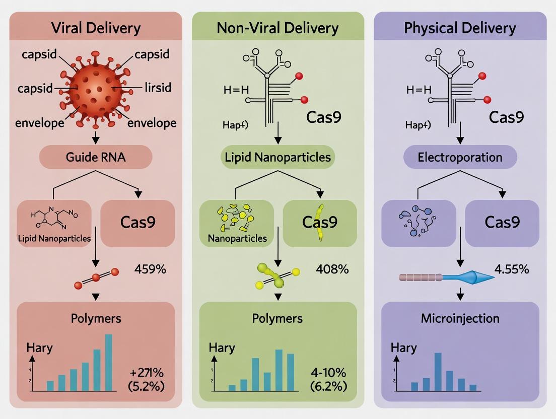
The therapeutic promise of CRISPR-Cas9 is contingent on the safe, efficient, and cell-specific delivery of its macromolecular components (Cas nuclease and guide RNA). Delivery strategies are broadly categorized into viral, non-viral, and physical methods, each presenting distinct trade-offs between efficiency, cargo capacity, immunogenicity, and manufacturability. The primary barriers are summarized below.
Table 1: Quantitative Comparison of Primary CRISPR-Cas9 Delivery Methods
| Method Category | Specific Vector/Technique | Typical Editing Efficiency (Ex Vivo) | Typical Editing Efficiency (In Vivo) | Key Advantages | Primary Barriers & Limitations |
|---|---|---|---|---|---|
| Viral | Adeno-Associated Virus (AAV) | 20-70% (in permissive cells) | 1-30% (tissue-dependent) | High tropism, long-term expression. | Cargo limit (~4.7 kb), pre-existing immunity, persistent nuclease expression raising off-target risks. |
| Viral | Lentivirus (LV) | 60-90% in hematopoietic stem/progenitor cells (HSPCs), T cells. | Limited use due to insertional mutagenesis risk. | Large cargo capacity, integrates into dividing cells. | Random genomic integration safety concerns, complex production. |
| Non-Viral | Lipid Nanoparticles (LNPs) | 70-95% in primary hepatocytes, T cells. | 10-60% in liver; <5% in extra-hepatic tissues (current data). | High payload, transient expression, scalable manufacturing. | Liver-tropic (systemic), inefficient targeting of non-hepatic tissues, potential cytotoxicity at high doses. |
| Non-Viral | Electroporation (Ex Vivo) | 50-90% in immune cells, HSPCs. | Not applicable. | High efficiency in amenable cell types. | High cell mortality, limited to ex vivo applications, requires specialized equipment. |
| Physical | Microinjection | >80% in zygotes. | Not applicable. | High precision, direct delivery. | Low throughput, technically demanding, only for ex vivo/embryonic use. |
Application Notes & Detailed Protocols
Protocol 2.1: Ex Vivo Editing of Human T Cells via Electroporation of RNP
This protocol details high-efficiency knockout in primary human T cells using Cas9 ribonucleoprotein (RNP) electroporation, a gold standard for ex vivo therapies like CAR-T engineering.
Research Reagent Solutions:
- Neon Transfection System (Thermo Fisher) or Nucleofector (Lonza): Electroporation device for hard-to-transfect cells.
- Chemically modified sgRNA (Synthego or Trilink): Enhances stability and reduces immunogenicity.
- Alt-R S.p. HiFi Cas9 Nuclease (IDT): High-fidelity Cas9 variant to minimize off-target effects.
- ImmunoCult Human CD3/CD28 T Cell Activator (STEMCELL): For pre-stimulation to enhance editing and viability.
- IL-2 (PeproTech): Cytokine for T cell expansion post-editing.
Procedure:
- T Cell Isolation & Activation: Isolate CD3+ T cells from PBMCs using a Ficoll gradient and magnetic separation. Activate cells with CD3/CD28 activator and 100 IU/mL IL-2 for 48 hours.
- RNP Complex Formation: For a single reaction targeting the TRAC locus, combine 60 pmol of HiFi Cas9 protein with 120 pmol of target-specific sgRNA in sterile duplex buffer. Incubate at room temperature for 10-20 minutes.
- Electroporation Preparation: Harvest activated T cells, count, and centrifuge. Resuspend 1x10^6 cells in 20 µL of Buffer R (Neon system) or SE Cell Line Solution (Nucleofector).
- Electroporation: Mix cell suspension with prepared RNP complex. Transfer to a 100 µL electroporation tip. Electroporate using the appropriate pulse parameters (e.g., Neon: 1600V, 10ms, 3 pulses).
- Recovery & Culture: Immediately transfer electroporated cells to pre-warmed culture medium (RPMI-1640 + 10% FBS + 100 IU/mL IL-2) in a 24-well plate. Culture at 37°C, 5% CO2.
- Analysis: Assess editing efficiency at 72-96 hours post-electroporation via flow cytometry for protein knockout or T7 Endonuclease I assay/TIDE analysis on genomic DNA.
Protocol 2.2: In Vivo Liver Editing via Systemic LNP Delivery
This protocol describes targeted in vivo knockout in hepatocytes using systemically administered, sgRNA-loaded LNPs.
Research Reagent Solutions:
- LNP Formulation (Pre-formulated): Commercially available LNP reagents optimized for mRNA delivery (e.g., GenVoy-ILM, Precision NanoSystems). Contains ionizable lipid, phospholipid, cholesterol, and PEG-lipid.
- Cas9 mRNA (TriLink CleanCap): 5-methoxyuridine-modified for enhanced stability and reduced immunogenicity.
- sgRNA (Chemical Modification): Chemically synthesized with 2'-O-methyl and phosphorothioate modifications.
- Animal Model: Ai14 reporter mice or other relevant disease models.
Procedure:
- mRNA-LNP Formulation: Co-encapsulate Cas9 mRNA and target-specific sgRNA at a defined mass ratio (typically 1:1 w/w) using a microfluidic mixing device according to the manufacturer's protocol. Purify via dialysis or tangential flow filtration. Characterize particle size (should be ~80-100 nm) and encapsulation efficiency (>90%).
- Animal Dosing & Administration: Anesthetize 8-12 week old mice. Administer LNP formulation via a single, slow bolus tail-vein injection at a dose of 1-3 mg mRNA/kg body weight in a total volume of 100-200 µL sterile PBS.
- Tissue Harvest & Analysis: Euthanize animals 3-7 days post-injection. Perfuse liver with cold PBS, then harvest and snap-freeze for genomic analysis or fix for histology.
- Efficiency Assessment: Isolate genomic DNA from liver lobes. Quantify indel frequency at the target locus via next-generation sequencing (NGS) amplicon analysis. Confirm protein-level knockout via immunohistochemistry or Western blot if antibodies are available.
Visualizing Delivery Pathways & Workflows
The Scientist's Toolkit: Essential Reagents
Table 2: Key Research Reagent Solutions for CRISPR Delivery
| Reagent Category | Specific Item | Function & Rationale |
|---|---|---|
| Nuclease & Guide | Alt-R S.p. HiFi Cas9 Nuclease (IDT) | High-fidelity Cas9 variant for reduced off-target editing, suitable for sensitive therapeutic applications. |
| Nuclease & Guide | Chemically Modified sgRNA (Synthego) | 2'-O-methyl, 2'-fluoro, and phosphorothioate backbone modifications increase nuclease resistance and reduce innate immune responses. |
| Delivery Vehicle | GenVoy-ILM Lipid Nanoparticles (Precision NanoSystems) | Pre-formed, ionizable lipid-based nanoparticles optimized for mRNA delivery, enabling rapid in vivo screening. |
| Delivery Vehicle | P3 Primary Cell 4D-Nucleofector X Kit (Lonza) | Cell-type specific electroporation buffer/cuvette kit designed for high viability and efficiency in primary human cells (T cells, HSPCs). |
| Cell Culture | ImmunoCult Human CD3/CD28 T Cell Activator (STEMCELL) | Provides a standardized, soluble stimulus for robust T cell activation, a critical pre-step for ex vivo editing. |
| Analysis | T7 Endonuclease I (NEB) | Enzyme for mismatch cleavage assay, a rapid, cost-effective method for initial assessment of editing efficiency at a target locus. |
| Analysis | Illumina CRISPR Amplicon Sequencing Assay | Provides a gold-standard, quantitative measure of on-target editing and off-target profiling via next-generation sequencing. |
Within the CRISPR-Cas9 delivery research landscape, viral vectors remain indispensable tools for achieving high-efficiency gene transfer, stable genomic integration, or transient expression. This Application Note details the core biology, quantitative performance, and specific experimental protocols for the three predominant viral vector platforms: Adeno-Associated Virus (AAV), Lentivirus, and Adenovirus. Their distinct mechanisms of action directly inform their selection for in vitro and in vivo CRISPR delivery, balancing payload capacity, tropism, immunogenicity, and expression kinetics.
Natural Biology & Quantitative Comparison
Adeno-Associated Virus (AAV)
Natural Biology: AAV is a non-enveloped, single-stranded DNA parvovirus requiring a helper virus (e.g., Adenovirus, Herpesvirus) for productive replication. Wild-type AAV preferentially integrates into a specific locus on human chromosome 19 (AAVS1), but recombinant vectors used for gene delivery persist primarily as episomal circular concatemers in post-mitotic cells, leading to long-term transgene expression without genomic integration.
Lentivirus (LV)
Natural Biology: Lentiviruses are a genus of complex, enveloped retroviruses (e.g., HIV-1). They can infect both dividing and non-dividing cells by actively importing the pre-integration complex through the nuclear pore. The viral RNA genome is reverse-transcribed into DNA and stably integrated into the host genome by the viral integrase enzyme, enabling permanent genetic modification.
Adenovirus (AdV)
Natural Biology: Adenoviruses are non-enveloped, double-stranded DNA viruses. They infect cells by receptor-mediated endocytosis, escape the endosome, and deliver their linear DNA genome to the nucleus, where it remains episomal. This triggers robust but transient transgene expression, as the viral DNA is not replicated during cell division and elicits strong innate and adaptive immune responses.
Quantitative Vector Comparison
Table 1: Key Quantitative Parameters for Viral Vectors in CRISPR Delivery
| Parameter | AAV | Lentivirus | Adenovirus |
|---|---|---|---|
| Genome Type | ssDNA (rAAV: self-complementary dsDNA available) | ssRNA (reverse transcribed to dsDNA) | dsDNA |
| Packaging Capacity | ~4.7 kb (scAAV: ~2.4 kb) | ~8-10 kb | ~8-36 kb (depending on gutless design) |
| Integration Profile | Predominantly episomal (low-risk random integration at high MOI) | Stable, random integration | Episomal |
| Typical In Vitro Titer | 10^12 – 10^13 vg/mL | 10^8 – 10^9 TU/mL | 10^10 – 10^11 PFU/mL |
| In Vivo Immunogenicity | Low | Low to Moderate | Very High |
| Expression Onset | Slow (weeks to peak) | Moderate (days) | Rapid (24-48 hours) |
| Expression Duration | Long-term (months-years in post-mitotic cells) | Permanent (through cell divisions) | Short-term (days-weeks) |
| Common CRISPR Use Case | In vivo gene editing/therapy (non-dividing cells) | In vitro screening, ex vivo cell engineering, in vivo targeting dividing cells | In vitro high-efficiency transduction, in vivo vaccination/oncolysis |
Experimental Protocols for CRISPR Vector Production & Titering
Protocol: Production of CRISPR-Ready AAV via Triple Transfection in HEK293T Cells
Purpose: Generate high-titer, recombinant AAV serotype 2 (AAV2) vector packaging a SpCas9 and gRNA expression cassette. Materials: See "Scientist's Toolkit" Section 4. Procedure:
- Day 1: Seed HEK293T cells in ten 15-cm dishes at 70% confluency in DMEM + 10% FBS.
- Day 2: Prepare transfection mix per dish: In 1.5 mL Opti-MEM, combine 10 µg AAV transfer plasmid (with ITRs, CRISPR payload), 20 µg pHelper plasmid (Adenoviral helper genes), and 10 µg pRep-Cap (AAV2 serotype) plasmid. Add 1 mL Opti-MEM with 90 µL PEI MAX (1 mg/mL), mix, incubate 15 min, add dropwise to cells.
- Day 3: Replace medium with fresh DMEM + 2% FBS.
- Day 5 (72h post-transfection): Harvest cells and media. Pellet cells, resuspend in lysis buffer (150 mM NaCl, 50 mM Tris-HCl, pH 8.5), and perform three freeze-thaw cycles (liquid nitrogen/37°C water bath).
- Purification: Treat lysate with Benzonase (50 U/mL, 37°C, 30 min). Clarify by centrifugation. Purify vector via iodixanol gradient ultracentrifugation (15%, 25%, 40%, 60% layers) at 350,000 x g for 1.5h. Collect the 40% iodixanol interface.
- Concentration & Buffer Exchange: Concentrate and exchange into PBS-MK (PBS with 1 mM MgCl2, 2.5 mM KCl) using a 100 kDa MWCO centrifugal filter. Aliquot and store at -80°C.
- Titering: Determine viral genome titer (vg/mL) by quantitative PCR (qPCR) against the ITR region using a standard curve.
Protocol: Functional Titering of CRISPR Lentivirus via Transduction Unit (TU) Assay
Purpose: Determine the functional titer (TU/mL) of a lentiviral vector encoding Cas9 and a puromycin resistance gene. Procedure:
- Day 1: Seed HEK293T cells in a 24-well plate at 5x10^4 cells/well.
- Day 2: Prepare serial 10-fold dilutions (10^-3 to 10^-6) of the lentiviral supernatant in complete medium containing 8 µg/mL Polybrene.
- Aspirate medium from cells and add 0.5 mL of each dilution to triplicate wells. Include a no-virus control.
- Day 3: Replace transduction medium with fresh complete medium.
- Day 4: Begin selection with puromycin (2 µg/mL). Change medium with puromycin every 2-3 days.
- Day 10-12: After control cells are dead, aspirate medium, fix cells with 4% PFA for 10 min, and stain with 0.1% crystal violet for 15 min. Wash and air dry.
- Quantification: Count the number of colonies (clusters of stained cells) in the wells from the optimal dilution (typically 10^-5 or 10^-6 where colonies are distinct and countable). Calculate TU/mL: (Average colony count x Dilution Factor) / Volume of inoculum in mL.
Visualization of Mechanisms & Workflows
Diagram Title: AAV Cellular Pathway for Episomal Delivery
Diagram Title: Lentiviral Integration Mechanism
Diagram Title: Adenovirus CRISPR Workflow
The Scientist's Toolkit
Table 2: Essential Research Reagent Solutions for Viral Vector CRISPR Delivery
| Reagent/Material | Function & Application |
|---|---|
| HEK293T/HEK293 Cells | Standard packaging cell line; expresses SV40 T-antigen (for plasmid amplification) and Adenovirus E1 genes (for AAV/AdV helper function). |
| Polyethylenimine (PEI MAX) | Cationic polymer for high-efficiency, low-cost transient transfection of packaging plasmids in producer cells. |
| Polybrene (Hexadimethrine) | Cationic polymer used to reduce charge repulsion between lentiviral particles and target cell membranes, enhancing transduction efficiency. |
| Iodixanol (OptiPrep) | Density gradient medium for the purification of AAV and Lentiviral vectors via ultracentrifugation, offering high recovery and purity. |
| Benzonase Nuclease | Degrades unpackaged and residual DNA/RNA in vector lysates, reducing viscosity and improving purity for downstream applications. |
| Puromycin Dihydrochloride | Selective antibiotic for enriching transduced cells when the lentiviral construct contains a puromycin resistance gene. |
| QuickTiter AAV Quantitation Kit | Commercial ELISA-based kit for rapid quantification of AAV particle titers, measuring intact capsids. |
| Lenti-X p24 Rapid Titer Kit | ELISA kit for quantifying HIV-1 p24 capsid protein concentration, used for estimating lentiviral vector physical particle count. |
| pAdVantage or pHelper | Plasmid providing Adenoviral helper genes (E2A, E4, VA RNA) necessary for AAV vector production in non-AdV-infected cells. |
| psPAX2 & pMD2.G | Common second-generation lentiviral packaging plasmids providing gag/pol and VSV-G envelope proteins, respectively. |
Within the broader thesis on CRISPR-Cas9 delivery, non-viral vectors offer critical advantages over viral methods, including reduced immunogenicity, larger cargo capacity, and simpler manufacturing. This document details application notes and protocols for the three leading non-viral platforms: Lipid Nanoparticles (LNPs), Polymers, and Inorganic Carriers. Their primary application is the safe and efficient intracellular delivery of CRISPR-Cas9 ribonucleoproteins (RNPs) or plasmid DNA for therapeutic gene editing.
Application Note: LNP-CRISPR for In Vivo Liver Targeting LNPs, particularly those incorporating ionizable lipids, are the frontline non-viral delivery system. Following systemic administration, they predominantly accumulate in the liver via ApoE-mediated uptake by hepatocytes. Recent advances involve incorporating novel biodegradable ionizable lipids (e.g., LP01, 5A2-SC8) that enhance potency and tolerability. A key application is the knockdown of Ttr for treating hereditary transthyretin amyloidosis, with clinical-stage candidates demonstrating >90% target protein reduction after a single dose.
Application Note: Polymer-Based Delivery for Ex Vivo Cell Engineering Cationic polymers, such as poly(β-amino esters) (PBAEs) and polyethylenimine (PEI), form polyplexes with nucleic acids. Their modular synthesis allows for fine-tuning of properties. A major application is the ex vivo engineering of primary T-cells or hematopoietic stem cells (HSCs). For instance, PBAE polymers optimized for endosomal escape can deliver mRNA encoding Cas9 and a sgRNA to primary human T-cells, achieving high-efficiency knockout of the PDCD1 gene (encoding PD-1) for next-generation CAR-T therapies.
Application Note: Inorganic Mesoporous Silica Nanoparticles (MSNs) for Co-Delivery Inorganic carriers like MSNs offer exceptional stability and precise control over pore architecture. A significant application is the co-delivery of Cas9 protein and multiple donor DNA templates for homology-directed repair (HDR). The high surface area allows for sequential loading strategies—adsorbing Cas9 RNP on the surface and loading donor DNA within the pores—enabling coordinated delivery to the nucleus for precise gene insertion, which is valuable for in vitro disease modeling.
Table 1: Performance Comparison of Non-Viral CRISPR-Cas9 Delivery Systems
| Platform | Typical Payload | Editing Efficiency (In Vitro) | Key Advantage | Major Limitation | In Vivo Efficacy (Liver) |
|---|---|---|---|---|---|
| LNPs | mRNA/sgRNA or RNP | 70-95% (hepatocytes) | Clinical validation, high in vivo efficacy | Primarily hepatic tropism | >90% protein knockdown |
| Polymers (PBAE) | plasmid DNA or RNP | 40-80% (primary T-cells) | Tunable structure, good for ex vivo use | Variable cytotoxicity, batch variability | Limited efficacy |
| Gold Nanoparticles | Cas9 RNP | 20-60% (cell lines) | Nuclease-resistant, precise RNP delivery | Lower efficiency, complex functionalization | Moderate (local delivery) |
| MSNs | RNP + donor DNA | 10-30% HDR (cell lines) | Co-delivery, high stability | Low transfection efficiency in some cells | Research stage |
Table 2: Formulation Components and Their Functions
| Component Class | Example Reagents | Function in Formulation |
|---|---|---|
| Ionizable Lipid | DLin-MC3-DMA, SM-102, ALC-0315 | Encapsulation, endosomal escape via protonation in acidic endosome. |
| Helper Lipid | DSPC, DOPE | Stabilizes bilayer structure; DOPE promotes membrane fusion. |
| Cholesterol | Animal-derived, Phytosterol | Modulates membrane fluidity and stability. |
| PEG-lipid | DMG-PEG2000, ALC-0159 | Provides stealth, controls nanoparticle size and shelf-life. |
| Cationic Polymer | Branched PEI (25kDa), PBAE variants | Condenses nucleic acids via charge, facilitates endosomal escape via "proton-sponge". |
| Inorganic Core | Mesoporous silica, Gold nanorod | Provides a rigid scaffold for adsorption/loading of cargo. |
Detailed Experimental Protocols
Protocol 1: Formulation of CRISPR mRNA LNP via Microfluidic Mixing Objective: Prepare LNPs encapsulating Cas9 mRNA and sgRNA. Materials: Ionizable lipid (e.g., SM-102), DSPC, Cholesterol, DMG-PEG2000, Cas9 mRNA, sgRNA, 25 mM sodium acetate buffer (pH 4.0), 1x PBS (pH 7.4), microfluidic device (e.g., NanoAssemblr Ignite), dialysis cassettes (MWCO 10kDa). Procedure:
- Prepare the lipid mixture in ethanol: Combine ionizable lipid, DSPC, cholesterol, and PEG-lipid at a molar ratio 50:10:38.5:1.5. Final total lipid concentration: 12.5 mM.
- Prepare the aqueous phase: Dilute Cas9 mRNA and sgRNA in sodium acetate buffer to a final total RNA concentration of 0.2 mg/mL.
- Set up the microfluidic device with a total flow rate (TFR) of 12 mL/min and a flow rate ratio (FRR, aqueous:organic) of 3:1.
- Load the aqueous and organic phases into separate syringes and initiate mixing. Collect the formed LNP suspension in a vial.
- Immediately dialyze the crude LNP against 1x PBS (pH 7.4) for 2 hours at room temperature using a dialysis cassette to remove ethanol and perform buffer exchange.
- Filter the LNPs through a 0.22 µm sterile filter. Characterize size (Z-average, DLS) and encapsulation efficiency (RiboGreen assay).
Protocol 2: Polyplex Formation with PBAE Polymers for Plasmid DNA Delivery Objective: Formulate polyplexes of PBAE polymer and CRISPR plasmid (encoding Cas9 and sgRNA). Materials: PBAE polymer (e.g., 447 polymer) in DMSO, CRISPR plasmid DNA in 25 mM sodium acetate buffer (pH 5.0), Opti-MEM reduced serum medium. Procedure:
- Dilute the PBAE polymer stock in sodium acetate buffer to a working concentration of 1 mg/mL. Vortex briefly.
- Dilute plasmid DNA in the same buffer to 0.05 mg/mL.
- For an N/P ratio of 20 (polymer nitrogen to DNA phosphate), add the calculated volume of polymer solution directly to the DNA solution. Vortex immediately for 10 seconds.
- Allow the polyplexes to form by incubating at room temperature for 15-20 minutes.
- Prior to transfection, dilute the formed polyplexes 5-fold with Opti-MEM. Add the diluted polyplexes dropwise to cells cultured in antibiotic-free medium.
- Replace medium after 4-6 hours of incubation.
Protocol 3: Loading of Cas9 RNP onto Gold Nanoparticles (AuNPs) Objective: Conjugate Cas9 ribonucleoprotein (RNP) to functionalized AuNPs for direct delivery. Materials: 20 nm PEGylated-AuNPs (with terminal -COOH groups), Cas9 protein, sgRNA, EDC, Sulfo-NHS, MES buffer (pH 6.0), PBS. Procedure:
- Form Cas9 RNP by incubating Cas9 protein with sgRNA at a 1:1.2 molar ratio in PBS for 10 min at room temperature.
- Activate the carboxyl groups on AuNPs: Mix 1 mL of AuNPs (1 nM) with 5 mM EDC and 10 mM Sulfo-NHS in MES buffer. React for 15 min on a shaker.
- Purify activated AuNPs via centrifugation (10,000 x g, 15 min) and resuspend in PBS.
- Add the pre-formed Cas9 RNP to the activated AuNPs. Allow conjugation to proceed for 2 hours at 4°C on a rotator.
- Block unreacted sites by adding 100 mM glycine and incubating for 30 min.
- Purify AuNP-RNP conjugates by centrifugation (2,500 x g, 10 min) and resuspend in sterile PBS. Characterize by UV-Vis spectroscopy and gel electrophoresis.
Diagrams
Diagram 1: LNP Mediated CRISPR Delivery Pathway
Diagram 2: Microfluidic LNP Formulation Workflow
The Scientist's Toolkit: Key Reagent Solutions
| Reagent / Material | Function / Explanation |
|---|---|
| Ionizable Lipid (e.g., SM-102) | Critical for mRNA encapsulation and endosomal escape. Protonates in endosome, destabilizing the endosomal membrane. |
| CleanCap Cas9 mRNA (5moU) | Chemically modified mRNA for enhanced stability and reduced immunogenicity; enables transient Cas9 expression. |
| In Vitro Transcribed sgRNA | Target-specific guide RNA; can be modified with 2'-O-methyl analogs at 3 terminal bases to reduce immune sensing. |
| Poly(β-amino ester) (PBAE) 447 | Biodegradable, cationic polymer that self-assembles with DNA; exhibits high transfection in many primary cells. |
| RiboGreen Assay Kit | Quantifies both encapsulated and free RNA in LNP formulations to determine encapsulation efficiency (%). |
| NanoAssemblr Microfluidic Instrument | Enables reproducible, scalable nanoprecipitation with precise control over LNP physicochemical properties. |
| Dynamic Light Scattering (DLS) Instrument | Measures hydrodynamic diameter (nm), polydispersity index (PDI), and zeta potential (mV) of nanoparticles. |
| EndoPort Peptide (e.g., ppTG21) | A fusogenic peptide that can be co-formulated to enhance endosomal escape of polyplexes or inorganic NPs. |
In the context of CRISPR-Cas9 delivery, physical disruption methods offer a direct, non-viral, and often transient means of breaching the cell membrane to introduce genetic cargo. These techniques circumvent the limitations of viral vectors (e.g., immunogenicity, insertional mutagenesis) and chemical non-viral methods (e.g., low efficiency in primary cells). This application note details the principles and protocols for three key physical methods: electroporation, microinjection, and sonoporation, positioning them as essential tools for ex vivo and in vivo research and therapeutic development.
Electroporation
Principle
Electroporation utilizes short, high-voltage electrical pulses to create transient, nanometer-scale pores in the cell plasma membrane. This allows the diffusion of nucleic acids (e.g., Cas9 ribonucleoprotein (RNP), plasmid DNA) into the cytoplasm. The critical parameters are electric field strength (V/cm), pulse length, and number of pulses, which must be optimized to balance delivery efficiency and cell viability.
Key Quantitative Data
Table 1: Typical Electroporation Parameters for CRISPR-Cas9 Delivery in Different Cell Types
| Cell Type | Format | Voltage / Field Strength | Pulse Length | Number of Pulses | Efficiency (Indel%) | Viability | Key Instrument |
|---|---|---|---|---|---|---|---|
| Primary T Cells | Cuvette | 500 V (1,500 V/cm) | 5 ms | 1 | 70-90% | 40-60% | Bio-Rad Gene Pulser Xcell |
| HEK293T | 96-well Plate | 170 V | 20 ms | 1 | 80-95% | 70-85% | BTX Harvard ECM 830 |
| iPSCs | Cuvette | 250 V (750 V/cm) | 10 ms | 2 | 50-70% | 50-70% | Lonza 4D-Nucleofector |
| K562 | Microfluidic | 500 V/cm | 10 ms | 10 | >90% | >80% | Thermo Fisher Neon |
Detailed Protocol: CRISPR RNP Electroporation of Primary Human T Cells
Objective: To introduce CRISPR-Cas9 RNPs into primary human CD4+ T cells for targeted gene knockout.
Materials (Research Reagent Solutions):
- Primary Human CD4+ T Cells: Isolated target cells.
- Cas9 Nuclease, high-fidelity: Recombinant protein for complexing with gRNA.
- sgRNA (crRNA+tracrRNA): Synthetic RNA guiding Cas9 to target locus.
- P3 Primary Cell 4D-Nucleofector X Kit (Lonza): Cell-type optimized buffer and cuvettes.
- Pre-complexed RNP: Formed by incubating Cas9 protein with sgRNA at 37°C for 10-20 minutes.
- 4D-Nucleofector System (Lonza): Instrument for precise pulse delivery.
- RPMI-1640 + 10% FBS: Recovery medium.
- IL-2 (200 U/mL): Cytokine to support T cell survival and proliferation post-electroporation.
Workflow:
- Isolate and activate CD4+ T cells using CD3/CD28 beads for 48-72 hours.
- On the day of electroporation, count cells and pellet 1-2 x 10^6 cells.
- Resuspend cell pellet in 100 µL of room temperature P3 Nucleofector Solution.
- Add 5-10 µg of pre-complexed RNP to the cell suspension. Mix gently.
- Transfer the entire mixture into a certified Nucleofector cuvette, ensuring no air bubbles.
- Select the appropriate program (e.g., "EO-115" for primary T cells) on the 4D-Nucleofector device and run the program.
- Immediately after pulsing, add 500 µL of pre-warmed RPMI-1640 medium to the cuvette.
- Carefully transfer the cells to a 24-well plate containing 1.5 mL of pre-warmed RPMI-1640 + 10% FBS + IL-2.
- Culture cells at 37°C, 5% CO2. Assess viability at 24 hours and gene editing efficiency (via T7E1 assay or NGS) at 72-96 hours post-electroporation.
Diagram 1: Electroporation Workflow for T Cell Gene Editing.
Microinjection
Principle
Microinjection is a direct mechanical method using a fine glass needle (0.5-5 µm tip) to inject CRISPR components directly into the cytoplasm or nucleus of a single cell. It offers precise control over the delivered dose and is the gold standard for delivery into large cells like oocytes, zygotes, and some stem cells. However, it is low-throughput and requires specialized micromanipulation equipment.
Key Quantitative Data
Table 2: Microinjection Parameters for Embryonic CRISPR-Cas9 Delivery
| Target | Injected Compartment | Needle Diameter | Injection Pressure (psi) | Injection Time (ms) | Cargo Concentration | Survival Rate | Typical Efficiency |
|---|---|---|---|---|---|---|---|
| Mouse Zygote | Pronucleus/Cytoplasm | 0.5 µm | 5-10 psi | 100-300 ms | Cas9 mRNA (50 ng/µL) + sgRNA (25 ng/µL) | 80-90% | 20-80% (Founder) |
| Zebrafish Embryo | Cytoplasm/1-cell | 1-2 µm | 10-20 psi | 50-100 ms | Cas9 Protein (300 ng/µL) + sgRNA | >90% | 50-90% (F0) |
| Human iPSC Nucleus | Nucleus | 0.7 µm | 7-12 psi | 200 ms | Cas9 RNP (10 µM) | 70-85% | >90% (Clonal) |
Detailed Protocol: Pronuclear Injection for Mouse Zygote Genome Editing
Objective: To generate knockout mice by injecting CRISPR-Cas9 components into the pronucleus of a fertilized mouse egg.
Materials (Research Reagent Solutions):
- Mouse Zygotes: B6D2F1/J, collected from superovulated females.
- Microinjection Buffer: 10 mM Tris, 0.1 mM EDTA, pH 7.4 (filter-sterilized).
- Cas9 Expression Vector or mRNA: Encoding SpCas9, purified and diluted.
- Target-specific sgRNA: In vitro transcribed and purified.
- Inverted Microscope with DIC/Nomarski Optics: For visualizing pronuclei.
- Micromanipulators & Microinjectors: Eppendorf TransferMan NK2 and FemtoJet 4i.
- Capillary Glass Needles: For holding (25 µm inner diameter) and injection (<1 µm tip).
- M2 and KSOM Embryo Culture Media: For handling and post-injection culture.
Workflow:
- Prepare the injection mixture: Dilute Cas9 mRNA and sgRNA in microinjection buffer to final concentrations of 50 ng/µL and 25 ng/µL, respectively. Centrifuge at 100,000 x g for 10 min to remove aggregates.
- Back-fill a clean injection needle with ~2 µL of the injection mixture. Mount on the injector.
- Place a group of 20-30 zygotes in a drop of M2 medium under mineral oil on the injection chamber. Mount the chamber on the microscope stage.
- Using the micromanipulators, orient a zygote so the larger male pronucleus is adjacent to the injection needle.
- Bring the needle tip against the zona pellucida and apply a brief pulse of the "Clean" function to pierce it. Advance the needle into the pronucleus.
- Apply a single injection pulse (Pi: 5-10 psi, Time: 100-300 ms). Visible swelling of the pronucleus (~50% increase) indicates successful delivery.
- Withdraw the needle carefully. Repeat for all zygotes.
- Wash injected zygotes in KSOM medium and culture at 37°C, 5% CO2, 5% O2 until the 2-cell stage (24h).
- Transfer viable 2-cell embryos into pseudo-pregnant foster females. Genotype resulting pups for targeted mutations.
Diagram 2: Microinjection into Mouse Zygote Pronucleus.
Sonoporation
Principle
Sonoporation employs ultrasound waves, typically in the presence of microbubble contrast agents, to induce cavitation and subsequent membrane perforation. The oscillating microbubbles generate localized shear stress and transient pores, enabling intracellular delivery. This method is promising for in vivo, non-invasive targeted delivery to tissues like liver, muscle, and brain.
Key Quantitative Data
Table 3: Sonoporation Parameters for In Vivo CRISPR-Cas9 Delivery
| Target Tissue | Ultrasound Frequency | Mechanical Index | Microbubble Type | Cargo Format | Delivery Route | Editing Efficiency In Vivo |
|---|---|---|---|---|---|---|
| Mouse Liver | 1 MHz | 0.8 - 1.2 | Lipid-shelled (Definity) | Plasmid DNA (pX330) | Tail vein + US | 5-10% (hepatocytes) |
| Mouse Skeletal Muscle | 2.25 MHz | 1.5 | Polymer-shelled | Cas9 RNP | Intramuscular + US | 2-5% (myofibers) |
| Mouse Brain (Focused) | 0.5 MHz | 0.7 | Custom cationic MB | siRNA/miRNA | Intravenous + FUS | N/A (BBB opening) |
Detailed Protocol: Ultrasound-Mediated CRISPR Delivery to Mouse Liver
Objective: To deliver CRISPR-Cas9 plasmid DNA to hepatocytes in vivo for gene disruption.
Materials (Research Reagent Solutions):
- Cas9/sgRNA Expression Plasmid (e.g., pX330): Purified endotoxin-free.
- Microbubble Contrast Agent (e.g., Definity): Lipid-shelled perfluoropropane bubbles.
- In Vivo Imaging System (IVIS) or Small Animal Ultrasound System: e.g., VisualSonics Vevo 2100 with a transducer.
- Medical Grade Ultrasound Gel: For acoustic coupling.
- Heating Pad: To maintain mouse body temperature during procedure.
- Animal Anesthesia System: Isoflurane vaporizer.
Workflow:
- Dilute plasmid DNA (50 µg) in 100 µL of sterile saline. Mix gently with 10 µL of activated microbubbles just before injection.
- Anesthetize the mouse and secure it in a supine position on a heating pad. Apply depilatory cream to the abdominal area to remove fur.
- Inject the DNA-microbubble mixture via the tail vein as a slow bolus.
- Immediately apply a thick layer of ultrasound gel over the liver region.
- Position the ultrasound transducer (e.g., MS250, 21 MHz for imaging) over the liver. Switch to therapy mode (1 MHz, MI 1.0).
- Apply pulsed ultrasound (1 MHz, MI 1.0, 20% duty cycle) for 5 minutes, scanning the entire liver lobe.
- Monitor microbubble destruction and replenishment in real-time using contrast imaging mode.
- After sonication, wipe off gel, and allow the mouse to recover in a warm cage.
- Harvest liver tissue 3-7 days post-treatment. Analyze editing efficiency via next-generation sequencing of the target locus from extracted genomic DNA.
Diagram 3: Sonoporation Mechanism for In Vivo Liver Delivery.
Table 4: Strategic Selection Guide for Physical CRISPR-Cas9 Delivery Methods
| Criterion | Electroporation | Microinjection | Sonoporation |
|---|---|---|---|
| Throughput | High (bulk cells) | Very Low (single cell) | Medium (localized region) |
| Primary Application | Ex vivo cells (immune cells, stem cells) | Large cells/embryos (zygotes, oocytes) | In vivo targeted tissues (liver, muscle) |
| Delivery Efficiency | Very High | Extremely High | Low to Moderate |
| Cell Viability | Moderate (optimization critical) | High (for skilled operator) | High (with optimized parameters) |
| Technical Complexity | Moderate | Very High | Moderate to High |
| Cost | Moderate (instrument, consumables) | High (instrument, skilled labor) | High (specialized US equipment) |
| Suitability for In Vivo | Limited (ex vivo only) | No (embryonic only) | Excellent |
| Key Advantage | High efficiency in hard-to-transfect cells | Precise dose control, works in any cell | Non-invasive, tissue-targeted |
The Scientist's Toolkit: Essential Research Reagent Solutions
- 4D-Nucleofector System & Kits (Lonza): Gold-standard for electroporation of primary and difficult-to-transfect cells. Cell-type specific kits optimize buffer conditions.
- Neon Transfection System (Thermo Fisher): Pipette-tip based electroporation for small sample sizes and adherent cells.
- Cas9 Electroporation Enhancer (IDT): A small molecule that improves editing efficiency and cell health post-electroporation when added to RNP complexes.
- Eppendorf InjectMan & FemtoJet System: Precise micromanipulation and pressure-controlled injection for microinjection.
- Definity or SonoVue Microbubbles: Clinically approved ultrasound contrast agents used as cavitation nuclei for sonoporation.
- pX330-U6-Chimeric_BB-CBh-hSpCas9 (Addgene): A widely used all-in-one CRISPR-Cas9 expression vector suitable for microinjection and sonoporation delivery.
- Alt-R S.p. Cas9 Nuclease V3 (IDT): High-fidelity, recombinant Cas9 protein for forming RNP complexes with synthetic sgRNA, ideal for electroporation and microinjection.
- Vevo Imaging Systems (FujiFilm VisualSonics): High-resolution micro-ultrasound platforms enabling image-guided sonoporation in small animals.
CRISPR-Cas9 genome editing requires efficient intracellular delivery of the editing machinery. The choice of payload format—plasmid DNA (pDNA), messenger RNA (mRNA), or pre-assembled ribonucleoprotein (RNP)—profoundly impacts editing efficiency, specificity, kinetics, and immunogenicity. This application note details protocols and comparative data for these three primary non-viral CRISPR payloads, framed within the broader thesis of optimizing delivery for research and therapeutic development.
Key Payload Formats: Comparison and Applications
Quantitative Comparison of CRISPR Payload Formats
Table 1: Characteristics of Major CRISPR-Cas9 Payload Formats
| Parameter | Plasmid DNA (pDNA) | mRNA | Pre-assembled RNP |
|---|---|---|---|
| Onset of Activity | Slow (24-48h) | Moderate (4-8h) | Very Fast (1-4h) |
| Duration of Activity | Long (days) | Short (<48h) | Very Short (<24h) |
| Transfection Method | Lipid nanoparticles (LNPs), electroporation, polymers | LNPs, electroporation | Electroporation, lipid-based, direct delivery |
| Immunogenicity Risk | High (TLR9 recognition) | Moderate (TLR3/7/8 recognition) | Low (No nucleic acid) |
| Off-Target Risk | Higher (sustained expression) | Moderate | Lower (transient presence) |
| Manufacturing Complexity | Moderate (bacterial fermentation) | High (in vitro transcription) | High (protein purification + synthesis) |
| Typical HDR Efficiency | Moderate | Moderate | High |
| Common Application | Stable cell line generation, in vivo with sustained expression | Therapeutic in vivo delivery, primary cell editing | High-fidelity editing, clinical ex vivo therapies (e.g., CAR-T) |
Table 2: Representative Editing Efficiencies in HEK293T Cells (via Electroporation)
| Payload Format | Delivery Method | Target Gene | Indel Efficiency (%) | HDR Efficiency (%) | Citation (Year) |
|---|---|---|---|---|---|
| pDNA (SpCas9) | Neon Electroporation | AAVS1 | 65% ± 8 | 32% ± 5 | Raguram et al. (2022) |
| mRNA (SpCas9) | Neon Electroporation | EMX1 | 78% ± 6 | 28% ± 4 | Raguram et al. (2022) |
| RNP (SpCas9) | Neon Electroporation | EMX1 | 92% ± 3 | 42% ± 6 | Raguram et al. (2022) |
| RNP (SpCas9) | Lipofection | HEK Site 4 | 70% ± 10 | N/A | Kim et al. (2024) |
Detailed Protocols
Protocol 1: CRISPR RNP Assembly and Delivery via Electroporation for Primary T Cells
Application: High-efficiency editing for ex vivo cell therapies (e.g., TRAC disruption for CAR-T).
Materials (The Scientist's Toolkit)
- Recombinant SpCas9 Protein: Purified, carrier-free, endonuclease activity verified.
- Synthetic crRNA & tracrRNA: Chemically modified for stability or unmodified. Alternatively, use synthetic sgRNA.
- Electroporation Buffer: Opti-MEM or specialized electroporation buffer (e.g., P3).
- Electroporator: 4D-Nucleofector (Lonza) or Neon (Thermo Fisher).
- Recovery Medium: Pre-warmed RPMI-1640 with 10% FBS and IL-2 (100 U/mL).
- Nuclease-Free Duplex Buffer: (e.g., IDT) for complexing RNA components.
Methodology
- RNP Complex Assembly:
- Resuscribe crRNA and tracrRNA in nuclease-free duplex buffer to 100 µM.
- Mix equimolar ratios of crRNA and tracrRNA (e.g., 3 µL each). Heat at 95°C for 5 min, then cool to room temperature to form guide RNA (gRNA).
- Combine purified SpCas9 protein with the formed gRNA at a molar ratio of 1:1.2 (Cas9:gRNA). Example: For a 10 µL reaction, mix 3 µg Cas9 (20 pmol) with ~1.5 µL of 100 µM gRNA (24 pmol).
- Incubate at room temperature for 10-20 minutes to form the active RNP complex.
Cell Preparation and Electroporation:
- Isolate and activate primary human T cells (e.g., using CD3/CD28 beads) 48-72 hours prior.
- Harvest cells, wash with PBS, and resuspend in the appropriate electroporation buffer at a concentration of 1-2 x 10^7 cells/mL.
- Mix 20 µL of cell suspension with 2-5 µL of the assembled RNP complex (final RNP dose 2-5 µM).
- Transfer mixture to a certified electroporation cuvette or strip.
- Electroporate using a pre-optimized program (e.g., for Lonza 4D-Nucleofector, use program EO-115 for primary T cells).
- Immediately add 80 µL of pre-warmed recovery medium to the cuvette and transfer cells to a plate with complete medium containing IL-2.
Post-Transfection Analysis:
- Culture cells and harvest genomic DNA 48-72 hours post-electroporation.
- Assess editing efficiency via T7 Endonuclease I assay, TIDE analysis, or next-generation sequencing (NGS) of the target locus.
Protocol 2: LNP Formulation and Delivery of CRISPR mRNA
Application: In vivo or in vitro delivery of Cas9 mRNA and sgRNA.
Materials (The Scientist's Toolkit)
- Cas9 mRNA: Chemically modified (e.g., pseudouridine, 5-methylcytidine) for enhanced stability and reduced immunogenicity.
- Ionizable Lipid: e.g., DLin-MC3-DMA, SM-102, or proprietary lipids.
- Helper Lipids: Cholesterol, DSPC, and PEG-lipid (DMG-PEG 2000).
- Microfluidic Mixer: e.g., NanoAssemblr or staggered herringbone micromixer.
- Dialysis Cassettes: For buffer exchange and purification of formed LNPs.
- sgRNA or Encoding Plasmid: Co-encapsulated or separately delivered.
Methodology
- LNP Formulation via Microfluidic Mixing:
- Prepare an Ethanol Phase containing the ionizable lipid, cholesterol, DSPC, and PEG-lipid at molar ratios (e.g., 50:38.5:10:1.5) dissolved in ethanol.
- Prepare an Aqueous Phase containing the CRISPR-Cas9 mRNA (and sgRNA if co-encapsulated) in citrate buffer (pH 4.0).
- Using a microfluidic mixer, rapidly mix the ethanol and aqueous phases at a 3:1 flow rate ratio (aqueous:ethanol). Total flow rate typically 12 mL/min.
- The resulting mixture contains self-assembled LNPs encapsulating the mRNA.
LNP Purification and Characterization:
- Dialyze the LNP suspension against a large volume of PBS (pH 7.4) for 18-24 hours at 4°C to remove ethanol and adjust pH.
- Optionally concentrate using centrifugal filter units.
- Characterize LNP size (via DLS, target 80-120 nm), polydispersity index (PDI < 0.2), encapsulation efficiency (using Ribogreen assay), and zeta potential.
In Vitro/In Vivo Delivery:
- For in vitro delivery, add LNPs to cells in serum-free medium, incubate 4-6 hours, then replace with complete medium.
- For in vivo delivery, administer via intravenous injection (common) or local administration. Dose is mRNA-dependent (e.g., 0.5-2 mg/kg in mice).
Visualization of Workflows and Pathways
Diagram Title: CRISPR Payload Intracellular Processing Pathways
Diagram Title: RNP Electroporation Workflow for T Cells
Protocols in Practice: Implementing Delivery Methods for Ex Vivo & In Vivo Applications
Within the broader thesis on CRISPR-Cas9 delivery systems—encompassing viral, non-viral, and physical methods—viral vectors remain the most efficient vehicles for in vivo and ex vivo gene editing applications. Lentiviral (LV) and Adeno-Associated Viral (AAV) vectors are predominant for stable integration and transient expression, respectively. This document details the application notes and protocols for producing and characterizing these vectors, bridging research-grade to Good Manufacturing Practice (GMP)-compliant processes essential for therapeutic development.
Viral Vector Packaging Workflows
Research-Grade Lentiviral Vector Production (Third-Generation System)
A third-generation, split-packaging system is used to enhance biosafety.
Detailed Protocol:
- Day 0 – Seeding: Plate HEK293T cells (or derivative lines like Lenti-X 293T) in high-glucose DMEM with 10% FBS on poly-L-lysine coated dishes or cell factories. Target 70-80% confluency at transfection.
- Day 1 – Transfection: For a 10 cm dish, prepare a DNA mix containing:
- 10 µg Transfer Plasmid (encoding CRISPR-Cas9 and gRNA).
- 6.5 µg Packaging Plasmid (pMDLg/pRRE).
- 3.5 µg Rev-Encoding Plasmid (pRSV-Rev).
- 5 µg Envelope Plasmid (pMD2.G for VSV-G). Dilute DNA in 500 µL of serum-free medium. In a separate tube, dilute 50 µL of PEI Max (1 mg/mL) in 500 µL serum-free medium. Combine the two, vortex, incubate 15-20 min at RT, and add dropwise to cells with fresh medium.
- Day 2 – Media Change: ~16 hours post-transfection, replace medium with fresh complete medium to reduce toxicity.
- Day 3 & 4 – Harvest: Collect viral supernatant at 48 and 72 hours post-transfection. Pool harvests. Clarify through a 0.45 µm PES filter. Aliquot and store at -80°C or concentrate immediately.
GMP-Compliant AAV Production (Triple Transfection in Suspension)
Scalable, serum-free process suitable for clinical material.
Detailed Protocol:
- Cell Expansion: Grow suspension-adapted HEK293 cells in chemically defined, serum-free medium (e.g., FreeStyle 293) in a bioreactor to a target density of 2-3 x 10^6 cells/mL.
- Transfection: For a 1L bioreactor, complex 1 mg of total plasmid DNA (ratio: Rep/Cap plasmid:Helper plasmid:ITR-flanked GOI plasmid = 1:1:1) with PEIpro (Polyplus) at a 1:2 DNA:PEI mass ratio in a volume of basal medium equal to 10% of the culture volume. Incubate 15 min, then add to the bioreactor.
- Harvest & Lysis: 48-72 hours post-transfection, harvest cells by centrifugation. Resuspend cell pellet in lysis buffer (150 mM NaCl, 50 mM Tris, pH 8.5) and perform 3-5 freeze-thaw cycles between -80°C and 37°C.
- Purification: Treat lysate with Benzonase (50 U/mL, 37°C, 1 hr). Clarify by centrifugation. Purify via iodixanol density gradient ultracentrifugation or, for GMP, affinity chromatography (e.g., AVB Sepharose). Perform buffer exchange into formulation buffer (e.g., PBS + 0.001% Pluronic F-68) using tangential flow filtration (TFF).
Titering & Potency Assays
Accurate titering is critical for dosing in CRISPR-Cas9 experiments.
Physical vs. Functional Titers
Table 1: Comparison of Viral Vector Titering Methods
| Titer Type | Method | Principle | Typical Yield (Research vs. GMP) | Relevance to CRISPR Delivery |
|---|---|---|---|---|
| Physical Titer | qPCR/ddPCR (Genome Copies/mL) | Quantifies vector genomes using primers against the ITR or a conserved vector region. | LV: 10^8-10^9 GC/mL (R), >10^10 GC/mL (GMP) AAV: 10^12-10^13 GC/mL (R), >10^14 GC/mL (GMP) | Defines total vector dose. Essential for MOI calculation. |
| Functional Titer | Transduction (TU/mL) | Measures infectious units via flow cytometry for a reporter (e.g., GFP) or antibiotic selection. | LV: 10^7-10^8 TU/mL (R) | Reflects delivery efficiency of CRISPR components into target cells. |
| Functional Titer | TCID50 | Measures infectious units via serial dilution and cytopathic effect or immunoassay. | AAV: Often used for rep/cap-containing vectors. | Critical for assessing helper virus contamination in AAV preps. |
| Potency Assay | Editing Efficiency (%) | T7E1 assay, NGS, or flow cytometry for loss/gain of target protein. | Dependent on target locus and cell type (5-90%). | Ultimate critical quality attribute (CQA). Confirms biological activity of the CRISPR-viral vector product. |
Protocol 3.1: ddPCR for AAV Genome Titer (Physical Titer)
- DNase Treatment: Incubate 10 µL vector prep with 2 U DNase I (37°C, 30 min) to degrade unencapsidated DNA. Heat-inactivate (75°C, 10 min).
- Digestion: Add Proteinase K (final 0.5 mg/mL) and SDS (0.5%) and incubate (56°C, 60 min) to release viral genomes.
- ddPCR Setup: Prepare reaction mix with QX200 ddPCR EvaGreen Supermix, primers/probe targeting polyA signal or a specific transgene sequence. Combine with digested sample and droplet generation oil in a DG8 cartridge.
- Droplet Generation & PCR: Generate droplets using the QX200 Droplet Generator. Transfer to a 96-well plate, seal, and run PCR: 95°C (5 min); 40 cycles of 94°C (30 s) and 60°C (1 min); 4°C hold.
- Quantification: Read plate on QX200 Droplet Reader. Calculate genome copies/mL using Poisson statistics.
Protocol 3.2: Editing Efficiency via T7 Endonuclease I (T7E1) Assay
- Genomic DNA Extraction: 72-96 hours post-transduction with CRISPR vector, harvest and lyse cells. Extract gDNA.
- PCR Amplification: Amplify 200-300 bp region flanking the CRISPR target site using high-fidelity polymerase.
- Heteroduplex Formation: Denature PCR products (95°C, 5 min) and re-anneal (ramp from 95°C to 25°C at -0.1°C/sec).
- Digestion: Digest with T7E1 enzyme (NEB) for 30 min at 37°C.
- Analysis: Run products on 2% agarose gel. Cleavage bands indicate indels. Calculate editing efficiency: % Indels = 100 * (1 - sqrt(1 - (b+c)/(a+b+c))), where a=uncut band intensity, b+c=cut band intensities.
Visualization
Diagram 1: Viral vector production workflows for CRISPR delivery.
Diagram 2: Viral vector quality control and release testing pathway.
The Scientist's Toolkit: Key Research Reagent Solutions
Table 2: Essential Materials for Viral Vector Production & Titering
| Reagent/Material | Supplier Examples | Function in Workflow |
|---|---|---|
| HEK293T/293 Cells | ATCC, Thermo Fisher | Standard adherent cell line for research-grade LV/AAV production due to high transfection efficiency. |
| Suspension HEK293 Cells | Thermo Fisher, Sartorius | Scalable cell lines for GMP-compliant, serum-free viral vector manufacturing in bioreactors. |
| Polyethylenimine (PEI Max/PEIpro) | Polysciences, Polyplus | Cost-effective cationic polymer for transient plasmid DNA transfection at research and large scale. |
| Poly-L-Lysine | Sigma-Aldrich | Coating reagent to enhance cell adherence for transfection-based packaging steps. |
| Benzonase Nuclease | MilliporeSigma | Degrades unpackaged nucleic acids to reduce viscosity and improve purification purity (removes host cell DNA/RNA). |
| Iodixanol | Sigma-Aldrich | Medium for density gradient ultracentrifugation, enabling high-purity AAV separation based on buoyant density. |
| AVB Sepharose HP | Cytiva | Affinity chromatography resin for GMP-scale purification of specific AAV serotypes. |
| ddPCR Supermix | Bio-Rad | Enables absolute quantification of viral genome copies without a standard curve, essential for reproducible physical titering. |
| T7 Endonuclease I | New England Biolabs | Enzyme for detecting small insertions/deletions (indels) at CRISPR target sites, a key potency assay. |
| QuickTiter LV/AAV Kits | Cell Biolabs | Commercial kits for rapid quantification of physical and functional titers via ELISA or fluorescence. |
Within the broader thesis examining CRISPR-Cas9 delivery methods—spanning viral vectors, non-viral carriers, and physical techniques—lipid nanoparticles (LNPs) have emerged as the leading non-viral platform for systemic administration. These formulations encapsulate CRISPR-Cas9 ribonucleoproteins (RNPs) or mRNA/sgRNA, enabling targeted in vivo gene editing. This protocol details the formulation, physicochemical characterization, and preliminary in vitro assessment of CRISPR-LNPs, providing a standardized workflow for researchers and drug development professionals.
Formulation Protocol: Microfluidic Mixing of CRISPR-LNPs
This method describes the preparation of LNPs encapsulating Cas9 mRNA and sgRNA (or Cas9 RNP) using rapid, precise microfluidic mixing.
Materials & Reagents
- Ionizable Lipid: e.g., DLin-MC3-DMA or SM-102. Critical for endosomal escape.
- Helper Lipid: DSPC. Stabilizes the LNP bilayer.
- Cholesterol: Enhances structural integrity and stability in serum.
- PEGylated Lipid: e.g., DMG-PEG 2000. Controls particle size and prevents aggregation.
- Aqueous Phase: Cas9 mRNA (or RNP) and sgRNA in citrate buffer (pH 4.0). The acidic pH ensures ionization of the lipid.
- Organic Phase: Lipids dissolved in ethanol (typically 90-100%).
- Microfluidic Device: e.g., NanoAssemblr Ignite or similar.
- Dialysis Tubing or Tangential Flow Filtration (TFF) System: For buffer exchange.
Detailed Protocol
- Prepare Lipid Stock Solution: Combine ionizable lipid, DSPC, cholesterol, and PEG-lipid at a molar ratio of 50:10:38.5:1.5 in pure ethanol to a total lipid concentration of 5-10 mM. Warm slightly if needed to dissolve fully.
- Prepare Aqueous Phase: Dilute Cas9 mRNA (or RNP) and sgRNA in 25 mM citrate buffer (pH 4.0) to a final concentration of 0.1-0.2 mg/mL.
- Set Up Microfluidic Mixing: Prime the device channels with ethanol and water. Set the instrument parameters: Total Flow Rate (TFR): 12 mL/min, and Flow Rate Ratio (FRR, Aqueous:Organic): 3:1.
- Execute Mixing: Load the aqueous phase and lipid-ethanol phase into separate syringes. Initiate simultaneous pumping through the device's mixing channels. The instantaneous mixing induces lipid self-assembly around the nucleic acid payload.
- Buffer Exchange & Purification: Collect the crude LNP suspension and immediately dilute in 1x PBS (pH 7.4). Transfer to a dialysis cassette (MWCO 20 kDa) against 1x PBS for 2 hours at 4°C, or use TFF for larger volumes.
- Sterile Filtration: Filter the final formulation through a 0.22 µm sterile filter. Aliquot and store at 4°C for short-term use (<1 week) or at -80°C for long-term storage.
Characterization Protocols & Data Presentation
A. Physicochemical Characterization
Perform these analyses immediately after formulation.
| Parameter | Method | Target Specification | Typical Value (Mean ± SD) |
|---|---|---|---|
| Particle Size & PDI | Dynamic Light Scattering (DLS) | Size: 70-100 nm; PDI: <0.2 | 85 ± 10 nm; 0.15 ± 0.05 |
| Zeta Potential | Phase Analysis Light Scattering | Slightly negative in PBS (~ -5 to -15 mV) | -10 ± 5 mV |
| Encapsulation Efficiency (EE%) | Ribogreen Assay (pre/post cleanup) | >90% for mRNA; >80% for RNP | 92 ± 3% (mRNA) |
| RNA Concentration | UV-Vis Spectrophotometry (A260) | Dependent on formulation scale | 0.05-0.2 mg/mL |
| Morphology | Cryo-Electron Microscography | Spherical, uniform, non-lamellar | Qualitative assessment |
B. In Vitro Functional Assessment: GFP Knockout Assay
This protocol validates gene editing efficacy in a stably expressing GFP cell line (e.g., HEK293-GFP).
Materials
- HEK293-GFP cells
- CRISPR-LNPs targeting GFP (vs. non-targeting sgRNA control)
- Lipofectamine (for RNP transfection control)
- Flow cytometry buffer and analyzer
- Genomic DNA extraction kit, T7 Endonuclease I or next-generation sequencing (NGS) reagents
Detailed Protocol
- Cell Seeding: Seed 2e5 cells per well in a 24-well plate in complete medium 24 hours before treatment.
- Dosing: Treat cells with CRISPR-LNPs at an mRNA dose range of 10-100 ng/well. Include untreated and non-targeting LNP controls.
- Incubation: Incubate cells for 72 hours at 37°C, 5% CO₂.
- Harvest & Analysis:
- Flow Cytometry: Trypsinize, wash, and resuspend cells in PBS+2% FBS. Analyze GFP fluorescence intensity. Calculate % GFP-negative cells.
- Molecular Confirmation: Extract genomic DNA from the remaining cells. Amplify the GFP target locus by PCR. Digest with T7E1 or subject to NGS to calculate indel frequency.
Expected Results Table
| Treatment Group | Dose (ng mRNA/well) | % GFP-Negative Cells (Flow) | Indel Frequency (NGS) |
|---|---|---|---|
| Untreated Control | 0 | <1% | <0.1% |
| Non-targeting LNPs | 50 | <2% | 0.2% |
| GFP-targeting LNPs | 10 | 15 ± 5% | 12 ± 4% |
| GFP-targeting LNPs | 50 | 65 ± 10% | 58 ± 8% |
| GFP-targeting LNPs | 100 | 80 ± 7% | 75 ± 6% |
The Scientist's Toolkit: Research Reagent Solutions
| Item | Function & Rationale |
|---|---|
| Ionizable Lipid (SM-102/DLin-MC3-DMA) | Enables encapsulation at low pH and promotes endosomal escape via protonation in the acidic endosome. |
| DMG-PEG 2000 | Shields LNPs, reduces opsonization, increases circulation time, and prevents aggregation during formulation. |
| NanoAssemblr Microfluidic Device | Enables reproducible, scalable, and rapid mixing for forming homogeneous, small-diameter LNPs. |
| Quant-iT RiboGreen Assay | Fluorescent dye specifically quantifies encapsulated vs. free RNA to determine encapsulation efficiency. |
| T7 Endonuclease I | Detects mismatches in heteroduplex DNA formed after editing, enabling initial quantification of indel formation. |
| Guide-it sgRNA In Vitro Transcription Kit | Allows for flexible, in-house production of high-quality, sequence-specific sgRNA. |
| Recombinant Cas9 Protein | For formulating RNP-loaded LNPs, which can reduce off-target effects and immune activation vs. mRNA. |
Workflow & Pathway Visualizations
Title: CRISPR-LNP Formulation via Microfluidic Mixing
Title: LNP Cellular Uptake and Endosomal Escape Pathway
Title: CRISPR-LNP Characterization Workflow
Electroporation Parameters for Primary Cells and Hard-to-Transfect Cell Lines
Within the expanding CRISPR-Cas9 delivery landscape, non-viral physical methods like electroporation are critical for their safety and versatility. This protocol details optimized electroporation parameters for primary cells and hard-to-transfect cell lines (e.g., Jurkat, THP-1, primary T cells, iPSCs), enabling efficient delivery of CRISPR ribonucleoproteins (RNPs), plasmids, or mRNA for genome editing applications.
Table 1: Optimized Electroporation Parameters for Common Hard-to-Transfect Cells
| Cell Type | Application | Voltage (V) | Pulse Length (ms) | # of Pulses | Buffer System | Recommended Device |
|---|---|---|---|---|---|---|
| Primary Human T Cells | CRISPR RNP | 1200-1500 | 10-20 | 1 | P3 Primary Cell Solution | Lonza 4D-Nucleofector |
| Jurkat (T Cell Line) | Plasmid DNA | 130-150 | 10-20 | 1 | SE Cell Line Solution | Bio-Rad Gene Pulser MXcell |
| THP-1 (Monocytic) | CRISPR RNP | 1350 | 10 | 1 | P3 Primary Cell Solution | Lonza 4D-Nucleofector |
| Human iPSCs | mRNA | 1100-1300 | 10-30 | 1 | P3 Primary Cell Solution | Lonza 4D-Nucleofector |
| Primary Neurons | Plasmid DNA | 120-150 | 5 | 2-3 | Neuron-specific Nucleofector Kit | Lonza 4D-Nucleofector |
| HSC (CD34+) | CRISPR RNP | 1250-1400 | 10-20 | 1 | P3 Primary Cell Solution | Lonza 4D-Nucleofector |
Table 2: Critical Parameter Impact on Viability & Efficiency
| Parameter | Effect on Viability | Effect on Efficiency | Optimization Strategy |
|---|---|---|---|
| Voltage Increase | Decreases | Increases (plateaus) | Titrate to find "sweet spot" for cell type. |
| Pulse Length Increase | Decreases | Increases | Use shortest effective pulse. |
| Number of Pulses | Decreases | May increase | Rarely >1 for modern square-wave devices. |
| Buffer Conductivity | High impact | High impact | Always use cell-type-specific, low-conductivity buffers. |
| Cell Concentration | Low impact | Critical | 1-5e6 cells/mL optimal; too high causes arcing. |
| Cargo Amount | Low impact | Saturation possible | Titrate CRISPR RNP (e.g., 2-10 pmol per reaction). |
Detailed Experimental Protocol: CRISPR RNP Electroporation of Primary Human T Cells
Objective: To achieve high-efficiency knockout in primary human T cells using Cas9-gRNA RNP electroporation.
I. Reagent and Material Preparation
- Pre-warm RPMI-1640 complete medium (with 10% FBS, 1% Pen/Strep, IL-2 (100 IU/mL)) at 37°C.
- Thaw P3 Primary Cell Nucleofector Solution (Lonza) at room temperature.
- Prepare CRISPR RNP complex:
- Dilute 10 nmol of synthetic sgRNA in nuclease-free duplex buffer (IDT) to 100 µM.
- Mix 3 µL of 100 µM sgRNA with 3 µL of 100 µM Alt-R S.p. HiFi Cas9 Nuclease V3 (IDT) in a sterile microcentrifuge tube.
- Incubate at room temperature for 10-20 minutes to form the RNP complex.
- Keep on ice until use.
II. Cell Preparation
- Isolate CD3+ T cells from PBMCs using a negative selection kit.
- Activate T cells for 48-72 hours using CD3/CD28 Dynabeads in complete medium with IL-2.
- On the day of electroporation, count cells and ensure viability >95%.
- Pellet 1-2 x 10^6 activated T cells at 90 x g for 10 minutes.
- Aspirate supernatant completely.
III. Electroporation Procedure
- To the cell pellet, add 100 µL of room-temperature P3 Primary Cell Solution.
- Add 6 µL of the pre-formed RNP complex from Step I.3. Gently mix by pipetting. Do not vortex.
- Transfer the entire cell-RNP suspension into a certified 100 µL Nucleocuvette, avoiding air bubbles.
- Place the cuvette into the Lonza 4D-Nucleofector X unit and select the pre-optimized program "EO-115".
- Press "Start" to deliver the pulse. Immediate "sparking" in the cuvette is normal.
- Immediately after the pulse, add 500 µL of pre-warmed complete medium directly to the cuvette using the provided pipette.
- Gently transfer the cells (total ~600 µL) into a 12-well plate containing 1.4 mL of pre-warmed complete medium with IL-2.
- Place cells in a 37°C, 5% CO2 incubator.
- Critical Step: Remove CD3/CD28 activation beads 24 hours post-electroporation using a magnet.
IV. Post-Electroporation Analysis
- Viability Check: At 24 hours post-electroporation, assess viability using Trypan Blue exclusion or a flow cytometry-based viability dye (e.g., DAPI or 7-AAD).
- Efficiency Assessment: At 72-96 hours post-electroporation, harvest cells for genomic DNA extraction. Assess editing efficiency via T7 Endonuclease I assay, ICE analysis (Synthego), or next-generation sequencing of the target locus.
Visualizing the Experimental Workflow
Workflow for T Cell RNP Electroporation
The Scientist's Toolkit: Key Research Reagent Solutions
| Item | Function & Rationale |
|---|---|
| Cell-Type-Specific Nucleofector Kit (e.g., P3) | Low-conductivity, optimized electrolyte solution crucial for primary cell viability and delivery efficiency. Contains supplements to enhance recovery. |
| Alt-R S.p. HiFi Cas9 Nuclease V3 (IDT) | High-fidelity Cas9 enzyme with reduced off-target effects, ideal for therapeutically relevant editing in sensitive primary cells. |
| Chemically Modified sgRNA (Synthego/IDT) | Incorporation of 2'-O-methyl and phosphorothioate modifications increases stability and reduces immune activation post-electroporation. |
| Recombinant Human IL-2 | Essential cytokine for maintaining T cell proliferation and viability during post-electroporation recovery. |
| CD3/CD28 T Cell Activator Beads | Provides the necessary TCR and co-stimulatory signals to activate primary T cells, making them receptive to electroporation and editing. |
| DAPI or 7-AAD Viability Stain | Flow cytometry-compatible dyes for accurate quantification of post-electroporation cell death, superior to Trypan Blue for suspension cells. |
| Genomic DNA Extraction Kit (e.g., QuickExtract) | Rapid, column-free method for extracting PCR-ready DNA from cell pellets for quick editing efficiency analysis via T7E1 or PCR. |
| T7 Endonuclease I / ICE Analysis Tool | Accessible, cost-effective methods for initial quantification of indel formation efficiency at the target genomic locus. |
Application Notes
Effective CRISPR-Cas9 therapy requires precise delivery to target tissues. Viral and non-viral vectors, combined with physical methods, offer distinct advantages and challenges for targeting the liver, central nervous system (CNS), lungs, and hematopoietic system. This document synthesizes current strategies and protocols.
Liver Delivery: The liver is a prime target due to fenestrated endothelium and high biosynthetic activity. Hepatocyte tropism is leveraged by viral vectors like recombinant Adeno-Associated Virus (AAV), particularly serotypes 8 and 9, and lipid nanoparticles (LNPs) with ionizable lipids. Physical methods like hydrodynamic injection enable high transient transfection in preclinical models.
CNS Delivery: The blood-brain barrier (BBB) presents a significant hurdle. Intracranial or intrathecal injections of AAVs (e.g., AAV9, AAV-PHP.eB) enable direct CNS transduction. Focused ultrasound with microbubbles can temporarily disrupt the BBB for systemic vector passage. Non-viral polymers and exosomes are under investigation for less immunogenic delivery.
Lung Delivery: Local administration via inhalation or intranasal instillation is key. AAV6, lentivirus, and synthetic vectors like polyethylenimine (PEI) or LNPs are formulated for aerosol delivery. These strategies target airway epithelial cells or alveolar macrophages for diseases like cystic fibrosis.
Hematopoietic System Delivery: Ex vivo delivery to hematopoietic stem and progenitor cells (HSPCs) via electroporation of ribonucleoprotein (RNP) complexes is clinically established. For in vivo targeting, lentiviral vectors and ligand-conjugated nanoparticles aim for bone marrow or circulating cells.
Protocols
Protocol 1: Systemic LNP Delivery for Hepatocyte-Specific Gene Editing in Mice
Objective: To achieve CRISPR-Cas9 genomic editing in hepatocytes via systemic intravenous (IV) injection of sgRNA/Cas9 mRNA-loaded LNPs.
- LNP Formulation: Prepare LNPs using a microfluidic mixer. Combine an ethanol phase containing ionizable lipid (e.g., DLin-MC3-DMA), phospholipid, cholesterol, and PEG-lipid with an aqueous phase containing Cas9 mRNA and sgRNA in citrate buffer (pH 4.0). Use a 3:1 flow rate ratio (aqueous:ethanol).
- Dialysis & Characterization: Dialyze formed LNPs against PBS (pH 7.4) for 2 hours. Characterize particle size (should be 70-100 nm) and polydispersity index (PDI <0.2) via dynamic light scattering. Measure encapsulation efficiency (>90% target).
- Animal Administration: Anesthetize C57BL/6 mice (8-10 weeks old). Inject LNP formulation via tail vein at a dose of 0.5 mg mRNA/kg body weight in a total volume of 100-200 µL.
- Analysis: Harvest liver tissue 3-7 days post-injection. Isolate genomic DNA and assess editing efficiency via next-generation sequencing (NGS) of the target locus. For protein knockdown, perform western blot on liver lysates.
Protocol 2: Intracerebroventricular (ICV) Injection of AAV-CRISPR for CNS Editing in Neonatal Mice
Objective: To deliver CRISPR-Cas9 components to the CNS via direct ICV injection in neonates.
- Vector Preparation: Produce AAV9 vectors encoding SaCas9 and a target-specific sgRNA using HEK293T cell transfection, followed by iodixanol gradient purification. Titer should exceed 1x10^13 vg/mL.
- Neonatal Injection: Within 48 hours of birth, cryoanesthetize mouse pups. Using a 33-gauge Hamilton syringe mounted on a stereotaxic frame, inject 2 µL of AAV9 preparation (total dose 2x10^10 vg) into each lateral ventricle (coordinates from lambda: AP -1.0 mm, ML ±1.0 mm, DV -1.5 mm).
- Post-procedure: Allow pups to recover on a warm pad before returning to the dam.
- Analysis: At 4-6 weeks post-injection, perfuse animals, harvest brain tissue, and section. Assess transduction via immunohistochemistry for a reporter (e.g., GFP) or editing efficiency via NGS on microdissected regions.
Protocol 3: Ex Vivo RNP Electroporation of Human CD34+ HSPCs
Objective: To genetically edit human hematopoietic stem/progenitor cells for autologous transplantation.
- Cell Isolation: Isolate CD34+ HSPCs from mobilized peripheral blood or cord blood using magnetic-activated cell sorting (MACS). Maintain in serum-free expansion medium supplemented with cytokines (SCF, TPO, FLT3-L).
- RNP Complex Formation: Complex purified Cas9 protein (30 pmol) with chemically synthesized sgRNA (36 pmol) in electroporation buffer. Incubate at room temperature for 10 minutes.
- Electroporation: Wash 1x10^5 cells and resuspend in 20 µL electroporation buffer (P3 Primary Cell Solution, Lonza). Add pre-formed RNP complex and electroporate using the Lonza 4D-Nucleofector (Program DZ-100 or equivalent).
- Reculture & Analysis: Immediately transfer cells to pre-warmed culture medium. After 48 hours, assess viability (trypan blue) and editing efficiency (T7E1 assay or flow cytometry for a surface marker knockout). For functional studies, engraft edited cells into immunodeficient mice.
Protocol 4: Oropharyngeal Instillation for Lung Epithelial Delivery in Mice
Objective: To deliver CRISPR-Cas9 components to the lung epithelium via non-viral vectors.
- Vector Preparation: Formulate branched PEI (25 kDa)/DNA polyplexes at an N/P ratio of 10. Dilute 20 µg of plasmid DNA encoding Cas9 and sgRNA in 50 µL of 5% glucose. Mix with an equal volume of PEI solution (in 5% glucose) by vortexing. Incubate 15 minutes.
- Animal Instillation: Anesthetize mouse with ketamine/xylazine. Suspend the animal vertically. Using a pipette tip, slowly drip 100 µL of polyplex preparation onto the pharynx, ensuring the liquid is aspirated into the trachea.
- Monitoring: Allow animal to recover on a warm pad.
- Analysis: After 5 days, harvest lung tissue, homogenize, and extract genomic DNA and protein. Quantify editing by NGS and assess gene expression changes by qRT-PCR.
Data Presentation
Table 1: Comparison of Delivery Strategies by Tissue Target
| Target Tissue | Primary Vector/Strategy | Typical Administration Route | Key Advantage | Major Limitation | Representative Editing Efficiency* |
|---|---|---|---|---|---|
| Liver | AAV8 | Intravenous (IV) Systemic | High hepatocyte tropism; long-term expression | Preexisting immunity; cargo size limit | 30-60% (serum protein levels) |
| Liver | Ionizable Lipid LNPs | IV Systemic | High payload; transient activity; manufacturability | Primarily hepatotropic; potential reactogenicity | >90% (mRNA translation) |
| CNS | AAV9 (ICV/IT) | Direct Injection (Intracerebroventricular/Intrathecal) | Bypasses BBB; broad CNS transduction | Invasive procedure; risk of off-target CNS areas | 20-50% (brain region dependent) |
| Lung | AAV6 | Oropharyngeal/Aerosol | Localized delivery; targets airway epithelium | Immune response; transient in dividing cells | 10-30% (airway epithelia) |
| Lung | PEI/DNA Polyplexes | Oropharyngeal Instillation | Large cargo capacity; low cost | Lower efficiency; higher cytotoxicity than LNPs | 5-15% (alveolar cells) |
| Hematopoietic | Electroporation of RNP | Ex Vivo | High precision; no vector DNA integration | Requires cell harvesting & transplantation | 70-90% (in cultured CD34+ cells) |
| Hematopoietic | Lentiviral Vector | Ex Vivo Infection | Stable integration in dividing cells | Risk of insertional mutagenesis | 40-80% (transduction) |
*Efficiency ranges are approximate and highly dependent on target gene, model, and specific construct.
Table 2: Quantitative Summary of Key Physical Delivery Parameters
| Physical Method | Target Cell/Tissue | Typical Equipment | Key Parameter Settings | Cell Viability Post-Procedure | Throughput |
|---|---|---|---|---|---|
| Hydrodynamic Injection | Mouse Hepatocytes | Syringe Pump | Volume: 10% body weight (2 mL for 20g mouse); Duration: 5-7 sec | 70-90% (transient liver stress) | Low (in vivo) |
| Electroporation (4D-Nucleofector) | HSPCs (CD34+) | Lonza 4D-Nucleofector | Program: DZ-100; Cell Number: 1e5; RNP dose: 30 pmol Cas9 | 50-70% at 24h | Medium |
| Focused Ultrasound+Microbubbles | Brain Endothelium | MRI-guided FUS | Frequency: 1 MHz; Pressure: 0.5 MPa; MB dose: 1e8 | >95% (with optimized params) | Low |
| Microinjection | Zygotes (Transgenesis) | Micromanipulator | Injection vol: 1-2 pL; DNA conc: 1-5 ng/µL | 10-20% (embryo survival to term) | Very Low |
Diagrams
Title: CRISPR-Cas9 Delivery Strategy Decision Workflow
Title: LNP-Mediated CRISPR Delivery to Hepatocytes
The Scientist's Toolkit: Research Reagent Solutions
Table 3: Essential Reagents and Materials for Featured Protocols
| Item | Function/Application | Example Product/Catalog Number (Representative) |
|---|---|---|
| Ionizable Cationic Lipid | Core component of LNPs for nucleic acid encapsulation and endosomal escape. | DLin-MC3-DMA (MedChemExpress, HY-108027) |
| AAV Helper-Free System | For production of recombinant AAV vectors (serotypes 6, 8, 9, PHP.eB). | AAVpro Kit (Takara Bio, 6667) |
| Cas9 Nuclease, S. pyogenes | Purified protein for formation of RNP complexes for ex vivo electroporation. | TrueCut Cas9 Protein v2 (Thermo Fisher, A36498) |
| Chemically Synthesized sgRNA | High-purity, modified sgRNA for use with Cas9 protein or mRNA in LNPs. | Synthego Engineered Modified sgRNA |
| Lonza P3 Primary Cell Kit | Optimized nucleofection solution and cuvettes for HSPC electroporation. | P3 Primary Cell 4D-Nucleofector X Kit (Lonza, V4XP-3032) |
| Polyethylenimine (PEI), 25 kDa | Cationic polymer for forming DNA polyplexes for lung delivery. | Linear PEI "Max" (Polysciences, 24765) |
| CD34 MicroBead Kit, human | Magnetic beads for isolation of human CD34+ HSPCs from source material. | CD34 MicroBead Kit UltraPure (Miltenyi Biotec, 130-100-453) |
| In Vivo-JetPEI | In vivo-grade linear PEI formulation optimized for systemic or local delivery. | Polyplus-transfection, 201-50G |
| T7 Endonuclease I | Enzyme for detecting indel mutations via mismatch cleavage (T7E1 assay). | NEB, M0302S |
| Alt-R HDR Donor Oligo | Single-stranded DNA donor template for homology-directed repair. | Integrated DNA Technologies (IDT) |
| Cytokine Mix for HSPCs | Recombinant proteins (SCF, TPO, FLT3-L) to maintain HSPCs in culture. | StemSpan SFEM II (StemCell Tech, 09605) + Cytokine Additives (09655) |
The efficacy of CRISPR-Cas9-mediated gene therapy is fundamentally constrained by the delivery method. This case study is framed within a broader thesis positing that an optimal preclinical pipeline must systematically compare viral, non-viral, and physical delivery modalities to identify the optimal vector for a specific therapeutic target. A successful proof-of-concept (POC) requires a head-to-head evaluation of delivery efficiency, specificity, and cellular toxicity across these platforms.
Application Notes: Delivery Modality Comparison
A critical first step is the quantitative benchmarking of delivery systems for a model system, such as correcting a GFP-disruption mutation in HEK293T cells. The following table summarizes key performance metrics from recent literature.
Table 1: Quantitative Comparison of Delivery Methods for CRISPR-Cas9 RNP/DNA
| Delivery Method | Specific Modality | Avg. Editing Efficiency (%) | Cell Viability (%) | Key Advantage | Primary Limitation |
|---|---|---|---|---|---|
| Viral | AAV9 | 40-60 | >85 | High in vivo tropism | Limited cargo capacity (~4.7 kb) |
| Viral | Lentivirus | 70-90 | 70-80 | Stable integration, high efficiency | Insertional mutagenesis risk |
| Non-Viral | Lipid Nanoparticles (LNPs) | 50-80 | 60-75 | Large cargo capacity, rapid production | Immunogenicity, liver tropism |
| Non-Viral | Electroporation | 80-95 | 50-65 | High efficiency ex vivo | High cytotoxicity |
| Physical | Microinjection | >95 | >90 (for survivors) | Precise, direct delivery | Low throughput, technically demanding |
| Non-Viral | Polymeric Nanoparticles | 30-50 | 75-85 | Tunable polymer structure | Lower efficiency than LNPs |
Experimental Protocols
Protocol 3.1: Parallel Evaluation of Delivery Efficiency via NGS
Objective: Quantify indel formation at the target locus across different delivery methods. Materials: HEK293T cells with GFP-disruption mutation, CRISPR-Cas9 RNP (Alt-R S.p. Cas9 Nuclease V3 + sgRNA), delivery reagents (LNP formulation, Lentiviral particles, Lipofectamine CRISPRMAX), Nucleofector Kit. Procedure:
- Cell Seeding: Seed 2e5 cells/well in a 24-well plate 24h pre-transfection.
- Delivery Setup:
- LNP/CRISPRMAX: Complex 2 µg RNP with 3 µL reagent in Opti-MEM for 20 min. Add to cells.
- Lentivirus: Transduce cells at an MOI of 10 in the presence of 8 µg/mL polybrene.
- Electroporation: Use Nucleofector 4D, program CM-137, with 1 µg RNP per 1e6 cells.
- Harvest: Collect cells 72h post-delivery. Extract genomic DNA (gDNA) using a silica-membrane kit.
- PCR & NGS: Amplify target locus with barcoded primers. Purify amplicons and pool for Illumina sequencing.
- Analysis: Use CRISPResso2 to calculate % indels from sequencing reads.
Protocol 3.2: Assessment of Cellular Toxicity & Viability
Objective: Measure cell health and apoptosis post-delivery. Materials: Annexin V-FITC/PI Apoptosis Kit, flow cytometer, CellTiter-Glo Luminescent Viability Assay. Procedure:
- Treat Cells: As per Protocol 3.1, in triplicate.
- Viability Assay (48h): Add CellTiter-Glo reagent to one plate, incubate, and record luminescence. Normalize to untreated control.
- Apoptosis Assay (72h): Harvest cells, stain with Annexin V-FITC and Propidium Iodide per kit instructions.
- Flow Cytometry: Analyze 10,000 events per sample. Gate live (Annexin V-/PI-), early apoptotic (Annexin V+/PI-), late apoptotic (Annexin V+/PI+), and necrotic (Annexin V-/PI+) populations.
Visualized Workflows & Pathways
Title: Preclinical Delivery Pipeline Decision Workflow
Title: Non-Viral LNP Delivery Pathway for CRISPR-Cas9 RNP
The Scientist's Toolkit: Research Reagent Solutions
Table 2: Essential Reagents for Delivery Pipeline Experiments
| Item Name | Vendor (Example) | Function in the Pipeline |
|---|---|---|
| Alt-R S.p. Cas9 Nuclease V3 | Integrated DNA Technologies (IDT) | High-purity, recombinant Cas9 protein for RNP assembly. |
| Synthetic sgRNA (chemically modified) | Synthego or IDT | Enhances stability and reduces immunogenicity compared to in vitro transcribed RNA. |
| LNP Formulation Kit (GenVoy-ILM) | Precision NanoSystems | For reproducible, scalable preparation of CRISPR RNP-loaded LNPs. |
| Lipofectamine CRISPRMAX | Thermo Fisher Scientific | Cationic lipid transfection reagent optimized for RNP delivery. |
| Lentiviral CRISPR/Cas9 All-in-One Particles | VectorBuilder | Pre-packaged, ready-to-use viral delivery system for screening. |
| Nucleofector Kit for Primary Mammalian Cells | Lonza | Reagents for high-efficiency electroporation of hard-to-transfect cells. |
| CellTiter-Glo 2.0 Assay | Promega | Luminescent assay for quantifying viable cells based on ATP content. |
| Annexin V-FITC Apoptosis Detection Kit | BioLegend | Flow cytometry-based kit to distinguish apoptotic and necrotic cells. |
| CRISPResso2 Analysis Tool | Open Source (Broad Institute) | Bioinformatics pipeline for quantifying genome editing from NGS data. |
Optimizing Efficiency and Safety: Solving Common CRISPR Delivery Problems
The efficacy of CRISPR-Cas9-based therapeutics is critically dependent on the delivery vehicle's ability to evade host immune surveillance. Both viral and non-viral vectors trigger innate and adaptive immune responses, leading to vector neutralization, reduced transduction efficiency, and potential toxicity. This document details current strategies and protocols for engineering stealth into AAV capsids and non-viral lipid nanoparticles (LNPs) to enhance delivery for gene editing applications.
Key Immune Evasion Strategies and Quantitative Data
Table 1: Comparative Analysis of Stealth Strategies for Delivery Vectors
| Strategy Category | Specific Approach | Target Immune Mechanism | Key Quantitative Outcome(s) | Reported Efficiency Increase vs. Unmodified Control |
|---|---|---|---|---|
| AAV Capsid Engineering | Directed Evolution (in vivo selection) | Pre-existing & induced NAbs | Reduction in NAb-mediated neutralization by >100-fold in murine models. | Transduction in pre-immunized mice: 10-50x higher. |
| Rational Design: Peptide Insertion (e.g., HLA-G peptide) | Innate (NK cell) & Adaptive (T-cell) Recognition | Up to 90% reduction in CD8+ T-cell activation in vitro. | Liver transduction (C57BL/6): ~5x higher. | |
| Site-Specific Chemical PEGylation | Recognition by Anti-AAV Antibodies | Decrease in antibody binding affinity by ~70-80%. | In vitro neutralization resistance: 20-100x. | |
| LNP Surface Functionalization | Incorporation of PEG-lipids (PEGylation) | Protein Corona Formation, MPS Uptake | Increase in circulation half-life from <1 hr to >6 hrs in mice. | Splenic/hepatic macrophage uptake reduced by ~60%. |
| "Self" Peptide Display (e.g., CD47 mimetics) | Phagocytosis (via CD47-SIRPα "Don't Eat Me" signal) | Reduction in macrophage phagocytosis by 40-70% in vitro. | Tumor delivery efficiency: 3-5x increase in xenograft models. | |
| Polymer Coatings (e.g., Poly(2-oxazoline)) | Complement Activation (C3 opsonization) | Reduction in C3 deposition by >85% in vitro. | Plasma AUC increased by 4-8x in rodent models. |
Detailed Experimental Protocols
Protocol 3.1: Directed Evolution of AAV Capsids for Reduced Neutralization
Objective: Generate AAV capsid variants with enhanced ability to evade pre-existing neutralizing antibodies (NAbs). Materials: AAV capsid library (mutant), HEK293T cells, pooled human IVIG (source of NAbs), NGS reagents, PCR equipment. Procedure:
- In Vitro Selection Round: Incubate your AAV mutant library (1e11 vg) with a high-titer pool of human IVIG (1:50 final dilution) in PBS++ for 1h at 37°C.
- Neutralization Challenge: Apply the mixture to confluent HEK293T cells in 10-layer CellSTACKs. Allow transduction for 48h under standard conditions.
- Recovery & Amplification: Harvest cells, extract genomic DNA, and rescue packaged AAV genomes via PCR using primers flanking the variable region.
- In Vivo Selection Round: Package the recovered capsid genes into infectious particles. Administer intravenously to mice pre-immunized with a standard AAV serotype (e.g., AAV9). After 72h, isolate target tissue (e.g., liver), extract DNA, and amplify the capsid sequences.
- Iteration & Identification: Repeat steps 1-4 for 3-5 rounds. Submit final PCR products for next-generation sequencing (NGS). Identify enriched capsid mutations. Clone top candidates and produce high-titer stocks for validation in NAb inhibition assays.
Protocol 3.2: Functionalization of LNPs with CD47 Mimetic Peptides
Objective: Produce stealth LNPs by conjugating a "Self" peptide to the surface to minimize phagocytic clearance. Materials: DSPC, Cholesterol, Ionizable lipid (e.g., DLin-MC3-DMA), PEG-lipid (Maleimide-PEG-DMG), CD47 mimetic peptide (with C-terminal Cysteine), TCEP, Microfluidic mixer, Size Exclusion Chromatography (SEC) columns. Procedure:
- LNP Formulation: Prepare an ethanolic lipid mixture (Ionizable lipid:DSPC:Cholesterol:PEG-lipid at 50:10:38.5:1.5 molar ratio). Prepare an aqueous phase (10 mM citrate, pH 4.0) containing your CRISPR-Cas9 payload (mRNA/sgRNA or RNP).
- Formation: Use a microfluidic mixer (e.g., NanoAssemblr) to combine phases at a 3:1 aqueous-to-ethanol flow rate ratio to form bare LNPs. Dialyze against PBS, pH 7.4, overnight.
- Peptide Conjugation: Reduce the maleimide-PEG on the LNP surface by incubating with TCEP (1 mM final) for 15 min at RT. Purify LNPs via SEC (PBS buffer).
- Conjugation Reaction: Immediately incubate purified LNPs with the CD47 mimetic peptide (10-fold molar excess to maleimide groups) for 2h at RT under gentle agitation.
- Purification & Characterization: Remove unconjugated peptide via SEC (PBS). Characterize LNP size (DLS), peptide conjugation efficiency (fluorescence assay or HPLC), and validate "Don't Eat Me" signaling in a macrophage co-culture phagocytosis assay.
The Scientist's Toolkit: Research Reagent Solutions
Table 2: Essential Materials for Stealth Delivery Research
| Reagent / Material | Supplier Examples | Primary Function in Stealth Engineering |
|---|---|---|
| Ionizable Cationic Lipid (DLin-MC3-DMA) | MedChemExpress, Avanti Polar Lipids | Core component of LNPs for nucleic acid encapsulation; impacts endosomal escape and reactivity. |
| Maleimide-PEG-DSPE | Nanocs, Creative PEGWorks | Provides reactive moiety for post-formulation, site-specific conjugation of thiol-containing ligands (e.g., peptides). |
| AAVXpress Library Kit | VectorBuilder, Takara Bio | Pre-built diversified AAV capsid plasmid libraries for directed evolution campaigns. |
| Recombinant Human IVIG | Sigma-Aldrich, Lee BioSolutions | Source of pooled human neutralizing antibodies for in vitro selection and neutralization assays. |
| Anti-Human C3b/iC3b ELISA Kit | Hycult Biotech, Quidel | Quantitative measurement of complement activation and opsonization on nanoparticle surfaces. |
| THP-1 Monocyte Cell Line (Human) | ATCC | Differentiate into macrophages for standardized in vitro phagocytosis and immune activation assays. |
| CD47 (IAP) Recombinant Protein | R&D Systems, Sino Biological | Positive control for SIRPα binding assays to validate mimetic peptide function. |
Visualization of Key Pathways and Workflows
Diagram 1: Strategies for Vector Stealth Engineering
Diagram 2: CD47-SIRPα Stealth Pathway vs Opsonization
Within the broader thesis evaluating CRISPR-Cas9 delivery methods—viral, non-viral, and physical—the efficiency of non-viral vectors remains a primary bottleneck. The majority of internalized non-viral delivery vehicles (e.g., lipid nanoparticles, polymer complexes) are trafficked to endosomes and subsequently degraded in lysosomes. Enhancing endosomal escape is therefore a critical, rate-limiting step for achieving effective cytosolic delivery and subsequent nuclear translocation of CRISPR-Cas9 ribonucleoproteins or plasmids.
Table 1: Efficiency and Toxicity Profiles of Endosomal Escape Agents
| Strategy | Mechanism of Action | Reported Escape Efficiency (Cytosolic Delivery) | Key Limitations/Toxicity Notes | Primary Vector Type |
|---|---|---|---|---|
| Proton-Sponge Polymers (e.g., PEI) | Buffers endosomal pH, causes osmotic swelling & rupture. | 15-25% in vitro (varies with polymer weight, N/P ratio) | High cytotoxicity; aggregation with serum proteins. | Polyplexes |
| pH-Sensitive Lipids (e.g., DODAP, DLin-MC3-DMA) | Undergo phase transition at acidic pH, destabilizing endosomal membrane. | 20-30% (LNP mRNA delivery) | Formulation stability; can be immunogenic. | Lipid Nanoparticles |
| Cell-Penetrating Peptides (e.g., GALA, HA2) | pH-dependent conformational change, membrane pore formation or fusion. | 10-20% (peptide-dependent) | Susceptible to proteolysis; off-target membrane effects. | Peptide/DNA complexes |
| Photosensitizers (e.g., TPPS₂ₐ) | Light-induced ROS generation disrupts endosomal membrane. | Up to 40% (strictly light-controlled) | Requires precise light exposure; potential phototoxicity. | Porphyrin-based carriers |
| Viral-Derived Peptides (e.g., INF7, derived from influenza HA) | pH-dependent insertion and pore formation. | 12-22% (often requires high peptide density) | Immunogenicity concerns; synthesis complexity. | Peptide-modified vectors |
| Porphyrin-Based MOFs | Proton absorption & ROS generation under light. | ~35% (with optimal light dose) | New class; long-term biocompatibility under study. | Metal-Organic Frameworks |
Table 2: Impact of Escape Enhancement on CRISPR-Cas9 Gene Editing Efficiency
| Delivery System | Escape Enhancer | Model Cell Line | Reported Editing Efficiency (vs. No Enhancer) | Key Measurement Method |
|---|---|---|---|---|
| PEI polyplexes | Chloroquine (lysosomotropic agent) | HEK293T | 8% → 22% | T7E1 assay |
| Lipid Nanoparticles | Ionizable lipid (DLin-MC3-DMA) | Hepatocytes (primary) | ~45% in vivo (mRNA) | NGS of target locus |
| Polymer Micelles | pH-responsive benzoic-imine bonds | HeLa | 5% → 28% | Flow cytometry (GFP reporter) |
| Gold Nanoparticles | Photothermal membrane disruption | A375 | 15% → 60% (with laser) | Indel formation by ICE analysis |
| Peptide-DNA Nanocubes | Endosomolytic peptide (HA2) | U2OS | 3-fold increase | Luciferase reporter assay |
Experimental Protocols
Protocol 3.1: Quantitative Assessment of Endosomal Escape Using Fluorophore-Quencher (F-Q) Pairs
Objective: To measure the kinetics and efficiency of cytosolic release of cargo from endosomes. Principle: A double-labeled molecular beacon (e.g., siRNA or dextran) with a fluorophore (Cy5) and a quencher (BHQ-2) remains quenched when intact in endosomes. Proteolytic cleavage or dequenching upon release into the cytosol yields a measurable fluorescence increase.
Materials:
- F-Q labeled dextran (e.g., Cy5-BHQ2-Dextran, 10,000 MW)
- Transfection reagent (LNP or polymer of interest)
- Confocal live-cell imaging setup or plate reader
- Appropriate cell line (e.g., HeLa, HEK293)
- HBSS buffer
Procedure:
- Complex Formation: Formulate the F-Q dextran with the delivery vector at the optimal weight/weight or N/P ratio in serum-free buffer. Incubate for 20 min at room temperature.
- Cell Treatment: Seed cells in a 96-well black-walled imaging plate 24h prior. Wash with PBS and add F-Q complex formulations in serum-free medium. Incubate for 4h at 37°C, 5% CO₂.
- Quench External Fluorescence: After 4h, carefully aspirate media. Add acid-wash buffer (0.5% acetic acid, 0.5M NaCl, pH 3.0) for 1 min to quench any fluorescence from surface-bound or internalized but still-quenched probes. Wash 3x with HBSS.
- Imaging/Analysis:
- For kinetic reads: Immediately add pre-warmed imaging medium and place plate in a pre-equilibrated live-cell imager. Acquire Cy5 fluorescence (Ex/Em ~650/670 nm) every 10 min for 2-6h.
- For endpoint analysis: Measure fluorescence using a plate reader. Include control wells with free F-Q dextran (background) and cells treated with digitonin (10 µg/mL, 10 min) to release all cargo (maximum dequenching control).
- Data Calculation:
Escape Efficiency (%) = (F_sample - F_background) / (F_digitonin - F_background) * 100
Protocol 3.2: Co-Localization Analysis for Endosomal Entrapment
Objective: To quantify the fraction of delivery vector co-localized with endosomal/lysosomal markers. Materials:
- LNP or polyplexes labeled with Cy5
- Primary antibody for early endosome marker (EEA1) or lysosome marker (LAMP1)
- Corresponding fluorescent secondary antibody (e.g., Alexa Fluor 488)
- Fixative (4% PFA), permeabilization buffer (0.1% Triton X-100), blocking buffer (5% BSA)
- Confocal microscope with high-resolution Z-stack capability
- Image analysis software (e.g., ImageJ, Coloc2 plugin)
Procedure:
- Transfection & Fixation: Treat cells grown on coverslips with labeled delivery vectors for 2, 4, 6, and 24h. At each time point, wash cells and fix with 4% PFA for 15 min.
- Immunostaining: Permeabilize cells (0.1% Triton X-100, 10 min), block with 5% BSA (1h). Incubate with primary antibody (1:200 in blocking buffer) overnight at 4°C. Wash 3x with PBS, incubate with secondary antibody (1:500) for 1h at RT. Include DAPI for nuclei.
- Imaging: Acquire high-resolution Z-stack images (~5-10 slices per cell) using consistent laser power and gain settings across all samples.
- Quantitative Analysis:
- Open images in ImageJ. Split channels.
- Apply a manual threshold to each channel to define positive signals.
- Run the "Coloc2" plugin to calculate Manders' overlap coefficients (M1 & M2), representing the fraction of Cy5 signal overlapping with the organelle marker and vice versa.
- M1 (fraction of vector in endosomes) =
sum(Cy5_colocalized) / sum(Cy5_total). Report as mean ± SD across >50 cells per time point.
Protocol 3.3: Functional CRISPR Editing Assay to Correlate Escape Efficiency
Objective: To directly link endosomal escape enhancement to functional CRISPR-Cas9 gene knockout. Materials:
- LNP or polymer formulation containing SaCas9 mRNA and sgRNA targeting a genomic locus (e.g., AAVS1 safe harbor)
- Control formulation with a non-endosomolytic vector
- Cells with high transfection efficiency (e.g., HepG2)
- Genomic DNA extraction kit
- NGS library preparation reagents for the target site
- ICE analysis software (Synthego) or TIDE analysis web tool
Procedure:
- Transfection: Seed cells in a 24-well plate. At 70% confluency, transfert with CRISPR-LNP formulations at an optimal mRNA dose (e.g., 50 ng/well). Include positive (SpCas9 RNP via electroporation) and negative (non-targeting sgRNA) controls.
- Harvest & Extract DNA: 72h post-transfection, wash cells, trypsinize, and pellet. Extract genomic DNA using a commercial kit. Quantify DNA concentration.
- Amplify Target Locus: Perform PCR to amplify a ~300-500bp region surrounding the cut site using high-fidelity polymerase. Verify amplicon size on agarose gel.
- Next-Generation Sequencing (NGS): Purify PCR products, barcode samples, and pool for NGS on an Illumina MiSeq. Ensure >50,000 reads per sample.
- Analysis: Use ICE or CRISPResso2 to align sequences and quantify the percentage of indels at the target site.
- Correlation: Plot editing efficiency (%) against endosomal escape efficiency (%) as measured by Protocol 3.1 for the same formulations to establish correlation.
Visualizations
Diagram 1: Pathways of Non-Viral Vector Intracellular Trafficking
Diagram 2: Mechanisms of Major Endosomal Escape Agents
Diagram 3: Workflow for Evaluating Endosomal Escape
The Scientist's Toolkit: Key Research Reagent Solutions
Table 3: Essential Materials for Endosomal Escape Research
| Item | Function/Description | Example Product/Catalog # |
|---|---|---|
| pH-Sensitive Dye (e.g., pHrodo Dextran) | Fluoresces brightly only in acidic compartments (endosomes/lysosomes), used to track uptake and vesicle acidification. | Thermo Fisher Scientific, P10361 |
| Fluorophore-Quencher (F-Q) Pairs | For direct quantification of cytosolic release via dequenching (see Protocol 3.1). | Cy5-BHQ2 labeled siRNA, Custom from Sigma or IDT. |
| Ionizable Cationic Lipid | Core component of modern LNPs; enables encapsulation and pH-dependent endosomal escape. | DLin-MC3-DMA (MedKoo, 510039). |
| Proton-Sponge Polymer | Gold-standard polymer for benchmarking endosomal buffering capacity. | Branched PEI, 25kDa (Sigma, 408727). |
| Endosomolytic Peptide | Synthetic peptide to augment escape when conjugated to vectors. | HA2 (Infuenza-derived) peptide (GL Biochem). |
| Lysosomal Inhibitor (Control) | Positive control for enhancing functional delivery by blocking lysosomal degradation. | Chloroquine diphosphate (Sigma, C6628). |
| Antibody for EEA1 | Marker for early endosomes in co-localization studies. | Abcam, ab70521 (mouse monoclonal). |
| Antibody for LAMP1 | Marker for late endosomes/lysosomes. | Cell Signaling Technology, 9091S. |
| Membrane Integrity Dye | To assess potential cytotoxicity from escape mechanisms (e.g., pore formation). | Propidium Iodide (PI) (Sigma, P4170). |
| HDR Inhibitor (for NGS assays) | Used to suppress homology-directed repair, enriching for NHEJ indels in CRISPR editing analysis. | NU7026 (Sigma, SML0338). |
Within the broader research thesis comparing viral, non-viral, and physical CRISPR-Cas9 delivery methods, a critical parameter is their impact on editing fidelity. Off-target effects remain a significant barrier to therapeutic translation. This application note synthesizes current research, positing that the delivery modality directly dictates the cellular kinetics of Cas9/gRNA presence (e.g., duration, concentration, and localization), which in turn is a primary determinant of editing specificity. We provide protocols and data analysis frameworks to quantify this relationship.
Table 1: Comparative Analysis of Delivery Methods and Associated Off-Target Profiles
| Delivery Method | Typical Format | Editing Kinetics (Peak Expression) | Key Fidelity Advantage | Key Fidelity Risk | Primary Off-Target Assessment Method |
|---|---|---|---|---|---|
| Viral (AAV) | DNA vector for Cas9 + gRNA. | Slow (days to weeks), persistent. | Enables use of high-fidelity Cas9 variants; lower peak Cas9 levels. | Prolonged expression increases chance of off-target cleavage over time. | Targeted deep sequencing (CHIP-seq, GUIDE-seq) at predicted sites. |
| Viral (Lentivirus) | DNA vector, often integrates. | Moderate to fast, can be persistent. | Tunable via inducible promoters. | Genomic integration risks; long-term expression similar to AAV. | Genome-wide methods like CIRCLE-seq or DISCOVER-Seq. |
| Non-Viral (RNP) | Pre-complexed Cas9 protein + gRNA. | Very fast (hours), transient (<24-48h). | Short exposure window minimizes off-target activity; no DNA transcription. | Lower editing efficiency in some cell types can lead to selective pressure. | BLISS or SITE-seq for sensitive, unbiased detection. |
| Non-Viral (mRNA/LNP) | mRNA for Cas9 + synthetic gRNA. | Fast (hours to days), transient (days). | Tunable via mRNA engineering; transient expression. | High transient peak Cas9 from mRNA translation can increase risk. | Digenome-seq or GUIDE-seq post-transfection. |
| Physical (Electroporation) | Delivery of RNP, mRNA, or plasmid. | Defined by cargo (RNP=fast, plasmid=slow). | Precise dosage control; ideal for RNP delivery. | Cellular stress from procedure may alter DNA repair dynamics. | Targeted NGS or HTGTS. |
Table 2: Experimental Parameters for Kinetics-Fidelity Studies
| Controlled Variable | Experimental Manipulation | Measured Fidelity Output |
|---|---|---|
| Cas9/gRNA Exposure Time | Inducible systems (e.g., doxycycline-on), or RNP wash-out time courses. | Off-target vs. on-target ratio over time. |
| Cas9 Dosage | Titration of RNP complex amount, mRNA quantity, or viral MOI. | Off-target signal slope vs. on-target saturation point. |
| Nuclear Localization Timing | Use of NLS-engineered Cas9 or photoactivatable systems. | Correlation of nuclear concentration with indel formation at off-target sites. |
Detailed Experimental Protocols
Protocol 1: Kinetic Profiling of Cas9 Activity via RNP Electroporation and Time-Course Sampling Objective: To correlate transient RNP presence with on- and off-target editing rates. Materials: See "Scientist's Toolkit" below. Procedure:
- RNP Complex Formation: Complex HiFi Cas9 protein (pmol amounts as titrated) with chemically modified sgRNA (at a 1:1.2 molar ratio) in duplex buffer. Incubate 10 min at 25°C.
- Cell Electroporation: Harvest and wash 1e5 target cells (e.g., HEK293). Resuspend in P3 buffer with RNP complex. Electroporate using a 96-well shuttle system (Program: CM-150).
- Time-Course Harvest: Immediately post-pulse, plate cells in pre-warmed medium. Collect cell pellets at defined intervals (e.g., 2, 6, 12, 24, 48h).
- Genomic DNA Extraction: Use a silica-membrane column kit for all time points.
- NGS Library Prep for Parallel On/Off-Target Analysis: a. Perform a multiplexed PCR (20 cycles) to amplify all predicted off-target sites (from algorithms like CRISPRscan) and the on-target locus. b. Index with a second PCR (10 cycles). c. Pool, purify, and quantify libraries for Illumina sequencing.
- Data Analysis: Align sequences. Calculate indel % per site per time point. Plot kinetic curves for on-target vs. representative off-target sites.
Protocol 2: Evaluating AAV-Delivered High-Fidelity Cas9 Variant with Persistent Expression Objective: To assess if high-fidelity variants mitigate risks of long-term AAV expression. Materials: AAV9 vector encoding SpCas9-HF1 and sgRNA, target cells, DNeasy Blood & Tissue Kit. Procedure:
- Transduction: Transduce cells at an MOI of 1e5 vg/cell in the presence of 5μM HDR enhancer. Include untransduced control.
- Long-Term Passaging: Culture cells for 28 days, passaging every 3-4 days.
- Sampling: Harvest genomic DNA at day 3, 7, 14, and 28.
- Off-Target Detection via GUIDE-seq: a. At day 1 post-transduction, co-transfect cells with the AAV and the GUIDE-seq oligonucleotide duplex using a lipid transfector. b. At each sampling time point, extract gDNA. c. Perform GUIDE-seq library preparation as originally described (Tsai et al., Nat Biotechnol, 2015), including shearing, adapter ligation, and nested PCR to enrich for integration sites. d. Sequence and analyze with the GUIDE-seq computational pipeline.
- Analysis: Compare the number and indel frequency of off-target sites identified at each time point.
Mandatory Visualizations
Title: Delivery Method Determines Editing Kinetics and Fidelity Risk
Title: Workflow for Measuring Kinetics of Editing Fidelity
The Scientist's Toolkit: Essential Research Reagents & Materials
| Item | Function & Relevance to Fidelity Studies |
|---|---|
| High-Fidelity Cas9 Variant (e.g., SpCas9-HF1, eSpCas9) | Engineered protein with reduced non-specific DNA contacts, crucial for mitigating off-target effects with prolonged expression systems. |
| Chemically Modified sgRNA (e.g., 2'-O-Methyl, Phosphorothioate) | Enhances stability and reduces immunogenicity, allowing lower effective doses and improving RNP-based kinetic control. |
| GUIDE-seq Oligoduplex | Unbiased, genome-wide off-target detection method. Essential for comprehensive baseline mapping for any delivery method. |
| Electroporation System (e.g., 4D-Nucleofector) | Enables precise, high-efficiency delivery of RNP complexes for transient kinetic studies with minimal cargo modification. |
| Inducible Expression System (dCas9-KRAB or Cas9) | Allows precise temporal control over Cas9 activity initiation to study the impact of exposure duration in isolation. |
| Next-Generation Sequencing (NGS) Kit for Amplicons | Required for high-throughput, quantitative comparison of indel frequencies at multiple loci across time points. |
| Lipid Nanoparticles (LNPs) for mRNA/sgRNA | A clinically relevant non-viral delivery platform for studying the kinetics of mRNA-encoded Cas9 and its fidelity implications. |
| Doxycycline-Inducible AAV Vector | Enables study of prolonged but controllable Cas9 expression from viral vectors, separating delivery from kinetic variables. |
1. Introduction Within the thesis exploring CRISPR-Cas9 delivery methods, scaling from research-grade viral and non-viral systems to GMP production presents a critical bottleneck. This document details application notes and protocols for scaling AAV (viral) and lipid nanoparticle (LNP, non-viral) CRISPR delivery systems, highlighting key parameters, challenges, and quantitative comparisons.
2. Key Scaling Parameters & Quantitative Comparison The transition from bench to clinic involves orders-of-magnitude increases in scale, with corresponding shifts in critical process parameters (CPPs) and critical quality attributes (CQAs).
Table 1: Scaling Parameters for Viral (AAV) vs. Non-Viral (LNP) CRISPR Delivery
| Parameter | Bench-Scale (AAV) | Clinical-Scale (AAV) | Bench-Scale (LNP) | Clinical-Scale (LNP) |
|---|---|---|---|---|
| Typical Volume | 0.5 - 2 L (cell culture) | 200 - 2000 L | 1 - 10 mL (aqueous) | 10 - 100 L (aqueous) |
| Typical Yield | 1e13 - 1e14 vg total | 1e16 - 1e18 vg total | 0.1 - 1 g mRNA | 10 - 1000 g mRNA |
| Transfection Method | PEI-mediated (HEK293) | Baculovirus/Sf9 or Stable Cell Line | Microfluidic Mixing | Turbulent Jet or Confined Impingement Mixing |
| Purification | Ultracentrifugation, Benchtop Chromatography | Tangential Flow Filtration (TFF), Multi-column Chromatography | Dialysis, SEC Spin Columns | In-line TFF, Tangential Flow Diafiltration |
| Formulation Buffer Exchange | Centrifugal Concentrators | In-line Diafiltration (TFF) | Dialysis Cassettes | Tangential Flow Diafiltration |
| Key CQA | Full/Empty Capsid Ratio (<10% ideal), Potency, Genomic Titer | Full/Empty Capsid Ratio (<5% required), Potency, Purity (HCP/DNA) | Encapsulation Efficiency (>90%), Size (70-100 nm), PDI (<0.2) | Encapsulation Efficiency (>95%), Size Consistency, Residual Solvent |
| Primary Challenge | Empty capsid removal, vector potency retention. | Reproducible full/empty ratio at scale, clearance of host cell DNA. | Reproducible particle size & PDI. | Mixing efficiency & heat management, aseptic production. |
Table 2: Recent Clinical-Grade Production Yield Data (Representative Values)
| System | Scale | Reported Yield | Key Achievement | Reference Year |
|---|---|---|---|---|
| AAV8 | 500 L Sf9 | 1.2e17 vg/L | Consistent full capsid yield (~65%) | 2023 |
| AAV9 | 200 L HEK293 | 5e15 vg/L | Scalable transient transfection | 2024 |
| LNP (mRNA) | 30 L Mix | >95% Encapsulation | cGMP-compliant continuous process | 2023 |
| LNP (RNP) | Lab-scale | 80% RNP Encapsulation | Proof-of-concept for Cas9 RNP delivery | 2024 |
3. Experimental Protocols
Protocol 3.1: Scalable AAV Production via PEI-Mediated Transfection in Suspension HEK293 Cells Objective: Produce AAV vectors at 10L bioreactor scale for pre-clinical studies. Materials: HEK293 suspension cells, serum-free medium, pAAV-Rep/Cap, pHelper, pAAV-CRISPR-ITR, linear PEI (40 kDa), bioreactor, perfusion or fed-batch system. Procedure:
- Cell Expansion: Expand HEK293 cells in serum-free medium to a target density of 3-4e6 cells/mL in a 10L bioreactor. Maintain pH (7.1±0.1), DO (40%).
- Transfection Mix Preparation (Separate Vessels): Vessel A: Dilute DNA plasmid cocktail (1:1:1 mass ratio, total 1 mg/L culture) in 5% final volume fresh medium. Vessel B: Dilute PEI to 1 mg/mL in 5% final volume fresh medium. Incubate 5 min.
- Complexation: Rapidly mix Vessel B into Vessel A. Vortex immediately for 10 sec. Incubate 15-20 min at RT.
- Bioreactor Transfection: Add DNA:PEI complex dropwise to the bioreactor under moderate agitation.
- Harvest: 72 hours post-transfection, cool culture to 4°C. Separate cells and supernatant via continuous centrifugation. Retain both fractions for purification.
- Purification (Clarified Lysate): Subject cell pellet to freeze-thaw, benzonase treatment. Clarify. Use affinity chromatography (AVB Sepharose or POROS CaptureSelect), followed by ion-exchange chromatography. Formulate via TFF into final buffer (PBS + 0.001% Pluronic F-68).
Protocol 3.2: Clinical-Scale LNP Formulation via Confined Impingement Jet Mixing Objective: Manufacture CRISPR mRNA or RNP-loaded LNPs under cGMP-like conditions. Materials: Ionizable lipid (e.g., DLin-MC3-DMA), DSPC, Cholesterol, PEG-lipid, mRNA/Cas9 RNP, Ethanol, Sodium Acetate Buffer (pH 4.0), Confined Impingement Jet (CIJ) Mixer, TFF system. Procedure:
- Lipid Phase Preparation: Dissolve ionizable lipid, DSPC, cholesterol, PEG-lipid (50:10:38.5:1.5 molar ratio) in ethanol to a total lipid concentration of 25-50 mM. Filter (0.22 µm).
- Aqueous Phase Preparation: Dilute mRNA or Cas9 RNP in sodium acetate buffer (pH 4.0). For mRNA, use concentration of 0.2 mg/mL; for RNP, maintain a 1:2 molar ratio (sgRNA:Cas9). Filter (0.22 µm).
- Continuous Mixing: Using a CIJ mixer, pump the lipid phase (in ethanol) and aqueous phase (in buffer) at a 1:3 volumetric ratio (e.g., 15 mL/min lipid, 45 mL/min aqueous). Maintain total flow rate to achieve a total residence time < 0.1 seconds.
- Immediate Buffer Exchange: Direct the crude LNP mixture into a diafiltration vessel. Begin TFF immediately using a 100-500 kDa MWCO membrane cassette against PBS (pH 7.4) for 10 diavolumes to remove ethanol, adjust pH, and exchange buffer.
- Concentration & Sterile Filtration: Concentrate LNPs to target final RNA concentration (e.g., 1 mg/mL). Perform 0.22 µm sterile filtration. Store at 2-8°C.
4. Diagrams & Visualizations
Diagram 1: AAV Production Scale-Up Path & Challenges (100 chars)
Diagram 2: Clinical-Scale LNP Formulation Workflow (99 chars)
5. The Scientist's Toolkit: Research Reagent Solutions
Table 3: Essential Reagents & Materials for Scale-Up Studies
| Item | Function & Relevance | Example/Supplier |
|---|---|---|
| Linear PEI (40 kDa) | High-efficiency polycation for large-scale transient transfection of HEK293 cells. Scalable alternative to CaPO4. | Polyplus PEIpro |
| Baculovirus Expression System | Scalable, serum-free production of AAV in insect Sf9 cells. Reduces empty capsids. | Bac-to-Bac (Thermo), flashBAC (Oxford) |
| Ionizable Cationic Lipid | Key component of LNPs for encapsulating nucleic acids. Enables endosomal escape. | DLin-MC3-DMA, SM-102, ALC-0315 |
| Microfluidic/Jet Mixing Device | Enables reproducible, scalable nanoprecipitation of LNPs with controlled size and PDI. | NanoAssemblr, CIJ Mixer (Precision NanoSystems) |
| Affinity Chromatography Resin | Critical for high-purity AAV capture based on serotype-specific antibody binding. | AVB Sepharose, CaptureSelect AAVX |
| Tangential Flow Filtration (TFF) System | For concentration, buffer exchange, and diafiltration of both AAV and LNP products at scale. | Pellicon Cassettes (Merck), KrosFlo (Repligen) |
| Benzonase Nuclease | Digests unpackaged nucleic acids (host cell & plasmid DNA/RNA) during AAV purification to reduce viscosity and impurities. | Merck Millipore |
| Sterile Connectors & Tubing | Single-use, sterile fluid paths for aseptic processing in GMP-like manufacturing. | Colder Products (CPC), Saint-Gobain |
The therapeutic and research potential of CRISPR-Cas systems is often constrained by the physical cargo capacity of delivery vehicles. This challenge is compounded by two key trends: the adoption of large Cas variants (e.g., Cas9 orthologs, Cas12a, base editors, prime editors) and the frequent need to deliver multiple gRNAs for complex editing strategies. Within the broader thesis on delivery methods—encompassing viral (e.g., AAV, Lentivirus), non-viral (e.g., LNPs, polymers), and physical (e.g., electroporation) approaches—overcoming the cargo limit is a pivotal hurdle for efficacy.
Quantitative Analysis of Cargo Sizes and Vehicle Capacities
The following table summarizes the cargo requirements of common CRISPR components and the payload capacities of standard delivery vehicles.
Table 1: CRISPR Component Sizes and Delivery Vehicle Capacities
| Component / Vehicle | Typical Size / Capacity (kb) | Notes & Implications |
|---|---|---|
| CRISPR Effectors | ||
| SpCas9 (saCas9) | ~3.1 kb (~3.2 kb) | Standard effector; saCas9 is smaller but with different PAM. |
| Cas12a (AsCpfl) | ~3.9 kb | Requires shorter crRNA, no tracrRNA. |
| BE4max (Base Editor) | ~5.2 kb | Fusion of Cas9 nickase + deaminase + inhibitors. |
| PE2 (Prime Editor) | ~6.3 kb | Fusion of Cas9 nickase + reverse transcriptase. |
| Expression Elements | ||
| gRNA expression cassette | ~0.2 - 0.5 kb | Size varies with promoter and terminator. Multiplexing adds linearly. |
| Promoter (e.g., U6, Pol II) | ~0.2 - 0.5 kb | Constitutive or tissue-specific. |
| PolyA signal | ~0.1 - 0.2 kb | |
| Delivery Vehicle Max Payload | ||
| Adeno-Associated Virus (AAV) | ~4.7 kb | Strict limit; requires splitting or miniaturization. |
| Lentivirus (LV) | ~8-10 kb | Larger capacity, integrates into genome. |
| Lipid Nanoparticles (LNPs) | >10 kb (mRNA) | Encapsulates mRNA and gRNAs; no hard sequence limit, but efficiency decreases with size. |
| Polymers / Nanoparticles | Variable | Depends on formulation; can package large plasmid DNA. |
Strategic Approaches to Overcome Cargo Limits
Minimization and Optimization of CRISPR Components
- Cas Protein Engineering: Use of smaller Cas orthologs (e.g., S. aureus Cas9, ~3.2 kb) or ultra-compact variants (e.g., CasΦ, ~2.0 kb). Codon optimization and removal of non-essential sequences.
- Promoter/UTR Optimization: Employing minimal, yet efficient, promoters and regulatory elements to shrink DNA cassettes.
- gRNA Engineering: Using truncated gRNAs (tru-gRNAs) or optimized scaffolds.
Split Systems and Trans-Splicing
For AAV delivery, large genes are split into two halves, each packaged into separate virions. Co-infection leads to intracellular reconstitution via:
- Inteins: Self-splicing protein elements that ligate the two Cas protein fragments.
- Trans-Splicing: Using split inteins or homologous DNA recombination to reassemble the full coding sequence.
Multiplexed gRNA Delivery Strategies
- Single Transcript with Processing Units: Expressing multiple gRNAs from a single Pol II transcript, separated by ribozyme, tRNA, or Csy4 sequences that mediate cleavage into individual units.
- Multi-Promoter Arrays: Tandem arrangement of multiple Pol III (e.g., U6) promoters, each driving a single gRNA.
Exploiting Alternative Delivery Vehicles
- Lentiviral Vectors: For ex vivo applications where larger cargo and genomic integration are acceptable.
- Non-Viral Vectors (LNPs, Polymers): Ideal for delivering large mRNA or plasmid cargo without a strict size ceiling, though formulation optimization is critical for large molecules.
Detailed Experimental Protocols
Protocol 1: AAV Split-intein Reconstitution of a Large Cas Variant (e.g., Base Editor)
Objective: To deliver a base editor (BE) exceeding the AAV cargo limit via dual-AAV, intein-mediated reconstitution. Materials:
- Plasmids: pAAV containing 5’-BE-inteinN, pAAV containing inteinC-3’-BE, each with appropriate ITRs.
- Cells: HEK293T cells (for production), target cell line (e.g., HeLa).
- Reagents: PEI transfection reagent, AAVpro purification kit, target genomic DNA extraction kit, PCR/sequencing primers.
Procedure:
- AAV Production: Co-transfect HEK293T cells with pAAV-BE-inteinN, pAAV-inteinC-BE, and pHelper/pRepCap plasmids using PEI.
- Purification: Harvest cells and medium at 72h. Purify AAV serotype of choice (e.g., AAV9) using an iodixanol gradient or kit.
- Target Cell Transduction: Plate target cells. Co-infect with both AAVs at a 1:1 MOI ratio. Include single-AAV controls.
- Analysis (7-14 days post-transduction): a. Extract genomic DNA. b. PCR-amplify the target region. c. Perform next-generation sequencing (NGS) or T7E1 assay to quantify base editing efficiency.
Protocol 2: Multiplexed gRNA Delivery via a tRNA-gRNA Array in a Single Plasmid
Objective: To express four gRNAs from a single plasmid for combinatorial gene knockout. Materials:
- Plasmid Backbone: All-in-one CRISPR plasmid expressing SpCas9.
- Oligonucleotides: DNA oligos encoding each gRNA spacer sequence, with flanking tRNAGly sequences.
- Enzymes: BsmBI restriction enzyme, T4 DNA Ligase, Gibson Assembly Master Mix.
- Cells: HEK293FT cells, transfection reagent, genomic DNA extraction kit, surveyor nuclease assay kit.
Procedure:
- Array Construction: Synthesize or assemble by PCR a DNA fragment:
U6-promoter - [tRNAGly-gRNA1-spacer - tRNAGly-gRNA2-spacer - tRNAGly-gRNA3-spacer - tRNAGly-gRNA4-spacer] - terminator. - Cloning: Digest the all-in-one plasmid backbone (with Cas9) and the array fragment using appropriate enzymes (e.g., BsmBI for Golden Gate assembly). Ligate using T4 ligase.
- Validation: Sanger sequence the entire array to confirm spacer sequences and tRNA junctions.
- Transfection & Analysis: a. Transfect validated plasmid into target cells. b. Harvest genomic DNA after 72h. c. Perform multiplex PCR on all four target genomic loci. d. Analyze indels for each locus individually via NGS or the Surveyor nuclease assay.
Visualizations
Diagram 1: Dual-AAV Intein Reconstitution Strategy
Title: Dual-AAV Base Editor Delivery via Inteins
Diagram 2: Multiplexed gRNA Array Expression & Processing
Title: Multiplex gRNA Expression via tRNA Array
The Scientist's Toolkit: Key Research Reagent Solutions
Table 2: Essential Materials for Cargo-Limited CRISPR Delivery Research
| Reagent / Material | Function & Application | Example/Supplier Note |
|---|---|---|
| Miniaturized Cas Expression Plasmids | Provide codon-optimized Cas genes with minimal bacterial backbones for easier packaging. | Addgene # plasmids (e.g., pCMV-saCas9). |
| Dual-AAV Split-Intein System Vectors | Ready-to-use plasmids with optimized split sites for reconstituting large proteins (e.g., BE, PE). | Addgene #112867 (BE3 split), #132846 (PE2 split). |
| tRNA-gRNA Cloning Backbone | Plasmid for easy assembly of multiplex gRNA arrays processed by endogenous RNases. | Addgene #105839 (pTRI-gRNA). |
| Csy4-Ribozyme Multiplex System | Alternative array system using Csy4 nuclease or ribozymes for precise gRNA processing. | Addgene #113839 (Csy4-based). |
| High-Titer AAV Purification Kits | For producing clean, concentrated AAV for dual-vector experiments. | Takara Bio, Cell Biolabs. |
| Lipid Nanoparticle (LNP) Formulation Kits | For encapsulating large mRNA/crRNA cargoes for non-viral delivery screening. | Precision NanoSystems NanoAssemblr. |
| Next-Generation Sequencing (NGS) Library Prep Kits | Essential for quantifying on-target editing and off-target effects in multiplexed experiments. | Illumina, IDT xGen Amplicon. |
| RNP Electroporation Reagents | For physical delivery of pre-assembled Cas protein + gRNA complexes, bypassing cargo limits. | Neon Transfection System (Thermo Fisher), Lonza 4D-Nucleofector. |
Head-to-Head Analysis: Validating and Selecting the Optimal Delivery System
Application Notes
In the pursuit of effective CRISPR-Cas9 delivery for therapeutic and research applications, the selection of a method is governed by a critical balance of four interdependent metrics. This analysis, framed within a thesis on optimizing delivery systems, provides a comparative overview of major viral, non-viral, and physical methods to guide experimental design and clinical translation.
1. Quantitative Comparison of CRISPR-Cas9 Delivery Methods
| Delivery Method | Transfection Efficiency (Typical Range) | Durability of Expression | Cargo Capacity (Maximum Practical Load) | Relative Cost (Scale: $ - $$$$$) | Key Applications |
|---|---|---|---|---|---|
| Adeno-Associated Virus (AAV) | High in vivo (>70% in target cells) | Long-term (months-years), but episomal | Very Limited (<~4.7 kb) | $$$$$ | In vivo gene therapy, stable expression in post-mitotic cells. |
| Lentivirus (LV) | High in vitro/vivo (60-95%) | Stable genomic integration (permanent) | Moderate (~8 kb) | $$$$ | Genome-wide screens, stable cell line generation, ex vivo therapies. |
| Adenovirus (AdV) | Very High in vitro/vivo (up to ~95%) | Transient (days-weeks), episomal | Large (~8-10 kb) | $$$ | High-efficiency in vivo transduction, vaccine vectors, transient high-level expression. |
| Lipid Nanoparticles (LNPs) | Moderate-High in vitro (70-90%); Variable in vivo | Transient (days) | Moderate-High (~10 kb for mRNA) | $$$ | FDA-approved siRNA/mRNA delivery, rapid in vivo Cas9 mRNA/sgRNA delivery. |
| Polymeric Nanoparticles (e.g., PEI) | Moderate in vitro (40-80%); Lower in vivo | Transient (hours-days) | Moderate-High (~15 kb) | $ | In vitro transfection, high cargo capacity applications, low-cost screening. |
| Electroporation | Very High ex vivo (>80% in amenable cells) | Transient or stable (depends on cargo) | Very High (plasmid DNA) | $$ | Ex vivo manipulation of immune cells (e.g., CAR-T), hard-to-transfect primary cells. |
| Microinjection | Near 100% (per cell injected) | Depends on cargo DNA/RNA form | Very High | $$$$$ | Pronuclear injection for transgenic models, zygote editing. |
Metrics are generalized from recent literature and represent typical ranges; actual performance is highly dependent on cell type, formulation, and target tissue.
2. Experimental Protocols
Protocol 1: Evaluating LNP-mediated Cas9 mRNA/sgRNA Delivery In Vitro Objective: To assess the transfection efficiency and functional knockout efficacy of CRISPR-Cas9 ribonucleoprotein (RNP) delivered via lipid nanoparticles. Materials: Cas9 mRNA, sgRNA targeting gene of interest, commercial LNP formulation kit (e.g., based on ionizable lipids), HEK293T cells, flow cytometer, T7E1 or NGS assay kit. Procedure:
- Complex Formation: Dilute Cas9 mRNA and sgRNA in nuclease-free buffer. Mix with pre-formed empty LNPs according to the manufacturer's protocol for active loading. Incubate 15-30 min at room temperature.
- Cell Seeding & Transfection: Seed HEK293T cells in a 24-well plate 24h prior to achieve 70-80% confluency. Replace medium with fresh, serum-containing medium. Add the LNP-RNP complexes dropwise. Incubate cells at 37°C, 5% CO₂.
- Efficiency Analysis (48-72h post-transfection):
- Transfection Efficiency: If using a fluorescent reporter (e.g., GFP mRNA co-encapsulated), analyze percentage of GFP-positive cells via flow cytometry.
- Functional Knockout Efficacy: Harvest genomic DNA. Amplify the target region by PCR. Assess indel formation using the T7 Endonuclease I (T7E1) assay or next-generation sequencing (NGS) for quantitative results.
Protocol 2: Lentiviral Transduction for Stable CRISPR Knockout Cell Line Generation Objective: To create a polyclonal or monoclonal cell population with stable genomic integration of a CRISPR-Cas9/sgRNA construct. Materials: Lentiviral transfer plasmid (e.g., lentiCRISPRv2), psPAX2 (packaging plasmid), pMD2.G (envelope plasmid), HEK293FT packaging cells, polybrene (8 µg/mL), puromycin. Procedure:
- Virus Production: Co-transfect HEK293FT cells with the transfer, packaging, and envelope plasmids using a high-efficiency transfection reagent (e.g., PEI). Change medium 6-8h post-transfection. Collect viral supernatant at 48h and 72h, filter through a 0.45 µm filter.
- Target Cell Transduction: Seed target cells. Mix filtered viral supernatant with fresh growth medium containing polybrene. Replace target cell medium with the virus-containing medium. Centrifuge plates at 800 x g for 30 min at 32°C (spinoculation) to enhance infection.
- Selection & Validation: 48h post-transduction, replace medium with selection medium containing puromycin (concentration determined by kill curve). Maintain selection for 5-7 days. Validate knockout via western blot, functional assay, or sequencing of the target locus from the polyclonal pool or isolated clones.
3. Visualizations
Title: CRISPR-Cas9 Delivery Method Selection Workflow
Title: LNP Delivery Pathway for Cas9 mRNA
4. The Scientist's Toolkit: Essential Reagents for CRISPR Delivery Research
| Reagent/Material | Primary Function & Relevance |
|---|---|
| Ionizable Cationic Lipids (e.g., DLin-MC3-DMA) | Core component of LNPs; enables mRNA encapsulation, cellular uptake, and endosomal escape via pH-dependent charge shift. |
| Polyethylenimine (PEI), Branched | High cationic charge density polymer for complexing nucleic acids; common, cost-effective reagent for in vitro transfection and viral packaging. |
| Polybrene (Hexadimethrine Bromide) | Cationic polymer that reduces charge repulsion between viral particles and cell membrane, enhancing viral transduction efficiency. |
| Puromycin Dihydrochloride | Aminonucleoside antibiotic for selection of eukaryotic cells expressing a puromycin resistance gene (e.g., in lentiviral constructs). |
| T7 Endonuclease I (T7E1) | Enzyme that cleaves heteroduplex DNA formed from PCR products of indel-mutated loci; standard assay for initial knockout efficiency validation. |
| Next-Generation Sequencing (NGS) Library Prep Kit | For deep sequencing of target amplicons to quantitatively analyze indel spectrum and frequency with high accuracy. |
| sgRNA Synthesis Kit (or Custom Synthesis) | For production of high-purity, chemical sgRNA for RNP formation, critical for microinjection, electroporation, and some LNP protocols. |
| HEK293T/HEK293FT Cells | Standard mammalian cell line with high transfection efficiency, used for producing high-titer lentiviral and other viral particles. |
Application Notes
Understanding the safety profiles of CRISPR-Cas9 delivery systems is paramount for therapeutic translation. This analysis provides a comparative overview of key risks—immunogenicity, genotoxicity, and off-target effects—associated with viral, non-viral, and physical delivery methods, framed within current research paradigms for drug development.
Viral Vectors (AAV, Lentivirus): Exhibit high immunogenicity risks due to pre-existing and induced immune responses against the viral capsid and transgene products. Genotoxicity is a pronounced concern with integrating vectors (e.g., LV), while AAVs risk genotoxicity via rare, random integration and large DNA deletions. Off-target editing is contingent on sustained Cas9 expression.
Non-Viral Vectors (LNPs, Polymers): Typically show lower immunogenicity than viral methods, though cytokine responses can occur. Genotoxicity is primarily linked to plasmid DNA integration risks, which are low. Off-target effects are often transient due to short-lived Cas9 expression, but initial high payload delivery can pose a risk.
Physical Methods (Electroporation, Microinjection): Direct delivery of RNP complexes minimizes immunogenicity to the Cas9 protein and guide RNA alone. Genotoxicity is low, as no foreign DNA is introduced in RNP formats. Off-target profiles are favorable due to the rapid degradation of the RNP complex, limiting the window for aberrant editing.
Comparative Safety Data
Table 1: Quantitative Safety Profile Comparison by Delivery Method
| Delivery Method | Immunogenicity Risk (Incidence/Level) | Genotoxicity Risk (Integration Frequency) | Typical Off-Target Mutation Frequency | Primary Safety Concerns |
|---|---|---|---|---|
| Adeno-Associated Virus (AAV) | High (Pre-existing Abs ~30-70%; T-cell responses) | Low (~0.1% of genomes) | Variable (0.1% - 5% of on-target) | Capsid/Transgene immunity, Hepatotoxicity, Large deletions |
| Lentivirus (LV) | Moderate to High | High (Random integration) | Variable (0.1% - 5%) | Insertional mutagenesis, Chronic immunogenicity |
| Lipid Nanoparticles (LNP) | Moderate (Dose-dependent cytokine release) | Very Low (<0.001%) | <0.5% (with RNP or sa mRNA) | Infusion reactions, Liver tropism (systemic) |
| Electroporation (RNP) | Low (Anti-Cas9 Abs possible) | None (No DNA template) | <0.1% (rapid RNP decay) | Cell viability, Scalability challenges |
| Hydrodynamic Injection | Low to Moderate | Low | ~1-2% | High localized stress, Organ-specific damage |
Table 2: Mitigation Strategies for Key Risks
| Risk Category | Viral Vectors | Non-Viral Vectors | Physical Methods |
|---|---|---|---|
| Immunogenicity | Capsid engineering, Immunosuppression, Tissue-specific promoters | PEGylation, Optimized lipid chemistry, Use of purified RNP | Use of HiFi Cas9 variants, RNP delivery |
| Genotoxicity | Use of integrase-deficient LV, Self-inactivating designs | Use of Cas9 mRNA or RNP instead of plasmid DNA | Native RNP delivery (no DNA) |
| Off-Target | Control expression with inducible promoters; Use of sgRNA with high fidelity scores | Co-delivery with Alt-R HiFi Cas9 protein; Truncated sgRNAs | Use of RNP complexes with short half-life; GUIDE-seq validation |
Experimental Protocols
Protocol 1: Assessing Immunogenicity of AAV-CRISPR Vectors
Objective: To evaluate humoral and cellular immune responses against viral capsid and Cas9 protein in a murine model.
- Animal Dosing: Administer AAV9-Cas9-sgRNA (1e11 vg/mouse, i.v.) to C57BL/6 mice (n=10). Include PBS control group (n=5).
- Serum Collection: Collect blood via retro-orbital bleed at days 0 (pre-dose), 14, and 28. Isolate serum by centrifugation.
- Anti-Capsid ELISA: Coat 96-well plate with empty AAV9 capsids (1e9 vg/well). Add serial dilutions of mouse serum. Detect with anti-mouse IgG-HRP. Calculate titer as inverse of dilution giving signal >2x background.
- Anti-Cas9 ELISA: Repeat Step 3 using recombinant S. pyogenes Cas9 protein (1 µg/mL) as coating antigen.
- IFN-γ ELISpot: At day 28, isolate splenocytes. Plate 2e5 cells/well with pools of Cas9-derived peptides or AAV capsid peptides. Develop spots per manufacturer's protocol. Count spots using an automated ELISpot reader.
- Data Analysis: Compare antibody titers and T-cell responses between treated and control groups using unpaired t-test.
Protocol 2: GUIDE-seq for Genome-Wide Off-Target Detection
Objective: To identify off-target sites of a given sgRNA delivered via a specified method.
- Cell Transfection: Seed HEK293T cells (5e5/well in 6-well plate). Co-transfect with:
- 1 µg plasmid expressing Cas9 and sgRNA of interest (or 2 µg RNP complex).
- 100 pmol of GUIDE-seq oligonucleotide duplex (annealed).
- Use preferred delivery method (e.g., Lipofectamine 3000 for plasmid/oligo).
- Genomic DNA Extraction: Harvest cells 72h post-transfection. Extract gDNA using DNeasy Blood & Tissue Kit. Quantity.
- Library Preparation: Digest 2 µg gDNA with MmeI (NEB). Ligate adaptors with T4 DNA Ligase. Perform PCR (18 cycles) with barcoded primers to enrich for integration sites.
- Sequencing & Analysis: Purify PCR product and sequence on Illumina MiSeq (2x150 bp). Analyze reads using the published GUIDE-seq computational pipeline (https://github.com/aryeelab/guideseq). Identify off-target sites with ≥5 unique reads.
Protocol 3: Integrated DNA Analysis for Genotoxicity (LAM-PCR)
Objective: To detect and quantify vector integration events in target cell genomes.
- Cell Preparation & DNA Extraction: Transduce target cells (e.g., primary T-cells) with lentiviral CRISPR vector at MOI 5. Culture for 14 days. Extract high-molecular-weight gDNA.
- Linear Amplification-Mediated PCR (LAM-PCR):
- Digestion: Digest 500 ng gDNA with MspI or Tsp509I (frequent cutters).
- Linker Ligation: Ligate a biotinylated linker cassette to digested ends.
- Linear PCR: Perform primer extension from a vector-specific primer (e.g., within LV LTR) using a biotinylated primer.
- Capture & Purification: Bind biotinylated products to streptavidin magnetic beads. Wash.
- Nested PCR: Elute and perform two sequential PCRs with nested vector-specific and linker-specific primers.
- Sequencing & Mapping: Purify final PCR products. Sequence via NGS. Map reads to the reference genome (hg38) to identify integration sites. Calculate frequency relative to total sequenced reads.
Diagrams
Title: Immunogenicity Cascade After Delivery
Title: Integrated Safety Assessment Workflow
The Scientist's Toolkit
Table 3: Key Research Reagent Solutions for Safety Profiling
| Reagent / Kit | Vendor Examples | Function in Safety Assessment |
|---|---|---|
| HiFi Cas9 Protein | IDT (Alt-R), Thermo Fisher (TrueCut) | High-fidelity nuclease variant to reduce off-target editing in RNP deliveries. |
| GUIDE-seq Oligo Duplex | Synthesized commercially (IDT) | A short, double-stranded tag that integrates at double-strand breaks for genome-wide off-target identification. |
| Anti-Cas9 Antibody (mAb) | Diagenode (7A9-3A3), MilliporeSigma | Detection of Cas9 protein in cells/tissues (IHC, flow) and for anti-Cas9 ELISA development. |
| LAM-PCR Kit | Roche, Eurofins Genomics | Streamlined kit for linear amplification-mediated PCR to identify viral integration sites. |
| IFN-γ ELISpot Kit | Mabtech, BD Biosciences | Sensitive detection of Cas9 or capsid-specific T-cell responses from splenocytes or PBMCs. |
| NGS Library Prep Kit | Illumina (Nextera), NEB (NEBNext) | Preparation of sequencing libraries from GUIDE-seq or LAM-PCR amplicons. |
| AAV Neutralizing Ab Assay | Promega (Rapid titer kit), GeneCopoeia | Measures pre-existing neutralizing antibodies against specific AAV serotypes in serum. |
| Cell Viability Assay | Promega (CellTiter-Glo) | Assesses cytotoxicity of delivery methods (e.g., electroporation, lipids) in target cells. |
1. Introduction & Context The optimization of CRISPR-Cas9 therapeutic applications is fundamentally constrained by delivery efficacy. This protocol, situated within a broader thesis evaluating viral, non-viral, and physical delivery methods, provides a standardized framework for benchmarking in vivo delivery performance. Direct comparison across disparate animal models is critical for translating preclinical findings.
2. Key Quantitative Data Summary
Table 1: Benchmarking Delivery Modalities Across Common Animal Models
| Delivery Method | Animal Model | Common Route | Typical Target Organ Efficiency* | Peak Expression/Duration | Key Limitations |
|---|---|---|---|---|---|
| Adeno-Associated Virus (AAV) | Mouse, NHP | Intravenous, Local | Liver (10-40%), Muscle (10-60%) CNS (varies) | Weeks to months (persistent) | Packaging limit (~4.7 kb), pre-existing immunity, hepatotoxicity risk |
| Lentivirus | Mouse | Intravenous, In vivo/Ex vivo | Hematopoietic cells, CNS | Stable integration | Insertional mutagenesis risk, lower in vivo systemic tropism |
| Lipid Nanoparticles (LNPs) | Mouse, Rat, NHP | Intravenous | Liver (5-90%, dose-dependent) | Days to weeks (transient) | Liver-tropic (standard formulations), reactogenicity, rapid clearance |
| Polymeric Nanoparticles | Mouse | Intravenous, Local | Tumor, Spleen, Liver (1-20%) | Days (transient) | Lower efficiency vs. LNPs, potential polymer toxicity |
| Electroporation (Physical) | Mouse | Local (e.g., muscle, skin) | Local tissue (10-30% of cells) | Days to weeks | Highly invasive, limited to accessible tissues |
| Hydrodynamic Injection | Mouse | Intravenous (rapid, high-volume) | Liver (>90% hepatocytes) | Days | Only suitable for small rodents, high mortality risk |
Note: Efficiency = % of target cells transfected/transduced showing Cas9/repoter expression. Varies widely with construct design, dose, and specific formulation.
Table 2: Critical Readout Parameters for Benchmarking
| Parameter Category | Specific Metrics | Primary Assays |
|---|---|---|
| Biodistribution | Vector genome copies per µg DNA in tissue, % of dose/organ | qPCR/ddPCR, In vivo imaging (luciferase) |
| Delivery Efficiency | % Transduced/Transfected cells in target tissue, mean fluorescence intensity | Flow cytometry, IHC/IF microscopy |
| Functional Editing | Indel frequency (%), specific HDR or knockout rate | NGS (amplicon-seq), T7E1/SURVEYOR, RFLP |
| Safety & Toxicity | Serum cytokines (IL-6, IFN-γ), ALT/AST levels, histopathology | ELISA, clinical chemistry, H&E staining |
3. Experimental Protocols
Protocol 1: Standardized In Vivo Benchmarking of Systemic LNP and AAV Delivery Objective: Compare liver-targeted delivery efficiency of CRISPR-Cas9 mRNA/LNP vs. AAV-Cas9/gRNA in C57BL/6 mice. Materials: CRISPR-Cas9 mRNA, sgRNA (targeting Pcsk9), ionizable cationic LNP formulation, AAV8 expressing Cas9/sgRNA, C57BL/6 mice (6-8 weeks), IV injection setup.
Formulation & Dose Preparation:
- Prepare LNP solution containing 0.5 mg/kg Cas9 mRNA and 0.25 mg/kg sgRNA via microfluidic mixing. Dilute in sterile PBS.
- Prepare AAV8 dose at 1x10^11 vg/mouse in sterile PBS.
- Keep LNPs at 4°C and use within 6 hours. Thaw AAV on ice.
Animal Dosing (n=5/group):
- Anesthetize mice with isoflurane.
- Administer total injection volume of 100 µL via tail vein (slow, steady push for LNP; controlled push for AAV).
- Include a PBS-injected control group.
Tissue Collection & Processing (Day 7 post-injection):
- Euthanize mice. Collect liver, spleen, and blood.
- Perfuse livers with cold PBS via portal vein to remove blood.
- Divide liver: one piece in 10% NBF for IHC, one piece snap-frozen in liquid N2 for NGS/qPCR, one piece homogenized for flow cytometry.
Analysis:
- qPCR: Extract genomic DNA. Quantify vector genomes (AAV) or biodistribution (LNP-derived RNA/DNA).
- NGS Editing Analysis: Amplify target Pcsk9 locus, prepare libraries, and sequence on a MiSeq. Analyze indel % with CRISPResso2.
- Serum PCSK9/ALT: Use commercial ELISA kits to assess functional protein knockout and liver toxicity.
Protocol 2: Ex Vivo Immune Cell Editing & Re-Implantation Benchmarking Objective: Compare lentiviral vs. electroporation-based delivery for editing murine hematopoietic stem/progenitor cells (HSPCs). Materials: C57BL/6 mouse bone marrow, lentiviral sgRNA/Cas9 vector, Cas9 RNP (recombinant Cas9 + sgRNA), electroporator (e.g., Neon), cytokine mix (SCF, TPO, Flt3L).
HSPC Isolation & Culture:
- Isolate lineage-negative (Lin-) cells from bone marrow using a magnetic bead kit.
- Pre-stimulate cells in cytokine-supplemented media for 24-48 hours.
Delivery:
- Lentiviral: Transduce cells with LV at an MOI of 50 in the presence of 8 µg/mL polybrene. Spinoculate at 800xg for 30 mins.
- Electroporation: Form Cas9 RNP complex (30 pmol Cas9: 90 pmol sgRNA). Electroporate 1e5 cells using manufacturer's optimized pulse conditions.
Assessment & Transplantation:
- Culture cells for 72 hours. Analyze viability (trypan blue) and editing efficiency (flow cytometry for a surface target or PCR-based assay).
- Transplant 5e4 edited cells along with helper bone marrow into lethally irradiated syngeneic recipients.
- Monitor engraftment and editing persistence in peripheral blood at 4, 8, and 12 weeks via flow cytometry and NGS.
4. Visualization: Workflow and Pathway Diagrams
Title: In Vivo Delivery Benchmarking Workflow
Title: Key Intracellular Delivery Pathways
5. The Scientist's Toolkit: Research Reagent Solutions
Table 3: Essential Materials for In Vivo Delivery Benchmarking
| Reagent/Material | Function & Application | Example/Note |
|---|---|---|
| Ionizable Cationic Lipid | Core component of LNPs; enables mRNA encapsulation and endosomal escape. | DLin-MC3-DMA, SM-102. Critical for liver-targeted LNP formulation. |
| Polyethyleneimine (PEI) | Cationic polymer for nucleic acid complexation; used for in vivo DNA delivery. | Branched PEI (25 kDa). High transfection but associated cytotoxicity. |
| Recombinant Cas9 Protein | For forming Ribonucleoprotein (RNP) complexes for ex vivo or local delivery. | High-purity, endotoxin-free, NLS-tagged protein essential for RNP use. |
| AAV Serotype Kit | Screening tool to identify optimal AAV capsid for specific tissue tropism. | AAV-DJ or AAV-PHP.eB for enhanced CNS tropism in mice. |
| In Vivo Transfection Reagent | Ready-to-use formulations for localized (e.g., intramuscular) nucleic acid delivery. | In vivo-jetPEI, in vivo-jetRNA. Requires optimization for each tissue. |
| Luciferase Reporter Plasmid/mRNA | Bioluminescent tracer for real-time, non-invasive biodistribution and kinetics. | Firefly or Gaussia luciferase. Allows longitudinal imaging in same animal. |
| Next-Generation Sequencing Kit | For deep sequencing of target loci to quantify indel spectrum and frequency. | Illumina MiSeq compatible amplicon-seq kits (e.g., from Integrated DNA Tech). |
| Multiplex Cytokine ELISA Panel | Quantifies immune response (e.g., IL-6, TNF-α) to viral/non-viral delivery vectors. | Critical for assessing immunogenicity and injection-related toxicity. |
Within the broader research thesis evaluating CRISPR-Cas9 delivery methods—viral (e.g., AAV, lentivirus), non-viral (e.g., lipid nanoparticles, electroporation of RNP), and physical (e.g., microinjection)—validation assays are critical for comparative analysis. The efficacy and safety of any gene editing application hinge on rigorous quantification of on-target editing efficiency, characterization of the resulting mutation profiles (indel spectra), and confirmation of the intended functional outcome. This document provides detailed application notes and protocols for these essential validation steps.
Application Note: Quantifying On-Target Editing Efficiency
Following CRISPR-Cas9 delivery, the first validation step is to measure the frequency of insertions and deletions (indels) at the intended genomic target site. The choice of assay depends on required sensitivity, throughput, and need for sequence resolution.
Quantitative Data Comparison of Common On-Target Assays
| Assay Name | Sensitivity (Lower Limit of Detection) | Throughput | Quantitative? | Sequence Resolution? | Primary Use Case |
|---|---|---|---|---|---|
| T7 Endonuclease I (T7E1) / Surveyor | ~1-5% | Low | Semi-Quantitative | No | Rapid, low-cost initial screening. |
| Fragment Length Analysis (e.g., CE, ARMS-qPCR) | ~0.1-1% | Medium | Yes | No (size-based only) | High-sensitivity quantification without sequences. |
| Sanger Sequencing + Deconvolution (e.g., ICE, TIDE) | ~1-5% | Medium | Yes | Indirect Inference | Cost-effective for low-complexity edits. |
| Next-Generation Sequencing (NGS) | ~0.01% | High (with multiplexing) | Yes | Full Sequence Data | Gold standard for comprehensive validation and spectra. |
Protocol 1.1: NGS-Based On-Target Editing Analysis Objective: To precisely quantify editing efficiency and obtain sequence-level data from genomic DNA samples post-CRISPR-Cas9 delivery. Materials: Isolated genomic DNA, PCR primers with overhangs for Illumina adapters, high-fidelity PCR master mix, NGS library prep kit (e.g., Illumina), size selection beads. Procedure:
- Amplicon PCR: Design primers flanking the target site (~200-300 bp product). Perform PCR with 50-100 ng gDNA using a high-fidelity polymerase.
- Library Preparation: Clean amplicons with magnetic beads. Perform a limited-cycle PCR to add full Illumina adapter sequences and sample-specific dual indices.
- Pooling & Quantification: Quantify libraries by qPCR, pool equimolarly, and load onto an Illumina MiSeq or similar platform (2x250 or 2x300 bp chemistry recommended).
- Data Analysis: Process FASTQ files through a pipeline (e.g., CRISPResso2, MAGeRECKO). Key output: % Indels = (1 - (Aligned Reads with Perfect Reference Match / Total Aligned Reads)) * 100.
Application Note: Characterizing Indel Spectra
The distribution of specific insertion and deletion mutations (the "indel spectrum") is influenced by the delivery method, cell type, and local DNA sequence. It is a critical safety readout, as certain profiles may have higher oncogenic risk.
Protocol 2.1: Analysis of Indel Spectra from NGS Data Objective: To categorize and visualize the types and frequencies of mutations induced at the target locus. Procedure:
- Utilize the aligned data from Protocol 1.1 processed through CRISPResso2.
- Extract the "Allelesfrequencytable.txt" output, which lists each unique sequence variant and its frequency.
- Categorize: Manually or via script, group alleles as: -1 bp deletions, -2 bp deletions, -3+ bp deletions, +1 bp insertions, complex indels, etc.
- Visualize: Create a stacked bar chart showing the proportion of each indel category or a table of the top 10 most frequent alleles.
Representative Indel Spectrum Data Post-Delivery
| Delivery Method (Example) | Most Frequent Indel (-1 bp) | % of Total | Predicted Frameshift % | Predicted In-Frame % |
|---|---|---|---|---|
| Lentiviral sgRNA + Cas9 | ΔT (Deletion of Thymine) | 42% | 88% | 12% |
| Electroporation of RNP | ΔAG (2 bp deletion) | 35% | 65% | 35% |
| AAV Homology-Directed Repair | Precise Insertion (e.g., 12 bp donor) | 78% | 0% | 100% |
Application Note: Assessing Functional Outcomes
Editing validation is incomplete without confirming the intended functional change, which may be gene knockout, correction, or activation.
Protocol 3.1: Functional Knockout Validation via Flow Cytometry Objective: To measure loss of target protein expression following editing of a gene encoding a surface or intracellular protein. Materials: Edited cells, antibody against target protein, isotype control, fixation/permeabilization kit (if intracellular), flow cytometer. Procedure:
- Harvest cells 5-7 days post-editing to allow for protein turnover.
- Stain cells with fluorescently conjugated antibody according to standard protocols.
- Acquire data on a flow cytometer. Compare to non-edited control cells.
- Analysis: Calculate % Knockout Efficiency = (1 - (MFI of Edited Sample / MFI of Control Sample)) * 100, where MFI is median fluorescence intensity for the target channel.
Protocol 3.2: Functional Correction Assay (e.g., for a Metabolic Gene) Objective: To confirm restoration of enzymatic activity in edited cells. Materials: Edited cell lysates, enzyme-specific fluorogenic or chromogenic substrate, microplate reader. Procedure:
- Lyse cells and quantify total protein concentration.
- In a 96-well plate, mix equal amounts of lysate with reaction buffer containing the substrate.
- Monitor product formation kinetically (absorbance/fluorescence) for 30-60 minutes.
- Normalize activity to total protein. Express data as % Wild-type Activity Restored compared to isogenic untreated control and healthy positive control cells.
Diagrams
Title: CRISPR Validation Workflow from Delivery to Report
Title: DNA Repair Pathways & Corresponding Validation Assays
The Scientist's Toolkit: Key Research Reagent Solutions
| Item | Function in Validation | Key Considerations |
|---|---|---|
| High-Fidelity DNA Polymerase (e.g., Q5, KAPA HiFi) | Amplifies target locus for NGS or other assays with minimal error. | Critical for accurate representation of true editing variants. |
| NGS Library Prep Kit for Amplicons (Illumina, IDT) | Attaches sequencing adapters and indices to PCR amplicons. | Choose kits with low sample-handling steps to reduce bias. |
| CRISPResso2 Software | Primary bioinformatics tool for analyzing NGS data from editing experiments. | Provides efficiency, indel spectrum, and HDR analysis in one suite. |
| T7 Endonuclease I | Detects heteroduplex DNA formed by indel mixtures. | Cost-effective for quick screens but lacks sensitivity and resolution. |
| Fragment Analysis Size Standards | Used with capillary electrophoresis to size PCR fragments precisely. | Essential for accurately calling indel sizes in fragment analysis. |
| Validated Antibodies (Flow Cytometry) | Detect protein loss (knockout) or restoration (correction). | Requires antibody validated for the specific application (surface/intracellular). |
| Fluorogenic Enzyme Substrates | Measure functional enzymatic activity in cell lysates. | Enables quantitative kinetic readout of gene correction efficacy. |
| Magnetic Beads for Size Selection | Clean up PCR products and perform precise NGS library size selection. | Improves NGS library quality and sequencing performance. |
The therapeutic and research potential of CRISPR-Cas9 is critically dependent on the delivery technology used to introduce its components into target cells. The choice between viral, non-viral, and physical delivery methods must be guided by the specific application—whether it is high-throughput basic research or a clinical therapy aimed at human patients. This application note provides a structured framework for this decision, supported by current data and detailed protocols.
Comparative Analysis of Delivery Technologies
The selection criteria pivot on key parameters: cargo capacity, delivery efficiency, immunogenicity, manufacturing scalability, and risk of off-target integration.
Table 1: Quantitative Comparison of Primary CRISPR-Cas9 Delivery Methods
| Method | Typical Delivery Efficiency (In Vitro) | Cargo Capacity | Immunogenicity | Scalability & Cost | Primary Use Case |
|---|---|---|---|---|---|
| Adenoviral Vectors (AV) | 70-95% | ~8 kb (High) | High | Moderate cost, scalable | Clinical: Ex vivo cell therapy, vaccines. |
| Adeno-Associated Viral Vectors (AAV) | 60-90% | ~4.7 kb (Low) | Low to Moderate | High cost, complex GMP | Clinical: In vivo gene therapy (e.g., LCA, SMA). |
| Lentiviral Vectors (LV) | 80-95% | ~8 kb (High) | Moderate | Moderate cost, scalable | Research: Stable cell line generation; Clinical: Ex vivo (e.g., CAR-T). |
| Lipid Nanoparticles (LNP) | 50-85% | High (mRNA/sgRNA) | Low (but reactogenic) | High scalability, moderate cost | Clinical: In vivo systemic delivery (e.g., NTLA-2001). |
| Electroporation / Nucleofection | 60-90% | High | None (physical) | Low throughput, high cell mortality | Research/Clinical: Ex vivo delivery to primary cells (T cells, HSCs). |
| Polymeric Nanoparticles | 40-80% | High | Very Low | Scalable, low cost | Research: In vitro screening; Preclinical: In vivo targeted delivery. |
Table 2: Decision Framework Matching Application to Technology
| Application Goal | Key Requirements | Recommended Delivery Method(s) | Rationale |
|---|---|---|---|
| Basic Research: High-Throughput Screening | High efficiency, scalability, cost-effectiveness. | LV for stable integration; Polymeric NPs/LNPs for transient screens. | LV enables permanent genomic modification for long-term studies. NPs offer flexible, transient cargo delivery. |
| Basic Research: Primary Cell Manipulation | High efficiency in hard-to-transfect cells, cell viability. | Electroporation (Nucleofection). | Proven high efficiency in primary T cells, HSCs, and neurons. |
| Clinical Therapy: Ex Vivo (e.g., CAR-T, HSC) | High efficiency, clinical-grade manufacturing, safety. | Electroporation of RNP; LV for stable CAR integration. | Electroporation of Cas9 RNP minimizes off-targets. LV provides durable transgene expression. |
| Clinical Therapy: In Vivo Systemic (e.g., Liver) | Targeted delivery, low immunogenicity, high payload. | LNP (for mRNA/sgRNA). | Clinically validated (NTLA-2001). Efficient liver tropism, transient action reduces off-target risk. |
| Clinical Therapy: In Vivo Local (e.g., Eye, Brain) | Long-term expression, localized transduction, safety. | AAV. | Established clinical path for ocular diseases. Serotypes available for targeted tissue tropism. |
Detailed Experimental Protocols
Protocol 1: High-Throughput Knockout Screening Using Lentiviral Delivery
Objective: To generate a genome-wide knockout library for functional genomics screening in mammalian cells. Workflow:
- Library Cloning: Clone the pooled sgRNA library (e.g., Brunello) into a lentiviral sgRNA expression plasmid (e.g., lentiCRISPRv2).
- Virus Production: Co-transfect HEK293T cells with the library plasmid and packaging plasmids (psPAX2, pMD2.G) using PEI transfection reagent. Harvest viral supernatant at 48 and 72 hours.
- Cell Transduction: Incubate target cells (e.g., HeLa) with viral supernatant and polybrene (8 µg/mL) for 24h. Apply selection pressure (e.g., Puromycin, 2 µg/mL) for 5-7 days to establish the library pool.
- Screening: Split the library pool into control and experimental arms (e.g., drug treatment). Culture for 14-21 population doublings.
- Genomic DNA Extraction & NGS: Isolate gDNA from ≥1e7 cells per arm. Amplify integrated sgRNA sequences via PCR and subject to Next-Generation Sequencing.
- Analysis: Use MAGeCK or similar algorithm to identify sgRNAs enriched or depleted in the experimental condition.
Protocol 2:Ex VivoGenome Editing of Human T Cells via Electroporation
Objective: To generate TRAC-knockout CAR-T cells for therapy. Workflow:
- RNP Complex Formation: Incubate 30 µg of recombinant SpCas9 protein with 30 µg of TRAC-targeting sgRNA (chemically modified) at room temperature for 10-20 minutes.
- T Cell Activation: Isolate PBMCs from leukapheresis product. Activate CD3+ T cells with CD3/CD28 Dynabeads (bead:cell ratio 3:1) in IL-2 (100 IU/mL) supplemented media for 48 hours.
- Electroporation: Wash and resuspend 1e6 activated T cells in P3 buffer (Lonza). Mix with RNP complex and electroporate using a Lonza 4D-Nucleofector (program EO-115). Immediately add pre-warmed media.
- CAR Transduction (Optional): 24h post-electroporation, transduce cells with a lentiviral vector encoding the CAR.
- Analysis & Expansion: Assess editing efficiency at 72h via T7EI assay or NGS. Expand cells in IL-2/IL-15 media for 10-14 days before functional assays.
Visualizations
Title: Decision Framework for CRISPR Delivery Technology Selection
Title: CRISPR-Cas9 Delivery Method Selection Algorithm
The Scientist's Toolkit: Research Reagent Solutions
| Item | Function & Application | Example Vendor/Product |
|---|---|---|
| Recombinant Cas9 Protein | Pre-formed RNP complex for electroporation; reduces off-target effects and DNA vector persistence. | Integrated DNA Technologies (IDT) Alt-R S.p. Cas9 Nuclease V3. |
| Chemically Modified sgRNA | Enhanced nuclease stability and reduced immunogenicity for in vivo and RNP-based applications. | Synthego Synthetic sgRNA with 2'-O-methyl analogs. |
| Lentiviral Packaging Mix | Third-generation systems for safer production of high-titer lentivirus for stable delivery. | Addgene psPAX2 & pMD2.G packaging plasmids. |
| Lipid Nanoparticle Formulation Kit | For encapsulating CRISPR mRNA/sgRNA; enables in vivo systemic delivery. | Precision NanoSystems NanoAssemblr Ignite. |
| Nucleofector Kit & Device | Electroporation solutions optimized for primary cell types (T cells, HSCs, neurons). | Lonza 4D-Nucleofector System & P3 Primary Cell Kit. |
| Next-Gen Sequencing Library Prep Kit | For quantifying editing efficiency and analyzing sgRNA enrichment in pooled screens. | Illumina Nextera XT DNA Library Prep Kit. |
| Cell Selection Antibiotics | For selecting successfully transduced cells after viral delivery of antibiotic-resistance genes. | Thermo Fisher Puromycin Dihydrochloride. |
| T7 Endonuclease I (T7EI) | Fast, affordable assay for initial assessment of indel formation efficiency at target locus. | New England Biolabs T7 Endonuclease I. |
Conclusion
The choice of CRISPR-Cas9 delivery method is a fundamental determinant of experimental and therapeutic success, requiring a careful balance of efficiency, specificity, safety, and practicality. Viral vectors offer high efficiency and permanence but face immune and cargo constraints. Non-viral methods, particularly LNPs, provide versatility and improved safety profiles, with physical methods remaining gold standards for ex vivo manipulation. The future lies in hybrid and smart systems that combine viral tropism with non-viral safety, exploit cell-specific targeting ligands, and allow for temporal control of editor activity. As clinical trials advance, the convergence of delivery engineering with novel CRISPR editors (base, prime, epigenomic) will be pivotal in realizing the full therapeutic potential of precise genome editing across diverse genetic diseases.