From Bacterial Immunity to Biotech Revolution: The Comprehensive History and Discovery of CRISPR Technology
This article traces the journey of CRISPR technology from its initial discovery as a bacterial immune mechanism to its current status as a revolutionary gene-editing tool.
From Bacterial Immunity to Biotech Revolution: The Comprehensive History and Discovery of CRISPR Technology
Abstract
This article traces the journey of CRISPR technology from its initial discovery as a bacterial immune mechanism to its current status as a revolutionary gene-editing tool. Aimed at researchers, scientists, and drug development professionals, it explores the foundational science behind CRISPR, details its methodological evolution and diverse therapeutic applications, analyzes key challenges and optimization strategies, and provides a comparative assessment against traditional gene-editing platforms. Synthesizing historical context with the latest 2025 clinical advancements, this review serves as a critical resource for understanding both the technical trajectory and future potential of CRISPR in biomedicine.
The Accidental Discovery: Tracing CRISPR's Origins from Bacterial Defense to Genetic Scissors
The period between 1987 and 2000 marks the nascent stage of one of the most significant biological discoveries of the 21st century: the Clustered Regularly Interspaced Short Palindromic Repeats (CRISPR) system. This era was characterized by the initial observation of mysterious repetitive sequences in prokaryotic genomes, whose biological function remained elusive for over a decade. Within the broader context of CRISPR technology research, this foundational period represents the critical first encounter with a genomic structure that would eventually revolutionize genetic engineering [1] [2]. Researchers during this time meticulously documented these unusual patterns without understanding their profound implications as an adaptive immune system in prokaryotes, setting the stage for a paradigm shift in molecular biology.
The significance of these early observations lies in their cross-domain conservation. The independent discovery of similar repetitive architectures in both bacteria and archaea suggested a fundamental biological role, driving persistent investigation despite the absence of immediate functional explanations [1] [3]. This chapter delineates the key observations, methodological challenges, and evolving hypotheses that characterized the early CRISPR research landscape, providing the essential groundwork for the transformative applications that would follow.
Key Discoveries and Chronological Development
The elucidation of CRISPR's identity and significance was a gradual process, spanning multiple research groups and continents between 1987 and 2000. The table below summarizes the pivotal milestones during this initial observation period.
Table 1: Key Discoveries in CRISPR Research (1987-2000)
| Year | Key Discovery/Event | Lead Researcher(s) | Organism(s) Studied | Significance |
|---|---|---|---|---|
| 1987 | First accidental discovery of unusual interrupted repeats [1] | Yoshizumi Ishino [1] [3] | Escherichia coli | Initial observation of a CRISPR-like sequence; function unknown. |
| 1993 | Identification of similar repeats in archaea [4] [1] | Francisco Mojica [4] [3] | Haloferax mediterranei | Showed conservation across domains of life; suggested possible role in gene regulation or DNA partitioning. |
| 1993-2000 | Systematic identification across diverse prokaryotes [4] [3] | Multiple groups [4] [3] | Various bacteria and archaea | Recognition of a common family of sequences; given multiple names (SRSRs, SPIDRs, LCTRs). |
| 2000 | Proposal of the acronym CRISPR [4] [3] | Francisco Mojica & Ruud Jansen [4] [3] | N/A (Comparative genomics) | Standardized the nomenclature, unifying the field. |
| 2002 | Identification of cas genes [5] [3] | Ruud Jansen et al. [5] [3] | Multiple prokaryotes | Found conserved genes adjacent to CRISPR loci, hinting at a functional complex. |
The Initial Discovery in E. coli (1987)
The first documented encounter with a CRISPR sequence occurred in 1987 when Yoshizumi Ishino and colleagues at Osaka University were conducting a routine analysis of the iap gene (isozyme conversion of alkaline phosphatase) in Escherichia coli [1]. Their objective was unrelated to immune systems or repeats; they were studying the gene responsible for alkaline phosphatase isozyme conversion in the bacterium's periplasm [1]. During sequencing of a 1.7-kbp DNA fragment spanning the iap gene region, they stumbled upon a puzzling downstream sequence [1] [3].
This enigmatic region contained five highly homologous, 28-base-pair repeats, each separated by non-repeating, variable sequences of 32 base pairs (spacers) [1]. The repeats were arranged in a direct, clustered fashion and displayed palindromic characteristics [1] [3]. The authors noted the uniqueness of this structure in their publication, explicitly stating that "five highly homologous sequences of 29 nucleotides were arranged in tandem as direct repeats with 32 nucleotides as spacing" but could not assign any function to it [1]. The technological limitations of the time, which relied on the Klenow fragment for sequencing reactions, made resolving these palindromic sequences particularly challenging due to secondary structure formation, requiring several months of meticulous work to decipher accurately [1].
Expansion to Archaea and Comparative Genomics (1993-2000)
A critical expansion of the field occurred in 1993 when Francisco Mojica, then a PhD student, identified similar sequences in the genome of the halophilic archaeon Haloferax mediterranei [4] [1]. His initial research focused on how this organism adapts to high-salt environments, and the discovery of these repeats was, like Ishino's, serendipitous [1]. This finding was pivotal because it demonstrated that these unusual sequences were not an anomaly limited to E. coli but were conserved across two fundamental domains of life—Bacteria and Archaea—implying they held a important and evolutionarily significant biological function [1].
Throughout the 1990s, as more microbial genomes were sequenced, similar repeat clusters were identified in an increasing number of organisms [1] [3]. Different research groups used different names for these structures, including Short Regularly Spaced Repeats (SRSRs) by Mojica, Spacers Interspersed Direct Repeats (SPIDRs), and Large Cluster of Tandem Repeats (LCTRs) [1]. This period was characterized by widespread confusion in nomenclature but growing interest in the sequences' potential role. Early functional hypotheses were speculative, ranging from involvement in gene regulation—potentially by facilitating a B-to-Z DNA transition in halophiles—to playing a role in chromosome partitioning during cell division, given the symmetric location of some clusters around the origin of replication in certain Pyrococcus species [1].
The turning point in nomenclature came around 2000-2002. Francisco Mojica, recognizing that all described sequences belonged to a single family, proposed the acronym CRISPR through correspondence with Ruud Jansen, who first used the term in print in 2002 [4] [3]. This standardized naming convention was crucial for unifying the field and enabling efficient literature searches and comparative analyses.
Structural Characterization of the Repeats
The early research period yielded a detailed, albeit purely descriptive, understanding of the CRISPR locus architecture. The defining features, derived from comparative genomic studies, are summarized below.
Table 2: Structural Characteristics of Early CRISPR Loci
| Structural Component | Description | Functional Implication (Theorized 1987-2000) |
|---|---|---|
| Repeats | Short (24-47 bp), direct, often palindromic sequences; highly conserved within a locus [1] [3]. | Suggested formation of secondary RNA structures; possible protein-binding sites. |
| Spacers | Variable sequences of similar length (∼30-40 bp) interspersed between repeats [1] [3]. | Hypothesized to confer specificity, but the source and target were unknown. |
| Leader Sequence | An AT-rich, non-repetitive region of several hundred base pairs located upstream of the repeat-spacer array [1]. | Proposed to serve as a promoter for transcription of the entire locus. |
| Locus Location | Typically found in intergenic regions [1]. | Supported a potential regulatory role rather than encoding proteins. |
The structural analysis confirmed that CRISPR loci were not random repetitions but highly organized genetic elements. The conservation of the repeat sequences within a given locus was striking, while the spacer sequences were unique, suggesting a mechanism for generating diversity [1]. The presence of a common leader sequence was another key hallmark, strongly implying that the entire array was transcribed as a single unit [1]. The palindromic nature of many repeats led to early suggestions that the transcribed RNA could form stable secondary structures, such as stem-loops, which might be critical for function [3].
Experimental Protocols and Methodologies
The research conducted during this era relied heavily on foundational molecular biology techniques. The following workflow diagram and detailed protocol outline the key methodological approaches used to identify and characterize these mysterious repeats.
Diagram 1: Experimental workflow for early CRISPR identification
Detailed Protocol: Identification and Initial Characterization of CRISPR Loci
Objective: To isolate, sequence, and perform initial characterization of unknown repetitive genomic sequences from prokaryotes.
Materials and Reagents:
- Bacterial/Archaeal Strains: Source of genomic DNA (e.g., E. coli K-12, H. mediterranei).
- Cloning Vectors: M13 mp18 and mp19 phage vectors for single-stranded DNA production [1].
- Enzymes: Restriction enzymes, Klenow fragment of E. coli DNA Polymerase I [1].
- Radioisotopes: [α³²P]-dATP for radioactive labeling of sequencing products [1].
- Membranes: Nitrocellulose or nylon membranes for Southern and Northern blotting.
- Oligonucleotides: Designed based on initial repeat sequences for use as hybridization probes.
Methodology:
Genomic DNA Extraction and Cloning: High-molecular-weight genomic DNA was extracted from the prokaryotic strain of interest. The DNA was partially digested with restriction enzymes and size-fractionated. Fragments of interest (e.g., containing a known gene like iap) were cloned into M13 vectors for sequencing [1].
DNA Sequencing via Sanger Method: Template single-stranded DNA was prepared from the M13 clones. The dideoxy chain termination reaction was performed using the Klenow fragment at 37°C, with reaction products labeled by incorporating [α³²P]-dATP. Sequence ladders were visualized by autoradiography [1]. The palindromic nature of the repeats often caused nonspecific termination, complicating sequencing and requiring manual resolution and subcloning of smaller fragments, a process that could take several months [1].
Sequence Analysis and Comparative Genomics: Manual analysis of sequence chromatograms revealed the direct repeat structures. With the growth of public databases in the 1990s, sequences were compared using early bioinformatic tools (e.g., BLAST) to identify homologous repeat clusters in other sequenced prokaryotes [4] [3].
Experimental Confirmation of Conservation (Southern Blot): To verify the presence of similar repeats in related strains without full sequencing, genomic DNA was digested, electrophoresed, and transferred to a membrane (Southern blotting). The membrane was hybridized with a radioactively labeled probe complementary to the conserved repeat sequence, revealing a characteristic banding pattern in positive strains [1].
Transcriptional Analysis (Northern Blot): To investigate if the loci were transcribed, total RNA was extracted, separated by electrophoresis, and transferred to a membrane. Hybridization with a repeat-specific probe revealed multiple RNA transcripts, providing the first evidence that CRISPR loci were functional at the RNA level [1].
The Scientist's Toolkit: Essential Research Reagents
The investigation of CRISPR repeats during this period relied on a suite of core molecular biology reagents. The following table catalogs the key solutions and their specific functions in these early studies.
Table 3: Key Research Reagent Solutions for Early CRISPR Characterization
| Research Reagent / Solution | Function in CRISPR Discovery |
|---|---|
| M13 Bacteriophage Vectors (mp18/mp19) | Essential for generating single-stranded DNA templates required for the Sanger sequencing method of the era [1]. |
| Klenow Fragment | The primary enzyme used in dideoxy sequencing reactions before the advent of thermostable polymerases. Its suboptimal processivity at palindromic repeats made sequencing CRISPR loci challenging [1]. |
| [α³²P]-dATP | Radioactive label for visualizing DNA sequencing ladders via autoradiography and for preparing high-sensitivity probes for Southern/Northern blotting [1]. |
| Oligonucleotide Probes | Synthetic DNA sequences complementary to the conserved repeat regions, used as hybridization probes to identify homologous sequences across different species via blotting techniques [1]. |
| Bioinformatic Databases & Tools | Emerging public genome databases and sequence alignment tools (e.g., BLAST) were crucial for comparing newly found repeats and recognizing CRISPR as a widespread, conserved family [4] [3]. |
Evolving Hypotheses and Functional Speculations
Prior to the seminal realization in 2005 that spacers were derived from foreign genetic elements, several hypotheses were proposed to explain the function of CRISPR loci. The following diagram illustrates the logical progression of these early theories.
Diagram 2: Logical flow of early functional hypotheses for CRISPR
The most prominent early hypotheses included:
- Role in DNA Repair: Eugene Koonin's group initially hypothesized, based on genomic context analysis, that Cas proteins specific to thermophilic archaea and bacteria might constitute a novel DNA repair system [6]. This was a logical inference given the nuclease and helicase motifs found in some Cas proteins [5].
- Involvement in Chromosome Partitioning: The symmetric location of some CRISPR arrays around the chromosomal origin of replication in Pyrococcus species led to the theory that they might serve as loading sites for proteins involved in segregating replicated DNA into daughter cells [1]. However, this model did not hold for the many loci found elsewhere in genomes.
- Gene Regulation: The transcription of the CRISPR loci, confirmed by Northern blotting, and their conservation in halophiles led Francisco Mojica to suggest a potential role in regulating gene expression, possibly by modulating DNA topology [1].
None of these hypotheses fully explained all the observed characteristics, particularly the variable spacer sequences. The field remained in a state of intriguing uncertainty until the next phase of research, which would leverage the growing power of genomics to compare spacer sequences with public databases, leading to the groundbreaking immune system hypothesis.
The period from 2000 to 2007 marked a paradigm shift in understanding CRISPR (Clustered Regularly Interspaced Short Palindromic Repeats), transitioning from observation of mysterious genetic repeats to experimental validation of their function as an adaptive immune system in prokaryotes. This foundational chapter examines the crucial hypothesis—primarily advanced by Francisco Mojica, Alexander Bolotin, and Eugene Koonin, and later validated by Rodolphe Barrangou and Philippe Horvath—that CRISPR-Cas systems provide heritable, sequence-specific immunity against viruses and plasmids. We detail the key bioinformatic predictions and decisive experimental evidence that established this biological function, which subsequently paved the way for the development of CRISPR-Cas9 as a revolutionary genome-editing tool.
The journey to hypothesizing CRISPR as an adaptive immune system began with the identification of an unusual genetic architecture. Initially discovered in 1987 in the E. coli genome, CRISPR loci consisted of short, palindromic repeats separated by unique "spacer" sequences of similar length [6] [3]. For over a decade, the function of these structures remained enigmatic, with proposed roles ranging from DNA repair to chromosome segregation [3].
A critical turning point emerged circa 2000, when Francisco Mojica, analyzing genomes across multiple microbial species, recognized that these sequences constituted a distinct family [4] [7]. He subsequently named them "Clustered Regularly Interspaced Short Palindromic Repeats" (CRISPR) [3]. The subsequent hypothesis that CRISPR constitutes an adaptive immune system represents a quintessential example of scientific discovery, integrating comparative genomics, bioinformatic analysis, and ultimately, rigorous experimental validation.
The Core Hypothesis: CRISPR as an Adaptive Immune System
The adaptive immune system theory posited that CRISPR-Cas systems allow prokaryotes to acquire resistance to invading genetic elements, such as viruses and plasmids, in a sequence-specific and heritable manner. The table below summarizes the key postulates of this hypothesis and the initial evidence supporting them.
Table 1: Core Postulates of the CRISPR Adaptive Immune System Hypothesis and Initial Evidence
| Postulate | Description | Supporting Initial Evidence (2000-2005) |
|---|---|---|
| Spacer Origin | Spacer sequences are derived from foreign genetic elements (phages, plasmids). | Bioinformatic analyses showed spacer sequences matched viral and plasmid DNA [4] [3]. |
| Adaptive Immunity | Integration of new spacers confers resistance to specific pathogens. | Observation that microbes with spacers matching a virus were resistant to infection [6]. |
| Memory & Heritability | The spacer array provides a genetic record of past infections, passed to progeny. | The CRISPR locus is part of the genome, ensuring heritability of resistance [8]. |
| Specificity | Immunity is highly specific to the pathogen whose DNA matches the spacer. | Specificity was inferred from the precise sequence homology between spacers and foreign DNA [3]. |
The conceptual breakthrough came from simultaneously recognizing the origin of the spacer sequences and their potential function. In 2005, three research groups independently reported the key bioinformatic evidence that spacers often exhibited sequence homology to bacteriophage genomes and other mobile genetic elements [4] [3]. This led to the direct proposition that CRISPR is a prokaryotic immune system capable of adaptive, sequence-based recognition of pathogens [3].
Key Theoretical and Experimental Milestones (2000-2007)
The establishment of the CRISPR adaptive immunity theory was driven by a series of interconnected discoveries between 2000 and 2007.
Bioinformatic Predictions and Mechanism Proposals (2000-2005)
2000-2005: Francisco Mojica's Insight: Mojica's group was instrumental in recognizing the foreign origin of spacers. His observation that viruses could not infect bacteria possessing homologous spacer sequences directly suggested an immunological function [7] [6]. He also hypothesized that the RNA transcript of these spacers could guide the immune response, a mechanism analogous to RNA interference in eukaryotes [3].
May 2005: Alexander Bolotin and the Discovery of Cas9: While studying Streptococcus thermophilus, Bolotin and colleagues identified a unique CRISPR locus lacking some known cas genes but containing a novel, large gene encoding a protein with predicted nuclease activity—later named Cas9 [4]. They also noted a crucial pattern: the viral DNA sequences matching the spacers all shared a common adjacent sequence, which they identified as the Protospacer Adjacent Motif (PAM) [4]. This finding was critical for understanding how the system distinguishes between foreign DNA and the bacterial genome's own CRISPR array.
March 2006: Eugene Koonin's Hypothetical Scheme: Using computational analysis, Eugene Koonin and his team formally proposed a hypothetical model for CRISPR-Cas as a bacterial immune system. They abandoned the earlier DNA repair hypothesis and outlined a functional framework based on the inserts derived from phage DNA, providing a theoretical foundation for the field [4].
Experimental Validation and Functional Elucidation (2005-2007)
2005/2007: Rodolphe Barrangou and Philippe Horvath's Definitive Proof: The most compelling experimental evidence came from a Danisco team studying S. thermophilus, a bacterium used in yogurt production. In a landmark 2007 study, they demonstrated that when challenged with a bacteriophage, the bacteria integrated new spacers derived from the phage genome into their CRISPR array [4] [6]. Crucially, they showed that removing these spacers eliminated phage resistance, while adding specific spacers engineered to match a phage sequence conferred resistance to that specific phage [4] [3]. This provided direct, causal evidence for the adaptive immune system hypothesis.
2008: Elucidation of the crRNA and DNA Targeting: Following the 2007 validation, John van der Oost's group identified that the spacer sequences are transcribed into small CRISPR RNAs (crRNAs) that guide Cas proteins to the target DNA [4]. Simultaneously, the team of Luciano Marraffini and Erik Sontheimer demonstrated that the target of the CRISPR-Cas system is DNA, not RNA, a finding that was pivotal for its future application in genome editing [4] [3].
The following diagram synthesizes the core mechanism of the adaptive immune function as understood by the end of this pivotal period.
Diagram 1: The CRISPR Adaptive Immune Mechanism. This workflow illustrates the process from initial infection and spacer acquisition to conferred immunity upon re-infection, as validated between 2005 and 2007.
Table 2: Timeline of Critical Discoveries Establishing the Adaptive Immune Function (2000-2007)
| Year | Scientist(s) | Key Contribution | Nature of Evidence |
|---|---|---|---|
| 2000 | Francisco Mojica | Recognized CRISPR as a distinct family across microbes [4]. | Bioinformatic |
| 2005 | Mojica / Others | Reported spacer homology to phage/plasmid DNA [4] [3]. | Bioinformatic |
| 2005 | Alexander Bolotin | Identified Cas9 and the PAM sequence in S. thermophilus [4]. | Genomic Analysis |
| 2006 | Eugene Koonin | Proposed first formal hypothesis of CRISPR as an adaptive immune system [4]. | Computational/Bioinformatic |
| 2007 | Rodolphe Barrangou, Philippe Horvath | Experimentally demonstrated acquired resistance via spacer integration in S. thermophilus [4] [6]. | Experimental Validation |
Experimental Protocols: Validating the Hypothesis
The definitive proof in 2007 came from a series of elegant experiments in Streptococcus thermophilus. The methodology below outlines the core protocol used to test the adaptive immunity hypothesis.
Key Experimental Workflow: Establishing Acquired Resistance
Objective: To determine if exposure to a bacteriophage leads to the acquisition of new spacers in the CRISPR locus and if these new spacers are necessary and sufficient for phage resistance.
Materials:
- Bacterial strains: Streptococcus thermophilus (phage-sensitive starter strain).
- Bacteriophages: Target phages for infection.
- Growth media: Suitable for S. thermophilus culture.
- Molecular biology reagents: PCR primers flanking the CRISPR locus, DNA sequencing reagents, transformation tools for genetic manipulation.
Methodology:
- Phage Challenge: Expose a culture of phage-sensitive S. thermophilus to a high titer of a specific bacteriophage.
- Selection of Survivors: Plate the infected culture to isolate individual bacterial colonies that survive the phage challenge.
- Genetic Analysis:
- PCR and Sequencing: Amplify the CRISPR locus from the genomes of both the original sensitive strain and the surviving resistant colonies using PCR. Sequence the amplified DNA to compare the spacer content.
- Spacer Identification: Analyze the sequences to identify new spacers acquired in the resistant strains. Use bioinformatic tools (e.g., BLAST) to confirm the origin of these new spacers from the genome of the infecting phage.
- Functional Validation:
- Spacer Removal: Genetically remove the newly acquired spacer from the resistant strain. The resulting strain should become phage-sensitive again, demonstrating the spacer's necessity for immunity.
- Spacer Addition: Introduce a spacer sequence derived from a phage into the CRISPR locus of a naive, phage-sensitive strain. This engineered strain should gain resistance to that specific phage, demonstrating the sufficiency of a single spacer for specific immunity [4] [6] [3].
Interpretation: The acquisition of phage-derived spacers in resistant survivors, coupled with the loss of resistance upon spacer deletion and the gain of resistance upon spacer insertion, provides conclusive evidence that the CRISPR system functions as an adaptive immune mechanism.
The Scientist's Toolkit: Key Research Reagents & Materials
The experiments that validated the CRISPR immune hypothesis relied on a specific set of biological tools and reagents. The following table details these essential components.
Table 3: Essential Research Reagents for Early CRISPR Immunity Research
| Reagent / Material | Function in Research | Specific Example in Key Experiments |
|---|---|---|
| Model Bacteria | Provided the cellular system for studying CRISPR function in vivo. | Streptococcus thermophilus: Used by Barrangou and Horvath for its well-defined CRISPR loci and industrial relevance [4] [8]. |
| Bacteriophages | Acted as the foreign invaders (pathogens) to challenge the bacterial immune system. | Phages infecting S. thermophilus; their DNA was the source of new spacers [4] [6]. |
| CRISPR Locus Sequencing | Enabled the discovery and comparison of spacer composition before and after phage challenge. | PCR primers flanking the CRISPR array, followed by Sanger sequencing, identified newly acquired spacers [3]. |
| Bioinformatics Databases | Allowed for the identification of spacer origins by homology comparison. | BLAST searches confirmed spacer sequences matched viral or plasmid DNA [4] [3]. |
| Genetic Engineering Tools | Enabled the manipulation of the CRISPR locus to test necessity and sufficiency of spacers. | Tools for precise deletion or insertion of spacer sequences to prove causal relationships [4] [3]. |
The hypothesis that CRISPR is an adaptive immune system, solidified between 2000 and 2007, was not merely an answer to a fundamental biological question. It laid the complete groundwork for a technological revolution. The understanding that CRISPR-Cas systems are programmable, RNA-guided nucleases capable of making precise cuts in DNA molecules directly enabled their repurposing as genome engineering tools. The subsequent work in 2012 to simplify the system into a two-component tool (Cas9 and a single-guide RNA) and its application in eukaryotic cells in 2013 were built directly upon the foundational knowledge of its native biological function [5] [4]. Thus, this period of hypothesis-driven discovery exemplifies how understanding a fundamental microbial defense mechanism can unlock transformative technologies with profound implications for biological research, therapeutic development, and biotechnology.
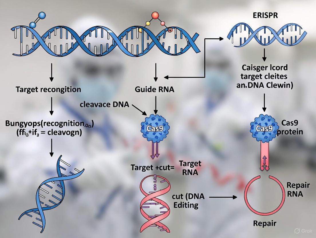
The CRISPR-Cas9 system, derived from an adaptive immune mechanism in prokaryotes, has revolutionized genome engineering due to its precision and programmability [9]. At its core, this system consists of three essential molecular components: the Cas9 endonuclease, which acts as the DNA-cutting machinery; the CRISPR RNA (crRNA), which provides the targeting specificity; and the trans-activating CRISPR RNA (tracrRNA), which serves as a critical scaffold and processing aid [10] [11]. These three elements form a functional complex capable of creating targeted double-strand breaks in DNA, enabling researchers to edit genomes with unprecedented ease [9]. The historical discovery and functional characterization of these components, particularly the relatively late identification of tracrRNA in 2011, were pivotal in transforming a bacterial defense system into a versatile technological platform [4] [11]. This guide examines the structure, function, and interplay of these core components, providing researchers and drug development professionals with a comprehensive technical understanding essential for advancing CRISPR-based therapeutics.
Historical Context of Component Discovery
The elucidation of CRISPR-Cas9's key components unfolded through decades of research, with critical discoveries building upon earlier observations to reveal the complete system. Table 1 summarizes the major milestones in identifying the core molecular components.
Table 1: Historical Timeline of Key CRISPR Component Discoveries
| Year | Discovery | Key Researchers | Significance |
|---|---|---|---|
| 1987 | Initial identification of CRISPR sequences | Yoshizumi Ishino | First observation of unusual repetitive sequences in E. coli [6] |
| 2000 | Term "CRISPR" coined | Francisco Mojica | Recognition that disparate repeat sequences shared common features [4] |
| 2005 | Identification of Cas9 and PAM sequences | Alexander Bolotin | Discovery of Cas9 protein and its PAM requirement for target recognition [4] |
| 2005 | CRISPR as adaptive immunity | Francisco Mojica | Hypothesis that CRISPR is an adaptive immune system based on spacer homology to phage [4] [7] |
| 2007 | Experimental demonstration of adaptive immunity | Philippe Horvath | Confirmed CRISPR provides acquired resistance against viruses; suggested Cas9 sufficient for interference [4] |
| 2008 | Spacers transcribed into guide RNAs | John van der Oost | Identification that spacer sequences are processed into small crRNAs [4] |
| 2011 | Discovery of tracrRNA | Emmanuelle Charpentier | Identification of tracrRNA as essential component for crRNA maturation in Type II systems [4] [11] |
| 2012 | Biochemical characterization; creation of single-guide RNA (sgRNA) | Virginijus Siksnys; Charpentier & Doudna | Reprogrammed Cas9 with custom guides; fused crRNA and tracrRNA into single guide RNA [4] |
The journey began with the initial discovery of CRISPR sequences in 1987, but their function remained mysterious for years [6]. Francisco Mojica's crucial insight in 2005 that these sequences matched bacteriophage DNA established CRISPR as a prokaryotic immune system [4] [7]. Simultaneously, Alexander Bolotin's discovery of the Cas9 protein and its associated PAM requirement laid groundwork for understanding targeting specificity [4]. The period from 2007-2008 yielded critical understanding of the mechanism, with researchers demonstrating that spacers are transcribed into small RNAs that guide the immune response [4]. The final piece came in 2011 when Emmanuelle Charpentier's team discovered tracrRNA, revealing the complete picture of how the system processes its guides and assembles into a functional complex [11]. This discovery directly enabled the engineering of the dual-RNA complex into a single-guide RNA (sgRNA), simplifying the system for widespread applications [4] [11].
Molecular Anatomy of the Core Components
Cas9: The DNA Cutting Machinery
The Cas9 endonuclease serves as the DNA-cutting enzyme within the CRISPR-Cas9 system. This multi-domain protein contains two distinct nuclease domains responsible for cleaving opposite strands of the target DNA: the HNH nuclease domain cleaves the DNA strand complementary to the crRNA guide sequence, while the RuvC-like nuclease domain cleaves the non-complementary strand [10] [9]. The resulting double-strand break occurs approximately 3-4 nucleotides upstream of the Protospacer Adjacent Motif (PAM), a crucial short DNA sequence adjacent to the target site that Cas9 requires for recognition and binding [10] [4].
Cas9's functional state is a DNA-bound complex where the protein undergoes significant conformational changes upon target recognition. In its apo state (unbound), the protein maintains a relatively open conformation; however, upon binding to both the guide RNA and the complementary DNA target, Cas9 shifts to a closed conformation that positions the nuclease domains for precise cleavage [9]. This structural rearrangement ensures that DNA cutting only occurs when the correct target sequence and PAM are present, providing an important layer of specificity to the system.
Table 2: Cas9 Variants and Their PAM Specificities
| Species/Variant of Cas9 | PAM Sequence | Notes |
|---|---|---|
| Streptococcus pyogenes (SpCas9) | 3' NGG | Most widely used variant; reduced NAG binding in enhanced versions [10] |
| Staphylococcus aureus (SaCas9) | 3' NNGRRT or NNGRR(N) | Smaller size beneficial for viral vector packaging [10] |
| xCas9 | 3' NG, GAA, or GAT | Engineered variant with increased PAM flexibility [10] |
| SpCas9-NG | 3' NG | Engineered variant with relaxed PAM requirements [10] |
| Acidaminococcus sp. (AsCpf1/LbCpf1) | 5' TTTV | Cas12 family nuclease; different cleavage mechanism [10] |
crRNA: The Targeting Guide
The CRISPR RNA (crRNA) provides the targeting specificity of the CRISPR-Cas9 system through its spacer sequence, which is complementary to the desired DNA target [10]. In native bacterial systems, multiple crRNAs are derived from a long precursor transcript (pre-crRNA) containing the entire CRISPR array, which includes direct repeats alternating with spacer sequences [11]. Each mature crRNA consists of a spacer region (approximately 20 nucleotides that define the genomic target) and repeat-derived sequences at both ends that contribute to the structural scaffold for Cas9 binding [10] [11].
The crRNA biogenesis pathway differs significantly between CRISPR system types. In Type I and III systems, the Cas6 nuclease processes pre-crRNA into individual units [9]. However, in Type II systems (including CRISPR-Cas9), crRNA maturation requires tracrRNA and RNase III, as detailed in Section 3.3 [11]. This fundamental difference in processing mechanisms reflects the evolutionary diversity of CRISPR systems, with the Type II pathway being particularly relevant for most genome engineering applications.
tracrRNA: The Scaffold and Processing Aid
The trans-activating CRISPR RNA (tracrRNA) is perhaps the most underappreciated yet essential component of the CRISPR-Cas9 system. Discovered in 2011 through small RNA sequencing in Streptococcus pyogenes, tracrRNA exists as multiple transcripts (171-nt and 89-nt primary transcripts processed to a mature 75-nt form) that are encoded adjacent to the cas9 gene [11]. The tracrRNA contains an anti-repeat region that base-pairs extensively with the repeat sequences in the pre-crRNA, forming a duplex that recruits the host endoribonuclease RNase III for processing [4] [11].
This RNA molecule serves two critical functions: first, it acts as a scaffold that links the crRNA to Cas9, facilitating the formation of a stable ribonucleoprotein complex; and second, it is essential for crRNA maturation in native Type II systems [11]. The tracrRNA's anti-repeat domain (approximately 24 nucleotides) base-pairs with the repeat regions of the pre-crRNA, creating a double-stranded RNA substrate that RNase III recognizes and cleaves [11]. After processing, the mature crRNA and tracrRNA remain associated as a dual-RNA complex that guides Cas9 to its DNA targets [11].
The discovery that tracrRNA and crRNA could be fused into a single-guide RNA (sgRNA) simplified the system for laboratory applications, but the native dual-RNA structure remains the biological functional unit [4] [11]. Bioinformatics analyses have revealed substantial diversity in tracrRNA structures across Type II systems, with at least 10 main groups identified based on predicted secondary structure features [11].
The Functional Complex: Component Interplay in DNA Targeting
The CRISPR-Cas9 DNA targeting mechanism involves a precisely orchestrated sequence of molecular interactions between the three core components and their DNA target. Figure 1 illustrates this process, from complex assembly through target cleavage.
Figure 1: CRISPR-Cas9 Complex Assembly and DNA Targeting Mechanism
Complex Assembly and crRNA Maturation
The process begins with the transcription of the CRISPR array to produce pre-crRNA, which contains multiple repeat-spacer units [11]. Simultaneously, tracrRNA is transcribed from its locus near the cas9 gene. The anti-repeat region of tracrRNA base-pairs with the repeat sequences in pre-crRNA, forming a double-stranded RNA structure that recruits the host RNase III enzyme [11]. In a process facilitated by Cas9, RNase III cleaves the pre-crRNA within the repeat regions, generating individual immature crRNAs that are subsequently trimmed to produce mature crRNAs of approximately 40 nucleotides [11]. The final functional complex consists of Cas9 bound to both the mature crRNA and the processed tracrRNA, forming a ribonucleoprotein ready for DNA target recognition [11].
PAM Recognition and DNA Binding
The Cas9 complex initially scans DNA for appropriate Protospacer Adjacent Motif (PAM) sequences through three-dimensional diffusion [10] [9]. Upon encountering a valid PAM (5'-NGG-3' for SpCas9), the protein undergoes conformational changes that promote DNA melting, enabling the formation of an "R-loop" structure where the target strand displaces to hybridize with the crRNA guide sequence [9]. This PAM recognition mechanism serves as a fundamental safeguard that prevents the CRISPR system from targeting its own CRISPR arrays, which lack PAM sequences [9].
DNA Cleavage Mechanism
Once specific complementarity between the crRNA spacer and target DNA is confirmed, Cas9 activates its nuclease domains to create a double-strand break [10]. The HNH domain cleaves the DNA strand complementary to the crRNA guide, while the RuvC domain cleaves the opposite strand [10] [9]. This coordinated cleavage results in a blunt-ended double-strand break approximately 3-4 base pairs upstream of the PAM sequence [10]. The cell then repairs this break through either error-prone non-homologous end joining (NHEJ) or homology-directed repair (HDR), enabling various genome editing outcomes including gene knockouts, insertions, or precise modifications [9].
Experimental Approaches for Studying Component Functions
Key Methodologies in CRISPR Research
Researchers have employed diverse experimental approaches to characterize the structure and function of CRISPR components. Biochemical reconstitution has been instrumental in defining the minimal requirements for DNA cleavage, with studies demonstrating that Cas9, crRNA, and tracrRNA constitute the essential components for targeted DNA interference [4]. Heterologous expression experiments, such as those showing that CRISPR systems function when transferred between bacterial species, confirmed these systems are self-contained units with all necessary components encoded within their loci [4].
Structural biology techniques including X-ray crystallography and cryo-electron microscopy have provided atomic-level insights into Cas9's architecture and its conformational changes during DNA binding and cleavage [9]. These approaches revealed how Cas9 positions its nuclease domains and how guide RNA binding activates the protein for DNA recognition [9]. Additionally, small RNA sequencing was critical for discovering tracrRNA and elucidating its role in crRNA processing, highlighting the importance of nucleic acid analysis in unraveling CRISPR mechanisms [11].
Essential Research Reagents
Table 3: Key Research Reagents for CRISPR-Cas9 Studies
| Reagent/Solution | Function/Application | Technical Notes |
|---|---|---|
| Guide RNA (synthetic) | Targets Cas9 to specific genomic loci | Can be dual-RNA (crRNA+tracrRNA) or single-guide RNA (sgRNA) format [10] |
| Recombinant Cas9 Nuclease | DNA cleavage enzyme | Purified protein for in vitro studies; codon-optimized versions for eukaryotic expression [10] |
| RNase III | Processes pre-crRNA in Type II systems | Host enzyme required for crRNA maturation in native bacterial systems [11] |
| PAM-containing DNA substrates | Target for Cas9 cleavage | Must contain appropriate PAM sequence adjacent to target site for recognition [10] [9] |
| Lipid Nanoparticles (LNPs) | In vivo delivery of CRISPR components | Encapsulate ribonucleoprotein complexes for therapeutic applications [12] |
The precise understanding of Cas9, crRNA, and tracrRNA structure and function has enabled the remarkable transition of CRISPR technology from basic bacterial immunity to transformative therapeutic applications. The discovery that these three components constitute a programmable DNA-targeting system paved the way for groundbreaking therapies now advancing through clinical trials, including Casgevy for sickle cell disease and beta-thalassemia, and multiple investigational treatments for genetic disorders like hereditary transthyretin amyloidosis and hereditary angioedema [12] [13]. As research continues to refine these core components through engineered Cas9 variants with altered PAM specificities, enhanced precision, and novel functionalities like base editing, the fundamental triad of Cas9, crRNA, and tracrRNA remains the foundation upon which next-generation genomic medicines are being built [10] [12]. For researchers and drug development professionals, deep knowledge of these key molecular components provides the essential framework for developing safer, more effective CRISPR-based therapeutics.
The Protospacer Adjacent Motif (PAM) is a critical short DNA sequence required for the function of CRISPR-Cas systems. This motif, typically 2–6 base pairs in length, is located directly adjacent to the DNA region targeted for cleavage by the CRISPR complex [14] [15]. The PAM sequence serves as a fundamental "self" versus "non-self" discrimination mechanism, enabling CRISPR systems to precisely identify and cleave foreign invading DNA while avoiding autoimmunity against the bacterial host's own genome [14]. For the commonly used Streptococcus pyogenes Cas9 (SpCas9), the PAM sequence is 5'-NGG-3' (where "N" can be any nucleotide base), located immediately downstream of the target DNA sequence [15] [16]. The discovery and understanding of the PAM have been instrumental in transforming CRISPR from a bacterial immune system into a revolutionary genome engineering tool.
Historical Discovery: The PAM in the Context of CRISPR Research
The history of PAM discovery is inextricably linked to the broader elucidation of the CRISPR-Cas system. The story begins not with PAM itself, but with the initial observation of unusual genetic structures in prokaryotes.
The Early Years: Identifying CRISPR Sequences
In 1987, Japanese researchers first discovered clustered regularly interspaced short palindromic repeats (CRISPR) in the Escherichia coli genome, though their function remained mysterious [5] [7]. Throughout the 1990s, Francisco Mojica at the University of Alicante characterized these sequences across multiple microorganisms and, in 2000, recognized that disparate repeat sequences shared common features, coining the term CRISPR [4]. The breakthrough came in 2005 when Mojica reported that these sequences matched snippets from bacteriophage genomes, correctly hypothesizing that CRISPR functions as an adaptive immune system in prokaryotes [4] [7].
The PAM Discovery and Its Significance
The PAM sequence was first identified in May 2005 by Alexander Bolotin and colleagues at the French National Institute for Agricultural Research (INRA) [4]. While studying the CRISPR locus in Streptococcus thermophilus, they noted that spacers homologous to viral genes all shared a common sequence at one end—the protospacer adjacent motif [4]. This observation was crucial because it revealed a fundamental aspect of the CRISPR targeting mechanism. Bolotin's team also discovered a novel Cas protein with predicted nuclease activity, now known as Cas9, which would become the cornerstone of CRISPR genome editing [5] [4].
Table 1: Key Historical Milestones in PAM and CRISPR Discovery
| Year | Discoverer(s) | Breakthrough | Significance |
|---|---|---|---|
| 1987 | Ishino et al. | First discovery of CRISPR sequences in E. coli | Initial observation of unusual genetic structures [5] |
| 2000 | Francisco Mojica | Coined the term CRISPR; recognized common features | Unified previously disparate observations [4] |
| 2005 | Alexander Bolotin | Identified PAM sequence and discovered Cas9 | Revealed key targeting mechanism and main editor enzyme [4] |
| 2005 | Francisco Mojica | CRISPR matches bacteriophage sequences | Established CRISPR as adaptive immune system [4] |
| 2007 | Barrangou & Horvath | Experimental proof of adaptive immunity in S. thermophilus | Confirmed CRISPR function; applied in bacterial vaccination [5] [4] |
The following timeline visualizes the key discoveries in CRISPR research that led to the identification and understanding of the PAM sequence:
The Functional Role and Mechanism of the PAM Sequence
Self vs. Non-Self Discrimination
The fundamental biological function of the PAM is to protect the host bacterium from its own CRISPR system. When the Cas1-Cas2 complex acquires spacers from invading viral DNA, it incorporates them into the bacterial CRISPR array without the adjacent PAM sequence [14] [15]. Consequently, when Cas effector proteins later scan the cell for foreign DNA, they only target sequences that are both complementary to the crRNA and adjacent to the correct PAM. The bacterial genome itself contains the spacers but lacks the flanking PAM, thus preventing autoimmune destruction [15]. This elegant mechanism ensures that the bacterial immune system exclusively attacks foreign genetic elements while preserving host genomic integrity.
The Molecular Mechanism of PAM Recognition
At the molecular level, PAM recognition initiates the process of DNA interrogation by Cas proteins. For Cas9, the PAM is recognized by a PI domain within the protein, which causes local DNA melting and facilitates the formation of an R-loop structure where the target DNA strand is displaced and the complementary strand pairs with the crRNA [14]. The PAM sequence is located directly downstream of the target DNA region (the protospacer), and the Cas9 nuclease creates a double-strand break approximately 3-4 nucleotides upstream of the PAM [15] [17].
The recognition process follows a specific sequence:
- PAM Binding: Cas proteins first scan DNA for compatible PAM sequences [14]
- DNA Melting: PAM binding induces local unwinding of the DNA duplex [14]
- Seed Sequence Interrogation: The "seed sequence" (8-10 bases at the 3' end of the gRNA) begins annealing to the target DNA [17]
- Full Complementarity Check: If seed matching is successful, complete pairing between gRNA and target DNA occurs [17]
- Cleavage Activation: Successful recognition triggers Cas nuclease activity [17]
Table 2: PAM Sequences and Properties of Commonly Used CRISPR-Cas Systems
| CRISPR Nucleases | Organism Isolated From | PAM Sequence (5' to 3') | Key Features |
|---|---|---|---|
| SpCas9 | Streptococcus pyogenes | NGG | Most widely used; first Cas9 adapted for eukaryotic editing [15] [17] |
| SaCas9 | Staphylococcus aureus | NNGRR(T) | Smaller size beneficial for viral packaging [15] |
| NmeCas9 | Neisseria meningitidis | NNNNGATT | Longer PAM; higher specificity [15] |
| Cas12a (Cpf1) | Lachnospiraceae bacterium | TTTV | Creates staggered cuts; processes its own crRNAs [15] [17] |
| Cas12b | Alicyclobacillus acidiphilus | TTN | Thermostable; used in diagnostic applications [15] |
| xCas9 | Engineered from SpCas9 | NG, GAA, GAT | Broad PAM recognition; increased fidelity [17] |
| SpRY | Engineered from SpCas9 | NRN (prefers NGN) | Near-PAMless variant; greatly expanded targeting range [17] |
Experimental Methods for PAM Identification and Characterization
As CRISPR technologies advanced, robust experimental methods emerged to characterize PAM requirements for both naturally occurring and engineered Cas nucleases. These methods have evolved from computational approaches to sophisticated high-throughput experimental techniques.
Evolution of PAM Determination Methods
Early PAM identification relied on in silico approaches through alignments of protospacers to identify consensus sequences [14]. While fast and accessible, these computational methods couldn't distinguish between functional PAMs for spacer acquisition versus target interference, and were limited by available phage genome sequences [14].
The plasmid depletion assay represented a significant experimental advancement. This negative selection approach involves transforming a host with an active CRISPR-Cas system with a plasmid library containing randomized DNA stretches adjacent to target sequences [14]. Plasmids with "inactive" PAMs that aren't recognized by the Cas nuclease are retained, allowing identification of functional PAMs through sequencing of the remaining plasmids [14].
More recently, PAM-SCANR (PAM screen achieved by NOT-gate repression) enabled high-throughput in vivo PAM screening using a catalytically dead Cas9 variant (dCas9). When dCas9 binds to a functional PAM, it represses GFP expression, allowing sorting by FACS and subsequent identification of functional PAM motifs [14].
Advanced Mammalian Cell-Based PAM Determination
A cutting-edge method published in 2025, PAM-readID, addresses the crucial need for PAM determination in mammalian cells, where cellular environment significantly influences PAM specificity [18]. This method uses double-stranded oligodeoxynucleotides (dsODN) integration to tag cleaved DNA ends bearing recognized PAMs.
The PAM-readID workflow consists of five key steps [18]:
- Library Construction: Create plasmids bearing target sequences flanked by randomized PAMs
- Transfection: Introduce plasmids expressing Cas nuclease, sgRNA, and dsODN into mammalian cells
- Cleavage & Integration: Allow 72 hours for Cas cleavage and NHEJ repair-mediated dsODN integration
- Amplification: PCR amplify integrated fragments using dsODN-specific and target-plasmid-specific primers
- Analysis: High-throughput sequencing and bioinformatic analysis to define PAM recognition profiles
This method successfully defined PAM profiles for SaCas9, Nme1Cas9, SpCas9, SpG, SpRY, and AsCas12a in mammalian cells, revealing non-canonical PAMs such as 5'-NNAAGT-3' for SaCas9 [18]. The experimental workflow is visualized below:
The Scientist's Toolkit: Essential Reagents for PAM Research
Table 3: Essential Research Reagents for PAM and CRISPR Experiments
| Reagent / Tool | Function / Application | Examples / Notes |
|---|---|---|
| Cas Nuclease Variants | DNA recognition and cleavage | SpCas9 (NGG), SaCas9 (NNGRR[T]), Cas12a (TTTV) [15] [17] |
| Guide RNA (gRNA) | Targets Cas nuclease to specific genomic loci | 20-nucleotide spacer defines target; scaffold binds Cas [17] |
| PAM Library Plasmids | Determining PAM recognition profiles | Contain randomized nucleotide regions adjacent to fixed target [18] |
| dsODN (double-stranded oligodeoxynucleotides) | Tagging cleaved DNA ends in PAM-readID | Integrated at cleavage sites during NHEJ repair [18] |
| High-Fidelity Cas Variants | Reduce off-target editing | eSpCas9(1.1), SpCas9-HF1, HypaCas9, evoCas9 [17] |
| PAM-Flexible Enzymes | Expand targetable genomic sites | xCas9 (NG), SpCas9-NG (NG), SpRY (NRN) [17] |
| Bioinformatics Tools | PAM sequence analysis and visualization | CRISPRTarget, CRISPRFinder, PAM Wheel visualization [14] |
PAM Engineering and Clinical Applications
Engineering Cas Proteins with Altered PAM Specificities
The limited targeting range imposed by strict PAM requirements has driven extensive protein engineering efforts to develop Cas variants with altered PAM specificities. These engineered enzymes significantly expand the targetable genome space for both basic research and therapeutic applications.
Key engineering approaches include:
- Directed Evolution: Using phage-assisted continuous evolution to generate Cas9 variants with broad PAM compatibility [17]
- Structure-Guided Engineering: Modifying PAM-interacting domains based on protein-DNA co-crystal structures [17]
- Domain Swapping: Creating chimeric Cas proteins with novel PAM specificities [19]
Notable successes include xCas9, which recognizes NG, GAA, and GAT PAMs; SpCas9-NG with NG PAM recognition; and SpRY, a nearly PAM-less variant that recognizes NRN (prefers NGN) and NYN sequences [17]. These advances have dramatically expanded the targeting range of CRISPR systems, enabling access to previously inaccessible genomic regions.
PAM Considerations in Clinical Applications
The translation of CRISPR technologies into clinical applications heavily depends on PAM availability near disease-relevant mutations. The first FDA-approved CRISPR-based medicine, Casgevy, for sickle cell disease and transfusion-dependent beta thalassemia, utilizes ex vivo editing where PAM constraints are managed during experimental design [12].
For in vivo therapeutic applications, PAM requirements present additional challenges. Successful clinical trials for hereditary transthyretin amyloidosis (hATTR) and hereditary angioedema (HAE) both target genes expressed in the liver, using lipid nanoparticles (LNPs) that naturally accumulate in hepatic tissue [12]. These therapies use CRISPR to disrupt disease-causing genes, requiring PAM sequences adjacent to the target sites.
Recent advances in personalized CRISPR therapy highlight both the promise and challenges of PAM-dependent genome editing. In 2025, physicians developed a bespoke in vivo CRISPR treatment for an infant with CPS1 deficiency, delivering the therapy via LNPs in just six months from design to administration [12]. This case demonstrates the critical importance of PAM availability when designing personalized therapies for rare genetic mutations.
The integration of artificial intelligence and machine learning is accelerating PAM and CRISPR research. AI models are being used to predict protein structures, optimize guide RNA designs, and engineer novel Cas proteins with desired PAM specificities [19]. These computational approaches complement experimental methods, enabling more rapid development of CRISPR tools with expanded targeting capabilities.
The history of PAM research demonstrates how understanding a fundamental biological mechanism—bacterial adaptive immunity—has enabled revolutionary technologies. From its initial discovery in 2005 by Bolotin to the current development of PAM-flexible Cas variants, the PAM sequence has remained a central consideration in CRISPR-based genome editing. As clinical applications expand, ongoing research continues to address the limitations imposed by PAM requirements through both protein engineering and improved delivery methods.
The future of CRISPR-based therapeutics will likely involve a combination of approaches: engineered Cas variants with relaxed PAM specificities for increased targeting range, improved delivery systems to reach target tissues, and advanced screening methods to identify optimal target sites within disease-associated genes. The PAM sequence, once a curiosity of bacterial immunity, now stands as a cornerstone of genome engineering with profound implications for medicine, biotechnology, and basic research.
The 2012 publication by Jinek et al., "A Programmable Dual-RNA–Guided DNA Endonuclease in Adaptive Bacterial Immunity," represents a pivotal moment in the history of CRISPR technology. While the CRISPR (Clustered Regularly Interspaced Short Palindromic Repeats) system was first discovered in 1987 [5] and its function as a prokaryotic immune system was elucidated in 2007 [8], the 2012 paper demonstrated for the first time that the CRISPR-Cas9 system could be engineered as a universal programmable tool for cutting DNA at precise locations in vitro [20] [8]. This work laid the essential foundation for all subsequent applications of CRISPR-Cas9 as a genome-editing technology, transforming biological research and therapeutic development.
The journey to this landmark discovery began with foundational research from multiple groups. Francisco Mojica was instrumental in recognizing CRISPR as an adaptive immune system in prokaryotes [5]. Later, the roles of the key molecular components were uncovered: the Cas9 nuclease was identified [5], the crRNA (CRISPR RNA) was shown to guide the system to foreign DNA [5], and finally, a second RNA, the tracrRNA (trans-activating CRISPR RNA), was discovered as being essential for processing crRNA and for Cas9 nuclease activity [5]. The critical insight of the 2012 study was the simplification of this natural, multi-component system into a single, programmable endonuclease.
Core Experimental Breakthrough
Research Objective and Hypothesis
The primary goal of the 2012 study was to reconstitute the Type II CRISPR system in vitro to determine whether the Cas9 protein could be programmed with engineered RNA components to cleave specific, pre-determined DNA sequences. The central hypothesis was that the Cas9 enzyme's DNA cleavage activity was RNA-programmable. The researchers proposed that by combining the naturally occurring crRNA and tracrRNA into a single chimeric "guide RNA" (gRNA), they could create a simplified, two-component system (Cas9 + gRNA) capable of inducing double-strand breaks in DNA targets matching the gRNA sequence [8].
Key Experimental System and Methodology
The experiments utilized a purified, recombinant Streptococcus pyogenes Cas9 protein and synthetic RNA components. The DNA targets were typically short, linear, double-stranded DNA fragments containing a target sequence adjacent to the requisite Protospacer Adjacent Motif (PAM), which for SpCas9 is 5'-NGG-3' [5] [8].
A key methodological innovation was the design of a single-chimeric guide RNA (sgRNA). This was created by fusing the 3' end of the crRNA (which contains the target-complementary "spacer" sequence) to the 5' end of a truncated tracrRNA via a synthetic loop sequence. This chimeric RNA molecule retained the essential functions of both natural RNAs, thereby simplifying the system [8].
The standard DNA Cleavage Assay protocol involved:
- Complex Formation: Incubating the Cas9 protein with the sgRNA (or with separate crRNA and tracrRNA) in a suitable reaction buffer to form the ribonucleoprotein (RNP) complex.
- Reaction Initiation: Adding the target DNA substrate to the pre-formed RNP complex.
- Cleavage Reaction: Conducting the reaction at 37°C in a buffer containing Mg2+ as an essential cofactor [20].
- Reaction Termination and Analysis: Stopping the reaction and analyzing the products using agarose gel electrophoresis to visualize and quantify the cleavage efficiency based on the appearance of shorter DNA fragments.
The following diagram illustrates this streamlined experimental workflow.
The in vitro cleavage assays provided robust quantitative data demonstrating the system's programmability and specificity. The table below summarizes the core findings.
Table 1: Summary of Key Quantitative Findings from the 2012 Study
| Experimental Parameter | Key Finding | Significance |
|---|---|---|
| DNA Cleavage Efficiency | High-efficiency, site-specific double-strand breaks (DSBs) were achieved. | Confirmed Cas9 functions as an RNA-programmable endonuclease. |
| Dual-RNA Requirement | Both crRNA and tracrRNA were necessary for efficient DNA cleavage in the natural system. | Elucidated the fundamental mechanism of the native bacterial immune system. |
| Chimeric gRNA Function | The single-guide RNA (sgRNA) was functionally equivalent to the natural dual-RNA complex. | Simplified the system from three components to two, enabling facile engineering. |
| PAM Specificity | Cleavage was strictly dependent on the presence of a 5'-NGG PAM sequence adjacent to the target site. | Defined a critical targeting constraint and revealed a key mechanism for self/non-self discrimination. |
| Mg2+ Dependence | Cleavage activity was absolutely dependent on the presence of Mg2+ ions. | Identified an essential catalytic cofactor for the Cas9 nuclease. |
A critical finding was the architecture of the Cas9 nuclease, which contains two distinct catalytic domains that cleave opposite DNA strands. The study demonstrated that these domains could be inactivated individually to create a "nickase" (creating single-strand breaks) or together to create a catalytically dead Cas9 (dCas9). The following diagram details this cleavage mechanism and its engineering.
The Scientist's Toolkit: Essential Research Reagents
The in vitro demonstration relied on a specific set of core reagents. The table below details these essential materials and their functions, which formed the basis for subsequent CRISPR tool development.
Table 2: Key Research Reagent Solutions for CRISPR-Cas9 Experiments
| Reagent / Material | Function in the Experiment |
|---|---|
| Purified Cas9 Nuclease | The effector enzyme that executes the DNA cleavage; its programmable specificity is the core of the technology. |
| crRNA (CRISPR RNA) | The guide RNA component that contains the ~20 nucleotide spacer sequence complementary to the target DNA. |
| tracrRNA (trans-activating crRNA) | Essential RNA component that base-pairs with the crRNA, facilitates processing, and is required for Cas9 nuclease activity. |
| Single-Guide RNA (sgRNA) | A chimeric, synthetic RNA molecule combining the essential parts of crRNA and tracrRNA, simplifying the system. |
| Target DNA Plasmid/Fragment | The double-stranded DNA substrate containing the target sequence and the requisite PAM site for cleavage. |
| Reaction Buffer with MgCl₂ | Provides the optimal ionic and pH conditions for Cas9 activity; Mg2+ is an essential catalytic cofactor. |
Impact and Evolution in Research and Drug Development
The immediate impact of this in vitro demonstration was profound. Within a year, multiple groups had adapted the CRISPR-Cas9 system for genome editing in mammalian cell culture [8], heralding a new era in genetic engineering. For researchers and drug development professionals, this provided a tool of unprecedented simplicity and programmability for gene knockout, knock-in, and modulation [21] [8].
The technology has since evolved dramatically, leading to:
- Advanced Editors: Development of base editors and prime editors for precise single-nucleotide changes without creating double-strand breaks [22].
- Diagnostic Tools: Creation of CRISPR-based diagnostic tests, including for viruses like SARS-CoV-2 [5] [12].
- Therapeutic Breakthroughs: The 2023 approval of Casgevy, the first CRISPR-based medicine for sickle cell disease and beta-thalassemia, marks the clinical culmination of this research [12]. Current clinical trials are exploring in vivo CRISPR therapies for diseases like hereditary transthyretin amyloidosis (hATTR) and hereditary angioedema (HAE) [12] [22].
- Functional Genomics: The development of CRISPR screening platforms, including hypothesis-driven custom libraries, for drug target discovery and dissecting the genetic basis of disease [23] [24].
The 2012 publication by Jinek et al. was a landmark proof-of-concept that successfully harnessed a bacterial defense mechanism, transforming it into a versatile, programmable, and efficient molecular tool. It laid the essential groundwork for a technology that continues to revolutionize basic research and is now delivering on its promise to treat human disease.
The 2020 Nobel Prize in Chemistry awarded to Emmanuelle Charpentier and Jennifer A. Doudna recognized their development of CRISPR-Cas9 as a method for genome editing, a discovery that has fundamentally reshaped the life sciences [25]. This whitepaper provides an in-depth technical examination of the CRISPR-Cas9 system, tracing its origins from a prokaryotic immune mechanism to a programmable genetic scissors. We detail the key molecular components, experimental workflows, and mechanistic insights that enabled the repurposing of this bacterial defense system into a versatile genome engineering platform. Furthermore, we present current clinical applications and quantitative data from recent trials, highlighting the transformative impact of this technology on therapeutic development.
The journey to the 2020 Nobel Prize began not with human genetics, but with fundamental microbiological research. The term CRISPR, an acronym for Clustered Regularly Interspaced Short Palindromic Repeats, was first coined in 2002 by Francisco Mojica, who identified these unusual DNA repeats in archaea and bacteria and correctly hypothesized their function in adaptive immunity [4] [5]. This built upon observations dating back to 1987, when unusual repetitive sequences in the E. coli genome were first documented [5] [26].
The timeline below summarizes the critical discoveries that led to the development of the CRISPR-Cas9 gene-editing tool:
Table 1: Historical Timeline of Key CRISPR Discoveries
| Year | Discovery | Key Researchers | Significance |
|---|---|---|---|
| 1987 | Unusual repetitive sequences in E. coli | Ishino et al. [5] | Initial observation of what would later be known as CRISPR |
| 2002 | Term "CRISPR" coined; Cas genes identified | Mojica, Jansen et al. [4] [5] | Naming and initial characterization of the system |
| 2005 | CRISPR spacers match phage DNA | Mojica et al.; Bolotin et al. [4] [5] | Identification of CRISPR as an adaptive immune system |
| 2007 | Experimental demonstration of adaptive immunity | Barrangou, Horvath et al. [4] | First functional proof that CRISPR provides resistance to viruses |
| 2011 | Discovery of tracrRNA | Charpentier et al. [4] | Identified a key RNA component essential for the Cas9 system |
| 2012 | In vitro reprogramming of CRISPR-Cas9 | Doudna, Charpentier et al. [25] [4] | Creation of a programmable gene-editing tool |
The period from 2005-2008 saw crucial advancements in understanding the system's mechanism. Researchers discovered that CRISPR systems require Cas (CRISPR-associated) genes [5], that the target molecule is DNA rather than RNA [4], and that spacer sequences are transcribed into small guide RNAs (crRNAs) that direct Cas proteins to complementary DNA sequences [4]. The stage was set for reprogramming this bacterial immune system into a universal gene-editing tool.
The Core Technology: Molecular Mechanism of CRISPR-Cas9
Key Molecular Components
The CRISPR-Cas9 system's functionality depends on a minimal set of molecular components that work in concert to achieve targeted DNA cleavage:
- Cas9 Nuclease: The effector protein that creates double-stranded breaks (DSBs) in DNA. It contains two nuclease domains: the HNH domain that cleaves the DNA strand complementary to the guide RNA, and the RuvC-like domain that cleaves the non-complementary strand [4] [27].
- crRNA (CRISPR RNA): A short RNA molecule containing a ~20 nucleotide guide sequence that is complementary to the target DNA site, providing the system's targeting specificity [5].
- tracrRNA (trans-activating crRNA): A necessary RNA component that facilitates the maturation of crRNA and forms a complex with Cas9 [4] [28].
- sgRNA (single-guide RNA): A synthetic fusion of crRNA and tracrRNA into a single RNA chimera, simplifying the system for practical applications [4] [27].
- PAM (Protospacer Adjacent Motif): A short (2-6 base pair) DNA sequence adjacent to the target site that is essential for Cas9 recognition and cleavage. For the commonly used Streptococcus pyogenes Cas9, the PAM sequence is 5'-NGG-3' [4] [27].
The following diagram illustrates the relationships and functions of these core components in the CRISPR-Cas9 complex:
The Three-Stage Mechanism of Action
The natural CRISPR-Cas9 system functions as an adaptive immune system in three distinct stages:
- Adaptation (Spacer Acquisition): When a bacterium survives a viral infection, it incorporates a short fragment of the viral DNA (∼20-40 bp) as a "spacer" into its CRISPR locus, creating a molecular memory of the infection [26].
- Expression and Maturation: The CRISPR locus is transcribed into a long precursor CRISPR RNA (pre-crRNA), which is processed into mature crRNAs with the help of tracrRNA and Cas9 [4] [5].
- Interference: The mature crRNA-tracrRNA complex guides Cas9 to complementary viral DNA sequences. Cas9 scans DNA for PAM sequences, unwinds the DNA adjacent to the PAM, and if the crRNA matches the target DNA, Cas9 introduces a double-strand break, thereby inactivating the pathogen [4] [27].
The key insight by Charpentier and Doudna was that the tracrRNA and crRNA could be combined into a single-guide RNA (sgRNA), and that by simply modifying the 20-nucleotide guide sequence of the sgRNA, the Cas9 nuclease could be programmed to target any DNA sequence of choice, provided it is adjacent to a PAM [25] [28].
Experimental Breakthrough: Reprogramming CRISPR-Cas9In Vitro
Key Experimental Workflow
The seminal experiment by Charpentier and Doudna that led to the reprogramming of CRISPR-Cas9 involved a systematic biochemical approach to reconstitute and simplify the system in vitro [4] [28]. The workflow can be summarized as follows:
Detailed Methodology
Objective: To reconstitute the Streptococcus pyogenes Type II CRISPR system in vitro and engineer it into a programmable gene-editing tool.
Materials and Reagents:
- Purified Cas9 protein from S. pyogenes
- DNA templates containing target sequences
- In vitro transcription system for RNA synthesis
- Synthetic crRNA and tracrRNA molecules
- Plasmid DNA substrates for cleavage assays
- Gel electrophoresis equipment for analyzing cleavage products
Procedure:
- Component Isolation: The Cas9 gene was cloned and the protein was expressed and purified. Native crRNA and tracrRNA components were identified and characterized [4] [28].
- System Reconstitution: The minimal components (Cas9, crRNA, tracrRNA) were combined in a buffer system with plasmid DNA targets to reconstitute DNA cleavage activity in vitro [4].
- RNA Engineering: The dual-RNA structure (crRNA:tracrRNA duplex) was redesigned into a single-guide RNA (sgRNA) by connecting the 3' end of the crRNA to the 5' end of the tracrRNA via a synthetic linker [4] [27].
- Reprogramming Validation: The guide sequence of the sgRNA was modified to target specific DNA sequences of interest. Cleavage activity was confirmed by incubating the programmed Cas9-sgRNA complex with DNA substrates containing the target sequence and the requisite PAM [25] [4].
- Specificity Verification: Target recognition specificity was validated through mismatch experiments, demonstrating that single-base pair mismatches between the sgRNA and target DNA could significantly reduce cleavage efficiency [27].
Key Outcome: The experiment demonstrated that:
- The CRISPR-Cas9 system could be reduced to two components: Cas9 protein and a programmable sgRNA.
- The system could be targeted to any DNA sequence of choice by simply modifying the 20-nucleotide guide sequence within the sgRNA.
- DNA cleavage required both complementarity between the sgRNA and target DNA, and the presence of a correct PAM sequence adjacent to the target site [4] [27].
The Researcher's Toolkit: Essential Reagents for CRISPR-Cas9 Experiments
Table 2: Essential Research Reagents for CRISPR-Cas9 Applications
| Reagent/Solution | Function | Technical Specifications |
|---|---|---|
| Cas9 Nuclease | Creates double-stranded breaks in target DNA | Wild-type SpCas9: 1368 amino acids; requires NGG PAM. Variants include high-fidelity (HF-Cas9), Cas9 nickase (nCas9), and catalytically dead Cas9 (dCas9) [27]. |
| Guide RNA (sgRNA) | Targets Cas9 to specific genomic loci | ~100 nt synthetic RNA or expressed from U6 promoter. 20-nt guide sequence must be complementary to target, with G at position 1 for U6 transcription [4] [27]. |
| Repair Templates | Enables precise genome editing via HDR | Single-stranded oligodeoxynucleotides (ssODNs, ~100-200 nt) or double-stranded DNA donors with ~800 bp homology arms [29]. |
| Delivery Vectors | Introduces CRISPR components into cells | Plasmids, mRNA, or ribonucleoprotein (RNP) complexes. Viral vectors (AAV, lentivirus) or non-viral methods (electroporation, lipid nanoparticles) [12] [27]. |
| Validation Tools | Confirms editing efficiency and specificity | T7 Endonuclease I or Surveyor assays for indel detection; Sanger sequencing; next-generation sequencing for off-target analysis [26]. |
Clinical Translation and Current Applications
The reprogramming of CRISPR-Cas9 has led to its rapid adoption in clinical research and therapeutic development. The following table summarizes key clinical areas where CRISPR-based therapies have shown significant progress:
Table 3: Current Status of CRISPR-Cas9 Clinical Applications
| Disease Area | Therapeutic Approach | Clinical Trial Status / Key Results |
|---|---|---|
| Genetic Blood Disorders | Ex vivo editing of hematopoietic stem cells | Approved: Casgevy (exa-cel) for sickle cell disease and β-thalassemia demonstrates sustained response >2 years [12]. |
| Hereditary Transthyretin Amyloidosis (hATTR) | In vivo knockdown of TTR gene in liver | Phase III: LNP-delivered therapy shows ~90% reduction in TTR protein sustained over 2 years [12]. |
| Hereditary Angioedema (HAE) | In vivo knockdown of kallikrein gene | Phase I/II: 86% reduction in kallikrein; 8 of 11 high-dose participants attack-free [12]. |
| Oncology | PD-1 knockout in T cells for enhanced immunotherapy | Early trials: First clinical trial for metastatic non-small cell lung cancer demonstrated safety and feasibility [26]. |
| Rare Genetic Diseases | Personalized in vivo CRISPR therapy | Proof-of-concept: Infant with CPS1 deficiency safely received 3 LNP doses with symptom improvement [12]. |
Delivery Technologies in Clinical Development
Effective delivery remains a critical challenge for CRISPR therapeutics. Current approaches include:
- Ex Vivo Editing: Cells are removed from the patient, edited in culture, and reintroduced (used in Casgevy for sickle cell disease) [12].
- In Vivo Viral Delivery: Adeno-associated viruses (AAVs) deliver CRISPR components but have limited packaging capacity and potential immunogenicity [27].
- In Vivo Non-Viral Delivery: Lipid nanoparticles (LNPs) have emerged as a promising vehicle, particularly for liver-targeted therapies, with potential for redosing [12] [27].
The therapeutic development pathway for CRISPR-based medicines involves careful consideration of both the editing strategy and delivery modality, as illustrated below:
The recognition of CRISPR-Cas9 with the 2020 Nobel Prize in Chemistry marks a pivotal moment in the history of biotechnology. The fundamental work by Charpentier and Doudna to elucidate the molecular mechanism of this bacterial immune system and reprogram it into a versatile genetic scissors has created a paradigm shift across the life sciences. The technology's simplicity, precision, and programmability have democratized gene editing, accelerating basic research and enabling the development of transformative therapies for previously untreatable genetic diseases.
While challenges remain in delivery efficiency, specificity, and ethical considerations, the rapid clinical translation of CRISPR-based therapies demonstrates the profound impact of this discovery. As delivery technologies advance and editing precision improves, CRISPR-Cas9 and its derivatives are poised to revolutionize therapeutic development, offering new hope for patients with genetic disorders, cancers, and other intractable diseases.
From Lab Bench to Clinic: CRISPR's Methodological Evolution and Breakthrough Applications
The CRISPR-Cas9 system represents a revolutionary advance in genome engineering, enabling precise manipulation of DNA sequences across diverse biological systems. At its core, this technology functions as a programmable DNA-targeting platform where a single guide RNA (gRNA) directs the Cas9 nuclease to create site-specific double-strand breaks (DSBs) in DNA. This mechanism has transformed basic biological research and therapeutic development, offering unprecedented control over genetic information [5]. The system's operation mirrors its natural function in prokaryotic immunity, where it protects bacteria and archaea from mobile genetic elements such as viruses by storing fragments of foreign DNA in genomic CRISPR arrays [4] [5]. When these sequences are transcribed and processed, they guide Cas proteins to recognize and cleave matching invading DNA, providing adaptive immunity [5]. The repurposing of this natural system for programmable genome editing has created a powerful toolkit that is reshaping medicine and biotechnology, with ongoing clinical trials demonstrating its potential to treat genetic disorders, infectious diseases, and cancer [12].
Historical Context of CRISPR Discoveries
The development of CRISPR-Cas9 technology stems from foundational discoveries by numerous researchers across three decades. Table 1 summarizes the key historical milestones that led to our current understanding of the CRISPR-Cas9 mechanism.
Table 1: Historical Timeline of Key CRISPR-Cas9 Discoveries
| Date | Discoverers | Key Finding | Significance |
|---|---|---|---|
| 1987 | Ishino et al. [30] | First discovery of unusual repetitive DNA sequences in E. coli | Initial characterization of what would later be recognized as CRISPR |
| 2000-2005 | Francisco Mojica et al. [4] [5] | Recognized CRISPR as a distinct class of sequences across prokaryotes; hypothesized function as adaptive immune system | Established biological significance and coined the "CRISPR" term |
| 2005 | Alexander Bolotin et al. [4] | Identified Cas9 and unique PAM sequence in S. thermophilus | Discovered key nuclease component and targeting requirement |
| 2007 | Philippe Horvath et al. [4] [5] | Experimental demonstration of CRISPR as adaptive immunity in bacteria | Provided direct evidence of CRISPR function in phage resistance |
| 2008 | John van der Oost et al. [4] | Discovery that spacers are processed into crRNAs | Elucidated the RNA-guided nature of the system |
| 2008 | Luciano Marraffini & Erik Sontheimer [4] | Demonstrated CRISPR targets DNA, not RNA | Corrected understanding of molecular target |
| 2011 | Emmanuelle Charpentier et al. [4] | Identification of tracrRNA essential for Cas9 function | Revealed second RNA component needed for DNA cleavage |
| 2012 | Virginijus Siksnys [4] [30] & Charpentier/Doudna [4] [30] | Biochemical characterization of Cas9-crRNA-tracrRNA complex; showed reprogrammability | Established foundation for engineering as genome editing tool |
| 2013 | Feng Zhang et al. [4] & George Church et al. | First demonstration of CRISPR-Cas9 genome editing in eukaryotic cells | Validated technology for human and mouse cell engineering |
The chronological progression of discoveries highlights how fundamental research into bacterial immunity naturally evolved into a transformative biotechnology. Early work focused on understanding the unusual genetic sequences, while later studies elucidated the molecular mechanism, ultimately enabling its repurposing for genome engineering [4] [5]. The convergence of bioinformatic, microbiological, and biochemical approaches was essential to developing the simplified two-component system (Cas9 and gRNA) used today [30].
The Molecular Mechanism of gRNA-Directed Targeting
Components of the CRISPR-Cas9 System
The CRISPR-Cas9 system requires two fundamental components to function: the Cas9 nuclease and a guide RNA (gRNA). Cas9 is a multidomain enzyme containing multiple functional regions, including the HNH and RuvC nuclease domains, which are responsible for cleaving the target and non-target DNA strands, respectively [5]. The guide RNA is a synthetic fusion molecule that combines two naturally occurring RNA elements: the CRISPR RNA (crRNA), which contains the target-specific spacer sequence, and the trans-activating CRISPR RNA (tracrRNA), which serves as a structural scaffold for Cas9 binding [4] [5]. This chimeric single-guide RNA (sgRNA) simplifies the system for experimental and therapeutic applications [4].
The Targeting Process
The process of gRNA-directed DNA cleavage follows a precise sequence of molecular events, beginning with complex formation and culminating in double-strand break formation. Figure 1 illustrates this complete targeting mechanism.
Figure 1: gRNA-Directed DNA Targeting by CRISPR-Cas9. The mechanism begins with Cas9-gRNA ribonucleoprotein complex formation, followed by PAM recognition, DNA duplex unwinding, gRNA-DNA hybridization, and sequential cleavage by HNH and RuvC nuclease domains.
The targeting process initiates with the formation of the Cas9-gRNA ribonucleoprotein complex. Once assembled, this complex surveys DNA molecules, rapidly scanning for short protospacer adjacent motifs (PAMs), which are typically 5'-NGG-3' sequences for the commonly used Streptococcus pyogenes Cas9 [4] [5]. PAM recognition is essential for initiation of the DNA targeting process and serves as a fundamental specificity checkpoint [5].
Upon identifying a compatible PAM sequence, Cas9 mediates local DNA melting, unwinding the double helix approximately 4-5 nucleotides upstream of the PAM [5]. This unwinding permits the spacer region of the gRNA to base-pair with the target DNA protospacer sequence. Successful hybridization between the gRNA spacer and DNA target activates Cas9's nuclease activity, leading to precise DSB formation 3 nucleotides upstream of the PAM site [4]. The HNH domain cleaves the DNA strand complementary to the gRNA spacer sequence, while the RuvC domain cleaves the opposite strand, generating a blunt-ended double-strand break [5].
Quantitative Aspects of CRISPR-Cas9 Targeting
Efficiency and Specificity Parameters
The efficiency and specificity of CRISPR-Cas9 targeting are influenced by multiple molecular and cellular factors. Table 2 summarizes key quantitative parameters that affect gRNA-directed cleavage, based on experimental characterizations from recent studies.
Table 2: Quantitative Parameters of CRISPR-Cas9 Targeting Efficiency and Specificity
| Parameter | Typical Range/Value | Impact on Targeting | Experimental Measurement |
|---|---|---|---|
| gRNA Spacer Length | 17-24 nucleotides [5] | Longer spacers increase specificity but may reduce efficiency; 20nt is standard | Systematic testing of editing efficiency with varying spacer lengths |
| PAM Requirement | 5'-NGG-3' (SpCas9) [4] | Absolute requirement for recognition; limits targetable sites | Bioinformatic analysis of targeting scope; engineering of PAM-relaxed variants |
| Editing Timecourse | Neurons: up to 16 days; Dividing cells: 1-3 days [31] | Varies by cell type and division status; impacts experimental timing | Time-series sequencing post-transduction [31] |
| Repair Pathway Distribution | Neurons: NHEJ-dominated; iPSCs: MMEJ-dominated [31] | Cell type affects repair outcomes from same DSB | ICE analysis; sequencing; repair pathway inhibition studies [31] |
| Off-target Frequency | Varies by gRNA; reducible with high-fidelity variants [32] | Primary safety concern for therapeutic applications | GUIDE-seq; CIRCLE-seq; whole-genome sequencing [32] |
The quantitative relationship between gRNA characteristics and targeting efficiency reveals that spacer sequence composition significantly influences cleavage success. GC content between 40-60% typically improves stability, while specific nucleotide preferences at positions near the PAM can enhance efficiency [33]. Additionally, the structural accessibility of the target DNA region, influenced by chromatin state and epigenetic modifications, contributes substantially to observed editing rates [34].
DNA Repair Outcomes Following DSB Formation
The biological consequences of CRISPR-Cas9 editing are determined not only by DSB formation but also by cellular repair pathways. Figure 2 illustrates the major DNA repair mechanisms that resolve Cas9-induced breaks, each producing distinct mutational outcomes.
Figure 2: DNA Repair Pathways Resolving Cas9-Induced Double-Strand Breaks. Cellular repair mechanisms compete to resolve DSBs, with pathway dominance influenced by cell type, cell cycle stage, and experimental conditions. NHEJ typically dominates in postmitotic cells, while MMEJ is more prominent in dividing cells [31].
The distribution of repair pathways varies significantly across cell types, influencing experimental and therapeutic outcomes. Recent research demonstrates that postmitotic cells like neurons predominantly utilize non-homologous end joining (NHEJ), resulting in smaller insertions and deletions, while dividing cells such as iPSCs frequently employ microhomology-mediated end joining (MMEJ), producing larger deletions [31]. Homology-directed repair (HDR), which enables precise genome editing, occurs primarily in cycling cells during S/G2 phases and typically represents the least frequent repair pathway [31] [32].
Experimental Protocols for gRNA-Cas9 Targeting
Core Methodology for gRNA Validation
Robust experimental validation of gRNA function follows a standardized workflow. The protocol begins with gRNA design using computational tools to identify candidate sequences with high predicted on-target efficiency and minimal off-target potential. Target sites must be adjacent to appropriate PAM sequences and should avoid regions with extensive homology to other genomic loci [33]. Following design, gRNA construction involves cloning oligonucleotides encoding the spacer sequence into appropriate expression vectors, which typically employ U6 or other RNA polymerase III promoters for gRNA expression [33].
The delivery phase introduces CRISPR components into target cells. Common approaches include viral delivery (lentivirus, AAV), virus-like particles (VLPs) for sensitive cells like neurons [31], lipid nanoparticles (LNPs) for in vivo applications [12], and electroporation for ex vivo editing. The validation stage assesses editing efficiency through tracking of indels by decomposition (TIDE) analysis, next-generation sequencing of target loci, or functional assays measuring phenotypic consequences [31]. For therapeutic applications, comprehensive safety assessment includes off-target profiling using methods like GUIDE-seq or CIRCLE-seq and evaluation of structural variations through karyotyping or CAST-Seq [32].
Advanced Technical Considerations
More sophisticated experimental applications require additional technical considerations. For in vivo therapeutic applications, delivery efficiency remains a primary challenge. Virus-like particles (VLPs) have successfully delivered Cas9 ribonucleoprotein to human iPSC-derived neurons with up to 97% efficiency [31], while lipid nanoparticles (LNPs) enable systemic delivery to the liver [12]. Recent advances demonstrate that LNP-mediated delivery allows redosing, unlike viral vector approaches [12].
For precision editing applications, HDR efficiency can be modulated through cell cycle synchronization or small molecule inhibitors of NHEJ pathway components. However, recent studies reveal that DNA-PKcs inhibitors, while enhancing HDR rates, can dramatically increase frequencies of kilobase- and megabase-scale deletions and chromosomal translocations [32]. Alternative approaches include base editing or prime editing systems that minimize DSB formation while enabling precise sequence changes [34].
Research Reagent Solutions
The effective implementation of CRISPR-Cas9 technology relies on a comprehensive toolkit of specialized reagents and systems. Table 3 catalogs essential research reagents for gRNA-directed Cas9 targeting experiments.
Table 3: Essential Research Reagents for CRISPR-Cas9 Experiments
| Reagent Category | Specific Examples | Function & Utility | Application Notes |
|---|---|---|---|
| Cas9 Expression Systems | High-fidelity Cas9 variants [32], Cas9 nickases [32] | DNA cleavage with reduced off-target effects; paired nicking for enhanced specificity | HiFi Cas9 maintains on-target efficiency while reducing off-target activity [32] |
| Delivery Technologies | Virus-like particles (VLPs) [31], Lipid nanoparticles (LNPs) [12], Electroporation | Enable CRISPR component delivery to diverse cell types | VLPs achieve >97% delivery to neurons [31]; LNPs allow redosing [12] |
| gRNA Design Tools | CRISPR-GPT [33], Various computational algorithms | AI-assisted gRNA selection with off-target prediction | CRISPR-GPT accelerates experimental design and troubleshooting [33] |
| Repair Pathway Modulators | DNA-PKcs inhibitors [32], 53BP1 inhibitors [32], PARP1 modulators [35] | Bias repair toward HDR or specific mutagenic outcomes | DNA-PKcs inhibitors increase HDR but also structural variations [32]; PARP1 depletion enhances both NHEJ and MMEJ [35] |
| Analysis & Detection | CAST-Seq [32], LAM-HTGTS [32], Amplicon sequencing | Detect on-target edits, off-target effects, and structural variations | Long-range methods essential for identifying large deletions missed by short-read sequencing [32] |
| Specialized Applications | Epigenetic editors [34], Compact Cas12f editors [34], Prime editors [34] | Enable chromatin modification, small-format delivery, precise editing without DSBs | Cas12f editors fit therapeutic viral vectors; epigenetic editors allow reversible gene regulation [34] |
The research reagent landscape continues to evolve with emerging technologies. AI-assisted tools like CRISPR-GPT can significantly accelerate experimental design, functioning as a "copilot" that suggests approaches, predicts potential problems, and explains methodological principles [33]. For therapeutic applications, compact editing systems such as Cas12f-based editors provide full editing capability within the size constraints of viral delivery vectors [34]. Additionally, epigenetic editors that fuse catalytically dead Cas9 to chromatin-modifying domains enable reversible gene regulation without permanent DNA changes [34].
The core mechanism of gRNA-directed Cas9 targeting represents a remarkable convergence of basic biological discovery and technological application. While the fundamental process of RNA-guided DNA cleavage is now well-characterized, significant challenges remain in optimizing specificity, delivery, and repair pathway control. Current research focuses on enhancing the precision and safety of CRISPR technologies through high-fidelity Cas variants [32], improved delivery methods [31] [12], and more sophisticated control over DNA repair outcomes [31] [35]. The integration of artificial intelligence tools like CRISPR-GPT promises to accelerate experimental design and optimization [33], potentially reducing development timelines for therapeutic applications. As these technologies mature, the precise mechanism of gRNA-directed targeting will continue to serve as the foundation for an expanding repertoire of genome engineering applications with profound implications for biological research and therapeutic development.
The CRISPR-Cas9 system has revolutionized genetic research by providing scientists with unprecedented precision in genome editing. However, the CRISPR-Cas9 enzyme itself functions merely as "molecular scissors" that create precise double-strand breaks (DSBs) in DNA [36] [37]. The actual genetic editing occurs through the cell's endogenous DNA damage repair (DDR) pathways, which join the two cut ends, leading to either a knockout, precise point mutation, or knockin [36] [38]. Understanding and harnessing these repair pathways—particularly non-homologous end joining (NHEJ) and homology-directed repair (HDR)—is fundamental to controlling genome editing outcomes.
The history of CRISPR technology reveals a remarkable journey from basic bacterial immunity to sophisticated genome engineering [27] [39]. The unusual repetitive sequences now known as CRISPR were first identified in 1987 in Escherichia coli [27]. By 2002, researchers had formally named these sequences "Clustered Regularly Interspaced Short Palindromic Repeats" [27]. The function of CRISPR as an adaptive immune system in bacteria and archaea was confirmed in 2007, demonstrating that these sequences provide resistance to specific bacteriophages [40] [27]. The subsequent adaptation of the CRISPR-Cas9 system for use in mammalian cells by 2012 established the foundation for modern gene editing applications [40] [27]. This historical context underscores how fundamental biological research into bacterial defense mechanisms naturally evolved into powerful tools for manipulating cellular repair pathways for precise genome engineering.
DNA Repair Mechanisms in CRISPR-Cas9 Genome Editing
Non-Homologous End Joining (NHEJ): The Default Repair Pathway
Non-homologous end joining represents the dominant and faster DNA repair pathway in mammalian cells, active throughout all phases of the cell cycle [38]. This pathway repairs DSBs by directly ligating the broken DNA ends without requiring a homologous template [36]. The process begins with the binding of Ku70-Ku80 (KU) heterodimers to the DNA ends, which then serve as a scaffold to recruit other core NHEJ factors, including DNA-PKcs (the catalytic subunit of DNA-dependent protein kinase) [38]. The aligned DNA termini are subsequently processed and ligated by the XRCC4-DNA ligase IV-XLF complex [38].
This speed and template-independence come at the cost of precision. The NHEJ pathway often results in small insertions or deletions (INDELs) at the repair site due to the activity of DNA nucleases and polymerases that process the broken ends before ligation [36] [38]. These INDELs can disrupt gene function by creating frameshift mutations, premature stop codons, or altered splicing patterns, making NHEJ ideal for gene knockout studies where the goal is to inactivate or disrupt a gene [36] [37].
Table 1: Key Characteristics of NHEJ and HDR Pathways
| Feature | Non-Homologous End Joining (NHEJ) | Homology-Directed Repair (HDR) |
|---|---|---|
| Template Requirement | No template required [36] | Requires homologous template (sister chromatid or donor DNA) [36] [37] |
| Cell Cycle Phase | Active throughout all phases [38] | Restricted to S and G2 phases [36] [38] |
| Repair Speed | Faster (default pathway) [38] | Slower [38] |
| Fidelity | Error-prone (often creates INDELs) [36] [38] | High-fidelity (precise repair) [38] |
| Primary Application in Gene Editing | Gene knockouts [36] [37] | Gene knockins, precise point mutations [36] [37] |
| Key Initiating Factors | KU70-KU80 complex [38] | MRN complex, CtIP [38] |
Homology-Directed Repair (HDR): The Precision Pathway
Homology-directed repair is a precise DNA repair mechanism that utilizes homologous sequences from a sister chromatid, donor plasmid, or single-stranded oligodeoxynucleotide (ssODN) as a template for accurate DSB repair [36] [37]. Unlike NHEJ, HDR is restricted to the S and G2 phases of the cell cycle when a sister chromatid is available [38]. The critical step committing a DSB to HDR is the 5'-to-3' resection of DNA ends to form 3' single-stranded DNA overhangs [38]. This process is initiated by the MRE11-RAD50-NBS1 (MRN) complex and CtIP, followed by long-range resection by exonuclease 1 (EXO1) and the DNA2/BLM complex [38].
The resulting 3' single-stranded DNA overhangs are bound by replication protein A (RPA) and subsequently replaced by RAD51 with the assistance of BRCA1, BRCA2, and PALB2 [38]. The RAD51 nucleoprotein filament then mediates strand invasion into the homologous DNA template, generating a displacement loop (D-loop) that serves as the foundation for precise DNA synthesis and repair [38]. In CRISPR/Cas9 gene editing, researchers leverage this pathway by providing a donor template containing the desired modification flanked by homology arms that match the sequences adjacent to the DSB [36] [37].
Diagram 1: HDR and NHEJ Cellular Repair Pathways. This diagram illustrates the key steps and major protein complexes involved in the NHEJ (yellow-to-red) and HDR (green-to-blue) pathways following a CRISPR-Cas9-induced double-strand break (DSB).
Experimental Design: Controlling Repair Pathway Choice
Strategic Selection for Research Goals
The decision to harness NHEJ or HDR depends primarily on the experimental objectives. NHEJ is the preferred pathway when the research goal is to create gene knockouts for loss-of-function studies, as its error-prone nature efficiently generates disruptive INDELs [36] [37]. This approach is particularly valuable for functional gene screening and establishing causal relationships between gene disruption and phenotypic outcomes.
In contrast, HDR is the mechanism of choice when precise genetic modifications are required, such as introducing specific point mutations to model human disease, inserting reporter genes (e.g., GFP tags), or creating conditional alleles [36] [37]. The high fidelity of HDR enables researchers to make controlled, predictable changes to the genome, which is essential for studying subtle genetic effects, correcting pathogenic mutations, or inserting therapeutic transgenes.
Methodologies to Favor HDR
Since mammalian cells preferentially employ NHEJ over HDR, researchers have developed several strategies to manipulate the repair pathway choice toward HDR for precise editing [38]. These approaches can be categorized based on their mechanism of action:
- Cell Cycle Synchronization: Restricting editing to S/G2 phases when HDR is active by using chemical inhibitors such as nocodazole or aphidicolin to synchronize cells [38].
- Modulating Repair Protein Activity: Using small molecule inhibitors or RNA interference to transiently suppress key NHEJ factors (e.g., KU, DNA-PKcs, DNA Ligase IV) or overexpress HDR-promoting factors (e.g., RAD51, BRCA1) [38].
- Cas9 Engineering: Utilizing Cas9 variants such as nickases that create single-strand breaks or fusing Cas9 to HDR-promoting domains to bias repair toward HDR [38].
- Optimized Donor Design: Designing donor templates with extended homology arms and strategically positioning the DSB site relative to the intended edit to enhance HDR efficiency [36].
Table 2: Research Reagent Solutions for CRISPR-Cas9 Genome Editing
| Reagent Category | Specific Examples | Function in Experiment |
|---|---|---|
| Nuclease Systems | Cas9 nuclease (protein/plasmid) [36] | Creates double-strand breaks at target genomic loci |
| Targeting Molecules | Single guide RNAs (sgRNA) [36] | Directs Cas9 to specific DNA sequences through complementary base pairing |
| Repair Templates | Single-stranded ODNs, donor homology plasmids [36] [37] | Provides homologous sequence for HDR-mediated precise editing |
| Delivery Tools | Lipid nanoparticles (LNPs) [12], Viral vectors [27] | Enables intracellular delivery of CRISPR components |
| Pathway Modulators | NHEJ inhibitors (e.g., DNA-PKcs inhibitors) [38], Cell cycle synchronization agents [38] | Manipulates cellular repair pathway choice to favor HDR over NHEJ |
| Validation Tools | PCR primers for amplification of target locus [36], Sequencing assays | Confirms successful gene editing and characterizes specific modifications |
Detailed Protocol for HDR-Based Precise Editing
The following protocol provides a methodology for achieving precise genome editing through HDR in mammalian cells, incorporating strategies to enhance HDR efficiency:
sgRNA Design and Validation: Design sgRNAs with high on-target efficiency and minimal off-target effects using established algorithms. Select target sites close to the intended modification to minimize the distance between the DSB and the edit. Validate sgRNA efficiency using a T7E1 assay or tracking indel activity through high-throughput sequencing.
Donor Template Construction: Design a single-stranded oligodeoxynucleotide (ssODN) or plasmid donor template with the desired modification flanked by homology arms. Optimal arm length is typically 30-90 nucleotides for ssODNs and 800-1000 bp for plasmid-based donors. Incorporate silent mutations in the PAM sequence when possible to prevent re-cleavage of successfully edited alleles.
Cell Synchronization (Optional): Synchronize cells in S/G2 phase 24 hours before transfection by treating with 2-4 μM aphidicolin or similar cell cycle inhibitors. Verify synchronization efficiency by flow cytometry.
CRISPR Component Delivery: Co-deliver Cas9 (as mRNA, protein, or plasmid), validated sgRNA, and donor template at optimal ratios. For mammalian cells, use electroporation for immune cells or lipofection for adherent cell lines. A typical RNP-to-donor ratio of 3:1 often yields optimal HDR efficiency.
NHEJ Inhibition (Optional): Add 5-10 μM DNA-PKcs inhibitor (such as NU7441) or 1 μM Ligase IV inhibitor (such as SCR7) immediately after transfection and maintain for 24-48 hours to suppress NHEJ activity.
Validation and Screening: Harvest cells 72-96 hours post-transfection. Extract genomic DNA and amplify the target region by PCR. Analyze editing efficiency using sequencing methods (Sanger or next-generation sequencing) and sequence alignment tools to quantify HDR rates.
Clinical Applications and Current Trends
The strategic harnessing of DNA repair pathways has enabled the development of innovative CRISPR-based therapies now advancing through clinical trials. As of 2025, the clinical landscape for CRISPR medicines represents both promising advances and significant challenges [12]. The first CRISPR-based medicine, Casgevy, received approval for sickle cell disease (SCD) and transfusion-dependent beta thalassemia (TBT), representing a landmark achievement for the field [12]. This ex vivo therapy primarily leverages NHEJ to disrupt regulatory elements or correct disease-causing mutations in hematopoietic stem cells.
Recent clinical progress has demonstrated the therapeutic potential of both NHEJ and HDR-based strategies. Intellia Therapeutics has reported promising results from clinical trials for hereditary transthyretin amyloidosis (hATTR) and hereditary angioedema (HAE) using in vivo CRISPR-Cas9 therapies delivered via lipid nanoparticles (LNPs) [12]. These treatments employ NHEJ to disrupt disease-causing genes, with trials showing rapid, deep, and sustained reductions in pathogenic proteins [12]. Notably, their hATTR treatment achieved approximately 90% reduction in TTR protein levels sustained over two years [12].
Alongside these NHEJ-based approaches, HDR-mediated precise editing continues to advance. In a remarkable demonstration of personalized medicine, researchers developed a bespoke in vivo CRISPR therapy for an infant with CPS1 deficiency in just six months [12]. The treatment was delivered via LNPs and administered by IV infusion, with the patient safely receiving multiple doses that progressively reduced symptoms [12]. This case establishes a proof-of-concept for on-demand gene-editing therapies for rare genetic diseases and illustrates the potential of HDR for precise genetic corrections.
Diagram 2: Therapeutic Development Workflow. This diagram outlines the decision-making process for developing CRISPR-based therapies, highlighting how therapeutic goals determine the choice between NHEJ-based knockout approaches and HDR-based precise editing strategies.
Emerging technologies are further expanding the clinical potential of CRISPR-based therapies. Base editing and prime editing technologies now enable precise single-nucleotide changes without creating DSBs, thereby bypassing the competitive balance between NHEJ and HDR [40] [27]. In a murine model of sickle cell disease, base editing outperformed conventional CRISPR-Cas9 in reducing red cell sickling, demonstrating higher editing efficiency with fewer genotoxicity concerns [34]. Additionally, the development of dramatically improved compact gene-editing enzymes such as Cas12f1Super and TnpBSuper, which are small enough for therapeutic viral delivery yet show substantially improved editing efficiency, addresses a significant hurdle in clinical gene therapy [34].
Recent advances in delivery systems, particularly lipid nanoparticles (LNPs), have been crucial for in vivo applications. LNPs have demonstrated excellent liver tropism and enable redosing—a significant advantage over viral delivery systems that often trigger immune responses preventing repeated administration [12]. The flexibility of LNP delivery was demonstrated in the CPS1 deficiency case, where the infant received multiple doses that progressively increased editing efficiency and therapeutic benefit [12].
The strategic harnessing of cellular DNA repair pathways represents a cornerstone of modern CRISPR-based genome editing. The distinct characteristics of NHEJ and HDR enable researchers to pursue different genetic outcomes—from complete gene knockouts to precise nucleotide-level corrections. As the field advances, ongoing research into manipulating repair pathway choice through chemical inhibition, cell cycle synchronization, and engineered Cas variants continues to improve the efficiency of precise editing.
The clinical translation of CRISPR technology demonstrates the therapeutic relevance of both pathways, with NHEJ-based disruption strategies showing promise for dominantly inherited disorders and HDR-based approaches enabling precise gene correction. The emergence of novel editing platforms like base editing and prime editing, coupled with advances in delivery systems such as LNPs, promises to further expand the therapeutic potential of genome editing. As these technologies continue to evolve within the rich historical context of CRISPR research, they offer unprecedented opportunities to address genetic diseases through targeted manipulation of the fundamental cellular repair mechanisms that govern genome integrity.
The discovery of the CRISPR-Cas9 system and its subsequent development into a programmable genome-editing tool has revolutionized therapeutic development, creating two distinct technological pathways for clinical implementation: in vivo and ex vivo gene therapy. The fundamental distinction between these approaches lies in the location where genetic modification occurs—either inside the patient's body (in vivo) or outside the body (ex vivo) followed by reinfusion of modified cells [41]. This paradigm emerged from decades of foundational research, beginning with Francisco Mojica's initial characterization of CRISPR loci in 1993 and his subsequent recognition in 2005 that these sequences functioned as an adaptive immune system in prokaryotes [4]. The transformative potential of this system was fully realized in 2012 when the teams of Emmanuelle Charpentier and Jennifer Doudna demonstrated that the CRISPR-Cas9 system could be engineered as programmable "genetic scissors" [5], a discovery that earned them the Nobel Prize in Chemistry in 2020 and established the technical foundation for both therapeutic approaches.
The subsequent first demonstration of CRISPR-mediated genome editing in eukaryotic cells by Feng Zhang's and George Church's teams in 2013 [4] accelerated the clinical translation of both ex vivo and in vivo strategies. These approaches now represent complementary pillars in the therapeutic landscape, each with distinct technical considerations, clinical applications, and developmental challenges. This whitepaper examines these contrasting methodologies within the context of ongoing clinical development, exploring their respective workflows, therapeutic applications, and technical requirements for researchers, scientists, and drug development professionals working in the genomic medicine sector.
Historical Foundation: From Bacterial Immunity to Therapeutic Genome Editing
The journey from fundamental bacterial research to therapeutic applications spans several decades of incremental discoveries across the global scientific community. The timeline below outlines key milestones that enabled the development of both ex vivo and in vivo CRISPR therapies:
Figure 1: Historical Timeline of CRISPR Technology Development
The elucidation of CRISPR's molecular mechanisms proceeded through several critical phases. Following the initial discovery of CRISPR sequences, Alexander Bolotin's 2005 research identified the Cas9 protein and its associated protospacer adjacent motif (PAM), revealing key components of the system's targeting mechanism [4]. In 2008, John van der Oost's team demonstrated that spacer sequences are transcribed into guide RNAs (crRNAs) [4], while Luciano Marraffini and Erik Sontheimer established that CRISPR systems target DNA rather than RNA [4], clarifying the fundamental editing mechanism. The final critical component—tracrRNA—was identified by Emmanuelle Charpentier's team in 2011 [4], completing the understanding of the natural system and setting the stage for its reengineering as a programmable tool.
The 2012 publication from Charpentier and Doudna detailing how the CRISPR-Cas9 system could be programmed to cleave specific DNA sequences in vitro [4] marked the transition from basic research to therapeutic tool development. This was followed rapidly in 2013 by the first demonstrations of CRISPR-mediated genome editing in human cells by teams led by Feng Zhang and George Church [4], establishing the technical foundation for both ex vivo and in vivo therapeutic approaches that would enter clinical development within the following three years.
Technical Comparison of Ex Vivo and In Vivo Approaches
Ex Vivo Gene Therapy: Methodology and Applications
Ex vivo gene therapy involves harvesting cells from a patient, genetically modifying them outside the body, and then reinfusing the edited cells back into the patient [41]. This approach is particularly suitable for conditions affecting tissues that can be easily accessed, manipulated, and transplanted, such as blood cells [41].
Experimental Protocol for Ex Vivo CRISPR Therapy:
- Cell Harvesting: Collection of specific cell types from the patient, typically hematopoietic stem cells or immune cells via apheresis [42]
- Cell Processing and Activation: Isolation and stimulation of target cells to enhance editing efficiency
- CRISPR Delivery: Introduction of CRISPR components (typically as ribonucleoprotein complexes) via electroporation [43]
- Editing Verification: Quality control steps including genomic sequencing to confirm on-target editing and assess off-target effects [41]
- Cell Expansion: Culture of successfully edited cells to achieve therapeutic quantities
- Patient Conditioning: Administration of chemotherapy (e.g., busulfan) to clear endogenous stem cells and create niche space [42]
- Reinfusion: Transplantation of edited cells back to the patient via intravenous infusion [42]
The most prominent example of ex vivo CRISPR therapy is exagamglogene autotemcel (Casgevy), which treats sickle cell disease and transfusion-dependent beta-thalassemia by editing the BCL11A gene in hematopoietic stem cells to increase fetal hemoglobin production [42]. This approach demonstrates the typical ex vivo workflow where cells are harvested, edited using CRISPR-Cas9, and rigorously tested before being reintroduced to the patient following conditioning chemotherapy [42].
In Vivo Gene Therapy: Methodology and Applications
In vivo gene therapy involves directly delivering the CRISPR gene-editing components to target cells inside the patient's body [44]. This approach is preferred for organs that cannot be easily removed or accessed, such as the liver, brain, or eyes [41].
Experimental Protocol for In Vivo CRISPR Therapy:
- Vector Preparation: Formulation of CRISPR components (typically mRNA encoding Cas9 and guide RNA) within delivery vehicles such as lipid nanoparticles (LNPs) or viral vectors [12]
- Administration: Systemic delivery via intravenous infusion or localized injection to target tissues [12]
- Cellular Uptake: Endocytosis of delivery vehicles by target cells
- Intracellular Release: Release of CRISPR components from delivery vehicles into the cytoplasm
- Nuclear Entry: Trafficking of CRISPR machinery to the nucleus
- Genome Editing: Cas9-mediated cleavage of target DNA followed by cellular repair via NHEJ or HDR [44]
A landmark example of in vivo CRISPR therapy is the recent development of a personalized treatment for an infant with CPS1 deficiency, where lipid nanoparticles were used to deliver CRISPR components directly to liver cells [12]. This case demonstrated the potential for rapid development of customized in vivo therapies, with the treatment developed, approved, and administered within just six months [12]. Additionally, Intellia Therapeutics' phase I trial for hereditary transthyretin amyloidosis (hATTR) represents the first systemic in vivo CRISPR therapy, achieving approximately 90% reduction in disease-related protein levels that remained sustained through two years of follow-up [12].
Figure 2: Comparative Workflows for Ex Vivo and In Vivo Therapies
Comparative Analysis of Technical and Clinical Parameters
The selection between ex vivo and in vivo approaches involves careful consideration of multiple parameters, including target tissues, delivery methods, scalability, and clinical implementation requirements. The tables below provide a structured comparison of these critical factors.
Table 1: Technical Parameter Comparison Between Ex Vivo and In Vivo Therapies
| Parameter | Ex Vivo Therapy | In Vivo Therapy |
|---|---|---|
| Editing Location | Outside body (laboratory) [41] | Inside body (target cells) [41] |
| Target Tissues | Blood cells, immune cells [41] | Liver, eye, brain, muscles [41] [45] |
| Delivery Methods | Electroporation [43] | Lipid nanoparticles, viral vectors [12] [41] |
| Editing Verification | Pre-reinfusion quality control possible [41] | Indirect monitoring via biomarkers [12] |
| Therapeutic Durability | Potentially lifelong with stem cell editing [42] | May require redosing [12] |
| Manufacturing Complexity | High (patient-specific) [41] | Lower (standardized formulations) [41] |
Table 2: Clinical Development Considerations for Therapeutic Approaches
| Consideration | Ex Vivo Therapy | In Vivo Therapy |
|---|---|---|
| Scalability | Patient-by-patient, less scalable [41] | Highly scalable (manufactured doses) [41] |
| Cost Factors | High (skilled labor, facilities) [41] | High (initially) but potential for reduction [41] |
| Treatment Timeline | Weeks (cell processing required) [42] | Days (direct administration) [12] |
| Conditioning Regimen | Required (myeloablative chemotherapy) [42] | Not required [12] |
| Approved Examples | Casgevy (SCD, TDT) [42] | Zolgensma (SMA), Luxturna (LCA) [41] |
| Current Clinical Trials | Multiple oncology and hematology trials [13] | hATTR, HAE, cardiovascular targets [12] |
The Scientist's Toolkit: Essential Reagents and Methodologies
The successful implementation of CRISPR-based therapies requires specialized reagents and delivery systems tailored to each approach. The table below details key research solutions and their applications in both ex vivo and in vivo contexts.
Table 3: Essential Research Reagents for CRISPR Therapy Development
| Research Reagent | Function | Ex Vivo Applications | In Vivo Applications |
|---|---|---|---|
| CRISPR-Cas9 Nucleases | Creates double-strand breaks in target DNA [42] | Gene knockout (BCL11A) [42] | Gene disruption (TTR, kallikrein) [12] |
| Guide RNAs (sgRNA) | Targets Cas nuclease to specific genomic loci [44] | Endogenous gene targeting | Tissue-specific gene regulation |
| Lipid Nanoparticles (LNPs) | Encapsulates and delivers nucleic acids | Limited use | Primary delivery vehicle for liver targets [12] |
| Viral Vectors (AAV, Lentivirus) | Delivers genetic material to cells | Cell engineering (CAR-T) [13] | In vivo gene editing (retinal diseases) [41] |
| Electroporation Systems | Creates temporary pores for reagent entry | Primary delivery method for hematopoietic cells [43] | Not applicable |
| Cell Culture Media | Supports cell growth and maintenance | Essential for ex vivo expansion | Not applicable |
| Cytokines/Growth Factors | Promotes cell proliferation and survival | Stem cell maintenance during editing | Limited application |
| Selection Markers | Enriches for successfully edited cells | Antibiotic resistance, fluorescence | Not typically used |
Current Clinical Landscape and Emerging Applications
Clinical Trial Progress and Therapeutic Areas
The clinical development of CRISPR therapies has expanded rapidly across diverse disease areas. Ex vivo approaches have demonstrated remarkable success in hematological disorders, with Casgevy showing durable responses in sickle cell disease and beta-thalassemia patients, with some participants followed for more than five years [42]. In vivo strategies have shown promising results in metabolic disorders, with Intellia's hATTR program reporting sustained ≈90% reduction in disease-related protein levels over two years [12] and their hereditary angioedema (HAE) trial demonstrating 86% reduction in kallikrein levels and significantly reduced attack frequency [12].
Recent innovations include the development of personalized in vivo therapies, exemplified by the case of an infant with CPS1 deficiency who received a bespoke CRISPR treatment developed within six months [12]. This achievement demonstrates the potential for rapid customization of in vivo approaches for rare genetic disorders. Additionally, the ability to safely administer multiple doses of LNP-delivered CRISPR therapies represents a significant advantage over viral vector approaches, which typically cannot be redosed due to immune reactions [12].
Technical Challenges and Solution Strategies
Both therapeutic approaches face distinct technical challenges that influence their clinical development:
Ex Vivo Challenges:
- Manufacturing Complexity: Patient-specific manufacturing requires significant resources and specialized facilities [41]
- Cell Viability: Extended ex vivo manipulation can compromise cell function and viability
- Conditioning Regimens: Myeloablative chemotherapy carries significant toxicity [42]
In Vivo Challenges:
- Delivery Efficiency: Achieving sufficient editing in target tissues while minimizing off-target distribution [44]
- Immune Responses: Potential immune reaction to CRISPR components or delivery vehicles [46]
- Off-Target Effects: Unintended editing at genomic sites with sequence similarity to targets [42]
Solution strategies include the development of novel delivery vehicles with improved tissue specificity, base editing systems that reduce off-target risks, and optimized gRNA designs that enhance editing specificity. For ex vivo approaches, automated closed-system manufacturing platforms are being developed to improve scalability and reduce costs.
The contrasting approaches of ex vivo and in vivo CRISPR therapies represent complementary pillars in the evolving landscape of genetic medicine. Ex vivo methodologies offer precise control over the editing process and have established proven clinical success for hematological disorders, while in vivo strategies present opportunities for treating a broader range of conditions affecting internal organs with potentially greater scalability. The ongoing refinement of both approaches—including improved delivery systems, enhanced editing precision, and streamlined manufacturing processes—will continue to expand the therapeutic scope of CRISPR-based interventions.
The historical trajectory of CRISPR technology, from fundamental bacterial immunity research to transformative clinical applications, demonstrates how basic scientific discoveries can catalyze entirely new therapeutic paradigms. As both ex vivo and in vivo approaches mature through ongoing clinical trials and technological innovation, they promise to address increasingly diverse medical needs, ultimately fulfilling the potential of genome editing as a modality for treating both rare genetic disorders and more common diseases with genetic components.
The discovery of the Clustered Regularly Interspaced Short Palindromic Repeats (CRISPR)-Cas system, awarded the Nobel Prize in Chemistry in 2020, represents a pivotal moment in modern biology, offering an unprecedented toolkit for precise genome engineering [47]. However, the transformative therapeutic potential of CRISPR has been constrained by a central challenge: the safe and efficient delivery of its molecular components—typically the Cas nuclease and a guide RNA (gRNA)—into the nuclei of target cells [48]. The solution to this "delivery problem" has become a major frontier of research, driving innovation in viral and non-viral vector technologies.
This whitepaper examines the leading delivery platforms that are enabling the clinical translation of CRISPR-based therapies: Lipid Nanoparticles (LNPs), Adeno-Associated Viruses (AAVs), and other Viral Vectors. We will explore their core mechanisms, present a comparative analysis of their capabilities and limitations, and detail the experimental workflows that underpin their application in preclinical and clinical research. As the field progresses beyond the initial discovery phase of CRISPR, these delivery system breakthroughs are proving critical for expanding the scope of treatable genetic disorders, from rare monogenic diseases to common conditions.
Historical Context: From CRISPR Discovery to Delivery Hurdles
The journey of CRISPR from a curious bacterial sequence to a revolutionary gene-editing technology involved key discoveries by several scientists. Francisco Mojica first identified and named the CRISPR sequences, while Emmanuelle Charpentier and Jennifer Doudna's collaboration elucidated the mechanism of the CRISPR-Cas9 system and reprogrammed it into a programmable gene-editing tool [47]. Feng Zhang's team subsequently demonstrated its application in eukaryotic cells, paving the way for therapeutic development [47].
This foundational work, however, immediately revealed a significant translational barrier. The CRISPR-Cas9 system is a large molecular complex that cannot passively cross cell membranes. Early delivery methods relied on physical techniques or simple chemical transfections, which were inefficient, cytotoxic, and unsuitable for in vivo applications. The immune response to foreign bacterial proteins and the potential for off-target editing further complicated the delivery challenge. Consequently, the development of sophisticated viral and non-viral vectors has become an integral part of the CRISPR research narrative, determining which diseases can be targeted and how effectively.
Platform Deep Dive: Core Delivery Technologies
Lipid Nanoparticles (LNPs)
LNPs are synthetic, spherical vesicles composed of ionizable lipids, phospholipids, cholesterol, and PEG-lipids that self-assemble to encapsulate nucleic acid or protein payloads [49] [50]. Their modular composition allows for precise tuning of their properties. The ionizable lipids are particularly critical, as they are neutral at physiological pH but become positively charged in the acidic environment of endosomes, promoting endosomal escape and the release of the CRISPR cargo into the cytoplasm [50].
Originally validated by their success in mRNA COVID-19 vaccines, LNPs have been rapidly adapted for CRISPR delivery. They are exceptionally suited for packaging CRISPR ribonucleoprotein (RNP) complexes or mRNA encoding the Cas protein [48]. A major advantage is their transient activity, which limits the window for off-target editing and reduces immunogenicity compared to viral vectors that can lead to prolonged Cas9 expression [12]. Furthermore, LNPs are not precluded by pre-existing host immunity, allowing for potential re-dosing, as demonstrated in recent clinical cases for hereditary transthyretin amyloidosis (hATTR) and a personalized therapy for CPS1 deficiency [12].
Recent innovations focus on overcoming the natural liver tropism of systemically administered LNPs. Strategies include ligand conjugation (e.g., using DARPins) for active targeting of specific cell types, such as T-lymphocytes, and engineering novel ionizable lipids that enhance delivery to extrahepatic tissues like the lungs [51]. For instance, Acuitas Therapeutics has reported novel LNP candidates that achieve highly targeted delivery to immune cells and airway epithelial cells in cystic fibrosis models [51].
Experimental Protocol: LNP Formulation and In Vivo Delivery
Objective: To formulate LNPs encapsulating CRISPR-RNP or mRNA and assess their editing efficacy in a mouse model.
Materials:
- CRISPR Cargo: Cas9 mRNA (or Cas9 protein) and sgRNA.
- Lipids: Ionizable lipid (e.g., ALC-315), phospholipid (e.g., DSPC), cholesterol, PEG-lipid.
- Formulation Apparatus: Microfluidic mixer.
- Animals: Disease-model mice.
Methodology:
- LNP Formulation: Using a microfluidic device, mix an aqueous buffer containing the CRISPR cargo with an ethanol solution of the lipid mixture at a controlled flow rate and ratio. The rapid mixing facilitates self-assembly into LNPs.
- Purification and Characterization: Purify the formed LNPs via tangential flow filtration or dialysis. Characterize the particles for size (e.g., 80-100 nm), polydispersity index, encapsulation efficiency, and zeta potential using dynamic light scattering.
- In Vivo Administration: Administer the LNP formulation to mice intravenously via tail vein injection. Include a control group receiving empty LNPs.
- Efficacy Analysis: After a predetermined period (e.g., 7-14 days), harvest the target tissue (e.g., liver). Isolate genomic DNA and assess editing efficiency using next-generation sequencing (NGS) of the target locus. Evaluate protein-level correction via Western blot or immunohistochemistry, if applicable.
Adeno-Associated Virus (AAV) Vectors
AAVs are small, non-enveloped, single-stranded DNA viruses that are non-pathogenic in humans. As vectors, they are engineered by replacing the viral genes with a therapeutic transgene, flanked by the essential inverted terminal repeats (ITRs) [52]. Recombinant AAV (rAAV) vectors are prized for their high tissue specificity (tropism), ability to transduce non-dividing cells, and capacity for long-term transgene expression without integrating into the host genome [52] [53]. These properties have made them the leading vehicle for in vivo gene therapy, with several FDA-approved products.
For CRISPR delivery, a significant limitation is the constrained packaging capacity of ~4.7 kb [53]. The commonly used Streptococcus pyogenes Cas9 (SpCas9) alone is about 4.2 kb, leaving insufficient space for the gRNA and regulatory elements. The field has developed multiple strategies to overcome this:
- Dual-AAV Systems: The Cas9 and gRNA expression cassettes are split across two separate AAV vectors. Co-infection of a cell with both vectors can reconstitute a functional CRISPR system [53].
- Compact Cas Orthologs: Using naturally smaller Cas proteins, such as Staphylococcus aureus Cas9 (SaCas9) or Campylobacter jejuni Cas9 (CjCas9), which can be packaged into a single AAV along with the gRNA [53].
- Cas Effector Engineering: Creating smaller, high-fidelity variants of Cas proteins, like Cas12Max (1080 aa), to fit within the packaging limit [48].
A primary safety concern with AAVs is the potential for pre-existing humoral immunity in patients and dose-dependent toxicities [52]. Furthermore, the persistence of AAV genomes and prolonged Cas9 expression can increase the risk of off-target effects and immune responses against the bacterial protein.
Experimental Protocol: AAV Vector Engineering and Tropism Analysis
Objective: To engineer a dual-AAV system for delivering a large Cas9 and evaluate its tropism and editing efficiency.
Materials:
- Plasmids: ITR-containing AAV backbone plasmids, rep/cap plasmid, adenoviral helper plasmid.
- Cells: HEK293T cells for virus production.
- Animals: Wild-type or disease-model mice.
Methodology:
- Vector Construction: Clone the coding sequence for a large Cas9 (e.g., SpCas9) into one AAV backbone plasmid under a specific promoter. Clone the sgRNA expression cassette into a second AAV backbone plasmid.
- Virus Production: Co-transfect HEK293T cells with the AAV backbone plasmids, a rep/cap plasmid (defining the serotype, e.g., AAV9 for broad tropism), and an adenoviral helper plasmid using polyethylenimine (PEI). Harvest cells and lysate 48-72 hours post-transfection.
- Virus Purification: Purify the crude lysate via iodixanol density gradient ultracentrifugation. Concentrate and buffer-exchange the virus using Amicon centrifugal filters. Titrate the viral genome concentration using quantitative PCR.
- In Vivo Administration and Analysis: Co-inject the two AAV vectors systemically (e.g., retro-orbital) into mice. After several weeks, analyze biodistribution by measuring vector genome copies in different tissues (liver, heart, muscle, brain) via qPCR. Assess on-target editing efficiency in each tissue by NGS.
Other Viral Vectors (Adenoviral and Lentiviral Vectors)
Beyond AAVs, other viral vectors play specific roles in CRISPR delivery.
- Adenoviral Vectors (AdVs): AdVs are double-stranded DNA viruses with a very large packaging capacity (~36 kb), enabling them to carry full-length Cas9, multiple gRNAs, and even donor DNA templates for homology-directed repair [48]. They efficiently transduce a wide range of dividing and non-dividing cells. However, they can elicit strong host immune responses, which may limit their re-administration and pose safety risks in clinical applications.
- Lentiviral Vectors (LVs): LVs are RNA viruses that integrate their cargo into the host genome, leading to stable, long-term expression of CRISPR components. This makes them highly suitable for ex vivo applications, such as engineering chimeric antigen receptor (CAR) T-cells or modifying hematopoietic stem cells [48]. The risk of insertional mutagenesis and the persistent expression of Cas9 (increasing off-target potential) are significant concerns for in vivo use.
Comparative Analysis of Delivery Platforms
The choice of delivery system is dictated by the specific therapeutic application. The table below provides a quantitative and qualitative comparison of the leading platforms.
Table 1: Comparative Analysis of CRISPR Delivery Systems
| Feature | Lipid Nanoparticles (LNPs) | Adeno-Associated Virus (AAV) | Adenovirus (AdV) | Lentivirus (LV) |
|---|---|---|---|---|
| Packaging Capacity | ~5-10 kbp (for nucleic acids) | Limited (~4.7 kb) | Very High (~36 kb) | High (~8 kb) |
| Cargo Type | mRNA, siRNA, RNP | DNA | DNA | RNA (integrates as DNA) |
| Expression Kinetics | Transient (days) | Slow onset, long-lasting (years) | Slow onset, long-lasting | Slow onset, permanent |
| Primary Applications | In vivo editing (especially liver), transient modulation | In vivo gene therapy, long-term editing | In vivo delivery of large cargos | Ex vivo cell engineering |
| Immunogenicity | Low, allows for re-dosing [12] | High, pre-existing immunity, limits re-dosing [52] | Very High | Moderate |
| Manufacturing | Scalable, defined chemical synthesis | Complex, low yields | Complex | Complex |
| Key Advantage | Transient action, re-dosability, clinical validation | High tropism, long-term expression in non-dividing cells | Massive cargo capacity | Stable genomic integration |
| Key Limitation | Predominant liver tropism | Small packaging capacity, immunogenicity | Strong immune response | Insertional mutagenesis risk |
Table 2: Quantitative Preclinical Data from Recent Studies
| Delivery System | Payload | Disease Model | Route of Administration | Editing Efficiency | Key Outcome |
|---|---|---|---|---|---|
| LNP [12] | CRISPR-Cas9 mRNA | Hereditary ATTR (hATTR) | Intravenous (IV) in humans | N/A | ~90% reduction in serum TTR protein |
| LNP [12] | CRISPR-Cas9 mRNA | Hereditary Angioedema (HAE) | IV in humans | N/A | 86% reduction in kallikrein; 8/11 patients attack-free |
| AAV9 [53] | Compact Nme2-ABE8e | Hereditary Tyrosinemia ( mice) | IV in mice | 0.34% (hepatocytes) | Restoration of 6.5% FAH+ cells |
| AAV8 [53] | IscB-ωRNA ABE | Hereditary Tyrosinemia ( mice) | IV in mice | 15% (hepatocytes) | Restoration of Fah expression |
| scAAV9 [53] | TnpB | High Cholesterol ( mice) | IV in mice | Up to 56% (liver) | Significant reduction in blood cholesterol |
The Scientist's Toolkit: Essential Research Reagents
Successful CRISPR delivery experiments require a suite of specialized reagents and tools.
Table 3: Essential Research Reagents for Delivery System Development
| Reagent / Tool | Function | Example Application |
|---|---|---|
| Ionizable Lipids | Core component of LNPs; enables encapsulation and endosomal escape [49]. | Formulating novel LNP compositions for improved potency and targeting [51]. |
| AAV Rep/Cap Plasmids | Provide replication and capsid proteins for AAV production; Cap defines serotype and tropism [52]. | Producing AAV vectors with specific tissue tropism (e.g., AAV9 for CNS). |
| Microfluidic Mixer | Provides controlled, rapid mixing for consistent and reproducible LNP formation [50]. | Manufacturing monodisperse LNP formulations for preclinical studies. |
| HEK293T Cells | A robust cell line for viral vector production; expresses adenoviral E1 genes needed for AAV/AdV replication. | Generating high-titer AAV, AdV, or LV stocks via transfection. |
| Next-Generation Sequencing (NGS) | Gold standard for quantifying on-target editing efficiency and detecting off-target effects [53]. | Assessing the specificity and efficacy of a CRISPR-LNP treatment in vivo. |
| DARPin Ligands | Designed ankyrin repeat proteins; used to functionalize LNP surfaces for active targeting of specific cell receptors [51]. | Creating T-cell or lung-targeted LNPs for extrahepatic delivery. |
Visualizing Workflows and Mechanisms
LNP-mediated CRISPR Delivery Workflow
Diagram 1: LNP CRISPR Delivery
Dual AAV Vector Strategy for Large CRISPR Cargo
Diagram 2: Dual AAV Strategy
The development of LNP, AAV, and other viral vector platforms has been instrumental in unlocking the therapeutic potential of CRISPR technology, moving it from a powerful laboratory tool to a clinical reality. Each platform offers a distinct set of advantages: LNPs excel in safety and transient delivery for precise editing, AAVs provide long-lasting expression with excellent tissue targeting, and lentiviruses remain the gold standard for ex vivo cell engineering.
The future of CRISPR delivery lies in the continued refinement of these platforms. Key areas of innovation include engineering next-generation LNPs with expanded tropism beyond the liver, designing novel AAV capsids with enhanced specificity and reduced immunogenicity through machine learning [52], and developing transient viral systems that combine the high efficiency of viruses with the safety profile of non-viral methods. Furthermore, the integration of anti-CRISPR proteins to deactivate Cas9 after editing, as demonstrated by new protein delivery systems [54], will add another layer of safety and precision. As these delivery breakthroughs continue to emerge, the scope of diseases addressable by CRISPR-based therapies will expand, ultimately fulfilling the promise of genome editing as a definitive treatment for a broad range of human genetic disorders.
The year 2025 stands as a watershed moment in the history of CRISPR-based medicine, marking the maturation of genome editing from a revolutionary laboratory tool to a established therapeutic modality. This progress is built upon decades of foundational research, beginning with the initial discovery of Clustered Regularly Interspaced Short Palindromic Repeats (CRISPR) in the E. coli genome in 1987 [7]. The biological function of these sequences remained enigmatic until 2005, when Francisco Mojica hypothesized correctly that CRISPR, together with CRISPR-associated (Cas) genes, constitutes an adaptive immune system in prokaryotes [5] [4]. A critical breakthrough came with the discovery of the Cas9 protein by Alexander Bolotin in 2005, who also noted the necessity of a protospacer adjacent motif (PAM) for target recognition [4]. The transformative potential of this system was unlocked in 2011-2012, following the identification of the trans-activating CRISPR RNA (tracrRNA) by Emmanuelle Charpentier [4], and the subsequent biochemical characterization and adaptation of the CRISPR-Cas9 system into a programmable gene-editing tool by the teams of Charpentier, Jennifer Doudna, and Virginijus Siksnys [5] [4]. This set the stage for its first demonstration in eukaryotic cells by Feng Zhang and George Church in 2013 [4]. Within little more than a decade of its repurposing as a gene-editing tool, CRISPR technology has yielded two landmark clinical achievements in 2025: the confirmation of the durable benefits of the first-approved CRISPR therapy, Casgevy, and the successful administration of the first fully personalized in vivo CRISPR therapy for a single patient with a rare, incurable disease.
Casgevy for Sickle Cell Disease: Confirming Durable Clinical Benefits
CASGEVY (exagamglogene autotemcel) is a non-viral, ex vivo CRISPR/Cas9 gene-edited cell therapy approved for patients aged 12 years and older with severe sickle cell disease (SCD) or transfusion-dependent beta thalassemia (TDT) [55] [56]. The therapy involves harvesting a patient's own hematopoietic stem and progenitor cells, which are then edited outside the body to disrupt a specific genetic target.
Mechanism of Action and Therapeutic Strategy
The therapeutic strategy of Casgevy centers on the BCL11A gene, which encodes a repressor protein that normally silences the gene for fetal hemoglobin (HbF) after birth [56]. In SCD, patients produce defective adult hemoglobin that causes red blood cells to sickle. Casgevy uses CRISPR-Cas9 to make a precise edit in the erythroid-specific enhancer region of the BCL11A gene [55] [57]. This edit reduces the expression of the BCL11A protein, leading to reactivated production of fetal hemoglobin (HbF). HbF does not sickle and its presence in red blood cells has been demonstrated to prevent the sickling of cells containing the defective adult hemoglobin, thereby addressing the root cause of the disease's pathophysiology [56].
Clinical Trial Design and Updated 2025 Efficacy Data
The safety and efficacy of Casgevy were established in global clinical trials (CLIMB-121 and CLIMB-131 for SCD; CLIMB-111 and CLIMB-131 for TDT) [55]. These are ongoing, open-label, single-arm studies. Patients undergo myeloablative conditioning with busulfan before the infusion of their own edited cells [55]. The 2025 longer-term follow-up data presented at the European Hematology Association Congress provides compelling evidence of the therapy's durability [55].
Table 1: Longer-term Efficacy Outcomes of Casgevy (Data as of June 2025)
| Disease | Efficacy Endpoint | Result (95% CI) | Mean Duration of Effect |
|---|---|---|---|
| Sickle Cell Disease (SCD) | Freedom from vaso-occlusive crises (VOCs) for ≥12 consecutive months (VF12) | 95.6% (43/45 evaluable patients) [55] | 35.0 months (range: 14.4 - 66.2 mo) [55] |
| Sickle Cell Disease (SCD) | Freedom from in-patient hospitalization for VOCs for ≥12 consecutive months (HF12) | 100% (45/45 evaluable patients) [55] | 36.1 months (range: 14.5 - 66.2 mo) [55] |
| Transfusion-Dependent Beta Thalassemia (TDT) | Transfusion-independence for ≥12 consecutive months (TI12) | 98.2% (54/55 evaluable patients) [55] | 40.5 months (range: 13.6 - 70.8 mo) [55] |
The longest follow-up data now extends to more than 5.5 years for SCD patients and more than 6 years for TDT patients, with mean follow-ups of 39.4 months and 43.5 months, respectively [55]. The safety profile of Casgevy remains consistent with that of myeloablative conditioning with busulfan and autologous hematopoietic stem cell transplant [55]. The most common side effects are related to the low blood cell counts that occur prior to engraftment, including low levels of platelets and white blood cells [56].
Figure 1: CASGEVY Mechanism and Workflow. This diagram illustrates the ex vivo process of CASGEVY therapy, from cell collection and BCL11A gene editing to the resulting physiological changes and clinical outcomes.
The First Personalized In Vivo CRISPR Therapy
In a landmark event for precision medicine, a research team supported by the National Institutes of Health (NIH) developed and safely delivered the first personalized in vivo CRISPR therapy to an infant with a life-threatening, incurable genetic disease in 2025 [58].
Patient Case and Therapeutic Imperative
The patient was a six-month-old infant diagnosed with carbamoyl phosphate synthetase 1 (CPS1) deficiency [58]. This rare disorder is characterized by a mutation in the CPS1 gene, which is essential for the urea cycle in the liver. The mutation leads to an inability to properly break down ammonia, resulting in its accumulation to toxic levels that can cause severe brain damage, liver failure, and death [58]. Conventional management involves a severely restricted protein diet and ammonia-scavenging medications until a liver transplant can be performed—a risky and often unavailable option for infants [58].
Protocol for a Bespoke Therapy
The research team at the Children's Hospital of Philadelphia (CHOP) and the University of Pennsylvania developed a customized therapy targeting the infant's specific CPS1 mutation using CRISPR [58]. The entire process, from diagnosis to treatment, was completed in a remarkable six months, establishing a new paradigm for rapid therapeutic development [58].
A critical feature of this approach was the use of lipid nanoparticles (LNPs) for delivery [12] [58]. Unlike viral vectors, LNPs do not typically trigger strong immune reactions, allowing for the possibility of multiple doses to optimize the level of gene correction. The infant received an initial low dose at six months of age, followed by a higher dose later, with the ability to administer a third dose if needed [58]. This redosing capability is a significant advantage of LNP-based in vivo delivery.
Efficacy and Safety Outcomes
Signs of therapeutic efficacy were observed shortly after treatment. The infant was able to tolerate more protein in their diet, and the care team could reduce the dosage of medications required to control ammonia levels [58]. A particularly telling indicator of improvement was the infant's ability to successfully overcome common childhood illnesses, such as a cold and a gastrointestinal bug, without the dangerous spikes in ammonia that would typically be life-threatening for a child with CPS1 deficiency [58]. The treatment was reported to be safe, with no serious side effects observed [12] [58].
Table 2: Key Characteristics of 2025's Landmark CRISPR Therapies
| Characteristic | CASGEVY (exa-cel) | Personalized CPS1 Therapy |
|---|---|---|
| Therapeutic Approach | Ex vivo cell therapy [56] | In vivo gene editing [58] |
| Target Disease | Sickle Cell Disease, Beta Thalassemia [55] | Carbamoyl Phosphate Synthetase 1 (CPS1) Deficiency [58] |
| Target Organ/Cells | Hematopoietic Stem Cells [56] | Liver Cells (Hepatocytes) [58] |
| Delivery Method | Electroporation (of edited cells) [56] | Lipid Nanoparticles (LNP) [58] |
| Dosing Regimen | Single-dose infusion [56] | Multiple doses possible [12] [58] |
| Development Model | "One-size-fits-all" for a genetic subgroup | Fully personalized for a single patient [59] [58] |
| Key Milestone | Durable efficacy >5.5 years [55] | First successful personalized in vivo CRISPR therapy [58] |
Figure 2: Personalized In Vivo Therapy Workflow. This diagram outlines the process for creating and delivering the first personalized in vivo CRISPR therapy, from mutation identification and LNP formulation to systemic delivery, gene correction in the liver, and the resulting clinical improvement.
The Scientist's Toolkit: Essential Research Reagents and Materials
The development and implementation of advanced CRISPR therapies rely on a suite of sophisticated research reagents and materials. The following table details key solutions essential for the workflows described in this review.
Table 3: Key Research Reagent Solutions for CRISPR Therapy Development
| Reagent/Material | Function | Application in Featured Therapies |
|---|---|---|
| CRISPR Nucleases (e.g., Cas9) | Engineered enzymes that create precise double-stranded breaks in DNA at locations specified by a guide RNA [5]. | The core editing machinery used in both Casgevy (ex vivo) and the personalized CPS1 therapy (in vivo) [55] [58]. |
| Guide RNA (gRNA) & tracrRNA | A synthetic RNA complex (or single-guide RNA) that directs the Cas nuclease to the specific target DNA sequence [5] [4]. | Designed to target the BCL11A enhancer in Casgevy and the specific patient CPS1 mutation in the personalized therapy [56] [58]. |
| Lipid Nanoparticles (LNPs) | Non-viral delivery vehicles that encapsulate CRISPR components and facilitate their entry into target cells upon systemic administration [12]. | Critical for the safe, effective, and redosable delivery of the CRISPR machinery to the liver in the CPS1 deficiency case [12] [58]. |
| Hematopoietic Stem Cell Mobilization Agents | Pharmaceuticals (e.g., plerixafor) that mobilize stem cells from the bone marrow into the peripheral blood for collection [56]. | Used in the initial apheresis step to collect a patient's own cells for the manufacture of Casgevy [56]. |
| Myeloablative Conditioning Agents (e.g., Busulfan) | Chemotherapeutic agents that clear the bone marrow of native cells to create space for the engraftment of newly infused, edited cells [55] [56]. | A mandatory step prior to Casgevy infusion to ensure successful engraftment of the edited hematopoietic stem cells [55]. |
| Homology-Directed Repair (HDR) Template | A designed DNA template that provides the correct genetic sequence for the cell to use when repairing the CRISPR-induced break, enabling precise gene correction [58]. | Used in the personalized CPS1 therapy to correct the specific mutation, rather than simply disrupt a gene as in Casgevy [58]. |
The clinical milestones of 2025 represent two divergent but complementary paths for the future of CRISPR-based medicine. Casgevy demonstrates the power of a standardized, "off-the-shelf" platform therapy for defined genetic subgroups, with proven long-term durability that solidifies its role in clinical practice. In contrast, the personalized CPS1 therapy showcases the ultimate in precision medicine: a bespoke treatment designed and delivered for a single individual at unprecedented speed. This achievement, facilitated by an LNP delivery platform, proves the feasibility of rapid, on-demand drug development for ultra-rare diseases and introduces redosing as a viable clinical strategy for in vivo editing.
However, these advances also highlight significant challenges. The high cost and complex logistics of both ex vivo and bespoke in vivo therapies present substantial barriers to widespread access [12]. The field must now focus on innovating in manufacturing, conditioning regimens, and delivery technologies to make these transformative treatments more accessible. As the pipeline expands—with promising candidates in oncology, cardiovascular disease, and other genetic disorders—the foundational knowledge and clinical proof-of-concept established by these 2025 milestones will undoubtedly accelerate the arrival of CRISPR as a mainstream therapeutic modality.
The discovery of the Clustered Regularly Interspaced Short Palindromic Repeats (CRISPR) system and its development into a precision gene-editing tool represents one of the most significant breakthroughs in modern biology. What began as a fundamental investigation into bacterial adaptive immunity has rapidly evolved into a transformative therapeutic platform with applications across human health. The journey commenced in 1987 with the accidental discovery of unusual repetitive sequences in the E. coli genome by Ishino and colleagues [5] [6], though their function remained mysterious for nearly two decades. Francisco Mojica at the University of Alicante played a pivotal role in characterizing these sequences across diverse prokaryotes and, in 2005, correctly hypothesized their function as an adaptive immune system after recognizing that spacers derived from viral DNA sequences [5] [4].
The subsequent elucidation of the CRISPR-Cas9 mechanism involved crucial contributions from multiple research groups. Alexander Bolotin's discovery of the Cas9 protein and the Protospacer Adjacent Motif (PAM) [4], John van der Oost's identification of CRISPR RNA (crRNA) [4], and Emmanuelle Charpentier's discovery of trans-activating CRISPR RNA (tracrRNA) [5] [4] collectively provided the essential components. The watershed moment arrived in 2012 when the teams of Charpentier and Doudna, and independently Siksnys, reconstituted the CRISPR-Cas9 system in vitro, demonstrating its programmable potential for targeted DNA cleavage [5] [4]. This was swiftly followed by its adaptation for genome editing in eukaryotic cells by Feng Zhang and George Church in 2013 [4], launching the current therapeutic revolution. This whitepaper examines how this technology is now being deployed against three major therapeutic challenges: oncology, rare genetic diseases, and cardiovascular disorders.
CRISPR in Oncology: Engineering the Immune Response
Oncology represents the most active frontier for CRISPR-based therapies, with strategies primarily focusing on engineering a patient's own immune cells to enhance their anti-tumor capabilities. The most advanced approach involves creating chimeric antigen receptor (CAR) T-cells that are precisely edited to improve their persistence, potency, and safety.
Clinical Trial Landscape and Key Methodologies
The clinical landscape for CRISPR-based cancer therapies is rapidly expanding, with numerous trials currently underway targeting various hematological malignancies and solid tumors [60]. These trials predominantly utilize ex vivo editing strategies, where T-cells are isolated from patients, genetically modified in the laboratory, expanded, and then reinfused into the patient.
Table 1: Selected CRISPR Clinical Trials in Oncology
| Therapy/Indication | Target(s) | Editing Approach | Phase | NCT Identifier |
|---|---|---|---|---|
| Relapsed/Refractory B-cell Acute Lymphoblastic Leukaemia (B-ALL) | CD19-specific CAR-T | Ex vivo cell therapy | Phase I/II | NCT02808442 [60] |
| Acute Myeloid Leukaemia (AML) | Not Specified | Ex vivo cell therapy | Phase I | NCT05066165 [60] |
| B-Cell Non-Hodgkin Lymphoma (NHL) | CD19-specific CAR-T | Ex vivo cell therapy | Phase I/II | NCT04213469 [60] |
| Multiple Solid Tumors (NSCLC, Melanoma, CRC) | SOCS1, Regnase-1 | CRISPR-Cas9–engineered TIL therapy (KSQ-004EX) | Phase I/II | Not Specified [61] |
A key experimental workflow in creating these therapies involves a multi-step process of cell isolation, activation, editing, and expansion. The following diagram visualizes a standard protocol for generating CRISPR-edited CAR-T cells:
Figure 1: Workflow for CRISPR-Edited CAR-T Cell Therapy. RNP: Ribonucleoprotein. [60]
Advanced Engineering Strategies
Beyond standard CAR-T engineering, next-generation approaches are emerging. For instance, KSQ Therapeutics is evaluating KSQ-004EX, a CRISPR-Cas9–engineered tumor-infiltrating lymphocyte (TIL) therapy that inactivates SOCS1 and Regnase-1—genes identified via their CRISPRomics platform as key limiters of TIL function [61]. Preclinical data demonstrated that this dual knockout enhanced T-cell persistence, tumor infiltration, and killing capacity [61]. This highlights a trend towards targeting multiple genetic pathways to overcome the immunosuppressive tumor microenvironment.
Conquering Rare Diseases: From One-Time Cures to Bespoke Therapies
CRISPR technology offers a paradigm shift for treating rare genetic diseases, moving from lifelong management of symptoms towards potential one-time curative treatments. The field has already witnessed its first historic approval with Casgevy (exagamglogene autotemcel) for sickle cell disease and transfusion-dependent beta thalassemia [12] [62].
In Vivo and Ex Vivo Therapeutic Approaches
Therapeutic strategies for rare diseases are bifurcated into in vivo and ex vivo approaches. In vivo therapy involves direct administration of the CRISPR editing components into the patient's body, often using lipid nanoparticles (LNPs) that naturally accumulate in the liver, making them ideal for targeting monogenic liver diseases [12]. Ex vivo therapy involves extracting a patient's cells (e.g., hematopoietic stem cells), editing them in the laboratory, and then reinfusing them back into the patient.
Table 2: Selected CRISPR Clinical Trials for Rare Diseases
| Therapy/Indication | Target Gene | Delivery Method | Phase | Key Results / Status |
|---|---|---|---|---|
| NTLA-2001 (Transthyretin Amyloidosis) | TTR | LNP (in vivo) | Phase III | ~90% reduction in disease-related protein; sustained 2+ years [12] |
| NTLA-2002 (Hereditary Angioedema) | KLKB1 | LNP (in vivo) | Phase I/II | 86% avg. reduction in kallikrein; 8/11 patients attack-free [12] |
| PM359 (Chronic Granomatous Disease) | NCF1 | Ex vivo HSC editing | Phase I (2025) | Prime editing to correct mutations; IND cleared [63] |
| Bespoke Therapy (CPS1 Deficiency) | CPS1 | LNP (in vivo) | N/A (Compassionate) | Infant treated; symptom improvement; multiple safe doses [12] |
A landmark case in 2025 demonstrated the potential for ultra-personalized CRISPR medicine. An infant with a lethal rare disease, CPS1 deficiency, was treated with a bespoke in vivo CRISPR therapy developed, FDA-approved, and delivered in just six months [12]. The therapy was administered via LNP, and crucially, the use of this non-viral delivery system allowed doctors to safely administer multiple doses to increase the percentage of edited cells, a significant advantage over viral vector approaches [12]. This case serves as a proof-of-concept for a future of on-demand gene-editing therapies for individuals with ultra-rare, untreatable genetic diseases.
Revolutionizing Cardiovascular Disease: A Single-Shot Solution
Cardiovascular disease, a leading cause of mortality worldwide, is being targeted by CRISPR therapies that aim to confer durable protection against key risk factors like elevated cholesterol with a single treatment. The strategy often involves mimicking naturally occurring human genetic variants that are associated with cardioprotection.
Targeting Lipid Metabolism
The most advanced programs in this area focus on genes expressed in the liver that regulate lipid metabolism. CTX310 (CRISPR Therapeutics) and VERVE-201 (Verve Therapeutics) both target the ANGPTL3 gene, loss-of-function of which is linked to lower LDL cholesterol, triglycerides, and reduced cardiovascular risk [62] [64] [63]. Another target is the PCSK9 gene, which has been successfully targeted by Verve's base editor therapy, VERVE-101 [63].
The mechanism of action for these in vivo liver-directed therapies can be visualized as follows:
Figure 2: Mechanism of In Vivo CRISPR Therapy for Cardiovascular Disease. LNP: Lipid Nanoparticle; LDL-C: Low-Density Lipoprotein Cholesterol. [62] [64] [63]
Clinical Progress and Safety
Recent clinical data for CTX310 from a Phase 1 trial demonstrated robust, dose-dependent reductions in both LDL cholesterol (mean reduction of -49% at highest dose) and triglycerides (mean reduction of -55%) following a single-course intravenous administration [62] [64]. The therapy was well-tolerated, with no treatment-related serious adverse events reported, supporting its continued advancement [62] [64]. These findings underscore the potential of a single-course treatment to durably manage cardiovascular risk factors, addressing the significant challenge of patient adherence to daily medications [64].
The Scientist's Toolkit: Essential Reagents and Platforms
The development and implementation of CRISPR-based therapies rely on a sophisticated suite of reagents and delivery technologies.
Table 3: Key Research Reagent Solutions for Therapeutic Development
| Tool / Material | Function | Example in Clinical Development |
|---|---|---|
| CRISPR-Cas9 Ribonucleoprotein (RNP) | Complex of Cas9 protein and guide RNA; enables precise DNA cleavage with reduced off-target effects compared to plasmid delivery. | Used in ex vivo cell engineering for CAR-T therapies (e.g., NCT02808442) [60]. |
| Lipid Nanoparticles (LNPs) | Non-viral delivery vehicles for in vivo administration; encapsulate and protect CRISPR components, with natural tropism for the liver. | Used in NTLA-2001, CTX310, and NTLA-2002 for liver-directed editing [12] [62]. |
| Adeno-Associated Virus (AAV) | Viral vector for in vivo delivery; offers efficient transduction of various tissues but has limitations in packaging size and potential immunogenicity. | Used to deliver base editors for ACTA2 R179H mutation correction in mice [61]. |
| Guide RNA (gRNA) | A synthetic RNA molecule that combines the functions of crRNA and tracrRNA to direct the Cas nuclease to a specific genomic locus. | Essential for all CRISPR therapies; design is critical for specificity and efficiency. |
| GalNAc-LNP Technology | Targeted delivery platform where LNPs are conjugated with N-Acetylgalactosamine (GalNAc) to enhance uptake by hepatocytes. | Used in Verve Therapeutics' VERVE-102 and VERVE-201 programs [63]. |
| Prime Editors | Versatile "search-and-replace" editing systems that can install all possible base-to-base conversions without causing double-strand breaks. | Used in PM359 for ex vivo correction of NCF1 mutations in chronic granulomatous disease [63]. |
CRISPR-based therapeutics have dramatically expanded the horizons of what is possible in medicine. From its origins as a prokaryotic immune system, CRISPR technology is now yielding clinical benefits across oncology, rare diseases, and cardiovascular disease. The ongoing evolution of the tool—including the development of base editing, prime editing, and novel delivery systems—promises to further enhance the safety, efficacy, and scope of these therapies. The successful deployment of the first approved CRISPR therapy and the emergence of bespoke treatments for single patients herald a new era of genetic medicine. While challenges remain, including delivery to non-liver tissues and managing immune responses, the progress to date provides a robust foundation for a future where a single treatment can potentially cure a wide range of devastating diseases.
Navigating the Hurdles: Technical Challenges and Optimization Strategies in CRISPR Therapy
The Clustered Regularly Interspaced Short Palindromic Repeats (CRISPR) system has revolutionized genetic engineering since its initial discovery in prokaryotic immune systems. First observed in Escherichia coli in 1987 and later identified as an adaptive immune mechanism in 2007, CRISPR-Cas technology has evolved into a powerful tool for precise genome editing [5] [7]. The 2020 Nobel Prize in Chemistry awarded to Emmanuelle Charpentier and Jennifer Doudna recognized the transformative potential of this "genetic scissors" technology [5]. However, the transition from bacterial defense system to therapeutic application in humans has highlighted a critical limitation: the efficient delivery of CRISPR components to specific tissues and ultimately into the nuclei of target cells.
The historical development of CRISPR reveals a technology that initially benefited from its bacterial origins—a simple system comprising a Cas nuclease guided by RNA to recognize and cleave specific DNA sequences. This simplicity enabled rapid adoption in research settings, but therapeutic applications face the complex biological barriers of the human body. The core challenge lies in transporting the relatively large CRISPR machinery (Cas proteins and guide RNA) through the bloodstream, across cellular membranes, and finally into the nucleus, all while avoiding off-target effects, immune recognition, and degradation [65].
This technical guide examines the current state of tissue-specific targeting and nuclear transport strategies for CRISPR therapeutics, framing these delivery challenges within the broader historical context of CRISPR technology development while providing researchers with practical experimental approaches and clinical perspectives.
Historical Context: From Bacterial Immunity to Therapeutic Application
The evolution of CRISPR from fundamental biological discovery to therapeutic tool provides critical insights into current delivery challenges. Following its initial discovery in 1987, key milestones included Francisco Mojica's characterization of CRISPR as a distinct family of DNA sequences in 1993-2000 and his seminal 2005 realization that these sequences functioned as an adaptive immune system by incorporating viral DNA fragments [4] [7]. This understanding paved the way for the 2012 demonstration by Charpentier, Doudna, and Siksnys that the CRISPR-Cas9 system could be programmed for precise DNA cleavage, establishing the foundation for genome editing [5] [4].
The translation to eukaryotic systems in 2013 by Feng Zhang and George Church's laboratories marked the beginning of therapeutic applications but immediately highlighted delivery as a primary bottleneck [4]. Early approaches relied on viral vectors, particularly adeno-associated viruses (AAVs), but these presented limitations including immunogenicity, constrained packaging capacity, and broad tropism. The historical progression of CRISPR technology has therefore increasingly focused on overcoming biological barriers, mirroring the evolution of the native bacterial system which developed sophisticated mechanisms for distinguishing self from non-self DNA through PAM sequences and precise spacer acquisition [5].
Table 1: Historical Evolution of CRISPR Delivery Approaches
| Time Period | Primary Delivery Strategies | Key Limitations | Major Advances |
|---|---|---|---|
| 2012-2015 | Viral Vectors (AAV), Electroporation | Immunogenicity, size constraints, lack of specificity | First in vitro demonstrations, initial eukaryotic cell editing |
| 2015-2018 | Lipid Nanoparticles (LNPs), Polymer-based NPs | Liver-dominated tropism, efficiency challenges | First in vivo systemic deliveries, initial clinical trials |
| 2018-2022 | Targeted LNPs, Virus-like Particles | Endosomal trapping, nuclear transport inefficiency | First approved therapies (Casgevy), tissue-specific ligands |
| 2022-Present | Peptide-based Systems, Hybrid Approaches | Manufacturing complexity, scalability | Redosable systems, personalized therapies (e.g., 2025 CPS1 deficiency case) |
Tissue-Specific Delivery Strategies
Viral Vector Engineering
Early CRISPR therapeutic approaches adapted viral delivery systems from gene therapy, primarily using adeno-associated viruses (AAVs). These vectors offer efficient transduction but face significant challenges including limited packaging capacity (~4.7kb), pre-existing immunity in populations, and broad tissue tropism that necessitates sophisticated targeting strategies. Engineering AAV capsids with specific peptide motifs has enabled improved tissue selectivity, while dual-vector approaches have been developed to circumvent cargo size limitations for larger Cas orthologs.
Recent clinical advances demonstrate the evolving role of viral vectors. The 2023 approval of Casgevy for sickle cell disease and beta-thalassemia utilized ex vivo delivery via electroporation rather than in vivo viral approaches, highlighting the persistent challenges with viral delivery for in vivo applications [12]. However, ongoing clinical trials, particularly for ocular diseases where local administration is feasible, continue to employ optimized AAV platforms.
Lipid Nanoparticles (LNPs)
Lipid nanoparticles have emerged as a leading non-viral delivery platform, particularly for systemic administration. LNPs are spherical vesicles composed of ionizable lipids, phospholipids, cholesterol, and lipid-anchored polyethylene glycol, which self-assemble into particles that encapsulate CRISPR components. The 2025 clinical landscape shows LNPs as the predominant delivery method for in vivo CRISPR therapies, with several key advantages:
- Redosability: Unlike viral vectors, LNPs do not typically induce strong immune responses that prevent repeated administration. Intellia Therapeutics reported in 2024-2025 the first successful redosing of in vivo CRISPR therapy for hATTR amyloidosis, with patients receiving second infusions without significant adverse effects [12].
- Liver tropism: Naturally accumulating in hepatocytes following systemic administration, LNPs have proven highly effective for liver-targeted therapies. Clinical results from Intellia's hATTR program demonstrate sustained ~90% reduction in disease-causing TTR protein levels over two years [12].
- Flexible payload capacity: LNPs can encapsulate various CRISPR components, including mRNA encoding Cas proteins and guide RNAs, without strict size limitations.
The historic case of infant KJ in 2025, who received a personalized CRISPR treatment for CPS1 deficiency, utilized LNP delivery administered via IV infusion, with the patient safely receiving three separate doses to achieve therapeutic editing levels [12]. This case established a precedent for personalized, redosable CRISPR therapies using LNP delivery.
Peptide-Based Delivery Systems
Peptide-based carriers represent an emerging approach that addresses specific limitations of both viral and LNP delivery systems. These platforms offer potential advantages including:
- Reduced immunogenicity compared to viral vectors
- Enhanced tissue specificity through targeted peptide motifs
- Improved endosomal escape capabilities
- Resistance to proteolysis and serum stability
Current research focuses on cell-penetrating peptides (CPPs) that facilitate cellular uptake through various mechanisms, and targeting peptides that bind specifically to receptors on particular cell types. Fusion peptides that combine multiple functions—targeting, membrane penetration, and endosomal release—show particular promise for overcoming the multiple barriers to efficient CRISPR delivery [65].
Table 2: Quantitative Comparison of Primary CRISPR Delivery Platforms
| Platform | Typical Size (nm) | Payload Capacity | Tropism/Specificity | Immunogenicity | Clinical Status |
|---|---|---|---|---|---|
| AAV Vectors | 20-25 | <4.7kb | Broad with some natural tropism; can be engineered | Moderate to high; pre-existing immunity in population | Multiple clinical trials (Phase I-III) |
| Lipid Nanoparticles | 60-100 | High, flexible | Primarily liver; other tissues with formulation optimization | Low; allows redosing | Approved therapies; multiple advanced trials |
| Polymer-based NPs | 50-200 | High, flexible | Variable; can be engineered | Low to moderate | Preclinical and early clinical development |
| Peptide-based Systems | 10-100 | Moderate | Highly specific with targeting ligands | Very low | Primarily preclinical research |
Nuclear Transport Mechanisms
Efficient nuclear import represents the final critical barrier for CRISPR genome editing, as the Cas protein and guide RNA must localize to the nucleus where genomic DNA resides. This challenge is particularly pronounced in non-dividing cells where the nuclear envelope remains intact.
Passive and Active Nuclear Import Strategies
The nuclear pore complex (NPC) regulates nucleocytoplasmic transport, permitting passive diffusion of small molecules (<40kDa) while requiring active, signal-mediated transport for larger cargoes. Since commonly used Cas9 proteins (e.g., SpCas9, ~160kDa) greatly exceed the passive diffusion limit, strategic engineering is required for efficient nuclear localization:
- Nuclear Localization Signals (NLSs): Classical NLS sequences, particularly the SV40 large T-antigen NLS, are routinely fused to Cas proteins to facilitate recognition by importin proteins and subsequent active transport through the NPC. Both single and bipartite NLS configurations are employed, with strategic positioning at either the N-terminus, C-terminus, or both ends of the protein.
- Cas9 Engineering: Optimization of NLS number, positioning, and composition has demonstrated significant improvements in nuclear import and editing efficiency. Some engineered variants incorporate nuclear export signals (NESs) to shuttle between cellular compartments, potentially reducing off-target editing in the cytoplasm.
- mRNA versus Protein Delivery: The choice of delivering Cas9 as protein or mRNA impacts nuclear localization. RNP delivery pre-complexes Cas9 protein with guide RNA and can be engineered with optimal NLS configurations, while mRNA delivery relies on endogenous translation machinery, after which the newly synthesized Cas9 must be imported into the nucleus.
Cell Cycle Considerations
Nuclear membrane breakdown during mitosis provides an alternative nuclear access mechanism that significantly enhances CRISPR editing efficiency in dividing cells. This biological phenomenon explains the notably higher editing rates observed in actively proliferating cells compared to post-mitotic cells. This consideration is particularly relevant for therapeutic applications targeting different tissue types—dividing hematopoietic stem cells versus non-dividing neurons, for example—and informs delivery strategy selection based on target cell proliferation status.
Experimental Protocols for Delivery Assessment
LNP Formulation and Testing Protocol
Materials Required:
- Ionizable lipid (e.g., DLin-MC3-DMA)
- Phospholipid (e.g., DSPC)
- Cholesterol
- PEG-lipid (e.g., DMG-PEG2000)
- CRISPR payload (mRNA encoding Cas protein, sgRNA)
- Microfluidic mixer or ethanol injection apparatus
- Dynamic light scattering (DLS) instrument
- Cell culture models (hepatocytes, target cells)
Methodology:
- Prepare lipid mixture in ethanol at specific molar ratios (typically 50:10:38.5:1.5 for ionizable lipid:DSPC:cholesterol:PEG-lipid)
- Dissolve CRISPR payload in aqueous buffer (pH 4.0 citrate buffer)
- Mix organic and aqueous phases using microfluidic device at 3:1 flow rate ratio
- Dialyze against PBS to remove ethanol and establish neutral pH
- Characterize LNP size (DLS), polydispersity, encapsulation efficiency (RiboGreen assay)
- Validate functionality in vitro using hepatocyte cell lines (HepG2, Huh7)
- Assess in vivo delivery in animal models via IV injection
Critical Steps:
- Maintain precise control over pH during formulation
- Optimize nitrogen-to-phosphate (N:P) ratio for nucleic acid encapsulation
- Include appropriate controls (empty LNPs, scrambled gRNA)
Nuclear Import Efficiency Assay
Materials Required:
- Fluorescently labeled Cas9 protein (e.g., Cy3, FITC)
- Target cells plated on chambered coverslips
- Confocal microscopy system with Z-stack capability
- Image analysis software (e.g., ImageJ, Imaris)
- Selective import inhibitors (e.g., importazole)
Methodology:
- Label Cas9 protein using amine-reactive fluorescent dyes
- Deliver labeled Cas9 to cells via preferred method (e.g., electroporation, transfection)
- At defined timepoints post-delivery, fix cells and stain nuclei (DAPI)
- Acquire high-resolution Z-stack images using confocal microscopy
- Quantify nuclear versus cytoplasmic fluorescence intensity
- Calculate nuclear import ratio (nuclear intensity/total cellular intensity)
- Validate specificity using import inhibitors
Analysis Parameters:
- Time-course assessment (15min to 24h post-delivery)
- Comparison of different NLS configurations
- Cell cycle synchronization to assess mitosis effects
Visualization of CRISPR Delivery Pathways
Diagram 1: CRISPR Delivery and Intracellular Trafficking Pathway. This workflow illustrates the major delivery methods and intracellular barriers encountered by CRISPR therapeutics, highlighting endosomal escape and nuclear import as critical bottlenecks.
The Scientist's Toolkit: Essential Reagents for Delivery Research
Table 3: Key Research Reagents for CRISPR Delivery Studies
| Reagent/Category | Specific Examples | Function/Application | Key Considerations |
|---|---|---|---|
| Ionizable Lipids | DLin-MC3-DMA, SM-102, ALC-0315 | LNP formulation backbone; enable nucleic acid encapsulation and endosomal escape | pKa optimization critical for endosomal escape; affects liver tropism |
| Cell-Penetrating Peptides | TAT, Penetratin, Transportan | Enhance cellular uptake of CRISPR cargoes; can be fused to Cas proteins or used in complexes | Varying mechanisms (energy-dependent vs. independent); potential cytotoxicity |
| Nuclear Localization Signals | SV40 NLS, cMyc NLS, nucleoplasmin NLS | Mediate active nuclear import via importin recognition | Position (N-/C-terminal), number, and spacing affect efficiency |
| Cas9 Orthologs | SaCas9, CjCas9, Nme2Cas9 | Smaller Cas variants for AAV packaging; different PAM requirements | Trade-offs between size, specificity, and editing efficiency |
| Fluorescent Reporters | eGFP-Cas9 fusions, Cy3-labeled sgRNAs | Track cellular uptake, localization, and kinetics | Large tags may alter protein function; consider self-labeling tags (SNAP, Halo) |
| Endosomal Escape Markers | Galectin-8-GFP, LysoTracker | Detect and quantify endosomal disruption and release | Galectin-8 recruitment indicates membrane damage; LysoTracker labels intact endosomes |
| gRNA Modification Reagents | 2'-O-methyl, phosphorothioate | Enhance nuclease resistance and stability of guide RNAs | Modifications at 5' and 3' ends protect without compromising RNP formation |
Clinical Applications and Future Directions
The clinical translation of advanced delivery systems is demonstrating remarkable success in addressing previously untreatable genetic disorders. The 2025 clinical landscape shows particular progress in:
- Hereditary Transthyretin Amylothosis (hATTR): Intellia Therapeutics' phase I results published in late 2024 demonstrated sustained ~90% reduction in disease-causing TTR protein levels following a single LNP-based CRISPR treatment, with effects maintained over two years of follow-up [12].
- Hereditary Angioedema (HAE): Recent clinical data shows 86% reduction in kallikrein levels and significant reduction in attacks following CRISPR-mediated knockdown, with 8 of 11 high-dose participants attack-free during the 16-week study period [12].
- Ultra-Rare Genetic Disorders: The landmark 2025 case of infant KJ with CPS1 deficiency established a precedent for personalized CRISPR therapeutics, with development and FDA approval of a bespoke therapy achieved in just six months using LNP delivery [12].
Future directions focus on expanding beyond liver-targeted applications through engineered delivery systems with enhanced tissue specificity. Research areas include:
- Selective Organ Targeting (SORT) LNPs: Incorporating additional lipid components to alter tissue tropism and enable extrahepatic delivery
- Barcoded Library Screening: High-throughput in vivo screening approaches to identify novel targeting ligands
- Hybrid Biomaterial Systems: Combining viral and non-viral strategies to leverage advantages of both platforms
- In Vivo Cell-Specific Targeting: Advanced receptor-ligand systems for precise cell type-specific editing
The ongoing maturation of CRISPR delivery platforms represents a critical frontier in genomic medicine, building on the historical foundation of CRISPR discovery while addressing the practical challenges of therapeutic application. As these delivery technologies evolve, they promise to expand the therapeutic landscape for genetic disorders, infectious diseases, and beyond, ultimately fulfilling the transformative potential first recognized in the bacterial immune systems where CRISPR originated.
The discovery of the CRISPR-Cas9 system has revolutionized genetic engineering, providing an unprecedented tool for precise genome manipulation. Originally identified as an adaptive immune system in bacteria and archaea that provides defense against viral infections [5], CRISPR was repurposed for genome editing in 2012 when researchers demonstrated that the Cas9 enzyme could be programmed with a synthetic guide RNA to create targeted double-strand breaks in DNA [66]. This breakthrough earned Emmanuelle Charpentier and Jennifer Doudna the Nobel Prize in Chemistry in 2020 and opened new frontiers in both basic research and therapeutic development [5].
However, a significant challenge that emerged alongside these advances is the phenomenon of off-target effects—unintended genetic alterations at sites other than the intended target [66] [67]. These effects occur when the Cas9 nuclease tolerates mismatches between the guide RNA and genomic DNA, potentially leading to cleavage at incorrect locations [66]. For research applications, such inaccuracies can confound experimental results, while in therapeutic contexts, they pose substantial safety risks, including the potential disruption of tumor suppressor genes or activation of oncogenes [67]. This technical guide examines the current strategies for mitigating off-target effects through high-fidelity Cas variants and optimized gRNA design, framed within the historical context of CRISPR technology development.
Understanding CRISPR Off-Target Effects
Molecular Mechanisms of Off-Target Editing
The CRISPR-Cas9 system functions as a ribonucleoprotein complex composed of the Cas nuclease and a single-guide RNA (sgRNA) that directs DNA cleavage adjacent to a protospacer adjacent motif (PAM) [66]. Off-target effects primarily stem from the system's tolerance for imperfect complementarity between the sgRNA and genomic DNA, particularly when mismatches occur outside the "seed sequence" adjacent to the PAM [68]. Several factors influence off-target activity:
- Mismatch tolerance: Cas9 can tolerate up to 3-5 mismatches between the sgRNA and target DNA, depending on their position and distribution [66]
- PAM flexibility: While SpCas9 primarily recognizes NGG PAM sequences, it can also interact with non-canonical PAMs such as NAG or NGA, albeit with lower efficiency [69]
- Chromatin accessibility: The structural organization of chromatin influences Cas9 binding, with open chromatin regions being more accessible and prone to off-target editing [67]
- Cellular delivery method: The format and concentration of CRISPR components (plasmid DNA, mRNA, or ribonucleoprotein complexes) affect editing specificity [70]
Historical Evolution of Specificity Concerns
The issue of off-target effects was recognized early in CRISPR development. Initial studies in 2013 demonstrated that CRISPR-Cas9 could induce mutations at off-target sites with sequences similar to the intended target [66]. This realization prompted two parallel approaches to improve specificity: engineering enhanced Cas nucleases with higher fidelity and developing computational and experimental methods for gRNA optimization and off-target detection [66] [67].
Computational gRNA Design and In Silico Prediction
Principles of Effective gRNA Design
The design of the guide RNA is a critical determinant of both on-target efficiency and off-target specificity. Key considerations include:
- Target position: For gene knockouts, target sites should be located within the 5-65% region of the protein-coding sequence to avoid functional truncated isoforms [71]
- Seed sequence optimization: The 10-12 nucleotides proximal to the PAM require perfect complementarity for efficient cleavage [68]
- GC content: Moderate GC content (40-60%) generally correlates with improved efficiency [66]
- Specificity measures: gRNAs should be designed to minimize similarity to other genomic sequences, especially in coding regions [71]
Figure 1: gRNA Design and Selection Workflow
In Silico Prediction Tools
Computational prediction represents the first line of defense against off-target effects. These tools can be categorized into alignment-based and scoring-based methods [66] [67]:
Table 1: In Silico Off-Target Prediction Tools
| Tool Name | Type | Key Features | Applications |
|---|---|---|---|
| Cas-OFFinder [66] | Alignment-based | Adjustable sgRNA length, PAM types, mismatch/bulge tolerance | Genome-wide off-target nomination |
| FlashFry [66] | Alignment-based | High-throughput analysis, provides GC content and on/off-target scores | Screening multiple gRNA candidates |
| CCTop [66] | Scoring-based | Considers mismatch distances to PAM | Prioritizing gRNAs with minimal off-target potential |
| DeepCRISPR [66] | Scoring-based | Incorporates sequence and epigenetic features | Enhanced prediction in chromatin context |
| CFD [66] | Scoring-based | Uses experimentally validated dataset | Cutting frequency determination |
These computational approaches primarily identify sgRNA-dependent off-target sites but often fail to account for the complex nuclear microenvironment, including epigenetic states and chromatin organization [66]. Therefore, their predictions require experimental validation, especially for therapeutic applications [67].
High-Fidelity Cas Variants
Engineering Strategies for Enhanced Specificity
The development of high-fidelity Cas variants represents a cornerstone of off-target mitigation. Based on the "excess energy" hypothesis—which posits that wild-type Cas9 possesses more binding energy than necessary for target recognition—researchers have engineered mutants with reduced non-specific DNA contacts [72]. Key engineering approaches include:
- Weakening non-specific interactions: Strategic mutations to residues that mediate contacts with the DNA phosphate backbone [72]
- Enhancing proofreading mechanisms: Modifications that improve discrimination between matched and mismatched targets [70]
- Directed evolution: Sequential screening for variants that maintain on-target activity while reducing off-target effects [70]
Comparison of High-Fidelity Cas Variants
Table 2: High-Fidelity Cas9 Variants and Their Characteristics
| Variant | Mutations | On-Target Efficiency | Specificity Improvement | Key Applications |
|---|---|---|---|---|
| SpCas9-HF1 [72] | N497A, R661A, Q695A, Q926A | >85% of sgRNAs comparable to wild-type | Near-elimination of off-targets for standard sites | Research and therapeutic applications |
| eSpCas9(1.1) [67] | M495A, Y515A, K526E, R661A | Moderate reduction | Significant reduction | Gene therapy development |
| HiFi Cas9 [67] [70] | R691A | Well-retained in RNP format | Improved on-to-off-target ratio | Therapeutic RNP delivery |
| Sniper2L [70] | F539S, M763I, K890N, E1007L | High (comparable to wild-type) | Superior mismatch discrimination | Applications requiring high efficiency and specificity |
| evoCas9 [67] | Multiple mutations from directed evolution | Moderate | Strong specificity | General genome editing |
| SuperFi-Cas9 [67] | Non-cleaving conformation | Low on-target activity | Dramatically reduced off-target binding | Specialized applications with fidelity priority |
Figure 2: Evolution of High-Fidelity Cas9 Variants
Alternative Cas Nucleases
Beyond engineered SpCas9 variants, naturally occurring Cas proteins from other bacterial species offer distinct advantages:
- SaCas9: Isolated from Staphylococcus aureus, this ortholog is approximately 1 kb smaller than SpCas9, facilitating delivery via adeno-associated viruses (AAVs) [69]
- Cas12 variants: Enzymes such as hfCas12Max demonstrate enhanced specificity with different PAM requirements (TN), expanding the targetable genomic space [69]
- ScCas9: Derived from Streptococcus canis, recognizes a less stringent PAM sequence (NNG), broadening potential target sites [69]
Experimental Detection and Validation Methods
Cell-Free Detection Methods
Biochemical approaches using purified genomic DNA provide high sensitivity for off-target nomination:
- CIRCLE-seq: Circularizes sheared genomic DNA followed by Cas9 cleavage and sequencing; highly sensitive but lacks chromatin context [66] [67]
- SITE-seq: Utilizes selective biotinylation and enrichment of Cas9-cleaved fragments; requires minimal sequencing depth [66]
- Digenome-seq: Digests purified genomic DNA with Cas9 ribonucleoprotein followed by whole-genome sequencing; sensitive but requires high coverage [66]
Cell-Based Detection Methods
These approaches capture off-target effects within the native cellular environment:
- GUIDE-seq: Integrates double-stranded oligodeoxynucleotides (dsODNs) into double-strand breaks genome-wide; highly sensitive with low false-positive rates [66] [67]
- BLISS: Directly captures double-strand breaks in situ using dsODNs with T7 promoter sequences; suitable for low-input samples [66]
- LAM-HTGTS: Detects chromosomal translocations resulting from double-strand breaks; specifically identifies structural variations [66] [67]
Figure 3: Comprehensive Off-Target Assessment Workflow
The Scientist's Toolkit: Essential Research Reagents
Table 3: Key Research Reagents for CRISPR Off-Target Assessment
| Reagent/Method | Function | Application Context |
|---|---|---|
| High-fidelity Cas9 variants (SpCas9-HF1, HiFi Cas9) [72] [67] | Reduce off-target editing while maintaining on-target activity | All therapeutic and precise research applications |
| CIRCLE-seq kit [66] [67] | Comprehensive nomination of potential off-target sites | Pre-clinical safety assessment |
| GUIDE-seq tag [66] | Double-stranded oligodeoxynucleotide tag integrates into DSBs | Genome-wide identification of off-target sites in living cells |
| ChIP-validified antibodies [66] | Recognize and bind catalytically inactive dCas9 for binding site analysis | Mapping Cas9 binding sites genome-wide |
| Validated gRNA libraries [71] | Pre-designed gRNAs with optimized on-target efficiency and minimized off-target potential | High-throughput genetic screens |
| RNP complexes [67] [70] | Preassembled Cas9-gRNA ribonucleoproteins for transient expression | Reduced off-target effects and improved editing consistency |
| ssODN repair templates [73] | Single-stranded oligodeoxynucleotides for precise genome editing via HDR | Introduction of specific point mutations or small inserts |
The journey to mitigate CRISPR off-target effects exemplifies the iterative nature of scientific advancement. From the initial characterization of the CRISPR bacterial immune system to the development of highly specific gene-editing therapeutics, researchers have employed a multi-faceted approach to address the challenge of specificity [5] [67]. The integration of computational prediction, protein engineering, and sophisticated detection methods has significantly reduced the risks associated with off-target effects, paving the way for clinical applications.
The recent approval of the first CRISPR-based medicine for sickle cell disease and beta-thalassemia marks a transitional moment—demonstrating that with rigorous off-target profiling and mitigation strategies, CRISPR therapeutics can meet regulatory safety standards [67]. As the field continues to evolve, the combination of improved gRNA design algorithms, next-generation high-fidelity Cas variants, and comprehensive off-target assessment protocols will further enhance the safety profile of CRISPR technologies, unlocking their full potential for treating genetic diseases.
Experimental Protocols
Protocol 1: gRNA Design and Validation Workflow
- Target Identification: Define the genomic target region, considering the biological context and potential confounding factors [71]
- PAM Site Localization: Identify available PAM sequences (NGG for SpCas9) adjacent to your target using sequence analysis tools [73]
- gRNA Candidate Generation: Select the 20 nucleotides immediately 5' to each PAM site as potential gRNAs [73]
- Specificity Screening: Input candidate gRNA sequences into prediction tools (e.g., Cas-OFFinder) to nominate potential off-target sites [66] [67]
- Efficiency Scoring: Use scoring algorithms (e.g., DeepCRISPR) to predict on-target efficiency [66] [68]
- gRNA Selection: Choose 2-3 top candidates based on optimal specificity and efficiency profiles [71]
- Experimental Validation: Test selected gRNAs using appropriate detection methods (e.g., GUIDE-seq) to confirm specificity [67]
Protocol 2: GUIDE-seq for Genome-Wide Off-Target Detection
- Cell Preparation: Culture HEK293T or other relevant cells to 70-80% confluency [66]
- Transfection Mixture: Co-transfect cells with:
- Cas9 expression plasmid (25-100 ng)
- sgRNA expression plasmid (25-100 ng)
- GUIDE-seq dsODN tag (50-100 pmol) [66]
- Genomic DNA Extraction: Harvest cells 72 hours post-transfection and extract genomic DNA [66]
- Library Preparation:
- Sequencing and Analysis:
- Perform high-throughput sequencing (Illumina)
- Align reads to reference genome
- Identify GUIDE-seq tag integration sites
- Nominate potential off-target sites [66]
- Validation: Verify top off-target sites using targeted amplicon sequencing [72]
The discovery of the CRISPR-Cas9 system and its development into a programmable genome-editing tool represents a paradigm shift in genetic engineering, awarded the Nobel Prize in Chemistry in 2020 [5]. This prokaryotic adaptive immune system, which protects bacteria and archaea from mobile genetic elements, was repurposed to enable precise manipulation of genomes across diverse organisms, from plants and animals to humans [5] [74]. However, as this revolutionary technology advances toward clinical applications, a significant challenge has emerged: the human immune system itself can recognize these bacterial-derived components, potentially jeopardizing the safety and efficacy of CRISPR-based therapies [75] [76].
A crucial consideration is the high prevalence of pre-existing immunity in the human population. The two most commonly used Cas9 proteins are derived from Staphylococcus aureus (SaCas9) and Streptococcus pyogenes (SpCas9)—bacteria that frequently colonize humans and cause common illnesses [76]. Studies have detected anti-Cas9 IgG antibodies in 79% and 65% of healthy human adults for SaCas9 and SpCas9, respectively [76]. Furthermore, cellular immunity mediated by Cas9-reactive T cells has been observed, raising concerns that these immune effectors could eliminate the very cells that CRISPR therapy aims to correct [76]. This whitepaper examines the mechanisms of anti-Cas9 immunity and details the cutting-edge strategies being developed to overcome this critical translational challenge.
The Immunological Challenge of Cas9
Mechanisms of Immune Recognition
The immune system recognizes Cas9 proteins through both humoral and cellular mechanisms. Antibodies can bind to Cas9 proteins, potentially marking them for destruction, but the more significant threat for cell-based therapies comes from CD8+ cytotoxic T lymphocytes (CTLs) that can identify and destroy cells expressing foreign Cas9 protein [76].
The immunogenicity of Cas9 stems from its bacterial origin. When Cas9 is presented to the human immune system, it is recognized as a foreign antigen. Antigen-presenting cells process the protein and present its peptides on Major Histocompatibility Complex (MHC) molecules, activating T-cell responses [76]. This process is particularly problematic for in vivo gene editing, where viral vectors like adeno-associated virus (AAV) deliver the Cas9 gene, leading to prolonged expression in the presence of an intact immune system [76].
Factors Influencing Immune Responses
The immunogenicity of Cas9 in therapeutic contexts depends on several factors:
- Delivery method: More inflammatory vectors and delivery routes heighten immune responses [76]
- Expression duration: Persistent Cas9 expression increases immunogenicity risk [76]
- Target tissue: Immune-privileged sites (e.g., eyes) or tolerogenic tissues (e.g., liver) exhibit reduced responses [76]
- Pre-existing immunity: Prior exposure to Cas9-source bacteria creates memory immune cells [76]
- Inflammatory context: Underlying patient inflammation or concurrent infections can amplify responses [76]
Strategic Approaches to Mitigate Anti-Cas9 Immunity
Researchers have developed multiple interconnected strategies to address Cas9 immunogenicity, focusing on evading, suppressing, or tolerizing immune responses.
Cas9 Protein Engineering
A primary approach involves engineering less immunogenic Cas9 variants through rational protein design. Researchers have identified specific immunogenic sequences on Cas9 and Cas12 proteins and used computational modeling to design versions that lack these immune triggers while retaining editing function [77].
In a landmark study, scientists used mass spectrometry to pinpoint exact immunogenic epitopes—short amino acid sequences approximately eight residues long—that are recognized by immune cells [77]. They then partnered with computational biologists to redesign Cas9 proteins without these epitopes. The resulting engineered enzymes demonstrated significantly reduced immune responses in mice with humanized immune systems while maintaining efficient gene-editing activity [77].
Table 1: Strategies to Overcome Anti-Cas9 Immunity
| Strategy Category | Specific Approaches | Key Features | Limitations |
|---|---|---|---|
| Protein Engineering | Epitope deletion/deimmunization [77]; Cas ortholog mining [76] | Directly removes immune triggers; uses novel Cas variants with lower seroprevalence | Potential impact on enzyme activity; need for extensive validation |
| Delivery Optimization | Tissue-specific promoters [76]; transient expression systems [76]; non-viral delivery [78] | Limits exposure to immune system; reduces duration of expression | Efficiency challenges with some delivery methods; potential for reduced editing persistence |
| Immunomodulation | Immune suppression [76]; tolerogenic induction [76] | Actively modulates immune response; can address pre-existing immunity | Safety concerns with long-term immunosuppression; complexity of tolerance induction |
| Administration Route | Immune-privileged sites (eye, CNS) [76]; intravascular delivery [76] | Exploits anatomical immune barriers; leverages natural tolerance mechanisms | Limited to specific diseases and tissues; potential for systemic exposure |
Delivery System Innovations
The choice of delivery method significantly impacts immune responses to Cas9. Key advancements include:
- Tissue-specific promoters: Using promoters that restrict Cas9 expression to target tissues (e.g., muscle-specific CK8 promoter) prevents expression in antigen-presenting cells, reducing immune activation [76]
- Transient delivery formats: Employing Cas9 mRNA or ribonucleoprotein (RNP) complexes rather than DNA vectors limits Cas9 exposure to days rather than weeks, reducing immunogenicity [76]
- Optimized viral vectors: Selecting less inflammatory vectors and administration routes; AAV vectors generally provoke milder immune responses compared to adenoviral vectors [76]
- Non-viral delivery systems: Lipid nanoparticles, extracellular vesicles, and polymer-based nanoparticles offer alternative delivery with potentially reduced immunogenicity [78]
Immunomodulatory Interventions
For patients with pre-existing anti-Cas9 immunity, researchers are exploring complementary immunomodulation strategies:
- Transient immunosuppression: Short-term use of corticosteroids or other immunosuppressants during initial Cas9 expression can prevent T-cell activation [76]
- Tolerance induction: Liver-directed AAV expression has been shown to induce antigen-specific tolerance, even in pre-sensitized individuals [76]
- Patient screening: Pre-treatment screening for anti-Cas9 antibodies and T cells allows for patient stratification or selection of alternative Cas9 orthologs [76]
- Targeting younger patients: Children are less likely to have developed robust anti-Cas9 immunity through bacterial exposures [76]
Experimental Models and Assessment Protocols
Evaluating Cas9 Immunogenicity
Robust experimental protocols are essential for assessing the immunogenicity of CRISPR components and the efficacy of mitigation strategies. Key methodologies include:
Immune Recognition Profiling
- Epitope mapping: Using mass spectrometry to identify Cas9-derived peptides presented on MHC molecules [77]
- T-cell activation assays: Measuring interferon-γ production or proliferation of Cas9-reactive T cells from donor blood samples [76]
- Antibody detection: ELISA-based detection of anti-Cas9 IgG in serum [76]
Functional Immune Response Assessment
- In vitro killing assays: Co-culturing Cas9-reactive T cells with Cas9-expressing target cells to measure cytotoxic activity [76]
- In vivo mouse models: Utilizing humanized immune system mice to evaluate immune responses to CRISPR components [77]
- Large animal studies: Employing non-human primates or dogs that better model human immune responses to gene therapies [76]
Table 2: Key Research Reagents for Studying Anti-Cas9 Immunity
| Reagent Category | Specific Examples | Research Application |
|---|---|---|
| Cas9 Variants | Wild-type SpCas9, SaCas9 [76]; Engineered low-immunogenicity Cas9 [77] | Benchmarking immunogenicity; testing deimmunized designs |
| Detection Assays | IFN-γ ELISpot [76]; MHC-peptide tetramers [77]; anti-Cas9 ELISA [76] | Measuring T-cell and antibody responses; tracking specific immune populations |
| Delivery Tools | AAV vectors [76] [78]; Lipid nanoparticles [78]; Electroporation systems [78] | Evaluating delivery-dependent immunogenicity; testing transient expression formats |
| Cell Culture Systems | Human PBMCs [76]; Antigen-presenting cells [77]; Cas9-expressing target cells [76] | In vitro immunogenicity assessment; antigen presentation studies |
| Animal Models | Humanized immune system mice [77]; Non-human primates [76] | Preclinical safety and immunogenicity testing |
Experimental Workflow for Immunogenicity Assessment
The following diagram illustrates a comprehensive workflow for evaluating Cas9 immunogenicity and testing mitigation strategies:
The journey from the initial discovery of CRISPR sequences in prokaryotes to the development of therapeutic genome editing has been remarkable [5] [4]. As the field addresses the challenge of anti-Cas9 immunity, researchers are building on this historic foundation to develop increasingly sophisticated solutions. The parallel development of multiple mitigation strategies—including protein engineering, delivery optimization, and immunomodulation—provides a robust toolkit for overcoming immunological barriers.
Looking forward, several emerging areas show particular promise. The integration of artificial intelligence with structural biology is accelerating the design of next-generation Cas variants with minimal immunogenicity [74] [78]. Advances in delivery technologies, particularly non-viral approaches, offer pathways for transient Cas9 expression that avoids prolonged immune exposure [78]. Furthermore, the growing understanding of immune tolerance mechanisms may enable specific protocols that prevent anti-Cas9 responses without generalized immunosuppression [76].
As CRISPR-based therapies continue their progression toward clinical application, addressing immunogenicity will remain a critical component of therapeutic development. By learning from the history of gene therapy and building on the fundamental understanding of immune recognition, researchers are developing comprehensive strategies to ensure that CRISPR's transformative potential can be safely realized for diverse therapeutic applications.
The discovery of the CRISPR-Cas microbial adaptive immune system and its development into a programmable genome-editing tool represents one of the most significant scientific breakthroughs of the 21st century [5]. The journey began in 1987 with the initial identification of unusual repetitive sequences in the E. coli genome, but it was not until 2005 that Francisco Mojica recognized these sequences as part of an adaptive immune system in prokaryotes, coining the term CRISPR [4] [7]. The subsequent discovery of the Cas9 protein by Alexander Bolotin, who also identified the protospacer adjacent motif (PAM) required for target recognition, laid essential groundwork for future applications [4]. Critical mechanistic insights followed, including the identification of trans-activating CRISPR RNA (tracrRNA) by Emmanuelle Charpentier in 2011, and the subsequent collaboration with Jennifer Doudna that fused crRNA and tracrRNA into a single-guide RNA (sgRNA), simplifying the system for programmable use [5] [4]. The final pivotal step came in 2013 when Feng Zhang and teams first successfully adapted CRISPR-Cas9 for genome editing in eukaryotic cells, opening the floodgates for biomedical applications [4].
Despite this rapid progress, the transformative potential of CRISPR-based therapeutics has been consistently challenged by a critical barrier: the efficient delivery of editing components to target cells and tissues in vivo [79] [80]. Viral vectors, particularly adeno-associated viruses (AAVs), have emerged as leading delivery vehicles due to their low immunogenicity, well-characterized safety profile, and the availability of multiple serotypes with distinct tissue tropisms [79]. However, AAVs possess a stringent packaging limit of approximately 4.7 kilobases (kb), which is severely challenged by the size of the commonly used Streptococcus pyogenes Cas9 (SpCas9), which alone spans ~4.2 kb, leaving minimal space for regulatory elements and sgRNAs [79] [81]. This review provides an in-depth technical guide to solving this critical size constraint through the discovery, engineering, and deployment of compact Cas proteins, enabling the full realization of AAV-mediated CRISPR therapeutics.
Viral Vector Delivery: The AAV Advantage and Its Limitation
Adeno-associated Viruses (AAVs) are small, non-enveloped, single-stranded DNA viruses from the Parvoviridae family that have become the gold standard for in vivo gene therapy delivery [79]. Their popularity stems from several advantageous characteristics: they are non-pathogenic, exhibit low immunogenicity and cytotoxicity compared to other viral vectors, and demonstrate persistent gene expression in non-dividing cells [79]. A critical feature is the existence of numerous natural and engineered serotypes—such as AAV8 for liver and AAV9 for crossing the blood-brain barrier—which enables tissue-specific targeting by selecting the appropriate capsid variant [79].
The primary challenge for CRISPR delivery is the limited cloning capacity of AAV at ~4.7 kb [79] [81]. The CRISPR-Cas9 system requires both the Cas nuclease and a guide RNA for function. As shown in the table below, the commonly used SpCas9, along with its sgRNA and necessary regulatory elements, easily exceeds this limit, making the development of smaller Cas effectors a critical research imperative.
Table 1: Size Constraints of Common CRISPR Effectors for AAV Packaging
| Effector Protein | Size (Amino Acids) | Coding Sequence (kb) | Packaging in AAV with sgRNA | Key References |
|---|---|---|---|---|
| SpCas9 | 1,368 | ~4.2 | Exceeds capacity (requires dual AAV or splitting) | [79] [81] |
| SaCas9 | 1,053 | ~3.2 | Fits comfortably | [81] |
| CjCas9 | 984 | ~3.0 | Fits comfortably | [81] |
| Nme2Cas12f | ~1,100 | ~3.3 | Fits comfortably | [19] |
| Cas12b | 1,108 | ~3.3 | Fits comfortably | [81] |
| CasX | 986 | ~3.0 | Fits comfortably | [81] |
| OpenCRISPR-1 | Varies (AI-designed) | ~3.1-3.5 | Designed to fit | [82] |
Strategies for Overcoming the AAV Size Barrier
Discovery and Characterization of Natural Compact Cas Orthologs
The search for naturally occurring, compact Cas proteins has been a highly successful strategy. By analyzing over 600 Cas9 orthologs, researchers identified a group of shorter proteins approximately 1,000 amino acids in size, compared to the 1,368 amino acids of SpCas9 [81]. The most prominent success from this effort is Staphylococcus aureus Cas9 (SaCas9). At 1,053 amino acids, SaCas9 can be packaged into AAV along with its sgRNA and regulatory elements. Feng Zhang's team demonstrated that AAV8-delivered SaCas9 could efficiently mediate genome editing in mouse liver, producing indels at a similar efficiency to SpCas9 without increased off-target effects or significant toxicity [81].
Further mining revealed even smaller variants. Campylobacter jejuni Cas9 (CjCas9) at 984 amino acids is the smallest known Cas9 ortholog and has been successfully used with AAV to edit genes in mouse muscle and eye tissue [81]. Beyond Cas9, other compact CRISPR systems have been explored. Cas12b (1,108 aa) and the exceptionally small CasX (986 aa) represent promising alternatives with distinct PAM requirements and mechanistic properties that can be packaged into a single AAV vector [81]. Recent research has also highlighted Nme2Cas12f, a particularly small effector that has been engineered for enhanced activity in human cells, providing another powerful tool for AAV delivery [19].
Protein Engineering and Synthetic Biology Approaches
Split-Cas Systems
For applications that require the specific targeting ability of larger Cas proteins like SpCas9, split-intein systems provide an elegant solution. This method involves breaking the Cas9 gene into two segments, each packaged into separate AAVs. The fragments are designed to include split inteins—natural protein-splicing elements—that facilitate the reconstitution of the full-length, functional Cas9 protein when the two AAVs co-infect the same cell [79] [81]. Chew et al. developed a split-intein SpCas9-AAV toolbox that retains the gene-targeting capabilities of full-length SpCas9, enabling the use of this larger effector without compromising its coding sequence [81].
AI-Driven Protein Design
A revolutionary approach to generating novel compact editors is the use of artificial intelligence and large language models (LMs). In a landmark 2025 study, researchers curated a dataset of over 1 million CRISPR operons to create the "CRISPR–Cas Atlas" [82]. They then fine-tuned protein language models on this dataset to generate artificial CRISPR-Cas proteins that adhere to functional constraints while diverging significantly from natural sequences.
This AI-driven approach yielded OpenCRISPR-1, a generated gene editor that is highly functional and specific despite being "400 mutations away in sequence" from any known natural protein [82]. This demonstrates the potential of AI to bypass evolutionary constraints and design optimized editors de novo, opening a new frontier for developing bespoke compact Cas proteins tailored for viral delivery.
Advanced Editing Modalities: Base and Prime Editing
The cargo size challenge is further exacerbated for advanced editing techniques like base editing and prime editing, which fuse Cas proteins to additional enzyme domains. To deliver these larger payloads, researchers have turned to dual-AAV strategies and intein-mediated trans-splicing [79]. For instance, the delivery of a cytidine base editor via dual AAVs has been used to successfully treat a mouse model of amyotrophic lateral sclerosis (ALS) [79]. The same approach has been applied to prime editors, which offer even greater versatility but require larger genetic cargo [79].
Table 2: Comparison of Strategies for AAV Delivery of CRISPR Components
| Strategy | Mechanism | Advantages | Limitations | Key Applications |
|---|---|---|---|---|
| Natural Compact Cas Orthologs | Use of naturally small Cas proteins (e.g., SaCas9, CjCas9). | Simplicity; proven efficacy in vivo; lower immunogenicity than split systems. | May have restrictive PAM requirements; varying editing efficiencies. | Liver (SaCas9), muscle and eye (CjCas9) editing [81]. |
| Split-Intein Systems | Cas protein split into two fragments, reconstituted via protein trans-splicing. | Allows use of preferred large Cas proteins (e.g., SpCas9). | Reconstitution efficiency can be variable; requires co-delivery of two AAVs. | Delivery of SpCas9, base editors, and prime editors [79] [81]. |
| Dual AAV (Trans-splicing) | Large transgene split and packaged into two separate AAVs. | Expands total cargo capacity beyond 5 kb; useful for large editors. | Requires high viral doses; potential for incomplete reconstitution. | Base editing and prime editing for neurodegenerative & metabolic diseases [79]. |
| AI-Designed Editors | De novo design of novel compact Cas proteins using language models. | Can optimize for multiple properties (size, specificity, activity). | Early stage of development; long design-test cycles. | OpenCRISPR-1 for precise human genome editing [82]. |
Experimental Protocols for Characterizing Compact Cas Proteins
In Vitro Characterization of Novel Compact Cas Effectors
Objective: To biochemically characterize the nuclease activity, PAM specificity, and kinetics of a novel compact Cas protein (e.g., SaCas9 or an AI-designed variant).
Methodology:
- Protein Purification: Clone the codon-optimized gene for the compact Cas protein into an expression vector (e.g., pET-based). Express the protein in E. coli and purify using affinity chromatography (e.g., Ni-NTA for His-tagged proteins) followed by size-exclusion chromatography.
- In Vitro Transcription: Synthesize the sgRNA in vitro using T7 RNA polymerase and a DNA template containing the T7 promoter and the sgRNA sequence.
- Ribonucleoprotein (RNP) Complex Formation: Incubate the purified Cas protein with the in vitro-transcribed sgRNA at a molar ratio of 1:1.2 to form the RNP complex.
- In Vitro Cleavage Assay:
- Substrate Preparation: Generate linear DNA substrates (e.g., PCR amplicons or plasmids) containing the target sequence with the predicted PAM.
- Reaction Setup: Incubate the RNP complex with the target DNA substrate in a suitable reaction buffer. Include controls without RNP, without sgRNA, and with a catalytically dead (dCas) variant.
- Kinetics: Perform time-course experiments by stopping reactions at various time points (e.g., 0, 15, 30, 60 minutes).
- Analysis: Resolve the products on an agarose gel. Successful cleavage is indicated by the disappearance of the full-length substrate and the appearance of shorter DNA fragments.
In Vivo Efficacy and Specificity Assessment in Mouse Models
Objective: To evaluate the gene-editing efficiency and safety of an AAV-delivered compact Cas system in a live animal model.
Methodology:
- Vector Construction: Package the expression cassette for the compact Cas (e.g., SaCas9) and its sgRNA targeting a endogenous gene (e.g., Pcsk9 in liver for a therapeutic model) into an AAV vector with a tissue-specific promoter and the appropriate serotype (e.g., AAV8 for liver tropism).
- Animal Injection: Administer the AAV preparation (~1x10¹¹ to 1x10¹³ vector genomes per kg) to adult mice via systemic injection (e.g., retro-orbital or tail vein).
- Tissue Collection and Analysis:
- Efficacy: Harvest target tissues (e.g., liver) 1-4 weeks post-injection. Extract genomic DNA and assess indel formation at the target locus using next-generation sequencing (NGS) or the T7 Endonuclease I (T7E1) assay. For phenotypic readouts, measure relevant biomarkers (e.g., serum Pcsk9 or cholesterol levels).
- Specificity: Use unbiased methods like BLESS (direct in situ breaks labeling, enrichment on streptavidin, and next-generation sequencing) or GUIDE-seq to genome-widely profile potential off-target sites [81].
- Toxicity and Immune Response: Perform histological analysis (H&E staining) on tissue sections. Measure standard serum biomarkers for tissue injury (e.g., alanine aminotransferase for liver). Analyze serum for anti-Cas antibody production.
Diagram 1: Development workflow for compact Cas proteins, showing parallel paths of natural discovery, AI-driven design, and protein engineering, converging on in vitro characterization and eventual therapeutic application.
The Scientist's Toolkit: Essential Reagents for Compact Cas Research
Table 3: Key Research Reagent Solutions for Compact Cas Protein Development and Testing
| Reagent / Tool | Function | Example Use Case | Key References |
|---|---|---|---|
| AAV Serotype Library | Provides different tissue tropisms for targeted delivery. | AAV8 for liver-targeting; AAV9 for CNS delivery. | [79] |
| Split-Intein Plasmid Systems | Enables reconstitution of large Cas proteins from dual AAVs. | Delivery of full-length SpCas9 or prime editors via two co-infecting AAVs. | [81] |
| CRISPR–Cas Atlas | A curated dataset of >1M CRISPR operons for mining and AI training. | Served as the training set for AI models that designed OpenCRISPR-1. | [82] |
| BLESS / GUIDE-seq | Genome-wide methods for profiling Cas nuclease off-target activity. | Demonstrated that SaCas9 did not have higher off-target effects than SpCas9. | [81] |
| Protein Language Models (e.g., ProGen2) | AI models fine-tuned on CRISPR data to generate novel functional Cas proteins. | Generation of OpenCRISPR-1 and other artificial, highly functional editors. | [82] |
| Lipid Nanoparticles (LNPs) | Non-viral delivery alternative for CRISPR components; enables re-dosing. | Delivery of personalized in vivo CRISPR therapy for CPS1 deficiency. | [12] |
The journey from the initial discovery of peculiar bacterial repeats to the development of sophisticated genome-editing therapeutics has been remarkable. The size constraint imposed by optimal viral delivery vectors like AAV presented a significant roadblock, but scientific innovation has risen to the challenge through multiple, complementary pathways. The discovery of natural compact orthologs like SaCas9 and CjCas9 provided the first viable solutions, while protein engineering strategies like split-intein systems expanded the toolbox to include larger, more established effectors. Most recently, the integration of artificial intelligence has broken new ground, enabling the de novo design of novel editors like OpenCRISPR-1 that are tailored for optimal performance and deliverability [82]. As these technologies mature and converge, the future of CRISPR therapeutics appears bright, promising a new era of precise, effective, and deliverable genetic medicines for a wide range of human diseases.
The advent of CRISPR-Cas9 technology has revolutionized the potential for treating genetic disorders, cancer, and viral infections by enabling precise genomic modifications. However, the therapeutic impact of CRISPR is entirely dependent on safe and effective delivery systems to transport the gene-editing machinery to target cells. Two primary delivery platforms have emerged: viral vectors and lipid nanoparticles (LNPs). While viral vectors, particularly adeno-associated viruses (AAVs), have been widely used in clinical trials and offer high delivery efficiency, they present a critical limitation—high immunogenicity that prevents repeated administrations. This review explores how LNPs have emerged as a superior delivery platform for scenarios requiring multiple doses, framing this advancement within the broader historical context of CRISPR technology development. The ability to redose is particularly crucial for treating chronic genetic diseases, where sustained therapeutic effect or targeting different tissues over time may be necessary.
Historical Context: The Evolution of CRISPR-Cas9 Technology
The discovery and development of CRISPR-Cas9 represent a landmark achievement in molecular biology, culminating in the 2020 Nobel Prize in Chemistry awarded to Emmanuelle Charpentier and Jennifer Doudna [5]. The timeline below charts the key discoveries that transformed a bacterial immune mechanism into a versatile gene-editing tool.
The journey began in 1987 when unusual repetitive DNA sequences were first observed in Escherichia coli, though their biological function remained unknown [5]. The pivotal breakthrough came from Francisco Mojica at the University of Alicante, who recognized these sequences as a distinct class present in multiple microorganisms and later hypothesized they functioned as an adaptive immune system for prokaryotes [4]. The key molecular component, Cas9, was identified by Alexander Bolotin in 2005, who also noted the presence of a specific protospacer adjacent motif (PAM) required for target recognition [4].
Experimental demonstration of CRISPR as an adaptive immune system came in 2007 through Philippe Horvath's work at Danisco, showing how bacteria integrate new phage DNA into CRISPR arrays to combat future viral attacks [4]. The mechanistic understanding progressed with discoveries of guide RNAs—crRNA by John van der Oost in 2008 [4] and tracrRNA by Emmanuelle Charpentier in 2011 [5] [4]. The field culminated in 2012 when multiple groups demonstrated that CRISPR-Cas9 could be reprogrammed for precise gene editing [4], followed by Feng Zhang's first application in eukaryotic cells in 2013 [4], opening the floodgates for therapeutic applications.
Delivery Systems: LNPs vs. Viral Vectors
Viral Vector Delivery Limitations
Viral vectors, particularly adeno-associated viruses (AAVs), have been extensively used in gene therapy due to their high transduction efficiency and ability to provide long-term gene expression [83]. Their capsid proteins can be engineered for tissue-specific targeting, making them valuable for certain therapeutic applications [83]. However, viral vectors trigger significant immune responses, generating neutralizing antibodies that prevent effective re-administration [84]. This poses a particular challenge for treating genetic disorders requiring multiple doses or for pediatric patients where a single administration may not provide lifelong correction.
Additionally, certain viral vectors like lentiviruses integrate their genetic material into the host genome, creating risks of insertional mutagenesis that could potentially disrupt normal gene function or activate oncogenes [83]. The persistence of viral components and bacterial-derived Cas9 protein in the body also increases the risk of off-target effects and immune reactions against edited cells [84].
LNP Delivery Advantages
Lipid nanoparticles represent a non-viral delivery platform that has gained significant prominence following their successful use in COVID-19 mRNA vaccines. LNPs are typically composed of four lipid components: ionizable cationic lipids, polyethylene glycol (PEG) lipids, zwitterionic phospholipids, and cholesterol [85]. These components work synergistically to encapsulate nucleic acids, facilitate cellular uptake, and enable endosomal release [85].
Table 1: Key Components of CRISPR-LNP Formulations and Their Functions
| Component | Function | Examples |
|---|---|---|
| Ionizable Cationic Lipid | Encapsulates nucleic acids; promotes cellular uptake and endosomal release | DLin-MC3-DMA, TCL053 |
| PEG Lipid | Stabilizes particle; reduces clearance; modulates immunogenicity | DMG-PEG2000 |
| Phospholipid | Enhances structural integrity | DSPC |
| Cholesterol | Stabilizes bilayer structure; enhances fusion with cell membranes | Cholesterol |
The mechanism of LNP-mediated delivery involves encapsulation of CRISPR components (mRNA, sgRNA) within nanoparticles that protect them from degradation. Following cellular uptake via endocytosis, the acidic environment of the endosome protonates the ionizable lipids, disrupting the endosomal membrane and releasing the payload into the cytoplasm [84]. This efficient delivery mechanism, combined with low immunogenicity, makes LNPs particularly suitable for CRISPR-based therapies.
Redosing Potential: Direct Comparative Evidence
Immunogenicity Profile Comparison
The fundamental difference in immunogenicity between viral vectors and LNPs directly impacts their redosing potential. Viral vectors trigger strong immune responses against both the viral capsid proteins and the bacterial-derived Cas9 protein, leading to neutralizing antibodies that eliminate the effectiveness of subsequent administrations [84]. In contrast, LNPs exhibit low immunogenicity, allowing repeated administrations without significant loss of efficacy.
Critical research has demonstrated that the administration route significantly influences immune responses against LNPs. A 2023 study systematically compared antibody generation against PEGylated mRNA-carrying LNPs administered via different routes [86]. The findings revealed that intramuscular injections generated overall low and dose-independent levels of anti-LNP antibodies, while both intravenous and subcutaneous injections produced substantial and highly dose-dependent antibody responses [86]. This suggests that intramuscular delivery, commonly used for vaccines, may be optimal for scenarios requiring redosing.
Table 2: Comparison of Anti-Vector Immune Response by Administration Route
| Administration Route | Anti-LNP Antibody Generation | Dose Dependency | Suitability for Repeated Dosing |
|---|---|---|---|
| Intramuscular | Low | Dose-independent | High |
| Subcutaneous | High | Strongly dose-dependent | Low |
| Intravenous | Substantial | Strongly dose-dependent | Moderate |
Experimental Evidence from Disease Models
Compelling evidence for the redosing advantage of LNPs comes from studies on Duchenne muscular dystrophy (DMD) treatment. Researchers developed a novel LNP system containing Cas9 mRNA and sgRNAs to induce genomic exon skipping in a humanized DMD mouse model [84]. Unlike AAV-based approaches, the LNP system allowed repeated intramuscular injections that resulted in cumulative restoration of dystrophin protein without diminished efficacy [84].
The experimental protocol involved:
- LNP Formulation: CRISPR components were encapsulated in LNPs containing a novel ionizable lipid (TCL053) optimized for skeletal muscle delivery [84].
- Administration Schedule: Mice received multiple intramuscular injections of LNP-CRISPR at specified intervals [84].
- Assessment: Genome editing efficiency and dystrophin restoration were quantified through indel analysis, RT-PCR, and Western blotting over extended periods [84].
The results demonstrated that although Cas9 protein expression was transient (undetectable after 7 days), the genomic edits were stable and persisted for at least one year [84]. Most importantly, repeated administrations led to accumulative therapeutic benefits without triggering significant immune responses that would compromise efficacy [84]. This contrasts sharply with AAV vectors, where pre-existing immunity prevents effective redosing.
The Scientist's Toolkit: Essential Reagents for LNP-CRISPR Research
Table 3: Key Research Reagent Solutions for LNP-Mediated CRISPR Delivery
| Reagent/Category | Specific Examples | Function/Application |
|---|---|---|
| Ionizable Lipids | DLin-MC3-DMA, TCL053 | Core component enabling nucleic acid encapsulation and endosomal escape |
| PEG Lipids | DMG-PEG2000 | Enhances nanoparticle stability and circulation time |
| Cas9 mRNA | Modified mRNA | Template for Cas9 protein translation in target cells |
| Guide RNA | Chemically modified sgRNA | Directs Cas9 to specific genomic targets; enhanced stability |
| LNP Formulation Kits | Microfluidic mixing systems | Standardized protocols for reproducible LNP production |
| Analytical Tools | Single-particle antibody measurement (SPAM) assays | Quantifies antibody binding to authentic LNP surfaces |
The development of lipid nanoparticles as a delivery platform for CRISPR-Cas9 components represents a significant advancement in gene therapy, particularly for applications requiring repeated administrations. The low immunogenicity of LNPs compared to viral vectors, combined with their transient activity that reduces off-target risks, positions them as a superior delivery system for many therapeutic scenarios. As research progresses, optimizing LNP formulations for tissue-specific targeting and enhanced efficacy will further expand their applications. The successful preclinical demonstrations of repeated LNP administrations, particularly for musculoskeletal disorders, provide a promising foundation for clinical translation. The unique redosing capability of LNPs may unlock treatment paradigms for chronic genetic diseases that require sustained or sequential therapeutic interventions, ultimately fulfilling the promise of CRISPR-based medicine for a broader range of disorders.
The discovery of the Clustered Regularly Interspaced Short Palindromic Repeats (CRISPR) system and its development into a precise genome-editing tool represents one of the most significant biological breakthroughs of the 21st century. Awarded the Nobel Prize in Chemistry in 2020, the CRISPR-Cas9 system, often termed "genetic scissors," has fundamentally transformed biological research and therapeutic development [5]. The original biological function of the CRISPR-Cas9 system in prokaryotes is protection from mobile genetic elements, particularly viruses [5]. This adaptive immune system allows bacteria and archaea to capture and store snippets of viral DNA within their own genomes, which are then used to recognize and destroy subsequent viral invasions.
The journey from fundamental biological discovery to therapeutic application began with the seminal work of Francisco Mojica at the University of Alicante, who first identified and characterized CRISPR sequences in the 1990s and later hypothesized their function as an adaptive immune system in 2005 [4] [7]. This was followed by critical discoveries from numerous international research groups, including the characterization of the Cas9 protein by Alexander Bolotin in 2005, the discovery of tracrRNA by Emmanuelle Charpentier in 2011, and the foundational biochemical work by Charpentier and Jennifer Doudna that reconstituted the CRISPR-Cas9 system for programmable DNA cleavage [5] [4]. The first successful adaptation of CRISPR-Cas9 for genome editing in eukaryotic cells by Feng Zhang's lab in 2013 opened the floodgates for therapeutic applications [4].
This whitepaper examines the critical manufacturing and regulatory challenges that researchers and drug development professionals must navigate to translate this revolutionary technology into safe and effective human therapies.
Historical Context: Key Discoveries in CRISPR Research
The development of CRISPR into a tool for precise genome editing was built upon decades of fundamental research. The table below summarizes the pivotal discoveries that form the foundation of modern CRISPR applications.
Table 1: Key Historical Discoveries in CRISPR Research
| Year(s) | Key Discovery | Lead Researcher(s) | Significance |
|---|---|---|---|
| 1987/1993 | Initial discovery of CRISPR sequences | Ishino et al.; Mojica [5] [4] | First identification of unusual repetitive DNA structures in prokaryotes. |
| 2000-2005 | CRISPR as an adaptive immune system | Francisco Mojica [5] [4] [7] | Hypothesized and confirmed that CRISPR spacers derive from viral DNA, defining a prokaryotic immune mechanism. |
| 2005 | Identification of Cas9 and PAM | Alexander Bolotin [4] [7] | Discovered the Cas9 protein and the Protospacer Adjacent Motif (PAM) essential for target recognition. |
| 2007 | Experimental proof of adaptive immunity | Rodolphe Barrangou, Philippe Horvath [5] [4] | Demonstrated that CRISPR provides acquired resistance against viruses in Streptococcus thermophilus. |
| 2008 | CRISPR RNA (crRNA) guides interference | John van der Oost [4] | Showed spacer sequences are transcribed into small RNAs that guide Cas proteins to target DNA. |
| 2008 | DNA is the molecular target | Luciano Marraffini & Erik Sontheimer [4] | Established that the CRISPR system in their study cleaves DNA, not RNA. |
| 2011 | Discovery of tracrRNA | Emmanuelle Charpentier [5] [4] | Identified the trans-activating CRISPR RNA (tracrRNA), essential for crRNA maturation and Cas9 function. |
| 2012 | CRISPR-Cas9 as a programmable tool | Virginijus Siksnys; Charpentier & Doudna [4] | Reconstituted the CRISPR-Cas9 system in vitro, demonstrating programmable DNA cleavage and creating a single-guide RNA (sgRNA). |
| 2013 | Genome editing in eukaryotic cells | Feng Zhang; George Church [4] | Successfully adapted CRISPR-Cas9 for precise genome editing in human and mouse cells. |
The following diagram illustrates the logical progression of these key discoveries, from initial observation to a fully programmable genome-editing tool.
The Manufacturing Hurdle: From Research to GMP-Grade Reagents
The transition from a research-grade CRISPR system to a clinical-grade therapeutic necessitates a significant leap in quality, consistency, and documentation, primarily revolving around the production of Good Manufacturing Practice (GMP)-grade reagents.
Understanding GMP Requirements
Current Good Manufacturing Practice (cGMP) regulations, enforced by the FDA, establish the minimum requirements for the methods, facilities, and controls used in manufacturing drug products [87]. The primary goal is to ensure that a product is safe for use and possesses the ingredients and strength it claims to have [87]. For CRISPR-based therapies, the critical reagents that fall under GMP scrutiny are the Cas nuclease (or its coding sequence) and the guide RNA (gRNA), along with any donor DNA templates used for knock-in strategies [88]. The core GMP requirements applicable to these reagents are found in 21 CFR Parts 210 and 211 [87] [89].
Key Challenges in Producing GMP-Grade CRISPR Reagents
Manufacturers face several interconnected challenges in producing GMP-grade CRISPR components:
- Procurement of True GMP Reagents: A significant obstacle is the scarcity of suppliers offering true GMP-grade gRNAs and nucleases, as opposed to "GMP-like" research-grade materials. The complexity of GMP requirements, which demand scientific expertise, dedicated facilities, controlled cell lines, stringent quality control, and extensive documentation, limits the number of qualified producers. This limited supply is being outpaced by the rapidly growing demand from the cell and gene therapy sector [88].
- Ensuring Consistency and Standardization: CRISPR therapies are inherently complex and variable. Changing vendors of critical raw materials between research and clinical stages can introduce unintended process changes, leading to clinical results that are not comparable and posing risks to patient safety. Such discrepancies can force developers to repeat costly and time-consuming preclinical research, delaying therapies by months or years [88].
- Adapting to Advanced Manufacturing: The FDA encourages the use of advanced manufacturing technologies, which it defines as innovative approaches that can enhance drug quality and scale up production [89]. For CRISPR manufacturing, this includes continuous manufacturing and real-time quality monitoring. The FDA's recent draft guidance acknowledges the flexibility of cGMPs, noting that sampling for in-process controls does not always require physical removal of materials and can be achieved through in-line or on-line measurements [89]. However, the FDA currently advises against using process models alone without accompanying in-process material testing, as the predictive accuracy of such models must be rigorously validated [89].
The Regulatory Pathway: Navigating FDA Approval for CRISPR Therapies
The regulatory landscape for CRISPR-based therapies is evolving rapidly as agencies like the FDA adapt existing frameworks to novel technological challenges.
Evolving Regulatory Framework
The clinical development pathway for CRISPR therapies is primarily overseen by the Center for Biologics Evaluation and Research (CBER) under the Office of Therapeutic Products (OTP) [90]. The FDA has recognized that its existing framework, designed for small-molecule drugs, is a poor fit for the complexity and pace of innovation in the CRISPR field [88]. In response, it has released several key guidance documents:
- "Human Gene Therapy Products Incorporating Human Genome Editing": This final guidance provides recommendations on the information required in Investigational New Drug (IND) applications, including study design, safety, and manufacturing data [90].
- "Studying Multiple Versions of a Cellular or Gene Therapy Product in an Early-Phase Clinical Trial": This guidance clarifies the design of "umbrella trials," where multiple versions of a therapy (e.g., different viral capsids for delivery) can be tested under a single master protocol with separate INDs. This approach accelerates development by allowing direct comparison of different versions and using a shared control group, reducing the number of subjects needed for recruitment [90].
- "Considerations for the Development of Chimeric Antigen Receptor (CAR) T Cell Products": While focused on CAR-T, this draft guidance is broadly applicable to other gene-edited cell therapies, covering safety, manufacturing, and clinical study design [90].
Key Regulatory Hurdles and Considerations
- IND Application Complexity: Sponsors must provide comprehensive Chemistry, Manufacturing, and Controls (CMC) data, demonstrating strict control over the manufacturing process and final product quality. This includes whole genome sequencing of CRISPR-engineered cells to document precise on-target editing and the absence of unintended modifications [91].
- Demonstrating Product Safety and Durability: Regulators require robust data to assess long-term safety and the durability of the therapeutic effect. For CRISPR therapies, this involves extensive off-target editing analysis and long-term follow-up studies to monitor the stability of the genetic modification [88] [91].
- Precedent from Recent Approvals: The November 2023 approval of Casgevy (exagamglogene autotemcel) for sickle cell disease marked a historic milestone as the first CRISPR-based therapy to receive FDA approval. This approval established a critical regulatory precedent for the data and evidence required to demonstrate safety and efficacy for future CRISPR therapies [90] [91].
The Scientist's Toolkit: Essential Reagents and Experimental Workflows
Successfully developing a CRISPR-based therapeutic requires a deep understanding of its core components and their functions within a controlled workflow.
Table 2: Essential Components of a CRISPR-Based Therapeutic
| Component | Function | GMP-Grade Consideration |
|---|---|---|
| Cas Nuclease (e.g., Cas9) | An enzyme that creates a double-stranded break in the target DNA sequence. | Requires expression from a controlled cell line, with stringent purification and testing for endotoxin, sterility, and nuclease activity. |
| Guide RNA (gRNA) | A short RNA sequence that complexes with the Cas nuclease and directs it to the specific genomic target via complementary base pairing. | Must be synthesized with high purity, minimal off-target reactivity, and tested for identity, potency, and the absence of contaminants. |
| Delivery Vector (e.g., AAV, Lentivirus) | A vehicle used to deliver the CRISPR components into target cells (in vivo) or into cells ex vivo. | Viral vectors require production under GMP, with tight controls on titer, infectivity, and identity. The specific serotype or design (e.g., mutated capsid) may define a "different version" of the product from a regulatory perspective [90]. |
| Donor DNA Template | A DNA sequence providing a template for homology-directed repair (HDR) to insert a new gene or correct a mutation. | If used, must be a highly purified and characterized reagent, free of contaminants. |
The development of a CRISPR therapeutic from discovery to the clinic follows a multi-stage, iterative process that integrates research, manufacturing, and regulatory activities, as shown in the workflow below.
The path from the initial discovery of bizarre repetitive sequences in bacteria to the development of life-saving CRISPR therapies is a testament to decades of fundamental scientific inquiry. The historical context of CRISPR's discovery is not merely academic; it provides a crucial framework for understanding the biological mechanism that researchers now seek to harness and control for therapeutic purposes. As the field progresses, the major hurdles are no longer purely scientific but are increasingly centered on the complex tasks of manufacturing and regulatory navigation.
The successful translation of CRISPR technology into medicine depends on a synergistic approach. Researchers and developers must partner with reliable vendors capable of producing true GMP-grade reagents early in the development process to ensure a seamless transition from bench to bedside. Simultaneously, a proactive engagement with the evolving FDA guidance for cell and gene therapies is essential. By learning from the first wave of CRISPR clinical trials and the precedent set by approvals like Casgevy, and by leveraging new regulatory mechanisms such as umbrella trials, the scientific community can overcome these hurdles. The continued collaboration between researchers, manufacturers, and regulators will be the key to unlocking the full potential of CRISPR to treat and cure a vast array of human diseases.
CRISPR vs. Traditional Editing: A Comparative Analysis of Efficiency, Precision, and Clinical Utility
The ability to make precise changes to an organism's DNA represents one of the most significant technological advances in modern biology. Gene editing has evolved from a conceptual framework to a powerful toolkit that enables researchers to investigate gene function, develop therapeutic interventions for genetic disorders, and create genetically modified organisms for agricultural and industrial applications [92]. The trajectory of this field has been marked by the sequential development of three primary engineered nuclease systems: Zinc Finger Nucleases (ZFNs), Transcription Activator-Like Effector Nucleases (TALENs), and Clustered Regularly Interspaced Short Palindromic Repeats (CRISPR)-Cas systems [92] [93]. Each technology represents a distinct approach to solving the fundamental challenge in gene editing: how to create a double-stranded break in DNA at a precisely desired location within the vast expanse of the genome.
The discovery and development of CRISPR technology must be understood within this historical context. Before CRISPR's emergence, ZFNs and TALENs provided early breakthroughs in targeted genetic modifications but required intricate protein engineering and significant expertise [92]. The advent of CRISPR-Cas systems in 2012 marked a turning point, democratizing access to precision gene editing and accelerating advancements across scientific disciplines [92] [93]. This review provides a comprehensive technical comparison of these three gene-editing platforms, focusing on their mechanisms, performance metrics, and applications within the broader context of CRISPR's historical development.
Historical Context: The Journey to CRISPR
The history of gene editing reveals a progressive simplification of targeting mechanisms, culminating in the CRISPR-Cas9 system. The foundational concept—creating targeted double-strand breaks in DNA that harness cellular repair mechanisms to achieve desired genetic changes—remained consistent, but the methods for achieving this specificity evolved dramatically.
The timeline of key discoveries that led to the current gene-editing era began with foundational research into bacterial immune systems. Between 1993 and 2005, Francisco Mojica at the University of Alicante in Spain first characterized CRISPR sequences and recognized that they matched snippets from bacteriophage genomes, leading him to correctly hypothesize that CRISPR serves as an adaptive immune system in bacteria [4]. In 2005, Alexander Bolotin at the French National Institute for Agricultural Research discovered the Cas9 protein in Streptococcus thermophilus and noted the presence of a protospacer adjacent motif (PAM) required for target recognition [4].
Critical mechanistic insights followed between 2006 and 2011. In 2008, John van der Oost's team at Wageningen University showed that spacer sequences are transcribed into small guide RNAs (crRNAs) [4], while Luciano Marraffini and Erik Sontheimer at Northwestern University demonstrated that CRISPR systems target DNA rather than RNA [4]. In 2010, Sylvain Moineau's group established that Cas9 creates double-stranded breaks at precise positions [4], and in 2011, Emmanuelle Charpentier discovered the trans-activating CRISPR RNA (tracrRNA) essential for the Cas9 system [4].
The pivotal moment came in 2012 when multiple groups successfully harnessed this bacterial system for programmable gene editing. Virginijus Siksnys and colleagues conducted key biochemical characterization of Cas9-mediated cleavage [4], while Charpentier, collaborating with Jennifer Doudna, reported that crRNA and tracrRNA could be fused into a single guide RNA [4]. In January 2013, Feng Zhang at the Broad Institute became the first to successfully adapt CRISPR-Cas9 for genome editing in eukaryotic cells, demonstrating targeted genome cleavage in human and mouse cells [4].
This historical progression from protein-based targeting (ZFNs, TALENs) to RNA-based targeting (CRISPR) represents the fundamental shift that made gene editing more accessible and versatile [92].
Molecular Mechanisms: A Technical Comparison
Zinc Finger Nucleases (ZFNs)
ZFNs are fusion proteins composed of two functional domains: a DNA-binding domain comprised of zinc finger proteins and a cleavage domain from the FokI restriction enzyme [92] [93]. Each zinc finger domain recognizes a specific DNA triplet, and multiple fingers must be assembled in tandem to achieve sufficient specificity [92] [94]. The FokI domain functions as a dimer, meaning two ZFN molecules must bind to opposite strands of the DNA with correct spacing and orientation for the nuclease to become active and create a double-strand break [93]. This requirement for dimerization increases specificity but also complicates design, as both subunits must be successfully engineered to recognize adjacent target sites [92].
Transcription Activator-Like Effector Nucleases (TALENs)
Similar to ZFNs, TALENs are fusion proteins that combine a DNA-binding domain from transcription activator-like effectors (TALEs) with the FokI nuclease domain [92] [93]. The key distinction lies in the DNA recognition mechanism: each TALE repeat domain recognizes a single nucleotide rather than a triplet, following a simple code where specific amino acid residues correspond to specific DNA bases [95] [94]. This one-to-one correspondence makes TALEN design more straightforward than ZFNs. Like ZFNs, TALENs also require dimerization of the FokI domains to create double-strand breaks, necessitating pairs of TALENs targeting flanking sequences [93].
CRISPR-Cas9 System
The CRISPR-Cas9 system operates on a fundamentally different principle, utilizing RNA-DNA base pairing rather than protein-DNA interactions for target recognition [92] [93]. The system consists of two components: the Cas9 nuclease and a guide RNA (gRNA) [92]. The gRNA is a synthetic fusion of two natural RNA molecules - the CRISPR RNA (crRNA) that defines the target specificity, and the trans-activating crRNA (tracrRNA) that serves as a scaffold for Cas9 binding [4]. The gRNA directs Cas9 to a complementary DNA sequence adjacent to a protospacer adjacent motif (PAM), which for the commonly used Streptococcus pyogenes Cas9 is "NGG" [94]. Upon binding to a compatible DNA site, Cas9 creates a blunt-ended double-strand break three nucleotides upstream of the PAM sequence [4] [94].
Performance Metrics: Quantitative Comparison
The following tables provide a comprehensive comparison of the technical specifications, performance characteristics, and practical considerations for the three major gene-editing platforms.
Table 1: Technical Specifications and Design Parameters
| Parameter | CRISPR-Cas9 | TALENs | ZFNs |
|---|---|---|---|
| Targeting Mechanism | RNA-DNA hybridization [93] | Protein-DNA binding [93] | Protein-DNA binding [93] |
| Recognition Specificity | 20-nucleotide guide sequence + PAM [94] | 1 TALE repeat per nucleotide [95] [94] | 1 zinc finger per DNA triplet [92] [94] |
| Nuclease Component | Cas9 protein (single molecule) [4] | FokI dimer (requires pair) [93] | FokI dimer (requires pair) [93] |
| PAM Requirement | Yes (NGG for SpCas9) [94] | None | None |
| Target Sequence Length | ~20 bp + PAM [94] | Typically 14-20 bp per TALEN [94] | 9-18 bp [94] |
| Multiplexing Capacity | High (multiple gRNAs) [92] | Limited | Very Limited |
Table 2: Performance Metrics and Practical Considerations
| Metric | CRISPR-Cas9 | TALENs | ZFNs |
|---|---|---|---|
| Precision | Moderate to High [92] | High [92] [96] | High [92] |
| Off-Target Effects | Higher probability [95] [97] | Reduced off-target activity [97] | Lower off-target risks [92] |
| Ease of Design | Simple (programmable gRNA) [92] | Moderate (complex assembly) [92] [94] | Difficult (extensive protein engineering) [92] [94] |
| Development Time | Days [92] | Days [94] | Months [94] |
| Cost Efficiency | Low cost [92] | High cost [92] [98] | High cost [92] [98] |
| Scalability | High (ideal for high-throughput) [92] | Limited [92] | Limited [92] |
| Therapeutic Applications | Growing rapidly [99] | Preferred for clinical applications requiring high specificity [96] | Established track record [92] |
Table 3: Applications and Current Market Position
| Application Area | CRISPR-Cas9 | TALENs | ZFNs |
|---|---|---|---|
| Basic Research | Dominant platform [92] | Niche applications [92] | Limited use [92] |
| Therapeutic Development | Rapidly expanding [92] [100] | Preferred for specific clinical applications [96] | Established therapies [92] |
| Agricultural Biotechnology | Extensive applications [92] [94] | Limited use [98] | Limited use [98] |
| Market Share (2025) | ~50-60% [98] | ~10-15% [98] | ~5-10% [98] |
| Key Strengths | Versatility, ease of use, cost-effectiveness [92] [98] | Precision, lower off-target effects [96] [97] | Proven precision, regulatory familiarity [92] |
Experimental Protocols: Methodology for Technology Evaluation
Protocol for Assessing Editing Efficiency
To quantitatively compare the efficiency of CRISPR, TALEN, and ZFN platforms, researchers typically employ a standardized reporter assay in a controlled cell line (e.g., HEK293 cells) [92]. The protocol involves:
Vector Construction: For CRISPR, clone the 20-nucleotide target sequence into a gRNA expression vector with a U6 promoter and express Cas9 from a CMV promoter [94]. For TALENs and ZFNs, clone the engineered DNA-binding domains into vectors expressing the FokI nuclease domain [94].
Cell Transfection: Transfect cells using a consistent method (e.g., lipofection) with equal molar amounts of editing constructs alongside a GFP reporter for tracking transfection efficiency [94].
Harvest and Analysis: Harvest cells 72 hours post-transfection, extract genomic DNA, and amplify the target region by PCR [94]. Quantify editing efficiency using T7 Endonuclease I assay or next-generation sequencing to detect insertions/deletions (indels) [94].
Protocol for Off-Target Assessment
Comprehensive evaluation of off-target effects is essential for technology comparison:
In Silico Prediction: Identify potential off-target sites using computational tools based on sequence similarity to the intended target [92] [94].
Targeted Sequencing: Design primers for the top predicted off-target sites (typically 10-20 sites) and perform deep sequencing (≥10,000x coverage) to detect low-frequency mutations [94].
Genome-Wide Screening: For comprehensive assessment, utilize methods like GUIDE-seq or CIRCLE-seq that capture off-target sites in an unbiased manner [92].
Data Analysis: Calculate the off-target ratio by dividing the number of validated off-target sites by the on-target efficiency [94].
Research Reagent Solutions: Essential Materials for Gene Editing Studies
Table 4: Essential Research Reagents for Gene Editing Studies
| Reagent Category | Specific Examples | Function | Technology Application |
|---|---|---|---|
| Nuclease Components | SpCas9 protein, FokI nuclease domain [92] [93] | Creates double-strand breaks in DNA | All platforms |
| Targeting Modules | gRNA expression vectors, TALE repeat arrays, Zinc finger arrays [92] [94] | Provides target specificity | Platform-specific |
| Delivery Systems | Lentiviral vectors, Adenoviral vectors, Nanoparticles [92] | Introduces editing components into cells | All platforms (with size considerations) |
| Detection Assays | T7 Endonuclease I, Next-generation sequencing kits [94] | Measures editing efficiency and specificity | All platforms |
| Cell Culture Reagents | Transfection reagents, Cell culture media, Selection antibiotics [94] | Maintains cellular systems for editing | All platforms |
| Control Constructs | Validated gRNAs, TALEN pairs, ZFN pairs [94] | Serves as positive controls for experimental validation | Platform-specific |
The choice between CRISPR, TALENs, and ZFNs depends critically on the specific research requirements. CRISPR-Cas9 excels in scenarios requiring rapid prototyping, multiplexed editing, and applications where cost and ease of use are primary considerations [92] [94]. TALENs offer advantages for projects demanding high specificity with minimal off-target effects, particularly in therapeutic contexts where precision is paramount [96] [97]. ZFNs, while historically significant, are increasingly specialized for applications where their established regulatory history and proven precision offer distinct advantages [92].
The future of gene editing will likely see continued refinement of all platforms, with emerging technologies like base editing and prime editing building upon these foundational systems [92] [99]. The optimal selection strategy involves matching the technology's capabilities to the experimental goals, resource constraints, and regulatory requirements of the specific project. As the field advances, the complementary strengths of these technologies will continue to shape biological research and therapeutic development in the coming decade.
The advent of RNA-guided gene editing represents one of the most transformative technological breakthroughs in modern biology. While earlier genome-editing technologies like Zinc Finger Nucleases (ZFNs) and Transcription Activator-Like Effector Nucleases (TALENs) demonstrated the feasibility of targeted genetic modifications, they remained complex, expensive, and inaccessible to most research laboratories due to their protein-based targeting systems [101]. The emergence of the CRISPR-Cas9 system, with its unique RNA-guided mechanism, fundamentally altered this landscape by providing an unprecedented combination of precision, versatility, and accessibility [102].
This revolutionary system leverages a simple guide RNA (gRNA) molecule to direct the Cas9 nuclease to specific DNA sequences, enabling researchers to make precise alterations to genomes across diverse organisms and cell types with relative ease [102]. The implications of this technology extend far beyond basic research, offering powerful applications in drug discovery, disease modeling, and therapeutic development [103]. This review explores how the RNA-guided design of CRISPR-Cas9 democratized gene editing, focusing on its technical advantages, experimental applications, and profound impact on biomedical research.
Historical Context: From Bacterial Immunity to Genome Engineering
The development of CRISPR-Cas9 as a gene-editing tool stems from foundational discoveries made by researchers investigating bacterial adaptive immune systems. The journey began in 1987 when Yoshizumi Ishino and colleagues first identified unusual clustered repeats with interspersed spacers in the Escherichia coli genome, though their function remained mysterious at the time [5] [6].
Key Discoveries Paving the Way for CRISPR Applications
Table 1: Major Historical Milestones in CRISPR Development
| Year | Discovery | Key Researchers | Significance |
|---|---|---|---|
| 1987 | Identification of CRISPR sequences | Ishino et al. [5] | Initial discovery of unusual repetitive sequences in E. coli |
| 2002 | Naming of CRISPR and Cas genes | Jansen et al. [5] | Systematic characterization of CRISPR-associated components |
| 2005 | CRISPR as adaptive immunity; PAM identification | Mojica et al.; Bolotin et al. [5] [4] | Proposed biological function; discovered protospacer adjacent motif |
| 2007 | Experimental proof of adaptive immunity | Barrangou et al. [4] | Demonstrated bacteria acquire new spacers after viral exposure |
| 2011 | tracrRNA discovery | Charpentier et al. [5] [4] | Identified key RNA component for Cas9 function |
| 2012 | CRISPR-Cas9 as programmable gene editor | Doudna, Charpentier, Siksnys et al. [5] [4] | Repurposed bacterial system for RNA-guided DNA cleavage |
| 2013 | Adaptation in eukaryotic cells | Zhang, Church et al. [4] | Successful genome editing in human and mouse cells |
Francisco Mojica's pioneering work in the 1990s and early 2000s revealed that these sequences, which he named CRISPR, were present in diverse archaea and bacteria and functioned as an adaptive immune system [5] [4]. His crucial insight that spacer sequences matched viral genomes suggested these systems could recognize and destroy specific foreign genetic elements [7]. This concept was experimentally validated in 2007 by Rodolphe Barrangou and Philippe Horvath, who demonstrated that Streptococcus thermophilus could integrate new spacers from infecting viruses and use them to develop resistance [5] [4].
The mechanistic understanding of the Type II CRISPR system advanced significantly with Emmanuelle Charpentier's discovery of tracrRNA (trans-activating CRISPR RNA) in 2011, which works with crRNA to guide DNA cleavage [5] [4]. Shortly thereafter, collaborative work between Charpentier and Jennifer Doudna showed that crRNA and tracrRNA could be combined into a single-guide RNA (sgRNA) and that the Cas9 system could be programmed to cut specific DNA sequences in vitro [4]. These foundational discoveries, which earned Doudna and Charpentier the Nobel Prize in Chemistry in 2020, established the core principles for harnessing CRISPR-Cas9 as a programmable gene-editing tool [5].
Technical Comparison: RNA-Guided vs. Protein-Based Editing
The fundamental innovation of CRISPR-Cas9 lies in its RNA-guided targeting mechanism, which provides distinct advantages over previous protein-based systems. To appreciate the revolutionary impact of this approach, it is essential to compare the technical features and requirements of each platform.
Mechanism of Action Across Gene-Editing Platforms
Comparative Workflow and Efficiency Analysis
Table 2: Comparative Analysis of Major Genome Editing Technologies
| Feature | ZFNs | TALENs | CRISPR-Cas9 |
|---|---|---|---|
| Targeting Molecule | Protein (Zinc fingers) | Protein (TALE domains) | RNA (sgRNA) |
| Recognition Mechanism | Protein-DNA interaction | Protein-DNA interaction | RNA-DNA base pairing |
| Engineering Complexity | High (complex protein design) | Moderate (modular assembly) | Low (simple sgRNA design) |
| Construction Time | Several weeks | Several weeks | Several days |
| Multiplexing Capacity | Limited | Limited | High (multiple gRNAs) |
| Typical Editing Efficiency | <30% | <30% | >80% |
| Targeting Constraints | Limited by zinc finger availability | Requires TALE assembly | Requires PAM sequence (NGG for SpCas9) |
| Cost | High ($5,000-$25,000) | Moderate-high ($2,000-$10,000) | Low (<$100) |
The comparative data reveals why CRISPR-Cas9 has democratized gene editing. While ZFNs and TALENs require complex protein engineering for each new target—a process that can take weeks and demands specialized expertise—CRISPR targeting requires only the synthesis of a short 20-nucleotide guide RNA sequence through simple base-pairing rules [101]. This fundamental difference dramatically reduces the technical barriers, time, and cost associated with experimental design.
The scalability of CRISPR systems is another distinguishing advantage. While multiplexing with ZFNs or TALENs is technically challenging, CRISPR enables simultaneous editing of multiple genes by simply introducing several guide RNAs with a single Cas9 protein [102]. Early demonstrations showed that CRISPR could efficiently target multiple genomic loci in human cells with editing efficiencies exceeding 80%, significantly higher than the typically less than 30% efficiency achieved with earlier technologies [101].
Core Mechanism: The Molecular Architecture of RNA-Guided Targeting
Understanding the ease of CRISPR-Cas9 requires examining its molecular components. The system comprises two essential elements: the Cas9 nuclease and the guide RNA that programs its specificity [102].
Guide RNA Design and Function
The guide RNA is a chimeric molecule that combines the functions of the naturally occurring crRNA and tracrRNA into a single-guide RNA (sgRNA) [102] [4]. The 5' end of the sgRNA contains a ~20 nucleotide spacer sequence that defines the DNA target through Watson-Crick base pairing, while the 3' end forms a hairpin structure that facilitates Cas9 binding [102]. This elegant design means that retargeting the system requires only changing the 20-nucleotide guide sequence, a process that can be accomplished through simple oligonucleotide synthesis and molecular cloning.
Cas9 Nuclease and PAM Recognition
The Cas9 protein contains two nuclease domains: the HNH domain cleaves the DNA strand complementary to the guide RNA, while the RuvC-like domain cleaves the non-complementary strand [5] [102]. Target recognition requires not only guide RNA complementarity but also the presence of a short Protospacer Adjacent Motif (PAM) sequence immediately adjacent to the target site [102]. For the most commonly used Cas9 from Streptococcus pyogenes, the PAM sequence is 5'-NGG-3', where "N" represents any nucleotide [102]. This requirement helps distinguish self from non-self DNA in bacterial immunity and constrains potential target sites in editing applications.
DNA Repair Pathways and Editing Outcomes
After Cas9 creates a double-strand break, cellular repair mechanisms determine the editing outcome. The dominant Non-Homologous End Joining (NHEJ) pathway often results in small insertions or deletions (indels) that can disrupt gene function [102] [101]. The less frequent Homology-Directed Repair (HDR) pathway can be harnessed for precise gene modifications by providing a DNA repair template [102]. This dual repair capacity enables diverse applications, from gene knockouts to precise nucleotide changes.
Experimental Framework: Implementing CRISPR-Cas9 Workflows
The practical implementation of CRISPR-Cas9 editing involves several key steps, from target selection to validation. The following section outlines standard protocols for common gene-editing applications.
Guide RNA Design and Validation Protocol
Step 1: Target Site Selection
- Identify 20-nucleotide target sequences adjacent to PAM sites (5'-NGG for SpCas9)
- Avoid targets with potential off-site homology using genome-wide alignment tools
- Select targets with GC content between 40-80% for optimal efficiency
Step 2: sgRNA Construction
- Synthesize oligonucleotides encoding the target sequence
- Clone into appropriate expression vectors (e.g., U6 promoter-driven plasmids)
- Verify sequence integrity through Sanger sequencing
Step 3: Delivery System Preparation
- Choose delivery method based on cell type: lipid transfection, electroporation, or viral transduction
- For viral delivery, package sgRNA and Cas9 into lentiviral or AAV vectors
- Titrate viral particles to determine optimal multiplicity of infection
Gene Knockout via NHEJ Protocol
Step 1: Cell Transfection/Transduction
- Deliver Cas9 and sgRNA expression constructs to target cells
- Include fluorescent or antibiotic selection markers for enrichment
Step 2: Validation of Editing
- Harvest genomic DNA 72-96 hours post-delivery
- Amplify target region by PCR and analyze using mismatch detection assays (T7E1 or Surveyor)
- Confirm editing efficiency by next-generation sequencing
Step 3: Clonal Isolation and Characterization
- Isolate single cells by limiting dilution or FACS sorting
- Expand clonal populations and verify genotype by sequencing
- Characterize phenotypic consequences of gene knockout
Precise Gene Editing via HDR Protocol
Step 1: Donor Template Design
- Design single-stranded oligodeoxynucleotides (ssODNs) or double-stranded donors
- Include homologous arms (40-90 bp for ssODNs, >500 bp for plasmid donors)
- Incorporate desired modifications flanked by homology arms
Step 2: Optimization of HDR Conditions
- Co-deliver Cas9, sgRNA, and donor template simultaneously
- Consider cell cycle synchronization to enhance HDR efficiency
- Test small molecule enhancers of HDR (e.g., RS-1, SCR7)
Step 3: Screening and Validation
- Screen for precise edits using restriction fragment length polymorphism (RFLP) or digital PCR
- Sequence validated clones to confirm precise incorporation
- Exclude clones with random integrations or unintended mutations
Essential Research Reagents for CRISPR Experimentation
Table 3: Key Reagent Solutions for CRISPR-Cas9 Research
| Reagent Category | Specific Examples | Function & Application |
|---|---|---|
| Cas9 Expression Systems | SpCas9 expression plasmids, Cas9 mRNA, Recombinant Cas9 protein | Source of nuclease activity; choice affects delivery method and kinetics |
| Guide RNA Vectors | U6-driven sgRNA plasmids, crRNA-tracrRNA duplexes | Target specification; format affects ease of multiplexing |
| Delivery Tools | Lipid nanoparticles (LNPs), Electroporation systems, Lentiviral/AAV vectors | Cellular introduction of editing components; critical for efficiency |
| Donor Templates | Single-stranded ODNs, Double-stranded DNA plasmids with homology arms | Template for precise edits via HDR; design affects efficiency |
| Validation Reagents | T7 Endonuclease I, Surveyor nuclease, Sequencing primers | Detection and quantification of editing outcomes |
| Cell Culture Reagents | Selection antibiotics (puromycin, blasticidin), Clonal isolation media | Isolation of successfully edited cells |
| Control Elements | Non-targeting sgRNAs, Cas9-only controls, Mock treatment groups | Experimental normalization and specificity confirmation |
Advanced Applications: Scalability and High-Throughput Screening
The RNA-guided nature of CRISPR-Cas9 enables unprecedented scalability, transforming how researchers approach functional genomics and drug discovery.
Pooled CRISPR Screening Platforms
Large-scale pooled screens represent one of the most powerful applications of CRISPR technology. These approaches utilize comprehensive libraries of guide RNAs to systematically perturb genes across the entire genome in a single experiment [103]. The typical workflow involves:
- Designing a sgRNA library targeting thousands of genes (typically 3-10 guides per gene)
- Packaging the library into lentiviral vectors at low multiplicity of infection
- Transducing cells at high coverage to ensure representation of all guides
- Applying selective pressure (e.g., drug treatment, survival challenge)
- Sequencing guide abundance pre- and post-selection to identify enriched/depleted guides
These screens have identified essential genes in cancer cell lines, uncovered synthetic lethal interactions, and revealed mechanisms of drug resistance [103]. The scalability of this approach has enabled functional genomic screening in previously intractable cell types, including primary cells, stem cells, and animal models [103].
CRISPR Tool Expansion Beyond Gene Knockout
The modularity of the CRISPR system has spurred development of diverse engineered variants that expand its applications:
CRISPR Interference and Activation (CRISPRi/a) Catalytically dead Cas9 (dCas9) retains DNA-binding capability but lacks nuclease activity [103]. By fusing dCas9 to transcriptional repressors (KRAB) or activators (VP64, p65), researchers can precisely regulate gene expression without altering DNA sequence [103]. This enables reversible gene modulation and study of essential genes where knockout would be lethal.
Base Editing and Prime Editing More recent advancements include base editors that combine dCas9 with cytidine deaminase enzymes to directly convert C•G to T•A base pairs without double-strand breaks [103]. Prime editors further expand this capability using engineered reverse transcriptase domains to copy edited information from an extended guide RNA directly into the target site [29]. These technologies broaden the therapeutic potential of gene editing by enabling precise nucleotide conversions.
CRISPR-Based Diagnostic and Imaging Tools The programmability of Cas proteins has been leveraged for applications beyond editing. Cas13 systems target RNA rather than DNA, enabling transcript detection and manipulation [6]. Fusions between dCas9 and fluorescent proteins facilitate visualization of specific genomic loci in living cells, providing insights into nuclear organization and chromatin dynamics.
Therapeutic Translation: From Basic Research to Clinical Applications
The democratization of gene editing has accelerated the development of CRISPR-based therapies, with several reaching clinical trials and regulatory approval.
Landmark Clinical Successes
The most advanced application of CRISPR therapeutics is CASGEVY (exagamglogene autotemcel), the first CRISPR-based medicine approved for treating sickle cell disease (SCD) and transfusion-dependent beta thalassemia (TBT) [12] [104]. This ex vivo therapy involves:
- Harvesting patient CD34+ hematopoietic stem cells
- Editing the BCL11A gene to reactivate fetal hemoglobin production
- Reinfusing the edited cells after myeloablative conditioning
- Achieving durable production of edited blood cells capable of ameliorating disease symptoms
Clinical results have demonstrated that a single treatment can eliminate vaso-occlusive crises in SCD and transfusion requirements in TBT, representing a transformative advance for these genetic disorders [104]. As of late 2025, nearly 300 patients have been referred for treatment, with 39 patients already receiving infusions across authorized treatment centers [104].
Emerging In Vivo Applications
Beyond ex vivo approaches, in vivo CRISPR therapies are showing promising results. Intellia Therapeutics' phase I trial for hereditary transthyretin amyloidosis (hATTR) represents the first successful systemic in vivo administration of CRISPR-Cas9 therapy in humans [12]. The treatment uses lipid nanoparticles (LNPs) to deliver Cas9 mRNA and guide RNA to the liver, where it reduces production of the disease-causing TTR protein by approximately 90% [12]. This achievement demonstrates the potential for in vivo editing to treat genetic disorders without the complexity of cell extraction and reinfusion.
Oncology and Immunotherapy Applications
CRISPR has revolutionized cancer immunotherapy, particularly in engineering chimeric antigen receptor (CAR) T-cells [101]. Multiple clinical trials are investigating CRISPR-enhanced CAR-T therapies with improved persistence, potency, and safety profiles. These approaches include:
- Knocking out endogenous T-cell receptors to reduce graft-versus-host disease
- Deleting immune checkpoint genes (PD-1, CTLA-4) to enhance antitumor activity
- Inserting CAR constructs into specific genomic safe harbors for more predictable expression
The scalability of CRISPR has enabled rapid iteration and optimization of these therapeutic designs, accelerating the development of next-generation cancer immunotherapies.
The RNA-guided design of CRISPR-Cas9 has fundamentally transformed the landscape of genetic engineering, making powerful genome editing capabilities accessible to researchers across diverse disciplines and resource settings. By replacing complex protein engineering with simple RNA design, the technology has eliminated previous barriers of technical expertise, cost, and time investment [101]. This democratization has catalyzed a new era of biological discovery, enabling everything from large-scale functional genomics to personalized therapeutic development.
The scalability of CRISPR systems continues to drive innovation, with new engineered variants expanding the scope of editable sequences, improving specificity, and enabling novel applications beyond DNA modification [29]. As the technology matures, ongoing challenges including delivery optimization, off-target minimization, and ethical considerations will shape its future development. Nevertheless, the RNA-guided revolution has firmly established gene editing as a standard research tool rather than a specialized technique, empowering scientists to address fundamental biological questions and develop transformative therapies with unprecedented speed and precision.
The discovery of CRISPR (Clustered Regularly Interspaced Short Palindromic Repeats) represents a paradigm shift in genetic engineering, offering unprecedented precision in genome manipulation. First identified in bacterial immune systems, this technology has evolved from basic microbial defense mechanism to a versatile genome editing platform with profound economic implications for large-scale applications [5] [4]. The CRISPR-Cas9 system, awarded the Nobel Prize in Chemistry in 2020, has democratized genetic engineering by providing a more efficient, accurate, and cost-effective alternative to previous technologies like ZFNs and TALENs [8].
This whitepaper analyzes the economic advantages of CRISPR technology through a cost-benefit framework, examining how its unique technical attributes translate into significant financial benefits across research, therapeutic development, and agricultural applications. We present quantitative data on market growth, detailed experimental protocols, and visualizations of key workflows to provide researchers, scientists, and drug development professionals with a comprehensive economic analysis of CRISPR implementation for large-scale projects.
Historical Development of CRISPR Technology
The development of CRISPR technology spans several decades of incremental discoveries culminating in a revolutionary genome engineering tool. Understanding this historical context is essential for appreciating the economic advantages it presents.
Table 1: Key Historical Milestones in CRISPR Development
| Year | Discoverer/Research Group | Breakthrough | Significance |
|---|---|---|---|
| 1987 | Yoshizumi Ishino et al. [5] | First discovery of unusual repetitive sequences in E. coli | Initial observation of what would later be recognized as CRISPR |
| 2002 | Ruud Jansen et al. [4] | Coined the term "CRISPR" and identified cas genes | Formal naming and association with specific genetic elements |
| 2005 | Francisco Mojica et al. [5] [4] | Identification of CRISPR as an adaptive immune system | Conceptual understanding of biological function |
| 2012 | Emmanuelle Charpentier, Jennifer Doudna et al. [4] [8] | Biochemical characterization of Cas9-mediated DNA cleavage | Foundation for programmable genome editing |
| 2013 | Feng Zhang, George Church et al. [4] | First demonstration in eukaryotic cells | Proof-of-concept for mammalian genome editing applications |
The period from 2012 to 2015 witnessed rapid refinement of the CRISPR-Cas9 system, including the development of high-fidelity Cas9 variants, novel delivery methods, and specialized applications like base editing and epigenetic modulation [8]. This accelerated development timeline, compared to previous gene-editing technologies, has significantly compressed research and development cycles, contributing to substantial cost reductions in basic research and therapeutic development.
Economic Analysis of CRISPR Applications
Market Growth and Investment Trends
The economic impact of CRISPR technology is reflected in substantial market growth across multiple sectors. Current analyses project significant expansion driven by therapeutic applications, agricultural biotechnology, and research tools.
Table 2: CRISPR-Based Gene Editing Market Projections [105] [106]
| Market Segment | 2024 Market Size (USD Billion) | Projected 2034 Market Size (USD Billion) | CAGR (%) |
|---|---|---|---|
| Total CRISPR Market | $4.04 - $6.15 | $13.39 - $24.37 | 13.00% - 14.76% |
| Therapeutic Applications | $2.59 (64.05% of market) | Significant expansion expected | Not specified |
| Agriculture & Livestock | Smaller segment | Fastest growing segment | 14.3% |
| North America | $1.66 (41.15% of market) | Continued dominance | Not specified |
| Asia Pacific | Smaller segment | Fastest growing region | 15.18% |
This robust market growth is fueled by several economic factors: the high prevalence of genetic disorders affecting approximately 10 in 1000 people globally (70-80 million people) [105], continuous technological advancements improving efficiency and safety, and increasing investment from both public and private sectors [106]. North America currently dominates the market due to strong R&D infrastructure, presence of key industry players, and favorable regulatory environments, while Asia-Pacific exhibits the highest growth rate driven by expanding biotechnology sectors and government initiatives [105].
Cost-Benefit Analysis of CRISPR Versus Alternative Technologies
CRISPR technology demonstrates significant economic advantages over previous genome editing approaches across multiple parameters:
Reduced Development Timelines: The simplicity of CRISPR design and implementation has compressed research timelines from years to months in many applications. For example, the first personalized CRISPR treatment for an infant with CPS1 deficiency was developed, approved, and delivered in just six months—an unprecedented timeline for a targeted genetic therapy [12].
Lower Implementation Costs: The modular nature of CRISPR systems (with guide RNA determining specificity separate from the Cas enzyme) eliminates the need for protein engineering for each new target, significantly reducing both time and cost compared to ZFNs and TALENs [8]. This standardization has democratized access to precision gene editing across research institutions.
Therapeutic Development Economics: While current CRISPR-based therapies carry high price tags (e.g., Casgevy at $2.2 million per treatment), they often provide long-term economic benefits compared to conventional treatments. For sickle cell disease and beta-thalassemia, CRISPR-based cures eliminate recurring costs associated with lifelong disease management, including hospitalizations, transfusions, and medications [107] [108].
CRISPR Workflows and Methodologies
Generalized CRISPR Experimental Workflow
The following diagram illustrates the standard workflow for CRISPR-based gene editing experiments, highlighting key decision points and methodological considerations:
Detailed Methodologies for Key Applications
Therapeutic Development Protocol: In Vivo CRISPR Treatment
Recent advances in in vivo CRISPR therapies represent a significant economic opportunity for large-scale therapeutic development. The following protocol outlines the methodology used in recent successful clinical trials:
Target Selection and Validation: Identify disease-driving genes with expression primarily in accessible tissues (e.g., liver). For hereditary transthyretin amyloidosis (hATTR), the TTR gene is primarily expressed in the liver, making it an ideal candidate [12].
Guide RNA Design: Design gRNAs with high on-target efficiency and minimal off-target effects. Computational tools predict optimal gRNA sequences targeting the specific gene of interest. For hATTR, gRNAs are designed to disrupt the TTR gene [12].
Delivery System Preparation: Formulate CRISPR-Cas9 components (mRNA or ribonucleoprotein) into lipid nanoparticles (LNPs) optimized for hepatic uptake. LNPs preferentially accumulate in the liver after intravenous administration, providing organ-specific targeting [12].
Administration and Monitoring: Administer via single IV infusion. Monitor patients for infusion-related reactions and assess therapeutic efficacy through protein level quantification (e.g., TTR reduction in serum). In trials, participants showed ~90% reduction in TTR protein levels sustained over two years [12].
Dose Optimization: Explore multiple dosing regimens enabled by LNP delivery. Unlike viral vectors, LNPs don't trigger significant immune reactions, allowing for redosing if needed [12].
Agricultural Application Protocol: Crop Improvement
CRISPR applications in agriculture demonstrate significant economic advantages through improved yields, disease resistance, and environmental adaptability:
Trait Identification: Identify genes controlling desirable traits such as disease resistance, drought tolerance, or nutritional content. For example, researchers have developed disease-resistant cacao plants and crops with improved climate resilience [109].
Plant Transformation: Deliver CRISPR components to plant cells using established methods (e.g., Agrobacterium-mediated transformation or biolistics).
Regeneration and Selection: Regenerate whole plants from edited cells and select successfully modified lines through molecular screening.
Field Testing and Regulatory Compliance: Conduct field trials to evaluate performance under real-world conditions and navigate regulatory pathways which vary by jurisdiction [109].
Research Reagent Solutions for Large-Scale Projects
Table 3: Essential Research Reagents for CRISPR Experiments
| Reagent Category | Specific Examples | Function | Economic Advantage |
|---|---|---|---|
| Cas Enzymes | SpCas9, SaCas9, Cas12a | DNA cleavage at target sites | Standardized reagents reduce development costs |
| Delivery Systems | LNPs, AAV vectors, Electroporation systems | Transport CRISPR components into cells | LNPs enable redosing and reduce immunogenicity [12] |
| Validation Tools | Next-generation sequencing, T7E1 assay | Confirm editing efficiency and specificity | Prevent costly failed experiments |
| Cell Culture Systems | Primary cells, cell lines, organoids | Provide cellular context for editing | Enable high-throughput screening |
| Bioinformatics Tools | gRNA design software, off-target prediction algorithms | Optimize experimental design | Reduce experimental optimization time |
The availability of standardized, commercial CRISPR reagents has significantly reduced barriers to entry for large-scale projects. The market for CRISPR kits and reagents accounted for 77% of the total market share in 2024, reflecting high demand for these essential research tools [105].
Economic Challenges and Implementation Barriers
Despite significant advantages, CRISPR implementation faces several economic challenges that must be addressed in cost-benefit analyses:
High Development Costs: Bringing a gene therapy to market is estimated to cost approximately $5 billion, more than five times the average cost of developing traditional drugs [110]. These costs include research, clinical trials, manufacturing, and regulatory compliance.
Manufacturing Complexities: Current good manufacturing practice (cGMP) facilities for cell and gene therapies require substantial capital investment—potentially billions of dollars for dedicated facilities [108]. Distributed manufacturing models offer potential cost savings, with point-of-care manufacturing reducing costs from $475,000 to $40,000 per dose for some cell therapies [108].
Pricing and Reimbursement Challenges: The high prices of approved CRISPR therapies (e.g., $2.2 million for Casgevy) create accessibility issues and strain healthcare systems [107] [110]. Alternative pricing models, including subscription models and health care utility models, are being explored to improve affordability [108].
Regulatory Hurdles: The complex regulatory pathway for genomic therapies increases development time and cost. Streamlined regulatory processes could significantly reduce these barriers [108].
Future Directions and Economic Opportunities
The integration of artificial intelligence with CRISPR technology represents a significant emerging economic opportunity. AI algorithms enhance CRISPR precision by predicting optimal guide RNA sequences, reducing off-target effects, and accelerating therapeutic development [105] [106]. This convergence is expected to further reduce development costs and improve success rates.
Additionally, the expansion of CRISPR into new therapeutic areas—including neurodegenerative, cardiovascular, and autoimmune diseases—promises to address larger patient populations and create substantial economic value [106]. Continued innovation in delivery technologies, particularly LNPs optimized for different tissues, will further broaden applications and improve cost-effectiveness.
The following diagram illustrates the development pathway for CRISPR therapies, highlighting key stages from research to commercialization:
CRISPR technology presents substantial economic advantages for large-scale projects across research, therapeutic development, and agricultural applications. The cost-benefit analysis reveals significant reductions in development timelines, improved efficiency compared to previous genome editing technologies, and potential for long-term economic benefits despite high upfront costs. The projected market growth of 13-15% CAGR through 2034 reflects strong confidence in the continued economic viability of CRISPR applications [105] [106].
For researchers, scientists, and drug development professionals, the strategic implementation of CRISPR technologies requires careful consideration of both technical and economic factors. Optimization of delivery systems, leveraging of AI-assisted design tools, and exploration of alternative business models will be essential for maximizing economic returns while ensuring broad access to these transformative technologies. As CRISPR continues to evolve, its economic impact is expected to expand across multiple sectors, potentially revolutionizing how we approach genetic diseases, agricultural challenges, and industrial biotechnology.
The field of functional genomics relies on the ability to link genotypes to phenotypes, a process that has been revolutionized by technologies that enable targeted gene perturbation. For decades, RNA interference (RNAi) served as the primary method for gene silencing, providing crucial insights into gene function through mRNA knockdown. The discovery and development of the CRISPR-Cas9 system has introduced a powerful alternative for permanent gene knockout at the DNA level. Understanding the historical context, comparative advantages, and practical applications of these technologies is essential for researchers engaged in target identification and validation, particularly in drug discovery pipelines. The journey from the initial discovery of CRISPR components to its application as a precise genome-editing tool represents one of the most rapid and transformative trajectories in modern science, beginning with fundamental observations in bacterial immunity systems and culminating in Nobel Prize-winning technology that has reshaped genetic research [5] [4].
Historical Context and Technological Development
The Discovery and Maturation of RNA Interference
The foundation of RNAi was laid in the early 1990s when researchers observed unexpected gene silencing phenomena in plants, fungi, and nematodes—termed "co-suppression," "quelling," and "RNA interference," respectively [111]. The pivotal breakthrough came in 1998 when Andrew Fire and Craig Mello demonstrated that double-stranded RNA (dsRNA) was the triggering molecule for sequence-specific gene silencing in Caenorhabditis elegans, a discovery that earned them the Nobel Prize in Physiology or Medicine in 2006 [112] [111]. This work established RNAi as a conserved biological mechanism with immense potential as an experimental tool. The natural function of RNAi involves the regulation of endogenous gene expression and defense against viral pathogens through a conserved cellular machinery that includes the Dicer enzyme and the RNA-induced silencing complex (RISC) [112].
The CRISPR Revolution: From Bacterial Immunity to Genome Engineering
The history of CRISPR spans several decades of incremental discoveries before its explosive application as a genome-editing tool:
- 1987-2005: Discovery and Characterization - Unusual repetitive sequences in bacterial genomes were first reported in 1987 [5]. Francisco Mojica at the University of Alicante characterized these sequences throughout the 1990s, recognizing they represented a common feature (coining the term CRISPR) and hypothesizing their function as an adaptive immune system after finding matching viral sequences in 2005 [5] [4].
- 2005-2007: Key Components and Mechanisms - Alexander Bolotin discovered the Cas9 protein and the essential PAM sequence [4], while Philippe Horvath experimentally demonstrated CRISPR's function as an adaptive immune system in bacteria [5] [4].
- 2008-2011: Functional Elucidation - Research teams led by John van der Oost, Luciano Marraffini, Erik Sontheimer, and Sylvain Moineau established that CRISPR systems target DNA and create double-strand breaks [4]. Emmanuelle Charpentier discovered tracrRNA, completing the understanding of the natural Cas9 system [5] [4].
- 2012: Engineering Breakthrough - The teams of Charpentier and Jennifer Doudna collaboratively reconstituted the CRISPR-Cas9 system in vitro, demonstrating programmable DNA cleavage and creating a single-guide RNA (sgRNA) by fusing crRNA and tracrRNA [5] [4]. This engineering feat earned them the Nobel Prize in Chemistry in 2020.
- 2013: Eukaryotic Genome Editing - Feng Zhang and George Church independently demonstrated CRISPR-Cas9 function in human and mouse cells, establishing its use for genome engineering in eukaryotes [112] [4].
Table 1: Key Historical Milestones in CRISPR and RNAi Development
| Year | Discovery | Key Researchers | Significance |
|---|---|---|---|
| 1998 | RNAi mechanism | Fire & Mello | Established dsRNA-mediated gene silencing |
| 2006 | RNAi Nobel Prize | Fire & Mello | Recognized foundational discovery of RNAi |
| 2005 | CRISPR adaptive immunity | Mojica, Horvath | Identified CRISPR as bacterial immune system |
| 2012 | Programmable Cas9 | Doudna, Charpentier | Engineered CRISPR-Cas9 for programmable DNA cleavage |
| 2013 | Eukaryotic genome editing | Zhang, Church | Adapted CRISPR for use in human and mouse cells |
| 2020 | CRISPR Nobel Prize | Doudna, Charpentier | Recognized development of genome editing method |
Fundamental Mechanisms: Knockdown vs. Knockout
RNAi Mechanism: Transcriptional-Level Knockdown
RNAi functions at the mRNA level to reduce gene expression through a multi-step process:
- Initiation: Exogenous double-stranded RNA (dsRNA) or endogenous microRNA (miRNA) precursors are introduced into cells.
- Processing: The RNase III enzyme Dicer cleaves dsRNA into small fragments of 21-23 nucleotides, creating small interfering RNAs (siRNAs) or miRNAs.
- RISC Loading: These small RNAs load into the RNA-induced silencing complex (RISC), where the antisense (guide) strand is retained.
- Target Recognition and Silencing: The guide RNA directs RISC to complementary mRNA sequences. With perfect complementarity, the Argonaute protein within RISC cleaves the mRNA. With imperfect matching, translation is physically blocked without cleavage [112].
The result is a transient reduction in protein expression without permanent genomic alteration. This "knockdown" approach allows for studying genes where complete knockout would be lethal and enables reversible phenotype analysis [112].
CRISPR-Cas9 Mechanism: DNA-Level Knockout
CRISPR-Cas9 creates permanent genetic modifications through a targeted DNA break and repair process:
- Complex Formation: The Cas9 nuclease complexes with a guide RNA (gRNA) comprising both crRNA (targeting sequence) and tracrRNA (scaffold).
- Target Identification: The gRNA directs Cas9 to genomic loci complementary to its 20-nucleotide targeting sequence adjacent to a PAM sequence.
- DNA Cleavage: Cas9 creates a precise double-strand break (DSB) 3 base pairs upstream of the PAM site.
- Cellular Repair: The cell repairs the DSB through:
- Non-Homologous End Joining (NHEJ): An error-prone process that often results in insertions or deletions (indels) that disrupt the coding sequence, creating gene knockouts.
- Homology-Directed Repair (HDR): A precise pathway that can be harnessed to introduce specific sequence changes using a donor template [112].
This mechanism results in permanent gene knockout at the DNA level, completely abolishing protein production rather than merely reducing it [112].
Comparative Performance in Genetic Screens
Specificity and Off-Target Effects
A critical consideration in functional genomics is the specificity of genetic perturbation:
RNAi Off-Target Effects: RNAi suffers from significant sequence-dependent and sequence-independent off-target effects. siRNAs can target mRNAs with partial complementarity, silencing unintended transcripts. Additionally, dsRNA can trigger interferon responses in certain cell types, creating confounding phenotypic effects [112]. A comparative study demonstrated CRISPR has far fewer off-target effects than RNAi [112].
CRISPR Specificity: While early CRISPR systems had some sequence-specific off-target effects, improved gRNA design tools, chemically modified sgRNAs, and high-fidelity Cas variants have substantially reduced these concerns. The requirement for precise PAM adjacency and full complementarity in the seed region provides an additional specificity layer [112].
Functional Screening Performance
Systematic comparisons in human cell lines have revealed important performance differences:
Table 2: Comparative Performance of RNAi vs. CRISPR in Genetic Screens
| Parameter | RNAi | CRISPR-Cas9 | Implications for Screening |
|---|---|---|---|
| Mechanism | mRNA knockdown (post-transcriptional) | DNA knockout (genetic) | CRISPR produces complete loss-of-function |
| Phenotype Penetrance | Partial (~70-90% reduction) | Complete (100% ablation) | Stronger phenotypic signals with CRISPR |
| Duration of Effect | Transient (days to weeks) | Permanent and stable | CRISPR allows longer observation windows |
| Off-Target Rates | High (both sequence-dependent and independent) | Lower (primarily sequence-dependent) | Fewer false positives with CRISPR |
| Essential Gene Detection | 60% of gold standard essentials at 1% FPR | 60% of gold standard essentials at 1% FPR | Similar precision for core essentials |
| Additional Hits | ~3,100 genes at 10% FPR | ~4,500 genes at 10% FPR | CRISPR identifies more potential targets |
| Biological Process Bias | Identifies chaperonin-containing T-complex | Identifies electron transport chain | Technologies reveal different biological processes |
A landmark study comparing shRNA and CRISPR-Cas9 screens in the K562 human chronic myelogenous leukemia cell line found that while both technologies effectively identified essential genes, they showed surprisingly low correlation and enriched for different biological processes [113]. This suggests these technologies provide complementary rather than redundant information for target identification.
Practical Implementation: Screening Methodologies and Workflows
Screening Formats: Pooled vs. Arrayed Approaches
Both RNAi and CRISPR screens can be implemented in two primary formats:
Pooled Screens: A heterogeneous population of cells is transduced with a viral library containing a mixture of sgRNAs or shRNAs, followed by selection and phenotypic analysis. The specific genetic perturbations are deconvoluted through next-generation sequencing [114].
Arrayed Screens: Each genetic perturbation is performed separately in individual wells of multiwell plates, enabling direct association between genotype and phenotype without requiring sequencing [114].
Table 3: Comparison of Pooled vs. Arrayed Screening Formats
| Consideration | Pooled Screening | Arrayed Screening |
|---|---|---|
| Throughput | Very high (entire genome in one tube) | Moderate (limited by plate density) |
| Assay Compatibility | Binary assays (viability, FACS sorting) | Multiparametric assays (imaging, high-content) |
| Experimental Complexity | Higher (requires sequencing/deconvolution) | Lower (direct genotype-phenotype link) |
| Cost | Lower per target | Higher per target |
| Equipment Needs | Sequencing infrastructure | Automation/robotics |
| Data Analysis | Complex bioinformatics | Simpler, well-based analysis |
Experimental Protocols for CRISPR Screening
A typical CRISPR knockout screening workflow involves these critical steps:
Library Design and Selection
- gRNA Design: Select 4-6 gRNAs per gene targeting early exons to maximize frameshift probability.
- Library Type: Choose between genome-wide (~20,000 genes) or focused (pathway-specific) libraries.
- Control Design: Include non-targeting gRNAs and targeting essential/non-essential genes as controls [114].
Library Delivery
- Format Selection: Use lentiviral delivery for stable integration or synthetic ribonucleoprotein (RNP) complexes for higher editing efficiency and reduced off-target effects [112].
- Multiplicity of Infection (MOI): Maintain MOI <0.3 to ensure most cells receive single integrations.
- Cell Type Considerations: Adapt delivery method to cell sensitivity (electroporation for difficult-to-transfect cells) [115].
Phenotypic Selection and Analysis
- Selection Method: Apply selective pressure (e.g., drug treatment, nutrient deprivation) or use FACS-based sorting for specific phenotypes.
- Time Point Selection: Harvest cells at multiple timepoints to capture dynamic phenotypes.
- Sequencing and Hit Calling: Extract genomic DNA, amplify integrated gRNAs, and sequence to determine gRNA abundance changes compared to baseline [114].
The Scientist's Toolkit: Essential Research Reagents
Table 4: Key Research Reagents for CRISPR and RNAi Screening
| Reagent Type | Specific Examples | Function and Application |
|---|---|---|
| CRISPR Nucleases | SpCas9, SaCas9, Cas12a | DNA cleavage with different PAM requirements and sizes |
| CRISPR gRNA Libraries | Brunello, GeCKO, Human CRISPR Knockout Library | Pre-designed gRNA sets for specific genomic coverage |
| RNAi Triggers | siRNA, shRNA, miRNA mimics | Induce gene silencing through RNAi pathway |
| Delivery Vehicles | Lentivirus, AAV, Lipofectamine, Electroporation | Introduce genetic material into cells |
| Editing Detection | T7E1 assay, ICE Analysis, NGS | Quantify editing efficiency and specificity |
| Selection Markers | Puromycin, Blasticidin, GFP | Enumerate successfully transfected/transduced cells |
| Validation Tools | Antibodies, qPCR primers, cDNA | Confirm target protein or transcript reduction |
Applications in Target Identification and Validation
Drug Discovery Pipeline Integration
Functional genomic screens play pivotal roles throughout the drug discovery process:
- Target Identification: Primary screens systematically perturb genes to identify those whose modulation produces therapeutic-like phenotypes in disease models [114].
- Target Validation: Secondary screens using orthogonal methods (e.g., CRISPR for RNAi hits) establish confidence in candidate targets through mechanistic studies [114].
- Mechanism of Action Studies: Screens identify genes that confer resistance or sensitivity to existing therapies, informing combination strategies [114].
- Biomarker Discovery: Genetic screens reveal synthetic lethal interactions that can guide patient stratification.
Case Studies and Applications
- Schistosomiasis Drug Targets: RNAi screening identified protein kinases essential for parasite survival, validating potential drug targets for this neglected tropical disease [116].
- Insect Pest Control: Both RNAi and CRISPR have been applied to identify essential genes in agricultural pests, enabling development of species-specific control strategies [117].
- Cancer Dependencies: Large-scale CRISPR screens have identified novel cancer essential genes and context-specific vulnerabilities across hundreds of cell lines [113] [114].
CRISPR and RNAi represent complementary rather than competing technologies in the functional genomics toolkit. While CRISPR generally offers superior specificity and more complete gene disruption, RNAi remains valuable for studying essential genes where complete knockout is lethal, and when transient knockdown is preferable. The combination of both technologies provides the most robust approach for target identification and validation, as evidenced by improved performance when data from both methods are integrated [113].
Future directions in functional genomics include the development of more sophisticated CRISPR systems such as base editing for single-nucleotide changes, CRISPR interference/activation (CRISPRi/a) for precise transcriptional control, and single-cell CRISPR screening technologies that combine genetic perturbation with transcriptomic readouts [118]. These advances will further enhance our ability to systematically map gene function and identify novel therapeutic targets, ultimately accelerating drug discovery and personalized medicine approaches.
The historical trajectory from basic observations of bacterial immunity to programmable genome editing exemplifies how fundamental biological research can unexpectedly transform entire fields. As CRISPR technology continues to evolve, it will undoubtedly uncover new dimensions of gene function and regulation, further solidifying its role as an indispensable tool in functional genomics and therapeutic development.
The discovery of the CRISPR-Cas system represents a watershed moment in molecular biology, fundamentally altering our approach to genome manipulation. The journey began in 1987 with the initial identification of unusual repetitive sequences in prokaryotic genomes, though their function remained mysterious for years [5]. Francisco Mojica's pioneering work in the 1990s and early 2000s revealed that these sequences, which he termed CRISPR, served as an adaptive immune system in bacteria and archaea [4] [5]. Critical breakthroughs followed, including Alexander Bolotin's 2005 discovery of the Cas9 protein and its associated PAM sequence, Emmanuelle Charpentier's 2011 identification of tracrRNA, and the seminal 2012 work by Charpentier and Jennifer Doudna that re-engineered the Cas9 system into a programmable gene-editing tool [4] [5]. The first demonstration of CRISPR-Cas9-mediated genome editing in eukaryotic cells by Feng Zhang's and George Church's teams in 2013 marked the beginning of the CRISPR revolution in genetic engineering [4].
While revolutionary, traditional CRISPR-Cas9 systems rely on creating double-strand breaks (DSBs) in DNA, engaging error-prone cellular repair mechanisms that can lead to unintended insertions, deletions, or chromosomal rearrangements [119] [5]. This genotoxic risk prompted the development of more precise "next-generation" editors that could correct genetic mutations without inducing DSBs. Base editing, introduced in 2016, and prime editing, developed in 2019, represent two transformative approaches that have dramatically expanded the precision and safety of genome editing, opening new therapeutic avenues for addressing genetic diseases [120] [121].
Technical Mechanisms of Precision Genome Editing
Base Editing Architecture and Molecular Mechanism
Base editors achieve precise chemical conversion of one DNA base pair to another without requiring double-strand DNA cleavage [120]. These molecular machines consist of three key components: a catalytically impaired Cas9 variant (nCas9) that nicks rather than cuts DNA, a deaminase enzyme that facilitates the chemical conversion of bases, and an inhibitor of base excision repair (such as uracil glycosylase inhibitor, UGI) to enhance editing efficiency [121].
- Cytosine Base Editors (CBEs) convert cytosine (C) to thymine (T) by leveraging cytidine deaminase enzymes. These enzymes deaminate cytosine to uracil (U) within a narrow editing window (typically 4-5 nucleotides). The cellular machinery then interprets U as T during DNA replication, effecting a C•G to T•A base pair transition [119] [121].
- Adenine Base Editors (ABEs) perform adenine (A) to guanine (G) conversions through a similar mechanism but using engineered adenosine deaminase enzymes that convert adenine to inosine (I), which is read as guanine by cellular polymerases, resulting in an A•T to G•C transition [119] [121].
The following diagram illustrates the working mechanism of a cytosine base editor:
Base editing's principal advantage lies in its precision—it avoids double-strand breaks and the resulting indels associated with traditional CRISPR systems [120]. However, its applications are constrained by several factors: it can only achieve four of the twelve possible base-to-base conversions (C-to-T, G-to-A, A-to-G, and T-to-C) and operates within a narrow editing window, which can lead to unwanted "bystander" edits at nearby bases within the window [119] [120].
Prime Editing Architecture and Molecular Mechanism
Prime editing represents a more versatile precision editing technology that functions as a "search-and-replace" genomic tool, capable of installing all 12 possible base-to-base conversions, small insertions, and small deletions without requiring double-strand breaks or donor DNA templates [119] [121]. The prime editing system comprises two core components:
- Prime Editor Protein: A fusion of a Cas9 nickase (H840A) with an engineered reverse transcriptase (RT) enzyme [119] [121].
- Prime Editing Guide RNA (pegRNA): A specialized guide RNA that both directs the complex to the target genomic locus and encodes the desired edit [119] [121].
The multi-step prime editing process occurs as follows:
- Target Recognition and Nicking: The prime editor complex binds to the target DNA sequence guided by the pegRNA. The Cas9 nickase component nicks the non-target DNA strand, exposing a 3'-hydroxyl group [119] [121].
- Reverse Transcription: The exposed 3'-OH end serves as a primer for reverse transcription. The RT enzyme uses the template region of the pegRNA to synthesize a new DNA strand containing the desired edit [121].
- Flap Resolution and Strand Integration: Cellular repair machinery resolves the resulting DNA flap structure, favoring the incorporation of the edited strand over the original non-edited flap [119].
- Complementary Strand Correction (PE3 System): To increase efficiency, an additional sgRNA can be used to nick the non-edited strand, encouraging the cell to use the edited strand as a template for repair, resulting in a permanently edited DNA duplex [119] [121].
The following diagram illustrates this multi-step process:
This sophisticated mechanism enables precise genomic modifications without double-strand breaks, significantly expanding the therapeutic potential of genome editing while minimizing unwanted byproducts [119] [121].
Comparative Performance Analysis
Quantitative Comparison of Editing Capabilities
Table 1: Performance Characteristics of Genome Editing Technologies
| Editing Feature | Traditional CRISPR-Cas9 | Base Editing | Prime Editing |
|---|---|---|---|
| DSB Formation | Yes | No | No |
| Base Conversions | Limited (via HDR) | 4 of 12 possible | All 12 possible |
| Insertions | Yes (with donor template) | No | Yes (small insertions) |
| Deletions | Yes (often imprecise) | No | Yes (small deletions) |
| Theoretical Coverage | Limited by PAM availability | ~30% of pathogenic SNPs | ~89% of known pathogenic variants |
| Bystander Edits | N/A | Yes (within editing window) | Minimal |
| Editing Efficiency | Variable | High (often >50%) | Moderate to High (10-50%+, version-dependent) |
Evolution of Prime Editing Systems
Since its initial development, prime editing has undergone significant optimization to improve efficiency and specificity. The evolution from PE1 to the latest systems demonstrates remarkable progress in addressing initial limitations:
Table 2: Evolution of Prime Editor Systems
| Editor Version | Key Components | Editing Efficiency | Notable Features |
|---|---|---|---|
| PE1 | nCas9(H840A) + M-MLV RT | ~10-20% | Initial proof-of-concept system |
| PE2 | nCas9(H840A) + engineered RT | ~20-40% | Optimized reverse transcriptase |
| PE3 | PE2 + additional sgRNA | ~30-50% | Additional nicking of non-edited strand |
| PE4/PE5 | PE3 + MLH1dn (MMR suppression) | ~50-80% | Inhibits mismatch repair to enhance editing persistence |
| PE6 | Compact RT variants, epegRNAs | ~70-90% | Improved delivery and pegRNA stability |
| PE7 | La protein fusion | ~80-95% | Enhanced pegRNA stability in challenging cell types |
Recent advancements have focused on improving prime editing efficiency through various strategies. PE4 and PE5 systems incorporate dominant-negative MLH1 (MLH1dn) to suppress mismatch repair pathways that often reverse prime edits, thereby increasing editing yields [119]. Engineered pegRNAs (epegRNAs) with structured RNA motifs significantly enhance stability and reduce degradation, leading to more consistent editing outcomes [119]. The development of Cas12a-based prime editors offers an alternative with different PAM requirements and smaller size, potentially improving delivery options [119]. Additionally, fusion proteins like La (in PE7) have been shown to improve pegRNA stability and editing efficiency, particularly in difficult-to-transfect cell types [119].
Clinical Applications and Current Trials
Therapeutic Development with Base Editors
Base editing technologies have rapidly advanced into clinical testing, demonstrating promising therapeutic potential:
VERVE-102: A CRISPR base editing therapy developed by Verve Therapeutics (recently acquired by Eli Lilly) targets the PCSK9 gene in the liver to regulate low-density lipoprotein cholesterol [120]. Currently in a Heart-2 Phase 1b trial, this in vivo therapy is delivered via intravenous infusion. Early Phase 1 results have shown no clinically significant laboratory abnormalities or treatment-related serious adverse events, with plans to advance to Phase 2 upon completion of dose-escalation evaluation [120].
BEAM-101: Developed by Beam Therapeutics, this base editing treatment aims to address sickle cell disease by mimicking natural protective mutations in the hemoglobin gene [120]. Currently in the BEACON Phase 1/2 trial, updated data is scheduled for presentation at the American Society of Hematology Annual Meeting in December 2025 [120].
Therapeutic Development with Prime Editors
While prime editing is a younger technology, it has shown remarkable progress in preclinical studies and is advancing toward clinical applications:
Genetic Skin Disorders: Researchers have developed a prime editing strategy to correct pathogenic COL17A1 variants causing junctional epidermolysis bullosa, achieving up to 60% editing efficiency in patient keratinocytes and successfully restoring functional type XVII collagen [34]. In xenograft models, gene-corrected cells demonstrated a strong selective advantage, expanding from 55.9% of input cells to populate 92.2% of the skin's basal layer after six weeks, suggesting prime editing could provide an efficient treatment for this genetic skin disorder [34].
Rare Genetic Diseases: The versatility of prime editing makes it particularly suitable for addressing rare genetic mutations that are inaccessible to other editing approaches. Computational models suggest prime editing could theoretically correct up to 89% of known pathogenic human genetic variants, including single-nucleotide substitutions and small insertions/deletions [120].
Implementation Challenges and Experimental Considerations
Delivery Challenges and Solutions
The transition from theoretical potential to practical application of precision editors faces several implementation challenges, particularly regarding delivery:
Size Constraints: Prime editors are particularly large (~6.3 kb for PE2), creating packaging challenges for delivery vectors, especially adeno-associated viruses (AAVs) with limited cargo capacity (~4.7 kb) [120]. Solutions include developing dual-vector systems, engineering more compact editors (e.g., Cas12f-based systems), and exploring non-viral delivery methods such as lipid nanoparticles (LNPs) [119] [121].
Delivery Efficiency: Achieving efficient co-delivery of all components remains challenging. Optimized approaches include using lipid nanoparticles (LNPs) that show natural tropism for the liver, engineered viral vectors with enhanced tissue specificity, and non-viral delivery methods such as electroporation for ex vivo applications [12] [121].
Immune Considerations: The bacterial origin of Cas proteins may trigger immune responses in clinical applications. Mitigation strategies include using transient delivery methods (e.g., mRNA), engineering less immunogenic Cas variants, and employing patient screening for pre-existing immunity [121].
The Research Toolkit: Essential Reagents and Methodologies
Table 3: Essential Research Reagents for Precision Editing Experiments
| Reagent Category | Specific Examples | Function & Application Notes |
|---|---|---|
| Editor Plasmids | PE2, PEmax, BE4max | Express the editor protein; codon-optimized versions enhance expression in target cells |
| Guide RNA Systems | pegRNAs, sgRNAs | Target specificity and edit encoding; epegRNAs improve stability |
| Delivery Vehicles | AAVs, LNPs, Electroporation | Cellular delivery; LNP-mRNA allows transient expression |
| Optimization Tools | MMR inhibitors (MLH1dn) | Enhance editing efficiency by suppressing mismatch repair |
| Analysis Reagents | Next-gen sequencing, T7E1 assay | Detect editing outcomes and quantify efficiency |
| Cell Culture Media | Specialized formulations | Support primary cell growth during editing procedures |
Experimental Protocol Framework
Implementing base or prime editing requires careful experimental design:
Target Selection and Analysis: Identify target sequence and verify PAM availability. Analyze potential off-target sites and bystander editing risks (for base editing).
pegRNA Design (Prime Editing):
- Spacer sequence: 20-nt guide sequence complementary to target DNA
- RT template: 10-16-nt homology arm + desired edit
- Primer binding site: 8-15-nt sequence to initiate reverse transcription
Delivery Method Optimization:
- For viral delivery: Package editor and guide components considering size constraints
- For LNP delivery: Optimize mRNA and guide RNA ratios
- For ex vivo applications: Use electroporation or nucleofection
Efficiency Assessment:
- Use next-generation sequencing to quantify editing efficiency
- Assess cell viability and proliferation post-editing
- Conduct functional assays to verify phenotypic correction
Safety Validation:
- Perform whole-genome sequencing to identify potential off-target effects
- Validate specificity through targeted deep sequencing of predicted off-target sites
- Assess genomic integrity through karyotyping or other genomic stability assays
Future Directions and Emerging Synergies
The future of precision genome editing lies at the intersection of multiple disciplines, with several emerging trends shaping the next wave of innovation:
AI-Powered Editing Optimization: Artificial intelligence tools like CRISPR-GPT are revolutionizing experimental design by helping researchers generate optimal editing strategies, predict potential off-target effects, and troubleshoot design flaws [33]. These AI systems, trained on years of published CRISPR data, can dramatically accelerate the therapeutic development timeline from years to months [33].
Novel Editing Systems: The discovery and engineering of novel compact editors, such as Cas12f-based systems, are overcoming delivery constraints while maintaining high editing efficiency [119]. These smaller editors (400-500 amino acids) are particularly valuable for therapeutic applications where viral packaging limitations present significant barriers [119].
Epigenome Editing: CRISPR-based technologies are expanding beyond DNA sequence modification to include precise epigenetic editing, enabling reversible gene regulation without altering the underlying DNA sequence [34]. This approach has demonstrated promise in areas such as memory formation research and treating imprinting disorders like Prader-Willi syndrome [34].
Therapeutic Pipeline Expansion: The success of early clinical trials is accelerating investment and development across a broader range of genetic disorders. Both base and prime editing are being explored for applications in cancer immunotherapy, infectious disease management, and complex polygenic disorders [120].
As these technologies continue to evolve, base editing and prime editing represent complementary rather than competing approaches in the precision editing toolkit. Base editing offers high efficiency for specific transition mutations, while prime editing provides unparalleled versatility for a broader range of genetic corrections. Together, they significantly expand the therapeutic landscape for addressing genetic diseases with unprecedented precision, bringing us closer to realizing the full potential of genomic medicine.
The landscape of CRISPR-based therapeutic development is undergoing a significant strategic transformation. Driven by evolving market forces and advancing technical capabilities, developers are shifting from broad pipeline development to a focused approach prioritizing assets with the highest potential for near-term commercialization. This analysis, framed within the historical context of CRISPR discovery, examines the quantitative data, underlying mechanisms, and experimental evidence shaping this strategic reorientation. The convergence of financial pressures, delivery system advancements, and clinical validation is creating a new paradigm for therapeutic development that balances innovation with commercial viability.
Historical Context of CRISPR Technology
The discovery and development of CRISPR-Cas systems provide essential context for understanding current therapeutic trends. First identified in Escherichia coli in 1987, CRISPR (Clustered Regularly Interspaced Short Palindromic Repeats) sequences were not functionally understood until 2005, when Francisco Mojica hypothesized they served as an adaptive immune system in prokaryotes [5] [7]. The subsequent identification of the Cas9 protein by Alexander Bolotin in 2005, along with the protospacer adjacent motif (PAM) necessary for target recognition, established core components of the system [4].
Critical mechanistic breakthroughs followed, including the 2011 discovery of trans-activating CRISPR RNA (tracrRNA) by Emmanuelle Charpentier, and the 2012 demonstration by Charpentier and Jennifer Doudna that CRISPR-Cas9 could be programmed for targeted DNA cleavage using a single guide RNA (sgRNA) [5] [4]. The adaptation of this system for genome editing in eukaryotic cells by Feng Zhang and George Church's teams in 2013 unleashed its therapeutic potential [4]. This rapid scientific progression from fundamental discovery to therapeutic tool set the stage for the current commercial landscape, where implementation challenges now drive strategic decisions.
Quantitative Analysis of Market Shifts
Clinical Trial and Pipeline Metrics
Table 1: CRISPR Therapeutic Development Metrics (2024-2025)
| Development Area | Key Metrics | Representative Programs |
|---|---|---|
| Approved Therapies | >75 activated treatment centers globally; ~115 patients with cells collected; 29 patients infused (as of June 2025) [122] | CASGEVY (exa-cel) for SCD/TDT [12] [122] |
| In Vivo Liver Editing | Dose-dependent reductions of up to 82% in triglycerides, 86% in LDL cholesterol; Phase 3 trials initiated [12] [122] | Intellia's hATTR program; CTX310 (ANGPTL3); CTX320 (LPA) [12] [122] |
| Oncology & Autoimmune | RMAT designation granted for CTX112; Phase 1 trials in autoimmune diseases; 10/10 patients showing significant improvement in SLE trial [34] [122] | CTX112 (CD19); CTX131 (CD70); FT819 for SLE [34] [122] |
| Rare Genetic Diseases | 86-90% reduction in disease-causing proteins; sustained response at 2-year follow-up [12] | Intellia's hATTR & HAE programs [12] |
Table 2: Research Publication Trends (2014-2024)
| Analysis Category | Trend Findings | Implications |
|---|---|---|
| Overall Publication Volume | 3,241 papers in PubMed; Increasing trend since 2014 with moderate fall after 2023 [123] | Field maturation and consolidation after initial explosive growth |
| Geographic Contribution | China and USA lead publications and international collaborations [123] | High-income nations dominate research investment and output |
| Disease Focus | Breast and colorectal cancers frequently studied; focus on common diseases alongside rare conditions [123] | Balance between high-prevalence diseases and clear monogenic targets |
| Leading Institutions | Harvard Medical School and Dana-Farber Cancer Institute most productive [123] | Academic-medical centers drive foundational discoveries |
Financial and Strategic Indicators
The CRISPR therapeutic landscape faces significant financial headwinds despite technical success. Venture capital investment in biotechnology has declined, with investors increasingly demanding demonstrable returns on investment [12]. This has created financial pressures leading to significant layoffs across CRISPR-focused companies and prompted strategic narrowing of therapeutic pipelines [12]. Companies are prioritizing faster-to-market products over broader early-stage pipelines, focusing resources on programs with clear regulatory pathways and commercial potential [12].
Simultaneously, U.S. government funding for basic and applied biomedical research has faced substantial cuts, with National Science Foundation funding cut by half and funding for undergraduate STEM education reduced by 71% [12]. These reductions threaten the foundational research ecosystem necessary for long-term innovation, potentially constraining future pipeline development.
Analysis of Preference Shift Drivers
Market Force Influences
Therapeutic developers are responding to multiple converging market pressures. The high costs of clinical trials and manufacturing, particularly for ex vivo therapies like CASGEVY, necessitate careful resource allocation [12] [27]. The establishment of reimbursement pathways with state Medicaid programs and health systems like the UK's National Health Service creates commercial incentives for therapies with strong health economic arguments [12]. Additionally, the first FDA approval of a CRISPR-based medicine (CASGEVY) in 2023 established regulatory precedents that subsequent programs can follow, reducing regulatory uncertainty for similar approaches [12].
Technology Adoption Factors
Technical considerations equally influence developer preferences. Lipid nanoparticle (LNP) delivery systems have demonstrated success in liver-targeted therapies, creating a platform approach applicable to multiple indications [12]. The demonstrated durability of CRISPR interventions—with effects sustained over two years in clinical trials—supports their value proposition for chronic diseases [12]. Furthermore, the emergence of redosable in vivo editing (enabled by LNP delivery that avoids viral vector immunogenicity) creates opportunities for dose optimization and chronic disease management not possible with earlier technologies [12].
Experimental Evidence and Protocols
Key Therapeutic Workflows
Protocol 1: In Vivo Liver-Directed Gene Editing
This methodology underpins leading clinical programs for hATTR, HAE, and cardiovascular targets [12] [122].
- Guide RNA Design: Design sgRNA complementary to target DNA sequence (e.g., TTR gene for hATTR) with computational verification of specificity [12]
- Formulation: Complex sgRNA and Cas9 mRNA (or ribonucleoprotein) with targetable lipid nanoparticles (LNPs) [12] [122]
- Administration: Intravenous infusion of LNP formulation; natural LNP tropism directs delivery to hepatocytes [12]
- Validation: Quantify reduction in target protein (e.g., serum TTR) via blood tests at regular intervals; monitor liver enzymes for toxicity [12]
- Redosing: For suboptimal response, administer additional doses (enabled by LNP delivery avoiding viral vector immunogenicity) [12]
Protocol 2: Next-Generation CAR-T Cell Engineering for Allogeneic Therapy
This approach enables off-the-shelf cell therapies for oncology and autoimmune applications [34] [122].
- Cell Sourcing: Isolate T cells from healthy donor [122]
- Multiplex Editing: Use CRISPR/Cas9 to simultaneously disrupt multiple genes:
- Expansion: Culture edited cells under optimized conditions [122]
- Formulation: Create frozen off-the-shelf product [122]
- Administration: Infuse into patients with minimal conditioning; monitor for expansion and persistence [122]
Mechanism of Action Studies
Recent research has elucidated precise CRISPR-Cas9 mechanisms with therapeutic implications. Using nuclear magnetic resonance spectroscopy, researchers identified a "surveillance" complex that acts as a proofreading step before DNA cleavage [124]. This gatekeeper structure discriminates between on-target and off-target sequences, with higher-fidelity Cas9 variants stabilizing this complex to improve specificity [124]. Understanding these mechanisms enables engineering of more precise therapeutic editors.
Research Reagent Solutions
Table 3: Essential Research Reagents for CRISPR Therapeutic Development
| Reagent/Category | Function | Application Examples |
|---|---|---|
| High-Fidelity Cas9 Variants | Engineered nucleases with reduced off-target effects through stabilized surveillance complex [124] | Therapeutic programs requiring maximal specificity (e.g., CTX112) [122] |
| Lipid Nanoparticles (LNPs) | Delivery vehicles for in vivo administration; hepatotropic for liver targets [12] | Intellia's hATTR program; CTX310/320 cardiovascular programs [12] [122] |
| Guide RNA Libraries | Genome-wide screening sets for target identification [34] | CRISPR screens identifying SETDB1, XPO7-NPAT as cancer targets [34] |
| Base Editing Systems | CRISPR-derived editors enabling precise single-base changes without double-strand breaks [34] | Sickle cell disease approaches showing reduced genotoxicity concerns [34] |
| Epigenetic Editors (dCas9) | Catalytically dead Cas9 fused to epigenetic modifiers for gene regulation [34] | Pcsk9 silencing for cholesterol reduction; Prader-Willi syndrome models [34] |
| Compact Cas Proteins | Smaller Cas variants (Cas12f) enabling packaging into viral vectors [34] | Gene therapy applications with size constraints for delivery [34] |
Emerging Frontiers and Future Directions
The next horizon of CRISPR therapeutics includes several promising areas. In vivo editing of hematopoietic stem cells aims to eliminate the need for conditioning chemotherapy, dramatically expanding access to therapies for hemoglobinopathies [122]. Enhanced delivery systems beyond liver tropism are in development to target neurological, musculoskeletal, and other tissues [12] [27]. Multiplexed editing approaches simultaneously address multiple disease mechanisms, particularly in complex conditions like cancer and autoimmune diseases [122]. Artificial intelligence-driven design improves guide RNA specificity and predicts off-target effects, accelerating therapeutic optimization [34].
Despite promising advances, challenges remain in minimizing off-target effects, ensuring safe in vivo delivery, and scaling manufacturing processes. However, the current strategic shift toward prioritizing programs with clear clinical and commercial pathways reflects the field's maturation from proof-of-concept demonstrations to delivering transformative medicines to patients.
Conclusion
The history of CRISPR technology demonstrates a remarkable trajectory from a fundamental biological curiosity to a tool that is actively reshaping therapeutic development. The foundational science, now refined through sophisticated delivery systems and enhanced editors, has successfully transitioned into the clinic, as evidenced by approved therapies and a robust pipeline targeting diverse diseases. However, the path forward requires continued innovation to overcome persistent challenges in delivery efficiency, specificity, and manufacturing scalability. Future directions will likely focus on integrating AI for gRNA design and outcome prediction, expanding the toolkit of compact and precise editors, and navigating the complex ethical and regulatory landscape. For researchers and drug developers, CRISPR's versatility positions it not merely as a gene-editing tool, but as a foundational platform capable of unlocking a new era of precision medicine, from one-time cures for genetic disorders to complex multiplexed cell therapies for oncology.
References
- 1. History of CRISPR-Cas from Encounter with a Mysterious ... [https://pmc.ncbi.nlm.nih.gov/articles/PMC5847661/]
- 2. The Astonishing History, Controversies, and Game ... [https://oligotherapeutics.org/the-astonishing-history-controversies-and-game-changing-uses-of-crispr/]
- 3. crispr [https://en.wikipedia.org/wiki/CRISPR]
- 4. CRISPR Timeline [https://www.broadinstitute.org/what-broad/areas-focus/project-spotlight/crispr-timeline]
- 5. CRISPR–Cas9: A History of Its Discovery and Ethical ... [https://pmc.ncbi.nlm.nih.gov/articles/PMC9377665/]
- 6. A Brief History of CRISPR-Cas9 Genome-Editing Tools [https://bitesizebio.com/47927/history-crispr/]
- 7. Origins of CRISPR: How it Came to Be [https://www.bioagilytix.com/blog/origins-of-crispr-how-it-came-to-be/]
- 8. CRISPR History and Development for Genome Engineering [https://www.addgene.org/crispr/history/]
- 9. The Structural Biology of CRISPR-Cas Systems - PMC [https://pmc.ncbi.nlm.nih.gov/articles/PMC4417044/]
- 10. Components of CRISPR/Cas9 [https://blog.addgene.org/components-of-crispr/cas9-our-new-crispr-101-ebook]
- 11. The tracrRNA in CRISPR biology and technologies - PMC [https://pmc.ncbi.nlm.nih.gov/articles/PMC7614092/]
- 12. CRISPR Clinical Trials: A 2025 Update [https://innovativegenomics.org/news/crispr-clinical-trials-2025/]
- 13. The History of CRISPR Cell and Gene Therapies [https://www.synthego.com/blog/history-crispr-cell-and-gene-therapies/]
- 14. PAM identification by CRISPR-Cas effector complexes [https://pmc.ncbi.nlm.nih.gov/articles/PMC6546366/]
- 15. Importance of the PAM Sequence in CRISPR Experiments [https://www.synthego.com/guide/how-to-use-crispr/pam-sequence/]
- 16. What Is the PAM Sequence for CRISPR and Where Is It? | IDT [https://www.idtdna.com/pages/support/faqs/what-is-a-pam-sequence-and-where-is-it-located]
- 17. CRISPR Guide [https://www.addgene.org/guides/crispr/]
- 18. PAM-readID is a rapid, simple, and accurate ... [https://www.nature.com/articles/s42003-025-08984-y]
- 19. Harnessing artificial intelligence to advance CRISPR ... [https://www.nature.com/articles/s41576-025-00907-1]
- 20. Programmable DNA cleavage in vitro by Cas9 [https://pubmed.ncbi.nlm.nih.gov/24256227/]
- 21. CRISPR-Cas9: Tool for Qualitative and Quantitative Plant ... [https://pmc.ncbi.nlm.nih.gov/articles/PMC5116475/]
- 22. Ten CRISPR companies to watch in 2025 [https://www.labiotech.eu/best-biotech/crispr-companies/]
- 23. Spatial CRISPR genomics identifies regulators of the tumor ... [https://pmc.ncbi.nlm.nih.gov/articles/PMC8992964/]
- 24. Designing custom CRISPR libraries for hypothesis-driven ... [https://www.sciencedirect.com/science/article/pii/S2001037020303627]
- 25. Press release: The Nobel Prize in Chemistry 2020 [https://www.nobelprize.org/prizes/chemistry/2020/press-release/]
- 26. Research progress and application of the CRISPR/Cas9 ... [https://pmc.ncbi.nlm.nih.gov/articles/PMC10424087/]
- 27. CRISPR/Cas9 therapeutics: progress and prospects [https://www.nature.com/articles/s41392-023-01309-7]
- 28. The Nobel Prize in Chemistry 2020 - Popular information [https://www.nobelprize.org/prizes/chemistry/2020/popular-information/]
- 29. Advances in large-scale DNA engineering with the ... [https://www.nature.com/articles/s12276-025-01530-0]
- 30. Timeline: CRISPR-Cas9 [https://www.whatisbiotechnology.org/index.php/timeline/science/CRISPR-Cas9]
- 31. Characterizing and controlling CRISPR repair outcomes in ... [https://www.nature.com/articles/s41467-025-66058-3]
- 32. The hidden risks of CRISPR/Cas: structural variations and ... [https://pmc.ncbi.nlm.nih.gov/articles/PMC12325979/]
- 33. AI-powered CRISPR could lead to faster gene therapies ... [https://med.stanford.edu/news/all-news/2025/09/ai-crispr-gene-therapy.html]
- 34. CMN Weekly (31 October 2025) [https://crisprmedicinenews.com/news/cmn-weekly-31-october-2025-your-weekly-crispr-medicine-news/]
- 35. Influence of PARP1 on CRISPR/Cas9 induced Double ... [https://www.sciencedirect.com/science/article/pii/S2001037025004039]
- 36. HDR vs NHEJ: DNA Repair Pathway Comparison [https://invivobiosystems.com/crispr/hdr-vs-nhej/]
- 37. HDR vs. NHEJ: CRISPR Gene Editing Techniques [https://www.zeclinics.com/blog/hdr-vs-nhej-in-crispr-gene]
- 38. Methods Favoring Homology-Directed Repair Choice in ... [https://pmc.ncbi.nlm.nih.gov/articles/PMC7555059/]
- 39. CRISPR History: Discovery, Characterization, and Prosperity - PubMed [https://pubmed.ncbi.nlm.nih.gov/29150001/]
- 40. Current trends of clinical trials involving CRISPR/Cas ... [https://pmc.ncbi.nlm.nih.gov/articles/PMC10666174/]
- 41. In vivo and ex vivo gene therapies explained [https://www.genomicseducation.hee.nhs.uk/blog/in-vivo-and-ex-vivo-gene-therapies-explained/]
- 42. CRISPR Technologies for In Vivo and Ex Vivo Gene Editing [https://www.ncbi.nlm.nih.gov/books/NBK609557/]
- 43. Advancing gene editing therapeutics: Clinical trials and ... [https://www.sciencedirect.com/science/article/pii/S2162253125002203]
- 44. Revolutionizing in vivo therapy with CRISPR/Cas genome ... [https://pmc.ncbi.nlm.nih.gov/articles/PMC10867117/]
- 45. In Vivo vs. Ex Vivo Gene Therapy - QPS [https://www.qps.com/2023/08/30/in-vivo-vs-ex-vivo-gene-therapy/]
- 46. Applications and challenges of CRISPR-Cas gene-editing to ... [https://pmc.ncbi.nlm.nih.gov/articles/PMC8444435/]
- 47. Nobel Prize Awarded to Jennifer Doudna And Emmanuelle ... [https://www.synthego.com/blog/gene-editing-nobel-prize/]
- 48. CRISPR Delivery Methods: Cargo, Vehicles, and Challenges [https://www.synthego.com/blog/delivery-crispr-cas9/]
- 49. Expanding LNP Applications Beyond mRNA Vaccines [https://www.pharmasalmanac.com/articles/expanding-lnp-applications-beyond-mrna-vaccines]
- 50. Translational Advances in Lipid Nanoparticle Drug Delivery ... [https://pmc.ncbi.nlm.nih.gov/articles/PMC12567512/]
- 51. Acuitas Therapeutics Unveils Next-Generation Lipid ... [https://crisprmedicinenews.com/press-release-service/card/acuitas-therapeutics-unveils-next-generation-lipid-nanoparticle-advancements-at-the-2025-mrna-health/]
- 52. Revolution of AAV in Drug Discovery: From Delivery System to ... [https://pmc.ncbi.nlm.nih.gov/articles/PMC12178111/]
- 53. innovations and challenges in rAAV vector-based CRISPR ... [https://www.nature.com/articles/s41434-025-00573-2]
- 54. Scientists develop technology that brings new precision to ... [https://www.broadinstitute.org/news/technology-brings-new-precision-to-genome-editing]
- 55. Vertex Presents Longer-Term Data at the 2025 European ... [https://investors.vrtx.com/news-releases/news-release-details/vertex-presents-longer-term-data-2025-european-hematology]
- 56. How Does CASGEVY® Work? [https://www.casgevy.com/sickle-cell-disease/how-casgevy-works]
- 57. CRISPR Therapeutics Highlights Strategic… [https://crisprtx.com/about-us/press-releases-and-presentations/crispr-therapeutics-highlights-strategic-priorities-and-anticipated-2025-milestones]
- 58. Infant with rare, incurable disease is first to successfully ... [https://www.nih.gov/news-events/news-releases/infant-rare-incurable-disease-first-successfully-receive-personalized-gene-therapy-treatment]
- 59. World's first personalized CRISPR therapy given to baby ... [https://www.nature.com/articles/d41586-025-01496-z]
- 60. Overview CRISPR Clinical Trials 2025 - Learn | Innovate [https://crisprmedicinenews.com/clinical-trials/]
- 61. CMN Weekly (12 September 2025) [https://crisprmedicinenews.com/news/cmn-weekly-12-september-2025-your-weekly-crispr-medicine-news/]
- 62. Press Release [https://ir.crisprtx.com/news-releases/news-release-details/crispr-therapeutics-announces-positive-phase-1-clinical-data]
- 63. CRISPR Clinical Trials to Follow [https://www.synthego.com/blog/crispr-clinical-trials/]
- 64. Cleveland Clinic First-In-Human Trial of CRISPR Gene ... [https://newsroom.clevelandclinic.org/2025/11/08/cleveland-clinic-first-in-human-trial-of-crispr-gene-editing-therapy-shown-to-safely-lower-cholesterol-and-triglycerides]
- 65. Tissue-Specific Delivery of CRISPR Therapeutics [https://pmc.ncbi.nlm.nih.gov/articles/PMC7583726/]
- 66. Off-target effects in CRISPR/Cas9 gene editing - PMC [https://pmc.ncbi.nlm.nih.gov/articles/PMC10034092/]
- 67. Frontiers | Beyond the promise: evaluating and mitigating off-target effects in CRISPR gene editing for safer therapeutics [https://www.frontiersin.org/journals/bioengineering-and-biotechnology/articles/10.3389/fbioe.2023.1339189/full]
- 68. gRNA Design: How Its Evolution Impacted on CRISPR ... [https://pmc.ncbi.nlm.nih.gov/articles/PMC10741458/]
- 69. How to Choose the Right Cas Variant for Every CRISPR ... [https://www.synthego.com/guide/how-to-use-crispr/cas-nuclease-variants/]
- 70. Sniper2L is a high-fidelity Cas9 variant with high activity [https://www.nature.com/articles/s41589-023-01279-5]
- 71. How to Design Your gRNA for CRISPR Genome Editing [https://blog.addgene.org/how-to-design-your-grna-for-crispr-genome-editing]
- 72. High-fidelity CRISPR-Cas9 variants with undetectable ... [https://pmc.ncbi.nlm.nih.gov/articles/PMC4851738/]
- 73. CRISPR: Guide to gRNA design [https://www.snapgene.com/guides/design-grna-for-crispr]
- 74. Revolution of Biotechnology with CRISPR [https://www.nature.com/articles/s12276-025-01452-x]
- 75. Peeling back the layers of immunogenicity in Cas9-based ... [https://www.sciencedirect.com/science/article/pii/S1525001625005350]
- 76. Cas9 immunity creates challenges for CRISPR gene ... [https://pmc.ncbi.nlm.nih.gov/articles/PMC6115392/]
- 77. Scientists engineer CRISPR enzymes that evade ... [https://www.broadinstitute.org/news/scientists-engineer-crispr-enzymes-evade-immune-system]
- 78. Challenges and Opportunities in the Application of CRISPR ... [https://pmc.ncbi.nlm.nih.gov/articles/PMC12503735/]
- 79. Viral Vectors for the in Vivo Delivery of CRISPR Components [https://pmc.ncbi.nlm.nih.gov/articles/PMC9133430/]
- 80. CRISPR/Cas9 systems: Delivery technologies and ... [https://www.sciencedirect.com/science/article/pii/S1818087623000818]
- 81. Overcoming the AAV Size Limitation for CRISPR Delivery [https://blog.addgene.org/a-match-made-in-heaven-crispr/cas9-and-aav]
- 82. Design of highly functional genome editors by modelling ... [https://www.nature.com/articles/s41586-025-09298-z]
- 83. Lipid Nanoparticles vs. Viral Vectors: A Comparison for ... [https://www.helixbiotech.com/post/lipid-nanoparticles-vs-viral-vectors-a-comparison-for-gene-therapy]
- 84. Low immunogenicity of LNP allows repeated ... [https://www.nature.com/articles/s41467-021-26714-w]
- 85. Lipid-Nanoparticle-Based Delivery of CRISPR/Cas9 Genome ... [https://pmc.ncbi.nlm.nih.gov/articles/PMC9176214/]
- 86. Studying how administration route and dose regulates ... [https://pmc.ncbi.nlm.nih.gov/articles/PMC10220314/]
- 87. Current Good Manufacturing Practice (CGMP) Regulations [https://www.fda.gov/drugs/pharmaceutical-quality-resources/current-good-manufacturing-practice-cgmp-regulations]
- 88. CRISPR Cell and Gene Therapies: Key Challenges in ... [https://www.synthego.com/blog/crispr-cell-gene-therapy-obstacles/]
- 89. FDA Issues New Guidance on Current Good Manufacturing ... [https://www.arnoldporter.com/en/perspectives/advisories/2025/01/fda-guidance-good-manufacturing-practices-for-drugs]
- 90. Understanding FDA Cell and Gene Therapy Guidance [https://www.synthego.com/blog/fda-cell-and-gene-therapy-guidance/]
- 91. CRISPR and Gene Editing: Next-Gen Manufacturing Impacts [https://www.worldpharmatoday.com/biopharma/crispr-and-gene-editing-next-gen-manufacturing-impacts/]
- 92. Comparing Gene Editing Platforms: CRISPR vs. Traditional ... [https://blog.crownbio.com/comparing-gene-editing-platforms-crispr-vs-traditional-methods]
- 93. Gene editing | Definition, History , & CRISPR -Cas9 | Britannica [https://www.britannica.com/science/gene-editing]
- 94. A Comparative Review of Genetic Modification Tools for ... [https://nhsjs.com/2025/a-comparative-review-of-genetic-modification-tools-for-crop-improvement-efficiency-scalability-and-usability/]
- 95. Gene Editing Techniques: ZFNs , TALENs or CRISPR ? [https://www.news-medical.net/life-sciences/Gene-Editing-Techniques-ZFNs-TALENs-or-CRISPR.aspx]
- 96. TALENs—an indispensable tool in the era of CRISPR [https://www.sciencedirect.com/science/article/pii/S1687157X23006571]
- 97. CRISPR-Cas9 vs TALEN vs ZFN: Precision Gene Editing ... [https://synapse.patsnap.com/article/crispr-cas9-vs-talen-vs-zfn-precision-gene-editing-compared]
- 98. Gene Editing Kit Market Growth Outlook for 2024 [https://exactitudeconsultancy.com/reports/65919/gene-editing-kit-market]
- 99. Recent advances in therapeutic gene-editing technologies [https://www.sciencedirect.com/science/article/pii/S152500162500200X]
- 100. Genome Editing Market Report 2025 : CRISPR and Beyond - Emerging... [https://finance.yahoo.com/news/genome-editing-market-report-2025-151500990.html]
- 101. CRISPR/Cas9 technology in tumor research and drug ... [https://www.frontiersin.org/journals/pharmacology/articles/10.3389/fphar.2025.1552741/full]
- 102. CRISPR–Cas9-mediated genome editing and guide RNA ... [https://pmc.ncbi.nlm.nih.gov/articles/PMC4602062/]
- 103. Cornerstones of CRISPR-Cas in drug development and therapy [https://pmc.ncbi.nlm.nih.gov/articles/PMC5459481/]
- 104. Press Release [https://ir.crisprtx.com/news-releases/news-release-details/crispr-therapeutics-provides-business-update-and-reports-third-6]
- 105. CRISPR-Based Gene Editing Market Size 2025 to 2034 [https://www.precedenceresearch.com/crispr-based-gene-editing-market]
- 106. CRISPR-based Gene Editing Market Size 2024 To 2034 [https://www.novaoneadvisor.com/report/crispr-based-gene-editing-market]
- 107. Affordable Pricing of CRISPR Treatments is a Pressing ... [https://pubmed.ncbi.nlm.nih.gov/39392045/]
- 108. Towards affordable CRISPR genomic therapies: a task ... [https://www.nature.com/articles/s41434-023-00392-3]
- 109. 5 ways CRISPR gene editing is positively impacting the world [https://www.weforum.org/stories/2024/04/crispr-gene-editing-better-world/]
- 110. Paying for CRISPR Cures: The Economics of Genetic ... [https://innovativegenomics.org/news/paying-for-crispr-cures/]
- 111. RNA interference: historical overview and significance [https://pubmed.ncbi.nlm.nih.gov/15103066/]
- 112. RNAi vs. CRISPR: Guide to Selecting the Best Gene ... [https://www.synthego.com/blog/rnai-vs-crispr-guide/]
- 113. Systematic comparison of CRISPR-Cas9 and RNAi ... [https://pmc.ncbi.nlm.nih.gov/articles/PMC4900911/]
- 114. CRISPR Screens: Approaches, Strategies, and Workflow [https://www.synthego.com/guide/crispr-methods/crispr-screen/]
- 115. A simple validation and screening method for CRISPR ... [https://pmc.ncbi.nlm.nih.gov/articles/PMC11949363/]
- 116. Application of RNAi to Genomic Drug Target Validation in ... [https://pmc.ncbi.nlm.nih.gov/articles/PMC4438872/]
- 117. RNAi and CRISPR/Cas9 as Functional Genomics Tools in ... [https://pmc.ncbi.nlm.nih.gov/articles/PMC7761266/]
- 118. CRISPR-based functional genomics tools in vertebrate ... [https://www.nature.com/articles/s12276-025-01514-0]
- 119. Emerging trends in prime editing for precision genome ... [https://pmc.ncbi.nlm.nih.gov/articles/PMC12322275/]
- 120. The Arrival of Precision Gene Therapy with Base and ... [https://hp-ne.com/blog/the-arrival-of-precision-gene-therapy-with-base-and-prime-editing/]
- 121. Prime Editing as a Precision Gene Editing Tool [https://www.synthego.com/guide/crispr-methods/prime-editing/]
- 122. Press Release [https://ir.crisprtx.com/news-releases/news-release-details/crispr-therapeutics-provides-business-update-and-reports-13]
- 123. A decade of scientific advancements and collaborations on ... [https://pmc.ncbi.nlm.nih.gov/articles/PMC12214226/]
- 124. Researchers uncover key mechanisms of CRISPR gene ... [https://www.news-medical.net/news/20241028/Researchers-uncover-key-mechanisms-of-CRISPR-gene-editing.aspx]