Mastering CRISPR HDR: Advanced Troubleshooting Guide to Boost Low Editing Efficiency
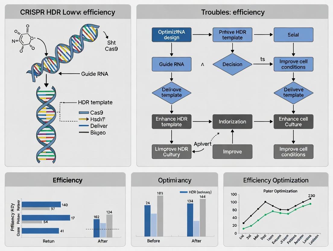
This comprehensive guide addresses the persistent challenge of low Homology-Directed Repair (HDR) efficiency in CRISPR-Cas9 genome editing.
Mastering CRISPR HDR: Advanced Troubleshooting Guide to Boost Low Editing Efficiency
Abstract
This comprehensive guide addresses the persistent challenge of low Homology-Directed Repair (HDR) efficiency in CRISPR-Cas9 genome editing. Targeting researchers and drug development professionals, it explores the foundational biology of DNA repair pathways, examines critical methodological variables in HDR experiment design, provides a systematic, step-by-step troubleshooting framework for optimizing outcomes, and details robust validation strategies to confirm precise edits. The article synthesizes current best practices and emerging solutions to empower scientists in achieving reliable, high-efficiency precision genome editing.
Understanding the HDR Bottleneck: Why CRISPR Precision Editing Often Fails
Technical Support Center: Troubleshooting Low HDR Efficiency in CRISPR Genome Editing
Frequently Asked Questions (FAQs) & Troubleshooting Guides
Q1: Why is my HDR efficiency so low compared to NHEJ-mediated indels? A: NHEJ is the dominant, fast-acting repair pathway active throughout the cell cycle, while HDR is restricted to the S/G2 phases and requires a homologous template. The competition is inherently biased toward NHEJ. Key factors include: cell cycle stage, template delivery/design, and Cas9 nuclease kinetics.
Q2: What are the most critical experimental parameters to optimize for improving HDR? A: Focus on these three areas:
- Template Design: Use single-stranded oligodeoxynucleotides (ssODNs) with homology arms optimized in length (35-90 nt) and ensure the template is delivered in high molar excess over the RNP.
- Cell Cycle Synchronization: Favor HDR by synchronizing cells in S/G2 phase or using small molecules.
- Inhibition of NHEJ Key Proteins: Transiently inhibit proteins like 53BP1 or DNA-PKcs to tilt the balance toward HDR.
Q3: Which small molecule inhibitors can boost HDR rates, and what are their caveats? A: Common inhibitors and their considerations are summarized in the table below.
Q4: How do I choose between ssODN and double-stranded DNA (dsDNA) donor templates? A: ssODNs are ideal for short edits (<100 nt) and show higher efficiency in many systems. dsDNA donors (e.g., plasmid, AAV) are necessary for large insertions. Ensure your dsDNA has long homology arms (>500 bp).
Q5: My HDR edits are correct but my cell viability is very low. What might be causing this? A: High cytotoxicity often stems from prolonged Cas9/sgRNA activity or the toxicity of NHEJ inhibitors. Use transient delivery methods (RNP electroporation) and titrate inhibitor concentrations/duration. Consider "hit-and-run" strategies with self-inactivating Cas9 systems.
Table 1: Efficacy of Small Molecule Modulators in Shifting Repair Pathway Balance
| Small Molecule | Target Pathway | Typical Concentration | Reported HDR Increase (Fold) | Key Caveats |
|---|---|---|---|---|
| SCR7 | NHEJ (Ligase IV inhibitor) | 1-10 µM | 2-5x | Variable activity; specific formulation is critical. |
| NU7026 | NHEJ (DNA-PKcs inhibitor) | 10 µM | 3-7x | Can be cytotoxic with prolonged exposure. |
| RS-1 | HDR (Rad51 stimulator) | 5-10 µM | 2-4x | May increase off-target integration events. |
| AZD-7648 | NHEJ (DNA-PKcs inhibitor) | 0.1-0.3 µM | Up to 10x | Potent and specific; newer compound with less in vivo data. |
| Brefeldin A | Undefined, cell cycle | 50 nM | 2-3x | Mild effect; may work via cell cycle modulation. |
Table 2: Donor Template Design Parameters and Recommended Use
| Template Type | Optimal Homology Arm Length | Optimal Size for Insertion | Recommended Molar Excess vs. RNP | Primary Delivery Method |
|---|---|---|---|---|
| ssODN (asymmetric) | 90 nt total (e.g., 35nt-55nt) | 1-100 bp | 100:1 to 1000:1 | Co-electroporation with RNP. |
| dsDNA Plasmid | >500 bp each arm | >100 bp | 10:1 to 50:1 | Transfection or electroporation. |
| AAV Vector | ~400-800 bp each arm | < 2 kb | N/A (MOI-based) | Viral transduction. |
Experimental Protocols
Protocol 1: Synchronizing Cells in S/G2 Phase for Enhanced HDR
- Cell Treatment: Treat adherent cells with a 24-hour incubation of 2 mM Thymidine.
- Release: Wash cells 3x with PBS and add fresh medium.
- Timing: Wait 3-5 hours post-release. At this point, a majority of cells will be in S/G2 phase.
- Editing: Perform CRISPR/Cas9 delivery (e.g., RNP electroporation) and donor template delivery during this window.
- Validation: Use flow cytometry with FUCCI reporters or staining for cell cycle markers (e.g., Cyclin B1) to confirm synchronization efficiency.
Protocol 2: RNP + ssODN Electroporation with NHEJ Inhibition
- Prepare RNP: Complex purified Cas9 protein (30 pmol) and sgRNA (36 pmol) in nuclease-free duplex buffer. Incubate 10 min at room temperature.
- Prepare Electroporation Mix: For a 20 µL reaction, combine RNP complex, ssODN donor template (3-6 nmol), and 1 µL of 10 µM NU7026 (or vehicle control).
- Harvest Cells: Trypsinize and wash 1e5 - 2e5 target cells. Resuspend in pre-warmed electroporation buffer.
- Electroporation: Add cell suspension to the mix, transfer to a cuvette, and electroporate using manufacturer's optimized protocol (e.g., Neon: 1400V, 20ms, 2 pulses).
- Recovery: Immediately transfer cells to pre-warmed medium. After 24 hours, replace medium to remove inhibitor.
Pathway and Workflow Diagrams
Diagram Title: The HDR vs. NHEJ Competition and Intervention Points
Diagram Title: Stepwise Troubleshooting Workflow for Low HDR
The Scientist's Toolkit: Key Research Reagent Solutions
| Reagent / Material | Supplier Examples | Function in HDR Enhancement |
|---|---|---|
| High-Purity Cas9 Nuclease | IDT, Thermo Fisher, Synthego | Ensures high cutting efficiency and minimal toxicity for "hit-and-run" editing. |
| Chemically Modified sgRNA | Synthego, Trilink | Increases stability and cutting efficiency, improving donor template engagement. |
| Ultramer ssODN Donors | IDT | Long, high-fidelity single-stranded DNA templates with precise homology arms. |
| DNA-PKcs Inhibitor (AZD-7648) | Selleckchem, MedChemExpress | Potently and selectively inhibits key NHEJ kinase, tilting balance toward HDR. |
| Cell Cycle Synchronization Agents | Sigma-Aldrich (Thymidine, Nocodazole) | Arrests cells at specific phases to enrich for HDR-competent populations. |
| Electroporation System (Neon/4D-Nucleofector) | Thermo Fisher, Lonza | Enables efficient, transient co-delivery of RNP and donor template into difficult cells. |
| HDR Reporter Cell Line (e.g., Traffic Light) | Addgene, generated in-house | Provides a rapid, fluorescence-based quantitative readout of HDR vs. NHEJ activity. |
| Next-Generation Sequencing Kit | Illumina, Pacific Biosciences | Allows precise, quantitative measurement of HDR and NHEJ outcomes at the target locus. |
CRISPR HDR Troubleshooting Support Center
Troubleshooting Guides & FAQs
Q1: Why is my HDR efficiency so low even with high-quality donor template and sgRNA? A: The most common cause is performing the experiment on an asynchronous cell population. HDR is primarily active during the S and G2 phases of the cell cycle, while NHEJ dominates in G1. If your cells are mostly in G1, NHEJ will be the predominant repair outcome.
Q2: How can I synchronize my cell cycle to improve HDR? A: Implement chemical synchronization. See the protocol below.
- Protocol: Double-Thymidine Block for S-Phase Synchronization
- Grow cells to ~70% confluence.
- Add fresh medium containing 2 mM thymidine. Incubate for 18 hours.
- Wash cells twice with 1X PBS and add fresh, pre-warmed medium.
- Incubate for 9 hours to release cells into the cycle.
- Add 2 mM thymidine again for 17 hours to collect cells at the G1/S boundary.
- Release cells into S-phase by washing with PBS and adding fresh medium. Transfert with CRISPR components immediately upon release.
Q3: What are the key cell cycle markers to check before CRISPR editing? A: The table below summarizes key markers and their interpretation for repair pathway choice.
Table 1: Cell Cycle Stage Markers and Dominant DNA Repair Pathways
| Cell Cycle Stage | Key Molecular Markers | Dominant DSB Repair Pathway | Approximate % of Asynchronous Population* |
|---|---|---|---|
| G1 | pRB hypophosphorylated, Low Cyclin A/E, High p27 | NHEJ (Canonical) | 40-50% |
| S | DNA replication (EdU+), High Cyclin A, Histone H3 phosphorylated (Ser31) | HDR (Primary) | 30-40% |
| G2 | Cyclin B1 accumulation, Histone H3 phosphorylated (Ser10), 4N DNA content | HDR & alt-EJ (Microhomology-Mediated) | 10-20% |
| M | pH3 (Ser10) positive, Condensed Chromosomes | NHEJ (Suppressed) | <5% |
*Percentages are typical for rapidly dividing immortalized cell lines (e.g., HEK293, U2OS).
Q4: Can I use inhibitors to bias repair toward HDR? A: Yes, transiently inhibiting key NHEJ factors during S/G2 can improve HDR outcomes.
- Protocol: SCR7 Treatment to Inhibit NHEJ
- Prepare a 10 mM stock of SCR7 (DNA Ligase IV inhibitor) in DMSO.
- 1-2 hours post-transfection/transduction of CRISPR components, add SCR7 to culture media at a final concentration of 1-10 µM.
- Incubate cells for 24-48 hours before changing to inhibitor-free media. Note: Titrate dose for your cell line to minimize toxicity.
Q5: How do I quantify cell cycle distribution in my sample? A: Flow cytometry analysis of DNA content is the standard method.
- Protocol: Propidium Iodide (PI) Staining for Cell Cycle Analysis
- Harvest and fix 1e6 cells in 70% ice-cold ethanol overnight at -20°C.
- Wash cells with PBS and resuspend in 500 µL PI/RNase Staining Buffer (e.g., from BD Biosciences).
- Incubate for 15 minutes at room temperature, protected from light.
- Analyze on a flow cytometer using a 488 nm laser and a 585/42 nm filter. Use software (e.g., ModFit) to deconvolute G1, S, and G2/M populations.
Experimental Protocol: Cell Cycle-Specific HDR Efficiency Measurement
Objective: To directly correlate HDR efficiency with cell cycle stage. Workflow:
- Transfect cells with CRISPR-Cas9 components (Cas9, sgRNA, fluorescent protein-tagged donor).
- At 24-48 hours post-transfection, pulse-label cells with 10 µM EdU for 30 minutes.
- Harvest cells, fix, and process using a "Click-It" EdU detection kit (Alexa Fluor 647).
- Stain DNA with FxCycle Violet Stain.
- Analyze by flow cytometry using three channels:
- FxCycle Violet: DNA content to gate G1, S, G2/M.
- Alexa Fluor 647: EdU signal to identify S-phase cells.
- Fluorescent protein (e.g., GFP): HDR reporter knock-in signal.
- Calculate HDR efficiency as (% GFP+ cells) within each cell cycle gate (G1/EdU-, S/EdU+, G2/M/EdU-).
Title: Workflow for Measuring Cell Cycle-Specific HDR Efficiency
Signaling Pathways Governing Repair Pathway Choice
Title: Cell Cycle Regulation of DSB Repair Pathway Choice
The Scientist's Toolkit: Research Reagent Solutions
Table 2: Essential Reagents for Cell Cycle-Aware CRISPR-HDR Experiments
| Item | Function / Rationale | Example Product/Catalog Number |
|---|---|---|
| Cell Cycle Synchronization Agents | Arrest cells at specific cycle stages to enrich for HDR-competent (S/G2) populations. | Thymidine (T1895, Sigma), Nocodazole (M1404, Sigma), Palbociclib (CDK4/6i) |
| EdU (5-Ethynyl-2’-deoxyuridine) | A thymidine analog for labeling S-phase cells via "click" chemistry, enabling cell cycle sorting/analysis. | Click-iT EdU Alexa Fluor 647 Imaging Kit (C10340, Thermo Fisher) |
| FxCycle Violet Stain | A low-wavelength, fixable DNA stain for cell cycle analysis by flow cytometry. Compatible with GFP and EdU-AF647. | FxCycle Violet Stain (F10347, Thermo Fisher) |
| NHEJ Inhibitors | Transiently suppress the competing NHEJ pathway to favor HDR in S/G2 cells. | SCR7 (HY-15678, MedChemExpress), NU7026 (DNA-PKi) |
| CDK1/2 Inhibitors | Chemical tools to probe CDK's essential role in promoting DNA end resection and HDR initiation. | RO-3306 (CDK1i, HY-12529), CVT-313 (CDK2i) |
| Live-Cell Cycle Reporters | Fluorescent biosensors (e.g., Fucci) to monitor cell cycle phase in real-time prior to editing. | FUCCI Cell Cycle Sensor (AAV, #C30056, Sirion Biotech) |
| HDR Reporter Cell Lines | Stable lines with integrated, broken fluorescent proteins to quantitatively measure HDR efficiency upon repair. | U2OS-DR-GFP (e.g., NCI-60) |
Welcome to the Technical Support Center for CRISPR HDR Efficiency Optimization. This guide is framed within a broader thesis research context focused on troubleshooting low Homology-Directed Repair (HDR) efficiency. Below are common issues and solutions related to donor DNA template design.
Frequently Asked Questions (FAQs) & Troubleshooting
Q1: My HDR efficiency is consistently below 5%. Could the issue be with my donor template's homology arm length? A: Yes, insufficient homology arm length is a primary cause of low HDR. The optimal length is cell-type and locus-dependent.
- Recommended Action: Use the following table as a starting guide for your design.
| Cell Type | Recommended Homology Arm Length (each arm) | Supported by Experimental Citation (Protocol Below) |
|---|---|---|
| Immortalized Mammalian Cells | 400 - 800 bp | Protocol 1 |
| Primary Cells & Stem Cells | 800 - 1000+ bp | Protocol 2 |
| Yeast | 35 - 60 bp | N/A |
| In vitro biochemical assays | 30 - 90 bp (ssODN) | Protocol 3 |
Q2: I am designing a single-stranded oligodeoxynucleotide (ssODN) template. What are the critical design rules? A: For ssODN templates, symmetry and modification are key.
- Issue: Unmodified ssODNs are degraded quickly, reducing availability.
- Solution: Order ssODNs with phosphorothioate (PS) bonds at the 2-3 terminal bases on both ends. This prevents exonuclease degradation.
- Design Rule: Center your desired edit. Ensure homology arms are symmetric (e.g., 60bp left + 60bp right) unless species-specific data suggests asymmetry.
Q3: Should I use a single-stranded or double-stranded donor template? A: The choice depends on the edit size and cell type. See comparative data below.
| Template Type | Ideal For | Typical Efficiency Range* | Key Advantage | Key Limitation |
|---|---|---|---|---|
| ssODN (<200 nt) | Point mutations, small tags | 0.5% - 10% | High cellular uptake, less indel formation | Limited cargo capacity |
| dsDNA (plasmid, PCR) | Large insertions (>200 bp) | 1% - 20% | Large cargo capacity, long homology arms | Higher risk of random integration |
| Viral Vectors (AAV) | In vivo or hard-to-transfect cells | 5% - 60%+ | High transduction efficiency | Complex production, size limit (~4.7kb) |
*Efficiency is highly variable and depends on locus, cell type, and delivery.
Q4: How do I prevent unintended integration of my plasmid donor backbone? A: This is a common issue that increases background noise.
- Troubleshooting Step: Linearize your plasmid donor template before transfection. Use a restriction enzyme site outside the homology arms. Alternatively, use a PCR-amplified dsDNA fragment containing only the homology arms and the payload, with no bacterial backbone sequence.
Q5: Does the strandedness of an ssODN relative to the cut strand matter? A: Recent data suggests it does.
- Observation: ssODNs complementary to the non-cut (PAM-containing) strand often show higher HDR efficiency in many systems.
- Recommended Test: If efficiency is low, design and test both "sense" and "antisense" ssODNs for your target. Use the table below to track results.
| ssODN Name | Strand Relative to Cut Site | Sequence (5'->3') [Partial] | HDR Efficiency (%) |
|---|---|---|---|
| ssODN_AS | Complementary to non-cut strand | [Sequence] | [Result] |
| ssODN_S | Complementary to cut strand | [Sequence] | [Result] |
Experimental Protocols Cited
Protocol 1: Testing Homology Arm Length in Mammalian Cells.
- Design: For your target locus, design PCR primers to generate dsDNA donors with varying homology arm lengths (e.g., 200bp, 500bp, 800bp).
- Prepare Donors: Amplify donors via PCR using a high-fidelity polymerase. Purify using a column-based kit.
- Co-transfect: Co-transfect HEK293T cells (in triplicate) with a fixed amount of Cas9/gRNA RNP and 100ng of each donor variant.
- Analyze: Harvest cells 72h post-transfection. Isolate genomic DNA and assess HDR efficiency by targeted NGS or droplet digital PCR (ddPCR).
- Key Reagent: High-fidelity PCR enzyme, ddPCR assay for HDR detection.
Protocol 2: Assessing ssODN Modification in Primary Cells.
- Design: Order two ssODNs for the same point mutation: one unmodified, one with PS modifications on three terminal bases at both ends.
- Electroporation: Use a nucleofection system optimized for your primary cell type (e.g., human T-cells).
- Delivery: Co-deliver 2µg of Cas9 protein, 2µg of synthetic gRNA, and 100pmol of ssODN per 100,000 cells.
- Culture & Analyze: Culture cells for 7 days, allowing for phenotype expression. Analyze HDR via flow cytometry (if tag introduced) or NGS of amplicons.
- Key Reagent: Phosphorothioate-modified ssODN, Primary Cell Nucleofector Kit.
Protocol 3: In vitro HDR Kinetics Assay.
- Prepare Components: Purify Cas9 protein, transcribe gRNA, and synthesize fluorescently labeled dsDNA target (100bp) and ssODN donors (90nt).
- Reaction Setup: In a buffer, mix 50nM Cas9:gRNA, 10nM target DNA, and 100nM ssODN donor.
- Incubate & Stop: Incubate at 37°C. Aliquot reactions at time points (0, 5, 15, 30, 60 min) into STOP buffer (EDTA, Proteinase K).
- Visualize: Run products on a capillary electrophoresis instrument (e.g., Fragment Analyzer). Calculate the percentage of target DNA converted to HDR product.
- Key Reagent: Recombinant Cas9 protein, Fluorescent DNA labeling kit.
Visualizations
Title: Donor Template Selection Workflow
Title: Key Steps in HDR Using a Donor Template
The Scientist's Toolkit: Research Reagent Solutions
| Item | Function & Rationale |
|---|---|
| High-Fidelity DNA Polymerase | To generate error-free PCR-amplified dsDNA donor fragments, preventing unwanted mutations in homology arms. |
| Phosphorothioate-modified ssODNs | Chemically stabilized single-stranded donors resist nuclease degradation, increasing intracellular half-life and HDR substrate availability. |
| Recombinant Cas9 Protein | For RNP formation and in vitro assays. Offers rapid kinetics and reduced off-target effects compared to plasmid DNA. |
| ddPCR HDR Detection Assay | Provides absolute quantification of low-efficiency HDR events (<1%) with high precision, superior to traditional qPCR or gel analysis. |
| Nucleofector Kit for Primary Cells | Enables efficient co-delivery of RNP and donor template into difficult cell types (T-cells, HSPCs, neurons) where lipofection fails. |
| Magnetic Beads for DNA Cleanup | For rapid, high-throughput purification of PCR donors and genomic DNA, essential for consistent NGS library prep. |
Troubleshooting Guides & FAQs
HDR Efficiency: General Issues
Q1: Why is my overall HDR efficiency low regardless of nuclease choice? A: Low HDR efficiency is often due to competition from the dominant non-homologous end joining (NHEJ) pathway and insufficient template delivery. Ensure you are using a high-fidelity repair template with sufficient homology arm length (typically 60-120 nt per arm for ssODNs, >500 bp for dsDNA). Cell cycle synchronization is critical, as HDR is most active in S/G2 phases. Consider using small molecule inhibitors like Scr7 or NU7441 to transiently inhibit NHEJ.
Q2: How do I choose the optimal nuclease platform for my specific HDR goal? A: Selection depends on your desired edit. See the decision table below.
Nuclease-Specific Issues
Cas9 Troubleshooting: Q3: My Cas9-mediated HDR creates unwanted indels at the target site. How can I reduce this? A: This is common due to persistent nuclease activity after HDR. Strategies include:
- Using high-fidelity Cas9 variants (e.g., SpCas9-HF1, eSpCas9).
- Delivering Cas9 as a pre-formed RNP complex for faster degradation.
- Employing a "self-inactivating" Cas9 system via targeted cleavage of its own expression cassette.
Cas12a Troubleshooting: Q4: I switched to Cas12a for its staggered cut, but HDR efficiency didn't improve. What's wrong? A: Cas12a's T-rich PAM (TTTV) limits targetable sites. Ensure your target locus has an optimal PAM. The 5' staggered ends may require different repair template design optimization compared to Cas9's blunt ends. Also, verify Cas12a expression and activity in your cell type, as it can be variable.
Base Editor Troubleshooting: Q5: My base editor is causing high levels of unintended off-target edits (bystander mutations). How do I mitigate this? A: Bystander mutations occur within the editing window (typically ~5 nucleotides). Solutions include:
- Using narrower-window base editors (e.g., ABE8e with engineered deaminases).
- Redesigning your gRNA to position the target base in a less mutation-prone position within the window.
- Using a "dual-AAV" split-intein system for BE delivery to reduce persistent expression.
Prime Editor Troubleshooting: Q6: Prime editing efficiency is very low in my primary cells. How can I optimize it? A: Prime editing efficiency is highly sequence-context dependent. Optimize by:
- Testing multiple pegRNA designs with varying primer binding site (PBS) length (8-18 nt) and RT template length (10-25 nt).
- Using engineered PE variants (PEmax, hyPE) with enhanced stability and nuclear localization.
- Including a nicking sgRNA (pegRNA+ngRNA strategy) to bias the repair outcome towards the edited strand.
Experimental Protocol: Comparing HDR Outcomes Across Nucleases
Objective: To systematically compare HDR efficiency, purity, and indel rates when introducing a specific point mutation using SpCas9, Cas12a, a Base Editor, and a Prime Editor.
Materials:
- Cell Line: HEK293T cells (or relevant cell type).
- Target Locus: A well-characterized genomic site amenable to all nucleases.
- Nuclease Delivery: Plasmids or RNP complexes for each editor.
- Repair Templates: For Cas9 and Cas12a, design ssODN donors with 90-nt homology arms. For PE, design pegRNAs. For BE, design the appropriate sgRNA.
- Analysis: Next-generation sequencing amplicon panel for the target region.
Method:
- Design & Cloning: Design and validate all gRNAs, pegRNAs, and repair templates for the same target base.
- Cell Transfection: Seed cells in 24-well plates. Transfect in triplicate using a consistent method (e.g., lipofection) with:
- Condition A: SpCas9 + sgRNA + ssODN donor.
- Condition B: Cas12a + crRNA + ssODN donor.
- Condition C: ABE8e (or CBE) plasmid + sgRNA.
- Condition D: PEmax plasmid + pegRNA (+ ngRNA if using).
- Include controls (nuclease only, donor only).
- Harvest & Extract: Harvest cells 72-96 hours post-transfection. Extract genomic DNA.
- Amplification & Sequencing: PCR-amplify the target region and subject to NGS.
- Data Analysis: Use bioinformatics tools (e.g., CRISPResso2, BE-Analyzer, pe-analyzer) to quantify:
- HDR/Editing Efficiency: (% reads with desired edit).
- Indel Rate: (% reads with insertions/deletions).
- Purity: (Desired edits / (Desired edits + Undesired edits)) * 100.
- Bystander Edits: For BE and PE.
Table 1: Comparative Performance of CRISPR Editors for a Model Point Mutation (Theoretical Data)
| Editor | Typical Efficiency Range | Indel Byproduct | Key Advantages | Key Limitations |
|---|---|---|---|---|
| SpCas9 + HDR | 1-30% (highly variable) | High (10-60%) | Large insertions, precise deletions | Dominant NHEJ, cell-cycle dependent |
| Cas12a + HDR | 2-25% | Moderate (5-40%) | Staggered cut, less off-target in some contexts | Restricted T-rich PAM, lower activity in some cells |
| Base Editor (CBE/ABE) | 10-80% (avg ~50%) | Very Low (<1%)* | High efficiency, no DSB, no donor required | Transition mutations only, bystander edits, size limits |
| Prime Editor | 5-50% (avg ~20-30%) | Very Low (<1%) | All 12 possible base changes, small insertions/deletions | Complex design, lower efficiency in some cells |
*Indels can occur if nick is converted to DSB; Bystander edits are the primary byproduct.
Table 2: Troubleshooting Matrix for Low HDR Outcomes
| Symptom | Cas9 | Cas12a | Base Editor | Prime Editor |
|---|---|---|---|---|
| Low Editing | Optimize donor design, use NHEJi, synchronize cell cycle. | Verify PAM, test crRNA design, optimize RNP ratio. | Check editing window, use engineered deaminase variant. | Optimize PBS & RT length, use PEmax/hyPE, add ngRNA. |
| High Indels | Use Hi-Fi Cas9, deliver as RNP, use "kill-switch". | Titrate nuclease amount, use high-fidelity variant. | Ensure nicking sgRNA is not creating a DSB. | Monitor for pegRNA-independent nicking. |
| Off-Target | Use Hi-Fi variant, predict with in silico tools, do GUIDE-seq. | Generally lower reported off-target than Cas9. | Perform wide-scale sequencing (e.g., Digenome-seq). | Current data suggests high specificity; profile with PE-specific tools. |
Visualizations
Title: CRISPR Nuclease Selection Decision Flowchart
Title: HDR vs NHEJ Pathway Competition After CRISPR DSB
The Scientist's Toolkit: Research Reagent Solutions
| Item | Function & Application |
|---|---|
| High-Fidelity Cas9 (e.g., SpCas9-HF1) | Reduces off-target cleavage while maintaining on-target activity for cleaner HDR. |
| AsCas12a (Cpfl) Nuclease | Provides staggered double-strand breaks (5' overhangs) which may alter HDR outcomes vs blunt cuts. |
| ABE8e & BE4max Plasmids | Next-generation base editors with improved efficiency and purity, reducing bystander edits. |
| PEmax & hyPE Plasmids | Engineered prime editor systems with improved stability and nuclear localization for higher editing rates. |
| Chemically Modified sgRNAs (e.g., Alt-R) | Enhance nuclease stability and reduce immune responses, especially in primary cells. |
| Electroporation Enhancer (e.g., Alt-R Cas9 Electroporation Enhancer) | Improves delivery and HDR outcomes when using RNP electroporation. |
| NHEJ Inhibitors (SCR7, NU7441) | Small molecules that transiently inhibit key NHEJ proteins (DNA Ligase IV, DNA-PK), tilting balance toward HDR. |
| Cell Cycle Synchronization Agents (e.g., Nocodazole, Aphidicolin) | Arrest cells at specific phases (M/G1-S) to enrich for HDR-competent (S/G2) populations post-release. |
| ssODN HDR Donor with Phosphorothioate Modifications | Protects single-stranded DNA donors from exonuclease degradation, increasing HDR template availability. |
| Next-Gen Sequencing Analysis Suites (CRISPResso2, BE-Analyzer) | Essential software for accurate, quantitative analysis of editing outcomes, indels, and HDR efficiency from NGS data. |
Blueprint for Success: Designing and Executing a Robust HDR Experiment
Troubleshooting Guides & FAQs
Q1: My HDR efficiency is consistently low (<1%) with ssODNs, even with optimized Cas9 RNP delivery. What could be the issue? A: This is a common challenge. Low ssODN efficiency often stems from rapid degradation or insufficient concentration. Ensure you are using high-quality, HPLC-purified ssODNs. The optimal concentration is typically 1-10 µM (10-100x the molar amount of Cas9 RNP). Also, verify that your ssODN is designed with homology arms of appropriate length (30-90 bases total) and that it is targeting the correct strand (the non-Cas9 nicked strand for best results). Consider adding a 5’-phosphate modification to enhance stability and engagement with the cellular repair machinery.
Q2: When should I choose a dsDNA donor over an ssODN? A: Choose dsDNA (e.g., plasmid, PCR fragment, or viral vector) when your edit requires the insertion of large sequences (>200 bp). dsDNA templates are also less prone to degradation and can be engineered with longer homology arms (200-1000 bp), which can significantly boost absolute HDR efficiency for complex knock-ins, especially when using selection strategies or when working with recalcitrant cell types.
Q3: I get high HDR efficiency but also excessive random integration of my dsDNA plasmid donor. How can I mitigate this? A: Random integration is a major drawback of plasmid donors. To mitigate this:
- Linearize the donor: Always use a linear dsDNA template (e.g., a PCR-amplified fragment with homology arms). This dramatically reduces non-homologous end joining (NHEJ)-mediated random integration compared to circular plasmids.
- Optimize donor amount: High concentrations increase off-target integration. Titrate your linear dsDNA donor (common range: 1-100 ng for plasmid-derived PCR fragments).
- Use inhibition strategies: Incorporate small molecule inhibitors of NHEJ, such as Scr7 or NU7026, during the first 24-48 hours post-transfection to favor HDR. (Note: These can be cytotoxic; titrate carefully.)
Q4: For precise point mutations in primary cells, which donor is recommended? A: For point mutations or small tag insertions (<100 bp) in sensitive primary cells, high-quality ssODNs are generally the superior choice. They exhibit lower cytotoxicity and minimal risk of random genomic integration. Use electroporation with Cas9 RNP and a chemically modified, phosphorothioate-protected ssODN to maximize delivery and template stability in these challenging cell types.
Data Presentation: Quantitative Comparison of Donor Templates
Table 1: Strategic Comparison of ssODN vs. dsDNA Donor Templates
| Parameter | ssODN (Single-Stranded Oligodeoxynucleotide) | dsDNA (Double-Stranded DNA - e.g., PCR fragment) |
|---|---|---|
| Optimal Insert Size | < 200 bp (best for point mutations, small tags) | > 200 bp (large genes, reporters, conditional alleles) |
| Typical Homology Arm Length | 30-60 bases (each arm) | 200-1000 bp (each arm) |
| Common Concentration | 1-10 µM (10-100x molar excess over RNP) | 1-100 ng (for PCR fragments in a standard transfection) |
| HDR Efficiency (Range) | 0.5% - 20% (highly cell-type dependent) | 1% - 50% (can be higher with long arms/selection) |
| Random Integration Risk | Very Low | Moderate to High (mitigated by linearization) |
| Cellular Toxicity | Low | Moderate (increases with size and amount) |
| Primary Cell Suitability | High (low toxicity, easy delivery with RNP) | Low to Moderate (challenging delivery, higher toxicity) |
| Key Advantages | Fast, cheap synthesis; low integration risk; ideal for SNPs. | High efficiency for large inserts; long homology arms boost accuracy. |
| Major Drawbacks | Limited insert size; rapid degradation; strand bias. | Cloning required; high random integration (plasmids); more immunogenic. |
Experimental Protocols
Protocol 1: HDR using ssODN with Cas9 RNP Electroporation
- Design: Design ssODN with ~30-60 nt homology arms. Order with 5’-phosphate and 3’-phosphorothioate bonds (2-3) at each end.
- Preparation: Complex chemically modified sgRNA with Cas9 protein to form RNP (3:1 molar ratio, 20 min, RT).
- Electroporation: Mix 1e5 cells, RNP (5-20 pmol), and ssODN (final 1 µM) in electroporation buffer. Electroporate using a cell-type optimized program (e.g., Neon or 4D-Nucleofector).
- Post-Transfection: Immediately transfer cells to pre-warmed media. Optionally, add 1 µM Scr7 (NHEJ inhibitor) for 24h.
- Analysis: Harvest cells 48-72h post-editing. Assess efficiency via NGS or droplet digital PCR (ddPCR).
Protocol 2: HDR using Linear dsDNA Donor via Lipofection
- Donor Preparation: Amplify donor cassette (insert + 500-800 bp homology arms) via high-fidelity PCR from a plasmid template. Purify using a silica-column kit. Quantify by fluorometry.
- Transfection: Seed HEK293T or similar cells to reach 70-80% confluency at transfection. For a 24-well, complex 500 ng linear dsDNA donor, 250 ng Cas9 expression plasmid (or 15 pmol RNP), and 1.5 µL of a transfection reagent (e.g., Lipofectamine 3000) in Opti-MEM.
- Inhibition: Replace media after 6h. Add NU7026 (final 10 µM) to fresh media for 24h to inhibit NHEJ.
- Selection & Analysis: After 48h, begin puromycin selection (if donor contains a selection marker) for 5-7 days. Isolate single clones and screen by genomic PCR and Sanger sequencing.
Mandatory Visualizations
Diagram 1: CRISPR HDR Donor Selection Workflow
Diagram 2: HDR Competitor Pathways with Donors
The Scientist's Toolkit: Research Reagent Solutions
| Reagent / Material | Function in HDR Experiment |
|---|---|
| HPLC-purified ssODN | Ensures high-purity, single-stranded template; critical for reproducible efficiency and reducing toxicity. |
| Phosphorothioate Modifications | Protects ssODN ends from exonuclease degradation, increasing intracellular half-life. |
| High-Fidelity PCR Enzyme | For error-free amplification of long, complex dsDNA donor fragments with extended homology arms. |
| Cas9 Nuclease (WT), protein | For RNP complex formation, enabling rapid, DNA-free delivery and high cleavage activity. |
| NHEJ Inhibitors (e.g., Scr7, NU7026) | Temporarily suppresses the dominant NHEJ pathway to increase the HDR/NHEJ ratio post-editing. |
| Electroporation System (4D-Nucleofector) | Enables high-efficiency delivery of RNP and donor templates into hard-to-transfect cell types. |
| ddPCR Assay for HDR | Provides absolute quantification of low-frequency HDR events with high precision and sensitivity. |
Technical Support Center
Troubleshooting Guides
Guide 1: Low HDR Efficiency Despite High Cas9 Cleavage Activity
- Problem: Confirmed high INDEL formation via T7E1 assay, but HDR knock-in remains low.
- Diagnosis: Likely due to suboptimal donor template design, improper component stoichiometry, or cell cycle mismatch.
- Solution Steps:
- Verify Donor Template: Ensure donor DNA (ssODN or dsDNA) has sufficient homology arm length (70-90 bp for ssODN, >500 bp for dsDNA). Check for silent mutations in PAM site to prevent re-cleavage.
- Optimize Ratios: Titrate the molar ratio of sgRNA:Cas9:Donor. Start with 1:1:5 (for plasmid) or 1:1:10 (for RNP with ssODN).
- Synchronize Cell Cycle: Utilize cell cycle inhibitors like nocodazole to enrich for cells in S/G2 phase, where HDR is active.
Guide 2: High Cellular Toxicity Post-Transfection
- Problem: Excessive cell death 24-48 hours after transfection with CRISPR components.
- Diagnosis: Often caused by transfection reagent cytotoxicity, overloading of RNP complexes, or excessive nuclease activity.
- Solution Steps:
- Titrate Transfection Reagent: Reduce reagent volume by 25-50% increments while maintaining complex stability.
- Switch Delivery Method: For sensitive cells, consider electroporation (e.g., Neon, Amaxa) with optimized voltage/pulse settings, or switch to a different lipid-based polymer.
- Modify RNP Amount: Lower total RNP amount delivered. Use a fluorescently tagged tracer sgRNA to monitor delivery efficiency independently.
Guide 3: Inconsistent Results Across Replicates
- Problem: High variability in HDR efficiency between experimental repeats.
- Diagnosis: Inconsistent reagent quality, cell passage number/health, or transfection mixture preparation.
- Solution Steps:
- Standardize RNP Formation: Always prepare RNP complexes fresh. Incubate sgRNA and Cas9 protein at room temperature for exactly 10-20 minutes before use.
- Quality Control Cells: Use cells at low passage (<25), ensure consistent confluency (70-80%) at transfection, and maintain stable cell line karyotypes.
- Master Mix Preparation: Prepare a single master mix of CRISPR components for all replicates to minimize pipetting error.
Frequently Asked Questions (FAQs)
Q1: What is the optimal ratio of sgRNA to Cas9 protein for RNP complex formation? A: A molar ratio of 1:1.2 to 1:1.5 (sgRNA:Cas9) is typically optimal for complete complex formation and activity. Excess sgRNA can lead to off-target effects, while excess Cas9 may increase toxicity.
Q2: When should I use lipid-based transfection vs. electroporation for CRISPR delivery? A: Use lipid-based reagents (e.g., Lipofectamine CRISPRMAX) for adherent, easy-to-transfect cell lines. Use electroporation for primary cells, stem cells, or difficult-to-transfect cell lines (e.g., Jurkat, macrophages). Electroporation generally offers higher efficiency but requires optimization of pulse parameters.
Q3: How does donor template form (ssODN vs. dsDNA) affect HDR efficiency and choice? A: ssODNs are ideal for short insertions (<60 bp) and point mutations, offering faster kinetics and lower toxicity. dsDNA donors (e.g., PCR fragments, plasmids) are necessary for larger insertions (>200 bp). dsDNA can yield higher absolute HDR but with higher background of random integration.
Q4: What are key additives to improve HDR efficiency? A: Small molecule additives can be crucial. NHEJ inhibitors like SCR7 or Ligase IV inhibitor enhance HDR relative to error-prone repair. Cell cycle synchronizers (e.g., nocodazole, RO-3306) and HDR enhancers like RS-1 are commonly used. See Table 2.
Q5: How long should I wait to analyze HDR efficiency post-transfection? A: Allow at least 48-72 hours for repair. For puromycin/mixed population analysis, wait 5-7 days post-selection. For single-cell cloning, allow 10-14 days for colony formation before screening.
Table 1: Optimized Component Ratios for Common Delivery Methods
| Delivery Method | Cas9 Form | sgRNA:Cas9 Molar Ratio | Donor Template Ratio (Molar) | Typical HDR Efficiency Range* |
|---|---|---|---|---|
| Lipid (Plasmid) | Expression Plasmid | 1:1 (plasmid) | 3:1 to 5:1 | 5-20% |
| Lipid (RNP) | Recombinant Protein | 1:1.2 to 1:1.5 | 5:1 to 10:1 (ssODN) | 10-40% |
| Electroporation (RNP) | Recombinant Protein | 1:1.5 | 10:1 to 50:1 (ssODN) | 20-60% |
| AAV (In vivo) | mRNA/protein + donor | N/A | Co-packaging critical | 1-10% |
*Efficiency varies significantly by cell type and locus.
Table 2: Common HDR-Enhancing Compounds and Protocols
| Compound | Target/Function | Recommended Concentration | Treatment Timing | Key Consideration |
|---|---|---|---|---|
| SCR7 | Ligase IV inhibitor (NHEJ) | 1-10 µM | Add during/after transfection, maintain 24-72h | Can be cytotoxic; use dose titration. |
| RS-1 | Rad51 stimulator (HDR) | 5-10 µM | Pre-treat 1h before transfection, include in media 24h | Optimize per cell line; may not work universally. |
| Nocodazole | Cell cycle synchronizer (G2/M) | 100 ng/mL | Treat 12-16h before transfection, wash out | Can stress cells; monitor viability closely. |
| Alt-R HDR Enhancer | Proprietary small molecule | 1X (per kit) | Include in RNP complex formulation | Optimized for IDT's Alt-R system. |
Experimental Protocols
Protocol 1: RNP Complex Assembly and Lipid-Based Transfection
- Complex Assembly: Dilute chemically modified sgRNA (e.g., Alt-R CRISPR-Cas9 sgRNA) and purified Cas9 nuclease (e.g., Alt-R S.p. Cas9 Nuclease V3) separately in sterile duplex buffer or Opti-MEM.
- Mix at a 1:1.2 molar ratio. For example, for a 10 µL reaction: 6 pmol sgRNA + 7.2 pmol Cas9.
- Incubate at room temperature for 15 minutes to form RNP.
- Transfection Mix: Dilute lipid-based transfection reagent (e.g., Lipofectamine RNAiMAX) in a separate tube of Opti-MEM. Incubate 5 minutes.
- Combine the RNP complex with the diluted transfection reagent. Incubate for 10-20 minutes at room temperature.
- Add the complete complex dropwise to cells in a culture plate with antibiotic-free medium.
- Replace medium after 4-6 hours.
Protocol 2: RNP Electroporation for Difficult-to-Transfect Cells (e.g., Jurkat T-cells)
- Cell Preparation: Harvest and wash 1x10^6 cells per condition with PBS. Resuspend in 20 µL of proprietary electroporation buffer (e.g., Neon Buffer R, SE Cell Line Solution).
- RNP Assembly: Prepare RNP complex as in Protocol 1, using a final amount of 30-60 pmol Cas9 protein per reaction.
- Mix the cell suspension with the pre-assembled RNP complexes and any donor DNA (e.g., 200 pmol ssODN).
- Electroporation: Load mixture into a tip. Apply pulse(s) (e.g., Neon System: 1400V, 10ms, 3 pulses for Jurkat).
- Immediately transfer cells to pre-warmed complete medium in a 24-well plate.
- Analyze or expand cells after 48-72 hours.
Visualizations
Title: CRISPR HDR Delivery Optimization Workflow
Title: DNA Repair Pathway Competition Post-CRISPR Cut
The Scientist's Toolkit: Research Reagent Solutions
| Item | Function & Rationale |
|---|---|
| Chemically Modified sgRNA (e.g., Alt-R CRISPR-Cas9 sgRNA) | Incorporates 2'-O-methyl and phosphorothioate modifications at terminal nucleotides to enhance nuclease stability, reduce immune responses, and improve RNP formation efficiency. |
| High-Fidelity Cas9 Nuclease (e.g., HiFi Cas9, eSpCas9) | Engineered Cas9 variant with reduced off-target cleavage activity while maintaining robust on-target efficiency, crucial for therapeutic applications. |
| Electroporation System (e.g., Neon, Amaxa 4D-Nucleofector) | Enables physical delivery of RNP complexes and donor DNA into hard-to-transfect cell types via optimized electrical pulses, often yielding the highest editing rates. |
| HDR Enhancer (e.g., Alt-R HDR Enhancer V2, RS-1) | Small molecule additives that shift the DNA repair balance towards HDR by stimulating key mediators like Rad51 or transiently inhibiting the competing NHEJ pathway. |
| Homology-Directed Donor Template (ssODN or dsDNA) | Provides the correct template for repair. ssODNs are synthetically accessible for short edits; long dsDNA donors (e.g., AAV, PCR amplicons) are needed for larger inserts. Critical silent mutations in the PAM prevent re-cutting. |
| Cell Cycle Synchronization Agent (e.g., Nocodazole, RO-3306) | Chemicals used to arrest cells in HDR-permissive phases (S/G2) prior to transfection, thereby increasing the proportion of cells competent for precise editing. |
| Next-Generation Sequencing (NGS) Validation Kit (e.g., Illumina CRISPR Amplicon) | Provides the most comprehensive and quantitative analysis of on-target editing efficiency, HDR/INDEL ratios, and off-target profiling, essential for robust data. |
Technical Support Center
Troubleshooting Guide: Common HDR Efficiency Issues
Issue 1: Low HDR efficiency despite high cutting efficiency.
- Potential Cause: Cas9 cutting occurs predominantly in G1 phase, but HDR repair is only active in S/G2 phases.
- Troubleshooting Steps:
- Quantify cell cycle distribution of your target cell population using flow cytometry (e.g., propidium iodide staining).
- Synchronize cells at the G1/S boundary using a thymidine block or a CDK4/6 inhibitor (e.g., Palbociclib).
- Deliver the Cas9 RNP and donor template immediately after release from synchronization.
- Compare HDR rates to an unsynchronized control.
Issue 2: High cytotoxicity post-editing.
- Potential Cause: Prolonged Cas9 expression leads to persistent double-strand breaks (DSBs) and activation of p53-mediated cell death or cell cycle arrest.
- Troubleshooting Steps:
- Switch from plasmid-based Cas9 expression to a transient delivery method (e.g., Cas9 protein RNP electroporation).
- Use a cell cycle-specific promoter (e.g., Geminin for S/G2 phases) to limit Cas9 expression to HDR-permissive phases.
- Co-deliver a p53 inhibitor transiently, but assess long-term genomic stability implications.
Issue 3: Poor donor template integration fidelity.
- Potential Cause: The donor template is degraded or not nuclear-localized when the cell is in S/G2 phase.
- Troubleshooting Steps:
- For ssODN donors, ensure chemical modifications (e.g., phosphorothioate) to increase stability.
- For viral donors (AAV), confirm viral tropism and entry kinetics align with your synchronized cell cycle window.
- For plasmid donors, use a cell cycle-regulated promoter to delay expression until S phase.
Frequently Asked Questions (FAQs)
Q1: What is the optimal cell cycle phase for achieving high HDR rates? A: The S and G2 phases are optimal because the homologous recombination (HR) machinery requires sister chromatids as templates, which are only present after DNA replication. Targeting Cas9 activity to these phases maximizes HDR over error-prone non-homologous end joining (NHEJ).
Q2: How can I synchronize my cell culture without causing excessive stress or apoptosis? A: Chemical synchronization is most common. A double thymidine block is effective for many immortalized lines. For primary or sensitive cells, consider a reversible CDK4/6 inhibitor (e.g., Palbociclib) for a gentler G1 arrest. Always titrate the inhibitor and duration to minimize stress.
Q3: Can I use Cas9 ribonucleoprotein (RNP) complexes for cell cycle-synchronized editing? A: Yes, RNP delivery is ideal for this approach. The rapid activity and degradation of Cas9 protein create a short editing window. By delivering the RNP at a specific time after releasing cells from a G1 block, you can align the peak of DSB formation with the S/G2 phases.
Q4: Are there specific promoters I can use to restrict Cas9 expression to S/G2? A: Yes. The Geminin promoter is activated at the G1/S transition and remains active through S, G2, and M phases, making it an excellent choice for driving Cas9 expression in an HDR-permissive window.
Q5: What quantitative metrics should I track to validate successful synchronization and improved HDR? A: Key metrics include:
- Synchronization Efficiency: % of cells in target phase via flow cytometry.
- Editing Efficiency: % INDELs via NGS or T7E1 assay.
- HDR Efficiency: % of alleles with precise donor integration via NGS or allele-specific qPCR.
- Cell Viability: Pre- and post-editing viability counts.
Table 1: HDR Efficiency Across Cell Cycle Phases (Representative Data)
| Cell Cycle Phase | Primary Repair Pathway | Typical HDR Efficiency (%) | Key Regulating Factor |
|---|---|---|---|
| G1 | NHEJ, MMEJ | < 1% | 53BP1, Shieldin Complex |
| S | HDR, SSA | 5-20%* | BRCA1, Rad51, CtIP |
| G2 | HDR, NHEJ | 5-15%* | BRCA1, Rad51 |
| M | NHEJ (limited) | Negligible | Cyclin B, CDK1 Activity |
*Efficiency is highly dependent on cell type, locus, and donor design.
Table 2: Synchronization Methods Comparison
| Method | Target Phase | Mechanism | Duration | Key Consideration |
|---|---|---|---|---|
| Serum Starvation | G0/G1 | Growth factor deprivation | 48-72 hrs | Can induce quiescence, not reversible. |
| Double Thymidine Block | G1/S | Inhibits DNA synthesis | ~16 hrs | Can cause replication stress. |
| Nocodazole | M | Disrupts microtubules | 8-12 hrs | High cytotoxicity risk. |
| CDK4/6 Inhibitor (e.g., Palbociclib) | G1 | Reversible kinase inhibition | 12-24 hrs | Gentler; suitable for primary cells. |
Experimental Protocols
Protocol 1: Cell Synchronization using a Double Thymidine Block
- Seed cells at 40-50% confluence.
- First Block: Add thymidine to culture medium to a final concentration of 2 mM. Incubate for 18 hours.
- Release: Wash cells 3x with 1x PBS and add fresh, pre-warmed complete medium. Incubate for 9 hours.
- Second Block: Add thymidine again to 2 mM. Incubate for 17 hours.
- Final Release & Transfection: Wash cells 3x with PBS. Add complete medium. Immediately proceed with delivery of CRISPR editing components (e.g., RNP electroporation). Harvest cells for analysis 24-72 hours post-release.
Protocol 2: Flow Cytometry for Cell Cycle Analysis (Propidium Iodide)
- Harvest & Fix: Trypsinize and collect ~1e6 cells. Wash with PBS. Resuspend pellet in 200 µL PBS. Fix by adding 800 µL of ice-cold 70% ethanol drop-wise while vortexing. Incubate at -20°C for ≥1 hour.
- Stain: Pellet fixed cells, wash with PBS. Resuspend in 500 µL staining solution (PBS with 50 µg/mL Propidium Iodide, 100 µg/mL RNase A, 0.1% Triton X-100).
- Analyze: Incubate at 37°C for 30 min in the dark. Analyze on a flow cytometer using a 488 nm laser and a 585/42 nm (or similar) filter. Use FL2-Area vs. FL2-Width to exclude doublets. Model cell cycle distribution using appropriate software (e.g., ModFit, FlowJo).
Visualizations
Title: Cell Cycle Phase Determines DNA Repair Pathway Choice
Title: Experimental Workflow for Cell Cycle-Synchronized HDR
The Scientist's Toolkit: Research Reagent Solutions
| Item | Function & Rationale |
|---|---|
| Palbociclib (CDK4/6 Inhibitor) | Reversibly arrests cells in G1 phase by inhibiting cyclin D-CDK4/6, enabling gentle synchronization for primary cells. |
| Thymidine | Inhibits DNA synthesis by depleting dCTP pools, causing a reversible arrest at the G1/S boundary for robust synchronization of cell lines. |
| Cas9 Nuclease (RNP format) | Pre-complexed guide RNA and protein. Enables rapid, transient activity, allowing precise temporal alignment of DSBs with S/G2 phase. |
| Chemically Modified ssODN Donor | Single-stranded oligodeoxynucleotide donor template with phosphorothioate bonds for nuclease resistance, increasing stability during the S/G2 window. |
| Cell Cycle Phase-Specific Reporter (FUCCI) | Fluorescent ubiquitination-based cell cycle indicator. Allows real-time monitoring and sorting of live cells based on their cell cycle phase (G1: red, S/G2/M: green). |
| Propidium Iodide / RNase A Staining Solution | For flow cytometry-based cell cycle analysis. RNase A removes RNA, and PI intercalates into DNA, providing a histogram of DNA content per cell. |
| Geminin-Promoter Driven Cas9 Plasmid | Restricts Cas9 expression to the S, G2, and M phases of the cell cycle, inherently biasing repair toward HDR by reducing Cas9-induced DSBs in G1. |
| p53 Inhibitor (e.g., Pifithrin-α, transient) | Temporarily suppresses p53 activation to reduce cell death following DSB induction, potentially improving survival of edited cells. Use with caution. |
Technical Support Center: Troubleshooting CRISPR HDR Efficiency
Frequently Asked Questions (FAQs)
Q1: Why is my HDR efficiency in my cancer cell line (e.g., HEK293) significantly higher than in my primary human T-cells? A: This is expected due to fundamental biological differences. Cancer cell lines have dysregulated DNA repair pathways, often favoring HDR, and are actively cycling. Primary T-cells are predominantly in G0/G1 phase, where NHEJ dominates. Quantitative data below summarizes key differences.
Q2: My iPSC clones show mosaicism after HDR editing. How can I reduce this? A: Mosaicism occurs because editing happens after the single cell has begun dividing. To mitigate, use cell cycle synchronization (e.g., nocodazole or RO-3306) to enrich for cells in S/G2 phase just before editing, or use a CRISPR-Cas9 ribonucleoprotein (RNP) delivery method for faster action.
Q3: I am getting no HDR in my primary neurons, despite high Cas9 cutting efficiency. What are the main barriers? A: Primary neurons are post-mitotic (non-dividing). The canonical HDR pathway is largely inactive in non-cycling cells. Consider alternative strategies like:
- Using NHEJ-mediated targeted integration with a "landing pad" or uni-directional donor.
- Employing Cas9-fused base editors or prime editors for point mutations without requiring a donor template.
- Using virus-derived recombinases (e.g., AAV-mediated delivery) if applicable.
Q4: What is the most critical factor for improving HDR across all model systems? A: Controlling the competition between HDR and NHEJ. The consistent strategy is the pharmacological or genetic inhibition of key NHEJ proteins (e.g., using SCR7, KU-0060648 to inhibit DNA-PK, or siRNA against Ku70/80) during the editing window. This must be optimized per cell type due to toxicity.
Troubleshooting Guides
Issue: Low HDR Efficiency in iPSCs
- Check 1: Cell State. Ensure iPSCs are in a pristine, undifferentiated state with high viability. Passage cells 24-48 hours before editing for optimal health.
- Check 2: Donor Design. For iPSCs, use long single-stranded DNA (ssDNA) donors (>200 nt) or Cas9-Triggered Linearization (CTL) donors. Ensure sufficient homology arm length (typically 800-1000 bp for plasmid donors, 50-100 nt for ssDNA).
- Check 3: Transfection Method. Electroporation of Cas9 RNP complexed with ssDNA donor is highly effective. For plasmid donors, nucleofection is preferred over lipofection.
- Protocol: HDR in iPSCs via RNP Electroporation
- Culture and passage iPSCs using standard methods.
- Design and synthesize crRNA, tractRNA, Cas9 protein, and long ssDNA donor.
- Complex crRNA and tractRNA to form guide RNA (gRNA). Incubate gRNA with Cas9 protein to form RNP (20 min, RT).
- Mix 1-2 million dissociated iPSCs with RNP (30-60 pmol) and ssDNA donor (200-400 pmol) in nucleofection solution.
- Electroporate using manufacturer's (e.g., Lonza) recommended program for human stem cells.
- Recover cells in pre-warmed medium with ROCK inhibitor (Y-27632).
- After 72 hours, begin antibiotic selection or single-cell sorting for clonal expansion.
Issue: High Cytotoxicity in Primary Cells During HDR Protocols
- Check 1: Delivery Toxicity. Primary cells are sensitive to transfection. Titrate RNP and donor amounts to the minimum required for detectable editing. Use high-viability electroporation buffers.
- Check 2: Inhibitor Toxicity. NHEJ inhibitors can be toxic. Perform a dose-response curve (e.g., SCR7 from 1-10 µM) and limit exposure time to 24-48 hours post-editing.
- Check 3: Cell Health. Use low-passage, freshly isolated primary cells whenever possible. Use culture media and supplements optimized for the specific primary cell type.
Table 1: Comparative HDR Efficiency and Characteristics Across Model Systems
| Model System | Typical HDR Efficiency Range* | Dominant Repair Pathway | Cell Cycle Dependence | Common Delivery Method | Key Consideration |
|---|---|---|---|---|---|
| Immortalized Cell Lines (HEK293, HeLa) | 5% - 40% | HDR (in S/G2) | High - Actively cycling | Lipofection, Electroporation | Easiest model; high transfection efficiency. |
| Induced Pluripotent Stem Cells (iPSCs) | 1% - 20% | HDR (but cell cycle varies) | Moderate - Can be synchronized | Nucleofection (RNP) | Risk of mosaicism; require clonal isolation. |
| Primary Cells (T-cells, HSCs) | 0.1% - 5% | NHEJ (predominantly in G0/G1) | Very High - Mostly quiescent | Nucleofection (RNP) | Low viability post-editing; sensitive to manipulation. |
*Efficiency for precise, reporter-integration edits. Highly variable based on locus, donor design, and protocol.
Table 2: Efficacy of Common NHEJ Inhibitors in Different Cell Types
| Compound | Target | Effective Conc. in Cell Lines | Effective Conc. in iPSCs | Tolerated in Primary T-cells? | Notes |
|---|---|---|---|---|---|
| SCR7 | Ligase IV | 5 - 10 µM | 1 - 5 µM | Marginally (≤ 2µM, 24h) | Broadly used but specificity debated. |
| NU7026 | DNA-PKcs | 5 - 20 µM | Not Recommended | No | High cytotoxicity in sensitive cells. |
| KU-0060648 | DNA-PKcs | 0.1 - 1 µM | 0.05 - 0.5 µM | Yes (≤ 0.5µM, 48h) | More potent and selective; better for primary cells. |
Experimental Workflow Diagram
Title: CRISPR HDR Strategy Tailoring Workflow
DNA Repair Pathway Decision Logic
Title: Cell Cycle and Donor Presence Dictate Repair Pathway
The Scientist's Toolkit: Research Reagent Solutions
Table 3: Essential Reagents for CRISPR HDR Optimization
| Reagent Category | Specific Item/Example | Function in HDR Experiment | Key Consideration |
|---|---|---|---|
| CRISPR Components | High-fidelity Cas9 (HiFi Cas9, SpCas9) | Creates the target double-strand break (DSB). | Reduces off-target effects; critical for therapeutic applications. |
| Chemically modified sgRNA (synthego) | Guides Cas9 to genomic locus. | Enhanced stability and cutting efficiency, especially in primary cells. | |
| Donor Template | Single-Stranded Oligodeoxynucleotide (ssODN) | Template for short edits (<100 bp). | Ideal for point mutations; high HDR rates in amenable cells. |
| Long ssDNA (lssDNA, >200 nt) | Template for larger edits. | Better for iPSCs and cell lines than dsDNA; reduces toxicity. | |
| AAVS1 Targeting Donor Plasmid | Plasmid donor with long homology arms. | Common safe-harbor locus knock-in protocol; requires careful delivery. | |
| Delivery Tools | Neon/4D-Nucleofector System | Electroporation device for hard-to-transfect cells. | Gold standard for RNP delivery into iPSCs and primary cells. |
| Lipofectamine CRISPRMAX | Lipid-based transfection reagent. | Simple option for highly transfertable cell lines (e.g., HEK293). | |
| Small Molecule Enhancers | KU-0060648 (DNA-PKcs inhibitor) | Inhibits canonical NHEJ to favor HDR. | More potent/selective than SCR7; better for primary cell work. |
| Nocodazole (M-phase sync) / RO-3306 (G2 sync) | Cell cycle synchronizing agents. | Enriches population in HDR-permissive phases (S/G2) prior to editing. | |
| ROCK inhibitor (Y-27632) | Inhibits Rho-associated kinase. | Improves survival of single dissociated iPSCs/post-electroporation. | |
| Validation & Isolation | Fluorescent Reporter Cassettes | Co-reporter (eGFP) or dual-reporter systems. | Enables FACS-based enrichment of successfully edited populations. |
| Antibiotics (Puromycin, Hygromycin) | Selection antibiotics. | Allows selective pressure for donor-integrated clones. | |
| CloneSelect Single-Cell Printer | Instrument for single-cell deposition. | Ensures clonality of derived iPSC or cell line colonies. |
The HDR Efficiency Toolkit: A Step-by-Step Guide to Diagnostic and Corrective Actions
Technical Support Center
Troubleshooting Guide & FAQs
Q1: My T7E1 or Surveyor mismatch cleavage assay shows no detectable indels, but my positive control works. What could be wrong? A: This typically indicates low cutting efficiency at your target locus. First, verify gRNA activity with a validated positive control target. If that works, the issue is likely locus-specific. Consider:
- gRNA Design: Secondary structure or chromatin inaccessibility can impede Cas9 binding. Use in silico tools to predict and redesign.
- Delivery Efficiency: Ensure your RNP or plasmid is efficiently delivered. For transfection, check cell viability and optimization.
- Assay Sensitivity: T7E1 has a detection limit of ~2-5%. For low-efficiency editing, use digital PCR (dPCR) or Next-Generation Sequencing (NGS).
Q2: My NGS data shows high indel rates but very low HDR rates (<1%) when using a ssODN donor. How can I improve HDR? A: High indel formation confirms cutting, but HDR is outcompeted by Non-Homologous End Joining (NHEJ). To bias repair toward HDR:
- Cell Cycle Synchronization: HDR is most active in S/G2 phases. Use chemicals (e.g., nocodazole, RO-3306) to synchronize cells.
- NHEJ Inhibition: Consider transient use of small molecule inhibitors (e.g., SCR7, NU7026).
- Donor Design & Delivery: Ensure your ssODN has sufficient homology arms (typically 60-90 nt each). For plasmid donors, increase homology arm length to >500 bp. Co-deliver the donor with the RNP complex.
Q3: I get highly variable HDR rates between technical replicates in my flow cytometry readout. What are the sources of this variability? A: Variability often stems from donor delivery inconsistency and sampling error.
- Donor Transfection: Ensure your donor nucleic acid is co-transfected or electroporated with maximal efficiency and reproducibility. Titrate donor amounts.
- Cell Clumping: For fluorescence-based sorting or analysis, filter cells to prevent clumps that cause aberrant signals.
- Gating Strategy: Use stringent, consistent gating controls (untreated and transfected negative controls). Low signal-to-noise requires careful threshold setting.
- Alternative Quantification: Confirm with a second method, like droplet digital PCR (ddPCR), which is less prone to sampling error at low frequencies.
Q4: What is the most accurate method to quantify low-frequency HDR events (<0.5%)? A: For very low frequencies, bulk NGS and digital PCR are preferred due to high sensitivity and precision.
- ddPCR: Provides absolute quantification without standards, ideal for detecting rare variants. Design probes specific to the HDR allele.
- Amp-Seq (Amplicon Sequencing): Use tightly designed amplicons around the target site. Sequence depth >100,000x is recommended for 0.1% sensitivity. Include unique molecular identifiers (UMIs) to correct for PCR amplification bias.
Table 1: Comparison of Common Quantification Methods for CRISPR Editing
| Method | Typical Detection Limit | Key Metric Measured | Throughput | Cost | Key Advantage | Key Limitation |
|---|---|---|---|---|---|---|
| T7E1/Surveyor Assay | ~2-5% | Indel frequency (indirect) | Medium | Low | Simple, low-cost | Indirect, low sensitivity, qualitative. |
| Sanger Sequencing + Deconvolution (e.g., ICE, TIDE) | ~5-10% | Indel frequency & spectrum | Low | Low | Provides indel patterns | Low sensitivity, unreliable for complex outcomes. |
| Flow Cytometry (Fluorescent Reporter) | ~0.1-0.5% | HDR efficiency (live cells) | High | Medium | High-throughput, live-cell enrichment | Requires integrated reporter; not endogenous. |
| Droplet Digital PCR (ddPCR) | ~0.01-0.1% | Absolute HDR or indel allele count | Medium | Medium-High | High sensitivity & precision, absolute quantitation | Requires specific probe/assay design per target. |
| Next-Gen Sequencing (NGS) | ~0.01-0.1% | Full sequence resolution of all edits | High (post-prep) | High | Comprehensive, detects all variants | Complex data analysis, higher cost per sample. |
Table 2: Example HDR Optimization Reagents & Their Effects
| Reagent/Approach | Function/Mechanism | Typical Effect on HDR | Potential Drawback |
|---|---|---|---|
| Nocodazole | Synchronizes cells in G2/M phase. | Can increase HDR 2-3 fold. | Cytotoxic, requires careful timing. |
| RS-1 (RAD51 stimulator) | Enhances RAD51-mediated strand invasion. | Reported 2-5 fold increase. | Can be cell-type specific; may increase off-target integration. |
| SCR7 | Ligase IV inhibitor; suppresses NHEJ. | Can increase HDR 2-4 fold. | Specificity and potency vary between SCR7 formulations. |
| ssODN with Phosphorothioate Bonds | Nuclease-resistant donor template. | Increases donor stability, can improve HDR 1.5-2x. | Costly for long oligos; potential toxicity. |
| 5'-Modifications on ssODN (e.g., 5'-Biotin) | Blocks resection, directs polarity. | Can improve HDR rate and bias. | Effect varies by locus and modification. |
Experimental Protocols
Protocol 1: ddPCR for Absolute Quantification of HDR Efficiency Principle: Partitions sample into ~20,000 droplets for endpoint PCR with fluorescent probes specific to wild-type (FAM) and HDR-edited (HEX) alleles.
- Genomic DNA Isolation: Harvest cells 72h post-editing. Use a column-based kit for clean gDNA. Elute in low-EDTA buffer.
- Restriction Digest: Digest 200 ng gDNA with a restriction enzyme that cuts between the probe binding site and the edit to linearize DNA and improve amplification. Incubate at 37°C for 30 min.
- ddPCR Reaction Setup:
- Prepare reaction mix: ddPCR Supermix for Probes (1X), HDR-specific HEX probe (900 nM final), WT-specific FAM probe (900 nM final), primer pair (250 nM each), and digested gDNA (~10-20 ng/μL final).
- Generate droplets using a QX200 Droplet Generator.
- PCR Amplification:
- Thermal cycling: 95°C for 10 min; 40 cycles of 94°C for 30s and 60°C for 60s; 98°C for 10 min (ramp rate: 2°C/s). Store at 4°C.
- Droplet Reading & Analysis:
- Read droplets on a QX200 Droplet Reader.
- Analyze with QuantaSoft software. Set thresholds based on no-template and negative control clusters.
- Calculate HDR %: (Concentration of HEX-positive droplets / Concentration of FAM-positive droplets) * 100.
Protocol 2: Amplicon-Seq (Amp-Seq) with UMIs for NGS-Based Quantification Principle: Deep sequencing of a target amplicon with UMIs to correct for PCR duplicates and polymerase errors.
- gDNA Extraction & QC: Extract high-quality gDNA. Measure concentration by fluorometry.
- First PCR (Target Amplification with UMI Addition):
- Use primers containing 5' universal handles and a random UMI (8-12 nt). Use a high-fidelity polymerase.
- Cycle: Use minimal cycles (10-15) to just detect product on agarose gel.
- Purification: Clean up PCR product with magnetic beads (0.8X ratio).
- Second PCR (Illumina Adapter Addition):
- Amplify the purified product with primers containing full Illumina P5/P7 adapter sequences and sample indexes.
- Run 8-12 cycles.
- Purification & Pooling: Clean up indexed libraries, quantify by qPCR, and pool equimolarly.
- Sequencing: Run on a MiSeq or similar with paired-end reads (2x150 bp or 2x250 bp) to achieve >100,000x coverage per sample.
- Bioinformatic Analysis:
- Demultiplex. Group reads by UMI to create consensus sequences, removing PCR errors.
- Align. Align consensus reads to reference genome.
- Variant Calling. Detect HDR-specific sequences and indels. HDR Rate = (UMI-corrected reads with perfect HDR incorporation / total UMI-corrected reads) * 100.
The Scientist's Toolkit
Table 3: Key Research Reagent Solutions for Diagnostic Assays
| Item | Function/Application | Example Product/Note |
|---|---|---|
| High-Fidelity DNA Polymerase | Accurate amplification for sequencing and cloning. | Q5 (NEB), KAPA HiFi. |
| Droplet Digital PCR Supermix | Enables absolute quantification of editing events. | Bio-Rad ddPCR Supermix for Probes. |
| NGS Library Prep Kit with UMI | Adds unique molecular identifiers for error correction. | NEBNext Ultra II Q5, IDT xGen UDI-UMI adapters. |
| T7 Endonuclease I | Detects heteroduplex DNA from indel mutations. | NEB T7E1. |
| RAD51 Stimulator (RS-1) | Small molecule to enhance HDR pathway. | Sigma-Aldrich SML0754. |
| NHEJ Inhibitor (SCR7) | Suppresses the competing NHEJ repair pathway. | TargetMol L755507 (active form). |
| Phosphorothioate-Modified ssODN | Nuclease-resistant single-stranded DNA donor template. | IDT Ultramer DNA Oligo. |
| Cell Cycle Synchronization Agent | Arrests cells in HDR-preferred phases (S/G2). | Nocodazole (G2/M), RO-3306 (G2). |
| Fluorescent Reporter Plasmid | Positive control for HDR efficiency and gRNA activity. | e.g., GFP reporter with target sequence. |
Visualizations
Title: CRISPR HDR Low Efficiency Troubleshooting Flowchart
Title: DNA Repair Pathway Competition at CRISPR-Induced Break
Technical Support Center
Troubleshooting Guides & FAQs
Q1: After treating cells with an NHEJ inhibitor (e.g., SCR7), I observe high toxicity and cell death. What could be the cause and how can I mitigate this?
A1: Excessive cytotoxicity is commonly due to inhibitor concentration or timing. NHEJ inhibitors can be toxic, especially in rapidly dividing cells.
- Troubleshooting Steps:
- Perform a dose-response curve (e.g., 1 µM to 100 µM) to determine the IC50 for your specific cell line.
- Titrate the duration of exposure. Pre-treat cells for 2-6 hours before transfection/transduction instead of continuous treatment.
- Ensure the inhibitor is prepared in the correct solvent (e.g., DMSO) and that final DMSO concentrations are ≤0.1% v/v.
- Combine with a cell cycle synchronizing agent (e.g., nocodazole) to enrich for S/G2 phase, where HDR is more active, potentially allowing for lower inhibitor doses.
Q2: I am using an HDR promoter (e.g., RS-1), but my HDR efficiency remains low. What other factors should I check?
A2: RS-1 enhances Rad51-mediated strand invasion, but efficiency depends on multiple upstream factors.
- Troubleshooting Steps:
- Verify sgRNA activity: Ensure your sgRNA has high cutting efficiency (>70%) via T7E1 or NGS assay. Low cutting is a primary bottleneck.
- Check donor template design: For knock-ins, ensure sufficient homology arm length (typically 70-120 bp per arm for ssODNs). For plasmid donors, check purity and supercoiling.
- Assess donor delivery: The donor template must be co-localized with the Cas9-induced DSB. Electroporation or nucleofection often outperforms lipofection for RNP + donor co-delivery.
- Optimize timing: HDR promoters like RS-1 are typically added during or immediately after CRISPR delivery and maintained for 24-48 hours.
Q3: How do I choose between SCR7, NU7026, and KU-0060648 for NHEJ inhibition?
A3: The choice depends on the target and cellular context. Refer to the quantitative comparison table below.
Q4: Can I combine multiple small molecules to further boost HDR? What are the risks?
A4: Yes, combinations (e.g., NU7026 + RS-1) are common but require careful optimization.
- Protocol: Perform a matrix experiment titrating both agents independently and in combination. Monitor HDR (%) by flow cytometry and cell viability.
- Risks: Additive or synergistic toxicity is the major risk. Always include viability controls (ATP assay, live-cell count). The effect may be cell-type specific.
Table 1: Comparison of Pharmacological Modulators for Enhancing CRISPR HDR
| Compound | Primary Target | Typical Working Concentration | Reported HDR Increase (Fold) | Key Considerations |
|---|---|---|---|---|
| SCR7 | DNA Ligase IV | 1 – 10 µM | 2 – 5 | Can be cytotoxic; variable efficacy between cell lines; multiple isoforms exist. |
| NU7026 | DNA-PKcs | 5 – 20 µM | 3 – 8 | Potent NHEJ inhibition; higher toxicity risk; use pulsed treatment. |
| KU-0060648 | DNA-PKcs | 0.1 – 1 µM | 4 – 10 | Highly potent; expensive; significant cytotoxicity at higher doses. |
| RS-1 | Rad51 | 5 – 15 µM | 2 – 7 | Promotes strand invasion; generally less cytotoxic than NHEJ inhibitors. |
| L755507 | β3-AR / Rad51? | 5 – 10 µM | 1.5 – 4 | Less validated; mechanism not fully elucidated in all contexts. |
Table 2: Example Experimental Outcomes in Common Cell Lines
| Cell Line | Treatment (Conc.) | Baseline HDR% | Treated HDR% | Viability (%) | Citation Year |
|---|---|---|---|---|---|
| HEK293T | NU7026 (10 µM) + RS-1 (7.5 µM) | 12% | 45% | 78% | 2023 |
| iPSCs | KU-0060648 (0.5 µM, pulsed) | 8% | 32% | 65% | 2024 |
| Jurkat | SCR7-pyrazine (5 µM) | 15% | 28% | 85% | 2023 |
| U2OS | RS-1 (10 µM) alone | 20% | 48% | 92% | 2024 |
Experimental Protocols
Protocol 1: Optimized Co-treatment with NU7026 and RS-1 for Adherent Cells (HEK293T Example)
Objective: To enhance CRISPR-Cas9 mediated knock-in of a fluorescent reporter tag.
Materials: See "The Scientist's Toolkit" below. Method:
- Day -1: Seed cells in a 24-well plate to achieve ~70% confluency at transfection.
- Day 0: a. Pre-treatment (2 hrs prior): Replace medium with fresh medium containing 10 µM NU7026 (from 10 mM DMSO stock). b. Complex formation: Prepare RNP by incubating 2 µg Alt-R S.p. HiFi Cas9 with 60 pmol sgRNA (resuspended in IDTE buffer) for 10 min at room temperature. c. Donor addition: Add 1 µg of purified ssODN donor (with 100 bp homology arms) to the RNP complex. d. Transfection: Using Lipofectamine CRISPRMAX, dilute the RNP+donor mix in Opti-MEM. Dilute Lipofectamine reagent separately. Combine and incubate 10 min. Add complexes dropwise to cells. e. Post-treatment (4 hrs after transfection): Add RS-1 to culture medium to a final concentration of 7.5 µM.
- Day 1 (24 hrs post-transfection): Replace medium with standard growth medium to remove compounds.
- Day 3-5: Analyze by flow cytometry (for fluorescent reporter) or extract genomic DNA for PCR/sequencing analysis.
Protocol 2: Pulsed Inhibition for Sensitive Cells (iPSCs)
Objective: To minimize toxicity while improving HDR in induced Pluripotent Stem Cells. Method:
- Nucleofect cells with Cas9 RNP and donor template using the 4D-Nucleofector system.
- Immediately after nucleofection, plate cells in medium containing a low dose of KU-0060648 (e.g., 0.25 – 0.5 µM).
- Critical Pulsed Step: After 16-20 hours, carefully wash cells and replace with fresh, compound-free medium.
- Culture and analyze as usual. This short exposure window limits cumulative toxicity.
Pathway & Workflow Visualizations
Title: Pharmacological Modulation of CRISPR Repair Pathways
Title: Small Molecule HDR Enhancement Workflow & Troubleshooting
The Scientist's Toolkit: Research Reagent Solutions
| Reagent / Material | Function & Role in HDR Enhancement | Example Product/Catalog # |
|---|---|---|
| High-Fidelity Cas9 Nuclease | Generates the target DSB with reduced off-target effects, providing a cleaner substrate for repair. | Alt-R S.p. HiFi Cas9, TruCut v2 Cas9 |
| Chemically Modified sgRNA | Increases stability and cutting efficiency, improving the rate of the initial DSB. | Alt-R CRISPR-Cas9 sgRNA (2'-O-methyl analogs) |
| Single-Stranded Oligonucleotide (ssODN) | A synthetic donor template with homology arms for precise, short knock-ins or point mutations. | Ultramer DNA Oligo (IDT), Custom ssODN |
| NHEJ Inhibitor (e.g., NU7026) | Selectively inhibits DNA-PKcs, a critical kinase in the NHEJ pathway, shifting repair balance toward HDR. | NU7026 (Selleckchem, Cat# S2893) |
| HDR Promoter (e.g., RS-1) | Stabilizes Rad51 filaments on resected DNA ends, promoting strand invasion from the donor template. | RS-1 (Tocris, Cat# 5548) |
| Low-Toxicity Transfection Reagent | Efficiently co-delivers bulky RNP complexes and donor DNA with high viability. | Lipofectamine CRISPRMAX, Neon Nucleofector System |
| Cell Cycle Synchronizer (e.g., Nocodazole) | Enriches for S/G2 phase cells, where the sister chromatid is available and HDR is most active. | Nocodazole (Sigma, Cat# M1404) |
| Viability Assay Kit | Critical for quantifying compound cytotoxicity during optimization steps. | CellTiter-Glo Luminescent Assay |
Troubleshooting Guides & FAQs
Q1: My chemically modified HDR template shows poor nuclease resistance in cellular lysates, contrary to literature. What could be the issue? A: This often stems from incomplete modification or degradation during handling. Ensure your phosphorothioate (PS) backbone modifications are introduced at terminal bases correctly. Use HPLC purification post-synthesis and verify modification integrity by mass spectrometry. Store templates in nuclease-free, neutral pH buffers at -80°C in single-use aliquots.
Q2: How can I optimize symmetric versus asymmetric modification patterns for a single-stranded oligodeoxynucleotide (ssODN) template? A: Data suggests a hybrid approach is most effective. See Table 1 for a quantitative comparison.
Table 1: Comparison of ssODN Modification Patterns for HDR Efficiency
| Modification Pattern | HDR Efficiency (%)* | Relative Stability* | Cellular Uptake* |
|---|---|---|---|
| Unmodified ssODN | 5.2 ± 1.1 | 1.0 | 1.0 |
| Fully Symmetric (PS all ends) | 18.5 ± 2.3 | 8.5 | 3.2 |
| Asymmetric (3' PS only) | 12.1 ± 1.8 | 4.1 | 5.7 |
| Hybrid (Terminal 5 bases PS, internal 2'-O-Me) | 24.7 ± 3.1 | 9.8 | 4.5 |
*Data normalized to unmodified ssODN control; mean ± SD, n=4 experiments.
Protocol: Evaluating Template Stability in Cellular Extracts.
- Prepare HEK293T cell lysate via hypotonic lysis followed by centrifugation (10,000 x g, 10 min).
- Incubate 1 µg of your modified template with 20 µL of lysate at 37°C for 0, 15, 30, and 60 minutes.
- Halt reaction with 5 mM EDTA and Proteinase K treatment.
- Purify nucleic acids via phenol-chloroform extraction and analyze integrity on a 15% denaturing urea-PAGE gel.
- Quantify full-length product using image analysis software (e.g., ImageJ).
Q3: Despite using 5'- and 3'- end-blocking groups, I observe concatemerization of my dsDNA plasmid template. How do I prevent this? A: End-blocking (e.g., 5' phosphorylation, 3' C3 spacers) is essential but not always sufficient. Incorporate 5'-5' inverted nucleotides (e.g., 5'-inverted dT) at both termini. This creates two 3' ends facing each other, physically preventing ligase-mediated concatemerization. Combine this with adenine thiophosphate modifications at the terminal two bases on each strand for exonuclease resistance.
Q4: What sequence homology arm length is optimal when using chemically protected templates? A: Chemical protection allows for shorter homology arms without sacrificing efficiency. For ssODNs, arms of 30-40 bases are sufficient. For long dsDNA templates, 400-800 bp arms are optimal. See Table 2.
Table 2: HDR Efficiency vs. Homology Arm Length with Chemically Modified Templates
| Template Type | Homology Arm Length | HDR Efficiency (%) | Off-target Integration Events |
|---|---|---|---|
| ssODN (2'-O-Me/PS) | 30 nt | 22.4 ± 2.5 | 0.05 ± 0.01 |
| ssODN (2'-O-Me/PS) | 90 nt | 23.8 ± 3.1 | 0.21 ± 0.08 |
| dsDNA (Linearized plasmid) | 200 bp | 15.2 ± 2.8 | 0.18 ± 0.05 |
| dsDNA (Linearized plasmid) | 800 bp | 31.7 ± 4.2 | 0.09 ± 0.03 |
Protocol: Testing Cellular Uptake of Fluorescently-Labeled Templates.
- Synthesize your ssODN template with a 5' internal Cy5 label and planned chemical modifications.
- Electroporate 2 nmol of the template into 1e5 HEK293 cells (for ssODNs) or use lipofection for dsDNA templates.
- At 2, 6, and 24 hours post-delivery, harvest cells, wash with PBS, and analyze via flow cytometry.
- Compare the geometric mean fluorescence of the modified template to an unmodified Cy5-labeled control. Confirm nuclear localization using confocal microscopy on fixed samples.
Q5: Which chemical modification offers the best balance between HDR efficiency and template toxicity? A: Locked Nucleic Acids (LNAs) offer high stability and resistance but can be cytotoxic at high concentrations (>1 µM) and inhibit HDR. 2'-O-Methyl (2'-O-Me) and 2'-Fluoro (2'-F) ribose modifications, combined with terminal PS linkages, provide an excellent balance, significantly reducing innate immune activation (e.g., TLR9 response) while maintaining high efficiency.
The Scientist's Toolkit: Key Research Reagent Solutions
Table 3: Essential Reagents for Template Engineering & HDR Experiments
| Reagent / Material | Function & Purpose in Template Engineering |
|---|---|
| Phosphorothioate (PS) Nucleotides | Replaces non-bridging oxygen with sulfur in the phosphate backbone, conferring nuclease resistance. Used at terminal positions. |
| 2'-O-Methyl (2'-O-Me) Ribose Modifications | Replaces the 2'-OH group with -OCH3 on ribose. Increases duplex thermal stability and protects against nucleases. Used internally in ssODNs. |
| 5'-Inverted dT Nucleotide | A nucleotide inserted in the reverse orientation at the 5' end. Creates a terminal 3' end, preventing enzymatic ligation and concatemerization. |
| C3 Spacer (3' Blocking Group) | A carbon-based linker added to the 3' end to prevent polymerase-mediated extension. |
| HPLC Purification Columns (C18 or IE) | Essential for purifying synthesized oligonucleotides to >95% purity, removing failure sequences and salts. |
| Electroporation System (e.g., Neon, Nucleofector) | Physical delivery method crucial for introducing bulky, chemically modified templates into hard-to-transfect cells. |
| TLR9 Reporter Cell Line (e.g., HEK-Blue hTLR9) | Used to assay the immunostimulatory potential of modified templates. Unmodified DNA strongly activates TLR9 signaling. |
Visualizations
Diagram 1: Decision Flow for HDR Template Mod Selection
Diagram 2: Mechanism of Template Protection from Degradation
Technical Support Center: Troubleshooting Guides and FAQs
FAQs on Cas9 Variant Selection and Use
Q1: When should I choose eSpCas9 over Cas9-HF1 for my HDR experiment? A: eSpCas9 is engineered to reduce nonspecific DNA interactions via altered positive charge residues (Slaymaker et al., 2016). It is optimal when you observe high off-target effects with wild-type SpCas9 that are confounding your HDR analysis. Cas9-HF1 contains alterations (N497A/R661A/Q695A/Q926A) that weaken non-specific interactions with the DNA phosphate backbone (Kleinstiver et al., 2016). Choose Cas9-HF1 when you need maximal on-target specificity, even at a potential slight cost to on-target cleavage efficiency, which is critical for clean HDR outcomes.
Q2: My HDR efficiency remains low despite using high-fidelity Cas9 variants. What fusion protein strategy should I consider next? A: Persistent low HDR efficiency often points to dominant NHEJ repair or insufficient HDR template recruitment. The next logical step is to fuse your chosen high-fidelity Cas9 (eSpCas9 or Cas9-HF1) to a domain that recruits the HDR machinery. Direct fusion to the RAD51 nucleoprotein filament stabilizer, such as the RAD51/RecA domain from S. cerevisiae (e.g., Rad51-sgCBP), has been shown to enhance HDR rates by 2-5 fold in mammalian cells (Lin et al., 2014; Charpentier et al., 2018). This fusion locally enriches the key recombinase for homology-directed repair.
Q3: I am using a Cas9-RAD51 fusion, but I see increased cellular toxicity. How can I mitigate this? A: Constitutive, high-level expression of RAD51 fusions can disrupt normal cellular DNA repair homeostasis. Implement the following:
- Use a doxycycline-inducible or other inducible expression system to control the timing and level of fusion protein expression.
- Titrate the amount of plasmid or mRNA delivered to find the minimum effective dose.
- Consider a transient delivery method (e.g., ribonucleoprotein complexes, RNPs, with purified fusion protein) rather than plasmid transfection to shorten exposure time.
- Employ a cell cycle synchronization protocol (e.g., double thymidine block) to enrich for cells in S/G2 phases, as HDR is more active and RAD51 expression is naturally higher, potentially reducing off-phase toxicity.
Troubleshooting Guide: Low HDR Efficiency with Advanced Tools
| Symptom | Possible Cause | Diagnostic Experiment | Recommended Solution |
|---|---|---|---|
| High off-target editing alongside desired HDR | High-fidelity Cas9 variant (eSpCas9/HF1) not functioning optimally. | Perform deep sequencing on top 3 predicted off-target sites for your sgRNA. Compare to wild-type SpCas9 control. | 1. Verify plasmid sequence of high-fidelity variant.2. Use a freshly prepared, validated sgRNA with high on-target score.3. Switch from one high-fidelity variant to the other (e.g., eSpCas9 to Cas9-HF1). |
| No improvement with Cas9-RAD51 fusion vs. Cas9 only | Fusion protein may be improperly folded or localized. | Perform a Western blot for the fusion tag. Check subcellular localization with a fluorescent tag (e.g., GFP). | 1. Ensure fusion linker is correct (e.g., (GGGGS)2-3).2. Verify nuclear localization signal (NLS) is present and functional.3. Use a positive control HDR template and locus known to work with fusions. |
| Increased cell death upon transfection of Cas9-RAD51 construct | Overexpression toxicity or dominant-negative interference with endogenous repair. | Perform a dose-response viability assay (e.g., CellTiter-Glo). Compare expression levels via flow cytometry if using fluorescent tag. | 1. Reduce amount of transfected DNA/RNA.2. Use an inducible promoter system.3. Switch to delivery of purified Cas9-RAD51 RNP complexes. |
| HDR efficiency is high but mutation accuracy is low (indels at repair site) | NHEJ still occurring at cleaved site despite HDR. The HDR template may have micro-homologies leading to microhomology-mediated end joining (MMEJ). | Sequence >20 HDR clones. Look for consistent patterns of small deletions around the junction. | 1. Use an HDR template with symmetrical, long homology arms (≥800bp).2. Consider adding silent blocking mutations in the PAM or seed region of the sgRNA binding site on the donor template to prevent re-cleavage. |
| Low overall integration of long donor templates (>3kb) even with fusions | Physical barrier to cellular import or template recruitment. | Perform a qPCR for donor template copy number in the nucleus post-transfection. | 1. Linearize the donor template; avoid supercoiled plasmids.2. For viral vector donors (AAV), ensure high titer and appropriate serotype.3. Co-deliver a nicking version of Cas9 (Cas9n) to only nick the target strand, which can favor HDR for large integrations. |
Table 1: Quantitative Comparison of Advanced Cas9 Variants and Fusion Proteins for HDR
| Protein/Strategy | Key Mutations/Fusion | Reported On-Target Efficiency (vs. WT SpCas9) | Reported Off-Target Reduction (vs. WT SpCas9) | Typical HDR Increase (Context Dependent) | Primary Best Use Case |
|---|---|---|---|---|---|
| eSpCas9(1.1) | K848A/K1003A/R1060A (charge balance) | ~70-90% | 5-10 fold | 1-1.5x (via cleaner cleavage) | Experiments where off-target indels are a major concern for phenotype interpretation. |
| Cas9-HF1 | N497A/R661A/Q695A/Q926A (backbone interaction) | ~60-80% | >10 fold | 1-1.5x (via cleaner cleavage) | Applications requiring the utmost specificity, e.g., therapeutic or disease modeling. |
| SpCas9-RAD51 | Wild-type or HF variant + RAD51 (e.g., yeast) fusion | Dependent on base Cas9 | Dependent on base Cas9 | 2-5 fold | Boosting HDR efficiency for precise point mutations or small insertions when template delivery is optimal. |
| HypaCas9 | N692A/M694A/Q695A/H698A (reduced non-specific interaction) | ~80-95% | ~5-8 fold | 1-2x | A balanced high-fidelity option with minimal on-target trade-off. |
Detailed Experimental Protocol: Testing a Cas9-HF1-RAD51 Fusion for HDR Enhancement
Objective: To quantitatively compare HDR efficiency for a specific point mutation using Cas9-HF1 alone versus a Cas9-HF1-RAD51 fusion protein.
Materials:
- Cells: HEK293T (or your target cell line).
- Target: A genomic locus with a known, quantifiable HDR outcome (e.g., BFP-to-GFP conversion, PAM disruption).
- Plasmids:
- pX458-Cas9-HF1 (expresses Cas9-HF1, sgRNA, and GFP).
- pX458-Cas9-HF1-RAD51 (custom clone, expresses fusion, sgRNA, GFP).
- HDR Donor Template: ssODN or plasmid donor with ~80bp homology arms on each side, containing the desired mutation and a silent restriction site for downstream screening.
- Reagents: Lipofectamine 3000, FACS sorting media, cell culture materials, lysis buffer, restriction enzymes for RFLP analysis.
Methodology:
- Cell Seeding: Seed 2.0 x 10^5 HEK293T cells per well in a 24-well plate 24 hours before transfection.
- Transfection Complex Formation (per well):
- Dilute 500 ng of Cas9-HF1 OR Cas9-HF1-RAD51 plasmid and 200 ng of HDR donor template in 25 µL Opti-MEM.
- Dilute 1.5 µL Lipofectamine 3000 in 25 µL Opti-MEM. Incubate 5 min.
- Combine dilutions, mix, incubate 15-20 min at RT.
- Transfection: Add complex dropwise to cells. Incubate at 37°C, 5% CO2.
- Cell Harvest & Sorting: 48 hours post-transfection, harvest cells by trypsinization. Resuspend in PBS + 2% FBS. Use FACS to sort GFP-positive cells (successfully transfected) into collection tubes.
- Genomic DNA Extraction: Extract gDNA from sorted cell pools using a commercial kit.
- HDR Efficiency Analysis (RFLP):
- Perform PCR (~500bp amplicon) spanning the HDR integration site using 100ng gDNA.
- Purify PCR product.
- Digest 50% of the product with the restriction enzyme whose site is introduced by the HDR donor. Run digested and undigested samples on a 2% agarose gel.
- Quantification: Measure band intensities. HDR efficiency (%) = (Intensity of Cut Band) / (Intensity of Cut + Uncured Bands) x 100.
- Validation: Clone PCR products from the sorted pool and Sanger sequence 20-50 clones to confirm precise editing and check for indels.
The Scientist's Toolkit: Research Reagent Solutions
Table 2: Essential Reagents for Advanced Cas9 HDR Experiments
| Item | Function | Example/Supplier Consideration |
|---|---|---|
| High-Fidelity Cas9 Expression Plasmid | Provides the nuclease with reduced off-target activity. | Addgene: #71814 (Cas9-HF1), #71814 (eSpCas9). Verify version and promoter (EF1α, CAG, etc.) for your cell type. |
| sgRNA Cloning Vector | Allows for specific sgRNA expression from a U6 promoter. | pX458/459/461 backbone or custom into your Cas9 plasmid. Use BbsI/BsaI sites. |
| HDR Donor Template | Provides the homology-directed repair blueprint. | ssODNs: IDT ultramers for <200bp edits. dsDNA: PCR fragments or gel-purified linear plasmids with long homology arms. |
| Cas9-RAD51 Fusion Plasmid | Recruits cellular HDR machinery to the DSB site. | Often requires custom cloning. Key components: Cas9 (HF variant), flexible linker (GGGGS)x2-3, RAD51 domain (e.g., yeast), NLS, expression tag. |
| Transfection Reagent | Delivers genetic material into cells. | Lipofectamine 3000 (HEK293), Nucleofector (primary cells), or PEI (cost-effective for HEK). |
| Cell Cycle Synchronization Agents | Enriches for S/G2 phase cells where HDR is active. | Thymidine, Nocodazole, or specific CDK inhibitors (e.g., RO-3306). |
| NHEJ Inhibitor (Optional) | Temporarily shifts repair balance toward HDR. | SCR7 or NU7026. Use with caution due to cytotoxicity; titrate carefully. |
| FACS Sorter | Enriches transfected/edited cell population for analysis. | Critical for clean comparison. Sort based on a co-expressed fluorescent marker (e.g., GFP from pX458). |
| High-Sensitivity Genotyping Kit | Detects low-frequency HDR events. | Restriction Enzyme-based (RFLP), T7 Endonuclease I (surveyor) for indels, or digital PCR/ddPCR for absolute quantification. |
Visualization: Experimental Workflow and Strategy Logic
Diagram 1: Systematic Troubleshooting Workflow for Low HDR Efficiency
Diagram 2: Mechanism of Cas9-RAD51 Fusion for HDR Enhancement
Technical Support Center: CRISPR HDR Troubleshooting
Troubleshooting Guides & FAQs
Q1: Despite high cutting efficiency confirmed by T7E1 assay, my HDR knock-in efficiency remains low (<5%). What are the primary concentration parameters to refine? A: This is a common issue where NHEJ outcompetes HDR. Focus on refining the following concentrations:
- Donor DNA Concentration: A linear dsDNA donor typically requires a 10:1 to 100:1 molar ratio over the target genomic locus. For ssODN donors, concentrations of 10-200 pmol per transfection in mammalian cells are common starting points.
- Cas9 RNP Concentration: High Cas9/sgRNA amounts increase double-strand breaks (DSBs) but can favor NHEJ. Titrate RNP complex from 10 to 100 nM. A moderate concentration (e.g., 30-50 nM) often provides a better balance.
- HDR Enhancer Additives: Small molecules like Rad51 stimulators (e.g., RS-1) or NHEJ inhibitors (e.g., Scr7) require precise titration. Start with literature values (e.g., 5-10 µM for RS-1) and titrate down to minimize cytotoxicity.
Q2: What is the optimal timing for delivering the HDR donor template relative to CRISPR-Cas9? A: Synchronizing donor availability with the DSB repair window is critical. The recommended protocol is:
- For RNP + ssODN co-delivery: Complex the Cas9 RNP with the ssODN donor in vitro prior to transfection to ensure simultaneous delivery.
- For plasmid donors or separate delivery: Transfect the donor DNA no more than 2-6 hours after delivering the CRISPR-Cas9 components. Delayed donor addition (>24h) results in missed repair windows.
- If using adenoviral vectors (AV) or adeno-associated viral vectors (AAV) for donor delivery, infect cells 24-48 hours prior to CRISPR-Cas9 delivery to ensure sufficient donor template is present at the time of DSB generation.
Q3: I am working with hard-to-transfect primary cells. How do I refine delivery conditions for HDR? A: Physical delivery methods often yield better HDR outcomes in sensitive cells.
- Electroporation: Use a high-fidelity nucleofector system. Critical parameters to optimize include voltage, pulse length, and cell density. For many primary human T-cells, a protocol using 1500V, 20ms pulse width, and 1-2 million cells per reaction in specific commercial buffers (e.g., P3 buffer) has shown success.
- Microinjection: For zygotes or single cells, direct co-injection of Cas9 protein, sgRNA, and donor DNA into the nucleus offers the highest control over stoichiometry.
- Avoid prolonged in vitro culture post-delivery, as this can lead to silencing or loss of edited cells. Analyze and sort cells as early as viability permits.
Q4: I observe high cytotoxicity upon adding HDR enhancers (e.g., RS-1, L755507). How can I mitigate this? A: Cytotoxicity negates HDR benefits. Implement this protocol:
- Pulse Treatment: Expose cells to the enhancer for a short, defined window (6-24 hours) immediately after CRISPR delivery, then replace with standard medium.
- Dose Titration: Perform a viability assay (e.g., CellTiter-Glo) with a concentration series. The table below summarizes data from recent studies:
| HDR Enhancer | Typical Range | Refined Low-Toxicity Protocol | Key Consideration |
|---|---|---|---|
| RS-1 (Rad51 stimulator) | 5 - 10 µM | 2.5 µM for 12-hour pulse | Cell type-sensitive; test below 1 µM for iPSCs. |
| L755507 (β3-adrenergic agonist) | 5 - 20 µM | 5 µM for 6-hour pulse | Can be highly toxic; avoid in primary cells. |
| SCR-7 (Ligase IV inhibitor) | 0.5 - 2 µM | 1 µM for 24-hour pulse | Efficacy varies between cell lines. |
Key Experimental Protocol: Titrating Donor DNA and RNP via Electroporation
Objective: Systematically determine optimal HDR conditions for a fluorescent protein knock-in in a mammalian cell line.
Materials:
- Cells (e.g., HEK293T, K562)
- Cas9 protein and chemically synthesized sgRNA
- Linear dsDNA donor template (with 800bp homology arms)
- Nucleofection System & appropriate kit
- Flow cytometer for analysis
Methodology:
- Complex Formation: Pre-complex Cas9 protein and sgRNA at a 1:2 molar ratio to form RNP. Incubate at room temperature for 10 minutes.
- Donor Mixing: Mix the pre-formed RNP with varying amounts of donor DNA. Set up the following conditions in a 96-well plate:
- Constant RNP (40 nM final), variable donor: 20, 60, 180 ng (for a 2kb donor).
- Constant donor (60 ng), variable RNP: 10, 30, 90 nM final.
- Electroporation: Combine each mixture with 2e5 cells in nucleofection buffer. Electroporate using a pre-optimized program (e.g., CM-150 for K562).
- Post-Transfection: Immediately add pre-warmed medium. Add HDR enhancer (e.g., 2.5 µM RS-1) to one set of conditions as a control.
- Analysis: At 72 hours post-delivery, harvest cells and analyze by flow cytometry for fluorescent signal to quantify HDR efficiency. Perform genomic DNA PCR and Sanger sequencing on pooled edited cells to confirm precision.
Experimental Workflow Diagram
Title: Workflow for HDR Protocol Refinement via Titration
The Scientist's Toolkit: Research Reagent Solutions
| Reagent / Material | Function in HDR Refinement |
|---|---|
| High-Purity ssODN (Ultramer) | Single-stranded oligodeoxynucleotide donor; reduces toxicity vs. plasmid, ideal for point mutations and short tags. Requires precise concentration optimization. |
| Cas9 Nuclease (WT), recombinant | Wild-type Cas9 protein for generating clean DSBs. Titratable to minimize off-targets and NHEJ bias compared to plasmid Cas9. |
| Chemical sgRNA (2'-O-methyl modified) | Enhances stability and reduces immune response in primary cells. Allows exact control of RNP stoichiometry. |
| NHEJ Inhibitor (e.g., SCR-7 pyrazine) | Temporarily inhibits the classical NHEJ pathway, shifting repair balance toward HDR. Requires pulsed application. |
| Nucleofection Kit (Cell-type specific) | Buffer solutions optimized for specific cell types (e.g., Primary Cell, Mammalian Stem Cell kits) to maximize viability and delivery efficiency during electroporation. |
| Recombinant Rad51 Protein | Can be used in in vitro reconstitution experiments to study and potentially boost the core HDR machinery activity. |
| AAV Serotype DJ Donor Vector | Provides high-efficiency, persistent donor template delivery with low immunogenicity for hard-to-transfect cells. Homology arm design is critical. |
Confirming Precision: Validating HDR Edits and Comparing Next-Generation Solutions
Troubleshooting Guides & FAQs
Q1: My Sanger sequencing chromatogram shows a clean edit after HDR, but my PCR screening suggests mixed populations. How do I resolve this discrepancy? A: This indicates potential co-occurrence of HDR and Non-Homologous End Joining (NHEJ) events in your cell population. Sanger sequencing of bulk PCR products can mask minority alleles.
- Troubleshooting Step: Clone your PCR product into a sequencing vector or perform TOPO TA cloning. Sequence 20-50 individual bacterial colonies. Calculate the percentage of clones with perfect HDR versus those with indels (NHEJ) or random integration.
- Protocol - TA Cloning & Colony Sequencing:
- Perform PCR around the target site using high-fidelity polymerase.
- Gel-purify the PCR product.
- Ligate the purified product into a linearized, blunt-end or TA-cloning vector per manufacturer's instructions.
- Transform competent E. coli, plate on selective media, and pick individual colonies.
- Perform colony PCR or plasmid prep, followed by Sanger sequencing of each clone.
- Analyze sequences for precise HDR, NHEJ indels, or presence of donor vector sequence (random integration).
Q2: I suspect random integration of my donor DNA template. What is the definitive assay to check for this? A: Quantitative PCR (qPCR) or Digital PCR (dPCR) targeting the donor-specific sequence (e.g., a synthetic tag or drug resistance cassette not present in the genome) is essential.
- Troubleshooting Step: Design TaqMan probes or primers specific to a unique region of your donor template. Normalize signal to a reference genomic locus. A significant signal indicates random integration.
- Protocol - qPCR for Random Integration Detection:
- Primer/Probe Design: Design a FAM-labeled TaqMan probe and primers specific to the exogenous sequence in the donor (e.g., GFP, PuroR). Design a VIC-labeled assay for a reference gene (e.g., RNase P, Actin).
- Genomic DNA Isolation: Isolate high-quality gDNA from edited and control cells.
- qPCR Run: Set up reactions in triplicate: 20ng gDNA, 1x TaqMan Master Mix, 1x each assay. Use a standard thermal cycling protocol.
- Analysis: Use the ΔΔCq method. Compare the exogenous assay Cq in edited cells versus a negative control (wild-type cells). A ΔCq < 10 suggests potential random integration.
Q3: What orthogonal methods, beyond sequencing the target locus, can confirm on-target editing and rule out large deletions? A: Employ a combination of Restriction Fragment Length Polymorphism (RFLP) and long-range PCR.
- Troubleshooting Step: If your HDR edit creates or destroys a restriction site, use RFLP for quick population assessment. Follow with long-range PCR (5-10kb) across the edited region to detect large, unexpected deletions or rearrangements.
- Protocol - RFLP & Long-Range PCR Analysis:
- RFLP: Amplify a 300-800bp region around the edit. Digest the PCR product with the appropriate restriction enzyme. Run on a high-percentage agarose gel (2-3%). The cleavage pattern will indicate the presence/ratio of edited vs. unedited alleles.
- Long-Range PCR: Using long-fidelity polymerase, design primers 5-10kb upstream and downstream of the cut site. Amplify gDNA from edited clones. Run products on a 0.8% agarose gel. A single band of expected size suggests no large deletions; multiple or shifted bands indicate structural variations.
Q4: How can I efficiently screen for off-target integrations of my donor template when using viral or long single-stranded DNA donors? A: Perform a Donor-Specific PCR (DS-PCR) screen against common genomic repeat elements and known high-activity genomic loci (e.g., AAVS1 safe harbor).
- Troubleshooting Step: Use one primer in the donor sequence and another primer in the suspected genomic repeat (e.g., Alu, LINE) or safe harbor locus. Amplification suggests donor integration near that element.
- Protocol - DS-PCR Screening:
- Primer Design: Create a panel of "outward-facing" primers within the donor template. Pair each with primers for repetitive elements (Alu, LINE-1) or known safe harbors.
- PCR Setup: Perform touchdown PCR on edited cell pool gDNA to increase specificity.
- Analysis: Run products on a gel. Sequence any specific bands to confirm the exact junction and location of integration.
Table 1: Comparison of Validation Techniques for CRISPR HDR Editing
| Technique | Primary Purpose | Detects | Approximate Time | Cost | Sensitivity | Throughput |
|---|---|---|---|---|---|---|
| Sanger Sequencing (Bulk PCR) | Confirm edit sequence | Point mutations, small indels | 1-2 days | Low | Low (≥15-20% allele freq.) | Low |
| TA Cloning + Colony Seq | Quantify HDR vs. NHEJ frequency | Precise HDR %, NHEJ spectrum | 3-5 days | Medium | High (<1% allele freq.) | Medium |
| qPCR/dPCR | Quantify random integration | Copy number of donor sequence | 1-2 days | Medium | High (dPCR: <0.1%) | High |
| RFLP Analysis | Rapid population screening | Presence of edit (if R site changed) | 1 day | Very Low | Moderate (≥5-10%) | High |
| Long-Range PCR | Detect structural variations | Large deletions, rearrangements | 1-2 days | Low | Low (large events) | Medium |
| DS-PCR | Screen for off-target integration | Donor integration at specific loci | 1-2 days | Low | Moderate | Medium |
| Whole Genome Sequencing | Comprehensive genomic analysis | All integration events, off-targets | Weeks | Very High | High | Low |
Experimental Protocols in Detail
Protocol 1: Digital PCR for Absolute Quantification of Random Integration Objective: To determine the absolute copy number of randomly integrated donor DNA per genome. Materials: ddPCR Supermix, droplet generator, droplet reader, donor-specific and reference assay. Steps:
- Digest 50ng of gDNA with a restriction enzyme that does not cut within the donor amplicon for 1 hour.
- Prepare 20µL reaction: 1x ddPCR Supermix, 1x each FAM (donor) and HEX (reference) assay, ~20ng digested gDNA.
- Generate droplets using the droplet generator.
- PCR amplify: 95°C for 10min; 40 cycles of 94°C for 30s, 60°C for 60s; 98°C for 10min (ramp rate 2°C/s).
- Read droplets on the droplet reader.
- Analyze using manufacturer's software. Copy number = (FAM concentration / HEX concentration) x 2.
Protocol 2: Junction PCR for Validating On-Target Integration Objective: To confirm precise integration of the donor at the intended genomic locus. Materials: High-fidelity PCR master mix, primers external to homology arms, internal donor primers. Steps:
- Design four primers:
- FwdExternal (FE): Upstream of 5' homology arm.
- RevExternal (RE): Downstream of 3' homology arm.
- FwdInternal (FI): Within the donor insert.
- RevInternal (RI): Within the donor insert.
- Perform two PCR reactions on edited clonal gDNA:
- Reaction A (5' Junction): Use primers FE + RI.
- Reaction B (3' Junction): Use primers FI + RE.
- Use a touchdown PCR program to ensure specificity.
- Gel-purify and sequence any products of the expected size. Correct integration shows clean sequences from genome into the donor and vice-versa.
Visualizations
Diagram 1: CRISPR HDR Validation Decision Tree
Diagram 2: Key Validation Pathways for Ruling Out NHEJ & Random Integration
The Scientist's Toolkit: Research Reagent Solutions
Table 2: Essential Materials for HDR Validation Experiments
| Item | Function in Validation | Example Product/Catalog | Key Consideration |
|---|---|---|---|
| High-Fidelity PCR Mix | Amplify target locus with minimal error for sequencing/cloning. | Q5 Hot Start (NEB), KAPA HiFi. | Critical for TA cloning and long-range PCR fidelity. |
| TA Cloning Kit | Clone mixed-population PCR products for single-allele analysis. | TOPO TA Cloning Kit (Thermo). | Efficiency of ligation and transformation affects throughput. |
| ddPCR/dPCR Master Mix | Absolute quantification of donor copy number for random integration. | ddPCR Supermix for Probes (Bio-Rad). | Requires specialized droplet generator/reader equipment. |
| TaqMan Probe Assays | Specific detection of donor sequence in qPCR/dPCR. | Custom TaqMan Assays (Thermo). | Design probe to unique donor region not in genome. |
| Restriction Enzymes | RFLP analysis if HDR creates/destroys a restriction site. | FastDigest enzymes (Thermo). | Check for star activity; use isoschizomers if needed. |
| Long-Range PCR Enzyme | Amplify large (5-20kb) genomic regions to check for deletions. | PrimeSTAR GXL (Takara). | Optimize extension time and template quality. |
| Gel Extraction Kit | Purify specific PCR products for downstream steps. | Monarch Gel Extraction Kit (NEB). | High recovery and purity essential for sequencing/cloning. |
| Sanger Sequencing Service | Final confirmation of edit sequence at target and junction sites. | In-house or commercial provider. | Request "long-read" (~900bp) for covering entire edit + flanks. |
Technical Support Center
Troubleshooting Guides & FAQs
Q1: After HDR-mediated knock-in, my genotyping confirms the insertion, but the target protein expression is absent. What could be wrong? A: This indicates a potential issue with functional validation. Common causes include:
- Disruption of the endogenous open reading frame (ORF): The knock-in may have introduced cryptic splice sites, frameshifts, or premature stop codons upstream of your insert. Solution: Perform RNA-seq or RT-PCR to analyze transcript variants and Sanger sequencing of cDNA.
- Silencing of the promoter: If inserting a reporter or selectable marker, the new genetic context may lead to epigenetic silencing. Solution: Treat cells with a histone deacetylase inhibitor (e.g., Trichostatin A) transiently and re-assay, or consider using an alternative promoter.
- Misfolding or instability of the fusion protein: The tagged protein may be degraded. Solution: Perform a cycloheximide chase assay to measure protein half-life and use proteasome inhibitors (e.g., MG132) to check for degradation.
Q2: My homozygous edited clonal line shows the expected protein expression but has an unexpected proliferation or morphological phenotype. Is this due to the edit? A: Not necessarily. This is a classic off-target or clone-specific artifact concern.
- Troubleshooting Steps:
- Assess multiple clones: Analyze at least 3-5 independent homozygous clones. If the phenotype is consistent across all, it is likely edit-specific.
- Rescue experiment: Re-introduce the wild-type allele via cDNA expression in the mutant clone. If the phenotype reverts, it confirms the edit is causative.
- Off-target analysis: Use GUIDE-seq or CIRCLE-seq to identify potential off-target sites. Genotype the top predicted sites in your problematic clone.
Q3: For point mutation corrections, how do I distinguish between HDR and a functional outcome caused by NHEJ-induced mutations that alter the protein sequence? A: This requires careful analysis beyond standard junction PCR.
- Protocol: Perform Sanger sequencing of the entire modified locus from genomic DNA of your purified clones. For critical applications, use TA cloning and sequence multiple sub-clones to ensure the purity and sequence of the allele. Functional assays must be paired with this precise sequence validation.
Q4: My fluorescent reporter knock-in shows mosaic expression (only some cells fluoresce) after puromycin selection. What does this mean? A: Mosaicism suggests either mixed clonal population or variegated transgene expression.
- Solutions:
- Re-clone: Re-plate the population at a very low density (0.5 cells/well) in a 96-well plate to isolate true single-cell clones.
- Verify genomic integration site: Use Southern Blot or long-range PCR to confirm a single, correct integration event. Multiple integrations can lead to silencing.
- Check reporter functionality: Treat cells with a known agonist/stimulus that should activate your reporter pathway to see if fluorescence uniformity improves.
Key Experimental Protocols Cited
Protocol 1: cDNA Rescue Experiment to Confirm Phenotype Specificity
- Clone the wild-type cDNA of your target gene into a mammalian expression vector with a selectable marker (e.g., blasticidin) different from your initial editing selection.
- Transfect this plasmid into your homozygous mutant clonal line using a standard method (e.g., lipofection, electroporation).
- Select with the appropriate antibiotic for 5-7 days to generate a polyclonal rescued population.
- Assay the phenotype (e.g., proliferation, differentiation, signaling response) in parallel across: a) Parental wild-type line, b) Mutant clone, c) Mutant + rescue clone.
- Quantify results. Restoration of the wild-type phenotype in the rescue line confirms the observed defect is due to the loss-of-function edit.
Protocol 2: Cycloheximide Chase Assay for Protein Stability
- Plate your edited and control cell lines in 6-well plates.
- Treat cells with cycloheximide (CHX) at a final concentration of 100 µg/mL to inhibit de novo protein synthesis.
- Harvest cells at defined time points (e.g., 0, 1, 2, 4, 8 hours) post-CHX addition.
- Lyse cells and perform a Western Blot for your target protein and a stable loading control (e.g., GAPDH, Tubulin).
- Densitometry: Quantify band intensity. Plot relative protein level (normalized to t=0) vs. time to calculate half-life.
Data Presentation: Common Functional Validation Assays
Table 1: Comparison of Phenotypic Validation Methods
| Assay Type | What It Measures | Key Advantage | Typical Timeline | Throughput |
|---|---|---|---|---|
| Western Blot | Protein expression/size/ modification | Direct, quantitative, widely accessible | 2-3 days | Low (1-12 samples/blot) |
| Flow Cytometry | Surface marker or reporter expression | Single-cell resolution, high-throughput analysis | 1 day | High (96-well plate) |
| Seahorse Assay | Cellular metabolic function (Glycolysis, OXPHOS) | Functional live-cell readout | 1 day per plate | Medium (20-30 samples) |
| Proliferation (CTG) | Cell growth/viability over time | Simple, scalable, inexpensive | 3-5 days | High (384-well plate) |
| Migration/Invasion | Cell motility (e.g., Transwell) | Relevant for cancer/metastasis research | 1-2 days | Medium (24-well plate) |
| RNA-seq | Global transcriptional changes | Unbiased, hypothesis-generating | 1-2 weeks | Low (cost-limited) |
The Scientist's Toolkit: Research Reagent Solutions
Table 2: Essential Reagents for Functional Validation after CRISPR HDR
| Reagent / Material | Function / Purpose | Example Product/Catalog |
|---|---|---|
| Puromycin Dihydrochloride | Selection for integrated resistance markers (e.g., PuroR) post-HDR. | Thermo Fisher, A1113803 |
| Blasticidin S HCl | Alternative selection agent for rescue experiments or dual selection strategies. | Thermo Fisher, A1113903 |
| Cycloheximide | Inhibits eukaryotic protein synthesis for protein turnover (chase) assays. | Sigma, C7698 |
| MG132 (Proteasome Inhibitor) | Blocks proteasomal degradation; confirms if protein absence is due to instability. | Cayman Chemical, 10012628 |
| Trichostatin A (TSA) | Histone deacetylase inhibitor; tests for epigenetic silencing of inserted cassettes. | Cayman Chemical, 89730 |
| CellTiter-Glo Luminescent Assay | Quantifies ATP as a proxy for viable cell count in proliferation/phenotype assays. | Promega, G7572 |
| ClonaCell-TCS Medium | Semi-solid methylcellulose medium for efficient single-cell cloning post-editing. | STEMCELL Tech, 03814 |
| Sanger Sequencing Service | Gold standard for final confirmation of DNA sequence at the edited locus. | Eurofins Genomics, Mix2Seq |
| Anti-Cas9 Antibody | Detects residual Cas9 protein; confirms editing window has closed for stable lines. | Cell Signaling Tech, 14697 |
Visualizations
Diagram 1: HDR Edit Functional Validation Workflow
Diagram 2: Troubleshooting Unexpected Post-Edit Phenotypes
Technical Support Center: HDR Efficiency Troubleshooting
Thesis Context: This support center is developed as part of a thesis research project focused on systematically diagnosing and resolving the pervasive challenge of low efficiency in CRISPR-Cas9 mediated Homology-Directed Repair (HDR). It provides a framework for evaluating both standard and emerging precision editing techniques.
Troubleshooting Guide: Common HDR Failure Points & Solutions
| Symptom | Potential Cause | Standard HDR Fix | Advanced/Alternative Solution |
|---|---|---|---|
| Low editing efficiency (<5%) | Dominant NHEJ pathway outcompetes HDR. | Synchronize cells in S/G2 phase. Use NHEJ inhibitors (e.g., Scr7, NU7026). | Use HDR enhancers like small molecules (RS-1, L755507) or fusions (Cas9-Rad52, Cas9-Mediator subunit). |
| High cell toxicity/apoptosis | Persistent DSB activation, p53 response. | Optimize Cas9/gRNA dose (use high-fidelity Cas9). Shorten RNP electroporation pulse. | Switch to Prime Editing (PE) which creates nicks or minimal DNA flaps, drastically reducing p53 activation. |
| Poor integration of large inserts (>1kb) | Chromatin inaccessibility, HDR machinery limitation. | Use chromatin-modulating drugs (e.g., Trichostatin A). Extend homology arm length (>800bp). | Use the PASTE system, which leverages a programmable integrase for large, multi-kilobase insertions without a persistent DSB. |
| High indel byproduct formation | Prolonged Cas9 activity on-target, re-cleavage of edited product. | Use timed, transient Cas9 expression (e.g., mRNA). Employ self-inactivating Cas9 systems. | Prime Editing inherently produces fewer indels. Use PEgRNA designs with optimized RTT length. |
| Inefficient in non-dividing/primary cells | HDR is restricted to S/G2 phases. | Consider alternative delivery (e.g., virus-like particles, VLPs). | PASTE and Prime Editing (PE3 systems) show enhanced activity in some non-dividing cell types vs. standard HDR. |
Frequently Asked Questions (FAQs)
Q1: We've optimized our HDR donor design and cell synchronization, but efficiency remains stagnant at ~10%. What are the next strategic steps? A: Consider integrating HDR enhancers. A recommended protocol is to co-deliver a plasmid expressing the Cas9-DN1S fusion protein (a dominant-negative 53BP1 fragment) alongside your standard RNP and donor. Alternatively, add 30µM RS-1 (an RAD51 stimulator) to the culture media 2 hours before transfection and maintain for 24 hours post-transfection. This can boost HDR rates 2-5 fold in many cell lines.
Q2: For inserting a 5kb fluorescent reporter cassette, standard HDR failed entirely. Is PASTE a reliable alternative, and what are its key constraints? A: Yes, PASTE (Programmable Addition via Site-specific Targeting Elements) is specifically designed for large integrations. Key constraints to be aware of:
- Payload Size: Demonstrated for inserts up to 36kb, but efficiency inversely correlates with size.
- Sequence Context: Requires a target "landing site" (a protospacer) and an attached attB site. The genomic locus must be permissive to serine integrase activity (Bxb1 or phiC31).
- Complexity: Requires delivery of four components: Cas9, pegRNA, Bxb1 integrase, and the donor attP-Payload cassette.
Q3: When moving from standard HDR to Prime Editing, our pegRNA designs are inefficient. What are the critical design parameters? A: Prime Editing efficiency is highly sensitive to pegRNA design. Follow this protocol:
- Primer Binding Site (PBS) Length: Test lengths between 10-16nt. Use 13nt as a starting point.
- Reverse Transcriptase Template (RTT) Length: Should match the length of the edit plus any template overhangs. Typically 10-25nt.
- 3' Extension Mismatches: Avoid more than 2-3 mismatches in the last 5 bases of the 3' extension.
- Secondary Structure: Use prediction tools (e.g., pegRNA Designer from the Broad Institute) to minimize folding in the spacer and RTT regions.
- Dual pegRNA Strategy: For larger edits, design two pegRNAs facing opposite strands to create a double-flap intermediate.
Q4: How do I quantitatively choose between these emerging techniques for my specific application? A: Base your decision on the core edit type, as summarized in this table:
| Edit Type | Recommended Primary Technique | Key Quantitative Advantage (vs. Std HDR) | Major Trade-off |
|---|---|---|---|
| Point mutations, small indels (<100bp) | Prime Editing (PE2/PE3) | Indel byproducts reduced by >10-fold; HDR:NHEJ ratio greatly improved. | Lower absolute efficiency in some cell types; design complexity. |
| Point mutations with maximal purity | HDR + Enhancers (e.g., RS-1) | Can boost HDR efficiency 2-5x without changing core protocol. | Does not fully suppress NHEJ; still yields indels. |
| Large, multi-kb insertions | PASTE | Enables integrations impossible for standard HDR; efficiency can be >10x higher for >3kb inserts. | Large cargo limit; complex 4-component delivery; potential for genomic attB site insertion. |
| Knock-in in non-dividing cells | Combined Approach | Prime Editing or PASTE show more promise than standard HDR, but data is emerging. | Very low absolute efficiency remains a universal challenge. |
Experimental Protocols
Protocol 1: Evaluating HDR Enhancers (Small Molecule Screen) Objective: Systematically test small molecules to boost standard HDR knock-in efficiency.
- Seed HEK293T cells in a 96-well plate.
- Transfect with a constant dose of SpCas9 RNP and an ssODN HDR donor template using your standard method (e.g., lipofection).
- Apply Small Molecules: 2 hours post-transfection, add media containing one of the following compounds (n=4 wells per condition):
- Control: DMSO only.
- RS-1 (RAD51 stimulator): 30µM.
- L755507 (β3-AR agonist): 7.5µM.
- SCR7 (DNA Ligase IV inhibitor): 1µM.
- NU7026 (DNA-PKcs inhibitor): 10µM.
- Incubate for 48 hours.
- Harvest & Analyze by targeted NGS (amplicon sequencing) of the locus. Calculate %HDR and %Indel for each condition.
Protocol 2: Prime Editing Workflow for a Point Mutation Objective: Introduce a specific point mutation using the PE2 system.
- Design pegRNA: Using the reference sequence, design a pegRNA with a 13nt PBS and an RTT containing the desired edit + 5-10nt of flanking homology.
- Clone pegRNA: Synthesize and clone the pegRNA sequence into a PE-adapted sgRNA expression vector (e.g., pU6-pegRNA-GG-acceptor).
- Cell Transfection: Co-transfect HEK293T cells (in a 24-well plate) with:
- 500ng pCMV-PE2 (express prime editor).
- 250ng pegRNA plasmid.
- Harvest: 72 hours post-transfection, harvest genomic DNA.
- Analysis: PCR amplify the target region and subject to Sanger sequencing. Decompose traces using inference of CRISPR Edits (ICE) or TIDE analysis to quantify editing efficiency.
Visualizations
Diagram 1: CRISPR Editing Pathway Decision Logic
Diagram 2: HDR Enhancer Mechanism of Action
The Scientist's Toolkit: Research Reagent Solutions
| Reagent / Material | Function in HDR Research | Example Product / Note |
|---|---|---|
| High-Fidelity Cas9 | Reduces off-target effects, minimizing unrelated cellular stress that can impact HDR. | Alt-R S.p. HiFi Cas9 Nuclease V3. |
| Chemically Modified ssODN Donor | Enhances donor stability and increases HDR efficiency. Resists exonuclease degradation. | Ultramer DNA Oligos (IDT) with 5' and 3' phosphorothioate bonds. |
| NHEJ Inhibitors | Temporarily suppress the competing NHEJ pathway to favor HDR. | Scr7 (DNA Ligase IV inhibitor), NU7026 (DNA-PKcs inhibitor). |
| HDR Enhancing Small Molecules | Pharmacologically modulate DNA repair pathways to favor HDR. | RS-1 (RAD51 stimulator), L755507 (β3-adrenergic receptor agonist). |
| Cell Cycle Synchronization Agents | Enrich for cells in S/G2 phase where HDR is active. | Aphidicolin, Nocodazole, or serum starvation protocols. |
| Prime Editor 2 (PE2) Plasmid | All-in-one expression vector for the nickase Cas9 (H840A) fused to engineered reverse transcriptase. | pCMV-PE2 (Addgene #132775). |
| pegRNA Cloning Backbone | Vector optimized for efficient pegRNA expression with necessary structural elements. | pU6-pegRNA-GG-acceptor (Addgene #132777). |
| Bxb1 Serine Integrase | Essential enzyme component for the PASTE system, catalyzing attB/attP recombination. | pCMV-Bxb1 (Addgene #51271). |
| Long-read Sequencing Service | Critical for validating large knock-ins and checking for structural anomalies post-PASTE or large HDR. | PacBio HiFi or Oxford Nanopore sequencing. |
Troubleshooting Guides & FAQs
FAQ 1: Why is my HDR efficiency so low despite using a high-activity Cas9 variant? Low HDR efficiency often stems from an imbalance between the competing NHEJ and HDR repair pathways. Using a high-activity Cas9 can create an excess of double-strand breaks (DSBs), overwhelming the cell's HDR machinery and favoring the faster NHEJ pathway. The trade-off here is between cutting efficiency (high) and HDR outcome (low). Solutions involve modulating the timing and method of DSB introduction relative to HDR template delivery or using NHEJ inhibitors.
FAQ 2: How can I increase the purity of my HDR edits without sacrificing too much efficiency? Purity refers to the percentage of edited cells with the precise desired edit, uncontaminated by indels or random integrations. To enhance purity, you must bias the repair toward HDR. This often involves reducing overall throughput (number of cells processed) to apply more stringent selection or screening. Key strategies include using chemically modified, single-stranded oligonucleotides (ssODNs) as donors, incorporating silent blocking mutations against re-cutting, and applying optimal selection protocols post-editing.
FAQ 3: My experimental scale-up (throughput) has caused a drop in both efficiency and purity. What went wrong? Scaling up, such as moving from a 24-well plate to a 6-well plate or flask, changes critical kinetic parameters. The concentrations of reagents (RNP, donor) per cell may become suboptimal, and transfection uniformity can suffer. The trade-off between throughput and control is evident. To troubleshoot, systematically re-optimize reagent ratios and delivery methods (e.g., electroporation parameters) at the new scale and consider using specialized high-throughput nucleofection kits designed for consistency.
Experimental Protocol: HDR Optimization with Small Molecule Modulators
- Objective: To enhance HDR efficiency and purity by temporally controlling DNA repair pathways.
- Materials: Target cell line, Cas9 protein, sgRNA, ssODN donor template, electroporation device, small molecules (e.g., SCR7, RS-1, NU7026), culture media.
- Procedure:
- Complex Formation: Form ribonucleoprotein (RNP) complexes by incubating Cas9 protein with sgRNA (3:1 molar ratio) at 25°C for 10 minutes.
- Electroporation Mix: Combine 2x10^5 cells, 5 µL of RNP complex (30 pmol), and 2 µL of ssODN donor (100 pmol) in a 20 µL total volume using an appropriate electroporation buffer.
- Delivery: Electroporate using a pre-optimized program (e.g., for HEK293T: Pulse Code CM-130, 1100V, 30ms, 2 pulses).
- Small Molecule Treatment: Immediately post-electroporation, resuspend cells in pre-warmed medium containing the chosen small molecule inhibitor (e.g., 1 µM SCR7 for NHEJ inhibition). Incubate for 48-72 hours.
- Analysis: Harvest cells and assess editing efficiency and purity via NGS amplicon sequencing.
Data Presentation
Table 1: Impact of Common Interventions on HDR Trade-offs
| Intervention | Typical Effect on HDR Efficiency | Typical Effect on Purity (Precise Edit) | Impact on Throughput (Scalability) | Key Trade-off Consideration |
|---|---|---|---|---|
| High-fidelity Cas9 variant | Slight decrease (~10-30%) | Increase | Minimal | Efficiency vs. Purity (reduced off-targets) |
| NHEJ Inhibitor (e.g., SCR7) | Increase (1.5-3x) | Significant Increase | Can complicate scale-up | Purity gain vs. Added cost/complexity |
| HDR Enhancer (e.g., RS-1) | Increase (2-4x) | Moderate Increase | Can complicate scale-up | Efficiency gain vs. Cell toxicity risk |
| ssODN vs. Plasmid Donor | Lower for ssODN | Higher for ssODN | Higher for ssODN (easier delivery) | Efficiency vs. Purity & Simplicity |
| Stringent FACS/Selection | Net decrease (population loss) | Large Increase | Large Decrease | Purity vs. Throughput & Efficiency |
| Scale-up (Bulk Culture) | Often Decreases | Often Decreases | Increases | Throughput vs. Control (Efficiency/Purity) |
Table 2: Quantitative Outcomes from a Model HDR Optimization Experiment (HEK293T, PPIB Locus)
| Condition | Total Editing (%) | HDR Efficiency (%) | Indel Background (%) | Purity (HDR/(HDR+Indel)) |
|---|---|---|---|---|
| RNP + Donor Only (Control) | 85.2 | 12.1 | 73.1 | 14.2% |
| Control + 1µM SCR7 | 80.5 | 28.7 | 51.8 | 35.6% |
| Control + 5µM RS-1 | 88.3 | 31.4 | 56.9 | 35.6% |
| Control + SCR7 & RS-1 | 82.7 | 41.2 | 41.5 | 49.8% |
Diagrams
Title: CRISPR-Induced DNA Break Repair Pathway Competition
Title: Systematic Troubleshooting Logic for Low HDR Efficiency
The Scientist's Toolkit: Research Reagent Solutions
| Item | Function & Rationale |
|---|---|
| High-Fidelity Cas9 (e.g., SpyFi Cas9) | Reduces off-target cleavage, improving the purity of edits by minimizing background mutations at unintended genomic sites. |
| Chemically Modified ssODNs | Single-stranded oligonucleotide donors with phosphorothioate bonds resist exonuclease degradation, increasing donor stability and HDR efficiency. |
| NHEJ Inhibitors (e.g., SCR7, NU7026) | Temporarily suppress the dominant NHEJ pathway, biasing repair toward HDR to increase the purity and proportion of precise edits. |
| HDR Enhancers (e.g., RS-1, Rad51-stimulatory compound) | Stabilizes Rad51 filaments on resected DNA ends, promoting strand invasion and homology search to boost HDR efficiency. |
| Cell Cycle Synchronization Agents (e.g., Aphidicolin, Nocodazole) | Enrich for cells in S/G2 phase where HDR is active, improving the baseline cellular capacity for precise editing. |
| Next-Generation Sequencing (NGS) Kit for Amplicon Sequencing | Enables quantitative, high-throughput analysis of editing outcomes (HDR vs. Indel percentages) to accurately measure efficiency and purity. |
| Clinical-Grade Electroporation Kit | Designed for high viability and consistent delivery in primary or difficult-to-transfect cells, crucial for scaling up throughput reliably. |
Conclusion
Achieving high-efficiency CRISPR HDR requires a multifaceted strategy that addresses the inherent cellular competition from NHEJ. Success hinges on a deep understanding of DNA repair biology, meticulous experimental design centered on donor template delivery and cell cycle synchronization, and a systematic troubleshooting approach that leverages small molecule inhibitors, optimized reagents, and advanced nuclease variants. Robust validation is non-negotiable to confirm precise, on-target edits. As the field advances, next-generation tools like prime editing offer promising alternatives with potentially higher fidelity and lower indel rates. Mastering these principles is critical for advancing fundamental genetic research and developing the next wave of precise gene therapies and cellular medicines, moving the promise of precision genome editing closer to reliable clinical application.