Minimizing CRISPR Off-Target Effects: A 2024 Guide to Screening Strategies, Best Practices, and Clinical Relevance
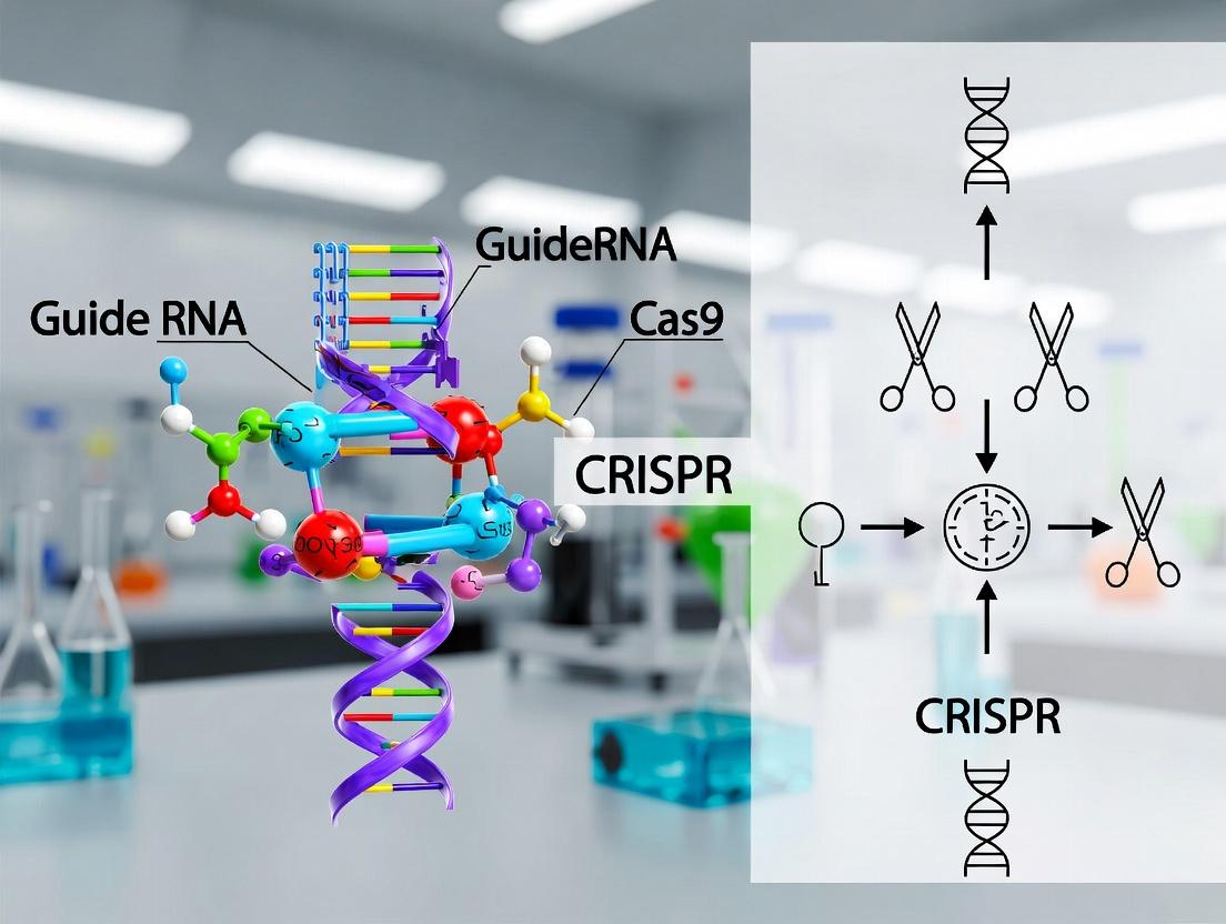
This comprehensive guide details the critical strategies for identifying and reducing CRISPR-Cas off-target effects, a primary safety concern for research and therapeutic applications.
Minimizing CRISPR Off-Target Effects: A 2024 Guide to Screening Strategies, Best Practices, and Clinical Relevance
Abstract
This comprehensive guide details the critical strategies for identifying and reducing CRISPR-Cas off-target effects, a primary safety concern for research and therapeutic applications. We explore the foundational mechanisms of off-target cleavage, including guide RNA-dependent and -independent events. The article provides a methodological deep dive into cutting-edge in silico prediction tools, high-fidelity Cas variants, and experimental screening techniques like CIRCLE-seq and GUIDE-seq. We cover troubleshooting protocols for assay optimization and data interpretation, followed by a comparative analysis of validation methods and their relative strengths. Aimed at researchers and drug developers, this resource synthesizes current best practices to enhance the precision and safety of genome editing projects.
Understanding the Problem: Mechanisms and Risks of CRISPR Off-Target Effects
Welcome to the Technical Support Center for CRISPR Off-Target Analysis. This resource is designed to help researchers troubleshoot key challenges in defining and mitigating off-target effects within the context of precision genome editing and therapeutic development.
Troubleshooting Guides & FAQs
Q1: Our targeted deep sequencing of predicted off-target sites shows no variants, but whole-genome sequencing (WGS) reveals unexpected editing events. What went wrong? A1: This is a common issue where computational prediction tools (e.g., Cas-OFFinder, CHOPCHOP) may miss valid off-target sites due to sequence complexity or mismatches beyond their search parameters.
- Solution: Implement an unbiased, genome-wide detection method. The current gold-standard protocol is CIRCLE-seq.
- Detailed CIRCLE-seq Protocol:
- Genomic DNA Isolation & Shearing: Extract high-molecular-weight gDNA from edited and control cells. Shear to ~300 bp using a focused-ultrasonicator.
- Circularization: Use ssDNA ligase to circularize sheared DNA fragments. This step removes free ends, preventing the subsequent amplification of linear, non-off-target fragments.
- In Vitro Cleavage: Incubate circularized DNA with the same RNP complex (Cas9+gRNA) used in your cellular experiment. Only DNA containing a functional off-target site will be linearized.
- Exonuclease Digestion: Treat the product with an exonuclease to degrade all remaining circular DNA, enriching for linearized, cleaved fragments.
- Library Prep & Sequencing: Prepare a next-generation sequencing library from the exonuclease-resistant DNA and perform paired-end sequencing.
- Bioinformatics Analysis: Map reads to the reference genome and identify sites with significant enrichment of breakpoints in the RNP-treated sample versus the control.
Q2: We observe high phenotypic noise in our negative control (non-targeting guide) screens, complicating hit identification. How can we reduce this background? A2: High noise often stems from copy-number effects and "passenger effects" from guide RNA activity variance.
- Solution: Utilize an optimized library design and analysis pipeline.
- Reagent Solution: Use a library with multiple independent guides per gene (e.g., 6-10) and a high diversity of negative control guides (at least 1000).
- Analysis Protocol: Process your screen data with the MAGeCK-VISPR pipeline. Specifically, use the Maximum Likelihood Estimation (MLE) algorithm within MAGeCK to model and regress out the variance explained by guide RNA efficiency and copy-number effects using the control guides.
- Key Step: Normalize read counts using the negative control guides and apply a false discovery rate (FDR) correction (Benjamini-Hochberg) on gene-level p-values. Genes are ranked by robust rank aggregation (RRA) scores.
Q3: For a novel Cas nuclease variant (e.g., Hi-Fi Cas9, eSpCas9), how do we empirically determine its off-target profile before committing to a large-scale screen? A3: A tiered validation approach is recommended.
- Solution Workflow:
- In Silico Prediction: Run comprehensive prediction for the wild-type Cas9 using your target sequence. This provides a baseline list of potential risky sites.
- Targeted Validation: Synthesize PCR amplicons covering the top 20-50 predicted sites. Perform an in vitro cleavage assay (T7 Endonuclease I or ICE analysis) using both the wild-type and novel variant RNPs. Quantify cleavage efficiency.
- Unbiased Discovery: Perform DIG-seq (Discovery of In-situ Genome-wide off-targets by sequencing) on a pool of edited cells. This method ligates a biotinylated adaptor in situ to Cas-induced double-strand breaks, allowing their pull-down and sequencing without in vitro cleavage steps, capturing cellular context.
| Method | Principle | Sensitivity | Throughput | Key Limitation | Best Use Case |
|---|---|---|---|---|---|
| Targeted Amplicon Seq | PCR & deep sequencing of predicted sites | Low (≥0.5% indels) | High (Multiplexed) | Relies on predictions; misses novel sites | Validating top predicted sites post-editing. |
| CIRCLE-seq | In vitro cleavage of circularized genomic DNA | Very High (~0.01% indels) | Medium | In vitro context may not reflect cellular state. | Gold-standard for unbiased, comprehensive in vitro off-target profiling. |
| GUIDE-seq | Tag integration at DSB sites in vivo | High (~0.1% indels) | Low to Medium | Requires transfection of a double-stranded tag. | Unbiased detection in cell cultures amenable to transfection. |
| DIG-seq | In situ biotinylation & capture of DSBs | High (~0.1% indels) | Medium | Requires in situ biotinylation steps. | Unbiased detection in primary cells or in vivo samples. |
| WGS | Sequencing of entire genome | Low-Medium (~5-10% indels) | Very Low | Costly; high background noise from natural variants. | Final safety assessment of clonal cell lines for therapy. |
Visualizations
Title: Off-Target Validation Tiered Workflow
Title: CIRCLE-seq Experimental Workflow
The Scientist's Toolkit: Key Research Reagent Solutions
| Item | Function/Description | Example Vendor/Product |
|---|---|---|
| High-Fidelity Cas9 Nuclease | Engineered protein variant with reduced non-specific DNA binding, decreasing off-target cleavage. | Integrated DNA Technologies (IDT): Alt-R Hi-Fi S.p. Cas9. |
| Chemically Modified sgRNA | Synthetic guide RNA with phosphorothioate bonds and 2'-O-methyl modifications; increases stability and can reduce off-target effects. | Synthego: Synthetic CRISPR guide RNAs. |
| T7 Endonuclease I | Enzyme that detects and cleaves mismatched DNA heteroduplexes, enabling quantification of indel frequencies at targeted loci. | New England Biolabs (NEB). |
| Next-Generation Sequencing Kit for Amplicons | Library preparation kit for targeted deep sequencing of PCR amplicons from potential off-target sites. | Illumina: DNA Prep with Enrichment. |
| Genome-Wide sgRNA Library | Lentiviral pooled library for negative selection screens; includes essential gene targeting and non-targeting controls for normalization. | Addgene: Brunello or Brie human genome-wide libraries. |
| Cas9 Electroporation Enhancer | Small molecule added during RNP electroporation to improve delivery efficiency, enabling lower RNP doses and potentially higher specificity. | IDT: Alt-R Cas9 Electroporation Enhancer. |
| Base Editors (e.g., BE4, ABE8e) | Fusion proteins that mediate direct chemical conversion of one base pair to another (C•G to T•A or A•T to G•C) without creating a double-strand break, significantly reducing indel-based off-targets. | Beam Therapeutics (licensed). |
CRISPR Off-Target Troubleshooting Center
Frequently Asked Questions (FAQs)
Q1: My CRISPR-Cas9 knockout screen shows high rates of phenotypic noise. Could this be due to off-target effects, and how can I confirm this?
A: Yes, phenotypic noise or inconsistent results across replicates can be a strong indicator of off-target cleavage. To confirm and diagnose:
- Perform targeted deep sequencing: Design primers for your top predicted off-target sites (using tools like MIT or CHOPCHOP) and for your on-target site. Sequence the genomic DNA from your pooled edited cell population. A significant rate of indels (e.g., >0.5%) at loci other than your intended target is confirmatory.
- Utilize GUIDE-seq or CIRCLE-seq: For an unbiased genome-wide assessment, adopt these experimental methods to identify off-target sites without prior prediction bias.
Q2: I am using a high-fidelity Cas9 variant (e.g., SpCas9-HF1), but I still observe off-target effects in my validation assays. What are the potential reasons?
A: Even high-fidelity variants are not infallible. The issue likely resides in the guide RNA (gRNA) design.
- Excessive guide homology: Your gRNA may have extended sequence homology (≥14 contiguous bases) with other genomic regions, overriding the nuclease's increased specificity.
- High gRNA expression levels: Overexpression from a strong U6 promoter can saturate the high-fidelity Cas9, leading to promiscuous binding. Consider using a weaker promoter or titrating gRNA amounts.
- Mismatch tolerance in the seed region: Re-evaluate your off-target predictions, paying special attention to the 10-12 bases proximal to the PAM. Some high-fidelity mutants retain tolerance for certain mismatches in this critical region.
Q3: What is the most effective strategy to minimize off-target cleavage for in vivo therapeutic development?
A: A multi-layered approach is critical for therapeutics:
- Nuclease Selection: Use engineered ultra-high-fidelity Cas9 variants (e.g., eSpCas9(1.1), HiFi Cas9) or switch to a more intrinsically precise nuclease like Campylobacter jejuni Cas9 (CjCas9).
- gRNA Optimization: Truncate gRNA length (17-18nt instead of 20nt) or incorporate chemical modifications (2'-O-methyl-3'-phosphorothioate) at terminal nucleotides to reduce binding energy and improve specificity.
- Delivery & Dosage Control: Use ribonucleoprotein (RNP) complexes instead of plasmid DNA to limit the duration of nuclease activity. Precisely control the RNP dose to the minimal effective concentration.
- Experimental Validation: Mandatorily use orthogonal off-target detection methods (e.g., combining GUIDE-seq with in silico prediction) before clinical candidate selection.
Troubleshooting Guides
Issue: High Background Noise in a CRISPRi/a Transcriptional Modulation Screen
- Symptoms: Weak or inconsistent gene activation/repression, high variance among sgRNAs targeting the same gene.
- Potential Cause: Off-target transcriptional modulation of genes involved in your phenotypic readout.
- Solution Steps:
- Re-analyze gRNA designs: Filter your library for guides with high off-target potential using updated algorithms (CRISPRoff, CRISPick).
- Employ a dual-sgRNA scoring approach: Only consider phenotypes where at least two independent, well-validated sgRNAs for the same gene show concordant results.
- Validate with orthogonal method: Use a small-molecule inhibitor or RNAi against your top hit to confirm the phenotype is not an artifact of CRISPR off-target effects.
Issue: Inconsistent Editing Outcomes in Clonal Cell Lines
- Symptoms: Genotypic and phenotypic heterogeneity among clones derived from the same transfection.
- Potential Cause: Off-target cleavages inducing varied chromosomal rearrangements or stress responses in different clones.
- Solution Steps:
- Karyotype analysis: Check for large-scale chromosomal abnormalities.
- Whole-genome sequencing (WGS): Perform low-coverage WGS on several aberrant clones to identify common off-target structural variations. This is a cornerstone for rigorous validation in therapeutic development.
- Modify protocol: Use RNP delivery and FACS sorting based on a co-transfected marker to reduce the time window for nuclease activity and isolate a more uniform population.
Table 1: Comparison of Wild-Type and High-Fidelity Cas9 Variants
| Nuclease | On-Target Efficiency (Relative to WT) | Off-Target Frequency Reduction (Fold) | Key Mechanism | Primary Use Case |
|---|---|---|---|---|
| SpCas9 (WT) | 100% (Baseline) | 1x (Baseline) | N/A | Initial screens, non-critical edits |
| SpCas9-HF1 | 60-80% | 10-100x | Weakened non-specific DNA contacts | Biochemical studies, sensitive backgrounds |
| eSpCas9(1.1) | 70-90% | 10-100x | Reduced non-specific electrostatic interactions | In vivo models, functional genomics |
| HiFi Cas9 | 80-95% | 50-200x | Altered DNA binding interface | Therapeutic development, clinical applications |
| CjCas9 | 40-70% | >100x (due to longer PAM) | Extended PAM requirement (NNNNRYAC) | Targets in dense genomic regions |
Table 2: Impact of Guide RNA Mismatches on Cleavage Efficiency
| Mismatch Position (5' → 3')* | Number of Mismatches | Average Cleavage Efficiency | Notes |
|---|---|---|---|
| Distal (positions 1-10) | 1-3 | 50-90% | Often tolerated, especially at PAM-distal end. |
| Seed (positions 11-20) | 1 | 10-40% | Drastic reduction, but not absolute. |
| Seed (positions 11-20) | 2-3 | <1% | Near-total ablation for most guides. |
| PAM-proximal (positions 16-20) | 1 (G-C, A-T) | 5-20% | Highly disruptive, but some non-canonical pairs may allow cleavage. |
Position 1 is the first base 5' of the spacer sequence. *Relative to perfect match guide. Data dependent on specific sequence context.
Experimental Protocols
Protocol 1: GUIDE-seq for Unbiased Off-Target Detection Principle: A short, double-stranded oligonucleotide tag is integrated into double-strand breaks (DSBs) generated during CRISPR editing, allowing for genome-wide amplification and sequencing of off-target sites. Materials: GUIDE-seq dsODN, transfection reagent, PCR reagents, next-generation sequencing platform. Methodology:
- Co-transfect cells with your Cas9/gRNA RNP (or expression plasmids) and the GUIDE-seq dsODN.
- Harvest genomic DNA 48-72 hours post-transfection.
- Perform tag-specific PCR to amplify genomic regions flanking integrated dsODN tags.
- Prepare sequencing library from PCR products and sequence using Illumina platforms.
- Analyze data using the published GUIDE-seq computational pipeline to map all DSB sites.
Protocol 2: CIRCLE-seq for In Vitro Off-Target Profiling Principle: Genomic DNA is circularized, digested with Cas9-gRNA complex in vitro, and linearized fragments (containing off-target cuts) are selectively sequenced. Materials: Purified genomic DNA, Cas9 nuclease, in vitro transcribed gRNA, DNA circularization enzymes, exonuclease. Methodology:
- Shear and circularize genomic DNA using a circligase.
- Treat circularized DNA with your Cas9-gRNA RNP complex to cleave at recognized sites.
- Digest with exonuclease to degrade all linear DNA (uncut circles remain).
- Fragment the nicked circles (from Cas9 cleavage) and prepare a next-generation sequencing library.
- Sequence and analyze to identify all potential Cas9 recognition sequences in the genome, ranked by cleavage efficiency.
Visualizations
Title: Off-Target Effect Diagnosis and Mitigation Workflow
Title: Guide RNA Mismatch Tolerance by Region
The Scientist's Toolkit: Research Reagent Solutions
| Item | Function & Rationale |
|---|---|
| High-Fidelity Cas9 Nuclease (e.g., Alt-R HiFi Cas9) | Engineered protein with dramatically reduced non-specific DNA binding, lowering off-target effects while maintaining robust on-target activity. Essential for sensitive applications. |
| Chemically Modified sgRNA (2'-O-methyl, 3' phosphorothioate) | Increases nuclease resistance and decreases immune activation. Terminal modifications can reduce off-target binding affinity, improving specificity. |
| GUIDE-seq Oligo Duplex | A short, blunt, double-stranded DNA oligo used as a tag for genome-wide, unbiased identification of CRISPR-Cas9 off-target cleavage sites. |
| Recombinant Cas9 Protein (for RNP formation) | Allows for transient, dose-controlled delivery of the nuclease. RNP complexes clear faster than plasmid DNA, reducing the window for off-target activity. |
| In Vitro Transcribed gRNA or Synthetic crRNA/tracrRNA | Provides flexibility in gRNA source. Synthetic RNAs offer high purity and the ability to incorporate precise chemical modifications. |
| Off-Target Prediction Software Subscription (e.g., Benchling, IDT) | Cloud-based platforms with constantly updated algorithms to design gRNAs with minimal predicted off-target effects across relevant genomes. |
| Positive Control Plasmid (e.g., with a known problematic off-target site) | Used to validate the sensitivity of your off-target detection methods in your specific cell line or model system. |
Technical Support & Troubleshooting Center
This support center is designed to assist researchers working on CRISPR screen off-target effect reduction, providing solutions to common experimental challenges related to the intrinsic fidelity of Cas9 and Cas12 nucleases.
Frequently Asked Questions (FAQs)
Q1: In our off-target validation assay for a SpCas9 guide, we observe significant cleavage at a site with a single-nucleotide mismatch at the PAM-distal end. This contradicts the established model that PAM-distal mismatches are well-tolerated. What could be causing this? A1: The established model is a general rule, but sequence context is critical. Certain "strong" mismatches, like a G:U wobble pair or a transversion mismatch in a high-GC context, can be tolerated even at PAM-distal positions. Furthermore, secondary DNA structure or protein co-factors in your cellular extracts may influence binding. We recommend:
- Perform an in vitro cleavage assay with purified Cas9 protein and synthetic DNA targets to isolate intrinsic nuclease behavior from cellular factors.
- Use a comprehensive mismatch tolerance profiling method like GUIDE-seq or CIRCLE-seq for that specific gRNA to map all potential off-target sites empirically.
- Check for seed region (positions 1-12) fidelity; a perfectly matched seed region with a distal mismatch can sometimes still permit cleavage.
Q2: For high-fidelity Cas12a (e.g., LbCas12a) screening, we are getting very low editing efficiency at our on-target site, undermining our screen's dynamic range. How can we improve on-target activity while maintaining specificity? A2: Low on-target efficiency is a common trade-off with high-fidelity variants. Please troubleshoot using this protocol:
- Protocol: Optimizing Cas12a RNP Delivery for High-Fidelity Screening
- gRNA Design: Verify your gRNA is the optimal length (typically 20-24 nt for Cas12a). Use a pre-validated design tool (e.g., from IDT or Benchling) and try 2-3 alternative gRNAs targeting the same locus.
- RNP Complex Formation: Increase the molar ratio of gRNA to Cas12a protein during ribonucleoprotein (RNP) complex assembly. Try ratios from 2:1 to 4:1 (gRNA:Cas12a). Incubate at 37°C for 10-20 minutes before delivery.
- Delivery: For electroporation, titrate the RNP concentration (e.g., from 2 µM to 10 µM final) and optimize pulse parameters. For lipofection, ensure you are using a reagent validated for RNP delivery.
- PAM Verification: Cas12a requires a T-rich PAM (TTTV). Ensure your target site has a canonical PAM and test sites with alternative T-rich PAMs (e.g., TTTV vs. TTCV).
Q3: When analyzing NGS data from a Cas12 off-target assessment (using SITE-Seq), how do we distinguish true, nuclease-dependent off-target sites from background genomic noise? A3: Implement the following bioinformatics and experimental pipeline:
- Bioinformatic Filtering: Set a minimum read-count threshold (e.g., ≥ 10 reads supporting a cut site). Normalize counts to reads per million (RPM). Use a matched "no nuclease" control sample and filter out any sites present in that control.
- Experimental Validation: For candidate off-target sites above your threshold, perform targeted amplicon sequencing across that locus from the original treated sample. Confirm indel frequency above background (typically >0.1%).
- Rule of Thumb: True off-targets usually show a characteristic signature: a sharp peak of sequence alignments centered on the predicted cut site (3-18 nt downstream of PAM for Cas12a).
Q4: We are transitioning from SpCas9 to a high-fidelity variant (e.g., SpCas9-HF1). Should we completely re-optimize our gRNA design parameters and delivery conditions? A4: Yes, a partial re-optimization is strongly advised. High-fidelity mutants often have altered kinetics. Key changes:
- gRNA Design: Prioritize gRNAs with higher on-target binding energy (use CFD or other scoring metrics). The tolerance for mismatches is drastically reduced, so in silico off-target predictions are more reliable.
- Delivery: Because these enzymes are less promiscuous, they may cleave slower. Consider increasing the concentration of the RNP or the time between transfection and analysis. A time-course experiment (e.g., harvest cells at 48h, 72h, 96h post-transfection) is recommended to find the peak editing window.
Quantitative Comparison of Fidelity Profiles
Table 1: Mismatch Tolerance and Cleavage Kinetics
| Parameter | SpyCas9 (WT) | SpCas9-HF1 / eSpCas9(1.1) | AsCas12a (LbCas12a) | AsCas12a Ultra (High-Fidelity Variant) |
|---|---|---|---|---|
| PAM Sequence | NGG (or NAG) | NGG | TTTV (V=A/C/G) | TTTV |
| Mismatch Tolerance | High. Tolerates up to 5+ mismatches, especially in PAM-distal region. | Severely Reduced. Tolerates typically ≤2 mismatches. | Moderate. Tolerant in PAM-distal region, but less than SpCas9. | Very Low. Extreme sensitivity to mismatches. |
| Primary Mismatch-Sensitive Region | Seed region (PAM-proximal 10-12 bases). | Entire guide RNA-target DNA heteroduplex. | PAM-proximal seed region and direct repeat sequence. | Entire guide-target interface. |
| Relative On-target Kcat | 1.0 (Reference) | 0.2 - 0.5x | ~0.8x (LbCas12a) | ~0.3 - 0.6x |
| Relative Off-target Kcat | 1.0 (Reference) | <0.1x | ~0.2x | <0.05x |
| Key Fidelity Mechanism | Excess energy provided by non-catalytic DNA-binding domains. | Mutations (N497A/R661A/Q695A/Q926A) disrupt non-specific contacts, raising energy penalty for mismatches. | RuvC nuclease domain activation requires stringent seed recognition. | Engineered mutations increase dependency on perfect guide-target complementarity for catalytic activation. |
Table 2: Guide RNA Design & Experimental Considerations
| Feature | Cas9 Systems | Cas12 Systems |
|---|---|---|
| Guide RNA Structure | Two-part: crRNA (targeting) + tracrRNA (scaffold), often fused as sgRNA. | Single, short crRNA (∼40 nt). No tracrRNA needed. |
| Optimal Target Length | 20 nucleotides (for SpCas9). | 20-24 nucleotides (longer can increase specificity for Cas12a). |
| Cleavage Pattern | Blunt ends, cut 3 bp upstream of PAM. | Staggered ends (5' overhang), cut 18-23 nt downstream of PAM. |
| Best Practice for Specificity Screening | Use Digenome-seq or GUIDE-seq for genome-wide profiling. High-fidelity variants make BLISS or SITE-Seq more effective. | Use SITE-Seq or CIRCLE-seq; these in vitro methods work well with Cas12's trans-cleavage property. |
| Common Specificity Challenge | PAM-distal mismatches can be tolerated, leading to numerous, unpredictable off-targets. | Secondary structure in the long crRNA direct repeat can impair on-target activity, leading to overestimation of specificity. |
Experimental Protocols
Protocol 1: In Vitro Mismatch Tolerance Profiling using Gel-Based Cleavage Assay Purpose: To quantitatively compare the intrinsic fidelity of Cas9 and Cas12 nucleases against a series of mismatched target DNA substrates. Materials: Purified Cas nuclease (WT and HiFi variants), synthetic gRNAs/crRNAs, fluorophore-quencher labeled double-stranded DNA substrates (on-target and 1-5 mismatched variants), reaction buffer. Method:
- Form RNP complexes by incubating 100 nM nuclease with 120 nM gRNA in reaction buffer for 10 min at 25°C.
- Start the reaction by adding DNA substrate to a final concentration of 50 nM.
- Aliquot reactions at time points (e.g., 0, 1, 5, 15, 30, 60 min) into a stop solution (EDTA/formamide).
- Denature samples and run on a high-resolution denaturing PAGE gel.
- Quantify cleaved vs. uncleaved substrate using fluorescence imaging. Calculate reaction velocities (v) for each substrate.
- Plot v(mismatch) / v(on-target) to generate a comparative mismatch tolerance profile.
Protocol 2: High-Throughput Specificity Verification using Dual Reporter System (HEK293T Cells) Purpose: To rapidly screen gRNA specificity for Cas9 vs. Cas12 in a cellular context. Materials: Dual-luciferase reporter plasmids (one with perfectly matched target site driving Firefly luciferase, another with a suspected off-target site driving Renilla luciferase), Cas9/Cas12 expression plasmids, gRNA expression plasmids, transfection reagent, dual-luciferase assay kit. Method:
- Co-transfect HEK293T cells in a 96-well plate with: a) Cas expression plasmid, b) gRNA plasmid, c) matched reporter plasmid, d) mismatched reporter plasmid.
- Include controls: No nuclease, nuclease without gRNA.
- At 48-72 hours post-transfection, lyse cells and perform dual-luciferase assay.
- Normalize Firefly and Renilla luminescence to respective controls.
- Specificity Score: Calculate the ratio: (Normalized Renilla Signal / Normalized Firefly Signal). A lower score indicates higher specificity (less off-target cleavage).
Visualizations
Diagram 1: Cas9 vs Cas12 Mismatch Sensitivity Workflow
Diagram 2: CRISPR Off-Target Validation Pathway for Research
The Scientist's Toolkit: Essential Research Reagents
Table 3: Key Reagents for Fidelity Profiling Experiments
| Reagent / Solution | Function in Experiment | Example Vendor/Product |
|---|---|---|
| High-Fidelity Nuclease Variants | Engineered proteins (e.g., SpCas9-HF1, eSpCas9, HiFi Cas12a) with reduced off-target activity while retaining robust on-target cleavage. | IDT Alt-R S.p. HiFi Cas9 Nuclease; ToolGen Wild-type & High-Fidelity AsCas12a. |
| Chemically Modified Synthetic gRNAs | Incorporation of 2'-O-methyl and phosphorothioate modifications at terminal nucleotides increases stability, RNP formation efficiency, and can reduce immunogenicity in cells. | Synthego CRISPR 2.0 gRNAs; IDT Alt-R Modified crRNAs. |
| Genome-Wide Off-Target Discovery Kits | All-in-one kits for methods like GUIDE-seq or CIRCLE-seq, providing optimized adapters, enzymes, and buffers for library preparation and NGS. | NEB GUIDE-seq Kit; CIRCLE-seq protocol reagents (commercially available components). |
| In Vitro Cleavage Assay Substrates | Fluorescently labeled (e.g., FAM/IBFQ) double-stranded DNA oligonucleotides containing matched and mismatched target sequences for precise kinetic measurements. | Custom synthesis from IDT or Eurofins. |
| Dual-Luciferase Reporter Assay Systems | Plasmid-based systems (e.g., pmirGLO backbone adapted) to co-express matched and mismatched targets for rapid, quantitative specificity screening in cell culture. | Promega Dual-Luciferase kits with custom reporter constructs. |
| Next-Generation Sequencing (NGS) Library Prep Kits for Amplicons | High-sensitivity kits (e.g., for Illumina) to prepare sequencing libraries from PCR amplicons of putative off-target loci for deep sequencing and indel analysis. | Illumina DNA Prep; Takara Bio SMARTer Amplicon kits. |
Technical Support Center: Troubleshooting CRISPR Screen Off-Target Effects
FAQs and Troubleshooting Guides
Q1: My CRISPR screen shows unexpectedly high rates of phenotypes in negative control cells. What are the primary gRNA-dependent off-target sources I should investigate? A: High background in controls often points to gRNA-dependent off-target effects. The key sources are:
- Sequence Homology: Mismatches and bulges in gRNA-DNA pairing, especially outside the seed region (positions 1-12 from PAM). Even 1-5 mismatches can permit cleavage.
- PAM Promiscuity: The Cas enzyme (e.g., SpCas9) may tolerate non-canonical PAM sequences (e.g., NAG, NGA instead of NGG for SpCas9), though at lower efficiency.
- Chromatin Accessibility: Open chromatin regions are more susceptible to off-target binding and cleavage.
Q2: I observe cytotoxic effects even in non-targeting control conditions. What gRNA-independent mechanisms could be at play? A: Cytotoxicity unrelated to gRNA sequence indicates gRNA-independent events:
- Cas9/12a/13d Protein Toxicity: Persistent, high-level expression of the nuclease can trigger DNA damage response (p53 activation) or innate immune responses.
- Delivery Vehicle Toxicity: High viral titer (for lentiviral delivery) or lipid nanoparticle (LNP) cytotoxicity can cause stress responses.
- Off-Target DNA/RNA Sensing: Non-specific nucleic acid binding can activate intracellular sensors like cGAS-STING (for DNA) or RIG-I/MDA5 (for RNA).
Q3: What are the most effective experimental methods to identify and quantify off-target effects in a pooled screen? A: The optimal method depends on your screen type. See Table 1 for a comparison.
Table 1: Quantitative Comparison of Major Off-Target Detection Methods
| Method | Primary Target | Detection Principle | Approx. Sensitivity (Variant Allele Frequency) | Key Advantage | Key Limitation |
|---|---|---|---|---|---|
| GUIDE-seq | DNA Breaks | Integration of double-stranded oligodeoxynucleotides at break sites | ~0.1% | Unbiased, genome-wide | Requires specialized oligo delivery; lower sensitivity for rare events. |
| CIRCLE-seq | DNA Breaks | In vitro cleavage of genomic DNA circles followed by sequencing | ~0.01% | Highly sensitive, no cellular context required | Purely in vitro; may not reflect chromatin state. |
| SITE-seq | DNA Breaks | In vitro Cas9 cleavage of genomic DNA fragments | ~0.1% | Sensitive, uses native chromatin | Complex protocol. |
| Digenome-seq | DNA Breaks | In vitro Cas9 digestion of whole genomic DNA followed by sequencing | ~0.1% | Comprehensive, computational analysis | High sequencing depth required; in vitro. |
| ONE-Seq (BLISS) | DNA Breaks | Direct labeling and sequencing of double-strand breaks | ~Single-cell | Can be used in situ and in single cells | Technically challenging. |
Q4: How can I minimize both gRNA-dependent and gRNA-independent off-targets in my experimental design? A: Employ a multi-pronged strategy:
- For gRNA-Dependent: Use high-fidelity Cas variants (e.g., SpCas9-HF1, eSpCas9(1.1)), truncated gRNAs (tru-gRNAs, 17-18nt), and predictive algorithms (e.g., CRISPOR, ChopChop) to select guides with minimal predicted off-targets.
- For gRNA-Independent: Use inducible expression systems (e.g., doxycycline-inducible) to limit Cas protein exposure time. Titrate delivery vectors (viral MOI, LNP concentration) to the minimum effective dose. Consider using dead-Cas (dCas) fused to base editors or prime editors, which have different off-target profiles.
Detailed Experimental Protocols
Protocol 1: Off-Target Validation Using GUIDE-seq Purpose: To empirically identify genome-wide off-target sites for a given gRNA in living cells. Materials: See "The Scientist's Toolkit" below. Method:
- Cell Preparation: Seed 500,000 HEK293T cells (or target cell line) per well in a 6-well plate.
- Co-transfection: At 60-80% confluency, co-transfect cells with:
- 500 ng plasmid expressing SpCas9 (or variant).
- 250 ng plasmid expressing your gRNA of interest.
- 100 pmol of GUIDE-seq oligo duplex (see Toolkit). Use a standard transfection reagent (e.g., Lipofectamine 3000).
- Harvest Genomic DNA: 72 hours post-transfection, harvest cells and extract genomic DNA using a silica-column-based kit.
- Library Preparation & Sequencing:
- Shear 1 µg genomic DNA to ~500 bp fragments.
- Perform end-repair, A-tailing, and ligation of Illumina sequencing adapters.
- Perform two nested PCRs using primers specific to the GUIDE-seq oligo and the Illumina adapters to enrich for integration events.
- Purify the final PCR product and sequence on an Illumina platform (2x150 bp recommended).
- Data Analysis: Use the published GUIDE-seq software suite to align reads, detect integration sites, and call significant off-target loci.
Protocol 2: Assessing Cellular Toxicity from Cas9 Expression (gRNA-Independent) Purpose: To quantify the impact of Cas9 delivery alone on cell health and proliferation. Materials: Cell line of interest, lentivirus for Cas9-only expression (no gRNA), empty vector control virus, puromycin (or appropriate selection agent), cell viability assay kit (e.g., CellTiter-Glo). Method:
- Viral Transduction: Infect target cells with a range of MOIs (e.g., 0.5, 1, 2, 5) of Cas9-only virus or control virus. Include an uninfected control.
- Selection: 48 hours post-infection, begin puromycin selection for 5-7 days to eliminate untransduced cells.
- Long-Term Growth Curve: After selection, re-seed equal numbers of cells from each condition into 12-well plates. Every 2-3 days for 2 weeks, trypsinize and count cells from triplicate wells. Plot population doublings over time.
- Endpoint Viability Assay: At the end of the growth curve, perform a CellTiter-Glo assay according to the manufacturer's protocol to measure ATP content as a proxy for metabolically active cells.
- Analysis: Compare the growth rates and final viability between Cas9-expressing and control cells. A significant decrease indicates gRNA-independent toxicity.
Visualizations
Title: gRNA-Dependent Off-Target Mechanisms
Title: gRNA-Independent Toxicity Pathways
The Scientist's Toolkit: Key Research Reagent Solutions
Table 2: Essential Materials for Off-Target Analysis Experiments
| Item | Function in Off-Target Research | Example Product/Catalog |
|---|---|---|
| High-Fidelity Cas9 Variant | Reduces gRNA-dependent off-target cleavage by weakening non-specific DNA interactions. | SpCas9-HF1 plasmid (Addgene #72247) |
| Truncated gRNA (tru-gRNA) Scaffold | 17-18nt guide sequence reduces off-target binding while often maintaining on-target activity. | pRG2 (tru-gRNA) backbone (Addgene #104174) |
| GUIDE-seq Oligo Duplex | Double-stranded, end-protected oligo that integrates into DNA breaks for genome-wide off-target discovery. | Custom synthesized (e.g., IDT): 5′-/5Phos/GTGTCAGTCACTTCCAGTTTA-3′, with complementary strand. |
| CIRCLE-seq Adapter | Specialized adapter for circularizing sheared genomic DNA for in vitro Cas9 digestion assays. | Circligase ssDNA Ligase & accompanying buffer (Lucigen). |
| Inducible Cas9 Expression System | Limits Cas9 exposure time to reduce gRNA-independent toxicity (e.g., Doxycycline-inducible). | pCW-Cas9 (Addgene #50661) |
| Cas9 Protein, Nuclease-Free | For in vitro validation assays (e.g., T7E1, Sanger sequencing) of suspected off-target sites. | Recombinant SpCas9 Nuclease (NEB #M0386) |
| Off-Target Prediction Software | Computational tool to design gRNAs with minimal predicted off-targets. | CRISPOR (crispor.tefor.net) |
| Sensitive DNA Cleavage Detection Kit | For validating individual suspected off-target sites (e.g., via mismatch detection). | T7 Endonuclease I (NEB #M0302) or ICE Analysis Tool (Synthego). |
Troubleshooting Guide & FAQs
Q1: Our CRISPR screen validation shows unexpected phenotypic hits. How do we determine if these are due to off-target effects? A: Unexpected hits are a primary symptom of off-target activity. Follow this protocol:
- Off-Target Prediction: Use updated bioinformatics tools (e.g., CRISPOR or CCTop) with the latest reference genome to predict potential off-target sites for your sgRNA.
- Site-Specific Validation:
- PCR Amplification: Design primers flanking the top 5-10 predicted off-target loci and the on-target site.
- Next-Generation Sequencing (NGS): Prepare amplicon libraries and sequence at high depth (>100,000x coverage).
- Analysis: Use a pipeline like CRISPResso2 to quantify insertion/deletion (indel) frequencies at each site. An indel frequency >0.5% at a predicted off-target site is commonly considered significant.
Q2: Our negative control (non-targeting sgRNA) samples show high toxicity or phenotypic effects in our cell viability screen. What could be the cause? A: This indicates a potential sequence-independent, off-target immune response or reagent toxicity.
- Troubleshooting Steps:
- Check sgRNA Design: Ensure your "non-targeting" control has no significant homology to the genome (BLAST it).
- Monitor Innate Immune Activation: Transfert cells with your CRISPR RNP (Cas9 + sgRNA) complex and a control (e.g., transfection reagent alone). 24 hours post-transfection, harvest RNA and perform qPCR for interferon-stimulated genes (ISGs) like IFIT1 or ISG15. Elevated levels indicate immune activation.
- Titrate Reagents: High concentrations of Cas9 protein or transfection reagent can cause cytotoxicity. Perform a dose-response experiment to find the minimum effective concentration.
Q3: How can we improve the reproducibility of our CRISPR knockout screens between biological replicates? A: Inconsistent off-target effects are a major source of irreproducibility.
- Solutions:
- Use High-Fidelity Cas9 Variants: Employ engineered Cas9 nucleases like SpCas9-HF1 or eSpCas9(1.1) which have reduced off-target activity while maintaining on-target efficiency.
- Implement Duplex sgRNAs: Use two sgRNAs (tracrRNA + crRNA) instead of a single-guide RNA (sgRNA). This format, especially when using Alt-R modified synthetic guides, can improve specificity.
- Employ Controlled Delivery: Use electroporation (e.g., Neon or Amaxa systems) instead of lipid-based transfection for more consistent delivery across replicates, especially in hard-to-transfect cells.
Q4: We observe different mutation patterns at the predicted off-target site compared to the intended target. Why does this happen? A: Mutation spectra are influenced by local sequence context and chromatin accessibility. This is expected but complicates prediction.
- Protocol for Characterization:
- Clone the PCR-amplified off-target locus from a pool of edited cells into a plasmid vector.
- Sequence 50-100 individual bacterial colonies.
- Categorize the exact indel sequences. You will often find a different distribution of deletions vs. insertions compared to the on-target site. Documenting this is crucial for understanding the potential functional impact of the off-target edit.
Data compiled from recent comparative studies (2023-2024).
Table 1: Comparison of Cas9 Nuclease Specificity
| Cas9 Nuclease | Relative On-Target Efficiency (%) | Reduction in Off-Target Activity vs. Wild-Type SpCas9 | Primary Use Case |
|---|---|---|---|
| Wild-Type SpCas9 | 100 | 1x (Baseline) | General use where ultimate on-target efficiency is critical and off-targets are screened for. |
| SpCas9-HF1 | 85-95 | 10-100x | High-specificity knockout screens and potential therapeutic applications. |
| eSpCas9(1.1) | 80-90 | 10-100x | Similar to HF1; choice often depends on specific guide sequence performance. |
| HypaCas9 | 70-80 | >100x | Applications requiring the highest possible specificity, accepting a moderate efficiency trade-off. |
| evoCas9 | 60-75 | >1000x | Ultra-specific editing for sensitive models (e.g., stem cells, clinical precursors). |
Table 2: Common Assays for Off-Target Detection
| Assay Name | Method | Detection Limit | Throughput | Key Advantage |
|---|---|---|---|---|
| GUIDE-seq | Integration of dsODN tags at DSB sites, followed by NGS. | ~0.1% | Low | Unbiased genome-wide discovery. |
| CIRCLE-seq | In vitro cleavage of genomic DNA circles, followed by NGS. | ~0.01% | Medium | Highly sensitive, cell-context independent. |
| SITE-Seq | In vitro cleavage of genomic DNA fragments, followed by NGS. | ~0.1% | Medium | Uses native chromatin structure. |
| Digenome-seq | In vitro cleavage of cell-free genomic DNA, followed by whole-genome sequencing. | ~0.1% | High | Comprehensive, but high cost and computational load. |
| Targeted Amplicon-Seq | PCR & deep sequencing of predicted off-target sites. | ~0.1-0.5% | High | Cost-effective for validating predicted sites. |
Experimental Protocols
Protocol 1: Off-Target Validation via Targeted Amplicon Sequencing Purpose: To quantify indel frequencies at predicted off-target loci. Materials: See "The Scientist's Toolkit" below. Steps:
- Genomic DNA Extraction: Harvest edited and control cells. Use a column-based gDNA extraction kit. Elute in nuclease-free water. Quantify via spectrophotometer.
- Primer Design & PCR: Design nested PCR primers for on-target and each off-target locus (amplicon size: 250-350 bp). Perform a first-round PCR (15 cycles) with outer primers. Use 1 µL of this product as template for a second PCR (20 cycles) with inner primers containing full Illumina adapter overhangs.
- Library Purification & Quantification: Clean PCR products with SPRI beads. Quantify using a fluorometric assay.
- Pooling & Sequencing: Equimolar pool of all amplicons. Sequence on an Illumina MiSeq or NovaSeq platform (2x250 bp or 2x300 bp chemistry recommended).
- Data Analysis: Demultiplex reads. Align to reference amplicon sequences using CRISPResso2 (default parameters). Report indel percentages for each site.
Protocol 2: Assessing Immune Activation by RNP Transfection Purpose: To measure sequence-independent, off-target interferon response. Steps:
- Cell Seeding & Transfection: Seed HEK293T or relevant cell line in a 24-well plate. The next day, complex 2 µg of Alt-R S.p. HiFi Cas9 protein with 2 µL of 100 µM synthetic sgRNA (targeting or non-targeting) in buffer. Add transfection reagent per manufacturer's instructions. Include a transfection-reagent-only control.
- RNA Harvest: 24 hours post-transfection, lyse cells directly in the well with 500 µL of TRIzol reagent. Isolate total RNA following the standard phase-separation protocol.
- cDNA Synthesis: Use 1 µg of total RNA for reverse transcription with a kit using random hexamers.
- qPCR: Prepare SYBR Green qPCR reactions with primers for IFIT1, ISG15, and a housekeeping gene (e.g., GAPDH). Run in triplicate.
- Analysis: Calculate ∆∆Ct values relative to the transfection-reagent-only control. A >2-fold increase in ISG expression indicates immune activation.
Visualizations
Title: Off-Target Analysis Workflow
Title: Off-Target Immune Response Pathway
The Scientist's Toolkit: Key Research Reagent Solutions
| Item | Function & Rationale |
|---|---|
| Alt-R S.p. HiFi Cas9 Nuclease V3 | Engineered high-fidelity Cas9 protein. Function: Significantly reduces off-target cleavage while maintaining robust on-target activity for more reproducible results. |
| Alt-R CRISPR-Cas9 Synthetic sgRNA (modified) | Chemically synthesized guide RNA with proprietary modifications. Function: Increases stability and reduces immune activation compared to in vitro transcribed (IVT) guides. |
| Alt-R HDR Enhancer V2 | Small molecule inhibitor of non-homologous end joining (NHEJ). Function: Improves homology-directed repair (HDR) efficiency in knock-in experiments, allowing use of lower, more specific RNP doses. |
| NEON Electroporation System & Kits | Electroporation-based delivery system. Function: Provides highly efficient and consistent delivery of RNP complexes into a wide range of cell types (including primary cells), minimizing reagent-dependent toxicity. |
| IDT xGen Amplicon Library Prep Kit | Library preparation for targeted sequencing. Function: Streamlined, high-fidelity PCR-based workflow for preparing NGS libraries from amplicons for off-target validation sequencing. |
| CRISPResso2 Software (Broad Institute) | Bioinformatics analysis tool. Function: Precisely quantifies genome editing outcomes from NGS data, providing clear metrics on indel percentages and types for on- and off-target sites. |
Strategic Toolkit: Proactive Methods for Off-Target Prediction and Reduction
Technical Support Center: Troubleshooting Guides & FAQs
FAQ 1: My designed sgRNA has a high predicted on-target score in one tool but a low score in another. Which should I trust? Answer: Discrepancies arise from different algorithms and training data. CRISPOR uses multiple scoring systems (e.g., Doench '16, Moreno-Mateos). CHOPCHOP primarily uses the Doench '16 efficiency score. Always consult multiple tools. For a CRISPR screen focused on off-target reduction, prioritize sgRNAs where both tools agree on high efficiency, then apply stringent off-target filtering.
FAQ 2: Cas-OFFinder returns thousands of potential off-target sites. How do I determine which are biologically relevant? Answer: Not all predicted off-targets are equal. Filter and prioritize using this protocol:
- Set a strict cutoff (e.g., ≤3 mismatches for SpCas9).
- Cross-reference with CRISPOR's off-target list, which includes genomic context (exonic, intronic, intergenic) and conservation scores.
- Rank off-targets by: a) Number of mismatches, b) Position of mismatches (PAM-distal worse), c) Presence in a coding or conserved region.
- For critical sgRNAs, experimentally validate the top 5-10 ranked sites.
FAQ 3: I need to design a genome-wide CRISPR knockout screen for my thesis on off-target reduction. What is the recommended in silico workflow? Answer: Follow this detailed methodology to maximize on-target and minimize off-target effects:
- Generate Candidates: Input your target gene list into CHOPCHOP for rapid, batch sgRNA generation.
- Comprehensive Scoring: Feed the candidate list into CRISPOR. Extract the Doench '16 and Moreno-Mateos '17 efficiency scores.
- Off-Tinterrogation: For each sgRNA, run Cas-OFFinder with parameters: Genomic sequence, mismatch number (e.g., 3-4), and your specific PAM (e.g., NGG for SpCas9).
- Final Selection: Apply the selection criteria in Table 1.
Table 1: sgRNA Selection Criteria for Off-Target Reduction Screens
| Criterion | Target Threshold | Rationale |
|---|---|---|
| On-Target Efficiency | Doench '16 score ≥ 50 | Ensures high knockout probability. |
| Specificity Score | CRISPOR CFD specificity score ≥ 50 | Higher score indicates lower predicted off-target activity. |
| Top Off-Target Hit | ≥3 mismatches | Minimizes risk of cleavage at the most likely off-target site. |
| Number of Off-Targets with 0-1 mismatches | 0 | Eliminates sgRNAs with near-perfect matches elsewhere in the genome. |
Workflow for sgRNA Design and Selection
FAQ 4: How do I handle predictions for non-canonical PAMs or engineered Cas variants (e.g., SpCas9-NG)? Answer: Tool capabilities vary. CRISPOR supports many variants (SpCas9-NG, xCas9, SpG, SpRY) in its advanced options. Cas-OFFinder allows you to input any user-defined PAM sequence via its "Pattern" field (e.g., "NG" for SpCas9-NG). CHOPCHOP has limited support for some variants—always verify in the tool's documentation. Consistency across tools may be lower for non-NGG PAMs, so empirical validation is even more critical.
Table 2: Tool Capabilities for Common Cas Variants
| Cas Variant | PAM Sequence | CRISPOR | CHOPCHOP | Cas-OFFinder |
|---|---|---|---|---|
| SpCas9 (Wild-type) | NGG | Full Support | Full Support | Use Pattern: NGG |
| SpCas9-NG | NG | Full Support | Limited Support | Use Pattern: NG |
| SpG | NGN | Full Support | No Support | Use Pattern: NGN |
| SpRY | NRN > NYN | Full Support | No Support | Use Pattern: N[AG]N or N[CT]N |
The Scientist's Toolkit: Research Reagent Solutions
| Item | Function in In Silico Prediction & Validation |
|---|---|
| CRISPOR.org Website/Command Line Tool | The central hub for integrating multiple on-target efficiency scores and curated off-target predictions with genomic annotations. |
| CHOPCHOP (Web or Python API) | Rapid, user-friendly batch design of sgRNAs for single genes or whole genomes. |
| Cas-OFFinder (Local Executable) | Flexible, high-speed genome-wide search for DNA sequences similar to your sgRNA, allowing full control over PAM and mismatch parameters. |
| Reference Genome FASTA File (e.g., hg38) | Essential local file for running Cas-OFFinder and other command-line tools. Ensures your predictions match your experimental genome build. |
| UCSC Genome Browser / IGV | Visualize predicted off-target sites within genomic context (genes, conserved regions, chromatin state) to assess potential functional impact. |
| Next-Generation Sequencing (NGS) Platform | Required for experimental validation of predictions via methods like GUIDE-seq or targeted deep sequencing of putative off-target loci. |
Pathway from Prediction to Experimental Validation
Technical Support Center: Troubleshooting High-Fidelity Cas9 Experiments
This support center is designed to assist researchers integrating high-fidelity Cas9 variants into their CRISPR screens, within the broader thesis context of systematically reducing off-target effects for more reliable therapeutic and functional genomics outcomes.
Frequently Asked Questions (FAQs)
Q1: Our SpCas9-HF1 screen shows significantly reduced editing efficiency at our intended on-target sites compared to wild-type SpCas9. What could be the cause and how can we mitigate this? A: This is a common trade-off. SpCas9-HF1 incorporates four mutations (N497A/R661A/Q695A/Q926A) that reduce non-specific interactions with the phosphate backbone of the DNA, increasing specificity but potentially lowering on-target activity with certain sgRNAs.
- Troubleshooting Steps:
- Validate sgRNA Design: Ensure your sgRNAs are 20nt in length. Consider using truncated sgRNAs (17-18nt) for SpCas9-HF1, as they can further reduce off-target effects but may require optimization of on-target efficiency.
- Check Delivery Ratios: Optimize the ratio of your sgRNA and Cas9 expression plasmids or viral titers. A slight increase in sgRNA concentration may help.
- Employ High-Fidelity Specific Enhancers: Co-deliver engineered E. coli RecA or Rad51 nucleoprotein filaments that bind to exposed single-stranded DNA at the Cas9 cleavage site, which have been shown to boost on-target efficiency of high-fidelity variants without compromising specificity.
- Verify Cell Health & Transfection Efficiency: Use a positive control (e.g., a highly efficient sgRNA with wild-type SpCas9) to confirm baseline cellular competency.
Q2: When using eSpCas9(1.1), we still detect off-target effects in our GUIDE-seq analysis. Is this expected? A: Yes, but at a reduced frequency. eSpCas9(1.1) contains three mutations (K848A/K1003A/R1060A) designed to reduce non-specific electrostatic interactions. It is not a perfect "off-switch" for off-targets.
- Troubleshooting Steps:
- Rank Off-Targets by Score: Analyze the predicted off-target sites. eSpCas9(1.1) is most effective at reducing off-target sites with 3-5 mismatches, particularly those with bulges or distal mismatches. Persistent sites often have 1-2 mismatches.
- Confirm Nuclease Purity: Ensure your preparation is not contaminated with wild-type SpCas9 protein or plasmid, which could account for the detected signal.
- Combine with Software Design: Re-design your sgRNAs using algorithms like ChopChop or CRISPick, selecting those with minimal predicted off-target sites even when using eSpCas9(1.1).
Q3: HypaCas9 is reported to have superior fidelity, but our NGS data shows variability in its performance across different genomic loci. What factors should we investigate? A: HypaCas9 (mutations: K848A/K1003A/R1060A/N692A/M694A/Q695A/H698A) combines features for reduced non-specific binding and stabilized proofreading conformation. Locus-specific variability is influenced by chromatin state and sgRNA sequence.
- Troubleshooting Steps:
- Assess Chromatin Accessibility: Check the ATAC-seq or histone modification profiles of your target loci. HypaCas9, like all Cas9 variants, is less efficient in heterochromatin. Consider using small molecule chromatin modulators (e.g., HDAC inhibitors) transiently.
- Analyze sgRNA Secondary Structure: Predict the secondary structure of your sgRNA transcripts. Stable hairpins in the seed region can severely impair the activity of all Cas9 variants, including HypaCas9.
- Titrate Expression Levels: Excessive HypaCas9 expression can saturate cellular repair mechanisms and increase background noise. Use a doxycycline-inducible or self-limiting system for tighter control.
Q4: For a large-scale positive selection screen aimed at drug target discovery, which high-fidelity variant should we prioritize to balance specificity and robust signal? A: For positive selection screens where false negatives are a major concern, the ranking often prioritizes on-target activity.
- Recommendation: HypaCas9 is frequently the first choice, as it is engineered to maintain near-wild-type on-target activity while achieving high fidelity. SpCas9-HF1 is a strong alternative if the sgRNA set is pre-validated for high efficiency.
- Protocol: Conduct a pilot validation screen comparing 50-100 known essential gene sgRNAs delivered with your chosen high-fidelity variant versus wild-type SpCas9. Calculate the dropout rate and signal-to-noise ratio to confirm performance before scaling.
Quantitative Comparison of High-Fidelity Cas9 Variants
Table 1: Biochemical and Cellular Performance Metrics
| Variant | Key Mutations | Reported Reduction in Off-Target Editing (vs. wtSpCas9) | Relative On-Target Efficiency (vs. wtSpCas9) | Primary Mechanism of Fidelity Enhancement |
|---|---|---|---|---|
| SpCas9-HF1 | N497A, R661A, Q695A, Q926A | >85% reduction (by GUIDE-seq) | 50-90%, highly sgRNA-dependent | Weakening non-catalytic DNA backbone interactions |
| eSpCas9(1.1) | K848A, K1003A, R1060A | >70% reduction (by BLISS assay) | 70-95% | Reducing non-specific electrostatic interactions with target DNA |
| HypaCas9 | K848A, K1003A, R1060A, N692A, M694A, Q695A, H698A | >93% reduction (by targeted NGS) | 80-100% | Combining eSpCas9 mutations with proofreading conformation stabilization |
Table 2: Experimental Application Guidance
| Application | Recommended Variant | Critical Consideration |
|---|---|---|
| Genome-wide knockout screens (negative selection) | HypaCas9 or eSpCas9(1.1) | Balance between fidelity and on-target power is critical for identifying essential genes. |
| Therapeutic allele correction | SpCas9-HF1 or HypaCas9 | Maximum fidelity is paramount; use with highly efficient, pre-validated sgRNAs. |
| CRISPR imaging/imaging | eSpCas9(1.1) fused to fluorescent protein | Sufficient on-target activity for signal generation with reduced background binding. |
| Multiplexed editing (e.g., saturation mutagenesis) | HypaCas9 | Maintains high activity across many targets while minimizing collateral off-target edits. |
Detailed Methodologies for Key Experiments
Protocol 1: Validating Off-Target Reduction Using Targeted Deep Sequencing Purpose: To quantitatively compare off-target editing frequencies between wild-type and high-fidelity Cas9 variants at predicted loci. Steps:
- sgRNA & Target Selection: Design sgRNAs targeting 2-3 standard genomic loci (e.g., EMX1, VEGFA). Identify top 5-10 predicted off-target sites for each using CAS-OFFINDER.
- Cell Transfection: Co-transfect HEK293T cells in triplicate with plasmids encoding your sgRNA and either wtSpCas9, SpCas9-HF1, eSpCas9(1.1), or HypaCas9.
- Genomic DNA Harvest: Extract gDNA 72 hours post-transfection using a silica-column-based kit.
- PCR Amplification: Design primers flanking each on-target and off-target site (amplicon size: 250-350 bp). Perform a first-round PCR, then add Illumina barcodes and adapters via a second, limited-cycle PCR.
- Sequencing & Analysis: Pool amplicons for Illumina MiSeq sequencing (2x250bp). Align reads to the reference genome. Use software (e.g., CRISPResso2) to calculate the percentage of Indels at each site. Normalize to transfection efficiency (e.g., via GFP+ percentage).
Protocol 2: Assessing On-Target Efficiency in a Pooled Screen Format Purpose: To evaluate the functional performance of a high-fidelity variant in a mock genetic screen. Steps:
- Library Design: Create a sub-pool of 200-500 sgRNAs from a genome-wide library targeting both positive control (essential) and negative control (non-essential) genes.
- Lentiviral Production: Package the sgRNA library with your chosen high-fidelity Cas9 variant expressed from a separate vector or stably expressed in the target cells.
- Cell Infection & Selection: Infect the target cell line (e.g., K562) at a low MOI (<0.3) to ensure single integration. Select with puromycin for 5-7 days.
- Timepoint Collection: Harvest genomic DNA at the end of selection (Day 7) and after 10-14 population doublings (Day 21).
- NGS Library Prep & Analysis: Amplify the integrated sgRNA cassettes from gDNA, sequence, and quantify sgRNA abundance. Calculate the log2 fold-change between Day 21 and Day 7. A robust screen will show clear depletion of essential gene sgRNAs.
Visualizations
Decision Workflow for High-Fidelity Cas9 Variant Selection
Experimental Workflow for Off-Target Validation
The Scientist's Toolkit: Key Research Reagent Solutions
Table 3: Essential Materials for High-Fidelity Cas9 Experiments
| Reagent/Material | Function & Purpose | Example/Note |
|---|---|---|
| Validated High-Fidelity Cas9 Plasmids | Source of nuclease expression. Must be sequence-verified. | Addgene: SpCas9-HF1 (#72247), eSpCas9(1.1) (#71814), HypaCas9 (#136076). |
| Chemically Competent E. coli (High-Efficiency) | For stable plasmid expansion and library cloning. | NEB Stable or Stbl3 cells are recommended for lentiviral library propagation. |
| Lentiviral Packaging Mix (3rd Gen) | For producing sgRNA or Cas9 lentivirus for delivery into difficult-to-transfect cells. | PsPAX2 and pMD2.G plasmids, or commercial kits like Lenti-X from Takara. |
| Next-Generation Sequencing Kit | For preparing amplicon-seq libraries from on/off-target PCR products. | Illumina TruSeq HT, NEBNext Ultra II, or Swift Biosciences Accel-NGS 2S. |
| Genomic DNA Extraction Kit | High-quality, PCR-ready gDNA is essential for NGS prep. | Silica-membrane based kits (e.g., Qiagen DNeasy) suitable for cultured cells. |
| CRISPR Analysis Software | For designing sgRNAs and analyzing NGS off-target data. | Design: CRISPick, ChopChop. Analysis: CRISPResso2, Galaxy/CRISPR. |
| Positive Control sgRNA/Cas9 | To validate baseline cellular editing competency. | A well-characterized sgRNA targeting a standard locus (e.g., EMX1 or AAVS1). |
| Cell Line with Stable Cas9 Expression | Enables focus on sgRNA variable; reduces delivery variables. | Generate via lentiviral transduction followed by blasticidin or puromycin selection. |
Technical Support & Troubleshooting Center
Frequently Asked Questions (FAQs)
Q1: What is the optimal length for a standard SpCas9 gRNA to minimize off-target effects while maintaining high on-target activity? A: Research indicates that shortening the gRNA from the canonical 20-nt spacer length can reduce off-target effects. A 17-18 nt "tru-gRNA" (truncated gRNA) often provides the best balance, significantly reducing off-target cleavage while retaining sufficient on-target activity for many targets. The optimal length is target-dependent and must be empirically validated.
Q2: Which gRNA chemical modifications are most effective for enhancing nuclease stability and reducing immunogenicity in primary cell screens? A: For stability, a common strategy involves incorporating 2'-O-methyl (M) analogs at the first and last 3-5 nucleotides of the gRNA, with phosphorothioate (PS) linkages at the terminal 1-3 bonds. To reduce immunogenicity, avoid 5'-triphosphate and use HPLC-purified, fully modified gRNAs. Recent studies favor 2'-O-methyl-3'-phosphonoacetate (MPA) and 2'-O-methyl-3'-thioPACE (MSP) modifications for superior performance.
Q3: My CRISPR screen with tru-gRNAs shows a drastic drop in on-target cutting efficiency. How can I troubleshoot this? A: This is a common issue. Please follow this troubleshooting guide:
- Verify Target Sequence: Re-check the protospacer adjacent motif (PAM) and the first 8-12 nt "seed" region of your target. Tru-gRNAs are less tolerant of mismatches in this region.
- Test a Length Series: For your target, chemically synthesize and test a series of gRNAs with spacer lengths from 16-20 nt. Activity often drops precipitously below a target-specific threshold.
- Check Delivery & Concentration: Ensure your gRNA delivery method (e.g., RNP transfection, lentiviral expression) is optimal for your cell type. Titrate the gRNA concentration; tru-gRNAs may require a higher molar ratio to Cas9 than full-length gRNAs.
- Validate with a Positive Control: Always include a well-characterized, full-length gRNA as a positive control for the experimental workflow.
Q4: How do I design an experiment to systematically compare off-target effects between modified full-length gRNAs and unmodified tru-gRNAs? A: You will need a combination of computational prediction and high-fidelity sequencing.
- Design: Select 3-5 target loci. For each, design: a) standard 20-nt gRNA, b) chemically modified 20-nt gRNA, c) 17-18 nt tru-gRNA.
- Prediction: Use tools like CHOPCHOP, Cas-OFFinder, or CRISPRitz to predict the top 10-50 potential off-target sites for each gRNA.
- Experimental Analysis: Transfert cells with SpCas9 and each gRNA variant. After 72 hours, harvest genomic DNA. Amplify the on-target and predicted off-target sites via PCR and analyze by next-generation sequencing (NGS) or T7E1 assay. Quantify indel frequencies.
Q5: Can I use truncated gRNAs (tru-gRNAs) with high-fidelity Cas9 variants like SpCas9-HF1 or eSpCas9? A: Yes, and this is a highly synergistic strategy for off-target reduction. High-fidelity Cas9 mutants are engineered to reduce non-specific interactions with the sugar-phosphate backbone of the target DNA. When paired with tru-gRNAs, which reduce the length of complementary interaction, their effects are often additive or multiplicative in minimizing off-target effects. However, on-target activity may be further attenuated, requiring careful optimization.
Table 1: Comparison of gRNA Engineering Strategies for Off-Target Reduction
| Strategy | Typical Design | Avg. On-Target Efficiency* | Avg. Off-Target Reduction* | Key Advantages | Key Limitations |
|---|---|---|---|---|---|
| Truncated gRNAs (tru-gRNAs) | 17-18 nt spacer length | 60-80% of full-length | 5,000-fold (at predicted sites) | Simple design, low cost, no protein engineering needed. | Highly sequence-dependent; can severely reduce on-target activity. |
| Chemical Modifications (Stability) | 2'-O-Me & PS at 5' & 3' ends | 90-100% of unmodified | Minimal reduction alone | Enhances serum stability, improves RNP activity in primary cells. | Does not directly address sequence-based off-target binding. |
| Chemically Modified Specificity | Full-length with MPA/MSP modifications | 85-95% of unmodified | Up to 100-fold | Can enhance specificity without changing length; good stability. | Synthetic cost is high; modification patterns require optimization. |
| tru-gRNA + HiFi Cas9 | 17-18 nt spacer + SpCas9-HF1 | 40-70% of WT combo | >10,000-fold | Maximum possible reduction with current tools. | Lowest on-target rates; requires expression of engineered protein. |
*Data is a composite average from recent key studies (Slaymaker, 2016; Yin et al., 2018; Ryan et al., 2018; Kocak et al., 2019). Actual results vary by genomic target.
Table 2: Troubleshooting Guide: Low On-Target Activity
| Symptom | Potential Cause | Solution |
|---|---|---|
| Very low indel rate with tru-gRNA (<10%) | Spacer too short for target site. | Increase spacer length incrementally (try 18, 19 nt). |
| Poor efficiency across all gRNA types | Suboptimal Cas9:gRNA ratio or delivery. | Titrate Cas9 and gRNA concentrations; switch to RNP delivery. |
| High efficiency in cell line A, none in cell line B | Chromatin inaccessibility or poor delivery in B. | Use chromatin accessibility data (ATAC-seq) to guide target selection; optimize transfection for cell line B. |
| Inconsistent results between replicates | gRNA synthesis or purification variability. | Use HPLC- or PAGE-purified synthetic gRNAs; ensure consistent handling. |
Detailed Experimental Protocols
Protocol 1: Empirical Testing of tru-gRNA Length Series for a Novel Target
Objective: To determine the optimal truncated gRNA length that maintains sufficient on-target activity for a specific genomic locus.
Materials: See "Scientist's Toolkit" below.
Method:
- Design: For your target sequence (N20-NGG), design DNA oligos for gRNA spacers of lengths 16, 17, 18, 19, and 20 nucleotides from the 5' end of the original spacer.
- Cloning/Synthesis: Clone these spacer sequences into your preferred gRNA expression vector (e.g., U6 promoter-driven) OR order chemically synthesized crRNA tracrRNA complexes for each length.
- Delivery: Co-transfect your target cell line (e.g., HEK293T) with:
- A plasmid expressing SpCas9 (or mRNA/protein).
- The equimolar amounts of each gRNA length variant (plasmid or synthetic RNA). Include a non-targeting gRNA control.
- A GFP reporter plasmid for normalization if needed.
- Harvest: At 72 hours post-transfection, harvest genomic DNA.
- Analysis: Amplify the on-target locus by PCR (amplicon size: ~300-500 bp). Quantify indel efficiency via T7 Endonuclease I (T7E1) assay or Sanger sequencing analyzed by Inference of CRISPR Edits (ICE) or Synthego's ICE Analysis tool.
- Validation: For the top-performing truncated length(s), proceed to off-target analysis (Protocol 2).
Protocol 2: GUIDE-seq for Unbiased Off-Target Profiling
Objective: To perform an unbiased, genome-wide identification of off-target sites cleaved by a given gRNA variant.
Materials: GUIDE-seq oligonucleotide duplex, high-fidelity PCR enzymes, NGS library prep kit.
Method:
- Delivery: Co-deliver into cells:
- SpCas9 (as protein or expressing plasmid).
- The gRNA of interest (full-length or tru-gRNA).
- The double-stranded GUIDE-seq oligonucleotide tag (100-500 nM).
- Genomic DNA Extraction: Harvest cells 72 hours later. Extract high-molecular-weight genomic DNA.
- Tag Enrichment & PCR: Fragment the DNA. Perform a first PCR to enrich for fragments containing the integrated tag using a tag-specific primer and an adapter primer. Follow with a second, barcoded PCR to create the sequencing library.
- Sequencing & Analysis: Perform paired-end NGS (Illumina MiSeq/NextSeq). Analyze reads using the standard GUIDE-seq analysis pipeline (e.g.,
guideseqpackage) to identify genomic sites with tag integration, which correspond to double-strand breaks.
Diagrams
Diagram 1: Workflow for Engineering gRNAs to Reduce Off-Target Effects
Diagram 2: Mechanism of Off-Target Reduction by tru-gRNAs
The Scientist's Toolkit
Table 3: Essential Research Reagents for gRNA Engineering Studies
| Reagent/Material | Function & Purpose | Example Product/Catalog |
|---|---|---|
| Chemically Modified Synthetic gRNAs | Directly test stability & specificity effects; essential for tru-gRNA series. | Synthego Synthetic gRNAs (with 2'-O-Me, PS); IDT Alt-R CRISPR-Cas9 crRNA (with modifications). |
| High-Fidelity Cas9 Protein | For RNP delivery; ensures consistent protein quality crucial for comparing gRNA variants. | Aldevron SpCas9 Nuclease; IDT Alt-R S.p. Cas9 Nuclease V3. |
| GUIDE-seq Oligo Duplex | Enables unbiased, genome-wide off-target detection for any gRNA design. | Integrated DNA Technologies (Custom duplex). |
| T7 Endonuclease I | Fast, cost-effective initial screening of on-target editing efficiency. | NEB T7 Endonuclease I (M0302). |
| Next-Generation Sequencing Kit | Required for definitive, quantitative on- and off-target analysis (amplicon sequencing). | Illumina MiSeq Reagent Kit v3; Swift Biosciences Accel-NGS 2S Plus DNA Library Kit. |
| Electroporation/Transfection Reagent | For efficient delivery of RNP or nucleic acids into relevant cell lines (especially primary cells). | Lonza Nucleofector Kit; Thermo Fisher Lipofectamine CRISPRMAX. |
| gRNA Cloning Vector | For stable, long-term expression of gRNA variants from a U6 promoter. | Addgene #41824 (px459 v2.0). |
| ICE Analysis Software | Web-based tool for rapid quantification of indel frequency from Sanger traces. | Synthego ICE Tool (free). |
Troubleshooting Guides & FAQs
Q1: In GUIDE-seq, I am observing very low or no integration of the dsODN tag into my target sites. What could be the cause? A: Low dsODN integration can be due to several factors. First, ensure the dsODN is correctly designed (typically 34-36 bp with phosphorothioate modifications on the 5' ends) and is used at an optimal concentration (commonly 50-100 nM). Second, low transfection efficiency of the RNP complex is a common culprit. Verify your transfection protocol and consider using a positive control (a validated gRNA with known high on-target activity). Third, insufficient nuclease activity will prevent the creation of the double-strand break needed for tag integration. Titrate your Cas9 concentration and check its quality.
Q2: During CIRCLE-seq library preparation, my final PCR amplification yields no product or multiple nonspecific bands. How can I resolve this? A: This often stems from issues with the circularization or exonuclease steps. Ensure complete digestion of linear genomic DNA by the plasmid-safe exonuclease; incomplete digestion leads to amplification of background, non-circularized DNA. Verify the enzyme activity and incubation time. For the PCR, use a high-fidelity polymerase and optimize the cycle number (typically 18-22 cycles) to prevent over-amplification artifacts. Always include a no-template control and a no-circularization control to diagnose the step where failure occurs.
Q3: Digenome-seq requires a large amount of high-quality genomic DNA. How do I prevent in vitro cleavage artifacts from degraded DNA? A: Genomic DNA integrity is paramount. Isolate DNA using a gentle method (e.g., phenol-chloroform with wide-bore tips) to avoid shearing. Always check DNA integrity on a pulsed-field or standard agarose gel; it should appear as a single, high-molecular-weight band. Artifacts from randomly fragmented DNA can be minimized by including a critical negative control: genomic DNA treated with Cas9 but without the gRNA. This control dataset must be subtracted bioinformatically from the experimental sample to identify true cleavage sites.
Q4: Bioinformatic analysis of these datasets is challenging. What are the key parameters for calling off-target sites, and how do I reduce false positives? A: Consistent parameters across methods are essential for comparison. Key filters include:
- Read Count/Minimum Reads: Set a threshold (e.g., ≥ 3-5 unique reads supporting a breakpoint).
- Genomic Alignment Quality: Filter out reads with low mapping quality (MAPQ score).
- Peak Calling/Stringency: Use established peak-calling algorithms (e.g., for GUIDE-seq) with appropriate false-discovery rate (FDR) corrections (e.g., FDR < 0.05).
- Control Subtraction: As noted for Digenome-seq, always subtract background signal using control sample data.
- Validation: Always plan to validate top-ranked off-target sites using an independent method like targeted deep sequencing or T7E1 assay.
Quantitative Data Comparison of Screening Workflows
Table 1: Comparison of Key In Vitro Off-Target Screening Methods
| Feature | GUIDE-seq | CIRCLE-seq | Digenome-seq |
|---|---|---|---|
| Primary Input | Cells (in vivo context) | Purified Genomic DNA (in vitro) | Purified Genomic DNA (in vitro) |
| Detection Principle | dsODN Tag Integration & Sequencing | Circularization & Amplification of Cleaved Ends | Whole Genome Sequencing of Cleaved DNA |
| Sensitivity | Moderate (detects sites in accessible chromatin) | Very High (theoretically unlimited) | High (depends on sequencing depth) |
| Throughput | Medium | High | High (but computationally intensive) |
| Key Advantage | Captures cellular context (chromatin, repair) | Extremely sensitive; comprehensive | Identifies cleavage sites without bias |
| Key Limitation | Requires efficient dsODN delivery | Purely in vitro; may overpredict | Requires high sequencing depth (>100x); computational cost |
| Typical Sequencing Depth | 20-50x on target regions | 50-100x whole genome equivalent | 100-200x whole genome |
Detailed Experimental Protocols
Protocol 1: GUIDE-seq Core Workflow
- Design & Synthesize dsODN: Design a 34-36 bp double-stranded oligodeoxynucleotide (dsODN) with phosphorothioate linkages at the 5' ends. Resuspend in nuclease-free buffer.
- Co-transfection: Co-transfect cultured cells with the following complexes: a) Cas9-gRNA RNP complex and b) the dsODN tag (final conc. 50-100 nM). Use a highly efficient method like nucleofection for primary or difficult-to-transfect cells.
- Genomic DNA Extraction: Harvest cells 48-72 hours post-transfection. Isolate genomic DNA using a kit that recovers large fragments.
- Library Preparation & Sequencing: Fragment DNA by sonication. Perform end-repair, A-tailing, and ligation of sequencing adapters. Enrich for dsODN-tagged fragments via PCR using one primer specific to the dsODN and another to the adapter. Sequence on a high-throughput platform.
- Bioinformatic Analysis: Map reads to the reference genome, identify dsODN integration sites, and cluster them to call off-target cleavage loci using tools like the GUIDE-seq software suite.
Protocol 2: CIRCLE-seq Core Workflow
- Genomic DNA Fragmentation & End Repair: Isolate high-molecular-weight genomic DNA. Shear it to ~300 bp via sonication. Repair the ends to make them blunt and phosphorylated.
- Adapter Ligation & Circularization: Ligate a biotinylated hairpin adapter to the repaired ends. Under highly dilute conditions, promote intramolecular circularization of the adapter-ligated fragments using a high-efficiency ssDNA ligase (e.g., CircLigase II).
- Digestion of Linear DNA: Treat the product with a plasmid-safe ATP-dependent exonuclease to degrade all remaining linear DNA, leaving only successfully circularized molecules.
- Cas9 In Vitro Cleavage: Incubate the circularized DNA library with pre-assembled Cas9-gRNA RNP complex. The complex will linearize circles that contain a cognate target sequence.
- PCR Amplification & Sequencing: Linearized circles are PCR-amplified using primers against the hairpin adapter sequence. The amplicons are sequenced, and reads are aligned to the reference genome to identify Cas9 cleavage sites.
Protocol 3: Digenome-seq Core Workflow
- In Vitro Cleavage of Genomic DNA: Incubate a large quantity (≥ 5 µg) of high-integrity genomic DNA with a high concentration of Cas9-gRNA RNP complex in a suitable reaction buffer. Include a matched control with Cas9 but no gRNA.
- Whole Genome Sequencing (WGS): Purify the DNA from both treatment and control reactions. Prepare standard, PCR-amplified WGS libraries. Sequence both libraries to ultra-high depth (≥ 100x genome coverage) on an Illumina platform.
- Read Alignment & Breakpoint Detection: Map all sequencing reads to the reference genome. Use a specialized algorithm (e.g., Digenome-seq tools, BLESS) to scan the entire genome for sites where the treated sample shows a sharp increase in reads starting (5' ends) relative to the control sample. These breakpoints indicate cleavage events.
- Peak Calling: Statistically compare treated and control read start profiles genome-wide to call significant off-target cleavage peaks.
Visualizations
Diagram 1: Off-Target Screening Workflow Selection Logic
Diagram 2: Comparative Experimental Pipeline Overview
The Scientist's Toolkit: Key Research Reagent Solutions
Table 2: Essential Materials for Off-Target Screening Experiments
| Reagent/Material | Function & Importance | Example/Notes |
|---|---|---|
| High-Fidelity Cas9 Nuclease | Catalyzes the DNA double-strand break. Batch-to-batch consistency and nuclease purity are critical for reproducible cleavage efficiency. | Recombinant Spy Cas9, HPLC-purified. |
| Phosphorothioate-Modified dsODN (GUIDE-seq) | Serves as the donor template for integration at break sites. End modifications prevent exonuclease degradation in cells. | 34-36 nt duplex, PS bonds on first 3-5 nucleotides at each 5' end. |
| High-Efficiency ssDNA Ligase (CIRCLE-seq) | Catalyzes the intramolecular circularization of adapter-ligated DNA fragments. Efficiency dictates library complexity. | CircLigase II. |
| Plasmid-Safe ATP-Dependent Exonuclease (CIRCLE-seq) | Digests linear DNA, enriching for successfully circularized molecules to drastically reduce background. | Must be ATP-dependent to avoid degrading circular DNA. |
| High-Molecular-Weight Genomic DNA | Starting material for in vitro assays (CIRCLE-seq, Digenome-seq). Integrity is non-negotiable to avoid false breakpoints. | Isolated via gentle lysis/phenol-chloroform; PFGE-checked. |
| Ultra-High Fidelity PCR Polymerase | For unbiased amplification of libraries. Reduces PCR errors that can create false-positive variant calls during sequencing. | KAPA HiFi, Q5. |
| Bioinformatics Pipeline Software | For raw read processing, alignment, peak/breakpoint calling, and off-target site annotation. | GUIDESeq (R package), CIRCLE-seq Mapper, Digenome-seq toolkit. |
Frequently Asked Questions (FAQs)
Q1: Our primary biochemical (Cas9 cleavage) assay shows high on-target activity, but the secondary cellular (survival/reporter) assay shows poor phenotype. What could be the cause? A1: This discrepancy is a key reason for layered screening. Potential causes include:
- Cellular Context: The gRNA may cleave the DNA in vitro, but chromatin inaccessibility in your specific cell line may prevent efficient cleavage.
- Biological Redundancy: The target gene may be part of a redundant pathway; knocking it out does not affect cell survival.
- Assay Sensitivity: Your cellular assay may not be sensitive enough. Consider using a more robust reporter (e.g., GFP disruption) or a longer time course.
- Solution: Proceed to tertiary phenotypic assays (e.g., NGS-based transcriptomics) to verify on-target biological impact. Re-check gRNA design for predicted chromatin accessibility.
Q2: In our orthogonal validation step, amplicon sequencing reveals unexpected indels at sites not predicted by in silico tools. How should we interpret these? A2: These are potential off-target sites. The layered approach is designed to catch these.
- Action Protocol:
- Rank sites by indel frequency.
- Validate top 3-5 sites using targeted Sanger sequencing.
- Cross-reference with data from biochemical methods like CIRCLE-seq or SITE-Seq if available.
- Correlate with cellular assay results: if a gRNA has high off-target indels and a strong phenotypic hit, the phenotype might be confounded by off-target effects. Such gRNAs should be deprioritized.
Q3: What are the critical controls for each layer in this integrated approach? A3:
| Screening Layer | Essential Negative Controls | Essential Positive Controls |
|---|---|---|
| Layer 1: Biochemical | Non-targeting scramble gRNA. | Validated high-efficiency gRNA for a standard target (e.g., EMX1). |
| Layer 2: Cellular | Non-targeting scramble gRNA; non-transfected cells. | gRNA for an essential gene (e.g., RPA3) in a survival screen. |
| Layer 3: Orthogonal NGS | Untreated wild-type sample. | Sample treated with a gRNA known to have a defined, common off-target site. |
Troubleshooting Guides
Issue: High background noise in the primary biochemical cleavage assay (e.g., T7E1 or Surveyor).
- Potential Cause 1: Poor quality or concentration of the synthesized DNA substrate.
- Solution: Re-purity the PCR-amplified target DNA fragment using a column-based kit. Verify concentration via spectrophotometry (e.g., Nanodrop).
- Potential Cause 2: Suboptimal Cas9:gRNA RNP complex formation or ratio.
- Solution Protocol: Standard RNP Complex Assembly
- Resuspend chemically synthesized crRNA and tracrRNA to 100 µM in nuclease-free duplex buffer.
- Mix equal volumes, heat to 95°C for 5 min, and cool slowly to room temperature to form gRNA.
- Dilute recombinant Cas9 nuclease to 10 µM in PBS.
- Incubate Cas9 with gRNA at a 1:2 molar ratio for 15-20 minutes at room temperature before adding to the reaction.
Issue: Low correlation between gRNA reads in the cellular screening library pre- and post-selection.
- Potential Cause: Insufficient library representation or PCR overamplification bias.
- Solution Protocol: Adequate Library Amplification for NGS
- Calculate representation: Use >500 cells per gRNA during transduction to ensure coverage.
- Genomic DNA extraction: Use a high-yield, column-based method. Pool samples if necessary.
- PCR Amplification: Use a high-fidelity polymerase. Perform the minimum number of PCR cycles required for detectable product (typically 14-18 cycles). Split into multiple parallel reactions to avoid overcycling.
- Purify the final product twice using size-selection SPRI beads.
The Scientist's Toolkit: Key Research Reagent Solutions
| Item | Function in Layered Screening | Example/Note |
|---|---|---|
| Recombinant HiFi Cas9 | High-fidelity variant for primary biochemical and cellular assays to reduce off-target cleavage. | e.g., Alt-R S.p. HiFi Cas9 Nuclease V3. |
| Chemically Modified sgRNA | Increases stability and reduces innate immune response in cellular assays, improving reliability. | e.g., crRNA with 2'-O-methyl 3' phosphorothioate modifications. |
| NGS-based Off-Target Discovery Kit | For orthogonal validation (Layer 3). Provides an unbiased genome-wide profile of off-target sites. | e.g., CIRCLE-seq kit or GUIDE-seq reagents. |
| Pooled CRISPR Library | Pre-designed, cloned gRNAs for cellular screening. Enables parallel assessment of hundreds of targets. | e.g., Brunello or Calabrese whole-genome knockout libraries. |
| Viral Transduction Reagents | For efficient delivery of the CRISPR library into difficult-to-transfect cell lines for cellular assays. | e.g., Polybrene or Lentiviral Transduction Enhancers. |
| Cell Viability Assay Reagent | Readout for cellular survival-based screens (Layer 2). | e.g., Luminescent ATP-based assays (CellTiter-Glo). |
Visualizations
Overcoming Hurdles: Optimizing Assays and Interpreting Complex Screening Data
Common Pitfalls in Off-Target Screening Assays and How to Avoid Them
Technical Support Center
Troubleshooting Guides & FAQs
Q1: Why is my GUIDE-seq experiment failing to detect off-target sites, even with positive controls? A: This is often due to suboptimal genomic DNA shearing or inefficient tag integration. Ensure dsODN tag is purified by HPLC and used at a concentration of 100 nM. Shearing should produce 300-500 bp fragments; verify size distribution on a Bioanalyzer. Use a positive control gRNA with known off-targets (e.g., VEGFA site 3) to validate the entire workflow.
Q2: In CIRCLE-seq, I am getting high background noise. How can I improve the signal-to-noise ratio? A: High background typically stems from incomplete circularization or non-specific linear DNA amplification. Follow this protocol:
- Purification: After in vitro cleavage, perform two sequential AMPure XP bead cleanups (0.8X ratio) to remove all proteins and salts.
- Circularization: Use Circligase II at 60°C for 2 hours. Include a no-circligase control to measure background.
- Exonuclease Digestion: Apply a cocktail of Exonuclease I and III for 1 hour at 37°C to degrade all linear DNA aggressively.
- Amplification: Limit PCR to 18-22 cycles. Use a high-fidelity polymerase.
Q3: For SITE-Seq, my off-target site validation rates via amplicon sequencing are very low (<20%). What step is critical? A: Low validation rates indicate poor in vitro cleavage or biased amplification. The most critical step is the repair of in vitro cleaved genomic DNA with a biotinylated duplex oligonucleotide. Use the following optimized mix:
- T4 DNA Polymerase: 0.5 U/µL
- T4 PNK: 0.5 U/µL
- Klenow Fragment (3'→5' exo-): 0.05 U/µL
- Biotin-dNTP mix: 25 µM each
- Incubate at 25°C for 1 hour. Post-capture, use stringent washing (2x SSC, 0.1% SDS at 55°C) to reduce non-specific pulls.
Q4: My Digeneseq experiment shows inconsistent off-target profiles between replicates. What could be the cause? A:* Inconsistency usually originates from incomplete Cas9 digestion or variable adapter ligation efficiency. Standardize the protocol:
- Cas9 Digestion: Use a 10:1 molar ratio of Cas9 RNP to genomic DNA. Perform digestion in a thermocycler: 37°C for 4 hours, then 65°C for 15 minutes to deactivate.
- Blunt-End Repair: Use the NEBNext Ultra II End Repair/dA-Tailing Module precisely according to the manufacturer's instructions. Do not extend times.
- Adapter Ligation: Keep DNA concentration below 50 ng/µL for efficient ligation. Use a unique dual-indexed adapter for each replicate to avoid index hopping.
- Cleanup: Perform a 1X AMPure bead cleanup after each enzymatic step.
Table 1: Comparison of Major Off-Target Screening Assays
| Assay | Required Input DNA | Theoretical Sensitivity | Key Limitation | Typical Validation Rate |
|---|---|---|---|---|
| GUIDE-seq | 1-5 µg genomic DNA (cells) | ~0.1% of reads at site | Requires dsODN delivery into cells | 60-90% |
| CIRCLE-seq | 100-500 ng genomic DNA | <0.01% of reads at site | In vitro only; may miss cellular context | 20-50% |
| SITE-Seq | 1-10 µg genomic DNA | ~0.1% of reads at site | Complex workflow; many steps | 40-70% |
| Digeneseq | 500 ng - 1 µg genomic DNA | ~0.1% of reads at site | Requires high-fidelity Cas9 digestion | 50-80% |
| BLISS | Single cells or 1 µg DNA | Single-cell resolution | Technically challenging; low throughput | N/A |
Table 2: Recommended Reagent Specifications for Key Steps
| Step | Reagent | Specification/Purity | Purpose |
|---|---|---|---|
| In vitro Transcription | NTP Mix | HPLC-purified, RNase-free | To generate high-activity gRNA with no truncations. |
| RNP Complex Formation | Cas9 Nuclease | Endonuclease-free, high concentration (>10 mg/mL) | Ensure complete complexation for predictable cleavage. |
| Adapter Ligation | T4 DNA Ligase | High-concentration (≥ 2,000,000 U/mL) | Maximize efficiency for low-input or damaged DNA ends. |
| Library Amplification | PCR Polymerase | High-fidelity, low-bias (e.g., KAPA HiFi) | Prevent chimera formation and maintain sequence diversity. |
| Size Selection | SPRIselect Beads | Pre-calibrated for precise size cutoffs | Remove adapter dimers and select optimal fragment length. |
The Scientist's Toolkit: Research Reagent Solutions
| Item | Function |
|---|---|
| HPLC-purified dsODN tag (GUIDE-seq) | Double-stranded oligodeoxynucleotide that integrates at double-strand breaks, providing a universal priming site for off-target amplification. |
| Circligase II ssDNA Ligase (CIRCLE-seq) | Enzymatically circularizes sheared genomic DNA post-in vitro cleavage, enabling removal of linear background DNA. |
| Biotinylated Duplex Repair Oligo (SITE-Seq) | Repairs Cas9-cleaved ends in vitro with a biotin tag, allowing streptavidin-based capture of off-target fragments. |
| Proteinase K, RNAse A (Digeneseq) | Thoroughly removes all Cas9 protein and guide RNA post-digestion to prevent interference with downstream library prep. |
| NEBNext Ultra II FS DNA Library Prep Kit | A robust, standardized kit for library construction from sheared DNA, recommended for consistency in adapter ligation. |
| IDT xGen Dual Index UMI Adapters | Adapters containing Unique Molecular Identifiers (UMIs) to correct for PCR duplication bias in sequencing. |
Experimental Protocols
Protocol 1: Optimized GUIDE-seq Workflow
- Cell Transfection: Co-transfect 500,000 HEK293T cells with 100 nM HPLC-purified dsODN tag and 50 nM pre-complexed Cas9 RNP (Cas9:gRNA molar ratio = 1:2.5) using a nucleofection system (e.g., Lonza 4D).
- Genomic DNA Extraction: Harvest cells 72 hours post-transfection. Extract gDNA using a silica-membrane column kit with RNAse A treatment. Elute in 50 µL TE buffer.
- Shearing & Size Selection: Shear 2 µg gDNA to a target peak of 400 bp (Covaris S220). Perform double-sided SPRI size selection (0.55X followed by 0.16X bead ratios) to isolate 300-500 bp fragments.
- Library Preparation: Use 500 ng sheared DNA with the NEBNext Ultra II DNA Library Prep Kit. During end-prep, spike in 2.5 µL of 10 µM GUIDE-seq primer (complementary to dsODN tag) to enrich for tag-integrated fragments.
- Sequencing: Pool libraries and sequence on an Illumina platform (PE 150 bp), aiming for 30-50 million reads per sample.
Protocol 2: High-Sensitivity CIRCLE-seq Assay
- Genomic DNA Isolation & Shearing: Extract high-molecular-weight gDNA (>50 kb) from target cells. Dilute 200 ng gDNA in 50 µL and shear to 200-300 bp (Covaris).
- In vitro Cleavage: Assemble Cas9 RNP (100 nM Cas9, 120 nM gRNA) in NEBuffer 3.1. Add 100 ng sheared gDNA. Incubate at 37°C for 4 hours.
- DNA End Repair & dA-Tailing: Purify DNA with 1.8X AMPure beads. Perform end-prep using the NEBNext Ultra II FS module (15 min at 20°C, then 15 min at 65°C).
- Circularization: Purify DNA. Set up 20 µL Circligase II reaction with 50 ng DNA. Incubate at 60°C for 2 hours, then 80°C for 10 minutes.
- Exonuclease Digestion: Add 20 U Exonuclease I and 40 U Exonuclease III directly to the reaction. Incubate at 37°C for 1 hour to degrade linear DNA.
- Rolling Circle Amplification & Digestion: Use Phi29 polymerase to amplify circular DNA for 8 hours at 30°C. Re-digest the amplified product with fresh Cas9 RNP (same conditions as step 2) to linearize off-target fragments.
- Library Prep & Sequencing: Prepare sequencing library from the re-cleaved product. Sequence to high depth (>100 million reads).
Diagrams
Off-Target Assay Selection Logic
GUIDE-seq Experimental Pipeline
Common Pitfalls & Mitigations
Optimizing NGS Library Prep for Sensitive Detection in Low-Abundance Events
Technical Support Center
Troubleshooting Guides & FAQs
Q1: During PCR amplification of low-input libraries, I observe high duplicate read rates and poor library complexity. What steps can I take? A1: This is a common issue when amplifying scarce material. Implement the following:
- Reduce PCR Cycles: Start with the minimum number of cycles (e.g., 8-10) and increment only as necessary. Use qPCR to quantify the library accurately before final amplification.
- Use High-Fidelity, Low-Bias Polymerases: Enzymes designed for NGS minimize amplification bias.
- Incorporate Unique Molecular Identifiers (UMIs): Adopt a UMI-based library prep protocol. UMIs tag each original molecule before amplification, allowing bioinformatic correction for PCR duplicates.
- Optimize Purification: Avoid excessive sample loss by using bead-based cleanups with a size-selective binding approach. Do not over-dry beads.
Q2: My negative control (no-template) shows detectable library post-amplification, indicating contamination. How do I resolve this? A2: Contamination undermines sensitivity for low-abundance events.
- Physical Separation: Perform pre- and post-PCR work in separate, dedicated rooms with separate equipment.
- Use UV-treated Tips and Filter Barriers: Always use sterile, filtered pipette tips.
- Implement Rigorous Decontamination: Regularly clean surfaces and equipment with DNA/RNA decontamination solutions (e.g., containing sodium hypochlorite or commercial DNA Away products).
- Include Multiple Controls: Run a no-template control (NTPC) and a no-amplification control (NAC) in every experiment to pinpoint the contamination stage.
Q3: After CRISPR off-target enrichment (e.g., GUIDE-seq, CIRCLE-seq), my NGS libraries have high adapter-dimer formation. How can I suppress this? A3: Adapter-dimers consume sequencing capacity and reduce library yield.
- Optimize Ligation Cleanup: Use a double-sided SPRI bead cleanup after adapter ligation. Optimize the bead-to-sample ratio to selectively bind the desired library fragments while leaving adapter-dimers in solution.
- Use Gel or Size-Selective Purification: For protocols like CIRCLE-seq, excise the correct size range from an agarose gel.
- Employ Suppression PCR: If using PCR, design adapters with hairpin structures or use PCR additives that suppress amplification of short, adapter-only products.
Q4: For detecting rare off-target cleavage events, what sequencing depth and controls are necessary? A4: Sensitive detection requires sufficient depth and robust controls.
- Sequencing Depth: Aim for a depth that provides statistical power to identify single-read events above background noise. For genome-wide methods, this often requires >100 million reads per sample. Use spike-in controls of known, low-abundance sequences to empirically determine the limit of detection.
- Essential Controls:
- Experimental Background: Process your unedited/non-treated cell sample through the entire workflow.
- Reagent Background: Include a "no-enzyme" control for the fragmentation or enrichment step.
- Spike-in Controls: Use synthetically generated, low-concentration DNA fragments containing known off-target sequences.
Table 1: Comparison of NGS Library Prep Kits for Low-Input Applications
| Kit Name | Recommended Input Range (DNA) | UMI Integrated? | Average Duplicate Rate at 10 ng input | Key Feature for Low-Abundance Detection |
|---|---|---|---|---|
| Kit A (Ultra Low-Input v3) | 0.1 ng - 10 ng | Yes | 15-25% | Pre-Capture PCR amplification |
| Kit B (Sensitive Prep HT) | 1 pg - 100 ng | No | 30-40% | Ligation-free, transposase-based |
| Kit C (xGen NGS) | 0.5 ng - 1 µg | Optional | 20-30% | High-efficiency blunt ligation |
Table 2: Impact of PCR Cycles on Library Complexity in CRISPR Off-Target Enrichment
| Starting Material (Post-Enrichment) | Number of PCR Cycles | Final Library Yield (nM) | Estimated Unique Molecules | % Duplicate Reads (Sequencing) |
|---|---|---|---|---|
| 5 ng | 8 cycles | 8.5 nM | 4.2 x 10⁷ | 22% |
| 5 ng | 12 cycles | 32.1 nM | 5.1 x 10⁷ | 68% |
| 5 ng | 15 cycles | 75.0 nM | 5.3 x 10⁷ | 85% |
| 1 ng | 10 cycles | 5.2 nM | 8.5 x 10⁶ | 35% |
Experimental Protocols
Protocol: UMI-Integrated Library Preparation for Sensitive Off-Target Detection
Context: This protocol follows the genomic DNA extraction after a CRISPR-Cas9 experiment and an off-target enrichment method (e.g., GUIDE-seq tag integration and extraction). It is designed to maximize the recovery of unique molecules for accurate, low-frequency event detection.
Materials: See "The Scientist's Toolkit" below.
Method:
- DNA End Repair & A-Tailing: Take 10-50 ng of enriched genomic DNA. Perform end-repair and A-tailing using a high-fidelity enzyme mix in a 50 µL reaction. Incubate at 20°C for 30 min, then 65°C for 30 min.
- UMI-Adapter Ligation: Dilute the recommended UMI adapter 1:10. Add 1.5 µL of diluted adapter and 15 µL of ligation master mix to the reaction. Incubate at 20°C for 15 min. Purify using a 1.8x SPRI bead ratio to remove excess adapters. Elute in 23 µL Buffer EB.
- Indexing PCR Amplification: Prepare a 50 µL PCR reaction: 23 µL ligated DNA, 25 µL PCR master mix, 1 µL each of unique dual index primers (i5 and i7). Use a high-fidelity polymerase. Cycle as follows:
- 98°C for 45 s (initial denaturation)
- 8-12 cycles of: 98°C for 15 s, 60°C for 30 s, 72°C for 30 s
- 72°C for 1 min (final extension)
- Double-Sided Size Selection:
- Add SPRI beads at a 0.6x ratio to the PCR product. Incubate, pellet, and keep the supernatant (contains large fragments).
- To the supernatant, add beads at a final 0.8x ratio (relative to original volume). Incubate, pellet, and discard supernatant.
- Wash beads twice with 80% ethanol. Elute in 25 µL Buffer EB. This selects against primer-dimers and large contaminants.
- Quality Control & Quantification: Assess library size distribution using a Bioanalyzer/TapeStation (peak ~350-450 bp). Quantify by qPCR using a library quantification kit for accurate molarity. Pool libraries at equimolar ratios for sequencing. Sequence on a platform capable of high depth (e.g., Illumina NovaSeq, 2x150 bp).
Mandatory Visualizations
Title: UMI NGS Library Prep Workflow for Sensitive Detection
Title: Sources and Mitigation of Background Noise in NGS Detection
The Scientist's Toolkit
Table 3: Essential Research Reagent Solutions for Sensitive NGS Library Prep
| Item | Function in Low-Abundance Detection | Example/Note |
|---|---|---|
| High-Fidelity DNA Polymerase | Minimizes errors during PCR amplification, crucial for accurate variant calling from rare events. | KAPA HiFi, Q5 Hot Start. |
| UMI-Adapters | Tags each original DNA molecule with a unique barcode, enabling bioinformatic removal of PCR duplicates. | Integrated in kits like Illumina TruSeq Nano, or sold separately (e.g., IDT Duplex Seq adapters). |
| SPRI (Solid Phase Reversible Immobilization) Beads | For precise, size-selective purification and cleanup, minimizing sample loss. Critical for adapter-dimer removal. | AMPure XP, Sera-Mag Select. |
| Library Quantification Kit (qPCR-based) | Accurately measures the concentration of amplifiable library fragments. More accurate than fluorometry for pool normalization. | KAPA Library Quantification Kit, Illumina Library Quantification Kit. |
| High-Sensitivity DNA Assay Kits | Accurately quantifies very low concentrations of input DNA prior to library prep. | Agilent High Sensitivity D5000, Qubit dsDNA HS Assay. |
| Negative Control Nucleic Acid | Used to monitor background contamination throughout the workflow. | Yeast tRNA, salmon sperm DNA, or commercial NTC solutions. |
Technical Support & Troubleshooting Center
FAQs & Troubleshooting Guides
Q1: My CRISPR screen shows high variability between replicates. How can I distinguish true off-target hits from technical noise? A: High inter-replicate variability often stems from insufficient sequencing depth or poor library complexity. First, calculate the coefficient of variation (CV) between replicates for your negative control (e.g., non-targeting sgRNAs). A CV > 0.5 suggests problematic noise. Implement the following steps:
- Increase Sequencing Depth: Aim for a minimum of 500-1000 reads per sgRNA in your plasmid library. For post-screen analysis, use tools like MAGeCK or CRISPResso2 to quantify read counts.
- Apply Robust Statistical Models: Use algorithms like MAGeCK-RRA (Robust Rank Aggregation) or BAGEL (Bayesian Analysis of Gene Essentiality) which are designed to be robust to outliers and technical noise.
- Validate with Orthogonal Methods: Correlate your screen hits with data from independent assays like CIRCLE-seq or SITE-seq for off-target site identification.
Q2: How do I filter out sequencing artifacts (e.g., PCR duplicates) that inflate off-target signal? A: PCR duplicates are a major source of false-positive signals in off-target detection.
- Experimental Protocol: During library preparation, incorporate Unique Molecular Identifiers (UMIs). Before PCR amplification, ligate UMIs to each DNA fragment. During bioinformatic analysis, collapse reads with identical UMIs and genomic coordinates into a single count.
- Analysis Workflow: Use a dedicated pipeline:
- Trim adapters (Trimmomatic).
- Align reads to reference genome (Bowtie2, BWA).
- Extract UMIs and deduplicate (UMI-tools, fgbio).
- Call variants/indels (CRISPResso2, GATK).
Q3: What are the key metrics to assess the quality of my NGS data for off-target analysis? A: Monitor these metrics at each step:
Table 1: Key NGS Quality Control Metrics for Off-Target Analysis
| Metric | Target Value | Tool for Assessment | Implication of Poor Score |
|---|---|---|---|
| Phred Score (Q30) | > 80% of bases | FastQC, MultiQC | High base-call error rate, false variant calls. |
| Alignment Rate | > 85% | Bowtie2/SAMtools logs | Poor library prep or contaminating DNA. |
| Insert Size | Matches expected library fragment size | Picard CollectInsertSizeMetrics | Issues during fragmentation or size selection. |
| Duplication Rate | < 20% (without UMIs) | Picard MarkDuplicates | Over-amplification, low complexity, artificial signal inflation. |
| On-Target Rate | Varies by enrichment method | Custom script (reads in target regions) | Inefficient capture or editing. |
Q4: Which computational tools are best for identifying off-target sites from different types of sequencing data? A: The optimal tool depends on your experimental method.
Table 2: Off-Target Detection Tools & Their Applications
| Tool Name | Best For Data From | Core Algorithm/Method | Key Output |
|---|---|---|---|
| CRISPResso2 | Targeted amplicon sequencing | Alignment and quantification of indels | Precise indel percentages at specified loci. |
| CIRCLE-seq | In vitro circularized genome sequencing | Identification of enzymatically cleaved ends | Genome-wide, high-sensitivity off-target site list. |
| Guide-seq | Integration of double-stranded oligos | Detection of integrated tag sequences | Unbiased in-cell off-target sites with genomic context. |
| DISCOVER-seq | MRE11 chromatin recruitment (ChIP-seq) | Peak calling on MRE11 binding sites | Off-target sites in a native chromatin context. |
| Cas-OFFinder | In silico prediction | Genome-wide search for sequence homology | Predicted potential off-target sites for validation. |
Essential Experimental Protocol: GUIDE-seq for Off-Target Detection
Objective: To identify genome-wide, unbiased off-target sites of a CRISPR-Cas9 nuclease in living cells.
Materials & Reagents:
- dsODN (double-stranded Oligodeoxynucleotide): 5'-phosphorylated, blunt-ended, with a 5' biotin tag. Serves as a repair template integrated at DSB sites.
- Streptavidin Magnetic Beads: For pull-down of dsODN-integrated genomic fragments.
- Nextera XT DNA Library Prep Kit: For preparing sequencing libraries.
- PCR reagents, primers specific to dsODN ends and for library amplification.
- Cell line of interest and sgRNA/Cas9 transfection reagents.
Methodology:
- Co-transfection: Co-transfect your target cells with plasmids/systems expressing Cas9, your sgRNA of interest, and the dsODN.
- Genomic DNA Extraction: Harvest cells 48-72 hours post-transfection. Extract high-molecular-weight genomic DNA.
- Shearing & Size Selection: Shear gDNA to ~500 bp fragments. Size-select to enrich for fragments containing the integrated dsODN.
- Streptavidin Pull-down: Incubate sheared DNA with streptavidin beads to capture biotinylated dsODN-containing fragments.
- Library Preparation & Sequencing: Process the pulled-down DNA with the Nextera XT kit to add sequencing adapters. Perform PCR with one primer specific to the dsODN and one to the Nextera adapter. Sequence on an Illumina platform.
- Bioinformatic Analysis: Map reads to the reference genome. Identify genomic loci with read pairs where one read maps to the genome and its mate maps to the dsODN. Cluster these sites to call off-target integration events.
The Scientist's Toolkit: Research Reagent Solutions
Table 3: Key Reagents for CRISPR Off-Target Characterization Experiments
| Reagent / Material | Function | Example Vendor/Catalog |
|---|---|---|
| UltraPure BSA (20mg/mL) | Reduces non-specific binding in enzymatic reactions (e.g., during GUIDE-seq pull-down). | Thermo Fisher, AM2618 |
| Proteinase K (PCR Grade) | Efficient digestion of nucleases/proteins post-DNA extraction for clean downstream NGS. | Roche, 03115879001 |
| Ampure XP Beads | For precise size selection and clean-up of NGS libraries; critical for removing adapter dimers. | Beckman Coulter, A63881 |
| KAPA HiFi HotStart ReadyMix | High-fidelity PCR for accurate amplification of NGS libraries with minimal bias. | Roche, KK2602 |
| Nucleofector Kit | High-efficiency transfection of difficult cells (e.g., primary cells) for CRISPR delivery. | Lonza, varies by cell type |
| RNase-free DNase Set | To remove plasmid DNA contamination from RNA preps in transcriptional off-target studies. | Qiagen, 79254 |
Visualizations
Diagram 1: Workflow for NGS-Based Off-Target Analysis
Diagram 2: CRISPR Off-Target Signal vs. Noise Sources
Diagram 3: Core Off-Target Reduction Strategy
Troubleshooting Guides & FAQs
Q1: Our CRISPR screen has a long list of predicted off-targets from in silico tools (e.g., Cas-OFFinder, CHOPCHOP). How do we prioritize which ones to validate experimentally? A: Prioritize based on a composite score. Use the following criteria, summarized in the table below.
| Priority Factor | High Priority Indicator | Recommended Tool/Action |
|---|---|---|
| Prediction Score | Off-target score > 0.5 (or top 10 ranked). | Tool-specific scores (e.g., CFD score, MIT specificity score). |
| Genomic Context | Located in exons, promoters, or known functional non-coding regions (e.g., enhancers). | UCSC Genome Browser, ENSEMBL. |
| Sequence Homology | Bulge-free mismatches in the seed region (nucleotides 1-12 proximal to PAM). | Manual review of alignment. |
| Phenotypic Read | sgRNA with strong phenotypic effect in screen. | Correlate off-targets with sgRNAs showing top/bottom 5% phenotype. |
Q2: During GUIDE-seq or CIRCLE-seq validation, we detect many more off-targets than predicted. How do we handle this data overload? A: Filter and triage the NGS data systematically.
| Filtering Step | Purpose | Typical Threshold |
|---|---|---|
| Read Depth | Remove sites with low coverage. | Minimum 10x reads. |
| Alignment Quality | Keep high-confidence alignments. | Mapping quality (MAPQ) > 20. |
| Edit Frequency | Focus on sites with measurable activity. | Indel frequency > 0.1%. |
| Uniqueness | Remove sites aligning to multiple genomic loci. | Unique alignment required. |
Q3: After identifying a list of potential off-target sites via NGS methods, what functional assay is most definitive for confirming biological impact? A: A targeted amplicon sequencing assay following transient transfection is the current gold standard.
- Design PCR primers flanking each prioritized off-target locus (amplicon size 200-300bp).
- Transfert your CRISPR-Cas9 components (sgRNA + Cas9 expression plasmids) into relevant cell lines (e.g., HEK293T, or your target cell line).
- Harvest genomic DNA 72 hours post-transfection.
- Amplify each target locus via PCR using barcoded primers.
- Pool and sequence amplicons on a high-throughput sequencer (MiSeq, NovaSeq).
- Analyze indel frequencies using tools like CRISPResso2 or ICE (Synthego). An indel frequency significantly above background (e.g., >0.5%) confirms it as a bona fide off-target.
Q4: What are common reasons for a predicted high-score off-target site to show zero activity in functional validation? A:
- Chromatin Inaccessibility: The site is in a tightly packed heterochromatin region. Check ATAC-seq or DNase-seq data for your cell type.
- Epigenetic Silencing: The locus is methylated. Consider bisulfite sequencing data.
- Cell-type Specificity: The off-target activity may be present in other cell types but not in your tested model.
- Transfection Inefficiency: Ensure your delivery method is efficient. Use a positive control GFP plasmid to assess rates.
Q5: How can we streamline the off-target validation workflow for multiple candidate therapeutic sgRNAs? A: Implement a tiered validation pipeline.
- Tier 1 (In Silico): Predict off-targets for all sgRNAs. Filter by composite score.
- Tier 2 (Unbiased Discovery): Perform GUIDE-seq or CIRCLE-seq for the top 2-3 lead sgRNAs.
- Tier 3 (Functional Confirmation): Validate all discovered sites from Tier 2 via targeted amplicon-seq.
- Tier 4 (Phenotypic Assessment): For off-targets in genes, perform RNA-seq or a focused cell viability assay to check for functional consequences.
Experimental Protocol: GUIDE-seq for Unbiased Off-Target Discovery
Objective: To identify genome-wide, Cas9-induced double-strand breaks (DSBs) in an unbiased manner.
Materials: GUIDE-seq oligonucleotide (dsODN), transfection reagent, genomic DNA extraction kit, PCR reagents, NGS library prep kit, sequencer.
Methodology:
- Co-transfection: Co-transfect cells (e.g., 2e5 HEK293T cells in a 24-well plate) with 250 ng of Cas9 expression plasmid, 250 ng of sgRNA expression plasmid, and 50 pmol of GUIDE-seq dsODN using a suitable transfection reagent (e.g., Lipofectamine 3000).
- Genomic DNA Extraction: Harvest cells 72 hours post-transfection. Extract gDNA using a column-based kit. Ensure DNA concentration >50 ng/µL.
- Sono-digestion: Shear 2 µg of gDNA via sonication (Covaris) to ~500 bp fragments or digest with MmeI (as per original protocol).
- End-Repair & A-tailing: Prepare blunted, A-tailed ends using a standard NGS library prep kit.
- Adapter Ligation: Ligate dsODN-specific adapters containing Illumina-compatible sequences.
- PCR Enrichment: Perform PCR (12-15 cycles) with primers specific to the GUIDE-seq adapter and a common Illumina primer. Include a sample without dsODN as a negative control.
- Sequencing & Analysis: Purify PCR product, quantify, and sequence on an Illumina MiSeq (2x150 bp). Analyze reads using the published GUIDE-seq analysis software (available on GitHub) to identify off-target integration sites.
Workflow Diagram
Title: Off-Target Validation & Prioritization Workflow
Signaling Pathway: DNA Damage Response at Validated Off-Target Sites
Title: DNA Damage Response from CRISPR Off-Target Cleavage
The Scientist's Toolkit: Key Research Reagent Solutions
| Item | Function in Off-Target Validation |
|---|---|
| GUIDE-seq dsODN | Double-stranded oligodeoxynucleotide that integrates into Cas9-induced DSBs, serving as a tag for unbiased off-target discovery via NGS. |
| Alt-R S.p. HiFi Cas9 | A high-fidelity variant of Cas9 nuclease engineered to reduce off-target cleavage while maintaining robust on-target activity. |
| CRISPResso2 Software | A computational tool for quantifying CRISPR-induced indel frequencies from targeted amplicon sequencing data. Critical for functional validation. |
| T7 Endonuclease I (T7EI) | A surveyor nuclease for detecting mismatches in heteroduplex DNA. A lower-cost, low-throughput method for checking a few suspected off-target sites. |
| Next-Generation Sequencer | Essential for running GUIDE-seq, CIRCLE-seq, and targeted amplicon-seq libraries to identify and quantify off-target edits. |
| Lipofectamine CRISPRMAX | A transfection reagent optimized for the delivery of CRISPR RNP complexes, often resulting in higher efficiency and reduced off-target effects vs. plasmid delivery. |
Troubleshooting Guide
Q1: What does "high background noise" in a CRISPR screen mean? A: High background noise refers to an experimental outcome where an unusually large number of genes appear as significant hits in a negative selection (dropout) screen, or where the distribution of guide RNA (gRNA) read counts lacks clear separation between essential and non-essential gene targets. This obscures true biological signals and invalidates the screen's results.
Q2: What are the primary technical causes of high background noise? A: The main causes fall into three categories, often interrelated.
Table 1: Primary Causes of High Background Noise
| Category | Specific Cause | Typical Manifestation |
|---|---|---|
| Library & Transduction | Inadequate library diversity / MOI > 1 | Multiple gRNAs per cell, causing conflated phenotypes. |
| Cell Culture & Selection | Insufficient selection pressure | Cells not undergoing effective negative selection, remaining in the population. |
| PCR & Sequencing | PCR over-amplification / Duplicate reads | Skewed representation of gRNA abundances, inflating variance. |
Q3: How do we systematically diagnose the cause? A: Follow this diagnostic workflow to identify the failure point.
Diagram Title: Diagnostic Workflow for High Background Noise
Q4: What are the detailed protocols to prevent or correct these issues? A: Implement these validated protocols at each stage.
Protocol 1: Ensuring Optimal Viral Transduction & Library Coverage
- Titer Virus: Perform a puromycin kill curve or flow cytometry (for fluorescent markers) to determine the functional titer.
- Calculate MOI: Transduce cells at a multiplicity of infection (MOI) of ~0.3-0.4 to ensure >95% of infected cells receive only one viral particle. Use the formula:
MOI = (Number of infected cells / Total cells) = (Transducing Units/mL * Volume) / Cell Number. - Scale for Coverage: Plate enough transduced cells to maintain a minimum 500x coverage of the gRNA library at all steps. For a 100,000-gRNA library, this means >50 million cells at the start of selection.
Protocol 2: Establishing Robust Selection Pressure
- Include Controls: Always include a non-targeting control gRNA pool and a known essential gene gRNA pool (e.g., RPA3).
- Pilot Kill Curve: Before the full screen, perform a time-course experiment with the essential gene pool. Determine the timepoint where >80% depletion of essential pool cells relative to non-targeting controls is achieved.
- Monitor Population: Use this pre-determined timepoint for harvesting genomic DNA from the full-scale screen.
Protocol 3: Optimizing PCR & Sequencing
- Two-Step PCR: Use a limited-cycle (≤18 cycles) first PCR to amplify gRNA cassettes from genomic DNA. Use unique dual indexing primers in a second, limited-cycle (≤12 cycles) PCR to add sequencing adapters.
- Quantify Carefully: Use fluorometric assays (e.g., Qubit) for precise quantification before sequencing.
- Bioinformatic Deduplication: Employ tools like
fastporUMI-tools(if using unique molecular identifiers) to remove PCR duplicates during sequencing data processing.
The Scientist's Toolkit: Research Reagent Solutions
Table 2: Essential Reagents for a Robust CRISPR Screen
| Reagent / Material | Function & Importance |
|---|---|
| High-Diversity Lentiviral gRNA Library | Ensures even representation of all guides; foundational for screen quality. |
| Validated Positive Control gRNAs (e.g., targeting RPA3) | Essential for titrating selection pressure and validating experimental conditions. |
| Non-Targeting Control gRNA Pool | Critical for establishing the baseline of no phenotype, used in hit scoring. |
| High-Efficiency Transduction Reagent (e.g., Polybrene) | Maximizes infection efficiency while maintaining low MOI. |
| Puromycin (or appropriate antibiotic) | For stable selection of successfully transduced cells. |
| High-Fidelity PCR Master Mix (e.g., KAPA HiFi) | Minimizes PCR errors and bias during gRNA amplification for sequencing. |
| Dual-Indexed Sequencing Adapters | Enables multiplexing and reduces index hopping errors in NGS. |
| gRNA Read-Counting Pipeline (e.g., MAGeCK) | Robust bioinformatics software for statistical analysis of screen results. |
FAQs
Q5: How does troubleshooting background noise relate to off-target effect research? A: High background noise can mask true on-target effects and create false positives that are indistinguishable from phenotypes caused by bona fide off-target cleavage. A clean, low-noise screen is a prerequisite for accurately quantifying and then applying strategies to reduce off-target effects, such as using high-fidelity Cas9 variants or optimized gRNA design algorithms.
Q6: Our screen passed all technical checks but still showed high noise. What else could it be? A: Consider biological confounders. High cell heterogeneity, slow growth kinetics, or an overly toxic treatment (in a combined screen) can induce stochastic dropout unrelated to gRNA identity. Performing a "screenability" test—measuring the correlation between replicates of control cells over time—before the main screen can predict this issue.
Q7: Can we salvage data from a screen with high background noise? A: Salvage is difficult but potential steps exist. Apply stringent, percentile-based cutoffs (e.g., top/bottom 1% of genes by rank) instead of p-value thresholds. Focus on the consistency of gRNAs targeting the same gene rather than individual gRNA significance. However, the most reliable action is to identify the root cause via the diagnostic workflow and repeat the screen with corrected parameters.
Benchmarking Accuracy: A Comparative Analysis of Validation and Mitigation Strategies
This technical support center is framed within ongoing thesis research dedicated to reducing CRISPR-Cas9 off-target effects. Accurate detection of these off-target sites is paramount. This guide provides a direct comparison of three prominent genome-wide detection methods—GUIDE-seq, CIRCLE-seq, and SITE-seq—focusing on their sensitivity and specificity, alongside troubleshooting for common experimental challenges.
Comparative Performance Data
The following table summarizes the key characteristics and reported performance metrics of each method, based on recent literature.
Table 1: Comparison of Off-Target Detection Methods
| Aspect | GUIDE-seq | CIRCLE-seq | SITE-seq |
|---|---|---|---|
| Core Principle | In vivo integration of oligonucleotide tag into DSBs | In vitro circularization & amplification of nicked genomic DNA | In vitro capture of Cas9-cleaved genomic DNA on streptavidin beads |
| Required Cellular Environment | Living cells | Cell-free (purified genomic DNA) | Cell-free (purified genomic DNA) |
| Sensitivity (Theoretical) | High; detects double-stranded breaks (DSBs) in situ | Very High; excels at detecting single-nucleotide mismatches | High; sensitive, but can miss low-frequency sites |
| Specificity (Signal-to-Noise) | High; tag integration requires a DSB | Moderate; requires careful enzymatic control to reduce background | Lower; prone to background from non-specific binding or shearing |
| Primary Advantage | Captures the cellular context (chromatin, repair) | Ultra-sensitive, scalable, high-depth sequencing | Protocol simplicity, no specialized oligo needed |
| Primary Limitation | Requires delivery of oligo into cells; lower multiplexing | Complex library prep; background noise from non-Cas9 nicks | Higher false-positive rate; requires high sequencing depth |
| Key Reported Metric | Detects 60-95% of known off-targets in validation studies | Can identify sites with <0.1% cleavage frequency | Effective but often yields more candidate sites requiring validation |
Experimental Protocols
GUIDE-seq Core Protocol
- Transfection: Co-deliver Cas9/sgRNA RNP and the double-stranded GUIDE-seq oligonucleotide tag into target cells (e.g., via nucleofection).
- Incubation: Culture cells for 48-72 hours to allow for DSB generation and tag integration via endogenous repair pathways.
- Genomic DNA Extraction: Harvest cells and extract high-molecular-weight genomic DNA.
- Library Preparation: Shear DNA, enrich for tag-containing fragments via PCR, and prepare sequencing libraries. A critical step is the use of a biotinylated primer specific to the integrated tag for pull-down.
- Sequencing & Analysis: Perform paired-end sequencing. Use the GUIDE-seq computational pipeline to identify tag integration sites, which correspond to DSB locations.
CIRCLE-seq Core Protocol
- Genomic DNA Isolation & Shearing: Extract genomic DNA from cells of interest and shear it to ~300 bp.
- Circularization: Use a splinter oligo and DNA ligase to circularize the sheared fragments. This step is crucial for removing linear DNA ends that are a source of background.
- Cas9 Cleavage In Vitro: Incubate circularized DNA with pre-assembled Cas9/sgRNA RNP. Only DNA linearized by Cas9 cleavage becomes substrates for the next step.
- Adapter Ligation & Linearization: Ligate sequencing adapters to the newly created ends, then linearize the DNA.
- Amplification & Sequencing: Amplify adapter-ligated fragments via PCR and sequence. Bioinformatic analysis maps cleavage sites to the reference genome.
SITE-seq Core Protocol
- In Vitro Cleavage: Incubate purified, sheared genomic DNA with Cas9/sgRNA RNP.
- Biotinylated End Capture: Label the newly created 3' ends via a terminal transferase reaction with biotin-dATP. Capture these biotinylated fragments on streptavidin magnetic beads.
- Wash Stringently: Perform rigorous washes to remove non-specifically bound DNA. This step is critical for reducing background.
- Elution & Library Prep: Elute the bound DNA, which represents Cas9-cut sites, and prepare sequencing libraries.
- Sequencing & Analysis: Sequence and analyze data to identify enrichment peaks corresponding to cleavage sites.
Troubleshooting Guides & FAQs
General Issues
Q: My assay yields an overwhelming number of off-target candidates, many of which appear to be false positives. How can I improve specificity?
- A: For CIRCLE-seq, ensure complete circularization and purification to eliminate linear DNA. For SITE-seq, increase stringency of washes (e.g., higher salt, detergent concentration) and validate top candidates with an orthogonal method like targeted amplicon sequencing. For all methods, apply stricter bioinformatic filters (e.g., requiring multiple read counts, specific cut-site motifs).
Q: My assay sensitivity seems low; known off-targets are not being detected.
- A:
- GUIDE-seq: Optimize the delivery efficiency of the oligonucleotide tag. Ensure tag is in excess relative to the RNP. Check cell viability post-transfection.
- CIRCLE-seq: Verify Cas9 nuclease activity on a control plasmid. Optimize the ratio of RNP to genomic DNA. Ensure the circularization step is efficient.
- SITE-seq: Confirm the activity of terminal transferase. Check that the biotin-streptavidin capture is efficient. Increase sequencing depth.
- A:
Method-Specific Issues
Q (GUIDE-seq): The oligonucleotide tag is toxic to my primary cells.
- A: Titrate the tag concentration to the lowest effective dose. Consider alternative delivery methods (e.g., electroporation with reduced voltage). A no-tag control is essential to identify background integration sites.
Q (CIRCLE-seq): My final library yield is very low after the circularization and linearization steps.
- A: This is a common bottleneck. Pre-qualify the ligase efficiency using a control linear DNA fragment. Optimize the splinter oligo concentration and the ligation time/temperature. Avoid over-purification between steps.
Q (SITE-seq): I see high background even after stringent washes.
- A: This often results from non-specific sticking of genomic DNA to beads. Include a high percentage of detergent (e.g., 0.1% SDS) in wash buffers, and perform a pre-clear step by incubating sheared genomic DNA with beads before the biotin capture step.
Visualizations
Diagram 1: High-level workflow comparison of three off-target detection methods.
Diagram 2: Logical relationships affecting method specificity and validation needs.
The Scientist's Toolkit: Research Reagent Solutions
Table 2: Essential Materials for Off-Target Detection Assays
| Reagent/Material | Primary Function | Key Consideration for Selection |
|---|---|---|
| High-Activity Cas9 Nuclease | Generates DSBs at on- and off-target sites. | Use HiFi or eSpCas9 variants to reduce off-target cleavage during the assay itself. |
| GUIDE-seq Oligonucleotide | Double-stranded, blunt-ended tag for integration into DSBs. | Must be phosphorothioated and HPLC-purified for stability and uptake. |
| T7 Endonuclease I or TIDE Kit | Validation tool. Detects insertions/deletions (indels) at predicted off-target sites. | Essential for orthogonal confirmation after GUIDE/CIRCLE/SITE-seq. |
| Next-Generation Sequencer | Provides deep sequencing to identify rare cleavage events. | High depth (>50M reads) is critical for SITE-seq and CIRCLE-seq sensitivity. |
| Streptavidin Magnetic Beads | Captures biotinylated DNA fragments in SITE-seq. | MyOne C1 beads are often used for their low non-specific binding. |
| CircLigase (ssDNA Ligase) | Circularizes single-stranded DNA for CIRCLE-seq. | Enzyme efficiency directly impacts library yield and background. |
| Terminal Deoxynucleotidyl Transferase (TdT) | Adds biotinylated nucleotides to 3' ends in SITE-seq. | A non-templated polymerase; activity must be freshly verified. |
| Cell Transfection/Nucleofection Kit | Delivers RNP and oligo into cells for GUIDE-seq. | Optimization for specific cell type (especially primary cells) is crucial. |
Technical Support Center
FAQ & Troubleshooting Guide: CRISPR Screen Off-Target Analysis Platforms
This support center addresses common technical challenges faced when performing CRISPR knockout or inhibition screens, specifically within the context of research focused on reducing off-target effects. The guidance is framed around the trade-offs between three core platform characteristics: ease-of-use, cost, and throughput.
Frequently Asked Questions (FAQs)
Q1: Our lab is new to CRISPR screening. We need a high-throughput method to identify genes affecting a pathway of interest, but we are concerned about cost and interpreting complex data. Which platform offers the best balance? A: For new users prioritizing ease-of-use and high-throughput data, lentiviral arrayed screens with readouts like cell viability (ATP-based luminescence) or a simple fluorescent reporter are recommended. Platforms like automated liquid handlers paired with plate readers are common. While the per-well reagent cost is higher than pooled screens, the data is immediately interpretable without next-generation sequencing (NGS), simplifying analysis. Start with a focused, validated library (e.g., 100-300 genes) to manage costs.
Q2: We are running a pooled CRISPR screen followed by NGS. Our sample multiplexing (demultiplexing) after sequencing has failed. What are the likely causes? A: Demultiplexing failure typically stems from issues with the unique dual indexes (UDIs) or sample indices added during PCR amplification.
- Cause 1: Index hopping or cross-contamination during pooled PCR. Solution: Use UDIs and limit PCR cycle number to minimize this risk.
- Cause 2: Incorrect index sequence entered in the demultiplexing software. Solution: Double-check the index sequences used for each sample against your laboratory record. Ensure the format (e.g., reverse-complement) matches the software requirement.
- Cause 3: Low sequencing quality at the index region. Solution: Inspure the base quality scores (Phred scores) for the index read. Re-sequence if quality is consistently low (
Q3: In our arrayed screen using a fluorescent reporter, we are seeing high signal variation and a low Z'-factor, making hits difficult to call. What steps should we take? A: Poor assay robustness jeopardizes any screen. Follow this troubleshooting protocol:
- Reagent Check: Ensure all cells are at similar passage number and viability >95% before assaying. Confirm reporter assay reagent stability and preparation.
- Protocol Optimization:
- Cell Density: Perform a cell titration experiment to find the optimal density for linear growth and signal detection.
- Timing: Re-optimize the incubation time between reagent addition and signal reading.
- Edge Effect: Use plates with evaporation lids, and consider excluding the outer well perimeter from your analysis if consistent edge effects are observed.
- Instrument Check: Calibrate plate reader or imager. Ensure consistent focal height for imaging.
Q4: For off-target validation, we plan to use targeted amplicon sequencing. How do we design effective primers and controls? A: Effective amplicon design is critical for accurate variant detection.
- Primer Design: Design primers 80-120bp upstream/downstream of the predicted cut site (from tools like CIRCLE-Seq or GUIDE-Seq). Amplicon size should be 200-300bp for optimal NGS. In silico check for off-target binding and polymorphisms.
- Essential Controls: Always include:
- Unedited Control: DNA from non-transfected/non-transduced cells.
- Positive Control: DNA edited with a highly efficient gRNA.
- No-Template Control (NTC): For the PCR step to detect contamination.
- Protocol: Use a high-fidelity polymerase for amplification. Clean up amplicons thoroughly before library preparation to prevent primer dimer carryover.
Experimental Protocols Cited
Protocol 1: Cell Viability Assay for Arrayed CRISPR Knockout Screens Objective: To assess gene knockout effects on cell proliferation/viability in a 384-well format. Materials: Arrayed CRISPR guide RNA library (e.g., in lentiviral format), HEK293T or relevant cell line, polybrene, culture media, ATP-based luminescence viability assay kit, white-walled 384-well plates, multichannel pipettes, plate reader. Method:
- Day 1: Cell Seeding. Seed cells in 384-well plates at 500-1000 cells/well in 20µL media.
- Day 2: Viral Transduction. Add lentiviral particles (MOI~0.3-0.5) and polybrene (final 8µg/mL) using an automated liquid handler. Include non-targeting gRNA and untreated controls. Centrifuge plates (1000xg, 30min, 32°C) to enhance transduction.
- Day 3: Puromycin Selection. Replace media with selection media containing puromycin (dose determined by kill curve).
- Day 7-10: Assay Endpoint. Equilibrate plates to room temperature. Add 20µL of reconstituted viability assay reagent. Incubate 10 min in dark, then read luminescence on a plate reader.
- Analysis: Normalize luminescence to control wells. Calculate Z'-factor. Use median absolute deviation (MAD) or Z-score to identify significant hits (e.g., Z-score > |2|).
Protocol 2: NGS Library Preparation from Pooled CRISPR Screen Genomic DNA Objective: To prepare sequencing libraries for gRNA abundance quantification from a pooled screen. Materials: Recovered genomic DNA (gDNA) from screen endpoint, Q5 High-Fidelity DNA Polymerase, custom PCR primers containing partial P5/P7 adapters and sample indices, AMPure XP beads, TapeStation. Method:
- PCR Amplification 1 (Add Handles): Set up 100µL PCR reactions per sample with ~1µg gDNA. Use primers that amplify the integrated gRNA sequence and add partial adapter sequences. Cycle number: As low as possible (12-18 cycles) to maintain representation.
- Purification: Clean up PCR1 product with 1.0x AMPure XP bead ratio. Elute in 30µL water.
- PCR Amplification 2 (Add Full Indices): Use 5µL of purified PCR1 product as template. Perform a second, limited-cycle (8-12 cycles) PCR with primers that add the full Illumina P5/P7 flow cell binding sites and unique dual indexes (UDIs).
- Final Purification & QC: Purify final library with 0.8x AMPure XP beads. Quantify by Qubit and analyze fragment size (~200-300bp) on TapeStation.
- Sequencing: Pool libraries equimolarly and sequence on an Illumina platform (MiSeq, NextSeq). Aim for >500 reads per gRNA in the initial library sample.
Data Presentation: Platform Trade-off Summary
Table 1: Comparison of Major CRISPR Screening Platforms
| Platform Type | Throughput (Genes/Screen) | Relative Cost (Reagents + Sequencing) | Ease-of-Use & Setup | Data Analysis Complexity | Best for Off-Target Research Phase |
|---|---|---|---|---|---|
| Arrayed (Lentiviral) | Low-Medium (100-5,000) | High (per-well reagents) | High (familiar plate formats) | Low-Medium (direct phenotypic readout) | Secondary Validation of hits from pooled screens; High-content off-target phenotyping. |
| Pooled (Lentiviral + NGS) | Very High (Whole genome) | Low (per gene) | Medium (viral handling, NGS required) | High (requires bioinformatics) | Primary Discovery of genes involved in off-target outcomes; Genome-wide modifier screens. |
| Arrayed (Synthetic sgRNA + Transfection) | Low (10s-100s) | Medium-High | Low (transfection optimization needed) | Low | Rapid Testing of candidate gRNAs and Cas9 variant performance against known off-target sites. |
Visualizations
Title: Workflow: Pooled vs. Arrayed CRISPR Screening
Title: Core Trade-offs in Screening Platform Selection
The Scientist's Toolkit: Research Reagent Solutions
Table 2: Essential Reagents for CRISPR Off-Target Screening Validation
| Item | Function & Relevance to Off-Target Research |
|---|---|
| High-Fidelity Cas9 (e.g., SpCas9-HF1, eSpCas9) | Engineered Cas9 variant with reduced off-target DNA binding and cleavage, used as a critical experimental control to benchmark reduction strategies. |
| Validated Negative Control gRNA (Non-targeting) | A gRNA with no perfect match in the host genome, essential for establishing baseline signal/noise in both pooled and arrayed screens. |
| Positive Control gRNA (e.g., for essential gene) | A gRNA with known strong phenotype (e.g., viability defect), used to validate screening protocol and assay sensitivity. |
| Pure-Grade Polybrene (or equivalent) | Enhances lentiviral transduction efficiency in hard-to-transduce cell lines, ensuring consistent library representation. |
| Next-Generation Sequencing Kit (Illumina-compatible) | For quantifying gRNA abundance in pooled screens. Accurate sequencing is paramount for detecting subtle fitness differences caused by off-target effects. |
| Targeted Amplicon Sequencing Panel | Custom-designed panel to deep-sequence predicted off-target sites from primary screens, enabling quantitative validation of indel frequencies. |
| ATP-based Viability Assay Reagent | A robust, homogeneous "add-mix-read" endpoint for arrayed viability screens, minimizing steps and variability during hit identification. |
Troubleshooting Guides & FAQs
FAQ: General Selection & Performance
Q1: What are the primary high-fidelity Cas9 variants currently available, and how do their fidelity improvements compare? A1: The most widely validated high-fidelity SpCas9 variants are engineered for reduced off-target effects while maintaining robust on-target activity. Key variants and their typical performance characteristics are summarized below.
Table 1: Comparison of High-Fidelity SpCas9 Variants
| Variant | Key Mutations (relative to SpCas9) | Reported Fidelity Increase (Fold Reduction in Off-Targets) | Typical On-Target Efficiency (vs. Wild-Type SpCas9) | Primary Citation |
|---|---|---|---|---|
| SpCas9-HF1 | N497A/R661A/Q695A/Q926A | 2-5x | ~70-100% (highly target-dependent) | Kleinstiver et al., Nature, 2016 |
| eSpCas9(1.1) | K848A/K1003A/R1060A | 2-5x | ~70-100% (highly target-dependent) | Slaymaker et al., Science, 2016 |
| HypaCas9 | N692A/M694A/Q695A/H698A | 2-10x | ~50-80% | Chen et al., Nature, 2017 |
| evoCas9 | M495V/Y515N/K526E/R661Q | 10-50x | ~60-70% | Casini et al., Nature Biotech, 2018 |
| Sniper-Cas9 | F539S/M763I/K890N | 5-20x | ~80-100% | Lee et al., Cell Reports, 2018 |
| xCas9 3.7 | A262T/R324L/S409I/E480K/E543D/M694I/E1219V | 10-100x (broad PAM: NG, GAA, GAT) | Highly variable; lower for non-NGG PAMs | Hu et al., Nature, 2018 |
| HiFi Cas9 | R691A | 1.5-10x | ~80-100% | Vakulskas et al., Nature Med, 2018 |
Q2: Why does my chosen high-fidelity Cas9 show poor on-target editing in my cell line, despite working well with wild-type SpCas9? A2: High-fidelity mutations often trade some catalytic rate for specificity, making them more sensitive to suboptimal sgRNA design, chromatin state, and delivery conditions.
- Troubleshooting Steps:
- Validate sgRNA Quality: Ensure high-activity sgRNA design. Use algorithms (e.g., from Doench et al.) and test 2-3 sgRNAs per target.
- Optimize Delivery Ratios: Titrate the plasmid or RNP ratio (Cas9:sgRNA:DNA template for HDR). For RNP delivery, a 1:2 to 1:5 molar ratio (Cas9:sgRNA) is often optimal.
- Check Expression Levels: Confirm robust Cas9 protein expression via Western blot. Some variants may have different codon optimization needs.
- Test a Positive Control Target: Use a well-validated, highly accessible genomic locus (e.g., AAVS1, EMX1) to benchmark system performance.
FAQ: Validation & Detection
Q3: What are the best methods to quantitatively assess off-target effects for validation in my specific system? A3: The choice depends on required sensitivity, throughput, and budget.
Table 2: Off-Target Detection Methods
| Method | Principle | Sensitivity | Throughput | Key Consideration |
|---|---|---|---|---|
| Targeted Amplicon Sequencing | Deep sequencing of predicted off-target sites. | High (≤0.1%) | Low to Medium | Relies on in silico prediction; can miss unpredicted sites. |
| GUIDE-seq | Captures double-strand breaks via integration of a double-stranded oligo. | Very High (≤0.01%) | Medium | Works best in cultured cells; requires tag integration. |
| CIRCLE-seq | In vitro circularization and sequencing of Cas9-cleaved genomic DNA. | Extremely High (≤0.0001%) | High | Cell-free; identifies genome-wide potential off-targets independent of cellular context. |
| Digenome-seq | In vitro Cas9 digestion of genomic DNA followed by whole-genome sequencing. | High (≤0.1%) | High | Cell-free; requires significant sequencing depth and bioinformatics. |
| SITE-seq | Biotinylated capture of Cas9-cleaved ends from genomic DNA. | High (≤0.1%) | Medium | Cell-based; captures cleavage in native chromatin. |
Q4: I have limited sequencing budget. What is a minimal validation protocol for comparing two Cas9 variants? A4: A tiered validation approach is recommended. Protocol: Tiered Off-Target Assessment
- In Silico Prediction: Use tools like CHOPCHOP, Cas-OFFinder, or CRISPRitz to identify top 10-20 predicted off-target sites for each sgRNA.
- Primary On-Target Screening: Transfect cells with each Cas9 variant + sgRNA. Harvest DNA at 72h. Assess on-target indels by T7EI or Surveyor assay. Proceed only if on-target efficiency is adequate.
- Targeted Amplicon Sequencing:
- PCR Amplification: Design primers flanking the on-target and top 3-5 predicted off-target sites. Perform multiplex PCR.
- Library Prep & Sequencing: Use a low-cost amplicon sequencing service (e.g., Illumina MiSeq).
- Analysis: Use CRISPResso2, ampliCan, or similar to quantify insertion/deletion frequencies at each site.
- Analysis: Calculate the Specificity Index (On-target % indels / (Sum of Off-target % indels)) for each variant at matched cutting efficiency, or compare off-target rates directly.
The Scientist's Toolkit: Research Reagent Solutions
Table 3: Essential Reagents for High-Fidelity Cas9 Validation
| Item | Function | Example Vendor/Product (for illustration) |
|---|---|---|
| High-Fidelity Cas9 Expression Plasmid | Mammalian codon-optimized vector for variant expression. | Addgene: pX458 (SpCas9-2A-GFP backbone). Clone variant sequences in. |
| Chemically Synthetic sgRNA | High-purity, ready-to-use sgRNA for RNP formation. | IDT, Synthego. |
| Lipofectamine CRISPRMAX | Lipid-based transfection reagent optimized for RNP delivery. | Thermo Fisher Scientific. |
| KAPA HiFi HotStart ReadyMix | High-fidelity PCR enzyme for accurate amplification of target loci for sequencing. | Roche. |
| T7 Endonuclease I | Enzyme for mismatch detection in Surveyor/T7EI assays for initial indel screening. | NEB. |
| Nextera XT DNA Library Prep Kit | For preparing multiplexed amplicon sequencing libraries. | Illumina. |
| Genomic DNA Extraction Kit | Rapid, pure gDNA extraction from cultured cells. | QIAGEN DNeasy Blood & Tissue Kit. |
| Recombinant Wild-Type SpCas9 Nuclease | Positive control for maximum on-target (and off-target) activity. | NEB, Thermo Fisher. |
| Control gRNA (Targeting AAVS1) | Validated high-efficiency positive control sgRNA. | Available from many synthetic providers. |
Experimental Workflow & Pathway Diagrams
Title: HiFi Cas9 Variant Validation Workflow
Title: HiFi Cas9 vs Wild-Type: Specificity Mechanism
Troubleshooting Guides & FAQs
Q1: In my GUIDE-seq experiment, I am detecting an overwhelming number of potential off-target sites, many with low read counts. How do I distinguish noise from biologically relevant off-targets? A1: This is a common challenge. Follow this decision tree:
- Apply a minimum read threshold: Filter sites with read counts below 5-10 (background level is experiment-specific).
- Check for genomic context: Use algorithms like CCTop or Cas-OFFinder to predict sites with up to 6 mismatches/gaps. Prioritize experimentally detected sites that are also in silico predicted.
- Validate top candidates: Use targeted amplicon sequencing or T7E1 assays on genomic DNA from treated cells to confirm the top 10-20 sites by read count.
- Assess functional impact: For high-confidence off-targets, check if they reside in coding exons, regulatory regions, or fragile sites.
Q2: My CIRCLE-seq experiment shows zero off-targets for my gRNA, which seems too good to be true. What could be wrong with my protocol? A2: A null result often indicates an assay failure. Troubleshoot these steps:
- Cell-free digestion efficiency: Ensure the Cas9 RNP complex is active by running a control digest on a plasmid containing the perfect target site. If digestion is poor, check gRNA synthesis purity and Cas9 protein activity.
- Circularization efficiency: Run a gel to confirm your genomic DNA libraries are successfully circularized before amplification. Low circularization yields in poor signal.
- Sequencing depth: Verify you achieved sufficient sequencing depth (>50 million reads per sample). Shallow sequencing misses rare events.
- Positive control gRNA: Always include a well-characterized gRNA with known off-targets (e.g., VEGFA site 3) to validate the entire CIRCLE-seq workflow.
Q3: When comparing off-target profiles from different assays (e.g., GUIDE-seq vs. Digenome-seq), the lists don't fully overlap. Which assay should I trust for my IND application? A3: Discrepancies are expected due to methodological differences. The current best practice is:
- Use a tiered, orthogonal approach: No single assay captures all off-targets. Rely on a combination of in silico prediction plus at least two cell-based (e.g., GUIDE-seq, SITE-seq) and one cell-free (e.g., Digenome-seq, CIRCLE-seq) method.
- Define your "acceptable profile" contextually: For preclinical research, a profile with limited, low-frequency off-targets outside of oncogenes/tumor suppressors may be acceptable. For clinical development, any off-target in a gene associated with a serious adverse event (e.g., TP53) must be thoroughly investigated and ideally eliminated via gRNA redesign or high-fidelity Cas9 variants.
Q4: How do I set a quantitative threshold for an "acceptable" off-target editing frequency in lead candidate selection? A4: There is no universal threshold, but emerging consensus from recent literature suggests the following framework for clinical candidates:
| Development Stage | Suggested Max Off-Target Frequency | Key Considerations |
|---|---|---|
| Preclinical (In Vitro) | < 0.5% (for any single site) | Focus on sites within genic regions; prioritize eliminating those with >0.1% frequency. |
| Preclinical (In Vivo) | < 0.1% (for any single site) | Must assess potential for clonal expansion. Any site >0.01% should be monitored in toxicology studies. |
| Clinical (IND-enabling) | < 0.01% (or below limit of detection) | Justify safety based on totality of evidence: editing frequency, location, and potential functional consequence of all off-targets. |
Q5: What are the critical steps for using high-fidelity Cas9 variants (e.g., SpCas9-HF1, eSpCas9) to reduce off-target effects, and why might they sometimes fail? A5: High-fidelity variants destabilize non-canonical DNA interactions. Key protocol adjustments:
- Optimize delivery: These variants often have reduced on-target activity. Use high-efficiency delivery methods (e.g., nucleofection) and potentially increase RNP concentration by 1.5-2x.
- Validate on-target efficiency: Always confirm on-target editing is sufficient for your therapeutic effect (e.g., >20% gene knockout) before proceeding with off-target analysis.
- Failure mode - altered PAM preference: Some engineered variants may have subtly altered PAM preferences. Re-check in silico predictions using the correct variant model.
- Re-profile off-targets: You must re-run your primary off-target assay (e.g., GUIDE-seq) with the high-fidelity variant. Do not assume the profile from wild-type SpCas9 applies.
Experimental Protocols
Protocol 1: Modified GUIDE-seq for Sensitive Off-Target Detection Principle: Captures double-strand breaks (DSBs) in situ via integration of a double-stranded oligodeoxynucleotide (dsODN) tag. Detailed Method:
- dsODN Design: Synthesize a 34-bp dsODN (non-homologous to the target genome) with phosphorothioate modifications on the last 3 bases at each end.
- Co-delivery: Co-nucleofect (e.g., using Lonza 4D-Nucleofector) 1x10^6 cells with:
- 150 pmol of Cas9 protein complexed with 150 pmol of synthetic gRNA (form RNP for 10 min at room temp).
- 150 pmol of dsODN tag.
- Harvest & Extract: Culture cells for 72 hours. Harvest and extract genomic DNA using a magnetic bead-based kit for high MW DNA.
- Library Preparation:
- Fragment 1.5 µg gDNA by sonication (Covaris) to ~500 bp.
- End-repair, A-tail, and ligate to Illumina adaptors.
- Perform two nested PCRs using primers specific to the dsODN tag and the Illumina adaptors to enrich for tagged genomic fragments.
- Sequencing & Analysis: Sequence on an Illumina MiSeq (2x150 bp). Map reads to the reference genome, identify dsODN integration sites, and cluster using the GUIDE-seq computational pipeline (available on GitHub).
Protocol 2: CIRCLE-seq for Comprehensive, Cell-Free Off-Target Profiling Principle: Circularized genomic DNA is cleaved in vitro by Cas9 RNP, linearizing off-target-containing circles, which are then amplified and sequenced. Detailed Method:
- Genomic DNA Preparation & Circularization: Extract gDNA from relevant cell type (e.g., HEK293, primary T-cells). Shear 3 µg gDNA to ~300 bp and blunt-end. Ligate using Circligase II ssDNA Ligase to create circular DNA libraries.
- Cas9 RNP Digestion: Incubate 500 ng of circularized DNA library with 100 nM purified Cas9 protein and 120 nM synthetic gRNA in NEBuffer 3.1 at 37°C for 16 hours.
- Digestion Product Processing: Treat with Exonuclease V and VIII to degrade linear DNA, enriching for successfully cleaved (now linear) circles.
- PCR Amplification & Sequencing: Amplify the remaining linear DNA with barcoded primers for Illumina. Purify and sequence on a NextSeq (2x75 bp).
- Analysis: Trim reads, map to reference genome, and identify breakpoints (junction of two non-contiguous genomic sequences) using the CIRCLE-seq analysis toolset. Sites are ranked by read count.
Visualizations
Title: Off-Target Analysis & gRNA Selection Workflow
Title: CIRCLE-seq Cell-Free Off-Target Profiling Protocol
The Scientist's Toolkit: Research Reagent Solutions
| Reagent / Material | Provider (Example) | Critical Function in Off-Target Analysis |
|---|---|---|
| Alt-R S.p. Cas9 Nuclease V3 | Integrated DNA Technologies (IDT) | High-activity, recombinant wild-type SpCas9 protein for forming RNP complexes in GUIDE-seq & CIRCLE-seq. |
| Alt-R CRISPR-Cas9 sgRNA | Integrated DNA Technologies (IDT) | Chemically modified synthetic sgRNA (2'-O-methyl, phosphorothioate) for enhanced stability and reduced immune activation in cellular assays. |
| GUIDE-seq dsODN Tag | Custom Synthesis (e.g., IDT, Eurofins) | Double-stranded oligo tag for marking DSBs in cells. Must have phosphorothioate bonds on ends to resist exonuclease degradation. |
| Circligase II ssDNA Ligase | Lucigen | Enzyme specifically designed to circularize single-stranded DNA, essential for CIRCLE-seq library preparation. |
| Nucleofector Kit & Device | Lonza | Enables high-efficiency delivery of RNP complexes and dsODN into hard-to-transfect primary cells for cell-based assays. |
| KAPA HyperPrep Kit | Roche | Used for efficient library construction from low-input DNA samples post-enrichment in GUIDE-seq or CIRCLE-seq. |
| SpCas9-HF1 Protein | ToolGen, Aldevron | High-fidelity Cas9 variant for validating that off-target effects can be minimized without significant loss of on-target activity. |
| T7 Endonuclease I | NEB | Enzyme for fast, low-cost validation of nuclease activity at predicted on- and off-target sites via mismatch cleavage assay. |
Technical Support Center
Troubleshooting Guides & FAQs
Q1: During single-cell CRISPR screen off-target analysis, I am observing low cell viability post-nucleofection. What could be the cause and how can I mitigate this? A: Low viability is often due to excessive electrical pulse duration or high voltage during electroporation. Optimize using a cell line-specific nucleofection kit. Reduce the number of CRISPR RNP complexes delivered per cell. Include a viability dye in your single-cell sequencing library prep to gate out dead cells computationally.
Q2: My long-read sequencing run for off-target amplicon validation shows very low yield. What are the primary troubleshooting steps? A: First, verify amplicon size. Long-read platforms (PacBio, Nanopore) require high molecular weight input; avoid excessive shearing. Use AMPure XP beads at a 0.6-0.8x ratio for size selection to remove short fragments. Ensure your polymerase is optimized for long PCR. Quantify DNA with a fluorescence-based assay (Qubit) not absorbance (Nanodrop).
Q3: In single-cell sequencing data analysis, I cannot confidently link gRNA identities to off-target mutation profiles in the same cell. What might be wrong?
A: This is often a issue of co-encapsulation efficiency. Ensure your single-cell platform's cell loading concentration is optimized for cell multiplet rate (<5%). Use a dual-indexed gRNA amplification strategy (like CROP-seq) to reduce index hopping. Bioinformatically, apply tools like CelliNGO or CITE-seq-Count with strict UMI thresholds.
Q4: When using GUIDE-seq for off-target identification, my background "noise" of non-specific integration events is very high. How can I improve signal-to-noise? A: High background is commonly due to excessive amounts of the dsODN (double-stranded oligodeoxynucleotide) tag. Titrate the dsODN:RNP complex ratio; start at 1:100 (dsODN:RNP). Ensure the dsODN is PAGE-purified. Increase the stringency of the post-hybridization capture wash steps before PCR amplification.
Q5: Long-read sequencing reveals what appear to be "chimeric" off-target amplicons. Are these real genomic rearrangements or artifacts? A: They may be both. To validate, perform orthogonal Sanger sequencing or short-read Illumina sequencing of the locus. True large deletions or translocations will be supported by split-read alignments. Artifacts often arise from incomplete PCR or template switching; using a high-fidelity, long-read polymerase mix (e.g., PrimeSTAR GXL) can reduce this.
Detailed Experimental Protocols
Protocol 1: Single-Cell CRISPR Off-Target Screening with 10x Genomics
- Cell Preparation: Nucleofect (Lonza 4D-Nucleofector) your target cell line with CRISPR-Cas9 RNP complex. Culture for 72 hours.
- Viability Staining: Harvest cells, resuspend in PBS with 0.04% BSA. Filter through a 40μm flow cytometry strainer. Stain with a viability dye (e.g., Zombie NIR).
- Single-Cell Library Prep: Use the 10x Genomics Chromium Next GEM Single Cell 5' Kit v2. Load up to 10,000 cells per lane. The kit captures poly-A mRNA and the gRNA transcript from the same cell.
- gRNA Amplification: Perform a targeted PCR amplification (25 cycles) of the gRNA construct from the cDNA using custom primers.
- Sequencing: Pool libraries and sequence on an Illumina NovaSeq (Read1: 28bp for gRNA; Read2: 90bp for transcriptome; i7 index: 10bp; i5 index: 10bp).
- Analysis: Use
Cell Rangerfor alignment and feature counting. UseMAGeCK-VISPRto associate gRNA presence with differential gene expression and aberrant splicing events indicative of off-target effects.
Protocol 2: Off-Target Validation using PacBio HiFi Long-Read Sequencing
- Amplicon Design: Design primers (with overhang adapters) flanking the top 20 predicted and GUIDE-seq-identified off-target sites. Keep amplicon length between 1.5 - 4 kb.
- PCR Amplification: Perform PCR using KAPA HiFi HotStart ReadyMix (Roche) with long-range cycling conditions. Pool equimolar amounts of each amplicon per sample.
- SMRTbell Library Prep: Use the SMRTbell Express Template Prep Kit 3.0 (PacBio). Damage repair, end repair/A-tail, and ligate universal hairpin adapters to the pooled amplicons.
- Size Selection: Perform two rounds of size selection with AMPure PB beads (PacBio): first a 0.45x cut to remove large fragments, then a 0.25x cut to retain the target amplicon range.
- Sequencing: Bind library to sequencing polymerase (Sequel II Binding Kit 3.2). Load on a SMRT Cell 8M and sequence on a PacBio Sequel II or Revio system with a 30-hour movie time.
- Analysis: Generate circular consensus sequences (CCS) with
ccstool (>Q20). Align to reference withpbmm2. Call variants and indels usingpbsvandDeepVariant. Quantify editing efficiency per site.
Data Presentation
Table 1: Comparison of Sequencing Technologies for Off-Target Analysis
| Feature | Short-Read Illumina | Single-Cell RNA-seq (10x) | PacBio HiFi | Oxford Nanopore |
|---|---|---|---|---|
| Read Length | 50-300 bp | 50-150 bp | 15-20 kb | 10 kb - 2 Mb+ |
| Accuracy | >99.9% (Q30) | >99.9% (Q30) | >99.9% (Q20) | ~97-99% (Q10-Q20) |
| Primary Off-Target Use | GUIDE-seq, BLISS | Linking gRNA to cell phenotype | Phasing complex variants, structural variants | Direct detection of base modifications |
| Time to Data | 1-3 days | 2-5 days | 0.5-2 days | 1-3 days |
| Cost per Sample | $ | $$ | $$$ | $$ |
| Best For | High-throughput, known sites | Heterogeneity, functional consequences | Definitive variant calling, haplotyping | Real-time, ultra-long repeats |
Table 2: Key Reagent Solutions for Integrated Off-Target Workflow
| Reagent/Material | Function in Experiment | Example Product |
|---|---|---|
| High-Fidelity Cas9 Nuclease | Ensures precise on-target cutting, reducing spurious off-target activity. | Alt-R S.p. HiFi Cas9 (IDT) |
| PAGE-Purified dsODN | Tag double-strand breaks for GUIDE-seq; purity reduces background noise. | Custom 5'-phosphorylated, 3'-protected dsODN (Integrated DNA Technologies) |
| Next GEM Chip & Gel Beads | Partitions single cells & gRNA for co-encapsulation in microdroplets. | 10x Genomics Chromium Next GEM Chip G |
| SMRTbell Prep Kit 3.0 | Prepares amplicon library for PacBio sequencing with hairpin adapters. | PacBio SMRTbell Express Template Prep Kit 3.0 |
| High-Fidelity Long-Range PCR Mix | Amplifies long genomic regions (>2kb) around off-target sites with low error rate. | KAPA HiFi HotStart ReadyMix (Roche) / PrimeSTAR GXL (Takara) |
| Magnetic Beads for Size Selection | Critical for removing short fragments to enrich long amplicons for long-read seq. | AMPure PB Beads (PacBio) / SPRIselect (Beckman Coulter) |
Mandatory Visualizations
Integrated Off-Target Analysis Workflow
DNA Repair Pathways After Off-Target Cleavage
Conclusion
Effectively managing CRISPR off-target effects is not a single-step solution but requires a multi-layered, integrated strategy. This involves beginning with careful in silico design, selecting high-fidelity nucleases, and employing sensitive, unbiased experimental screening methods. Troubleshooting and rigorous comparative validation are essential to confirm editing specificity. As CRISPR applications accelerate toward the clinic, establishing standardized, comprehensive off-target assessment protocols becomes paramount. Future directions point toward the development of even more precise editors, advanced computational predictors that integrate cellular context, and the establishment of regulatory-grade validation frameworks. By systematically applying the principles outlined here, researchers can significantly enhance the safety and reliability of their genome editing endeavors, paving the way for transformative advances in biomedicine.