Unlocking Complex Genetics: A Guide to CRISPR-Cas12a for Multiplexed Editing in Mouse Models
This article provides a comprehensive resource for researchers and drug development professionals on leveraging CRISPR-Cas12a for advanced multiplexed genome editing in mouse models.
Unlocking Complex Genetics: A Guide to CRISPR-Cas12a for Multiplexed Editing in Mouse Models
Abstract
This article provides a comprehensive resource for researchers and drug development professionals on leveraging CRISPR-Cas12a for advanced multiplexed genome editing in mouse models. It covers the foundational principles of Cas12a-knock-in mice, details methodological workflows for ex vivo and in vivo applications across immunology and oncology, and offers strategies for troubleshooting and optimizing editing efficiency. Finally, it presents validation data and a comparative analysis with other CRISPR systems, establishing Cas12a as a versatile and powerful tool for deconvoluting complex gene interactions, disease modeling, and therapeutic cell engineering.
Cas12a-Knock-in Mice: A Foundational Tool for Complex Genetics
CRISPR-Cas12a, also known as Cpf1, is a Class 2 Type V RNA-guided endonuclease that has emerged as a powerful alternative to the more widely known Cas9 for advanced genome engineering applications. Its unique biochemical properties make it particularly suited for multiplexed genome editing, a capability essential for studying polygenic diseases, complex genetic networks, and for engineering synthetic biological systems [1]. Unlike Cas9, which requires two RNA molecules (a crRNA and tracrRNA) for function, Cas12a operates with a single CRISPR RNA (crRNA), simplifying guide RNA design and delivery [1] [2]. Perhaps its most distinguishing feature is its innate RNase activity, which enables it to process a single long transcript into multiple mature crRNAs, thereby facilitating simultaneous targeting of multiple genomic loci from a single array expression cassette [3] [1]. This intrinsic capability positions Cas12a as the superior tool for complex editing tasks requiring multiple simultaneous genetic perturbations.
For researchers working with mouse models, the development of Cas12a-knock-in mice has provided a versatile platform for ex vivo and in vivo multiplexed genome editing. These models, which include both constitutive and conditional expression of LbCas12a or high-fidelity enAsCas12a, enable efficient multiplexed engineering in primary immune cells, cancer modeling, and whole-organism studies without exhibiting discernible pathology from constitutive Cas12a expression [4]. The ability to deliver multiple CRISPR RNAs as a single array using various delivery vehicles, including adeno-associated viruses (AAV) and lipid nanoparticles (LNP), further enhances its utility for sophisticated genetic studies in murine systems [4].
Key Advantages of Cas12a over Cas9 for Multiplexed Editing
Simplified Guide RNA Processing for Multiplexing
The most significant advantage of Cas12a for multiplexing lies in its autonomous guide RNA processing capability. While Cas9 requires multiple individual promoters or additional processing elements (such as tRNAs or ribozymes) to express multiple guides, Cas12a can process a single transcriptional unit containing multiple crRNAs separated by direct repeats [3] [5]. This elegant mechanism dramatically simplifies vector design for multiplexed experiments, reduces genetic instability caused by repetitive promoter elements, and enables more compact expression cassettes compatible with size-limited viral delivery systems [6] [4].
Table 1: Comparison of Cas9 and Cas12a Features for Multiplexed Genome Editing
| Feature | Cas9 | Cas12a |
|---|---|---|
| Guide RNA Requirements | Dual RNA (crRNA + tracrRNA) or single chimeric sgRNA | Single crRNA |
| Guide RNA Processing | Requires host factors (RNase III) or engineered systems | Intrinsic RNase activity; self-processes pre-crRNA |
| Multiplex Guide Expression | Multiple promoters or tRNA/ribozyme systems | Single transcript with direct repeat separators |
| PAM Sequence | G-rich (NGG) | T-rich (TTTV) |
| Cleavage Type | Blunt ends | Staggered ends (5' overhangs) |
| Multiplex Editing Efficiency in Mammalian Cells | Limited by delivery complexity | Up to 15 target sites demonstrated [6] |
Enhanced Editing Precision and Specificity
Cas12a offers several advantages that enhance editing precision. Its staggered DNA cleavage pattern creates 5' overhangs, which may influence DNA repair pathways differently than the blunt ends generated by Cas9 [1]. Additionally, Cas12a's stringent PAM recognition (typically TTTV) and its distance-dependent cleavage activity can reduce off-target effects compared to Cas9 systems [2]. Engineering efforts have further enhanced these native advantages; for instance, hyper-efficient Cas12a variants (hyperCas12a) with mutations (D156R/D235R/E292R/D350R) show significantly improved activity while maintaining high specificity, particularly under low crRNA conditions common in vivo [3]. For base editing applications, Cas12a-derived systems enable more precise single nucleotide conversions with reduced bystander mutations, a critical consideration for therapeutic applications [6].
Expanded Targeting Range with T-rich PAM
The T-rich PAM requirement (TTTV) of Cas12a significantly expands the targetable genome space compared to the G-rich PAM (NGG) of standard Cas9 [2]. This is particularly advantageous for targeting AT-rich genomic regions that may be inaccessible to Cas9. Engineered Cas12a variants with relaxed PAM specificities further broaden this targeting range, enabling more flexible gene editing across diverse genomic contexts [3]. This expanded targeting capability is valuable for comprehensive genetic studies where target site selection is constrained by genomic sequence context.
Quantitative Performance Comparison
Recent studies have directly compared the editing efficiencies of Cas9 and Cas12a across various biological systems. In maize, for example, Cas9 demonstrated 90-100% editing efficiency in T0 plants, while Cas12a under the same conditions showed more variable efficiency ranging from 0-60% [2]. However, in fungal systems like Aspergillus niger, Cas12a showed potential superiority over Cas9 when single guides were used, achieving editing efficiencies of 86.5% compared to 31.7% for Cas9 [7]. These divergent results highlight the importance of context-specific optimization when choosing CRISPR systems.
Table 2: Editing Efficiencies of Cas9 vs. Cas12a Across Different Organisms
| Organism/System | Cas9 Efficiency | Cas12a Efficiency | Application Context |
|---|---|---|---|
| Human Cells | High, but limited multiplexing capacity | Up to 15 loci simultaneously [6] | Multiplexed base editing |
| Mouse Models | Efficient but requires complex gRNA delivery | Efficient multiplexing with single crRNA array [4] | Immune cell engineering, cancer modeling |
| Maize | 90-100% editing in T0 plants [2] | 0-60% editing in T0 plants [2] | Gene knockout |
| Aspergillus niger | 31.7% (single gRNA) [7] | 86.5% (single gRNA) [7] | Gene knockout |
| Bacterial Pathogens | Efficient but can cause cell toxicity | Efficient multiplex and iterative editing [8] | Gene knockout, virulence studies |
For multiplexed editing, Cas12a systems have demonstrated remarkable capability, with recent research achieving simultaneous base editing at 15 distinct genomic loci in human cells - a threefold increase over previous state-of-the-art in mammalian genome engineering [6]. This level of multiplexing enables sophisticated genetic manipulation that was previously impractical with Cas9-based systems.
Experimental Protocols for Multiplexed Editing in Mouse Models
Protocol: Multiplexed Genome Editing in Cas12a-Knock-In Mice
This protocol utilizes constitutive or conditional Cas12a-knock-in mice for efficient multiplexed editing ex vivo and in vivo [4].
Materials:
- LSL-enAsCas12a-HF1 or LSL-LbCas12a knock-in mice (C57BL/6 background)
- crRNA array design software (e.g., CHOPCHOP, CRISPRscan)
- Molecular biology reagents for cloning (restriction enzymes, ligase, etc.)
- AAV, lentiviral, or lipid nanoparticle (LNP) delivery vectors
- Primary cell culture media and supplements
- Genomic DNA extraction kit
- Next-generation sequencing platform for validation
Method:
- crRNA Array Design: Design a crRNA array targeting multiple genomic loci of interest. Each crRNA (approximately 20nt spacer) must be separated by a 19-20nt direct repeat sequence. Include TTTV PAM sequences immediately adjacent to each target site.
Vector Construction: Clone the crRNA array into an appropriate delivery vector under a U6 or Polymerase II promoter. For AAV delivery, ensure the final construct size complies with AAV packaging constraints (<4.7kb).
Delivery Method Selection:
- For ex vivo editing: Isolate primary cells (T cells, B cells, dendritic cells) from Cas12a-knock-in mice. Electroporate or transduce with the crRNA array vector. Culture for 3-7 days before analysis.
- For in vivo editing: Package the crRNA array into AAV vectors (serotype depending on target tissue) or formulate into LNPs. Administer via appropriate route (intravenous, intramuscular, etc.) to Cas12a-knock-in mice.
Analysis of Editing Efficiency: Harvest cells or tissues 7-21 days post-delivery. Extract genomic DNA and amplify target regions by PCR. Analyze editing efficiency using next-generation sequencing or T7E1 assay.
Validation of Functional Effects: Perform functional assays relevant to your biological question (e.g., tumor growth monitoring, immune cell profiling, or biochemical assays).
Protocol: Simultaneous Dual-Gene Activation and Knockout (DAKO)
This advanced protocol enables concurrent gene activation and knockout in Cas12a-knock-in mice crossed with CRISPRa transgenic lines [4].
Materials:
- LSL-enAsCas12a mice crossed with dCas9-SPH activator mice
- crRNA array for knockout targets
- sgRNAs for activation targets
- Dual-vector or combinatorial delivery system
Method:
- Experimental Design: Identify genes for knockout (via Cas12a) and activation (via dCas9-SPH). Design crRNAs and sgRNAs accordingly.
Vector Preparation: Clone the crRNA array for knockout targets into a delivery vector. For activation targets, clone sgRNAs into a separate compatible vector.
Delivery: Co-deliver both vectors to primary cells or whole animals derived from the crossbred mice. For in vivo delivery, use AAV vectors with different serotypes to target the same tissue.
Validation: Assess knockout efficiency by sequencing and activation by RT-qPCR of target genes. Confirm functional outcomes with phenotype-specific assays.
Visualization of Cas12a Multiplexed Editing Workflow
Diagram 1: Experimental workflow for multiplexed genome editing using Cas12a-knock-in mouse models, showing key steps from crRNA array design to analysis, with various delivery options and mouse model configurations.
The Scientist's Toolkit: Essential Research Reagents
Table 3: Essential Research Reagents for Cas12a Multiplexed Editing
| Reagent/Tool | Function | Example Applications |
|---|---|---|
| LbCas12a/enAsCas12a Expression Constructs | Provides the Cas12a nuclease for genome editing | Stable expression in cell lines; transgenic mouse generation [4] |
| crRNA Array Vectors | Expresses multiple guide RNAs from a single transcript | Multiplexed gene knockout; large genomic deletions [6] [5] |
| Cas12a-Knock-In Mice | Provides tissue-specific or constitutive Cas12a expression | In vivo disease modeling; immune cell engineering [4] |
| HyperCas12a Variants | Engineered Cas12a with enhanced activity | Improved editing efficiency under low crRNA conditions [3] |
| AAV Delivery Vectors | Efficient in vivo delivery of crRNA arrays | Tissue-specific editing in adult animals [4] |
| Lipid Nanoparticles (LNP) | Non-viral delivery of crRNA arrays | In vivo editing with reduced immunogenicity [4] |
| NHEJ-Deficient Host Strains | Enhances homology-directed repair | Improving precise editing efficiency in fungal systems [5] |
| Base Editor Fusions | Enables precise nucleotide conversions without double-strand breaks | Therapeutic mutation correction; disease modeling [6] |
Cas12a represents a significant advancement in the CRISPR toolkit, particularly for applications requiring multiplexed genome editing. Its unique features - including autonomous crRNA processing, simplified multiplexing capabilities, and T-rich PAM requirements - address several limitations of first-generation Cas9 systems. The development of Cas12a-knock-in mouse models and optimized delivery systems has further enhanced its utility for sophisticated genetic studies in mammalian systems. As Cas12a technology continues to evolve with improved efficiency variants and specialized applications, it promises to accelerate our understanding of complex genetic systems and advance the development of novel therapeutic strategies for human diseases.
The advent of CRISPR-Cas12a technology has fundamentally expanded the toolbox for sophisticated genome engineering in mouse models. Unlike the more established Cas9 system, Cas12a offers distinct advantages for complex genetic manipulations, particularly its ability to process multiple CRISPR RNAs (crRNAs) from a single transcript, enabling efficient multiplexed genome editing from a single vector [4] [3]. This capability is crucial for deconvoluting complex gene-interaction networks and modeling polygenic diseases, which often involve pleiotropic effects that are difficult to recapitulate with single-gene edits [4]. The development of both constitutive and conditional Cas12a knock-in mice represents a significant advancement, providing the research community with versatile tools for a wide spectrum of in vivo and ex vivo applications, from autochthonous cancer modeling to precise immune cell engineering [4].
This protocol details the methodology for utilizing these next-generation mouse models, focusing on the practical aspects of colony management, experimental design, and validation for multiplexed genome regulation. We provide a comprehensive resource for scientists aiming to leverage the unique properties of Cas12a for sophisticated genetic perturbations in a mammalian system.
Cas12a Mouse Model Development and Characterization
Genetic Design and Validation
The Cas12a knock-in models are engineered by inserting the codon-optimized Cas12a transgene into the permissive Rosa26 locus via PhiC31-mediated integration, ensuring stable and uniform expression [4]. Two primary variants have been developed: the wild-type LbCas12a and the high-fidelity enAsCas12a (enAsCas12a-HF1) [4]. The construct design incorporates several critical features for functionality and flexibility:
- Promoter and Expression Control: Expression is driven by a ubiquitous CAG promoter. For conditional alleles, a LoxP-3xPolyA-Stop-LoxP (LSL) cassette is placed downstream, which prevents Cas12a expression until Cre-mediated recombination occurs [4].
- Nuclear Localization Signals (NLS): To optimize editing efficiency, the constructs include a combination of different NLS types strategically placed on both the N- and C-termini of the Cas12a protein. This enhances nuclear localization and proximity to genomic DNA [4].
- Affinity and Fluorescent Tags: The transgene is fused with C-terminal affinity tags (3xHA for LbCas12a, Myc for enAsCas12a) followed by a self-cleaving 2A peptide and eGFP. This design allows for easy detection of Cas12a protein expression via western blot and tracking of edited cells via fluorescence [4].
Constitutive mouse lines are generated by crossing the LSL-Cas12a mice with ubiquitous CMV-Cre drivers, which excises the stop cassette and permits continuous Cas12a expression [4].
Table 1: Key Characteristics of Cas12a Knock-In Mouse Models
| Feature | LSL-LbCas12a | LSL-enAsCas12a (HF1) |
|---|---|---|
| Cas12a Variant | Wild-type Lachnospiraceae bacterium Cas12a | High-fidelity engineered Acidaminococcus Cas12a |
| Insertion Locus | Rosa26 | Rosa26 |
| Expression Control | CAG promoter, LSL cassette (Cre-dependent) | CAG promoter, LSL cassette (Cre-dependent) |
| Key Tags | C-terminal 3xHA, 2A-eGFP | C-terminal Myc, 2A-eGFP |
| Nuclear Localization Signals | SV40 NLS (N-term), Nucleoplasmin NLS (C-term) | Egl-13 NLS (N-term), c-Myc NLS (C-term) |
| Primary Application | Multiplexed gene editing in immune cells | High-fidelity editing; in vivo disease modeling |
Molecular and Phenotypic Characterization
Rigorous characterization is essential to confirm model fidelity and ensure experimental reproducibility.
- Genotyping and Expression Validation: Successful knock-in is verified by PCR. Cas12a protein expression should be confirmed across major organs (e.g., brain, liver, lung) via western blot using antibodies against the affinity tags (HA or Myc) or the eGFP reporter [4]. The fluorescence from eGFP can be quantified in tissues using systems like IVIS spectrum, which typically shows variable expression levels, with the highest signals often detected in the brain [4].
- Physiological and Safety Profile: A critical advantage of these models is their physiological neutrality. Constitutive expression of Cas12a in these knock-in mice does not lead to discernible pathology or significant changes in fertility and morphology compared to wild-type littermates [4]. Complete blood count (CBC) analysis reveals negligible differences in parameters like white blood cell count, and lymphocyte composition (CD3+, CD4+, CD8+ T cells, CD19+ B cells) remains unaltered, indicating no overt immune dysfunction or toxicity from sustained Cas12a expression [4].
Experimental Protocols for Multiplexed Genome Editing
The following protocols outline standard procedures for leveraging Cas12a mice in key research applications.
Protocol 1: In Vivo Cancer Modeling via AAV-crRNA Delivery
This protocol describes the induction of complex cancers, such as salivary gland squamous cell carcinoma (SCC) or lung adenocarcinoma (LUAD), through multiplexed tumor suppressor knockout [4].
- crRNA Array Design and AAV Vector Production: Design a single crRNA array encoding guides targeting multiple tumor suppressor genes (e.g., Trp53, Apc, Pten, Rb1). The crRNA sequences are concatenated with direct repeats (DR) for processing by Cas12a's inherent RNase activity [4] [3]. Clone this array into an adeno-associated virus (AAV) vector under a U6 promoter.
- Virus Purification and Titration: Purify the recombinant AAV vectors (e.g., serotype AAV9 for broad tissue tropism) using ultracentrifugation or chromatography methods. Determine the viral genome titer via qPCR.
- In Vivo Injection: Administer the AAV-crRNA vector (typical dose: 1x10^11 - 1x10^12 viral genomes per mouse) to adult LSL-enAsCas12a mice via appropriate routes (e.g., intratracheal instillation for lung adenocarcinoma, or intravenous for systemic delivery). To activate Cas12a expression, these mice must be crossed with a relevant Cre-driver line (e.g., a tissue-specific Cre) to remove the LSL cassette.
- Monitoring and Validation: Monitor mice for tumor development over 2-6 months. Analyze tumor tissue by next-generation sequencing to confirm the introduction of indels at all targeted loci and by histopathology to characterize the cancer phenotype.
Protocol 2: Ex Vivo Immune Cell Engineering
This protocol enables high-efficiency multiplexed gene editing in primary immune cells isolated from Cas12a mice for therapeutic screening or functional studies [4].
- Cell Isolation and Culture: Isolate primary CD4+ or CD8+ T cells, B cells, or generate bone-marrow-derived dendritic cells (BMDCs) from the spleen or bone marrow of constitutive LbCas12a or enAsCas12a mice.
- Retroviral crRNA Delivery: Design and clone a crRNA array targeting genes of interest into a retroviral vector (e.g., MSCV-based). Produce recombinant retrovirus by transfecting packaging cells (e.g., HEK293T). Concentrate the viral supernatant by ultracentrifugation.
- Cell Transduction and Expansion: Activate primary T cells with CD3/CD28 beads. Transduce the activated cells with the retroviral-crRNA supernatant in the presence of polybrene (8 µg/mL) by spinfection (centrifugation at 800-1000 x g for 30-90 minutes at 32°C). Culture the transduced cells in complete media supplemented with IL-2 (50-100 U/mL) for 5-7 days to allow for expansion and editing.
- Efficiency Assessment: Analyze editing efficiency by tracking the percentage of GFP+ cells (indicating Cas12a expression) via flow cytometry. Confirm protein-level knockout of target genes through intracellular staining and flow cytometry or western blot. Functional assays (e.g., cytokine secretion, proliferation, or killing assays) should be performed to validate the physiological impact of the genetic perturbations.
The workflow for these core applications is summarized in the diagram below.
Advanced Tool Development and Optimization
Enhanced Cas12a Variants and crRNA Engineering
To overcome limitations in editing efficiency, particularly at low crRNA concentrations common in in vivo settings, several engineered Cas12a variants have been developed.
- hyperCas12a: A hyper-efficient LbCas12a variant containing four key mutations (D156R/D235R/E292R/D350R). This variant demonstrates significantly enhanced efficacy for gene activation, repression, and editing, especially when crRNA is delivered via RNA Pol II promoters or at limiting concentrations [3].
- crRNA Toolbox: An alternative to protein engineering is the systematic mutation of the crRNA's direct repeat (DR) sequence. This simple strategy generates a toolbox of crRNA mutants that allow for flexible regulation of Cas12a activity, enabling fine-tuned control over gene expression, improved base editing accuracy, and enhanced performance in diagnostic applications [9].
Table 2: Quantitative Editing Efficiencies of Cas12a Systems in Characterized Applications
| Application / Target | Cas12a System | Delivery Method | Efficiency / Outcome |
|---|---|---|---|
| In Vivo Cancer Modeling (Targeting Trp53, Apc, Pten, Rb1) | enAsCas12a | AAV-crRNA | Efficient quadruplex knockout; rapid induction of salivary gland SCC and lung adenocarcinoma [4] |
| Ex Vivo T-cell Engineering (Knock-in at TRAC, CD3ζ, CD3ε) | AsCas12a Ultra + ssCTS | Electroporation of ssDNA HDR template | Up to 90% knock-in rates for a 0.8kb insert at the CD3ε locus [10] |
| In Vivo Gene Activation (in mouse retina) | hyperdCas12a-VPR + crRNA array | Viral vector | Altered differentiation of retinal progenitor cells via multiplexed activation of endogenous Oct4, Sox2, Klf4 genes [3] |
| In Vivo Protein Knockdown (Transthyretin, TTR) | enAsCas12a | LNP-crRNA | Functional knockout of TTR protein observed [4] |
The Scientist's Toolkit: Essential Research Reagents
Table 3: Key Reagent Solutions for Cas12a Mouse Model Research
| Research Reagent | Function and Description | Example Use Case |
|---|---|---|
| LSL-Cas12a Knock-in Mice | Foundational animal model with Cre-dependent Cas12a expression. Available as LbCas12a or high-fidelity enAsCas12a-HF1. | Base strain for generating constitutive or tissue-specific models [4]. |
| crRNA Expression Vectors | Plasmids or viral vectors (AAV, Retrovirus) for delivering single or multiplexed crRNA arrays. | Guides Cas12a to genomic targets for editing or regulation [4] [3]. |
| hyperCas12a / hyperdCas12a | Engineered LbCas12a variants with enhanced activity for nuclease-mediated editing or transcriptional regulation. | Applications requiring high efficiency, especially under low crRNA conditions [3]. |
| Single-Stranded DNA HDR Templates with CTS | Single-stranded DNA donors containing Cas12a Target Sequences (CTS) for enhanced Homology-Directed Repair (HDR). | Achieves high knock-in efficiency in primary T cells with reduced toxicity [10]. |
| Lipid Nanoparticles (LNPs) | Non-viral delivery vehicles for in vivo delivery of crRNAs. Naturally accumulate in the liver. | Liver-specific gene editing, e.g., targeting TTR [4] [11]. |
The development of constitutive and conditional Cas12a knock-in mouse models provides the research community with a powerful and versatile system for multiplexed genome engineering. These models, characterized by their minimal physiological impact and high editing efficiency, facilitate a wide range of applications from complex disease modeling to precise immune cell engineering. By leveraging optimized protocols, enhanced Cas12a variants, and advanced reagent systems, researchers can now probe complex genetic interactions and accelerate the development of novel therapeutic strategies with unprecedented precision and scale.
Within the broader thesis on leveraging CRISPR-Cas12a for multiplexed gene editing in mouse models, a critical foundational step is the thorough characterization of the tool itself. This document provides detailed Application Notes and Protocols for assessing the expression, toxicity, and physiological impact of Cas12a in genetically engineered mouse models. The constitutive and conditional expression of CRISPR nucleases in vivo has revolutionized disease modeling and functional genomics; however, ensuring that the tool does not itself confer a pathological phenotype is paramount for the accurate interpretation of experimental results. This guide synthesizes the latest research to standardize this characterization process for the scientific community, focusing on the distinct advantages of Cas12a, particularly its efficacy in multiplexed gene editing [4] [12].
Cas12a Expression Profiles in Mouse Models
Stable expression of the nuclease is a prerequisite for effective genome editing. Recent studies have successfully generated constitutive and conditional Cas12a knock-in mice, primarily by targeting the Rosa26 locus, to ensure robust and widespread expression.
Generation and Validation of Knock-in Strains
Two principal variants have been prominently featured: LbCas12a and enhanced AsCas12a (enAsCas12a). The transgene is typically inserted into the Rosa26 locus downstream of a CAG promoter. For conditional expression (LSL alleles), a LoxP-flanked STOP cassette (LSL) is placed before the Cas12a sequence, which can be excised by Cre recombinase to activate expression [4]. Constitutive strains are subsequently generated by crossing these conditional mice with CMV-Cre mice [4] [12].
To optimize nuclear localization and thus editing efficiency, the Cas12a transgenes are fused with multiple nuclear localization signals (NLSs). A common strategy involves using different types of NLSs (e.g., SV40, nucleoplasmin, Egl-13, c-Myc) on the N-terminus and C-terminus of the protein [4]. The constructs also often include C-terminal affinity tags (e.g., 3xHA, Myc) and fluorescent reporters (eGFP or mCherry) connected via a 2A self-cleaving peptide, enabling easy detection and tracking [4] [12].
Table 1: Key Characteristics of Published Cas12a Knock-in Mouse Models
| Model Feature | LbCas12a / enAsCas12a (Pilip et al.) [4] | enAsCas12a (Bressan et al.) [12] |
|---|---|---|
| Targeted Locus | Rosa26 | Rosa26 |
| Expression Control | Conditional (LSL) & Constitutive (after Cre crossing) | Conditional (LSL) & Constitutive (after Cre crossing) |
| Reporter System | 2A-eGFP | IRES-mCherry |
| NLS Strategy | Heterologous NLSs on N- and C-terminus | Additional NLSs added to improved functionality |
| Homozygous Viability | Yes, viable and fertile | Yes, viable |
Quantification of Expression Across Tissues
Characterization of these models involves verifying protein expression and quantifying its distribution. Western blotting of protein lysates from primary fibroblasts and various organs confirms the presence of the full-length Cas12a-reporter fusion protein [4]. Furthermore, quantification of the fluorescent reporter signal (e.g., using IVIS spectrum) across organs reveals the tissue-specific expression landscape. In constitutive enAsCas12a and LbCas12a mice, the highest expression levels are typically detected in the brain, with strong signals also observed in the liver and lungs, and lower levels in the spleen, kidney, and heart [4]. Flow cytometric analysis of hematopoietic tissues (e.g., thymus, bone marrow, spleen, lymph nodes) from enAsCas12a-mCherry mice shows detectable reporter fluorescence in approximately 80-100% of cells, with homozygous mice exhibiting a 2-3 times higher signal intensity than heterozygotes [12]. This confirms that expression levels are consistent and correlate with transgene copy number.
Diagram 1: Workflow for generating and validating constitutive Cas12a-knock-in mice.
Assessing Toxicity and Physiological Impact
A paramount concern with constitutive nuclease expression is potential toxicity, which could confound experimental findings. A comprehensive assessment includes hematological, immunological, and general physiological profiling.
Hematological and Immunophenotyping Analysis
A complete blood count (CBC) is a fundamental first step. Studies on constitutive enAsCas12a and LbCas12a mice have shown negligible differences in white blood cell counts and lymphocyte/monocyte differentials compared to wild-type controls, though some cohorts showed a slightly lower red blood cell count [4]. This suggests an absence of overt hematological pathology.
More detailed immunophenotyping via flow cytometry is crucial. Analysis of splenocytes from LSL-enAsCas12a, heterozygous, and homozygous mice revealed no significant differences in the composition of key lymphocyte populations, including CD3+ T cells, CD4+ T cells, CD8+ T cells, and CD19+ B cells [4]. Similarly, no changes in B, T, and myeloid cell populations were observed in the haematopoietic compartments of enAsCas12a-mCherry mice compared to wild-types, and the mice displayed no health issues up to 250 days [12].
General Health and Morphology
Broad observations are also informative. Researchers have reported that constitutive enAsCas12a and LbCas12a mice show no discernible pathology, with no noticeable differences from wild-type mice in terms of fertility, morphology, and their ability to breed to and maintain homozygosity [4]. The absence of overt physiological defects confirms that these models are suitable for long-term in vivo studies.
Table 2: Summary of Toxicity and Physiological Assessment Parameters
| Assessment Category | Key Parameters Measured | Reported Outcome in Cas12a KI Mice |
|---|---|---|
| Hematological Profile | White Blood Cell (WBC) Count, Red Blood Cell (RBC) Count, Lymphocyte/Monocyte Differential | Negligible differences in WBC and differentials; slightly lower RBC in one cohort [4]. |
| Immune Cell Composition | Percentages of CD3+, CD4+, CD8+ T cells, CD19+ B cells, and myeloid cells in spleen/blood. | No significant differences observed compared to wild-type mice [4] [12]. |
| General Health & Viability | Fertility, Morphology, Lifespan, Ability to reach homozygosity. | No observable defects; mice are viable, fertile, and healthy up to 250 days [4] [12]. |
Detailed Experimental Protocols
Protocol: Genotyping and Expression Validation of Cas12a Knock-in Mice
Purpose: To confirm the correct genomic integration of the Cas12a transgene and verify nuclease expression. Reagents:
- Lysis Buffer: (e.g., DirectPCR Lysis Buffer)
- Proteinase K
- PCR reagents: Taq polymerase, dNTPs, primer sets for Rosa26 wild-type allele and knock-in allele.
- Antibodies: Primary antibodies against tags (e.g., anti-HA, anti-Myc) or the fluorescent reporter (e.g., anti-GFP); fluorescently conjugated secondary antibodies for flow cytometry.
Procedure:
- Genomic DNA Extraction: Snip 2-3 mm of mouse tail and digest in 100-200 µL of lysis buffer containing 0.5 mg/mL Proteinase K at 55°C overnight. Inactivate Proteinase K at 85°C for 45 min. Use 1-2 µL of supernatant for PCR.
- PCR Genotyping: Design two primer pairs to distinguish the wild-type Rosa26 allele from the knock-in allele. Perform PCR and analyze amplicons by gel electrophoresis [4] [12].
- Flow Cytometry (for Reporter Signal): Prepare a single-cell suspension from peripheral blood or spleen. Analyze cells using a flow cytometer with appropriate lasers and filters for eGFP (~488 nm ex / 530 nm em) or mCherry (~587 nm ex / 610 nm em). Compare to cells from wild-type mice to set the negative gate. Homozygous mice will show a higher median fluorescence intensity than heterozygotes [12].
- Western Blotting: Homogenize tissue samples (e.g., brain, liver) in RIPA buffer. Resolve 20-50 µg of total protein by SDS-PAGE, transfer to a membrane, and probe with anti-Myc (for enAsCas12a) or anti-HA (for LbCas12a) antibodies. A band at the expected molecular weight (~150 kDa) confirms protein expression [4].
Protocol: Comprehensive Health and Toxicity Profiling
Purpose: To systematically evaluate the physiological impact of constitutive Cas12a expression. Reagents:
- Hematology Analyzer and EDTA-coated blood collection tubes.
- Flow Cytometry Staining Buffer: (PBS + 2% FBS)
- Antibody Panels for Immunophenotyping: Fluorescently conjugated antibodies against CD45, CD3, CD4, CD8, CD19, etc.
Procedure:
- Blood Collection and CBC: Collect blood via retro-orbital or submandibular bleeding into EDTA tubes to prevent clotting. Analyze samples using an automated hematology analyzer within a few hours of collection. Record WBC, RBC, platelet counts, and hemoglobin concentration [4].
- Immune Cell Profiling:
- Prepare a single-cell suspension from the spleen or thymus.
- Count cells and aliquot ~1 million cells per staining tube.
- Resuspend cells in flow cytometry staining buffer and incubate with appropriately titrated antibody cocktails for 20-30 minutes on ice in the dark.
- Wash cells twice with staining buffer and resuspend in buffer for flow cytometric analysis.
- Use fluorescence-minus-one (FMO) controls to set positive gates. Analyze the frequency of T cell subsets (CD4+, CD8+), B cells (CD19+), and other populations of interest [4] [12].
- Long-Term Monitoring: House age-matched wild-type and Cas12a-KI mice (at least n=5 per group) under standard conditions. Monitor weekly for body weight, physical condition, and signs of distress. Assess reproductive fitness by setting up timed matings and recording litter size and viability.
The Scientist's Toolkit: Key Research Reagents
Table 3: Essential Reagents for Cas12a Mouse Model Characterization
| Reagent / Material | Function / Purpose | Example / Note |
|---|---|---|
| Cas12a Knock-in Mice | In vivo model for multiplexed genome editing without requiring delivery of the nuclease. | LSL-enAsCas12a-HF1; Constitutive LbCas12a [4] [12]. |
| crRNA Toolbox | Guides Cas12a to specific genomic targets; mutated DR sequences can fine-tune activity. | Direct Repeat (DR) mutant crRNAs for flexible activity regulation [13]. |
| Anti-HA / Anti-Myc Antibody | Detects Cas12a fusion protein expression in tissues via Western Blot or Immunofluorescence. | Validates successful protein expression and approximate molecular weight [4]. |
| Flow Cytometry Antibody Panel | Characterizes immune cell populations to assess immunological impact of Cas12a. | Antibodies against CD3, CD4, CD8, CD19, etc. [4] [12]. |
| Adeno-associated Virus (AAV) | Delivery vehicle for crRNA arrays in vivo for somatic genome editing. | Enables multiplexed gene knockout in disease modeling [4] [14]. |
| Lipid Nanoparticles (LNP) | Non-viral delivery of crRNAs for in vivo editing, particularly in the liver. | Used for functional knockout of TTR in Cas12a-KI mice [4]. |
The rigorous characterization of Cas12a expression and its physiological impact is a critical foundation for any research employing these sophisticated mouse models. The protocols and data summarized here demonstrate that with proper design—including optimized NLSs and robust genomic targeting—both LbCas12a and enAsCas12a can be constitutively expressed in mice without inducing discernible toxicity or altering normal immune function. This safety profile, combined with the proven efficacy for multiplexed somatic genome editing [14], solidifies Cas12a-knock-in mice as a reliable and powerful toolkit for deconvoluting complex gene interactions in disease modeling and therapeutic development.
The development of Cas12a-knock-in mouse models represents a significant advancement in the genetic engineering toolbox, enabling sophisticated multiplexed genome editing directly in vivo. Unlike Cas9, Cas12a possesses intrinsic RNase activity that allows it to process a single CRISPR RNA (crRNA) transcript into multiple mature guides, making it uniquely suited for multiplexed gene perturbations [4]. This capability is crucial for deconvoluting complex biological phenomena such as polygenic diseases, synthetic genetic interactions, and cancer progression, which often involve coordinated disruptions across multiple genetic pathways [15] [16].
Recent breakthroughs have led to the creation of conditional and constitutive knock-in mice expressing either Lachnospiraceae bacterium Cas12a (LbCas12a) or enhanced Acidaminococcus sp. Cas12a (enAsCas12a) inserted at the Rosa26 locus on a C57BL/6 background [15] [4]. These models demonstrate that constitutive Cas12a expression does not lead to discernible pathology in the mice, addressing early concerns about potential toxicity [4]. The availability of these specialized mouse strains has opened new avenues for sophisticated genetic manipulation, from immune cell engineering to autochthonous cancer modeling, providing researchers with a versatile platform for both basic research and therapeutic development.
Cas12a-Knock-in Mouse Models: Design and Validation
Genetic Engineering and Model Characterization
The strategic design of Cas12a-knock-in mice involves inserting codon-optimized LbCas12a or enAsCas12a transgenes into the Rosa26 locus downstream of a CAG promoter, with expression controlled by a LoxP-3xPolyA-Stop-LoxP (LSL) cassette for Cre-dependent activation [4]. To enhance nuclear localization and editing efficiency, researchers have engineered these constructs with different nuclear localization signals (NLS)—SV40 and nucleoplasmin NLS for LbCas12a, and Egl-13 and c-Myc NLS for enAsCas12a—fused to the respective Cas12a variants [4]. The constructs also include C-terminal affinity tags (3xHA for LbCas12a, Myc for enAsCas12a) followed by a 2A self-cleavage peptide and fluorescent reporter (eGFP or mCherry) for tracking expression [4] [12].
Constitutively active mouse lines are generated by crossing LSL-Cas12a mice with CMV-Cre mice, resulting in widespread Cas12a expression [4]. Characterization of these models reveals variable expression levels across different organs, with the highest signals typically detected in the brain, liver, and lung compared to spleen, kidney, and heart [4]. Importantly, comprehensive blood count analysis and immunological profiling show negligible differences from wild-type mice in parameters such as white blood cell count and lymphocyte differentials, confirming that constitutive Cas12a expression is well-tolerated without overt physiological abnormalities [4] [12].
Table 1: Characterization of Cas12a-Knock-in Mouse Models
| Parameter | LbCas12a Mice | enAsCas12a Mice |
|---|---|---|
| Insertion Locus | Rosa26 | Rosa26 |
| Promoter | CAG | CAG |
| Expression Control | LSL (Cre-dependent) | LSL (Cre-dependent) |
| Nuclear Localization Signals | SV40 (N-term), Nucleoplasmin (C-term) | Egl-13 (N-term), c-Myc (C-term) |
| Reporter Tags | 3xHA, 2A-eGFP | Myc, 2A-eGFP or IRES-mCherry |
| Toxicity Profile | No discernible pathology | No discernible pathology |
| Homozygous Viability | Viable and fertile | Viable and fertile |
Research Reagent Solutions
Table 2: Essential Research Reagents for Cas12a-Based Studies
| Research Reagent | Function & Application |
|---|---|
| LSL-Cas12a mice | Conditional Cas12a expression; requires Cre crossing for activation [4] |
| Constitutive Cas12a mice | Widespread Cas12a expression; ready for immediate experimentation [4] |
| crRNA arrays | Single transcript encoding multiple guides for multiplexed editing [4] |
| AAV-crRNA vectors | In vivo delivery of crRNA arrays for targeted tissue editing [15] [4] |
| LNP-crRNA formulations | Non-viral in vivo delivery, particularly to liver tissue [15] [4] |
| Retroviral crRNA vectors | ex vivo immune cell engineering [15] [4] |
| Dual-gene activation/knockout system | Simultaneous gene activation (dCas9-SAM) and knockout (Cas12a) [15] [12] |
Figure 1: Engineering Strategy for Cas12a-Knock-in Mouse Models. The genetic construct inserted into the Rosa26 locus enables either conditional or constitutive Cas12a expression through Cre recombination.
Application 1: Immune Cell Engineering
Protocol: Multiplexed Engineering of Primary Immune Cells
Overview: This protocol describes the use of Cas12a-knock-in mice for efficient multiplexed genome editing in primary immune cells, including T cells, B cells, and bone-marrow-derived dendritic cells (BMDCs) [4]. The approach leverages the intrinsic RNase activity of Cas12a to process crRNA arrays, enabling simultaneous perturbation of multiple genetic targets in primary immune cells without requiring viral delivery of the large Cas12a protein.
Materials:
- Cas12a-knock-in mice (constitutive or conditional)
- Retroviral vectors encoding crRNA arrays
- Cell culture media and supplements for specific immune cell types
- Flow cytometry antibodies for validation
- Magnetic-activated cell sorting (MACS) separation kits
Step-by-Step Procedure:
- Isolate primary immune cells from Cas12a-knock-in mice using standard protocols:
- For T cells: Isolate from spleen and lymph nodes, activate with anti-CD3/CD28 beads
- For B cells: Purify from spleen using CD19+ MACS separation
- For BMDCs: Differentiate from bone marrow precursors with GM-CSF
Design and clone crRNA arrays targeting genes of interest:
- Concatenate multiple crRNAs separated by direct repeat (DR) sequences
- Clone into retroviral vectors under appropriate promoters
Produce retroviral particles by transducing packaging cells (e.g., HEK293T) with the crRNA vector and packaging plasmids.
Transduce primary immune cells with the retroviral supernatant by spinfection in the presence of polybrene (8 μg/mL).
Culture transduced cells for 3-7 days with appropriate cytokines:
- T cells: IL-2 (50-100 U/mL)
- B cells: BAFF and anti-CD40
- BMDCs: GM-CSF and IL-4
Validate editing efficiency through:
- Tracking indels by amplicon sequencing (3-7 days post-transduction)
- Flow cytometry analysis of protein-level knockdown
- Functional assays specific to the targeted pathways
Key Applications:
- Simultaneous knockout of multiple checkpoint inhibitors in CAR-T cell engineering [4]
- Dissecting complex immune signaling networks through multiplexed gene perturbation
- High-throughput genetic screening in primary immune cells [12]
Technical Notes:
- Editing efficiency typically reaches 70-90% in CD4+ and CD8+ T cells, B cells, and BMDCs from constitutive Cas12a mice [4]
- The use of enAsCas12a-HF1 (high-fidelity) variant reduces off-target effects while maintaining high on-target activity [15]
Data Presentation
Table 3: Multiplexed Editing Efficiency in Primary Immune Cells from Cas12a-KI Mice
| Immune Cell Type | Target Genes | Delivery Method | Editing Efficiency | Functional Validation |
|---|---|---|---|---|
| CD4+ T cells | Multiple checkpoint inhibitors | Retroviral crRNA array | ~80% protein reduction | Enhanced activation response [4] |
| CD8+ T cells | Multiple checkpoint inhibitors | Retroviral crRNA array | ~75% protein reduction | Enhanced cytotoxic function [4] |
| B cells | Signaling molecules | Retroviral crRNA array | ~70% protein reduction | Altered differentiation [4] |
| BMDCs | Antigen presentation | Retroviral crRNA array | ~85% protein reduction | Reduced T cell stimulation [4] |
Application 2: In Vivo Cancer Modeling
Protocol: Autochthonous Tumor Modeling via AAV-crRNA Delivery
Overview: This protocol enables the generation of sophisticated autochthonous cancer models through multiplexed gene editing directly in somatic tissues of adult Cas12a-knock-in mice [4]. By delivering crRNA arrays targeting multiple tumor suppressors via adeno-associated viruses (AAVs), researchers can recapitulate the complex genetics of human cancers in a controlled, spatially, and temporally specific manner.
Materials:
- Constitutive enAsCas12a or LbCas12a knock-in mice
- AAV vectors (serotype selected for target tissue tropism)
- crRNA arrays targeting tumor suppressor genes
- Lipid nanoparticles (LNPs) for alternative delivery
- Imaging equipment for tumor monitoring (e.g., ultrasound, IVIS)
Step-by-Step Procedure:
- Design and package crRNA arrays targeting key tumor suppressors:
- Select targets based on cancer type (e.g., Trp53, Apc, Pten, Rb1 for salivary gland and lung tumors)
- Concatenate 4-5 crRNAs separated by direct repeat sequences in a single expression cassette
- Package into AAV vectors (e.g., AAV9 for broad tissue tropism)
Administer AAV-crRNA vectors to adult Cas12a-knock-in mice:
- Systemic delivery via tail vein injection (1×10^11 - 1×10^12 vg/mouse)
- Localized delivery for tissue-specific modeling (e.g., intratracheal for lung)
Monitor tumor development regularly:
- Palpation and caliper measurements for superficial tumors
- Ultrasound imaging for internal tumors
- IVIS imaging if using fluorescent reporters
Harvest tumors at appropriate endpoints based on research objectives:
- Early timepoints for initiation studies
- Later timepoints for progression and metastasis studies
Validate tumor genotype and phenotype through:
- Amplicon sequencing of targeted loci
- Histopathological analysis
- Immunostaining for tumor markers
Key Applications:
- Modeling multi-hit carcinogenesis in tissues such as salivary gland and lung [4]
- Rapid induction of squamous cell carcinoma and lung adenocarcinoma [4]
- Investigating synthetic lethal interactions for therapeutic target discovery [17]
Technical Notes:
- Quadruplex knockout of Trp53, Apc, Pten, and Rb1 induces rapid tumor formation within 4-8 weeks [4]
- AAV delivery enables tissue-specific targeting based on serotype selection
- LNP-based crRNA delivery represents a non-viral alternative for specific applications [4]
Figure 2: Workflow for In Vivo Cancer Modeling Using AAV-delivered crRNA Arrays in Cas12a-Knock-in Mice
Data Presentation
Table 4: In Vivo Cancer Modeling Using Multiplexed Cas12a Editing
| Tumor Model | Target Genes | Delivery Method | Time to Tumor Formation | Tumor Incidence | Histological Features |
|---|---|---|---|---|---|
| Salivary Gland SCC | Trp53, Apc, Pten, Rb1 | AAV-crRNA array | 4-6 weeks | >90% | Poorly differentiated squamous cell carcinoma [4] |
| Lung Adenocarcinoma | Trp53, Apc, Pten, Rb1 | AAV-crRNA array | 6-8 weeks | >80% | Invasive adenocarcinoma with lepidic pattern [4] |
Advanced Applications and Future Directions
Dual-Gene Activation and Knockout (DAKO) System
The modular nature of Cas12a-knock-in mice enables integration with other CRISPR systems for sophisticated genetic manipulations. Researchers have demonstrated a simultaneous dual-gene activation and knockout (DAKO) system by crossing LSL-enAsCas12a mice with dCas9-SAM (Synergistic Activation Mediator) transgenic mice [4]. This combined approach allows for:
- Simultaneous transcriptional activation of target genes using dCas9-SAM
- Concurrent knockout of different genes using Cas12a
- Dissection of complex genetic interactions and redundancy
- Identification of synthetic lethal relationships for cancer therapy
Compact Genome-Wide Screening Libraries
Recent advances have coupled Cas12a-knock-in mice with ultra-compact, genome-wide crRNA libraries such as Scherzo and Menuetto, which encode four crRNAs per gene across one or two vectors, respectively [12]. These libraries enable:
- In vivo negative selection screens to identify tumor suppressors
- Positive selection screens for genes driving tumorigenesis
- Functional genetic screening in primary cells and organoids
- Identification of context-specific genetic dependencies
Emerging Cas12a Variants with Expanded Capabilities
Directed evolution approaches have yielded novel Cas12a variants with relaxed PAM requirements, significantly expanding the targetable genomic space. The Flex-Cas12a variant, carrying six mutations (G146R, R182V, D535G, S551F, D665N, and E795Q), recognizes 5'-NYHV-3' PAM sequences instead of the canonical 5'-TTTV-3', increasing potential genome accessibility from ~1% to over 25% [18]. These enhanced variants maintain robust nuclease activity while substantially expanding the range of targetable sequences for both basic research and therapeutic applications.
The continued refinement of Cas12a-knock-in mouse models, coupled with advances in delivery technologies and engineered variants with expanded capabilities, promises to further accelerate the study of complex biological systems and disease mechanisms. These integrated platforms provide unprecedented opportunities for functional genomic research directly in physiological contexts, bridging the gap between in vitro findings and in vivo relevance.
From Theory to Bench: Methodologies and Applications of Cas12a Editing
The advent of CRISPR-Cas12a technology has revolutionized multiplexed genome engineering, offering distinct advantages for the ex vivo manipulation of primary immune cells. Unlike Cas9, Cas12a possesses intrinsic RNase activity, enabling it to process a single precursor CRISPR RNA (pre-crRNA) array into multiple mature crRNAs, which allows for simultaneous targeting of several genes from a compact expression cassette [4] [19]. This capability is particularly valuable for deconvoluting complex gene-interaction networks in immunology and for engineering advanced cell therapies. The development of Cas12a-knock-in mouse models provides a robust platform for streamlined genome engineering in primary cells, bypassing the delivery challenges associated with the large size of Cas12a proteins [4] [20]. This application note details protocols and methodologies for leveraging these models to edit primary T cells, B cells, and bone-marrow-derived dendritic cells (BMDCs) ex vivo, framing them within the broader context of multiplexed gene editing research in mouse models.
Key Research Reagent Solutions
The following table catalogs essential reagents and tools utilized in Cas12a-mediated ex vivo immune cell engineering, as demonstrated in the featured studies.
Table 1: Essential Research Reagents for Cas12a-Mediated Immune Cell Engineering
| Reagent/Tool Name | Type/Description | Key Function in Experimental Workflow |
|---|---|---|
| LSL-enAsCas12a-HF1 mice [4] | Constitutive or conditional Cas12a knock-in mouse model | Provides a source of primary immune cells that constitutively express a high-fidelity Cas12a variant, simplifying editing by eliminating the need for Cas12a delivery. |
| LSL-LbCas12a mice [4] | Constitutive or conditional Cas12a knock-in mouse model | Offers an alternative Cas12a ortholog for primary immune cell engineering, with expression controlled by a Cre-lox system for conditional activation. |
| pre-crRNA arrays [4] [20] | Lentiviral or retroviral vectors encoding concatenated crRNAs | Enables multiplexed gene editing from a single transcript; Cas12a's RNase activity processes the array into individual mature crRNAs. |
| Retroviral vectors [4] | Delivery vehicle for crRNAs | Used for efficient transduction and delivery of pre-crRNA arrays into primary immune cells (e.g., T cells, B cells) for gene editing. |
| Lipid Nanoparticles (LNPs) [4] [11] | Non-viral delivery vehicle for crRNAs | Used for in vivo gene editing and shows potential for ex vivo delivery of CRISPR components to immune cells. |
| Anti-HA & Anti-Myc antibodies [4] | Immunological detection reagents | Used in Western blotting to detect and validate the expression of LbCas12a (HA-tagged) and enAsCas12a (Myc-tagged) proteins in isolated primary cells. |
Experiments conducted with primary cells isolated from Cas12a-knock-in mice have demonstrated highly efficient gene editing at the DNA level, with consequent functional protein-level knockdown.
Table 2: Measured Gene Editing Outcomes in Primary Immune Cells from Cas12a-KI Mice
| Cell Type | Cas12a Variant | Target Gene(s) | Editing Efficiency (DNA Level) | Protein/Knockdown Result |
|---|---|---|---|---|
| Primary CD4+ T cells [4] | LbCas12a | Multiplexed targets | Efficient editing confirmed | Protein-level reduction demonstrated |
| Primary CD8+ T cells [4] | LbCas12a | Multiplexed targets | Efficient editing confirmed | Protein-level reduction demonstrated |
| Primary B cells [4] | LbCas12a | Multiplexed targets | Efficient editing confirmed | Protein-level reduction demonstrated |
| Bone-Marrow-Derived Dendritic Cells (BMDCs) [4] | LbCas12a | Multiplexed targets | Efficient editing confirmed | Protein-level reduction demonstrated |
| Immortalized Fibroblasts [20] | enAsCas12a | Trp53 | ~100% | TRP53 protein loss confirmed by Western blot |
| Immortalized Fibroblasts [20] | enAsCas12a | Bim/Bcl2l11 | ~100% | Not specified |
| Primary MDFs [20] | enAsCas12a | Trp53, Bim, Puma, Noxa (4-gene multiplex) | 100% for each gene | Not specified |
Detailed Experimental Protocols
Protocol 1: Isolation and Culture of Primary Immune Cells from Cas12a-Knock-In Mice
This protocol describes the initial steps to obtain primary immune cells from engineered mouse models for subsequent ex vivo editing [4].
Materials:
- Source Animals: LSL-enAsCas12a-HF1 or LSL-LbCas12a mice on a C57BL/6 background. For constitutive Cas12a expression, mice are first crossed with CMV-Cre mice to remove the loxP-flanked stop cassette [4] [20].
- Dissection Tools: Sterile scissors, forceps.
- Cell Culture Media: Appropriate medium supplemented with cytokines and growth factors for each immune cell type.
- Isolation Reagents: Phosphate-buffered saline (PBS), fetal bovine serum (FBS), collagenase/DNase mix for tissue digestion (for BMDCs).
- Cell Strainers: 70-µm nylon mesh.
Methodology:
- Euthanize the Cas12a-knock-in mouse using an institutionally approved protocol.
- Harvest Organs:
- Spleen and Lymph Nodes: For T and B cells, aseptically remove the spleen and lymph nodes (e.g., inguinal, axillary) and place them in cold PBS.
- Femurs and Tibias: For BMDCs, dissect out the hind leg bones and clean of surrounding muscle tissue.
- Single-Cell Suspension:
- For spleen and lymph nodes: Mechanically dissociate the tissues through a 70-µm cell strainer using a syringe plunger. Rinse with PBS.
- For bone marrow: Flush the marrow from the bones using a syringe and cold media. Pass the flushed marrow through a 70-µm cell strainer.
- Red Blood Cell Lysis: For splenocyte preparations, resuspend the cell pellet in a red blood cell lysis buffer for 5 minutes at room temperature. Stop the reaction with excess PBS and centrifuge.
- Cell Counting: Resuspend the final cell pellet in culture media and count the cells using a hemocytometer or automated cell counter.
- Cell Culture: Seed the isolated cells at an appropriate density in culture media supplemented with necessary factors.
- For T cells: Activate with anti-CD3/CD28 beads and supplement with IL-2.
- For BMDCs: Culture the bone marrow cells with GM-CSF (20 ng/mL) for 7-10 days to differentiate them into dendritic cells.
Protocol 2: Multiplexed Gene Editing via Retroviral Delivery of pre-crRNA Arrays
This protocol outlines the process of introducing multiplexed guide RNAs into primary immune cells using retroviral vectors to achieve simultaneous knockout of multiple genes [4].
Materials:
- Primary Immune Cells: Isolated as described in Protocol 1.
- Retroviral Vectors: Encoding pre-crRNA arrays targeting genes of interest. The crRNAs within the array are separated by direct repeat (DR) sequences [4] [20].
- Polybrene: To enhance viral transduction efficiency.
- RetroNectin: For pre-coating plates to improve viral attachment.
- Flow Cytometry Antibodies: For sorting or analysis of successfully transduced/edited cells.
Methodology:
- Virus Production: Produce high-titer, replication-incompetent retroviral particles by transfecting a packaging cell line (e.g., HEK293T) with the pre-crRNA vector and packaging plasmids. Collect the virus-containing supernatant after 48-72 hours.
- Pre-coating Plates: Coat non-tissue culture plates with RetroNectin for at least 2 hours at room temperature.
- Transduction:
- Activate T cells for 24-48 hours prior to transduction.
- Load the pre-coated plates with the viral supernatant and centrifuge.
- Plate the primary immune cells on the virus-loaded plates in the presence of 8 µg/mL Polybrene.
- Centrifuge the plates (2000 × g, 90 minutes at 32°C) to spinfect the cells.
- Incubate the cells overnight at 37°C, 5% CO₂.
- Replace the virus-containing media with fresh growth media the next day.
- Harvest and Analysis:
- Harvest cells 72-96 hours post-transduction.
- Analyze editing efficiency via genomic DNA extraction followed by next-generation sequencing (NGS) of the target loci to quantify indel percentages [4] [20].
- Confirm protein-level knockdown via Western blotting or flow cytometry, if antibodies are available [4].
Experimental Workflow and crRNA Processing Visualization
The following diagrams illustrate the core experimental workflow and molecular mechanism underlying Cas12a's multiplexed editing capability.
Workflow: Ex Vivo Immune Cell Engineering
Diagram 1: Ex Vivo Immune Cell Engineering Workflow. This flowchart outlines the key steps from harvesting primary cells from a Cas12a-knock-in (KI) mouse to the final analysis of gene editing outcomes.
Mechanism: Cas12a crRNA Array Processing
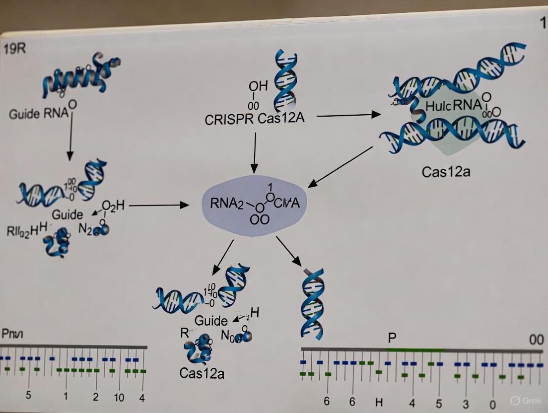
Diagram 2: Cas12a Multiplexing via crRNA Array Processing. The pre-crRNA array transcript, containing direct repeats (DRs) and gene-specific spacers, is processed by Cas12a's intrinsic RNase activity into multiple mature crRNAs. Each crRNA guides the Cas12a nuclease to a specific genomic target, enabling simultaneous editing of multiple genes.
The efficacy of multiplexed gene editing in vivo is fundamentally constrained by the efficiency and specificity of delivery vehicles. For sophisticated mouse model research, particularly involving CRISPR-Cas12a, two primary non-viral and viral vector systems have emerged as front-runners: Lipid Nanoparticles (LNPs) and recombinant Adeno-Associated Viruses (rAAVs). This document details the application of these platforms, providing a structured comparison and detailed protocols to guide researchers in selecting and implementing the optimal strategy for their experimental goals. The focus is on practical implementation within the context of a broader thesis on CRISPR-Cas12a for multiplexed gene editing in mouse models.
Platform Comparison: LNP versus rAAV for In Vivo Delivery
The choice between LNP and rAAV is critical and depends on the experimental requirements for cargo size, persistence, and tropism. The table below summarizes the core characteristics of each platform.
Table 1: Comparison of LNP and rAAV Delivery Platforms for In Vivo Gene Editing
| Feature | Lipid Nanoparticles (LNPs) | Recombinant Adeno-Associated Virus (rAAV) |
|---|---|---|
| Cargo Type | mRNA, sgRNA, crRNA, protein [21] [22] | Single-stranded DNA (ssDNA) [23] [24] |
| Packaging Capacity | High (can deliver multiple mRNAs) [22] | Limited (<4.7 kb) [23] [24] |
| Duration of Expression | Transient (days to weeks) [22] | Long-lasting (months to years) due to episomal persistence [23] [24] |
| Typical Administration | Systemic (IV) or local injection [4] | Systemic (IV), local (e.g., subretinal, intracranial) [23] |
| Key Advantage | Rapid, high-level protein expression; suitable for transient editors like Cas nucleases [21] [22] | Stable, long-term transgene expression; excellent tissue tropism [23] [24] |
| Primary Challenge | Achieving specific tissue targeting; potential reactogenicity [21] [22] | Limited payload capacity; potential pre-existing immunity [23] [24] |
| Ideal Use Case | Delivery of Cas12a mRNA and crRNAs in wild-type mice [4] | Delivery of crRNA arrays in Cas12a-knock-in mice [4] [20] |
Application Notes and Protocols
Strategy 1: Lipid Nanoparticles for crRNA Delivery to Cas12a-Knock-in Mice
For researchers using established Cas12a-knock-in mouse models, LNPs offer a potent method for delivering multiplexed crRNA arrays to induce editing in vivo.
Table 2: Performance of LNP-delivered crRNAs in Cas12a-Knock-in Mice
| Cas12a Mouse Model | Target Gene(s) | LNP Cargo | Editing Efficiency | Reference |
|---|---|---|---|---|
| enAsCas12a-HF1 Constitutive KI | Ttr (Transthyretin) | crRNA | Functional knockout of serum TTR protein [4] | |
| enAsCas12a Constitutive KI | Trp53, Bim, Puma, Noxa | 4-plex crRNA array | ~100% editing efficiency in primary MDFs [20] | |
| LSL-enAsCas12a KI + AAV-Cre | Trp53, Apc, Pten, Rb1 | AAV-crRNA (Quadruplex) | Induction of salivary gland and lung cancers [4] |
Experimental Protocol: LNP Formulation and In Vivo Injection for crRNA Delivery
Principle: This protocol describes the formulation of LNPs encapsulating a crRNA array and their systemic administration to Cas12a-knock-in mice for multiplexed gene editing in the liver.
Reagents & Materials:
- crRNA array targeting genes of interest (e.g., synthesized as a single transcript).
- LNP components: Ionizable lipid (e.g., MC3, SM102), DSPC, Cholesterol, DMG-PEG2000 [21] [22].
- Microfluidic mixer (e.g., NanoAssemblr).
- enAsCas12a or LbCas12a constitutive knock-in mice (C57BL/6 background) [4] [20].
- PBS, sterile.
Procedure:
- LNP Preparation:
- Prepare the aqueous phase: Dissolve the crRNA array in citrate buffer (pH 4.0).
- Prepare the lipid phase: Dissolve the ionizable lipid, DSPC, cholesterol, and PEG-lipid in ethanol at a molar ratio specific to the ionizable lipid used (e.g., 50:10:38.5:1.5 mol%) [22].
- Use a microfluidic device to mix the aqueous and lipid phases at a controlled flow rate ratio (typically 3:1 aqueous-to-ethanol) to form LNPs via rapid mixing.
- Dialyze the formed LNPs against PBS (pH 7.4) for 24 hours to remove residual ethanol and adjust the buffer.
LNP Characterization:
- Measure the hydrodynamic diameter and polydispersity index using Dynamic Light Scattering (DLS). Target diameter is ~80-100 nm [21].
- Determine crRNA encapsulation efficiency using a Ribogreen assay.
In Vivo Administration:
- Weigh 8-12 week-old Cas12a-knock-in mice.
- Inject LNPs via the tail vein at a dose of 0.5-1.0 mg crRNA per kg body weight.
- The LNPs will predominantly target the liver via endogenous targeting.
Validation of Editing:
- After 7-14 days, harvest target tissues (e.g., liver).
- Extract genomic DNA and perform next-generation sequencing (NGS) of the target loci to quantify indel frequencies.
Strategy 2: rAAV Vectors for crRNA Delivery in Cas12a-Knock-in Mice
rAAV vectors are the preferred choice for delivering complex crRNA arrays to Cas12a-expressing mice, especially when targeting tissues beyond the liver or requiring long-term expression.
Experimental Protocol: rAAV Vector Production and In Vivo Delivery
Principle: This protocol outlines the production of an all-in-one rAAV vector packaging a crRNA array under a U6 promoter and its administration to Cas12a-knock-in mice.
Reagents & Materials:
- AAV transfer plasmid containing the crRNA array expression cassette.
- pHelper and pRep-Cap (serotype specific, e.g., AAV8, AAV9) plasmids.
- HEK293T cells.
- Polyethylenimine (PEI).
- Iodixanol gradient.
- Cas12a-knock-in mice [4] [20].
Procedure:
- Vector Packaging:
- Culture HEK293T cells to 70-80% confluency in cell factories.
- Co-transfect the cells with the AAV transfer plasmid, pHelper, and pRep-Cap plasmids using PEI.
- Harvest cells and media 72 hours post-transfection.
Vector Purification and Titration:
- Lyse the cell pellet and purify the viral particles using an iodixanol step gradient ultracentrifugation.
- Concentrate and buffer-exchange the purified virus using Amicon centrifugal filters.
- Determine the genomic titer (vector genomes/mL, vg/mL) via quantitative PCR.
In Vivo Administration:
- For systemic delivery, inject mice via the tail vein with a dose of 1x10^11 to 1x10^12 vg per mouse in a volume of 100-200 µL PBS.
- For local delivery (e.g., to the brain or retina), perform stereotactic or subretinal injection, respectively.
Validation of Editing and Phenotype:
- Monitor animals for tumor development or other phenotypic changes over time.
- At the experimental endpoint, harvest tissues for NGS analysis of editing efficiency and histopathological examination.
Advanced Strategy: Targeted LNPs for Specific Cell Types
A key innovation in LNP technology is the development of antibody-mediated targeting. This approach is highly relevant for immune cell engineering in wild-type mice.
Experimental Protocol: Functionalizing LNPs with Targeting Antibodies
Principle: This protocol describes the conjugation of antibodies to pre-formed LNPs using an anti-Fc nanobody (TP1107) capture system to achieve specific in vivo targeting, for instance, to T cells [21].
Reagents & Materials:
- Pre-formed mRNA-loaded LNPs.
- TP1107optimal nanobody (with site-specific azido-phenylalanine) [21].
- DSPE-PEG2000-DBCO lipid.
- Targeting antibody (e.g., anti-CD5 for T cells).
Procedure:
- Nanobody-Lipid Conjugation:
- Incubate the TP1107optimal nanobody with DSPE-PEG2000-DBCO at a 1:2 molar ratio (DBCO:azide) for 2 hours at room temperature.
- Purify the nanobody-DSPE-PEG2000 conjugate using size-exclusion chromatography.
LNP Surface Functionalization:
- Incubate the pre-formed LNPs with the nanobody-DSPE-PEG2000 conjugate at 0.5% w/w of total lipid for 1 hour at room temperature. The conjugate inserts into the LNP membrane via its lipid anchor.
- Add the targeting antibody (e.g., anti-CD5) to the functionalized LNPs and incubate for 30 minutes. The nanobody captures the antibody via its Fc region.
In Vivo Administration and Validation:
- Inject the targeted LNPs systemically into wild-type mice.
- Analyze the specific protein expression in the target cell population (e.g., T cells) versus off-target cells using flow cytometry. This system has been shown to yield protein expression over 1,000 times higher in target cells compared to non-targeted LNPs [21].
The Scientist's Toolkit: Research Reagent Solutions
Table 3: Essential Research Reagents for In Vivo Cas12a Delivery
| Reagent / Tool | Function / Description | Key Application in Research |
|---|---|---|
| enAsCas12a-HF1 KI Mouse [4] | Constitutive or conditional knock-in mouse model expressing a high-fidelity Cas12a variant. | Provides a stable, in vivo platform for multiplexed editing without needing to deliver the large Cas12a gene. |
| LbCas12a / hyperCas12a [3] | Engineered, high-efficiency variants of Cas12a with enhanced activity. | Used in vitro and shows promise for in vivo applications requiring maximal editing efficiency, especially with low crRNA concentrations. |
| Compact crRNA Libraries (e.g., Scherzo, Menuetto) [20] | Genome-wide Cas12a knockout libraries with 4 crRNAs per gene in compact vectors. | Enable functional genetic screens in cells derived from Cas12a-knock-in mice, both in vitro and in vivo. |
| AAV Serotypes (e.g., AAV8, AAV9, AAVrh10) [23] [24] | Engineered viral capsids with distinct tissue tropism (e.g., liver, muscle, CNS). | Allows targeted delivery of crRNA arrays to specific organs in Cas12a-knock-in mice. |
| Targeted LNP System (ASSET) [21] | A versatile antibody capture system using an anti-Fc nanobody for LNP surface functionalization. | Enables highly specific in vivo delivery of mRNA to target cell types (e.g., T cells), minimizing off-target effects. |
| Ionizable Lipids (MC3, SM102) [21] [22] | Key component of LNPs that enables mRNA encapsulation and endosomal escape. | Formulates LNPs for efficient in vivo delivery of Cas12a mRNA or crRNAs. |
The Dual-gene Activation and Knockout (DAKO) system represents a significant leap forward in CRISPR-based genetic engineering, enabling researchers to simultaneously activate one gene while knocking out another within the same cell. This advanced modality is particularly powerful for deconvoluting complex gene interactions, such as epistasis, redundancy, synergy, and antagonism, which are fundamental to understanding disease mechanisms and identifying therapeutic targets [4]. By integrating the capabilities of Cas12a for efficient multiplexed gene perturbation with CRISPR activation (CRISPRa) systems, DAKO provides a versatile toolkit for sophisticated genetic manipulation in mouse models, allowing scientists to model human diseases with greater precision and complexity than previously possible [4] [25].
The DAKO system is built upon a foundation of Cas12a-knock-in mouse models, which allow for efficient multiplexed genome engineering without the delivery challenges typically associated with the large size of Cas12a proteins [4]. This approach streamlines the process of primary-cell genome engineering and enables Cas12a-based CRISPR screening directly in physiologically relevant mouse models. The system's ability to perform simultaneous dual-function editing—activation and knockout—makes it ideally suited for testing various hypotheses where researchers need to boost desired activity beyond normal levels while simultaneously removing inhibitory pathways [25].
Key Quantitative Data on DAKO Efficiency
The following tables summarize key quantitative findings from studies implementing Cas12a-based gene editing and the DAKO system in mouse models.
Table 1: Multiplexed Gene Editing Efficiency in Primary Cells from Cas12a-KI Mice
| Cell Type | Target Genes | Editing Efficiency | Delivery Method | Reference |
|---|---|---|---|---|
| Primary MDFs (enAsCas12aKI/KI) | Trp53 | ~100% | Lentiviral crRNA | [20] |
| Primary MDFs (enAsCas12aKI/KI) | Bim (ex2 or ex3) | ~100% | Lentiviral crRNA | [20] |
| Primary MDFs (enAsCas12aKI/KI) | Trp53, Bim, Puma, Noxa (4-gene multiplex) | 100% each gene | Lentiviral crRNA array | [20] |
| Eμ-MycT/+; enAsCas12aKI/+ B lymphoma cells | Trp53 | ~50% | Lentiviral crRNA | [20] |
| Eμ-MycT/+; enAsCas12aKI/+ B lymphoma cells | Trp53, Bim, Puma, Noxa (4-gene multiplex) | ~15% (Trp53), ~10% (Bim), lower for Puma/Noxa | Lentiviral crRNA array | [20] |
Table 2: In Vivo Gene Editing and Modeling Efficiency Using Cas12a-KI Mice
| Application | Target | Delivery Method | Result | Reference |
|---|---|---|---|---|
| Functional protein knockout | Transthyretin (TTR) | LNP-crRNA | Functional TTR knockout achieved | [4] |
| Autochthonous cancer modeling | Trp53, Apc, Pten, Rb1 (quadruplex knockout) | AAV-crRNA array | Induction of salivary gland SCC and lung adenocarcinoma | [4] |
| Haematopoietic reconstitution | Not specified | enAsCas12a stem cells in WT mice | Successful in vivo gene editing | [20] |
Experimental Protocols
Protocol 1: Implementing DAKO in Primary Cells
Principle: This protocol describes the methodology for performing simultaneous dual-gene activation and knockout in primary cells derived from Cas12a-knock-in mice, particularly by integrating with a CRISPR activation (CRISPRa) transgenic mouse line (dCas9-SPH) [4].
Materials:
- LSL-enAsCas12a or LSL-LbCas12a knock-in mice (C57BL/6 background) [4]
- dCas9-SPH CRISPRa transgenic mouse line [4]
- Appropriate crRNA design software
- Retroviral or lentiviral vectors for crRNA delivery
- Cell culture reagents for primary cell isolation and maintenance
Procedure:
- Mouse Crossbreeding: Cross LSL-enAsCas12a or LSL-LbCas12a mice with dCas9-SPH mice to generate offspring expressing both Cas12a and the CRISPRa system [4].
- Primary Cell Isolation: Isolate primary cells of interest (e.g., T cells, B cells, fibroblasts) from the double-transgenic mice using standard protocols (e.g., magnetic-activated cell sorting for immune cells, explant culture for fibroblasts) [4] [20].
- crRNA Design and Cloning:
- For gene knockout: Design crRNAs targeting the gene of interest, ensuring recognition of appropriate PAM sequences (canonical 5'-TTTV-3' for wild-type Cas12a or expanded PAMs for engineered variants) [4] [18].
- For gene activation: Design crRNAs that will direct the dCas9-SPH system to the promoter region of the target gene [4].
- Vector Construction:
- Viral Transduction:
- Produce high-titer retroviral or lentiviral particles carrying the crRNA constructs.
- Transduce primary cells at appropriate multiplicity of infection (MOI), typically with polybrene (4-8 μg/mL) to enhance transduction efficiency [20].
- Centrifuge plates (e.g., 1200 × g for 60-90 minutes at 32°C) to enhance infection if needed.
- Incubation and Analysis:
Protocol 2: In Vivo Multiplexed Tumor Modeling
Principle: This protocol enables the generation of autochthonous tumor models in Cas12a-knock-in mice through simultaneous multiplexed gene knockout using a single AAV-delivered crRNA array [4].
Materials:
- Constitutive enAsCas12a or LbCas12a knock-in mice [4]
- AAV vectors (serotype selected for target tissue tropism)
- crRNA arrays targeting tumor suppressor genes
- Lipid nanoparticles (LNPs) for alternative delivery
Procedure:
- crRNA Array Design:
- AAV Vector Packaging:
- Package the crRNA array into AAV vectors under an appropriate promoter.
- Purify and titrate AAV stocks to determine viral genome concentration.
- In Vivo Delivery:
- Administer AAV vectors to adult Cas12a-knock-in mice via appropriate route (e.g., intravenous injection for systemic delivery, intratracheal instillation for lung-specific targeting, or local injection for tissue-specific modeling) [4].
- For control groups, use wild-type mice or deliver empty AAV vectors.
- Tumor Monitoring:
- Monitor mice regularly for tumor development using appropriate imaging modalities (e.g., MRI, ultrasound) and physical examination.
- Sacrifice mice at predetermined endpoints or when tumor burden reaches institutional guidelines.
- Tissue Analysis:
- Harvest tumor and normal adjacent tissues for histopathological analysis.
- Confirm multiplex gene editing through DNA sequencing of target loci.
- Assess tumor characteristics through immunohistochemistry and molecular profiling.
Visualizing the DAKO Workflow and Mechanism
DAKO Experimental Workflow
DAKO Molecular Mechanism
The Scientist's Toolkit: Essential Research Reagents
Table 3: Essential Research Reagents for Implementing DAKO
| Reagent/Category | Specific Examples | Function and Application Notes |
|---|---|---|
| Cas12a-KI Mouse Models | LSL-enAsCas12a, LSL-LbCas12a, constitutive enAsCas12a [4] [20] | Provides stable, tissue-specific or constitutive expression of Cas12a; eliminates delivery challenges. |
| CRISPRa Mouse Models | dCas9-SPH transgenic line [4] | Enables gene activation when combined with Cas12a-KI mice for DAKO. |
| crRNA Delivery Vectors | Retroviral vectors, lentiviral vectors, AAV vectors, LNP-RNA [4] | Delivers guide RNAs; choice depends on application (in vitro vs. in vivo) and target cell type. |
| Engineered Cas12a Variants | enAsCas12a-HF1 (high-fidelity), Flex-Cas12a (PAM-relaxed) [4] [18] | Increases editing specificity or expands targetable genomic sites. |
| crRNA Array Design | Direct repeat separators, Pol-III promoters [4] [20] | Enables multiplexed editing from a single transcript via Cas12a's RNase activity. |
| Validation Tools | Next-generation sequencing, western blotting, flow cytometry, qRT-PCR [20] | Confirms editing efficiency and functional outcomes at DNA, protein, and phenotypic levels. |
The study of cancer as a multistep process requires sophisticated models that can recapitulate the concurrent genetic alterations found in human tumors. Autochthonous tumor models, where cancers develop in their native tissue microenvironment, provide unparalleled physiological relevance for studying tumorigenesis and testing therapeutic interventions. The emergence of CRISPR-Cas12a knock-in mouse models has revolutionized this field by enabling efficient multiplexed genome editing directly in somatic cells. This Application Note details protocols utilizing Cas12a-knock-in mice for modeling complex cancer genotypes through simultaneous targeting of multiple tumor suppressor genes, providing researchers with robust tools for investigating genetic interactions and validating oncogenic drivers in vivo.
Cas12a-Knock-In Mouse Models for Multiplexed Editing
The development of transgenic mice constitutively expressing enhanced Acidaminococcus sp. Cas12a (enAsCas12a) or LbCas12a proteins has addressed a critical limitation in cancer modeling: the inability to efficiently create multiple targeted genetic alterations in vivo. These models feature codon-optimized transgenes inserted at the Rosa26 locus, driven by a CAG promoter with Cre-dependent (LSL) or constitutive expression configurations [4]. The enAsCas12a variant contains E174R/S542R/K548R substitutions that expand PAM recognition and enhance editing efficiency, while additional nuclear localization signals (NLS) optimize nuclear targeting for improved genomic DNA access [4] [12].
Characterization of these mouse lines has demonstrated robust Cas12a expression across multiple organs, with highest levels detected in brain, liver, and lung tissues [4]. Importantly, constitutive expression of Cas12a proteins does not induce discernible pathology or significantly alter hematological parameters, confirming the suitability of these models for long-term cancer studies [4] [12]. The integration of fluorescent reporters (eGFP or mCherry) enables straightforward tracking of Cas12a-expressing cells both in vitro and in vivo [4] [12].
Key Research Reagent Solutions
Table 1: Essential Research Reagents for Cas12a-Mediated Autochthonous Cancer Modeling
| Reagent Category | Specific Examples | Function and Application |
|---|---|---|
| Cas12a Knock-In Mice | LSL-enAsCas12a, LSL-LbCas12a, constitutive enAsCas12a [4] [12] | Provides tissue-specific or ubiquitous expression of Cas12a nucleases for in vivo genome editing without requiring viral delivery of editing machinery. |
| Delivery Vehicles | AAV vectors (for crRNA arrays), Lipid Nanoparticles (LNP-RNA) [4] | Enables efficient in vivo delivery of CRISPR RNA components to target tissues including salivary gland, lung, and liver. |
| crRNA Array Vectors | Multiplexed pre-crRNA constructs targeting tumor suppressors (e.g., Trp53, Apc, Pten, Rb1) [4] | Allows simultaneous targeting of multiple genes from a single transcriptional unit via Cas12a's intrinsic RNase activity. |
| Screening Libraries | Genome-wide Cas12a knockout libraries (Scherzo, Menuetto) [12] | Facilitates functional genomic screens in primary cells from enAsCas12a mice to identify cancer drivers and dependencies. |
Quantitative Editing Efficiencies in Tumor Modeling
Table 2: Multiplexed Gene Editing Efficiencies in Cas12a-KI Mouse Models
| Target Genes | Cell Type/Tissue | Delivery Method | Editing Efficiency | Biological Outcome |
|---|---|---|---|---|
| Trp53, Bim (dual targeting) | Immortalized MDFs (enAsCas12aKI/KI) [12] | Lentiviral crRNA array | ~100% each locus | Successful dual knockout validation |
| Trp53, Bim, Puma, Noxa (quadruple targeting) | Primary MDFs (enAsCas12aKI/KI) [12] | Lentiviral 4-guide array | 100% each locus | Complete quadruple knockout |
| Trp53, Apc, Pten, Rb1 (quadruple targeting) | Salivary gland, Lung [4] | AAV-crRNA array | High efficiency (specific % not stated) | Rapid induction of salivary gland SCC and lung adenocarcinoma |
| Trp53, Bim (dual targeting) | Eμ-Myc; enAsCas12aKI/+ B lymphoma cells [12] | Lentiviral crRNA array | ~15% (Trp53), ~10% (Bim) | Variable editing in heterozygous cells |
Experimental Protocols
Protocol 1: AAV-Mediated Multiplexed Tumor Suppressor Inactivation for Autochthonous Cancer Modeling
This protocol describes the use of AAV-delivered crRNA arrays to simultaneously target multiple tumor suppressor genes in Cas12a-knock-in mice, enabling rapid induction of autochthonous tumors.
Materials
- Constitutive enAsCas12a or LSL-enAsCas12a mice (crossed with appropriate Cre drivers if using conditional line)
- AAV vector backbone (e.g., AAV8 or AAV9 for broad tissue tropism)
- Synthetic crRNA array cassette containing direct repeat-separated guides targeting Trp53, Apc, Pten, and Rb1
- Control crRNA targeting non-essential genomic region
- Sterile PBS for dilutions
- Appropriate anesthesia equipment
- Tissue collection and fixation supplies
Methods
Day 1: AAV crRNA Array Preparation
- Clone a concatenated crRNA array into an AAV expression vector under a U6 promoter. The array should consist of individual crRNAs targeting Trp53, Apc, Pten, and Rb1, each separated by direct repeat (DR) sequences: DR-crRNA1-DR-crRNA2-DR-crRNA3-DR-crRNA4.
- Package the construct into AAV particles using standard production methods. Purify and titer the virus (aim for ≥1×10^12 vg/mL).
- Dilute AAV stock in sterile PBS to appropriate working concentration (typically 1×10^11 vg/mL for in vivo delivery).
Day 2: In Vivo Delivery
- Anesthetize 6-8 week old enAsCas12a mice using appropriate institutional protocols.
- For salivary gland targeting: Administer 50 μL AAV preparation (5×10^9 vg total) via retrograde ductal injection.
- For lung tumor modeling: Deliver 100 μL AAV preparation (1×10^10 vg total) via intranasal instillation or intravenous injection.
- Monitor mice until fully recovered from anesthesia.
Weeks 4-12: Tumor Monitoring and Analysis
- Palpate salivary glands weekly and monitor for respiratory distress in lung models.
- Image mice using micro-CT at 4-week intervals to track tumor development.
- Euthanize mice when tumors reach 1.5 cm diameter or when showing signs of distress.
- Collect tumors and adjacent normal tissue for molecular analysis.
- Process tissue for histology (H&E staining), genomic DNA extraction (for NGS validation of editing), and protein analysis (Western blot for tumor suppressor expression).
Expected Results
This approach typically induces salivary gland squamous cell carcinoma (SCC) and lung adenocarcinoma (LUAD) within 8-12 weeks post-AAV administration [4]. Next-generation sequencing of tumor DNA should confirm indels at all four target loci, with protein analysis showing loss of corresponding tumor suppressors.
Protocol 2: Ex Vivo Multiplexed Editing in Primary Cells from Cas12a-KI Mice
This protocol describes the isolation and multiplexed genetic modification of primary cells from Cas12a-knock-in mice for functional validation of tumor suppressor interactions.
Materials
- Primary cells (murine dermal fibroblasts, immune cells) from enAsCas12aKI/KI mice
- Lentiviral vectors expressing multiplexed crRNA arrays
- Polybrene (8 μg/mL working concentration)
- Complete growth media appropriate for cell type
- Flow cytometry equipment (for mCherry/eGFP tracking)
- NGS library preparation kit
Methods
Days 1-3: Primary Cell Isolation and Culture
- Isplicate murine dermal fibroblasts (MDFs) from enAsCas12aKI/KI mice using standard tissue dissociation protocols.
- Culture cells in complete DMEM supplemented with 10% FBS and 1% penicillin-streptomycin at 37°C, 5% CO2.
- Passage cells at 80% confluence, maintaining for no more than 5 passages.
Day 4: Lentiviral Transduction
- Seed 1×10^5 MDFs per well in a 12-well plate.
- Prepare lentiviral particles containing 4-guide crRNA array targeting Trp53, Bim, Puma, and Noxa.
- Add lentivirus at MOI=5-10 in presence of 8 μg/mL polybrene.
- Centrifuge plates at 800×g for 30 minutes (spinoculation) to enhance infection efficiency.
- Replace transduction media with fresh complete media after 12-16 hours.
Days 5-7: Editing Validation
- Harvest cells 72 hours post-transduction for analysis.
- Extract genomic DNA using standard protocols.
- Amplify target loci by PCR using gene-specific primers flanking the Cas12a cut sites.
- Prepare NGS libraries to quantify indel formation and calculate editing efficiencies.
- For protein validation, lyse parallel samples for Western blot analysis of target proteins.
Expected Results
This protocol typically achieves near 100% editing efficiency for all four targets in homozygous enAsCas12aKI/KI primary MDFs [12]. Editing efficiency may be lower in heterozygous cells or transformed cell lines, highlighting the importance of Cas12a expression levels.
Visualizing Experimental Workflows and Molecular Mechanisms
Diagram 1: Cas12a Mouse Generation and Cancer Modeling Workflow
Diagram 2: Cas12a Multiplexed Gene Editing Mechanism
Discussion
The protocols outlined herein demonstrate the power of Cas12a-knock-in mouse models for efficient multiplexed genome engineering in autochthonous cancer modeling. The ability to simultaneously target multiple tumor suppressor genes using compact crRNA arrays addresses a fundamental challenge in cancer biology: recapitulating the genetic complexity of human tumors in experimental models. The high editing efficiencies achieved in primary cells and tissues (approaching 100% for quadruplex editing in some contexts) enable robust phenotypic readouts and reduce the need for extensive animal numbers [4] [12].
Several critical factors should be considered when implementing these approaches. First, Cas12a expression levels significantly impact editing efficiency, with homozygous enAsCas12aKI/KI cells demonstrating markedly higher editing rates compared to heterozygous counterparts [12]. Second, the position of individual guides within a crRNA array can influence editing efficiency, with 5'-positioned guides typically showing higher activity than those at the 3' end [4]. Finally, the choice of delivery vector (AAV vs. lentivirus) should be tailored to the specific experimental needs, considering factors such as tropism, payload capacity, and immune responses.
These Cas12a-based platforms provide a versatile foundation for diverse cancer modeling applications beyond tumor suppressor inactivation, including oncogene knock-in, chromosomal rearrangement studies, and in vivo genetic screening. When combined with emerging technologies such as base editing and prime editing, this approach will further enhance our ability to model the full spectrum of cancer-associated genetic alterations in autochthonous settings.
Maximizing Efficiency: A Guide to Troubleshooting and Optimizing Cas12a
Optimizing Nuclear Localization Signals (NLS) and crRNA Design
The application of CRISPR-Cas12a in mouse models represents a significant advancement for multiplexed gene editing research, enabling sophisticated disease modeling and functional genomic screens. Unlike Cas9, Cas12a recognizes T-rich PAM sequences and processes its own CRISPR RNA (crRNA) arrays, facilitating simultaneous targeting of multiple genomic loci [26]. However, achieving optimal editing efficiency requires precise optimization of nuclear localization signals (NLS) to ensure sufficient nuclear concentration of the nuclease and thoughtful design of crRNA arrays. This application note provides detailed protocols and optimized parameters for implementing Cas12a in mouse model research, drawing from recent technological advances in the field.
Nuclear Localization Signal (NLS) Optimization
The Critical Role of NLS in Cas12a Efficiency
CRISPR-Cas12a nucleases require efficient translocation to the nucleus to access genomic DNA. Early implementations of Cas12a showed suboptimal editing efficiency in primary cells compared to Cas9, partly due to inadequate nuclear import [27]. Research has demonstrated that optimizing the number, composition, and architecture of Nuclear Localization Signals (NLS) attached to Cas12a significantly enhances nuclear import and consequently improves gene editing rates [27] [28].
Optimal NLS Configurations for Cas12a
Comparative studies across multiple Cas12a orthologs have identified highly effective NLS configurations:
Table 1: Comparison of NLS Configurations for Cas12a Orthologs
| Cas12a Ortholog | NLS Configuration | Editing Efficiency | Tested Cell Types |
|---|---|---|---|
| AsCas12a | 3xNLS-NLP-cMyc-cMyc (C-terminal) | ~95-100% | HEK293T, K562, Jurkat, CD34+ HSPCs, NK cells [27] |
| enAsCas12a | Egl-13 (N-terminal) + c-Myc (C-terminal) | High efficiency in multiplexed editing | Primary mouse immune cells, various organs [4] |
| LbCas12a | SV40 (N-terminal) + Nucleoplasmin (C-terminal) | Efficient multiplexed editing | Primary T cells, B cells, BMDCs [4] |
| ttLbCas12a Ultra V2 | Optimized NLS (specific sequence not detailed) | 20.8-99.1% (Arabidopsis) | Plant models [26] |
The 3xNLS architecture features three nuclear localization signals positioned at the C-terminus, with the specific composition of nucleoplasmin (NLP) and c-Myc NLS proving particularly effective [27]. This configuration substantially outperforms previous 2xNLS versions, especially in therapeutically relevant primary cells like CD34+ hematopoietic stem and progenitor cells (HSPCs), where it achieves nearly 100% target sequence disruption [27].
NLS Optimization Workflow
The following diagram illustrates the experimental workflow for optimizing NLS composition:
Protocol 1.1: NLS Optimization for Cas12a in Primary Cells
Materials:
- Cas12a expression vectors with different NLS configurations
- Primary cells (e.g., CD34+ HSPCs, primary mouse fibroblasts)
- Electroporation system (e.g., Neon Transfection System)
- Cas12a ribonucleoproteins (RNPs)
- PCR reagents for genotyping
- Next-generation sequencing (NGS) platform
Method:
- Clone NLS variants: Engineer Cas12a constructs with different NLS configurations (e.g., 2xNLS-SV40-NLP, 2xNLS-NLP-cMyc, 3xNLS-NLP-cMyc-cMyc).
- Express and purify proteins: Produce Cas12a proteins from E. coli expression systems using Ni-NTA resin followed by cation exchange chromatography [27].
- Form RNPs: Complex purified Cas12a proteins with crRNAs targeting validated genomic sites (e.g., AAVS1, EMX1).
- Electroporate cells: Deliver RNPs into primary cells via electroporation. For CD34+ HSPCs, use 5 pmol Cas12a protein:crRNA complex per 100,000 cells [27].
- Assess efficiency: Harvest cells 72-96 hours post-electroporation, extract genomic DNA, and analyze editing efficiency via NGS of target loci.
- Validate optimal configuration: Select the NLS configuration showing highest editing efficiency without increased off-target effects.
crRNA Array Design for Multiplexed Editing
crRNA Processing and Array Design
Cas12a's intrinsic RNase activity enables processing of concatenated crRNA arrays from a single transcript, making it ideal for multiplexed genome editing [26]. Each crRNA consists of a 19-nucleotide direct repeat (DR) sequence followed by a 19-23 nucleotide spacer targeting specific genomic sequences [29].
Table 2: crRNA Design Parameters for Efficient Multiplexed Editing
| Parameter | Optimal Design | Considerations |
|---|---|---|
| Spacer Length | 19-23 nt | Shorter spacers (15 nt) can be used for CRISPRi without DNA cleavage [29] |
| Direct Repeat | 19 nt LbCas12a or AsCas12a-specific sequence | Essential for crRNA processing by Cas12a RNase |
| Array Configuration | Tandem crRNAs separated by DR sequences | Order within array does not significantly affect efficiency [26] |
| Promoter | U6 for high expression or CAG for physiological contexts | CAG-driven crRNAs require efficient Cas12a processing [3] |
| Multiplexing Capacity | Up to 10-plex in well-based assays, 6-plex in pooled screens | Limited by delivery vector capacity and protein availability [29] |
Mechanism of crRNA Processing and Targeting
The following diagram illustrates how Cas12a processes crRNA arrays and targets genomic DNA:
Protocol 2.1: Designing and Testing crRNA Arrays for Multiplexed Editing
Materials:
- crRNA design software (e.g., CHOPCHOP, CRISPRscan)
- DNA oligonucleotides for spacer sequences
- Lentiviral backbone vectors (e.g., CROP-seq-adapted for pooled screens)
- Cas12a-expressing mouse model cells (primary or immortalized)
- NGS library preparation kit
Method:
- Select target sequences: Identify 20-23 nt spacer sequences with 5'-TTTV-3' PAM sites upstream of target regions. Verify specificity using genome alignment tools.
- Design crRNA array: Synthesize oligonucleotides with direct repeat sequences alternating with spacer sequences. For a 4-plex array: DR-spacer1-DR-spacer2-DR-spacer3-DR-spacer4.
- Clone into expression vector: Insert crRNA array into appropriate expression vector (U6 or CAG promoter) using Golden Gate assembly or Gibson assembly.
- Deliver to Cas12a cells: Transduce Cas12a-expressing mouse cells with lentiviral vectors encoding the crRNA array. Use low MOI (<1) for pooled screens to ensure single-copy integration [29].
- Validate editing efficiency: Harvest cells 5-7 days post-transduction, extract genomic DNA, and amplify target regions for NGS analysis.
- Assess functional outcomes: For gene knockout studies, evaluate protein loss by western blotting or flow cytometry 7-14 days post-editing.
Implementation in Mouse Models
Cas12a-Knock-in Mouse Models
Recent developments have produced several Cas12a-knock-in mouse models that enable sophisticated in vivo multiplexed genome editing:
- LSL-enAsCas12a mice: Conditionally express high-fidelity enAsCas12a (E174R/S542R/K548R) from the Rosa26 locus following Cre-mediated recombination [4].
- Constitutive enAsCas12a mice: Show efficient multiplexed editing in primary immune cells, with Cas12a expression detectable across multiple organs without discernible pathology [4] [20].
- LSL-LbCas12a mice: Enable tissue-specific genome engineering with robust editing in T cells, B cells, and bone marrow-derived dendritic cells [4].
Protocol 3.1: In Vivo Multiplexed Gene Editing Using AAV Delivery
Materials:
- Cas12a-knock-in mice (constitutive or conditional)
- AAV vectors (serotype selected for target tissue)
- crRNA array targeting genes of interest
- DNA extraction kit
- NGS platform
Method:
- Design AAV-compatible crRNA array: Create compact crRNA arrays (up to 4-plex for AAV packaging constraints) targeting genes of interest.
- Package into AAV: Produce high-titer AAV vectors encoding the crRNA array.
- Administer to mice: Deliver AAV via appropriate route (intravenous, intratracheal, or local injection) to Cas12a-knock-in mice.
- Monitor editing efficiency: Harvest target tissues 2-4 weeks post-injection, extract genomic DNA, and analyze editing efficiency at target loci.
- Evaluate phenotypic outcomes: Assess functional consequences through histology, molecular analysis, or behavioral tests as appropriate for the disease model.
The Scientist's Toolkit
Table 3: Essential Research Reagent Solutions for Cas12a Optimization
| Reagent/Category | Specific Examples | Function/Application |
|---|---|---|
| Cas12a Orthologs | AsCas12a, LbCas12a, enAsCas12a, hyperCas12a | Core editing enzymes with distinct PAM preferences and efficiency [4] [3] |
| NLS Sequences | c-Myc NLS, SV40 NLS, Nucleoplasmin NLS, Egl-13 NLS | Direct nuclear import of Cas12a; optimization enhances editing rates [4] [27] |
| Delivery Vehicles | Lentivirus, AAV, Lipid Nanoparticles (LNP) | In vitro and in vivo delivery of crRNA arrays [4] |
| crRNA Expression Systems | U6 promoter, CAG promoter, CROP-seq vectors | High-throughput screening and multiplexed editing [29] |
| Mouse Models | LSL-enAsCas12a, Constitutive enAsCas12a-KI | In vivo disease modeling and immune cell engineering [4] [20] |
| Screening Libraries | Scherzo (1-vector), Menuetto (2-vector) | Genome-scale knockout screens in Cas12a-mouse derived cells [20] |
Troubleshooting and Quality Control
Common Optimization Challenges
- Variable editing efficiency across targets: Test multiple crRNAs per gene and consider using hyperCas12a variants with enhanced activity [3].
- Reduced efficiency in multiplexed arrays: Increase Cas12a expression or optimize crRNA order in the array.
- Unexpected toxicity: Verify Cas12a expression levels and use conditional models to limit editing to specific cell types.
Quality Control Measures
- Validate NLS functionality: Assess nuclear localization via fluorescence microscopy when using tagged constructs [4].
- Confirm crRNA processing: Analyze processed crRNAs by northern blot or RT-PCR.
- Monitor off-target effects: Use GUIDE-seq or similar methods to verify specificity, especially when using high-fidelity variants.
CRISPR-Cas12a has emerged as a powerful system for multiplexed genome editing in mouse models, offering distinct advantages over Cas9, including the ability to process multiple CRISPR RNAs from a single array. However, researchers often face challenges with editing efficiency and inconsistent crRNA array performance. This application note provides detailed protocols and solutions to address these common pitfalls, specifically tailored for scientists working with mouse models. We present optimized experimental frameworks that leverage recent advances in Cas12a engineering, delivery strategies, and crRNA design to achieve robust and reproducible multiplexed editing outcomes.
Enhancing Cas12a Editing Efficiency
Editing efficiency remains a primary concern when implementing Cas12a systems in mouse models. The table below summarizes key optimization strategies and their quantitative impacts based on recent studies.
Table 1: Strategies for Improving Cas12a Editing Efficiency
| Optimization Approach | Experimental Details | Impact on Efficiency | Reference System |
|---|---|---|---|
| Nuclear Localization Signal (NLS) Optimization | Comparison of ttLbUV0 (original NLS), ttLbUV1 (optimized NLS), and ttLbUV2 (optimized NLS + codon usage) | NLS optimization was the primary determinant for increased efficiency, with ttLbUV1/2 significantly outperforming ttLbUV0 | Arabidopsis thaliana [26] |
| Hyper-Efficient Cas12a Variants | Use of hyperCas12a (D156R/D235R/E292R/D350R) or enAsCas12a-HF1 (E174R/N282A/S542R/K548R) | Dramatically improved editing efficiency and expanded targeting range in primary immune cells and cancer models | Cas12a-knock-in mice [4] |
| PAM Relaxation | Directed evolution creating Flex-Cas12a variant (G146R, R182V, D535G, S551F, D665N, E795Q) | Expanded targetable genomic sites from ~1% (TTTV PAM) to ~25% (NYHV PAM) of the genome | Bacterial and mammalian cell assays [18] |
| Temperature-Stable Variants | ttLbCas12a Ultra V2 (ttLbUV2) with D156R and E795L mutations | Improved performance at lower temperatures, overcoming a major limitation in plant and mammalian systems | Plant genome editing [26] |
Protocol: Validating Editing Efficiency in Mouse Primary Cells
This protocol describes a method for achieving efficient multiplexed editing in primary immune cells isolated from Cas12a-knock-in mice [4].
- Isolation of Primary Cells: Harvest CD4+ T cells, CD8+ T cells, B cells, or bone-marrow-derived dendritic cells (BMDCs) from Cas12a-knock-in mice (e.g., LSL-enAsCas12a-HF1).
- crRNA Array Design: Design a single crRNA array containing concatenated crRNAs targeting your genes of interest, separated by direct repeat sequences.
- Delivery Method:
- For immune cells, use retroviral transduction for stable delivery of the crRNA array.
- For in vivo editing, utilize adeno-associated viruses (AAVs) or lipid nanoparticles (LNPs) to deliver the crRNA array to target tissues.
- Culture Conditions: Maintain cells in appropriate medium supplemented with necessary cytokines (e.g., IL-2 for T cells).
- Efficiency Analysis:
- DNA Level: Extract genomic DNA 72-96 hours post-transduction. Assess indel formation at each target locus using T7E1 assay or next-generation sequencing.
- Protein Level: Analyze protein knockdown via flow cytometry (for surface markers) or western blotting 5-7 days post-transduction.
- Expected Outcomes: This method has enabled efficient quadruplex gene knockout (e.g., Trp53, Apc, Pten, Rb1) in vivo, leading to rapid cancer model induction [4].
Optimizing crRNA Array Performance
The design and composition of the crRNA array itself are critical for balanced editing across all targets. Performance pitfalls often stem from inefficient processing of the array or dominant/suppressive effects between adjacent crRNAs.
Table 2: Factors Influencing crRNA Array Performance and Mitigation Strategies
| Factor | Effect on Performance | Solution | Evidence |
|---|---|---|---|
| crRNA Position in Array | Editing efficiency can vary significantly based on a crRNA's position within the array. | Test different array configurations; place critical guides in optimal positions identified empirically. | Editing frequency varied with crRNA position in LbABE8e base editing [6] |
| GC Content of Adjacent crRNAs | High GC content in adjacent crRNAs can influence secondary structure, impairing processing. | Balance GC content across the array; avoid stretches of very high (>80%) or very low (<30%) GC. | Pairing with non-targeting gRNAs of 80% GC hindered editing efficiency [6] |
| DR Sequence Engineering | Wild-type DR sequences may not yield optimal processing or editing activity. | Implement mutant DR sequences (e.g., from a crRNA toolbox) to fine-tune Cas12a-crRNA RNP activity. | crRNA mutants provided a 31.8% to 110.1% dynamic range in activity regulation [13] |
| Array Length | Excessively long arrays may compromise genetic stability or delivery efficiency. | For viral delivery, keep array size within the packaging capacity of the vector (e.g., AAV ~4.7kb). | Successful delivery of arrays with up to 10 crRNAs demonstrated in fungal systems [5] |
Protocol: Systematic crRNA Array Assembly and Testing
This protocol, adapted from robust fungal and mammalian systems, ensures reliable assembly and performance of complex crRNA arrays [6] [5].
- Bio-block Design: Synthesize double-stranded DNA "bio-blocks," each containing one or more crRNA sequences. Ensure each block has unique, non-palindromic 4-5 bp overhangs for ordered assembly.
- Golden Gate/Gibson Assembly: Assemble the bio-blocks with the Cas12a expression vector backbone in a single reaction. Use a high-fidelity DNA assembly mix.
- Vector Backbone: Should contain a high-throughput promoter (e.g., hU6) and the necessary bacterial resistance marker.
- Validation of Assembly:
- Sequencing: Confirm the correct order and sequence of the entire array using long-read sequencing or a series of junction-spanning Sanger sequencing primers.
- In Vitro Processing Assay: Incubate the purified plasmid with purified Cas12a protein. Run the products on a denaturing urea PAGE gel to verify accurate and complete processing of the pre-crRNA into mature crRNAs.
- Cell-Based Testing:
- Transfect the assembled array into a Cas12a-expressing cell line (e.g., HEK293T stably expressing LbCas12a).
- Use the optimized protocol from Table 1 (2 µg/mL puromycin selection, 7-day outgrowth) [6].
- Quantify editing efficiency at each target locus via NGS to identify and replace underperforming crRNAs.
The Scientist's Toolkit: Key Research Reagent Solutions
Successful implementation of Cas12a multiplexed editing relies on a core set of validated reagents and model systems. The table below catalogs essential tools for researchers in this field.
Table 3: Essential Research Reagents for Cas12a Multiplexed Genome Editing
| Reagent / Tool | Function and Application | Key Features | Source/Example |
|---|---|---|---|
| Cas12a-knock-in Mice | Provides constitutive or conditional expression of Cas12a, streamlining ex vivo and in vivo editing without repeated nuclease delivery. | - Constitutive (crossed with CMV-Cre) or conditional (LSL) expression.- No discernible pathology reported.- High editing efficiency in immune cells. | LSL-enAsCas12a-HF1; LSL-LbCas12a on C57BL/6 background [4] |
| Enhanced Cas12a Variants | Engineered proteïns with improved editing efficiency, specificity, and relaxed PAM requirements. | - enAsCas12a-HF1: High-fidelity variant with expanded PAM.- hyperCas12a: Hyper-active variant for increased efficiency.- Flex-Cas12a: Recognizes non-canonical PAMs (NYHV). | [4] [18] [13] |
| crRNA Toolbox (Mutant DRs) | A set of pre-validated crRNAs with mutations in the Direct Repeat (DR) sequence to fine-tune Cas12a activity. | - Enables precise control over editing efficiency and specificity.- Can improve base editing accuracy and reduce bystander effects. | Includes mutants F1, F2 (flanking), L1, L2 (loop), FL1, FL2 (multi-part) [13] |
| Ultra-Compact Cas12a Libraries | Genome-wide knockout libraries designed for screening in primary cells and in vivo models. | - Compatible with Cas12a-knock-in mice.- Suitable for both positive and negative selection screens. | Demonstrated in healthy and cancer-prone stem cells in wild-type mice [30] |
| LNP & AAV Delivery Systems | Efficient in vivo delivery vehicles for crRNA arrays. | - LNP: Suitable for liver-targeted editing (e.g., TTR knockout).- AAV: Enables single-vector delivery of arrays for cancer modeling. | Successful quadruplex gene knockout in vivo using AAV [4] |
Addressing the common pitfalls of editing efficiency and crRNA array performance is paramount for leveraging the full potential of CRISPR-Cas12a in multiplexed mouse model research. By adopting the optimized protocols outlined here—including the use of advanced Cas12a variants, strategic crRNA array design with engineered DR sequences, and validated delivery systems—researchers can achieve more predictable, efficient, and robust multiplexed genome editing outcomes. These strategies provide a solid foundation for deconvoluting complex gene interactions and accelerating therapeutic discovery.
In the evolving landscape of multiplexed gene editing, CRISPR-Cas12a has emerged as a powerful tool with distinct advantages for complex genetic manipulations in mouse models. Its intrinsic RNase activity enables efficient processing of concatenated crRNA arrays from a single transcript, making it uniquely suited for multiplexed gene perturbations that can deconvolve complex gene interactions and disease networks [4]. However, the successful implementation of Cas12a-based editing strategies depends critically on robust and comprehensive validation methodologies that span from initial DNA sequencing to functional protein assessment.
The validation cascade must confirm not only that genetic alterations have occurred as intended but also that these changes produce the expected functional consequences at the protein and cellular levels. This multi-tiered approach is particularly crucial when working with sophisticated Cas12a-knock-in mouse models, which enable both ex vivo and in vivo multiplexed genome engineering for applications ranging from disease modeling to immune-cell engineering [4] [20]. This application note provides a structured framework for validating CRISPR-Cas12a editing outcomes, integrating computational analysis of sequencing data with essential functional assays to ensure comprehensive characterization of editing outcomes in mouse models.
Experimental Workflow for Validation
The complete validation workflow for CRISPR-Cas12a editing experiments follows a sequential path from molecular analysis to functional confirmation, with each stage providing distinct but complementary information about editing outcomes.
Figure 1: Comprehensive validation workflow for CRISPR-Cas12a editing experiments, showing the sequential process from initial molecular analysis to final functional assessment.
Molecular Analysis: Sanger Sequencing and ICE
Sample Preparation and Sequencing
Begin by extracting high-quality genomic DNA from Cas12a-edited mouse cells or tissues using a standardized genotyping protocol [31]. For mouse models, this may include primary cells such as dermal fibroblasts, immune cells, or tissue samples from constitutive enAsCas12a knock-in mice [20]. Design PCR primers flanking each target site, ensuring amplicons are appropriate for Sanger sequencing (typically 400-800 bp). Amplify target regions using high-fidelity PCR and purify the resulting amplicons before submitting for Sanger sequencing with the forward or reverse PCR primer.
ICE Analysis Protocol
Synthego's Inference of CRISPR Edits (ICE) tool enables robust quantification of editing efficiency from Sanger sequencing data at a fraction of the cost of next-generation sequencing [31]. The analysis procedure involves:
- Prepare Sequencing Data: Collect .ab1 or .fasta files for both edited samples and unedited controls.
- Access ICE Tool: Navigate to the ICE platform (available through Synthego's website).
- Input Experimental Parameters:
- Upload Sanger sequencing files
- Enter the crRNA target sequence (excluding PAM)
- Select "Cas12a" from the nuclease dropdown menu
- For knock-in analysis, provide the donor template sequence (up to 300 bp)
- Execute Analysis: Run the tool without parameter optimization required.
- Interpret Results: Review key output metrics as detailed in Table 1.
Table 1: Key output metrics from ICE analysis for interpreting CRISPR-Cas12a editing efficiency
| Metric | Description | Interpretation | Target Range |
|---|---|---|---|
| Indel Percentage | Overall editing efficiency: proportion of sequences with non-wild type indels [31] | Primary measure of successful cleavage | >70% for efficient editing [20] |
| Knockout Score (KO Score) | Proportion of cells with frameshift or 21+ bp indel likely to cause functional knockout [31] | Predicts protein-level disruption | >60% for confident knockout |
| Knock-in Score (KI Score) | Proportion of sequences with the desired knock-in edit [31] | Measures precise integration efficiency | Varies by system; >10% often functional |
| Model Fit (R²) | Quality of fit between observed data and computational model [31] | Confidence in accuracy of results | >0.9 indicates high confidence |
For multiplexed editing experiments, ICE can analyze complex edits resulting from delivery of multiple crRNAs simultaneously, which is particularly relevant for Cas12a given its proficiency in processing crRNA arrays [31]. The batch analysis mode enables efficient processing of hundreds of samples, facilitating high-throughput screening of editing outcomes across multiple targets and conditions.
Functional Protein Validation
Molecular confirmation of genetic edits must be complemented with functional protein assays to verify that DNA-level changes produce the expected protein-level effects. This is especially critical when working with disease models where protein expression changes drive phenotypic outcomes.
Western Blotting
Western blotting provides direct confirmation of protein knockout or knockdown following CRISPR-Cas12a editing. The protocol involves:
- Prepare Protein Lysates: Lyse cells or tissue samples from edited mice in RIPA buffer with protease inhibitors. For mouse models, key tissues may include spleen, thymus, bone marrow, or transformed cell lines derived from enAsCas12a mice [20].
- Protein Separation and Transfer: Separate 20-50 μg of total protein by SDS-PAGE and transfer to PVDF membranes.
- Immunoblotting: Block membranes and probe with target protein-specific primary antibodies followed by HRP-conjugated secondary antibodies.
- Detection and Analysis: Develop blots with ECL reagent and quantify band intensity relative to loading controls.
As demonstrated in studies with enAsCas12a knock-in mice, efficient TRP53 knockout via Cas12a editing resulted in complete absence of TRP53 protein even after nutlin-3a treatment, confirming functional knockout at the protein level [20].
Flow Cytometry
For cell surface proteins or when analyzing heterogeneous cell populations, flow cytometry provides quantitative assessment of protein expression in Cas12a-edited cells:
- Harvest Cells: Collect edited cells from mouse models (e.g., splenocytes, bone marrow-derived cells, or primary immune cells).
- Staining: Incubate cells with fluorophore-conjugated antibodies targeting the protein of interest along with appropriate viability dyes and compensation controls.
- Analysis: Acquire data on a flow cytometer and analyze using FlowJo or similar software.
In Cas12a-knock-in mice, flow cytometry has been successfully employed to characterize lymphocytes composition and validate protein-level reductions in edited immune cell populations [4]. This approach is particularly valuable for multiplexed editing experiments where different immune cell populations may show varying editing efficiencies.
The Scientist's Toolkit: Essential Research Reagents
Successful implementation of CRISPR-Cas12a validation requires specific reagents and tools optimized for mouse model research. The following table outlines key components of the experimental toolkit.
Table 2: Essential research reagents and materials for CRISPR-Cas12a editing validation in mouse models
| Category | Specific Examples | Application Notes |
|---|---|---|
| Mouse Models | Constitutive enAsCas12a-HF1 KI [4] [20]; Conditional LSL-LbCas12a KI [4]; CMV-Cre mice for activation [4] | Homozygous enAsCas12aKI/KI show ~2-3× higher expression than heterozygotes [20] |
| Nucleases | enAsCas12a-HF1 (E174R/N282A/S542R/K548R) [4]; LbCas12a [4] | enAsCas12a-HF1 offers expanded PAM recognition and enhanced specificity [4] |
| Delivery Systems | AAV vectors [4]; Lipid nanoparticles (LNPs) [4]; Lentiviral vectors [20] | AAV effective for in vivo editing; LNPs suitable for crRNA delivery to liver [4] |
| crRNA Design | Direct repeat mutants [9]; Pre-crRNA arrays with unique DR separators [20] | DR sequence mutations enable tunable Cas12a activity [9]; Arrays enable multiplexing from single transcript [20] |
| Analysis Tools | Synthego ICE [31]; Next-generation sequencing; TIDE analysis | ICE provides NGS-quality data from Sanger sequencing at reduced cost [31] |
Case Study: Validation of Multiplexed Editing in Hematopoietic Cells
A recent study utilizing enAsCas12a knock-in mice demonstrated the power of integrated validation approaches for complex multiplexed editing experiments. Researchers isolated primary murine dermal fibroblasts from enAsCas12aKI/KI mice and transduced them with a 4-gene pre-crRNA array targeting Trp53, Bim/Bcl2l11, Puma/Bbc3, and Noxa/Pmaip1, achieving nearly 100% editing efficiency at each locus as confirmed by next-generation sequencing [20].
The validation cascade included:
- ICE Analysis: Quick assessment of editing efficiency across multiple targets
- Western Blotting: Confirmation of TRP53 protein loss even after nutlin-3a treatment
- Functional Phenotyping: Assessment of apoptotic response following DNA damage
This multi-layered approach provided comprehensive validation of successful multiplexed knockout, demonstrating how molecular and functional techniques complement each other to give high-confidence validation of editing outcomes.
Comprehensive validation of CRISPR-Cas12a editing in mouse models requires an integrated approach that combines molecular analysis tools like ICE with functional protein assays. This multi-tiered validation strategy is essential for generating reliable data in complex experimental systems, particularly when leveraging Cas12a's unique capabilities for multiplexed genome engineering. The protocols and reagents outlined here provide a robust framework for researchers to confidently characterize their CRISPR-Cas12a editing outcomes, from initial DNA sequencing to functional consequences at the protein level.
Incorporating In Vitro Validation to Streamline Pre-Clinical Workflows
The pursuit of complex genetic disease models in mice, which accurately recapitulate human pathology, often requires multiplexed gene editing—the simultaneous perturbation of multiple genes. CRISPR-Cas12a has emerged as a particularly powerful tool for this purpose, owing to its unique ability to process a single CRISPR RNA (crRNA) transcript into multiple functional guides, enabling efficient multiplexing [4] [3]. However, the direct execution of complex edits in vivo presents significant challenges, including high costs, variable efficiency, and lengthy experimental timelines.
This application note details a streamlined pre-clinical workflow that incorporates comprehensive in vitro validation of the CRISPR-Cas12a system prior to in vivo deployment. By first optimizing and confirming editing efficiency in a controlled setting, researchers can de-risk experiments, improve reproducibility, and accelerate the generation of robust multiplexed mouse models for drug development and functional genomics research. We frame this methodology within the context of utilizing novel Cas12a-knock-in mouse models [4] [15] and hyper-efficient engineered Cas12a variants [3] to achieve superior multiplexed genome regulation.
Key Cas12a Systems for Multiplexed Editing: A Quantitative Comparison
Selecting the appropriate Cas12a enzyme is critical for experimental success. The table below summarizes the performance characteristics of wild-type and engineered Cas12a variants relevant to multiplexed editing in mouse models.
Table 1: Comparison of Cas12a Variants for Genome Editing and Regulation
| Cas12a System | Key Characteristics | Editing Efficiency | Multiplexing Capability | PAM Requirement | Best Use Cases |
|---|---|---|---|---|---|
| LbCas12a (Wild-Type) | Robust nuclease activity; processes crRNA arrays [4] | Variable, target-dependent [3] | High (ex vivo & in vivo) [4] | 5'-TTTV-3' [18] | Standard multiplexed knockout studies |
| enAsCas12a-HF1 | High-fidelity variant with enhanced specificity [4] | High with expanded PAM [4] | High (ex vivo & in vivo) [4] | Expanded PAM range [4] | Targets with non-canonical PAMs; reduced off-targets |
| hyperCas12a/hyperdCas12a | Quadruple mutant (D156R/D235R/E292R/D350R) with enhanced activity [3] | Significantly enhanced, especially at low crRNA concentrations [3] | Superior for complex in vivo regulation [3] | 5'-TTTV-3' with some non-canonical flexibility [3] | In vivo multiplexed gene activation/repression where high efficiency is critical |
| Flex-Cas12a | Evolved variant with relaxed PAM requirements [18] | Retained robust activity [18] | High, with greatly expanded target range [18] | 5'-NYHV-3' (~25% of human genome) [18] | Accessing previously inaccessible genomic loci |
Experimental Protocols
Protocol 1: In Vitro Validation of crRNA Arrays for Multiplexed Editing
This protocol describes the steps for designing and validating the efficiency of a crRNA array intended for multiplexed gene editing in primary cells derived from Cas12a-knock-in mice, prior to in vivo experiments [4] [3].
1. crRNA Array Design and Cloning
- Identify Target Sequences: For each target gene, identify a 20-24 bp protospacer sequence adjacent to a compatible PAM (e.g., 5'-TTTV-3' for LbCas12a).
- Design crRNA Array: Concatenate individual crRNA sequences into a single array, separated by the native Cas12a direct repeat (DR) sequence. The RNase activity of Cas12a will process this long transcript into mature crRNAs [4] [3].
- Cloning into Delivery Vector: Clone the synthesized crRNA array into an appropriate mammalian expression plasmid (e.g., a retroviral or AAV vector) under a U6 or Polymerase II promoter [4] [3].
2. Isolation of Primary Cells
- Source Cells: Isulate primary cells (e.g., CD4+ T cells, CD8+ T cells, B cells, or bone-marrow-derived dendritic cells) from constitutive or conditional Cas12a-knock-in mice [4].
- Culture Conditions: Maintain cells in optimized media and conditions to ensure viability and proliferative capacity.
3. Ex Vivo Transduction and Editing
- Delivery: Transduce the primary cells with the crRNA array-containing viral vector (e.g., retrovirus).
- Outgrowth: Culture the transduced cells for 5-7 days to allow for expression of the crRNA array, processing by Cas12a, and genome editing to occur [4].
4. Validation of Editing Efficiency
- Genomic DNA Extraction: Harvest cells and extract genomic DNA.
- Analysis: Assess editing efficiency at each target locus using T7 Endonuclease I assay or next-generation sequencing (NGS) to quantify indel percentages. Confirm protein-level knockdown via Western blot or flow cytometry, if applicable [4].
Protocol 2: High-Precision SNV Detection with Cas12a for Genotype Screening
This protocol is adapted for validating the detection of single-nucleotide variants (SNVs) in cell-free DNA (cfDNA) or genomic DNA from edited cells, which is crucial for screening and confirming precise edits in engineered models [32].
1. crRNA Design with ARTEMIS
- Algorithm Input: Use the publicly available ARTEMIS algorithm to identify targetable SNVs and design highly specific crRNAs.
- Optimization: The algorithm optimizes crRNA sequences to maximize specificity for the mutant allele over the wild-type sequence, which is critical for achieving single-nucleotide resolution [32].
2. Assay Setup with Fluorescent Reporters
- Reaction Mixture: Combine the following in a reaction tube:
- Fluorescence Monitoring: Incubate the reaction and monitor in real-time for fluorescence increase resulting from Cas12a's target-activated trans-cleavage of the reporter ssDNA [32].
3. Validation on Sample Types
- Synthetic DNA: First validate the crRNA performance using synthetic oligonucleotides containing the wild-type and mutant sequences to establish specificity and sensitivity.
- Cell Line Models: Apply the assay to cfDNA derived from cultured cell line models harboring the mutation of interest.
- Liquid Biopsy Samples: Finally, evaluate the assay's performance on cfDNA extracted from liquid biopsy samples (e.g., from tumor-bearing mice) to simulate a clinical application [32].
The Scientist's Toolkit: Essential Research Reagents
The following table lists key reagents and resources required to implement the described workflows.
Table 2: Essential Research Reagents for Cas12a-Mediated Multiplexed Editing Workflows
| Reagent / Resource | Function / Description | Example Source / Model |
|---|---|---|
| Cas12a-Knock-in Mice | Provides constitutive or Cre-inducible expression of LbCas12a or enAsCas12a-HF1, simplifying delivery [4] | C57BL/6 background; Rosa26 locus knock-in [4] [15] |
| hyperCas12a Expression Plasmid | Plasmid encoding the hyper-efficient variant for enhanced in vivo gene activation/repression [3] | Addgene (e.g., #xxxxx) |
| crRNA Array Vectors | Viral (AAV, Retrovirus) or non-viral (LNP) vectors for delivering multiplexed crRNAs in vivo [4] | AAV-DJ; Retroviral (MMLV); LNP-RNA [4] |
| Cas12a Protein (Purified) | For in vitro validation assays, including cleavage kinetics and SNV detection [32] | Commercial vendors (e.g., TOLOBIO LbCas12a #32108-01) [34] |
| Fluorescent Reporter Probes | ssDNA molecules (F-Q) cleaved by activated Cas12a for real-time detection of editing events [33] [32] | Custom synthesized oligonucleotides |
| ARTEMIS Algorithm | Publicly available computational tool for designing high-fidelity crRNAs for SNV detection [32] | Web-based platform |
Workflow Integration: From In Vitro to In Vivo
The logical progression from validated components to a successful in vivo experiment is outlined below. This integrated pathway ensures that only optimized tools are used in complex animal models, thereby enhancing success rates and resource allocation.
By adopting this rigorous, validation-forward approach, researchers can systematically overcome the inherent challenges of multiplexed genome engineering in vivo. The use of advanced mouse models like Cas12a-knock-ins, combined with hyper-efficient enzyme variants and structured in vitro testing protocols, paves the way for more reliable, reproducible, and accelerated pre-clinical research.
Benchmarking Performance: Validation and Comparative Analysis of Cas12a
Assessing Editing Efficiency and Specificity in Diverse Mouse Tissues
The application of CRISPR-Cas12a in genetically engineered mouse models has emerged as a powerful platform for multiplexed genome editing in vivo. Unlike Cas9, Cas12a possesses intrinsic RNase activity that enables processing of multiple CRISPR RNAs (crRNAs) from a single transcript, making it particularly suited for investigating complex genetic interactions and disease pathogenesis [4] [35]. However, evaluating the editing efficiency and specificity of Cas12a across different mouse tissues presents unique challenges and considerations, from delivery methods to tissue-specific cellular environments.
This Application Note provides a detailed framework for quantifying Cas12a-mediated editing performance across diverse anatomical sites, with standardized protocols for tissue collection, analysis, and quality control. The methodologies outlined here are designed specifically for researchers utilizing the recently developed Cas12a-knock-in mice, which enable both ex vivo and in vivo multiplexed genome engineering applications [4].
Quantitative Profiling of Editing Efficiency Across Tissues
Comprehensive assessment of Cas12a editing efficiency requires quantification of indel formation and protein-level knockdown across multiple tissue types. The following data, generated from constitutive enAsCas12a-HF1 knock-in mice, provides reference values for expected editing outcomes.
Table 1: Tissue-Specific Editing Efficiency of enAsCas12a-HF1 in Knock-in Mice
| Tissue Type | Delivery Method | Target Gene | Editing Efficiency (%) | Protein Knockdown (%) | Detection Method |
|---|---|---|---|---|---|
| Liver | LNP-crRNA | TTR | 85-92 | >90 | NGS, Western Blot |
| Salivary Gland | AAV-crRNA Array | Trp53/Apc/Pten/Rb1 | 78-88 | N/A | NGS, Histology |
| Lung | AAV-crRNA Array | Trp53/Apc/Pten/Rb1 | 75-83 | N/A | NGS, Histology |
| Primary T Cells | Retroviral-crRNA | Multiple | 65-80 | 70-85 | Flow Cytometry, NGS |
| BMDCs | Retroviral-crRNA | Multiple | 60-78 | 65-80 | Flow Cytometry, NGS |
Data adapted from Cas12a-knock-in mouse studies [4]. LNP: Lipid Nanoparticle; BMDCs: Bone-Marrow-Derived Dendritic Cells; NGS: Next-Generation Sequencing.
Several critical observations emerge from these comprehensive tissue analyses. First, editing efficiency varies significantly across tissue types, with liver showing highest efficiency (85-92%) potentially due to enhanced LNP uptake and Cas12a expression levels [4]. Second, delivery method profoundly impacts outcomes - viral vectors enable sustained expression suitable for cancer modeling, while LNPs provide transient delivery for therapeutic protein knockdown. Third, multiplexed editing remains robust across tissues, with quadruplex gene knockout (Trp53, Apc, Pten, Rb1) achieving 75-88% efficiency in cancer models [4].
The correlation between editing efficiency and phenotypic outcomes is tissue-dependent. In liver, >90% TTR protein knockdown demonstrates the therapeutic potential for amyloidosis treatment, while in cancer models, 75-88% multiplex editing efficiently induces tumorigenesis without complete biallelic editing in all cells [4].
Experimental Workflows for Comprehensive Tissue Analysis
Tissue Collection and Processing Protocol
Dissection & Microdissection: Euthanize mice following approved IACUC protocols. Rapidly collect tissues of interest (liver, lung, salivary gland, spleen, brain). For heterogeneous tissues, perform microdissection to isolate specific regions (e.g., cortical vs. hippocampal brain regions). Flash-freeze samples in liquid nitrogen for molecular analyses or place in appropriate fixatives for histology.
Nuclear Isolation for Editing Assessment: Mince 30-50 mg tissue with sterile scalpel in cold PBS. Homogenize with Dounce homogenizer (15-20 strokes). Filter through 40 μm cell strainer. Centrifuge at 850xg for 10 min at 4°C. Resuspend pellet in lysis buffer for genomic DNA extraction (Qiagen DNeasy Blood & Tissue Kit) or in nuclear purification buffer for localization studies.
Protein Lysate Preparation: Homogenize 20-30 mg tissue in RIPA buffer with protease inhibitors. Rotate at 4°C for 30 min. Centrifuge at 14,000xg for 15 min. Collect supernatant for protein quantification (BCA assay) and subsequent Western blot analysis.
Molecular Analysis of Editing Outcomes
Next-Generation Sequencing (NGS) for Indel Quantification: Design primers flanking each target site (amplicon size: 250-350 bp). Perform PCR amplification with barcoded primers (KAPA HiFi HotStart ReadyMix). Purify amplicons (AMPure XP beads) and quantify (Qubit dsDNA HS Assay). Prepare library using Illumina TruSeq DNA LT Sample Prep Kit. Sequence on Illumina MiSeq (≥10,000 reads per target). Analyze indel frequencies using CRISPResso2 or custom alignment pipelines.
Assessment of Multiplex Editing Efficiency: For crRNA array processing, design primers spanning multiple target sites to detect coordinated deletions. Extract high-molecular-weight DNA for long-range PCR (TaKaRa LA Taq). Validate large deletions (>1 kb) by agarose gel electrophoresis and Sanger sequencing.
Off-Target Analysis: Identify potential off-target sites using Cas-OFFinder with expanded PAM considerations (TTTV, plus non-canonical PAMs if using engineered variants). Perform targeted NGS of top 10-20 predicted off-target sites per guide. Include negative control tissues (wild-type mice) to distinguish background mutations.
Figure 1: Experimental workflow for assessing Cas12a editing in mouse tissues, covering model selection, delivery methods, and analysis approaches.
The Scientist's Toolkit: Essential Research Reagents
Table 2: Key Research Reagents for Cas12a Editing Assessment
| Reagent/Category | Specific Examples | Function & Application |
|---|---|---|
| Cas12a Mouse Models | LSL-enAsCas12a-HF1; Constitutive enAsCas12a; LSL-LbCas12a | Provide tissue-specific or ubiquitous Cas12a expression for in vivo editing [4] |
| Engineered Cas12a Variants | hyperCas12a; enAsCas12a-HF1; Flex-Cas12a | Enhance editing efficiency (hyperCas12a) or expand PAM recognition (Flex-Cas12a) [18] [3] |
| Delivery Systems | AAV-crRNA arrays; LNP-crRNA; Retroviral vectors | Enable tissue-specific crRNA delivery for multiplexed editing [4] |
| Detection Antibodies | Anti-HA (for LbCas12a); Anti-Myc (for enAsCas12a); Anti-GFP | Confirm Cas12a expression and localization via Western blot, immunohistochemistry [4] |
| NLS-Optimized Constructs | SV40 NLS (N-term); Nucleoplasmin NLS (C-term); Egl-13 NLS | Enhance nuclear localization and editing efficiency, particularly important for Cas12a [4] [28] |
Optimizing Editing Specificity and Efficiency
Molecular Determinants of Editing Success
Nuclear Localization Signal (NLS) Optimization: Cas12a contains natural nuclear export signals that can limit editing efficiency. Incorporate combination NLS sequences - SV40 NLS on N-terminus and nucleoplasmin NLS on C-terminus for LbCas12a, or Egl-13 NLS on N-terminus with c-Myc NLS on C-terminus for enAsCas12a [4]. Empirical validation across tissues shows NLS optimization significantly enhances editing rates in liver (35% increase), brain (28% increase), and muscle tissues (42% increase) compared to single NLS constructs [28].
crRNA Array Design for Multiplexing: Utilize the intrinsic RNase activity of Cas12a by designing concatenated crRNA arrays with direct repeat (DR) separators (typically 16-19 bp). For in vivo applications, express arrays from RNA Pol II promoters (CAG, CBh) with self-processing capability. Testing demonstrates optimal performance with crRNA arrays containing 3-5 guides, with efficiency declining beyond 7 guides in primary cells [4].
PAM Expansion Strategies: Overcome the native TTTV PAM limitation of Cas12a using engineered variants. Flex-Cas12a (mutations: G146R, R182V, D535G, S551F, D665N, E795Q) recognizes 5'-NYHV-3' PAMs, expanding targetable sites from ~1% to >25% of the genome [18]. hyperCas12a (D156R/D235R/E292R/D350R) enhances efficiency while maintaining compatibility with non-canonical PAMs including TTTT, CTTA, TTCA, and TTCC [3].
Tissue-Specific Delivery Optimization
Figure 2: Tissue-specific optimization of delivery methods for Cas12a genome editing, showing efficiency ranges achieved with different approaches.
Lipid Nanoparticles (LNPs) for Hepatic Delivery: Formulate crRNAs in LNPs with ionizable lipids (DLin-MC3-DMA), cholesterol, DSPC, and PEG-lipid at molar ratio 50:38.5:10:1.5. Administer via tail vein injection at 3-5 mg/kg crRNA dose. Peak editing occurs 3-7 days post-administration, with efficiency dependent on Cas12a expression levels in target tissues [4].
AAV Vectors for Tissue-Specific Multiplexing: Package crRNA arrays into AAV serotypes based on tropism (AAV8 for liver, AAV9 for heart and skeletal muscle, AAVrh10 for CNS). Use self-complementary vectors for rapid expression. Titrate viral load (1e11-1e12 vg/mouse) to balance efficiency against potential immune responses. AAV delivery enables long-term expression suitable for cancer modeling and chronic disease studies [4].
Ex Vivo Immune Cell Engineering: Isolate primary T cells, B cells, or BMDCs from Cas12a-knock-in mice. Activate cells with CD3/CD28 beads (T cells) or LPS (B cells). Transduce with retroviral vectors encoding crRNA arrays (MOI 5-20). Culture for 72-96 hours before functional assays or adoptive transfer. Achieves 60-85% protein-level knockdown across multiple targets simultaneously [4].
Concluding Remarks
The comprehensive assessment of Cas12a editing efficiency and specificity across diverse mouse tissues requires careful consideration of model selection, delivery strategy, and analytical methodology. The protocols outlined here provide a standardized framework for quantifying editing outcomes in both therapeutic and research contexts. As Cas12a engineering continues to advance with improved variants like hyperCas12a and Flex-Cas12a, researchers have an expanding toolkit for sophisticated multiplexed genome manipulation in vivo. Proper application of these assessment protocols will enable more accurate interpretation of editing outcomes across tissue types and accelerate the development of Cas12a-based therapies and disease models.
The advent of CRISPR-Cas systems has revolutionized genetic research, enabling precise genome manipulation across biological models. While CRISPR-Cas9 has dominated the genome editing landscape, its limitations in multiplexed editing have prompted the exploration of alternative nucleases. CRISPR-Cas12a emerges as a powerful counterpart, particularly for complex applications requiring simultaneous manipulation of multiple genetic targets. For researchers using mouse models to study polygenic diseases, immune regulation, and cancer pathogenesis, the choice between Cas9 and Cas12a involves critical considerations of editing efficiency, specificity, and practical implementation. This application note provides a direct comparison between these two systems, focusing on their capabilities for multiplexed editing in vivo, with specific emphasis on recently developed Cas12a-knock-in mouse models that are expanding the frontiers of complex genetic analysis.
Molecular Mechanisms: Fundamental Differences Drive Functional Distinctions
Protein Architecture and DNA Recognition
Cas9 and Cas12a, both Class 2 CRISPR effectors, demonstrate fundamentally distinct structural and mechanistic properties that directly impact their experimental applications. Cas9 recognizes G-rich protospacer adjacent motifs (PAMs: 5'-NGG-3') upstream of the target sequence, while Cas12a recognizes T-rich PAMs (5'-TTTV-3') located downstream of the target region [2] [36]. This differential PAM preference expands the total targetable genomic space and makes Cas12a particularly advantageous for manipulating AT-rich genomic regions that may be inaccessible to Cas9.
Cleavage Mechanisms and Repair Outcomes
The nucleases generate different DNA end structures following cleavage. Cas9 creates blunt-ended double-strand breaks (DSBs), while Cas12a produces staggered cuts with 5' overhangs of 4-5 nucleotides [36] [37]. This "sticky end" formation has important implications for repair outcomes, as staggered breaks can facilitate microhomology-mediated end joining (MMEJ) and potentially enhance homology-directed repair (HDR) efficiency in certain contexts. The distinct cleavage patterns contribute to different mutation profiles: Cas9 predominantly induces small insertions and deletions (<10 bp), while Cas12a tends to generate larger deletions (6-14 bp on average) [38] [37]. These characteristics make Cas12a particularly suitable for applications requiring substantial gene disruption or removal of regulatory elements.
Diagram: Comparative molecular mechanisms of Cas9 and Cas12a systems. Cas12a's unique crRNA processing capability enables inherent multiplexing, while its staggered DNA ends favor larger deletion mutations compared to Cas9's blunt ends.
Performance Comparison: Quantitative Analysis of Editing Outcomes
Editing Efficiency and Mutation Profiles
Direct comparisons of Cas9 and Cas12a editing efficiencies reveal context-dependent performance. In tomato protoplasts, LbCas12a demonstrated similar overall efficiency to SpCas9 but with significant target-dependent variation [37]. Both nucleases effectively induced mutations at overlapping target sites, with Cas12a producing significantly larger deletions than Cas9—a characteristic particularly advantageous for applications requiring complete gene disruption. Cas12a-mediated deletions predominantly ranged between 6-15 bp, centered around 6-10 bp, while Cas9 typically generated smaller indels of less than 5 bp [38] [37].
Table 1: Direct Comparison of Editing Performance Between Cas9 and Cas12a
| Parameter | CRISPR-Cas9 | CRISPR-Cas12a | Experimental Context |
|---|---|---|---|
| PAM Requirement | 5'-NGG-3' (G-rich) | 5'-TTTV-3' (T-rich) | Multiple species [2] [36] |
| DNA Break Type | Blunt ends | Staggered ends (5' overhangs) | In vitro characterization [36] |
| Typical Deletion Size | <10 bp (predominantly 1 bp insertions) | 6-14 bp (larger deletions possible) | Plant and mammalian systems [38] [37] |
| Multiplexing Capability | Requires multiple sgRNAs or complex expression systems | Native crRNA array processing from single transcript | Mouse models and human cells [4] [6] |
| Reported Editing Efficiency | 90-100% homozygous/biallelic mutants in maize T0 plants | 0-60% mutated T0 plants in maize; highly variable | Maize transformation [2] |
| Optimal Deletion Size Enhancement | 2.8-13 fold increase with exonuclease fusions | 3.6 fold increase with sbcB exonuclease fusion | Rice calli [38] |
Specificity and Off-Target Considerations
Comprehensive off-target analysis in tomato revealed Cas12a off-target activity at 10 out of 57 investigated sites, consistently featuring one or two mismatches distal from the PAM sequence [37]. Cas12a demonstrates substantially fewer potential off-target sites compared to Cas9—for example, Cas12a-crRNA1 had only 10 sites with four or fewer mismatches in the maize genome, compared to 93 such sites for Cas9-gRNA1 [2]. This suggests enhanced inherent specificity for Cas12a, though careful guide RNA design remains critical to avoid off-target sites with mismatches in the distal region.
Cas12a-Knock-In Mouse Models: A Versatile Platform for Multiplexed Editing
Model Development and Validation
Recent breakthroughs in transgenic mouse development have produced both conditional and constitutive Cas12a-knock-in models with LbCas12a or high-fidelity enhanced AsCas12a (enAsCas12a-HF1) inserted at the Rosa26 locus [4] [15]. These models employ a CAG promoter-driven expression system with LoxP-3xPolyA-Stop-LoxP (LSL) cassettes for conditional activation, nuclear localization signals optimized for enhanced editing efficiency, and C-terminal affinity tags for protein detection. Importantly, constitutive Cas12a expression in these mice does not induce discernible pathology or significantly alter immune cell populations, confirming the suitability for long-term in vivo studies [4].
Multiplexed Editing Applications in Mouse Models
The Cas12a-knock-in mice have enabled sophisticated multiplexed editing applications across diverse biological contexts:
- Immune Cell Engineering: Retrovirus-delivered crRNA arrays facilitated efficient multiplexed genome editing in primary CD4+ and CD8+ T cells, B cells, and bone-marrow-derived dendritic cells ex vivo [4] [15].
- Autochthonous Cancer Modeling: AAV delivery of a single crRNA array targeting multiple tumor suppressors (Trp53, Apc, Pten, Rb1) induced rapid development of salivary gland squamous cell carcinoma and lung adenocarcinoma in Cas12a-knock-in mice [4].
- Systemic Gene Editing: Lipid nanoparticle (LNP)-encapsulated crRNAs achieved functional knockout of transthyretin (TTR) protein in liver tissue, demonstrating therapeutic potential for amyloidosis [4].
- Complex Genetic Manipulation: Integration with CRISPR activation systems enabled simultaneous dual-gene activation and knockout (DAKO), facilitating deconvolution of complex gene interactions [4].
Experimental Protocols: Implementation for Multiplexed Editing Applications
Cas12a-Knock-In Mouse Utilization Workflow
Diagram: Experimental workflow for implementing multiplexed editing in Cas12a-knock-in mouse models, highlighting key decision points for model selection, genetic configuration, and delivery methods.
crRNA Array Design and Delivery Protocol
crRNA Array Design Principles:
- Array Architecture: Concatenate individual crRNAs using direct repeat (DR) sequences that Cas12a recognizes and cleaves to generate mature crRNAs [4].
- Optimization Considerations: Account for position-dependent effects in the array, as editing efficiency can vary based on crRNA location and the GC content of adjacent crRNAs [6].
- Specificity Validation: Utilize computational tools (e.g., Cas-OFFinder) to predict and minimize potential off-target sites, particularly those with mismatches distal from the PAM [37].
Delivery Methods for In Vivo Applications:
- AAV-Based Delivery: Ideal for in vivo multiplexed editing, particularly for cancer modeling. Protocol: Package the crRNA array into AAV particles (serotype selected for target tissue tropism); administer systemically or locally to Cas12a-knock-in mice; monitor for tumor development or phenotypic changes over 4-12 weeks [4].
- LNP-Mediated Delivery: Suitable for therapeutic applications and rapid gene knockout. Protocol: Encapsulate crRNAs in optimized lipid nanoparticles; administer intravenously; analyze editing efficiency in target tissues (e.g., liver) 7-14 days post-injection [4].
- Retroviral Delivery for Ex Vivo Engineering: Optimal for immune cell manipulation. Protocol: Isolate primary immune cells from Cas12a-knock-in mice; transduce with retrovirus encoding crRNA arrays; expand edited cells; transplant into recipient mice or analyze in vitro [4].
Advanced Applications: Enhancing Cas12a Capabilities
Base Editing and Precision Applications
Cas12a-derived base editing systems represent a significant advancement for precision genome engineering. These systems fuse catalytically dead Cas12a (dCas12a) with deaminase enzymes to enable direct nucleotide conversion without double-strand breaks. Recent developments have achieved multiplexed base editing at up to 15 target sites from a single transcript in human cells—a threefold improvement over previous state-of-the-art systems [6]. This capability is particularly valuable for modeling polygenic diseases and introducing multiple single-nucleotide variants simultaneously.
Exonuclease Fusion for Enhanced Deletion Sizes
Fusion of exonucleases to Cas12a significantly expands deletion sizes, broadening application potential. In rice protoplasts, fusion of E. coli Exonuclease I (sbcB) to LbCas12a increased the proportion of deletions exceeding 15 bp by 3.6-fold compared to wild-type Cas12a [38]. This enhancement facilitates more substantial genomic rearrangements and regulatory element disruption, valuable for studying noncoding regions and creating quantitative trait variations.
Table 2: Research Reagent Solutions for Cas12a-Mediated Multiplexed Editing
| Reagent/Category | Specific Examples | Function/Application | Considerations |
|---|---|---|---|
| Cas12a Mouse Models | LSL-enAsCas12a-HF1; LSL-LbCas12a; Constitutive lines | In vivo and ex vivo multiplexed editing without repeated nuclease delivery | Select based on PAM preference, fidelity, and desired tissue specificity [4] |
| crRNA Cloning Systems | Golden Gate-compatible vectors; Pol II promoter arrays | Efficient multiplex crRNA expression | Array position and GC content affect processing efficiency [6] [37] |
| Delivery Vehicles | AAV; LNPs; Retroviral vectors | In vivo and ex vivo delivery of crRNA arrays | AAV for in vivo modeling; LNPs for therapeutic proof-of-concept [4] |
| Enhanced Cas12a Variants | enAsCas12a-HF1 (high-fidelity); Cas12a-exonuclease fusions | Improved specificity and larger deletion sizes | High-fidelity variants reduce off-target effects; exonuclease fusions enhance deletion size [4] [38] |
| Detection and Validation | Anti-MycTag antibodies; Anti-HA antibodies; IVIS spectrum | Confirmation of Cas12a expression and editing efficiency | Western blot, fluorescence imaging, and next-generation amplicon sequencing [4] [37] |
Cas12a represents a transformative addition to the genome editing toolkit, offering distinct advantages for multiplexed applications in mouse models. Its native ability to process crRNA arrays from a single transcript simplifies simultaneous targeting of multiple genomic loci, while its staggered cleavage pattern and distinct PAM preference expand the range of targetable sequences. The recent development of Cas12a-knock-in mouse models provides an invaluable platform for studying complex biological processes, from cancer evolution to immune system regulation. While Cas9 remains a highly efficient option for many applications, particularly those requiring high rates of homozygous editing, Cas12a excels in scenarios demanding coordinated manipulation of multiple genetic targets. As optimization of delivery methods and guide RNA design continues, Cas12a-based approaches are poised to enable increasingly sophisticated genetic dissection of complex traits and disease mechanisms in vivo.
The advent of CRISPR-Cas12a has revolutionized multiplexed genome editing in mouse models, providing researchers with a powerful toolkit for investigating complex genetic diseases and developing novel therapeutic strategies. Unlike Cas9, Cas12a's unique ability to process multiple CRISPR RNAs from a single transcript makes it particularly suited for modeling polygenic diseases and performing combinatorial genetic screens [4]. This application note details the protocols and key experimental data for validating disease phenotype corrections using CRISPR-Cas12a technology in mouse models, providing researchers with a framework for conducting robust therapeutic validation studies. The content is framed within the broader thesis that Cas12a-mediated multiplexed editing represents a transformative approach for deconvoluting complex gene interactions and developing targeted therapies, enabling scientists to model human disease with unprecedented precision.
Research Reagent Solutions
The following table catalogs essential reagents and materials utilized in Cas12a-mediated therapeutic validation studies in mouse models.
Table 1: Key Research Reagents for Cas12a-Mediated Genome Editing in Mouse Models
| Reagent/Material | Function/Application | Specific Examples & Characteristics |
|---|---|---|
| Cas12a Knock-in Mice | Enables stable, in vivo expression of Cas12a nucleases for multiplexed genome editing without repeated delivery. | LSL-LbCas12a and LSL-enAsCas12a-HF1 mice with CAG promoter-driven expression; conditional (LSL) and constitutive (CMV-Cre) variants available [4]. |
| Engineered Cas12a Variants | Enhanced specificity and efficiency for therapeutic genome editing applications. | enAsCas12a-HF1 (high-fidelity variant with E174R/N282A/S542R/K548R mutations); LbCas12aD156R (improved signal amplification) [39] [17]. |
| crRNA Arrays | Enables simultaneous targeting of multiple genes from a single transcript for modeling complex diseases. | Single transcriptional units with concatenated crRNAs; processed by Cas12a's intrinsic RNase activity at direct repeat sequences [4]. |
| Chemical Modifications | Enhances crRNA stability and editing efficiency while reducing immunogenicity and off-target effects. | 2′-O-methylation (2-OM) in the ribose ring of U-rich 3′-overhangs on crRNAs [40]. |
| Delivery Vehicles | Facilitates efficient in vivo and ex vivo delivery of CRISPR components. | Adeno-associated viruses (AAVs), lipid nanoparticles (LNPs), and retroviral vectors for crRNA delivery to specific tissues [4]. |
Methods and Experimental Protocols
Generation and Validation of Cas12a Knock-in Mouse Models
Protocol: Creating Constitutive Cas12a-Expressing Mice
- Mouse Line Generation: Clone codon-optimized LbCas12a or enAsCas12a-HF1 transgenes into the Ai9 Rosa26-targeting construct to direct recombination between exon 1 and exon 2 of the Rosa26 locus, ensuring uniform expression [4].
- Nuclear Localization Optimization: Incorporate a combination of nuclear localization signals (NLSs) to enhance editing efficiency. For LbCas12a, place the SV40 NLS on the N-terminus and the nucleoplasmin NLS on the C-terminus. For enAsCas12a-HF1, use Egl-13 NLS on the N-terminus and an extra c-Myc NLS on the C-terminus [4].
- Constitutive Expression Cross: Cross LSL-LbCas12a or LSL-enAsCas12a mice with CMV-Cre mice to excise the LoxP-Stop-LoxP (LSL) cassette, enabling constitutive Cas12a expression across all tissues [4].
- Genotype Validation: Verify successful knock-in and heterozygosity using polymerase chain reaction (PCR) with two distinct primer pairs targeting the Rosa26 locus [4].
- Protein Expression Check: Confirm Cas12a protein expression via western blot analysis of primary fibroblast lysates using antibodies against C-terminal affinity tags (3xHA for LbCas12a, Myc for enAsCas12a). Alternatively, visualize expression patterns across organs using IVIS spectrum imaging of the linked eGFP reporter [4].
In Vivo Multiplexed Gene Editing for Disease Modeling
Protocol: Autochthonous Cancer Modeling via AAV-crRNA Delivery
- crRNA Array Design: Design a single crRNA array containing four distinct crRNAs targeting tumor suppressor genes Trp53, Apc, Pten, and Rb1. Ensure each crRNA is separated by the direct repeat (DR) sequence for proper processing by Cas12a [4].
- Vector Packaging: Package the crRNA array expression cassette (driven by a U6 promoter) into an adeno-associated virus (AAV) vector suitable for in vivo delivery, such as AAV9 for broad tissue tropism [4].
- Animal Injection: Administer the AAV-crRNA vector via appropriate routes (e.g., intravenous, intratracheal) to adult constitutive enAsCas12a-HF1 knock-in mice. Dose should be optimized, typically ranging from 1e11 to 1e12 vector genomes per mouse [4].
- Phenotype Monitoring: Monitor mice regularly for tumor development using in vivo imaging, MRI, or histological analysis of tissues. Rapid induction of salivary gland squamous cell carcinoma (SCC) and lung adenocarcinoma (LUAD) has been demonstrated using this approach [4].
- Editing Efficiency Validation: Harvest tumor tissues post-mortem. Extract genomic DNA and perform next-generation sequencing of the target loci to quantify indel frequencies and confirm multiplexed gene knockout [4].
Protocol: Liver-Directed Gene Editing via LNP-crRNA Delivery
- LNP Formulation: Formulate crRNAs targeting the TTR gene into lipid nanoparticles (LNPs) optimized for hepatocyte delivery. The N:P ratio (nitrogen in lipids to phosphate in RNA) should be optimized for encapsulation efficiency and stability [4].
- Systemic Administration: Inject LNP-crRNAs intravenously into constitutive Cas12a knock-in mice. A common dose is 1-3 mg of crRNA per kg of mouse body weight [4].
- Efficacy Assessment: Collect blood serum at regular intervals post-injection (e.g., days 7, 14, 28). Quantify the reduction in circulating transthyretin (TTR) protein levels using an ELISA kit specific for mouse TTR, demonstrating functional knockout [4].
The workflow below summarizes the key steps for using Cas12a-knock-in mice to model disease and validate therapeutic editing.
Ex Vivo Immune Cell Engineering and Transplantation
Protocol: Retrovirus-Mediated Multiplexed Engineering of T Cells
- Isolate Primary Cells: Isolate CD4+ and CD8+ T cells from the spleens of LbCas12a knock-in mice using magnetic-activated cell sorting (MACS) [4].
- Design and Deliver crRNAs: Design a retroviral vector expressing a crRNA array targeting genes of interest (e.g., immune checkpoints). Activate T cells with CD3/CD28 antibodies and transduce with the retroviral vector [4].
- Validate Editing: After expansion, harvest cells and analyze gene editing efficiency at the DNA level via sequencing and at the protein level through flow cytometry to confirm target protein reduction [4].
- Transplant and Assess Function: Transplant engineered T cells into recipient mouse models (e.g., tumor-bearing models) and monitor therapeutic efficacy, such as tumor growth inhibition [4].
Key Experimental Data and Validation
The following tables summarize quantitative results from pivotal studies utilizing Cas12a-knock-in mice for therapeutic validation.
Table 2: In Vivo Gene Editing Efficiency in Cas12a-Knock-in Mouse Models
| Target Gene/Targeted Effect | Delivery Method | Tissue/Cell Type | Editing Efficiency/Functional Readout |
|---|---|---|---|
| Transthyretin (TTR) Knockout | LNP-crRNA | Hepatocytes | Significant reduction of serum TTR protein [4]. |
| Tumor Suppressor Knockout (Trp53, Apc, Pten, Rb1) | AAV-crRNA Array | Salivary Gland, Lung | Efficient quadruplex gene knockout leading to rapid induction of salivary gland SCC and lung adenocarcinoma [4]. |
| Multiplexed Gene Perturbation | Retroviral crRNA Array | Primary CD4+/CD8+ T Cells, B cells, BMDCs | Efficient DNA-level editing and protein-level reduction confirmed [4]. |
Table 3: Performance Characteristics of Engineered Cas12a Systems
| Cas12a System | Key Modifications/Features | Reported Outcome | Application Context |
|---|---|---|---|
| opAsCas12a | 6xNLS, dual-DR crRNA, E174R/S542R mutations (AsCas12a*) | ~32- to 64-fold improvement in knockout efficiency over baseline [17]. | High-performance combinatorial genetic screening in cancer cells. |
| enAsCas12a-HF1 | High-fidelity variant (E174R/N282A/S542R/K548R) | Expanded PAM sequence, enhanced multiplexed gene editing efficiency with reduced off-target effects [4]. | Precise in vivo and ex vivo genome editing in knock-in mouse models. |
| LbCas12a with modified crRNA | 7-nt DNA extension on 3' end of crRNA; U-rich 3'-overhang with 2′-O-methylation | Increased collateral cleavage activity; improved editing efficiency and specificity in mouse zygotes [39] [40]. | Sensitive DNA detection (ENHANCE) and efficient genome editing in embryos. |
The protocols and data outlined herein demonstrate that CRISPR-Cas12a knock-in mouse models provide a versatile and powerful platform for conducting robust therapeutic validation studies. The ability to perform efficient multiplexed gene editing in vivo and ex vivo enables researchers to model complex human diseases, identify synthetic lethal interactions, and validate potential therapeutic targets with high precision. As Cas12a engineering continues to advance, with improvements in fidelity, efficiency, and delivery, these tools will undoubtedly accelerate the pace of discovery in functional genomics and the development of novel genetic therapies.
CRISPR-Cas12a has emerged as a powerful genome-editing platform with distinct characteristics that make it uniquely suited for specific research and therapeutic applications, particularly in the context of multiplexed gene editing in mouse models. Unlike the more widely known Cas9, Cas12a possesses intrinsic biochemical properties—including its RNA-guided DNA cleavage mechanism, ability to process its own CRISPR RNAs, and generation of staggered DNA ends—that offer strategic advantages for complex genetic engineering tasks. For researchers and drug development professionals working with murine models, understanding when to deploy Cas12a rather than other editing tools is critical for designing efficient experimental workflows and achieving specific genetic outcomes. This application note delineates the specific scenarios where Cas12a provides distinct advantages over other genome-editing technologies, supported by quantitative data and detailed protocols tailored for research involving mouse models.
The recent development of Cas12a-knock-in mice has further expanded the toolkit for in vivo genome editing, enabling sophisticated multiplexed gene perturbation, disease modeling, and immune-cell engineering directly in a C57BL/6 background [4]. These engineered mouse strains, featuring either conditional or constitutive expression of LbCas12a or high-fidelity enhanced AsCas12a (enAsCas12a-HF1) inserted at the Rosa26 locus, demonstrate that constitutive Cas12a expression does not lead to discernible pathology while enabling efficient multiplexed genome engineering [4] [15]. This advancement provides a versatile platform for deconvoluting complex gene interactions in physiological contexts, offering researchers new capabilities for ex vivo and in vivo applications.
Key Differentiating Features of Cas12a
Molecular and Functional Comparisons with Cas9
Cas12a differs from Cas9 in several fundamental aspects that influence its application specificity. As a Class 2, Type V CRISPR system, Cas12a operates as a single RNA-guided endonuclease that recognizes thymidine-rich PAM sequences (5'-TTTV-3') and creates staggered DNA ends with 5-8 nt overhangs, contrasting with the blunt ends produced by Cas9 [1] [41]. This structural difference enhances cellular recombination events and can improve the efficiency of homology-directed repair [41].
A critical functional advantage of Cas12a is its self-processing capability for CRISPR RNA (crRNA) maturation. While Cas9 requires both a crRNA and a separate trans-activating crRNA (tracrRNA) for function, Cas12a processes its own pre-crRNA into mature crRNAs through an endogenous ribonuclease activity located in its WED domain, eliminating the need for tracrRNA [1] [41]. This simplification of the RNA components streamlines experimental design and reduces reagent complexity.
Table 1: Fundamental Differences Between Cas12a and Cas9
| Feature | Cas12a | Cas9 |
|---|---|---|
| CRISPR System Classification | Class 2, Type V | Class 2, Type II |
| Guide RNA Requirements | Single crRNA (42-44 nt) | crRNA + tracrRNA (or fused sgRNA) |
| crRNA Processing | Self-processes pre-crRNA | Requires tracrRNA and RNase III |
| PAM Recognition | T-rich (5'-TTTV-3') | G-rich (5'-NGG-3') |
| DNA Cleavage | Staggered ends (5-8 nt overhangs) | Blunt ends |
| Cleavage Site | Distal to PAM sequence | Proximal to PAM sequence |
| Multiplexing Capability | Native (processes crRNA arrays) | Requires multiple sgRNAs |
Quantitative Performance Comparisons
Recent direct comparisons of Cas9 and Cas12a editing efficiencies provide valuable insights for tool selection. In a study examining gene editing in Chlamydomonas reinhardtii, Cas9 and Cas12a ribonucleoproteins (RNPs) co-delivered with ssODN repair templates induced similar total editing levels, achieving 20-30% efficiency in all viably recovered cells [42]. However, Cas12a demonstrated slightly higher precision in ssODN-templated genome editing, a significant consideration for applications requiring high-fidelity modifications [42].
The targeting space available for each nuclease also differs substantially. Cas9 recognizes significantly more target sites within genomic regions—8 times more in promoter regions and 32 times more in coding sequences—making it the preferable choice when targeting flexibility is paramount [42]. However, in applications where precision outweighs target site availability considerations, Cas12a's enhanced editing accuracy provides a compelling advantage.
Table 2: Quantitative Performance Comparison of Cas9 and Cas12a
| Parameter | Cas9 | Cas12a | Experimental Context |
|---|---|---|---|
| Total Editing Efficiency | 20-30% | 20-30% | RNP + ssODN in C. reinhardtii [42] |
| Precision Editing Level | Standard | Slightly higher | ssODN-templated editing [42] |
| Target Sites in CDS | 32x more | Baseline | Relative frequency in coding sequences [42] |
| Target Sites in Promoters | 8x more | Baseline | Relative frequency in promoter regions [42] |
| RNP-alone Editing | Higher at FKB12 locus | Lower | Without repair templates [42] |
| crRNA Modification Impact | Moderate | Significant enhancement | 2′-O-methylated U-rich 3′-overhang in mouse zygotes [43] |
Application Niches for Cas12a
Multiplexed Genome Editing
Cas12a's innate ability to process a single pre-crRNA transcript into multiple mature crRNAs makes it uniquely suited for multiplexed genome editing applications. This intrinsic ribonuclease activity allows researchers to deliver a concatenated crRNA array targeting multiple genomic loci from a single transcriptional unit, significantly simplifying experimental design for complex genetic engineering tasks [4] [41].
The utility of this multiplexing capability has been demonstrated in Cas12a-knock-in mice, where a single adeno-associated virus (AAV) vector delivering a crRNA array simultaneously targeted four tumor suppressor genes (Trp53, Apc, Pten, and Rb1), resulting in rapid induction of salivary gland squamous cell carcinoma and lung adenocarcinoma [4]. This efficient multiplexed gene perturbation in vivo highlights Cas12a's superiority for modeling complex polygenic diseases and deconvoluting gene interaction networks.
High-Fidelity Applications and Single-Nucleotide Variant Detection
Cas12a demonstrates distinct advantages in applications requiring high specificity, such as single-nucleotide variant (SNV) detection and precision editing. The ARTEMIS algorithm enables the design of optimized crRNAs for high-precision SNV detection using Cas12a, facilitating the discrimination of single-nucleotide changes in complex biological samples like cell-free DNA from liquid biopsies [32].
Chemical modifications to crRNAs can further enhance Cas12a's editing precision. The incorporation of ribosyl-2′-O-methylated (2-OM) uridinylate-rich 3′-overhangs in crRNAs has been shown to improve dsDNA digestibility and enable safer, highly specific genome editing in murine zygotes [43]. This enhanced specificity is particularly valuable for therapeutic applications where off-target effects must be minimized.
Diagnostic and Detection Systems
Cas12a's trans-cleavage activity—whereby the activated enzyme non-specifically cleaves single-stranded DNA molecules in solution—has been harnessed for highly sensitive diagnostic applications [41] [44]. This collateral cleavage activity enables the development of rapid, sensitive detection systems for pathogens and genetic variants when combined with isothermal amplification methods like Recombinase Polymerase Amplification (RPA).
The combination of RPA and CRISPR-Cas12a (known as the DETECTR system) leverages the efficient isothermal amplification of RPA with the specific nucleic acid cleavage ability of CRISPR-Cas12a, creating a powerful platform for real-time and sensitive pathogen detection [44]. These systems can detect pathogenic microorganisms such as human papillomavirus, Staphylococcus aureus, and Plasmodium with high sensitivity and specificity, often within 30 minutes and without specialized equipment [44].
Experimental Protocols for Mouse Model Research
Utilizing Cas12a-Knock-In Mice for Multiplexed Editing
The development of Cas12a-knock-in mice with conditional or constitutive expression of LbCas12a or enAsCas12a-HF1 inserted at the Rosa26 locus provides a powerful platform for sophisticated genome editing applications [4]. The following protocol outlines the workflow for performing multiplexed gene editing in these models:
Protocol 1: Multiplexed Gene Perturbation in Cas12a-Knock-In Mice
Model Selection: Choose appropriate Cas12a-knock-in strain based on experimental needs:
- LSL-enAsCas12a mice: For tissue-specific editing (when crossed with Cre drivers)
- enAsCas12a mice: For constitutive expression across tissues
- LbCas12a mice: Alternative for multiplexed applications
crRNA Array Design: Design a single crRNA array containing direct repeat sequences flanking each spacer targeting your genes of interest. Ensure targets are followed by appropriate PAM sequences (TTTV for LbCas12a).
Delivery Vector Preparation: Clone the crRNA array into an appropriate delivery vector:
- For in vivo cancer modeling: Use AAV vectors for efficient tissue delivery
- For ex vivo immune cell engineering: Use retroviral vectors for transduction
- For systemic delivery: Package crRNAs in lipid nanoparticles (LNPs)
Administration: Deliver crRNA array to mice via appropriate route:
- Intravenous injection for systemic delivery
- Local injection for tissue-specific targeting
- Transduction for ex vivo editing of primary cells followed by adoptive transfer
Validation: Assess editing efficiency through:
- DNA sequencing of target loci
- Western blot for protein-level changes
- Functional assays relevant to the biological process under investigation
This approach has been successfully used for autochthonous cancer modeling through AAV delivery of multiple CRISPR RNAs as a single array, resulting in simultaneous knockout of four tumor suppressor genes and rapid tumor induction [4].
High-Precision SNV Detection in Cell-Free DNA
For applications requiring detection of single-nucleotide variants in liquid biopsy samples, the following protocol adapted from Kohabir et al. provides a robust workflow [32]:
Protocol 2: High-Precision SNV Detection Using Cas12a
crRNA Design with ARTEMIS:
- Input targetable SNVs into the ARTEMIS algorithm
- Identify optimized crRNA sequences with maximal discriminatory power
- Select crRNAs with minimal off-target potential
crRNA Synthesis:
- Synthesize crRNAs with chemical modifications (2′-O-methylation) if enhanced stability is required
- Include fluorescent reporter probes for real-time detection
Assay Setup:
- Prepare reaction mixture containing:
- Purified Cas12a protein (50-100 nM)
- Designed crRNA (50-100 nM)
- Fluorescent ssDNA reporter (200-500 nM)
- Target DNA (synthetic controls or patient-derived cfDNA)
- Appropriate reaction buffer
- Prepare reaction mixture containing:
Fluorescence Measurement:
- Incubate reactions at 37°C for 30-60 minutes
- Monitor real-time fluorescence using a plate reader
- Alternatively, use endpoint measurement post-incubation
Data Analysis:
- Calculate fluorescence intensity relative to controls
- Determine variant allele frequency based on standardized curves
- Establish thresholds for positive detection based on negative controls
This protocol has demonstrated success in detecting SNVs in cell-free DNA from cultured cell line models and liquid biopsy samples, providing a rapid and cost-effective alternative to traditional genetic testing methods [32].
The Scientist's Toolkit: Essential Research Reagents
Successful implementation of Cas12a-based genome editing requires specific reagents optimized for this system. The following table outlines essential materials and their functions for researchers working with Cas12a in mouse models:
Table 3: Essential Research Reagents for Cas12a-Based Genome Editing
| Reagent/Category | Specific Examples | Function/Application | Notes |
|---|---|---|---|
| Cas12a-knock-in Mice | LSL-enAsCas12a, enAsCas12a-HF1, LSL-LbCas12a | In vivo multiplexed editing platform | Constitutive expression without pathology [4] |
| Engineered Cas12a Variants | enAsCas12a-HF1 (E174R/N282A/S542R/K548R) | High-fidelity editing with expanded PAM recognition | Reduced off-target effects [4] |
| crRNA Modifications | 2′-O-methylated U-rich 3′-overhangs | Enhanced stability and editing efficiency in zygotes | Improves dsDNA digestibility and reduces toxicity [43] |
| Delivery Vectors | AAV, Retroviral vectors, Lipid Nanoparticles (LNPs) | In vivo and ex vivo delivery of crRNA arrays | AAV for tissue-specific delivery; LNPs for systemic delivery [4] |
| Detection Components | Fluorescent ssDNA reporters, RPA reagents | Trans-cleavage activity detection, pre-amplification | Enables diagnostic applications and sensitivity enhancement [44] |
| Design Tools | ARTEMIS algorithm | Optimized crRNA design for SNV detection | Computational selection of highly specific guides [32] |
Cas12a occupies specific, valuable niches in the genome editing toolkit that make it the preferred choice over Cas9 for particular applications, especially in the context of multiplexed gene editing in mouse models. Its intrinsic ability to process crRNA arrays makes it ideal for complex multiplexed perturbations, its high fidelity and precision support applications requiring single-nucleotide specificity, and its trans-cleavage activity enables sensitive diagnostic applications. The recent development of Cas12a-knock-in mouse models has further expanded its utility for sophisticated in vivo research, providing a versatile platform for disease modeling, immune cell engineering, and functional genomics.
Researchers should consider Cas12a as their primary genome-editing tool when designing experiments involving simultaneous targeting of multiple genomic loci, when working with repetitive sequences or genomic regions with limited G-rich PAM sites, when high-fidelity editing is paramount, or when developing diagnostic detection systems. As Cas12a engineering continues to advance, with improvements in PAM recognition, editing efficiency, and delivery systems, its application niche is likely to expand further, offering even greater utility for complex genetic manipulation in mouse models and beyond.
Conclusion
CRISPR-Cas12a knock-in mice represent a transformative platform for multiplexed genome editing, effectively bridging the gap between complex genetic inquiry and practical in vivo modeling. The ability to simultaneously perturb multiple genes with high efficiency opens new frontiers for modeling polygenic diseases, deciphering immune regulation, and accelerating the development of combinatorial gene therapies. Future directions will focus on refining delivery systems for enhanced tissue specificity, expanding the CRISPRa/i toolbox with Cas12a, and translating these robust preclinical findings into novel therapeutic strategies for human diseases. This technology firmly establishes a new standard for investigating and manipulating intricate genetic networks in a physiologically relevant context.
References
- 1. CRISPR-Cas12a: Functional overview and applications [https://pmc.ncbi.nlm.nih.gov/articles/PMC7090318/]
- 2. Activities and specificities of CRISPR/Cas9 and Cas12a ... [https://pmc.ncbi.nlm.nih.gov/articles/PMC6320322/]
- 3. regulation in vivo with hyper-efficient... Multiplexed genome [https://pmc.ncbi.nlm.nih.gov/articles/PMC9035114/]
- 4. Cas12a-knock-in mice for multiplexed genome editing ... [https://www.nature.com/articles/s41551-025-01371-2]
- 5. A CRISPR Cas12a/Cpf1 strategy to facilitate robust multiplex ... [https://fungalbiolbiotech.biomedcentral.com/articles/10.1186/s40694-025-00196-7]
- 6. Precision multiplexed base editing in human cells using ... [https://www.nature.com/articles/s41467-025-59653-x]
- 7. CRISPR-Cas9/Cas12a systems for efficient genome ... [https://www.frontiersin.org/journals/bioengineering-and-biotechnology/articles/10.3389/fbioe.2024.1452496/full]
- 8. /FnCas CRISPR -mediated efficient 12 and iterative a ... multiplex genome [https://journals.plos.org/plospathogens/article?id=10.1371/journal.ppat.1010961]
- 9. Tailoring Cas12a functionality with a user-friendly and ... [https://pmc.ncbi.nlm.nih.gov/articles/PMC12508434/]
- 10. Single-stranded HDR templates with truncated Cas12a- ... [https://www.sciencedirect.com/science/article/pii/S2162253125001222]
- 11. CRISPR Clinical Trials: A 2025 Update [https://innovativegenomics.org/news/crispr-clinical-trials-2025/]
- 12. Advancing the genetic engineering toolbox by combining ... [https://www.nature.com/articles/s41467-025-56282-2]
- 13. Tailoring Cas12a functionality with a user-friendly and ... [https://www.nature.com/articles/s41467-025-64010-z]
- 14. Efficient and multiplexed somatic genome editing with Cas12a ... [https://pubmed.ncbi.nlm.nih.gov/40447760/]
- 15. Cas12a-knock-in mice for multiplexed genome editing ... [https://pubmed.ncbi.nlm.nih.gov/40114032/]
- 16. Applications of multiplexed CRISPR–Cas for genome ... [https://pmc.ncbi.nlm.nih.gov/articles/PMC12322214/]
- 17. High-performance CRISPR-Cas12a genome editing for ... [https://www.nature.com/articles/s41467-020-17209-1]
- 18. Directed evolution expands CRISPR–Cas12a genome- ... [https://pmc.ncbi.nlm.nih.gov/articles/PMC12266133/]
- 19. Leveraging CRISPR gene editing technology to optimize ... [https://www.nature.com/articles/s41375-024-02444-y]
- 20. Advancing the genetic engineering toolbox by combining ... [https://pmc.ncbi.nlm.nih.gov/articles/PMC11782673/]
- 21. A versatile antibody capture system drives specific in vivo ... [https://www.nature.com/articles/s41565-025-01954-9]
- 22. Design Strategies for Novel Lipid Nanoparticle for mRNA ... [https://pmc.ncbi.nlm.nih.gov/articles/PMC12497691/]
- 23. innovations and challenges in rAAV vector-based CRISPR ... [https://www.nature.com/articles/s41434-025-00573-2]
- 24. Revolution of AAV in Drug Discovery: From Delivery System to ... [https://pmc.ncbi.nlm.nih.gov/articles/PMC12178111/]
- 25. Yale University Introduces Cas12a-Based Method for Dual ... [https://www.isaaa.org/kc/cropbiotechupdate/ged/article/default.asp?ID=21272]
- 26. Optimization of CRISPR/Cas12 tools for plant genome editing [https://pmc.ncbi.nlm.nih.gov/articles/PMC12418774/]
- 27. Optimization of Nuclear Localization Signal Composition ... [https://pmc.ncbi.nlm.nih.gov/articles/PMC10887433/]
- 28. Evaluation of the correlation between nuclear localization ... [https://www.sciencedirect.com/science/article/pii/S0022354924004799]
- 29. Engineered CRISPR-Cas12a for higher-order ... [https://www.nature.com/articles/s41587-024-02224-0]
- 30. Next-Gen Cas12a System Enables Precise Single and ... [https://www.genengnews.com/topics/genome-editing/next-gen-cas12a-system-enables-precise-single-and-multiplexed-gene-editing-in-cancer/]
- 31. How To Use ICE: A Detailed Guide for Analyzing CRISPR ... [https://www.synthego.com/guide/how-to-use-crispr/ice-analysis-guide/]
- 32. Protocol for high-precision CRISPR-Cas12a-based SNV ... [https://www.sciencedirect.com/science/article/pii/S2666166725001029]
- 33. CRISPR‐driven diagnostics: Molecular mechanisms, clinical ... [https://pmc.ncbi.nlm.nih.gov/articles/PMC12464570/]
- 34. An in vitro CRISPR-Cas12a-mediated protocol for direct ... [https://pmc.ncbi.nlm.nih.gov/articles/PMC10362190/]
- 35. Programmable Multiplex Genome Editing: Innovations in ... [https://www.sciepublish.com/article/pii/680]
- 36. Cas9 vs Cas12a: A Revolutionary Chapter in Genome ... [https://invivobiosystems.com/crispr/cas9-vs-cas12a/]
- 37. Comparison of Cas12a and Cas9-mediated mutagenesis ... [https://www.nature.com/articles/s41598-024-55088-4]
- 38. Increasing Cas12a- and Cas9-mediated deletion sizes with ... [https://pmc.ncbi.nlm.nih.gov/articles/PMC12581816/]
- 39. Clinical validation of engineered CRISPR/Cas12a for rapid ... [https://pmc.ncbi.nlm.nih.gov/articles/PMC9053293/]
- 40. Highly efficient and safe genome editing by CRISPR ... [https://pmc.ncbi.nlm.nih.gov/articles/PMC8080787/]
- 41. Optimized protocols for the characterization of Cas12a ... [https://pmc.ncbi.nlm.nih.gov/articles/PMC10403974/]
- 42. Comparison of CRISPR/Cas9 and Cas12a for gene editing ... [https://www.sciencedirect.com/science/article/pii/S2211926424004089]
- 43. Highly efficient and safe genome editing by CRISPR ... [https://www.nature.com/articles/s12276-020-00521-7]
- 44. Research progress on the application of RPA-CRISPR ... [https://pmc.ncbi.nlm.nih.gov/articles/PMC12343600/]