Validating Therapeutic Gene Editing in Clinical Trials: A 2025 Guide to Methods, Metrics, and Regulatory Success
This article provides a comprehensive guide for researchers and drug development professionals on validating therapeutic gene editing in the clinical landscape of 2025.
Validating Therapeutic Gene Editing in Clinical Trials: A 2025 Guide to Methods, Metrics, and Regulatory Success
Abstract
This article provides a comprehensive guide for researchers and drug development professionals on validating therapeutic gene editing in the clinical landscape of 2025. It covers foundational principles, from CRISPR's mechanism to regulatory pathways, and details cutting-edge methodologies for assessing editing efficiency and safety. The content explores solutions for critical challenges like delivery and immunogenicity, and offers a comparative analysis of validation tools and platforms. By synthesizing the latest clinical data, technological advances, and evolving regulatory frameworks, this resource aims to equip scientists with the knowledge to robustly and efficiently translate gene-editing therapies from the lab to the clinic.
The Foundations of Therapeutic Gene Editing: From CRISPR Mechanisms to Clinical Trial Pathways
The advent of programmable nucleases has revolutionized biological research and therapeutic development, transforming gene editing from a theoretical concept into a powerful and versatile set of tools. These technologies enable precise, targeted modifications to the human genome, offering potential treatments for a broad spectrum of genetic disorders. The three foundational platforms—Zinc Finger Nucleases (ZFNs), Transcription Activator-Like Effector Nucleases (TALENs), and the CRISPR-Cas system—comprise a powerful class of tools that are redefining the boundaries of biological research and clinical applications [1]. These chimeric nucleases are composed of programmable, sequence-specific DNA-binding modules linked to a non-specific DNA cleavage domain, enabling efficient genetic modifications by inducing targeted DNA double-strand breaks (DSBs) that stimulate cellular DNA repair mechanisms [1].
The therapeutic potential of these technologies lies in their ability to induce DSBs at specific genomic loci, prompting cells to repair these breaks through endogenous pathways. The repair processes can be harnessed to disrupt gene function or to introduce specific genetic changes. With the recent FDA approval of the first gene therapy drug utilizing the CRISPR/Cas9 system (Casgevy) for sickle cell disease patients, genome editing has evolved from theoretical concept to clinical reality [2]. This review provides a comprehensive comparison of ZFNs, TALENs, and CRISPR-Cas systems, focusing on their mechanisms, relative advantages, and applications in validating therapeutic gene editing in clinical trials research.
Fundamental Mechanisms and Molecular Architectures
Zinc Finger Nucleases (ZFNs)
ZFNs represent one of the first engineered nuclease platforms for targeted genome engineering. These fusion proteins combine a DNA-binding zinc finger protein (ZFP) domain with the cleavage domain of the FokI restriction enzyme [2] [3]. The Cys2-His2 zinc-finger domain is among the most common DNA-binding motifs found in eukaryotes, with each individual zinc finger consisting of approximately 30 amino acids in a conserved ββα configuration that typically contacts three base pairs (bps) in the major groove of DNA [1]. ZFNs are designed to function as pairs, with each monomer recognizing a specific DNA sequence. The modular structure allows for the construction of zinc finger arrays containing 3 to 6 fingers, enabling recognition of 9 to 18 bp sequences [2] [3].
The FokI nuclease domain must dimerize to become active, meaning that two ZFN monomers must bind to opposite DNA strands in close proximity (typically 5-7 bp apart) to create a functional nuclease that introduces a DSB in the target DNA [4] [3]. This dimerization requirement enhances targeting specificity, as it effectively doubles the recognition length and requires simultaneous binding of two independent ZFNs. However, a significant challenge in ZFN engineering is that zinc finger motifs assembled in arrays can affect the specificity of neighboring fingers, making the design process complex and often requiring extensive optimization [4] [1].
Transcription Activator-Like Effector Nucleases (TALENs)
TALENs emerged as an alternative to ZFNs, sharing a similar general architecture with the FokI nuclease domain but employing a distinct class of DNA-binding domains derived from transcription activator-like effectors (TALEs) from plant pathogenic bacteria Xanthomonas spp. [2] [1]. TALEs consist of consecutive arrays of 33-35 amino acid repeat domains, with each repeat recognizing a single DNA base pair [1]. The nucleotide specificity of each repeat is determined by two hypervariable amino acids at positions 12 and 13, known as repeat-variable diresidues (RVDs) [2] [1].
The RVD code is remarkably simple and predictable: the most commonly used RVDs include Asn-Ile for adenine (A), His-Asp for cytosine (C), Asn-Asn for guanine (G), and Asn-Gly for thymine (T) [2]. Like ZFNs, TALENs function as pairs with FokI nuclease domains that require dimerization to create DSBs [4]. The one-to-one correspondence between TALE repeats and DNA base pairs makes TALEN design more straightforward than ZFN design, as each DNA-binding domain operates independently without significant context-dependent effects on neighboring domains [4] [3].
CRISPR-Cas9 System
The CRISPR-Cas9 system represents a fundamentally different approach to genome editing, utilizing an RNA-guided DNA targeting mechanism rather than protein-DNA recognition. Derived from an adaptive immune system in bacteria, the CRISPR-Cas9 system consists of two key components: the Cas9 nuclease and a guide RNA (gRNA) that directs Cas9 to specific DNA sequences [2] [5]. The natural system involves two RNA components - CRISPR RNA (crRNA) for target recognition and trans-activating RNA (tracrRNA) for Cas9 activation - but these are typically combined into a single guide RNA (sgRNA) for experimental applications [2].
Target recognition occurs through Watson-Crick base pairing between the 20-nucleotide guide sequence in the sgRNA and the complementary DNA target sequence [4]. A critical requirement for Cas9 cleavage is the presence of a short DNA sequence adjacent to the target site called the Protospacer Adjacent Motif (PAM). For the most commonly used Cas9 from Streptococcus pyogenes (SpCas9), the PAM sequence is 5'-NGG-3' [4] [6]. Once the Cas9-sgRNA complex binds to a target sequence with the appropriate PAM, the Cas9 enzyme cleaves both DNA strands using its two distinct nuclease domains (HNH and RuvC), generating a DSB [2].
Table 1: Comparison of Fundamental Characteristics of Gene Editing Platforms
| Feature | ZFNs | TALENs | CRISPR-Cas9 |
|---|---|---|---|
| DNA Recognition Mechanism | Protein-DNA interaction [4] | Protein-DNA interaction [4] | RNA-DNA hybridization [4] |
| DNA Binding Domain | Zinc finger proteins (3-6 fingers recognizing 9-18 bp) [3] | TALE repeats (each recognizing 1 bp) [1] | Guide RNA (20 nt sequence) [6] |
| Cleavage Domain | FokI endonuclease [4] | FokI endonuclease [4] | Cas9 nuclease [4] |
| Dimerization Required | Yes [3] | Yes [3] | No [4] |
| Target Sequence Length | 9-18 bp per monomer [4] | 30-40 bp per monomer [4] | 20 nt + PAM [4] |
| PAM Requirement | None | None | Yes (5'-NGG-3' for SpCas9) [6] |
| Targeting Specificity | High (with optimized designs) [1] | High [3] | Moderate to high (with optimization) [4] |
Diagram 1: Molecular architectures of ZFNs, TALENs, and CRISPR-Cas9 showing their fundamental structural differences and DNA recognition mechanisms.
Comparative Performance Analysis
Efficiency and Specificity Profiles
When comparing the three major gene editing platforms, distinct patterns emerge regarding their editing efficiency and specificity. CRISPR-Cas9 generally demonstrates higher editing efficiency in most cellular contexts compared to ZFNs and TALENs, particularly for multiplexed editing applications [7]. The system's efficiency stems from its simplicity - only the guide RNA needs to be redesigned for new targets, whereas both ZFNs and TALENs require complete redesign and optimization of protein-DNA binding domains [4] [8].
Specificity profiles vary significantly between platforms. ZFNs and TALENs both utilize the FokI nuclease domain that requires dimerization for activity, which naturally enhances specificity as it requires simultaneous binding of two independent nuclease pairs at adjacent target sites [3]. However, ZFNs can exhibit greater off-target effects due to context-dependent influences between neighboring zinc finger motifs, which can compromise specificity [4] [1]. TALENs generally show high specificity with minimal off-target effects, attributed to their longer recognition sequences and the independence of individual TALE repeat domains [3].
CRISPR-Cas9 initially faced significant challenges with off-target effects, as the system can tolerate mismatches between the gRNA and target DNA, particularly in the 5' region of the guide sequence [4]. However, numerous strategies have been developed to enhance CRISPR specificity, including the use of high-fidelity Cas9 variants (HF-Cas9, eCas9, HypaCas9), Cas9 nickases that create single-strand breaks rather than DSBs, and modified guide RNA designs [4] [2]. Additionally, fusion of catalytically dead Cas9 (dCas9) with the FokI nuclease domain creates a system that requires both gRNA binding and FokI dimerization for cleavage, significantly enhancing specificity [4].
Table 2: Performance Comparison of Gene Editing Platforms
| Performance Metric | ZFNs | TALENs | CRISPR-Cas9 |
|---|---|---|---|
| Editing Efficiency | Moderate [3] | Moderate to High [3] | High [7] |
| Specificity | Moderate (context-dependent effects) [1] | High [3] | Moderate (improved with engineered variants) [4] |
| Off-Target Effects | Moderate (can be reduced with optimized designs) [3] | Low [3] | Moderate to High (reduced with high-fidelity variants) [4] |
| Multiplexing Capacity | Limited | Limited | High (multiple gRNAs) [8] |
| Toxicity/Cytotoxicity | Can be significant [3] | Generally low [3] | Variable (depends on delivery method and cell type) |
| Delivery Efficiency | Challenging (protein size and complexity) | Challenging (large repeat arrays) | Moderate (multiple delivery options available) |
Experimental Design and Workflow Considerations
The practical implementation of these technologies in research settings reveals substantial differences in their experimental workflows. CRISPR-Cas9 offers significant advantages in design simplicity and cloning efficiency. Guide RNAs can be designed in days and synthesized rapidly, while CRISPR expression vectors are widely available and straightforward to construct [4] [6]. The availability of pre-cloned Cas9 expression vectors and the ability to deliver gRNAs as synthetic RNAs or DNA expression vectors further simplifies experimental setup [4].
In contrast, both ZFNs and TALENs present greater challenges in design and construction. ZFN engineering is particularly complex due to context-dependent effects between zinc finger modules, requiring specialized expertise and often months of optimization to develop functional nucleases with high specificity [1] [3]. While commercial ZFN modules are available, they can be costly and offer limited targeting density (approximately every 50-200 bp in random DNA sequences) [3].
TALEN design is more straightforward than ZFN design due to the simple RVD code, but cloning TALE repeat arrays remains technically challenging due to extensive sequence repetition [2] [1]. However, methods such as Golden Gate assembly have streamlined this process, enabling construction of custom TALENs within days [4] [1]. TALENs also offer greater flexibility in target site selection compared to ZFNs, with multiple possible TALEN pairs available for each base pair of random DNA sequence [3].
Diagram 2: Comparative experimental workflows for CRISPR-Cas9, TALENs, and ZFNs, highlighting differences in design complexity and timeline.
DNA Repair Mechanisms and Editing Outcomes
All three nuclease platforms function by inducing DNA double-strand breaks at targeted genomic locations, after which cellular DNA repair mechanisms determine the ultimate editing outcome. The two primary repair pathways are non-homologous end joining (NHEJ) and homology-directed repair (HDR) [2] [3].
NHEJ is an error-prone repair pathway that directly ligates broken DNA ends, often resulting in small insertions or deletions (indels) at the cleavage site [2]. When these indels occur within protein-coding sequences and disrupt the reading frame, they can effectively knockout gene function. NHEJ operates throughout the cell cycle and is generally more efficient than HDR in most cell types [2]. All three nuclease platforms can leverage NHEJ for gene disruption applications.
HDR is a more precise repair pathway that uses a homologous DNA template to repair the break, allowing for specific nucleotide changes or insertion of foreign DNA sequences [3]. While HDR occurs naturally during the S and G2 phases of the cell cycle when sister chromatids are available, researchers can provide exogenous donor templates containing desired modifications flanked by homology arms [2] [3]. The efficiency of HDR is generally lower than NHEJ and varies significantly between cell types, with embryonic stem cells typically showing higher HDR efficiency compared to somatic cells [2].
More recent advancements in gene editing technology include base editing and prime editing systems, which are primarily derived from CRISPR platforms. Base editors use catalytically impaired Cas proteins fused to nucleobase deaminase enzymes to directly convert one DNA base to another without creating DSBs, thereby minimizing indel formation [2]. Prime editors represent a further refinement, using a Cas9 nickase fused to a reverse transcriptase and a prime editing guide RNA (pegRNA) to directly write new genetic information into a target DNA site [2].
Diagram 3: DNA repair pathways and editing outcomes following nuclease-induced DNA damage, including error-prone NHEJ, precise HDR, and more recent DSB-free base editing approaches.
Therapeutic Applications and Clinical Trial Advancements
Clinical Progress and Regulatory Milestones
The therapeutic potential of gene editing technologies is being realized through an expanding pipeline of clinical trials across diverse disease areas. CRISPR-Cas9 has demonstrated particularly rapid clinical advancement, with recent FDA approval of Casgevy (exagamglogene autotemcel) for sickle cell disease and transfusion-dependent beta-thalassemia representing a landmark achievement [2]. This therapy involves ex vivo editing of autologous CD34+ hematopoietic stem cells to reactivate fetal hemoglobin production, demonstrating the feasibility of CRISPR-based therapies to address monogenic disorders [2].
ZFN-based therapies have also shown promising clinical results, particularly in the treatment of HIV. SB-728-T, a ZFN-modified T-cell product designed to disrupt the CCR5 co-receptor, has demonstrated potential in clinical trials to create HIV-resistant immune cells [9]. Additionally, in vivo delivery of ZFNs targeting the albumin locus has enabled therapeutic levels of protein replacement in clinical trials for hemophilia B [3].
TALEN-based approaches, while somewhat less represented in clinical trials compared to ZFNs and CRISPR, have shown success in generating universal chimeric antigen receptor (CAR) T-cells by disrupting the T-cell receptor alpha constant (TRAC) locus to reduce graft-versus-host disease risk [3]. The first TALEN-edited product (UCART19) received regulatory approval in Europe for treating relapsed/refractory B-cell acute lymphoblastic leukemia [3].
According to recent analyses, the CRISPR therapies pipeline shows robust growth with over 25 companies developing 30+ candidates across various clinical stages, including Intellia's Phase III hereditary angioedema therapy and Locus Biosciences' antimicrobial-resistant UTI treatment [9]. Recent industry milestones include Eli Lilly's acquisition of Verve Therapeutics for up to $1.3 billion and FDA Fast Track designation for Caribou's lupus therapy, reflecting strong industry momentum despite ongoing challenges with off-target effects and immune responses [9].
Delivery Strategies for Therapeutic Applications
Effective delivery of gene editing components remains a critical challenge for therapeutic applications. Current approaches can be broadly categorized into ex vivo and in vivo strategies. Ex vivo editing involves removing cells from the patient, editing them in culture, and reinfusing the modified cells back into the patient. This approach has been particularly successful for hematopoietic stem cells and T-cells in treatments for blood disorders and cancers [2] [7].
In vivo delivery requires direct administration of editing components to target tissues within the patient. Viral vectors, particularly adeno-associated viruses (AAVs), have been widely used for in vivo delivery due to their high transduction efficiency and tissue tropism [5] [10]. However, immunogenicity concerns and packaging size limitations present challenges for viral delivery approaches.
Non-viral delivery systems, particularly lipid nanoparticles (LNPs), have emerged as promising alternatives for in vivo delivery of CRISPR components [5] [10]. LNPs have been pivotal in delivering mRNA editors for liver-targeted metabolic diseases and have demonstrated success in clinical trials [10]. Other non-viral approaches include electroporation (particularly for ex vivo applications), virus-like particles (VLPs), and extracellular vesicles [9] [10].
Table 3: Therapeutic Delivery Systems for Gene Editing Platforms
| Delivery System | Mechanism | Advantages | Limitations | Therapeutic Examples |
|---|---|---|---|---|
| Viral Vectors (AAV, Lentivirus) | Packaging of editing components into viral particles for cell transduction | High efficiency, tissue-specific tropism, stable expression | Immunogenicity, limited packaging capacity, potential insertional mutagenesis | In vivo liver-directed therapies [10] |
| Lipid Nanoparticles (LNPs) | Encapsulation of mRNA or ribonucleoprotein complexes for cellular uptake | Reduced immunogenicity, large packaging capacity, modular design | Primarily hepatic tropism, optimization required for other tissues | CRISPR-mRNA delivery for metabolic diseases [10] |
| Electroporation | Electrical pulses to create temporary pores in cell membranes for nucleic acid or protein entry | High efficiency for ex vivo applications, direct delivery of ribonucleoproteins | Primarily suitable for ex vivo applications, cell toxicity concerns | Ex vivo editing of T-cells and HSCs [7] |
| Extracellular Vesicles | Natural membrane vesicles for intercellular communication | Low immunogenicity, natural targeting mechanisms, ability to cross biological barriers | Production complexity, loading efficiency challenges | Prostate cancer therapy [9] |
Research Reagent Solutions and Experimental Materials
Successful implementation of gene editing technologies requires careful selection of appropriate reagents and experimental materials. The following table outlines key solutions for researchers designing gene editing experiments.
Table 4: Essential Research Reagents for Gene Editing Experiments
| Reagent Type | Function | Platform Compatibility | Considerations |
|---|---|---|---|
| Nuclease Expression Vectors | Plasmid DNA encoding the nuclease (Cas9, FokI fusions) | All platforms | Choice of promoter (constitutive vs. inducible), nuclear localization signals, epitope tags |
| Guide RNA Vectors or Synthetic gRNAs | Target recognition components | CRISPR-Cas9 | Chemical modifications enhance stability; U6 promoter commonly used for expression vectors |
| Zinc Finger or TALE Repeat Arrays | Custom DNA-binding domains for target recognition | ZFNs, TALENs | Commercial libraries available; context-dependence important for ZFNs |
| Donor DNA Templates | Homology-directed repair templates for precise editing | All platforms | Single-stranded oligos for small edits; double-stranded with homology arms for larger insertions |
| Delivery Reagents | Transfection reagents, electroporation kits, viral packaging systems | All platforms | Cell-type specific optimization required; chemical transfection vs. physical methods |
| Validation Primers and Sequencing Reagents | PCR amplification and sequencing of target loci | All platforms | Amplicon size, positioning relative to cut site, deep sequencing for detecting low-frequency edits |
| Cell Culture Media and Supplements | Maintenance and expansion of edited cells | All platforms | Selection antibiotics, cytokine supplements for primary cells, serum requirements |
| GMP-Grade Editing Components | Clinically compliant nucleases and guide RNAs | All platforms | Required for therapeutic applications; stringent quality control and documentation [7] |
For researchers advancing toward clinical applications, obtaining true GMP-grade reagents is essential. This requires scientific expertise, dedicated production facilities, controlled and authenticated cell lines, precision sequencing technology, stringent purity and quality control testing, and extensive documentation [7]. The limited suppliers of true GMP CRISPR reagents and increasing demand present challenges for therapeutic developers [7].
The rapid evolution of gene editing technologies has transformed therapeutic development, with ZFNs, TALENs, and CRISPR-Cas9 each offering distinct advantages for specific applications. CRISPR-Cas9 currently dominates the therapeutic landscape due to its simplicity, efficiency, and versatility, though ZFNs and TALENs maintain relevance for applications requiring high specificity and well-characterized editing profiles.
Future directions in the field include the development of more precise editing tools such as base editors and prime editors that minimize unwanted byproducts [2], continued refinement of delivery systems to enhance tissue specificity and efficiency [10], and addressing immunogenicity concerns associated with bacterial-derived editing proteins [7]. Additionally, the emergence of novel CRISPR systems beyond Cas9, such as Cas12 and Cas13, expands the toolbox for targeting diverse genetic elements [4] [5].
As the field progresses, standardization of off-target assessment methods, long-term safety monitoring, and ethical considerations surrounding germline editing will remain critical areas of focus. The ongoing clinical success of gene editing therapies promises to unlock new treatment paradigms for genetic disorders, cancers, and infectious diseases, ultimately fulfilling the therapeutic potential of programmable nucleases.
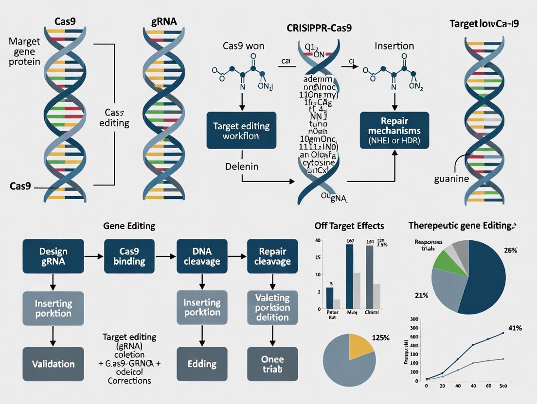
The molecular machinery of CRISPR-based gene editing represents a paradigm shift in biomedical science, offering unprecedented potential for treating genetic diseases at their source. At the core of this technology lies the precise interaction between guide RNA (gRNA) and Cas nucleases, which together enable targeted DNA cleavage. The cellular response to this cleavage—through DNA repair pathways such as Non-Homologous End Joining (NHEJ) and Homology-Directed Repair (HDR)—ultimately determines the editing outcome. For researchers and drug development professionals validating therapeutic gene editing in clinical trials, a sophisticated understanding of these components is not merely academic but fundamental to designing effective treatments. The ongoing clinical trials for conditions ranging from sickle cell disease to transthyretin amyloidosis demonstrate that the strategic manipulation of these molecular mechanisms can yield transformative therapies, making a thorough comparison of these systems essential for advancing the field.
The Core Components: gRNA and Cas Nucleases
Guide RNA (gRNA): The Targeting System
The guide RNA serves as the programmable homing device within the CRISPR system, dictating the precise genomic location where the Cas nuclease will create a double-strand break. In its natural setting in type II CRISPR-Cas systems, Cas9 requires two RNA molecules: the CRISPR RNA (crRNA), which contains the ~20 nucleotide spacer sequence complementary to the target DNA, and the trans-activating crRNA (tracrRNA), which facilitates complex formation [11]. For experimental and therapeutic applications, these are typically combined into a single guide RNA (sgRNA) that retains the targeting specificity of the crRNA and the structural functions of the tracrRNA [11] [12].
The gRNA can be produced through multiple methods, each with distinct implications for therapeutic development:
- In situ production: The gRNA is transcribed intracellularly from plasmid or viral DNA templates, enabling sustained expression but potentially increasing off-target risks [11].
- Ex situ production: gRNAs are synthesized in vitro then delivered to cells, allowing for chemical modifications that enhance stability, reduce immunogenicity, and improve targeting specificity [11]. These modified gRNAs can resist nuclease degradation and minimize unwanted immune responses mediated by Toll-like receptor 7 (TLR7) recognition [11].
Cas Nucleases: The Cutting Machinery
Cas nucleases are the effector proteins that create DNA double-strand breaks (DSBs) at locations specified by the gRNA. While Cas9 from Streptococcus pyogenes (SpCas9) remains the most widely used nuclease, numerous alternatives and engineered variants have been developed to address limitations in targeting range, specificity, and deliverability.
Table 1: Comparison of Key Cas Nuclease Variants for Research and Therapy
| Nuclease | PAM Requirement | Cleavage Pattern | Size (aa) | Key Features | Therapeutic Applications |
|---|---|---|---|---|---|
| SpCas9 | 5'-NGG-3' | Blunt ends | 1368 | Broadly characterized; many engineered variants available | Ex vivo therapies (e.g., Casgevy for SCD) |
| SaCas9 | 5'-NNGRRT-3' | Blunt ends | 1053 | Compact size suitable for AAV delivery | In vivo liver editing (preclinical) |
| Cas12a (Cpf1) | 5'-TTTV-3' | Staggered ends (5' overhangs) | ~1300 | Self-processes crRNAs; no tracrRNA needed | Potential for HDR-based approaches |
| hfCas12Max | 5'-TN-3' | Staggered ends | 1080 | Engineered high-fidelity variant; compact | Duchenne muscular dystrophy (HG-302 trial) |
| eSpOT-ON (ePsCas9) | Varies | Blunt ends | ~1100-1200 | Engineered for high fidelity without sacrificing efficiency | Clinical development stage |
The Cas protein structure consists of two primary lobes: the recognition (REC) lobe, responsible for binding the gRNA and target DNA, and the nuclease (NUC) lobe, which contains the RuvC and HNH nuclease domains that cleave the non-target and target DNA strands, respectively [11] [12]. Upon PAM recognition and successful DNA-RNA hybridization, Cas nucleases undergo a conformational change that activates their catalytic domains, resulting in a DSB approximately 3-5 base pairs upstream of the PAM site [11].
Diagram 1: gRNA-Cas Nuclease Target Recognition and Cleavage. The gRNA directs the Cas nuclease to a specific DNA sequence adjacent to a PAM site, resulting in a double-strand break (DSB).
DNA Repair Pathways: NHEJ vs. HDR
Following the creation of a DSB by Cas nucleases, cellular repair mechanisms are activated to restore genomic integrity. The competition between these pathways—primarily NHEJ and HDR—determines the editing outcome and must be carefully considered in therapeutic design.
Non-Homologous End Joining (NHEJ): The Dominant Pathway
Non-Homologous End Joining is an error-prone repair pathway that functions throughout the cell cycle by directly ligating broken DNA ends without requiring a homologous template [11] [13]. This pathway is favored in mammalian cells due to its speed and constant availability, but often results in small insertions or deletions (indels) at the repair site [13] [14]. These indels can disrupt gene function by introducing frameshift mutations or premature stop codons, making NHEJ particularly suitable for gene knockout strategies [13] [14].
In therapeutic contexts, NHEJ is harnessed for:
- Gene knockout: Introducing disruptive indels in disease-causing genes [13]
- Gene insertion: Facilitating integration of exogenous DNA fragments, though with less precision than HDR [13] [14]
- Exon skipping: Restoring reading frames in disorders like Duchenne muscular dystrophy [15]
Homology-Directed Repair (HDR): The Precision Pathway
Homology-Directed Repair is a precise, template-dependent repair mechanism that operates primarily during the S and G2 phases of the cell cycle when sister chromatids are available [13] [14]. HDR uses homologous sequences—either endogenous sister chromatids or exogenously supplied donor templates—to accurately repair DSBs without introducing errors [11] [13].
For precise genome editing, researchers provide a donor DNA template containing the desired modification flanked by homology arms complementary to the sequences surrounding the cut site [13] [14]. This approach enables a variety of precise edits:
- Gene correction: Fixing pathogenic point mutations [14]
- Gene insertion: Incorporating reporter genes or therapeutic transgenes [16] [14]
- Endogenous tagging: Adding epitope tags to study protein localization and function [16]
Despite its precision, HDR faces significant challenges in therapeutic applications due to its relatively low efficiency compared to NHEJ, restriction to specific cell cycle phases, and competition with other repair pathways [13] [16].
Table 2: Comparison of DNA Repair Pathways in CRISPR Genome Editing
| Characteristic | Non-Homologous End Joining (NHEJ) | Homology-Directed Repair (HDR) |
|---|---|---|
| Template Requirement | None | Homologous DNA template (endogenous or exogenous) |
| Cell Cycle Phase | All phases (especially G1) | S and G2 phases |
| Efficiency | High (dominant pathway in mammalian cells) | Low (0.5-20% in optimal conditions) |
| Fidelity | Error-prone (generates indels) | High-fidelity (precise editing) |
| Primary Applications | Gene knockouts, gene disruption | Gene correction, precise insertions, point mutations |
| Key Limitations | Lack of precision, unpredictable outcomes | Low efficiency, cell cycle dependence, donor delivery |
Alternative Repair Pathways: MMEJ and SSA
Beyond the major pathways, two alternative mechanisms—Microhomology-Mediated End Joining (MMEJ) and Single-Strand Annealing (SSA)—contribute to DSB repair outcomes and represent additional targets for optimizing editing precision.
Microhomology-Mediated End Joining (MMEJ) utilizes short homologous sequences (2-20 bp) flanking the DSB to facilitate repair, typically resulting in deletions [11] [16]. This POLQ-dependent pathway has been implicated in imprecise knock-in events, and its inhibition has been shown to improve HDR efficiency in some contexts [16].
Single-Strand Annealing (SSA) employs longer homologous sequences (typically >30 bp) and requires Rad52-mediated annealing [11] [16]. Recent research demonstrates that SSA inhibition reduces asymmetric HDR—a pattern of imprecise donor integration where only one side of the donor DNA is properly incorporated [16].
Diagram 2: Competing DNA Repair Pathways After CRISPR-Cas Cleavage. Double-strand breaks are repaired through multiple competing pathways, each producing distinct genetic outcomes.
Experimental Protocols for Pathway Analysis
Assessing DNA Repair Pathway Contributions in Knock-In Experiments
Understanding the complex interplay between DNA repair pathways requires carefully designed experimental approaches. Recent methodologies enable precise quantification of how different pathways contribute to editing outcomes.
Protocol: Long-Read Amplicon Sequencing for Repair Pattern Analysis [16]
Cell Preparation and Transfection:
- Use hTERT-immortalized RPE1 cells (human non-transformed diploid) maintained under standard conditions.
- Form ribonucleoprotein (RNP) complexes by mixing recombinant Cas nuclease (Cpf1 or Cas9) with in vitro transcribed gRNA.
- Electroporate RNP complexes along with PCR-amplified donor DNA containing 90 bp homology arms.
Pathway Inhibition:
- Immediately post-electroporation, treat cells with specific pathway inhibitors:
- NHEJ inhibition: Alt-R HDR Enhancer V2
- MMEJ inhibition: ART558 (POLQ inhibitor)
- SSA inhibition: D-I03 (Rad52 inhibitor)
- Maintain inhibitor treatment for 24 hours, covering the peak period of HDR activity.
- Immediately post-electroporation, treat cells with specific pathway inhibitors:
Sample Processing and Sequencing:
- Harvest cells 4 days post-electroporation for genomic DNA extraction.
- Amplify target loci using PCR with flanking primers.
- Perform long-read amplicon sequencing using PacBio Hi-Fi technology.
Data Analysis:
- Process sequencing data using the knock-knock computational framework to classify repair outcomes.
- Categorize sequences as: wild-type, indels, perfect HDR, or subtypes of imprecise integration (blunt, asymmetric HDR, imperfect).
- Quantify the frequency of each repair pattern under different inhibition conditions.
This protocol revealed that even with NHEJ inhibition, perfect HDR accounted for only about half of all integration events, with alternative pathways contributing to imprecise outcomes [16]. Simultaneous inhibition of multiple pathways may further enhance precise editing efficiency.
Strategies for Modulating Repair Pathway Choice
Researchers have developed multiple strategies to steer DNA repair toward desired pathways:
Enhancing HDR Efficiency [13] [16] [14]:
- Cell cycle synchronization: Enrich for S/G2 phase cells when HDR is active
- Pharmacological inhibition: Target key NHEJ proteins (e.g., DNA-PKcs inhibitors)
- Timed delivery: Introduce editing components when HDR machinery is most active
- SSA and MMEJ suppression: Inhibit Rad52 and POLQ to reduce competing pathways
Optimizing NHEJ-Mediated Integration [13] [14]:
- Donor design: Utilize double-stranded DNA donors with minimal homology
- Cas variant selection: Choose nucleases with cleavage profiles favoring NHEJ
- Pathway enhancement: Modulate NHEJ factors to improve integration efficiency
Clinical Validation and Therapeutic Applications
The strategic manipulation of DNA repair pathways has enabled the development of CRISPR-based therapies now advancing through clinical trials. These applications demonstrate how understanding the molecular machinery translates to therapeutic outcomes.
NHEJ-Dominated Therapeutic Approaches
Gene Disruption Strategies:
- Casgevy (exa-cel): The first FDA-approved CRISPR therapy for sickle cell disease and transfusion-dependent beta thalassemia uses ex vivo editing to disrupt the BCL11A gene, a repressor of fetal hemoglobin [17] [18]. This approach employs NHEJ to create disruptive indels that permanently inactivate the gene, leading to increased fetal hemoglobin production and reduced disease symptoms.
- NTLA-2001: Intellia Therapeutics' treatment for transthyretin amyloidosis (ATTR) utilizes LNP-delivered CRISPR-Cas9 to disrupt the TTR gene in the liver [17] [15]. This in vivo editing approach has demonstrated sustained reduction (>90%) of TTR protein levels in clinical trial participants, with functional improvement in disease symptoms [17].
HDR-Based Therapeutic Approaches
Precision Gene Editing Applications:
- PM359: Prime Medicine's candidate for chronic granulomatous disease (CGD) uses prime editing to correct mutations in the NCF1 gene ex vivo in hematopoietic stem cells [15]. This approach requires precise, HDR-like repair mechanisms to install specific nucleotide changes without creating DSBs.
- CTX211/VCTX210A: CRISPR Therapeutics' approach for type 1 diabetes involves HDR-mediated editing of allogeneic pancreatic endoderm cells to create immune-evasive insulin-producing cells for transplantation [15].
Advanced Clinical Trial Outcomes
Recent clinical results highlight the therapeutic potential of pathway-manipulated editing:
Intellia's hATTR Program: Phase I results published in 2024 demonstrated that a single dose of NTLA-2001 produced rapid, deep (>90%), and durable reduction of TTR protein levels, sustained over two years of follow-up [17]. The treatment was generally well-tolerated, supporting the advancement to global Phase III trials.
Intellia's HAE Program: October 2024 results from the Phase I/II trial of NTLA-2002 for hereditary angioedema showed that the higher dose group experienced an 86% reduction in kallikrein levels and a significant reduction in inflammatory attacks, with 8 of 11 participants attack-free during the 16-week observation period [17].
Personalized CRISPR Therapy: A landmark 2025 case reported the development of a bespoke in vivo CRISPR therapy for an infant with CPS1 deficiency, created and delivered in just six months [17]. The LNP-delivered treatment was safely administered in multiple doses, demonstrating the potential for personalized on-demand gene editing for rare genetic disorders.
Research Reagent Solutions
Table 3: Essential Research Reagents for Investigating CRISPR DNA Repair Pathways
| Reagent Category | Specific Examples | Research Application | Key Features |
|---|---|---|---|
| NHEJ Inhibitors | Alt-R HDR Enhancer V2 [16] | Enhancing HDR efficiency | Potent NHEJ pathway suppression |
| MMEJ Inhibitors | ART558 (POLQ inhibitor) [16] | Reducing MMEJ-mediated deletions | Specific targeting of POLQ polymerase |
| SSA Inhibitors | D-I03 (Rad52 inhibitor) [16] | Reducing asymmetric HDR | Inhibition of Rad52-mediated annealing |
| High-Fidelity Cas Variants | hfCas12Max [12], eSpOT-ON [12] | Reducing off-target effects | Engineered for enhanced specificity |
| Specialized Cas Nucleases | SaCas9 [19] [12], Cas12a [19] [20] | Expanding targeting range | Alternative PAM requirements; staggered cuts |
| Donor Template Systems | PCR-amplified donors with 90bp homology arms [16], ssODNs [14] | Facilitating HDR | Optimized homology for efficient recombination |
| Analysis Platforms | Knock-knock computational framework [16], long-read amplicon sequencing | Characterizing editing outcomes | High-resolution repair pattern classification |
The sophisticated interplay between gRNA-guided Cas nucleases and cellular DNA repair pathways represents both the challenge and promise of therapeutic gene editing. As clinical trials progress, it becomes increasingly evident that successful therapeutic outcomes depend on strategic pathway manipulation—whether harnessing NHEJ for efficient gene disruption or optimizing conditions for precise HDR-mediated correction. The expanding toolkit of Cas variants, pathway modulators, and delivery systems continues to address fundamental limitations in efficiency and specificity. For researchers and drug development professionals, a nuanced understanding of these molecular mechanisms provides the foundation for developing the next generation of CRISPR-based therapeutics, moving beyond proof-of-concept to durable treatments for genetic diseases.
The path from a therapeutic concept to an approved treatment is a meticulously structured journey designed to ensure safety and efficacy. For emerging fields like therapeutic gene editing, navigating the clinical trial pipeline is paramount to validating revolutionary technologies and bringing them to patients. This guide objectively compares the performance of gene editing therapies against traditional drug modalities across each clinical phase, providing a framework for researchers and drug development professionals.
The clinical trial pipeline is a multi-stage process that every new therapeutic must successfully pass through to gain regulatory approval. Its primary purpose is to systematically evaluate a drug's safety and efficacy in humans through a series of phased studies [21]. The pipeline begins after extensive preclinical research in labs and animal models, which provides initial data on a candidate's safety, toxicology, and biological activity [22] [23].
For therapeutic gene editing, this process validates not just a compound, but an entire technological platform. The pipeline is governed by strict regulatory standards, primarily enforced in the United States by the Food and Drug Administration (FDA). Researchers must submit an Investigational New Drug (IND) application to the FDA before initiating human trials. This application includes animal study data, manufacturing information, and clinical protocols [21] [24]. The subsequent clinical development is divided into three main phases (I, II, and III), followed by post-market monitoring (Phase IV) [21].
Detailed Breakdown of Clinical Trial Phases
Each phase of the clinical trial pipeline has a distinct objective, design, and success rate. The following table provides a comparative overview of these phases, including general success rates and specific performance data for gene therapies.
Table 1: Overview of Clinical Trial Phases and Success Metrics
| Trial Phase | Primary Objective | Typical Participants | Duration | General Industry Success Rate [25] | Gene Therapy Considerations |
|---|---|---|---|---|---|
| Phase 1 | Assess safety and dosage | 20-100 healthy volunteers or patients [21] | Several months [21] | ~70% move to next phase [21] | Often skips healthy volunteers; tests safety in patients with the target disease [22]. |
| Phase 2 | Evaluate efficacy and side effects | Up to several hundred patients [21] | Several months to 2 years [21] | ~33% move to next phase [21] | Phase I/II trials are often combined to accelerate development for serious rare diseases [22]. |
| Phase 3 | Confirm efficacy, monitor side effects | 300-3,000 patients [21] | 1 to 4 years [21] | 25-30% move to next phase [21] | Large, pivotal studies designed to provide definitive evidence for regulatory approval. |
| Phase 4 | Post-market safety monitoring | Several thousand patients [22] | Ongoing | N/A | Particularly critical for novel modalities like gene editing to track long-term safety [22]. |
Phase 1: Initial Safety and Dosing
- Core Objective: The primary goal of Phase 1 trials is to determine the safety profile of a new therapeutic and identify an appropriate dosage range. Researchers closely monitor how the drug interacts with the human body and assess any acute side effects [21].
- Protocol and Design: These studies are typically small-scale and tightly controlled. Dosing schemes are adjusted based on preclinical data to find the highest dose patients can tolerate without severe side effects [21]. While traditional drug trials often use healthy volunteers, gene and cell therapy trials are frequently more risky and specific, so they are typically tested directly in a small number of individuals with the disease [22].
- Gene Editing Performance: Early-phase trials for CRISPR therapies have demonstrated a favorable safety profile so far. A key advancement is the exploration of redosing. Unlike viral vector-based delivery, which can trigger dangerous immune reactions upon re-administration, Lipid Nanoparticle (LNP) delivery does not carry the same risk. For example, in a Phase I trial for hereditary transthyretin amyloidosis (hATTR), participants safely received a second infusion of the CRISPR therapy at a higher dose, and an infant with CPS1 deficiency safely received three doses, with each dose reducing symptoms further [17].
Phase 2: Therapeutic Efficacy and Expanded Safety
- Core Objective: Phase 2 trials aim to gather preliminary data on the therapeutic's efficacy for the targeted condition and to further evaluate its safety in a larger patient population [21].
- Protocol and Design: These studies involve more participants who have the disease or condition. They are not large enough to be definitive but provide critical data that researchers use to refine their methods and design robust Phase 3 protocols [21].
- Gene Editing Performance: For gene editing, efficacy is often measured through biomarkers that indicate target engagement. In Intellia Therapeutics' Phase I/II trial for hereditary angioedema (HAE), researchers used blood tests to measure the reduction of a disease-related protein (kallikrein). Participants receiving a higher dose had an average of 86% reduction in the protein, and the majority were attack-free in the 16 weeks following treatment [17]. This demonstrates a direct, measurable biological effect from the gene editing intervention.
Phase 3: Pivotal Confirmation of Safety and Efficacy
- Core Objective: Phase 3 trials are large-scale studies designed to definitively demonstrate whether a product offers a treatment benefit. They provide the comprehensive safety and efficacy data required for regulatory approval [21].
- Protocol and Design: These are often randomized, controlled, and double-blinded to eliminate bias. Because of their larger size and longer duration, Phase 3 trials are more likely to identify rare or long-term side effects that may not have been apparent in earlier phases [21].
- Gene Editing Performance: Success in Phase 3 leads to a Biologics License Application (BLA). The first CRISPR-based therapy to navigate this pipeline successfully was Casgevy, approved for sickle cell disease and transfusion-dependent beta thalassemia [17] [2]. For hATTR, the Phase III trial is designed to dose at least 500 participants and compare the effect to a placebo, with the data intended to support a commercialization application [17].
Regulatory Approval and Phase 4: Post-Market Surveillance
Following successful Phase 3 trials, developers submit a BLA to the FDA. Upon careful review, if the benefits are deemed to outweigh the risks, the treatment is approved for broader use [22]. Phase 4 studies, or post-market surveillance, are then required to monitor long-term safety and outcomes in the general patient population, which is especially important for novel therapies like gene editing [22].
Performance Comparison: Gene Editing vs. Traditional Drug Modalities
The clinical development of gene editing therapies exhibits distinct characteristics and challenges when compared to traditional small-molecule drugs. The data below highlight key comparative metrics.
Table 2: Performance and Development Metrics Comparison
| Development Metric | Traditional Small-Molecule Drugs | Gene Editing Therapies (CRISPR-based) |
|---|---|---|
| Typical Development Timeline | 10-15 years [23] | Accelerated pathways possible (e.g., first personalized CRISPR therapy developed and delivered in 6 months [17]) |
| Leading Cause of Clinical Failure | Lack of efficacy (~40-50%) [23] | Delivery challenges, long-term safety unknowns [17] [2] |
| Key Efficacy Measure | Symptom reduction, disease progression | Sustained reduction of disease-causing proteins, functional genetic correction [17] |
| Primary Safety Concerns | Off-target toxicity, side effects [23] | Off-target editing effects, immune responses to editing components or delivery vectors [2] |
| Representative Success Rate | Overall likelihood of approval from Phase 1 is less than 10-15% [23] [25] | Early successes in specific indications (e.g., Casgevy for sickle cell, hATTR with ~90% protein reduction [17]) |
The high failure rate for traditional drugs, largely due to a lack of efficacy, underscores a key potential advantage of gene editing: its rational design. By directly targeting the genetic root of a disease, it holds the promise of being more definitive. However, the field faces its own unique hurdles, with delivery being repeatedly cited as one of the biggest challenges—getting the editing machinery to the right cells in the body safely and efficiently [17] [2].
Essential Research Reagents and Experimental Protocols
The advancement of gene editing through the clinical pipeline relies on a specialized toolkit of reagents and standardized protocols.
Research Reagent Solutions
Table 3: Key Reagents for Therapeutic Gene Editing Research
| Research Reagent | Function in Gene Editing Experiments |
|---|---|
| CRISPR-Cas Nuclease (e.g., SpCas9, SaCas9) | The engine of the system; creates a double-strand break in the target DNA sequence [26] [2]. |
| Guide RNA (gRNA/sgRNA) | A synthetic RNA molecule that directs the Cas nuclease to a specific genomic locus via complementary base pairing [26] [2]. |
| Base Editors (e.g., ABE, CBE) | Chimeric proteins that enable the direct, irreversible conversion of one DNA base into another without causing a double-strand break, reducing genotoxicity [26] [2]. |
| Prime Editors (PE) | A versatile system that uses a Cas9 nickase-reverse transcriptase fusion and a prime editing guide RNA (pegRNA) to mediate all 12 possible base-to-base conversions, as well as small insertions and deletions, without double-strand breaks [26] [2]. |
| Lipid Nanoparticles (LNPs) | A non-viral delivery vehicle that encapsulates CRISPR components and facilitates their in vivo delivery, particularly to the liver [17]. |
| Viral Vectors (e.g., AAV) | Genetically engineered viruses used as vehicles to deliver gene editing machinery into cells, often used in ex vivo settings [17]. |
Key Experimental Protocols
To generate the data required for an IND submission and clinical trial progression, several core experiments must be conducted.
- In Vitro Efficacy and Specificity Assessment: The initial proof-of-concept involves delivering the CRISPR ribonucleoprotein (RNP) or genetic material into cultured human cells related to the target disease. The primary protocol involves transfection or electroporation, followed by next-generation sequencing (NGS) of the target locus to quantify editing efficiency and to screen for potential off-target edits across the genome [26] [2].
- In Vivo Safety and Efficacy in Animal Models: Following successful in vitro results, the therapeutic candidate is tested in animal models, such as mice or non-human primates. The protocol involves the systemic or localized administration of the therapy (e.g., via IV injection for LNP delivery). Researchers then analyze target tissue biopsies for editing efficiency and off-target effects. They also conduct comprehensive histopathology and blood chemistry analyses to assess overall animal health and detect any signs of toxicity or immune reaction [17] [2].
- Analytical Methods for Clinical Trials: In clinical trials, success is measured through specific, validated assays. For example, in trials for hATTR and HAE, blood tests are used to measure the reduction in disease-causing proteins (TTR and kallikrein, respectively). Additionally, functional and quality-of-life assessments are used to correlate biochemical changes with clinical improvement for patients [17].
Visualizing the Clinical Trial and Gene Editing Workflow
The diagram below illustrates the standard clinical trial pathway integrated with the critical research and development milestones specific to gene editing therapies.
Clinical Trial Pathway for Gene Editing Therapies
The diagram shows the standard phases (blue) and highlights critical, gene-editing-specific R&D activities (dashed outlines). Key differentiators include the early focus on delivery system optimization and off-target analysis, the central role of biomarker validation in establishing efficacy in mid-stage trials, and the essential long-term follow-up required after treatment.
The clinical trial pipeline provides the essential structured framework for validating the safety and efficacy of therapeutic gene editing. While this process shares the same rigorous phases as traditional drug development, the performance and considerations for CRISPR-based therapies are distinct. Current clinical data demonstrate remarkable successes, such as sustained reduction of disease-causing proteins and the first regulatory approvals, validating the potential of this modality [17].
However, the path forward is not without challenges. The field must continue to address hurdles related to delivery, long-term safety monitoring, and the economic pressures of drug development [17] [23] [25]. The ongoing evolution of the toolkit—with base editing, prime editing, and improved delivery systems—promises to expand the scope of treatable diseases [26] [2]. For researchers and drug developers, successfully navigating this complex pipeline requires a deep understanding of both its universal requirements and the unique demands of gene editing, ultimately ensuring these transformative therapies can safely reach the patients who need them.
The advent of CRISPR-Cas9 revolutionized genetic engineering by providing researchers with an unprecedented ability to target and cut specific DNA sequences. However, the reliance on double-strand breaks (DSBs) and the subsequent activation of DNA repair pathways introduced limitations, including unwanted indel formations and substantial off-target effects that pose significant challenges for therapeutic applications. The gene editing landscape has since evolved beyond cutting, with next-generation editors offering more precise and versatile solutions for modifying genetic information.
This evolution is particularly crucial within therapeutic gene editing, where the goal is to correct pathogenic mutations without introducing new genetic damage. Base editing and prime editing represent two transformative technological advances that address these challenges. These systems enable precise genome modification without requiring DSBs, thereby minimizing genotoxic risks and expanding the potential for clinical translation. As the field moves toward validating these tools in clinical trials, understanding their distinct mechanisms, capabilities, and experimental validation becomes essential for researchers and drug development professionals.
Understanding Base Editing
Molecular Mechanism
Base editors are sophisticated molecular machines that combine a catalytically impaired Cas nuclease with a deaminase enzyme to achieve single-nucleotide conversions without creating DSBs. The system operates through a coordinated multi-step process. The Cas nickase portion, still capable of binding DNA, is guided to a specific genomic locus by a gRNA. Once bound, the tethered deaminase enzyme catalyzes a chemical conversion on a single DNA strand within an accessible window of nucleotides, typically 3-5 bases wide.
Two main classes of base editors have been developed: Cytosine Base Editors (CBEs) convert C•G base pairs to T•A, while Adenine Base Editors (ABEs) convert A•T base pairs to G•C. Following deamination, the edited strand is processed by cellular repair machinery to permanently incorporate the base change, while the complementary strand is nicked to encourage repair that favors the edited base. This elegant mechanism enables efficient and precise nucleotide conversion with minimal indel formation compared to traditional CRISPR-Cas9 approaches [27].
Key Advances and Experimental Validation
Recent research has focused on enhancing the predictive accuracy and performance of base editing systems. A landmark November 2025 study in Nature Communications introduced CRISPRon, a deep learning framework that substantially improves prediction of base editing outcomes by training simultaneously on multiple experimental datasets while tracking their origins. This approach addresses a critical challenge in the field—the heterogeneity of data generated from different experimental conditions, editor variants, and cellular contexts [27].
The experimental methodology behind this advance involved generating substantial new data using SURRO-seq technology, which created libraries pairing gRNAs with their target sequences integrated into the genome. Researchers measured base-editing efficiency for approximately 11,500 gRNAs each for ABE7.10 and BE4-Gam base editors in HEK293T cells. Analysis revealed that ABE7.10 exhibited highly specific adenine-to-guanine transitions at 97%, while BE4 showed 92% cytosine-to-thymine specificity. Both editors displayed peak activity at positions four through eight in the protospacer sequence [27].
Notably, the team developed a novel training strategy that incorporated dataset origin as a feature vector, allowing the model to learn systematic differences across experimental conditions. This enabled users to tailor predictions to specific base editors and experimental setups—a crucial capability for therapeutic design. When validated on independent datasets, the CRISPRon models (CRISPRon-ABE and CRISPRon-CBE) demonstrated consistent superiority over existing methods, including DeepABE/CBE, BE-HIVE, BE-DICT, BE_Endo, and BEDICT2.0 [27].
Table 1: Key Base Editing Systems and Their Characteristics
| Editor Type | Conversion | Deaminase | Editing Window | Key Applications |
|---|---|---|---|---|
| BE4 (CBE) | C•G to T•A | rAPOBEC1 | ~5 nucleotides | Disease modeling, pathogenic variant correction |
| ABE7.10 | A•T to G•C | TadA-TadA* | ~5 nucleotides | Therapeutic correction of point mutations |
| ABE8e | A•T to G•C | Engineered TadA | ~5 nucleotides | Enhanced efficiency for therapeutic applications |
Understanding Prime Editing
Molecular Mechanism
Prime editing represents a more versatile genome editing platform that directly writes new genetic information into a specified DNA site without requiring DSBs or donor DNA templates. The system comprises two core components: a prime editing guide RNA (pegRNA) and a fusion protein consisting of a Cas9 nickase reverse transcriptase enzyme.
The prime editing process begins with the binding of the prime editor complex to the target DNA site. The Cas9 nickase then makes a single-strand cut at the target site, exposing a 3' DNA flap. The pegRNA serves a dual function: it directs the complex to the specific genomic locus and also serves as a template for the reverse transcriptase. The reverse transcriptase uses the 3' end of the nicked DNA strand as a primer and the pegRNA as a template to synthesize a DNA fragment containing the desired edit. Cellular repair mechanisms then resolve this intermediate structure to permanently incorporate the edit into the genome [28].
This sophisticated mechanism enables a wider range of precise edits—including all 12 possible base-to-base conversions, small insertions, and small deletions—with exceptionally high precision and minimal off-target effects compared to both traditional CRISPR-Cas9 and base editing systems.
Key Advances and Experimental Validation
Recent research has dramatically improved the efficiency and precision of prime editing systems. In September 2025, MIT researchers announced a breakthrough approach that lowered the error rate of prime editors from approximately one error in seven edits to one in 101 for the most-used editing mode, and from one error in 122 edits to one in 543 for a high-precision mode [29].
The experimental methodology behind this improvement involved engineering novel Cas9 mutations that created instability in the old DNA strands, making them more susceptible to degradation and thereby favoring incorporation of the newly edited strands. The researchers identified specific Cas9 mutations that dropped the error rate to 1/20th of the original value, and by combining pairs of these mutations, created a Cas9 editor that lowered the error rate further to 1/36th of the original. The final optimized editor, termed vPE, incorporated these modified Cas9 proteins with an RNA binding protein that stabilizes the ends of the RNA template more efficiently, achieving an error rate just 1/60th of the original prime editing system [29].
A groundbreaking application of prime editing was reported in November 2025 with the development of PERT (Prime Editing-mediated Readthrough of Premature Termination Codons). This innovative strategy addresses nonsense mutations, which account for approximately 24% of pathogenic alleles in the ClinVar database and cause about one-third of rare genetic diseases. Rather than correcting individual mutations, PERT uses prime editing to permanently convert a dispensable endogenous tRNA into an optimized suppressor tRNA (sup-tRNA) that enables readthrough of premature termination codons [30] [28].
The experimental protocol for PERT development involved iterative screening of thousands of variants of all 418 human tRNAs to identify those with the strongest sup-tRNA potential. Researchers optimized prime editing agents to install an engineered sup-tRNA at a single genomic locus without overexpression. In validation experiments using human cell models of Batten disease, Tay-Sachs disease, and cystic fibrosis, treatment with the same prime editor programmed to install the optimized sup-tRNA resulted in restoration of 20-70% of normal enzyme activity. In a mouse model of Hurler syndrome, in vivo delivery of a single prime editor that converts an endogenous mouse tRNA into a sup-tRNA extensively rescued disease pathology, demonstrating the therapeutic potential of this approach [30] [28].
Figure 1: Prime Editing Mechanism - The prime editor complex, consisting of a Cas9 nickase fused to reverse transcriptase, is guided to the target DNA by a pegRNA. The system nicks one DNA strand, then uses the reverse transcriptase to synthesize a new DNA flap containing the desired edit, which is subsequently integrated into the genome through cellular repair mechanisms.
Comparative Analysis: Base Editing vs. Prime Editing
Technical Comparison
When selecting an appropriate genome editing platform for therapeutic development, researchers must consider multiple technical parameters. The following table provides a systematic comparison of key characteristics between base editing and prime editing systems:
Table 2: Technical Comparison of Base Editing and Prime Editing Platforms
| Parameter | Base Editing | Prime Editing |
|---|---|---|
| DNA Break Mechanism | Single-strand nick | Single-strand nick |
| Editing Scope | Specific transitions (C→T, A→G) | All 12 base-to-base conversions, insertions, deletions |
| Theoretical Target Coverage | Limited by PAM and editing window constraints | Expanded targeting via pegRNA design flexibility |
| Maximum Efficiency | ~50-90% in optimized systems | ~20-50% in current systems |
| Off-Target Profile | Minimal DSB-related off-targets; potential RNA off-targets | Lowest reported off-target effects among editing platforms |
| Size Constraints | Limited by delivery vehicle capacity | Larger construct size may challenge viral packaging |
| Key Advantages | High efficiency for specific conversions, simplified design | Versatility, precision, minimal byproducts |
| Primary Limitations | Restricted to specific base changes, bystander edits | Complex pegRNA design, variable efficiency across loci |
Therapeutic Applications and Clinical Relevance
The distinct capabilities of base and prime editing platforms make them suitable for different therapeutic contexts. Base editors excel in scenarios requiring correction of specific point mutations that fall within its convertible bases, particularly for monogenic disorders caused by defined single-nucleotide polymorphisms. Their high efficiency and relatively straightforward design make them attractive for ex vivo therapeutic applications, such as engineering hematopoietic stem cells or immune cells for adoptive cell therapies.
Prime editing's broader editing scope positions it as a more versatile platform for addressing diverse genetic mutations, including those that base editors cannot correct. The PERT strategy exemplifies how prime editing can enable disease-agnostic therapeutic approaches—a single editing agent potentially treating multiple different genetic diseases caused by nonsense mutations. This has significant implications for drug development economics, as it could circumvent the need to develop individual therapies for each rare genetic disorder [30] [28].
Both platforms are progressing toward clinical validation. While CRISPR-based therapies have already reached patients—with the first FDA approval of Casgevy for sickle cell disease and beta-thalassemia in 2023—base and prime editing therapies are advancing through preclinical development. Recent clinical trial updates indicate growing industry investment in these technologies, with multiple programs expected to enter clinical testing in the coming years [17].
Experimental Design & Methodology
Essential Reagents and Tools
Implementing base or prime editing experiments requires careful selection of molecular tools and delivery systems. The following research reagent solutions are essential for successful experimental execution:
Table 3: Essential Research Reagent Solutions for Next-Generation Editing
| Reagent Category | Specific Examples | Function & Application Notes |
|---|---|---|
| Editor Plasmids | BE4max, ABE8e, PEmax | Engineered for enhanced efficiency and nuclear localization; codon-optimized for target species |
| Delivery Systems | AAV, LNPs, Electroporation | AAV for in vivo delivery; LNPs for clinical translation; electroporation for ex vivo applications |
| gRNA/pegRNA | Modified RNA, U6-driven expression | Chemically modified gRNAs enhance stability; pegRNA optimization critical for prime editing efficiency |
| Validation Tools | NGS panels, GUIDE-seq, Digenome-seq | Comprehensive off-target profiling essential for therapeutic development |
| Cell Culture Models | iPSCs, Primary cells, Organoids | Physiologically relevant models for evaluating therapeutic efficacy and safety |
Protocol Framework for Therapeutic Editing Validation
A robust experimental protocol for validating next-generation editors in therapeutic contexts should include these critical steps:
Target Selection and gRNA/pegRNA Design: Identify target sites with minimal predicted off-targets. For base editing, consider the editing window and potential bystander edits. For prime editing, optimize pegRNA length and secondary structure. Computational tools like CRISPRon can predict editing outcomes for specific base editor variants [27].
Editor Delivery: Select appropriate delivery method based on experimental system. For in vitro studies, transfection or electroporation of RNP complexes offers high efficiency with reduced off-target effects. For in vivo applications, lipid nanoparticles (LNPs) or viral vectors (AAV) are preferred. Recent clinical advances have demonstrated the safety and efficacy of LNP delivery for in vivo CRISPR therapies, with multiple doses possible due to reduced immunogenicity compared to viral vectors [17].
Efficiency Assessment: Quantify editing efficiency using next-generation sequencing (NGS) of the target locus. For therapeutic applications, aim for >20% efficiency for prime editing and >50% for base editing, though these thresholds vary by target and application.
Specificity Validation: Employ unbiased genome-wide methods like GUIDE-seq or CIRCLE-seq to comprehensively identify off-target edits. The improved specificity of next-generation editors should be confirmed through these sensitive detection methods.
Functional Validation: Assess functional correction in disease-relevant models. For the PERT system, this involved measuring enzyme activity restoration in cell models of genetic diseases and pathological rescue in animal models [30] [28].
Safety Profiling: Evaluate potential genotoxic effects through cell viability assays, karyotyping, and transcriptomic analysis. For clinical translation, comprehensive toxicology studies in relevant animal models are essential.
Figure 2: Therapeutic Editing Validation Workflow - A comprehensive framework for validating next-generation editors in therapeutic contexts, from target selection through safety assessment.
The development of base editing and prime editing technologies represents a paradigm shift in therapeutic genome engineering, moving beyond the limitations of traditional CRISPR-Cas9 systems. While base editors offer highly efficient correction of specific point mutations, prime editors provide unprecedented versatility in writing diverse genetic changes without DSBs. The recent advances in predictive algorithms, editing precision, and innovative strategies like PERT demonstrate the rapid maturation of these platforms toward clinical application.
For researchers and drug development professionals, the selection between these platforms depends heavily on the specific therapeutic objective. Base editing may be preferable for defined single-nucleotide corrections where its high efficiency and simpler design facilitate development. In contrast, prime editing offers a more flexible solution for diverse mutation types and enables innovative, disease-agnostic approaches. As both technologies continue to evolve—with improvements in efficiency, specificity, and delivery—they hold tremendous promise for expanding the scope of treatable genetic disorders and accelerating the development of transformative genetic medicines.
The ongoing clinical validation of CRISPR-based therapies provides a roadmap for the translation of these next-generation editors. With continued optimization and rigorous safety assessment, base and prime editing platforms are poised to significantly expand the therapeutic landscape for genetic diseases in the coming decade.
The development of transformative genetic medicines, particularly for rare diseases, is challenging the paradigms of traditional drug evaluation. Regulatory agencies, led by the U.S. Food and Drug Administration (FDA), are creating novel pathways to address the unique challenges posed by bespoke therapies and very small patient populations. These evolving regulatory foundations are critical for validating therapeutic gene editing in clinical research, balancing the need for robust evidence with the practical realities of treating ultra-rare conditions [31] [32].
For researchers and drug development professionals, understanding these pathways—from established expedited programs to emerging N-of-1 frameworks—is essential for navigating the development of precision genetic medicines. This guide objectively compares these regulatory options, their evidence requirements, and their application to gene editing therapies, providing a foundation for strategic development planning.
Established FDA Expedited Pathways: A Comparative Analysis
The FDA has long utilized specialized programs to accelerate therapies for serious conditions. These pathways reduce development timelines and improve success rates, particularly for rare diseases and oncology.
Table 1: Comparison of Key FDA Expedited Development Pathways
| Pathway Feature | Breakthrough Therapy (BTD) | Fast Track | Accelerated Approval | Priority Review |
|---|---|---|---|---|
| Purpose | Expedite development for substantial improvement over available therapies | Facilitate development for unmet medical needs | Approve based on surrogate endpoints likely to predict clinical benefit | Shorten review timeline for significant advances |
| Success Rate | 72% approval rate (2013-2022) [33] | 31 approvals in 2024 [34] | 80% of 2024 accelerated approvals were in oncology [34] | 96% for BTD, 98% for Accelerated Approval [34] |
| Key Benefits | Intensive FDA guidance, organizational commitment | Rolling review, early FDA communication | Approval based on surrogate endpoints | 6-month review (vs. 10-month standard) |
| Designation Timing | Requires preliminary clinical evidence | Can be based on nonclinical or clinical data | Can be requested after evidence generation | Determined during filing or with application |
| Therapeutic Area Prevalence | Oncology (46%), Infectious Disease (11%), Metabolic (8%) [33] | Across serious conditions with unmet needs | Primarily oncology and rare diseases | Across therapeutic areas with significant advances |
The data demonstrates that expedited pathways have become standard for innovative therapies, with 57% of 2024 applications utilizing at least one such designation [34]. Breakthrough Therapy Designation shows particularly strong correlation with ultimate approval, with 72% of designated products (2013-2022) achieving approval and another 13% still under review [33]. Rare disease products account for the majority of breakthrough designations (383 of 599 total between 2013-2025), highlighting FDA's focus on these conditions [33].
Table 2: Gene Editing-Specific Clinical Trial Designs and Evidence Generation Approaches
| Trial Design Element | Traditional RCT Approach | Adapted Designs for Rare Diseases | N-of-1/Few Considerations |
|---|---|---|---|
| Control Group | Concurrent placebo control | External controls, natural history comparisons | Patient as own control (pre-post) |
| Endpoint Selection | Clinical endpoints validated in large populations | Biomarkers, physiologic measures, clinical outcome assessments | Patient-specific clinical outcomes, biomarker correlation |
| Statistical Framework | Frequentist, p<0.05 significance | Bayesian approaches, disease progression modeling | Descriptive analysis, comparison to natural history |
| FDA Recognition | Gold standard but often infeasible | Supported in FDA's Innovative Designs guidance [31] | Emerging framework under Plausible Mechanism Pathway [31] |
The Plausible Mechanism Pathway: A New Framework for Ultra-Rare Diseases
In November 2025, FDA leadership unveiled the "Plausible Mechanism Pathway" targeting products for which randomized trials are not feasible, representing a significant shift in regulating bespoke therapies [31]. This pathway addresses the critical challenge that traditional development approaches are "failing" for ultra-rare diseases where the randomized controlled trial construct and p-value less than 0.05 are not "fit for purpose" [31].
Core Pathway Elements
The Plausible Mechanism Pathway requires satisfaction of five core elements [31]:
- Identification of a specific molecular or cellular abnormality, not a broad set of consensus diagnostic criteria
- The medical product targets the underlying or proximate biological alterations
- The natural history of the disease in the untreated population is well-characterized
- Confirmation exists that the target was successfully drugged, edited, or both
- Improvement in clinical outcomes or course of disease is demonstrated
The pathway leverages the expanded access single-patient IND paradigm as a vehicle for future marketing applications, treating successful single-patient outcomes as an evidentiary foundation rather than transforming expanded access directly into approval [31]. While initially focused on cell and gene therapies, the pathway remains available for common diseases with no proven alternatives or considerable unmet need [31].
Evidence Requirements and Postmarket Commitments
A crucial innovation of this pathway is how it aligns with statutory standards by permitting effectiveness to be demonstrated through confirmation that the target was successfully edited [31]. FDA will embrace non-animal models where possible and consider patients as their own controls [31].
The postmarketing framework requires collection of real-world evidence to demonstrate: (1) preservation of efficacy, (2) no off-target edits, (3) effect of early treatment on childhood development milestones, and (4) detection of unexpected safety signals [31]. This bears hallmarks of accelerated approval confirmatory trials but is adapted for bespoke therapies.
Diagram 1: Plausible Mechanism Pathway Flow
Regulatory Frameworks for N-of-1 and Bespoke Therapies
The most personalized end of the regulatory spectrum involves therapies developed for individual patients or very small groups (N-of-few). For conditions affecting fewer than 100 individuals globally—termed "nano-rare"—highly personalized approaches are often necessary [32].
Current Regulatory Mechanisms for Individualized Therapies
Table 3: Regulatory Pathways for N-of-1 and Bespoke Therapies
| Regulatory Aspect | Research IND (U.S.) | Expanded Access/Compassionate Use | Named Patient Program (EU) |
|---|---|---|---|
| Legal Basis | Investigational New Drug application [32] | Compassionate Use program [32] | Article 5(1) of Directive (EC) 2001/83 [32] |
| Administrative Process | Form 1571, full IRB review [32] | Streamlined Form 3926, administrative IRB review [32] | Physician request to manufacturer, ethics committee approval [32] |
| Review Timeline | 30 days (can be expedited) [32] | Few hours to 30 days based on urgency [32] | Varies by member state [32] |
| Intent | Non-commercial research [32] | Treatment outside clinical trials [32] | Treatment with unauthorized medicines [32] |
| Guidance Available | FDA draft guidance for ASO therapies [32] | Established procedures [32] | Limited specific guidance for N-of-1 [32] |
The FDA has issued specific guidance for antisense oligonucleotide (ASO) therapies, making them one of the few technologies with tailored regulatory advice for individualized therapies [32]. These guidelines specify that products should belong to well-characterized chemical classes with substantial clinical and nonclinical experience [32].
In Europe, no IND application is required for N-of-1 therapies, creating regulatory gaps in manufacturing standards, liability, and reimbursement [32]. The EMA has not provided specific guidance for N-of-1 therapies historically, though recent draft guidance on oligonucleotide development begins to address these treatments [32].
Case Study: Landmark Personalized CRISPR Intervention
The 2025 case of "Baby K.J." represents a landmark demonstration of personalized CRISPR therapy regulatory precedent [31] [17]. An infant with CPS1 deficiency received a bespoke in vivo CRISPR therapy developed and delivered in just six months [17].
Experimental Protocol and Methodology:
- Therapeutic Approach: Personalized CRISPR-Cas9 therapy delivered via lipid nanoparticles (LNPs) for in vivo editing [17]
- Dosing Strategy: Multiple doses administered via IV infusion to increase percentage of edited cells [17]
- Safety Monitoring: No serious side effects reported; improvement in symptoms and decreased medication dependence [17]
- Development Timeline: Six months from development to delivery [17]
This case established that LNP-delivered CRISPR therapies can be safely redosed, unlike viral vector approaches that typically trigger immune responses preventing readministration [17]. Each additional dose further reduced symptoms, suggesting additional editing with each administration [17].
Gene Editing vs. Gene Therapy: Regulatory Implications
Understanding the distinction between gene editing and gene therapy is crucial for regulatory planning, as these modalities face different evidence requirements and safety considerations.
Table 4: Regulatory Considerations for Gene Editing vs. Gene Therapy
| Consideration | Gene Editing | Gene Therapy |
|---|---|---|
| Mechanism | Direct modification of endogenous DNA sequence [35] | Introduction of functional gene copy [35] |
| Durability | Potentially permanent, especially in self-renewing cells [35] | Often temporary or variable; non-integrated transgenes may be lost [35] |
| Primary Safety Concerns | Off-target edits, genotoxicity, unpredictable long-term effects [35] | Immune responses to vectors, insertional mutagenesis, integration events [35] |
| Delivery Methods | Viral vectors, LNPs, or ex vivo modification [17] [35] | Typically viral vectors (AAV, lentiviral) [35] |
| Regulatory Precedents | Casgevy (CRISPR for SCD/TDT) [35] | Luxturna, Zolgensma [35] |
| Monitoring Requirements | Long-term follow-up for off-target effects, clonal expansion [35] | Long-term follow-up for immune complications, insertional mutagenesis [35] |
Diagram 2: Gene Therapy vs Gene Editing Pathways
Essential Research Reagents and Technologies
Advancing gene therapies through regulatory pathways requires specific research tools and platforms. The following table details key reagents and their functions in therapeutic development.
Table 5: Essential Research Reagent Solutions for Gene Editing Therapeutics
| Research Reagent/Category | Primary Function | Application in Development |
|---|---|---|
| Lipid Nanoparticles (LNPs) | In vivo delivery of editing components [17] | Liver-targeted therapies (e.g., hATTR, HAE) [17] |
| AAV Vectors | In vivo delivery of genetic payload [35] | Retinal diseases (Luxturna), neuromuscular disorders (Zolgensma) [35] |
| CRISPR-Cas Systems | Precise genome editing [35] | Gene disruption (Casgevy), correction of mutations [35] |
| Phosphorothioate Oligonucleotides | Stabilized ASO backbones [32] | Individualized antisense oligonucleotide therapies [32] |
| Single-Chain Variable Fragments (scFv) | Antigen recognition domain in CAR-T cells [36] | CAR-T therapies for oncology [36] |
| Clinical Outcome Assessments (COAs) | Measure patient-reported outcomes [37] | Patient-focused drug development, endpoint qualification [37] |
The regulatory landscape for gene editing therapies is rapidly evolving toward greater flexibility for ultra-rare diseases while maintaining rigorous evidence standards. Successfully navigating this landscape requires:
Early Regulatory Engagement: Proactively seeking FDA feedback through existing mechanisms like the Rare Disease Evidence Principles process, which clarifies evidence expectations for rare genetic conditions [31].
Platform Validation: Developing well-characterized platform technologies (LNP delivery, CRISPR systems, ASO chemistry) that can be leveraged across multiple individual therapies [31] [32].
Natural History Investment: Comprehensive natural history studies remain fundamental for establishing comparators for N-of-1 and small population studies [31] [32].
Postmarket Planning: Robust real-world evidence collection frameworks are increasingly integral to approval pathways, particularly for bespoke therapies [31].
The emergence of the Plausible Mechanism Pathway and refined approaches to N-of-1 therapies represents a pragmatic regulatory evolution to address the challenges of personalized genetic medicines. For researchers and developers, these frameworks offer promising routes to patients while maintaining scientific rigor and patient protection standards.
Methodologies and Real-World Applications: Measuring Success in Clinical Trials
Validating therapeutic gene editing in clinical trials research demands precise, reliable, and efficient assessment of on-target editing efficiency. The selection of an appropriate analytical method is paramount, as it directly influences the development and application of genome editing strategies, from initial proof-of-concept studies to critical quality control checks for clinical-grade therapies [38]. This guide provides a comparative analysis of five widely used techniques—T7 Endonuclease I (T7EI) assay, Tracking of Indels by Decomposition (TIDE), Inference of CRISPR Edits (ICE), droplet digital PCR (ddPCR), and live-cell reporter assays—to aid researchers and drug development professionals in selecting the optimal tool for their specific application.
The following table summarizes the core principles, key performance metrics, and primary applications of each method, providing a foundation for informed selection.
Table 1: Comprehensive Comparison of Gene Editing Efficiency Assessment Methods
| Method | Principle | Throughput | Quantitative Nature | Key Quantitative Performance | Key Strengths | Key Limitations | Ideal for Clinical Trial Phase |
|---|---|---|---|---|---|---|---|
| T7EI Assay | Cleaves heteroduplex DNA at mismatch sites; analysis by gel electrophoresis [38] | Medium | Semi-quantitative [38] | Lacks sensitivity of quantitative techniques [38] | Low cost, rapid, simple protocol [39] | Underestimates efficiency with single dominant indel; no sequence information [40] [39] | Pre-clinical, early screening |
| TIDE | Decomposes Sanger sequencing chromatograms to estimate indel frequencies [38] [40] | High | Quantitative [38] | Accuracy decreases with complex indels or low/high editing rates [40] | Cost-effective (uses Sanger data); user-friendly web tool [40] [39] | Limited accuracy for +1 insertions and complex indels [40] [39] | Pre-clinical, guide RNA screening |
| ICE | Decomposes Sanger sequencing traces to determine editing efficiency and indel spectrum [39] | High | Quantitative [39] | High correlation with NGS (R² = 0.96) [39] | Detects large indels; provides detailed indel distribution; high accuracy [39] | Accuracy dependent on sequencing quality [38] | Pre-clinical to manufacturing QC |
| ddPCR | Uses fluorescent probes to measure edit frequencies via droplet partitioning [38] | Medium | Highly quantitative and precise [38] | High precision for allelic modifications (e.g., NHEJ vs. HDR) [38] | Excellent quantitative precision; discriminates between edit types [38] | Requires specific probe design; limited to predefined edits [38] | Pre-clinical (mechanistic) to clinical (potency assays) |
| Live-Cell Reporter Assays | Fluorescent reporter activation upon successful editing; readout by flow cytometry [38] | High (with flow cytometry) | Quantitative (via fluorescence) [38] | Enables live-cell tracing and kinetic studies [38] | Functional readout; enables kinetic studies and cell sorting [38] | Assays engineered loci, not endogenous chromatin context [38] | Pre-clinical, tool development & screening |
A systematic comparison of computational tools like TIDE and ICE using artificial sequencing templates has demonstrated that while they perform well with simple indels, their estimated values can become more variable with complex indels [40]. Among these, DECODR was noted as providing the most accurate estimations for most samples in one study, though it was not a primary method requested for comparison [40].
Experimental Protocols for Key Assays
T7 Endonuclease I (T7EI) Assay Protocol
The T7EI assay is a cornerstone method for initial, rapid assessment of editing activity [38] [39].
- PCR Amplification: Amplify the target genomic region from both edited and wild-type control samples using high-fidelity PCR master mix. Standard reaction volumes are 25 µL [38].
- PCR Product Purification: Purify the resulting PCR products using a commercial gel and PCR clean-up kit [38].
- Heteroduplex Formation: Denature and re-anneal the purified PCR products to form heteroduplexes. A typical protocol involves heating to 95°C followed by slow cooling to room temperature [39].
- T7EI Digestion: Digest the heteroduplexed DNA by incubating with T7 Endonuclease I and the appropriate buffer (e.g., NEBuffer 2) at 37°C for 30 minutes [38].
- Analysis: Separate the digestion products by agarose gel electrophoresis (e.g., 1-2% agarose). Visualize DNA bands using an imaging system. Editing efficiency is estimated via densitometric analysis using the formula: % Indel = [1 - (1 / (a + b))] * 100, where a and b are the integrated intensities of the cleaved bands, and c is the intensity of the uncleaved band [38].
TIDE & ICE Protocol (Sanger Sequencing-Based Analysis)
TIDE and ICE both utilize Sanger sequencing data but employ different decomposition algorithms [40].
- PCR Amplification and Sequencing: Amplify the target region from test and control samples. Purify PCR products and submit for Sanger sequencing [38].
- Data Upload:
- TIDE: Upload the wild-type (reference) and edited sample sequencing chromatograms (in
.ab1format) to the TIDE web tool. Specify the cut site location (typically 3 bp upstream of the PAM sequence) and set the analysis window (e.g., 100-200 bp around the cut site) [38]. - ICE: Upload the Sanger sequencing
.ab1files (or FASTA sequences) for the edited sample and the reference sequence to the ICE web tool (e.g., Synthego ICE). Input the guide RNA sequence for analysis [39].
- TIDE: Upload the wild-type (reference) and edited sample sequencing chromatograms (in
- Analysis Execution: Run the decomposition analysis with default or customized parameters (e.g., indel size range). The tools will generate a report detailing editing efficiency (ICE score or indel frequency) and the spectrum of indel sequences present [38] [39].
ddPCR Assay Protocol for HDR Quantification
ddPCR offers absolute quantification of specific editing events, such as HDR or precise base edits [38].
- Assay Design: Design and validate two sets of hydrolysis probes with different fluorescent dyes (e.g., FAM and HEX/VIC). One probe targets the edited allele, the other targets the wild-type allele.
- Droplet Generation: Partition the PCR reaction mix, containing the genomic DNA sample, primers, probes, and ddPCR supermix, into thousands of nanoliter-sized droplets [38].
- Endpoint PCR Amplification: Perform PCR amplification on the droplet emulsion.
- Droplet Reading and Analysis: Read the droplets in a droplet reader to classify each as FAM-positive (edited), HEX/VIC-positive (wild-type), positive for both, or negative. The ratio of edited to total droplets provides an absolute count of the target molecule concentration, allowing for precise calculation of editing frequency [38].
Workflow and Selection Pathway Diagrams
The following diagram illustrates the logical decision-making process for selecting the most appropriate gene editing validation method based on research objectives and practical constraints.
Decision Workflow for Method Selection
The experimental workflow for validating gene editing efficiency, from initial cellular manipulation to final data analysis, is visualized below. This general framework applies across most methods, with key differences in the final analysis step.
General Experimental Workflow for Editing Validation
Essential Research Reagent Solutions
Successful execution of these validation assays requires high-quality, well-characterized reagents. The following table details key materials and their critical functions in the experimental workflow.
Table 2: Key Research Reagents for Gene Editing Validation
| Reagent / Material | Critical Function | Key Considerations for Clinical Trial Research |
|---|---|---|
| High-Fidelity PCR Master Mix | Amplifies the target genomic region with minimal errors, crucial for all PCR-based methods (T7EI, TIDE, ICE, ddPCR) [38] | Use of GLP/GMP-compliant reagents is recommended for pre-clinical and IND-enabling studies to ensure data integrity and regulatory compliance [41] |
| T7 Endonuclease I | Recognizes and cleaves mismatched DNA in heteroduplexes, forming the basis of the T7EI assay [38] | |
| Sanger Sequencing Services | Generates the chromatogram data files required for decomposition analysis by TIDE and ICE tools [38] [40] | |
| ddPCR Supermix & Probe Assays | Enables precise partitioning and absolute quantification of specific alleles in the ddPCR workflow [38] | Probe assays must be rigorously validated for specificity and efficiency. |
| Engineered Reporter Cell Line | Contains a construct that produces a fluorescent signal upon successful editing, used in live-cell assays [38] | The epigenetic context of the reporter may not fully reflect the endogenous target, a key limitation for clinical translation [38] |
| Synthego INDe gRNAs | High-quality guide RNAs for CRISPR editing | INDe gRNAs comply with Good Laboratory Practice (GLP) guidelines and are designed for use in IND-enabling studies [41] |
Strategic Application in Clinical Trial Workflow
The journey of a CRISPR therapy from the lab to the clinic is a multi-stage process, and the choice of editing validation method evolves with each stage [41].
- Discovery & Pre-Clinical Research: In initial proof-of-concept studies, T7EI and TIDE/ICE are ideal for rapid, cost-effective screening of multiple gRNAs and editing conditions in cells [39] [41]. Live-cell reporter assays are valuable for developing and optimizing editing tools, allowing for functional readouts and kinetic studies [38]. As research advances to animal models, ICE and ddPCR provide more quantitative and precise data required for IND-enabling studies [41].
- Clinical Trial Phases (I-III) and Manufacturing: During Phase I trials, which focus on safety and dosage, highly precise methods like ddPCR are critical for monitoring editing outcomes in patients and establishing correlations with clinical safety [41]. In later-phase trials (II & III) and for manufacturing quality control, ddPCR is well-suited for robust potency assays that quantitatively measure the product's specific biological activity, a key attribute for batch release [41].
The validation of therapeutic gene editing requires a toolkit, not a single instrument. The T7EI assay serves as a rapid and accessible first pass, while Sanger-based computational tools (TIDE and ICE) offer a powerful balance of detail and throughput for many pre-clinical applications. For clinical development, where precision and accuracy are non-negotiable, ddPCR emerges as a gold standard for quantifying specific edits. Live-cell reporter assays occupy a unique niche for functional and kinetic studies. By understanding the strengths, limitations, and optimal applications of each method, researchers can strategically select and deploy the right analytical tools to confidently advance gene editing therapies through the clinical pipeline.
Casgevy (exagamglogene autotemcel, or exa-cel) represents a watershed moment in therapeutic gene editing. As the first CRISPR/Cas9 gene-edited therapy to receive regulatory approval, its validation for treating severe sickle cell disease (SCD) and transfusion-dependent beta thalassemia (TDT) marks the transition of CRISPR technology from laboratory concept to clinical reality [42]. This case study examines the comprehensive clinical data and experimental methodologies that validated Casgevy, framing its development within the broader context of establishing definitive proof for genetic therapies. For researchers and drug development professionals, Casgevy's path from target identification to regulatory approval provides a foundational framework for the next generation of gene editing therapeutics.
Disease Context and Unmet Need
The Molecular Pathology of Hemoglobinopathies
SCD and TDT are monogenic inherited hemoglobinopathies that collectively affect millions worldwide [43]. SCD stems from an A•T point mutation in the hemoglobin-beta gene (HBB), which leads to the production of pathogenic hemoglobin (HbS) that polymerizes under low oxygen tension, causing red blood cells to assume a characteristic sickle shape [43] [44]. These sickled cells are responsible for vaso-occlusive crises (VOCs), the clinical hallmark of SCD characterized by episodes of severe pain, chronic hemolytic anemia, multi-organ damage, and significantly reduced life expectancy [42].
TDT, while also involving the HBB gene, results from different mutations that reduce or eliminate β-globin chain synthesis, causing ineffective erythropoiesis and severe anemia [42]. Patients with TDT require lifelong red blood cell transfusions every 2-5 weeks, leading to iron overload that necessitates iron chelation therapy and risks additional complications including endocrine dysfunction, hepatic fibrosis, and cardiac failure [42].
Limitations of Conventional Therapies
Prior to gene therapy approvals, treatment options were limited and inadequate for many patients:
Hydroxyurea, approved in 1998, was the first disease-modifying therapy for SCD, working primarily by inducing fetal hemoglobin (HbF) production to reduce sickling [43]. However, response is variable, requires lifelong daily administration, and is not effective for all patients.
L-glutamine and voxelotor provide additional symptomatic management but do not address the underlying genetic cause [43].
Allogeneic hematopoietic stem cell transplantation offered the only curative potential but was severely limited by the need for immunocompatible donors and risks of graft-versus-host disease (GVHD). Historically, fewer than 25% of patients could find a suitable matched related donor [43] [45].
Mechanism of Action: A Novel Genetic Approach
Casgevy employs a fundamentally different approach from previous treatments by directly targeting the genetic underpinnings of these diseases through ex vivo gene editing of autologous hematopoietic stem and progenitor cells (HSPCs).
Therapeutic Strategy: Reactivating Fetal Hemoglobin
Rather than correcting the disease-causing mutation itself, Casgevy's mechanism leverages natural human genetics. During development, fetal hemoglobin (HbF) - composed of two α- and two γ-globin subunits - is the primary oxygen carrier. After birth, a developmental switch occurs to adult hemoglobin (HbA), which contains β-globin chains [43]. This switch is mediated in part by BCL11A, a transcriptional repressor that silences the genes encoding γ-globin [44].
Casgevy disrupts this repression through non-viral, ex vivo CRISPR/Cas9 gene-editing of the erythroid-specific enhancer region of the BCL11A gene in a patient's own CD34+ HSPCs [42]. A precise double-strand break at this locus knocks out its enhancer function, thereby reducing BCL11A expression in erythroid lineages and reactivating HbF production [44]. The elevated HbF levels (≥20%) that result effectively compensate for the defective adult hemoglobin, reducing or eliminating the clinical manifestations of both SCD and TDT [42].
The diagram below illustrates this core mechanism of action.
Comparative Mechanism of Action
The table below contrasts Casgevy's mechanism with other available treatment modalities.
Table 1: Comparison of Therapeutic Mechanisms for Sickle Cell Disease
| Therapy | Modality | Molecular Target | Primary Effect | Administration |
|---|---|---|---|---|
| Casgevy | Ex vivo CRISPR/Cas9 gene editing | BCL11A erythroid enhancer | Reactivates fetal hemoglobin (HbF) production | One-time autologous transplant |
| Lyfgenia | Ex vivo lentiviral gene addition | HBB gene locus | Adds functional β-globin gene (HbA) | One-time autologous transplant |
| Hydroxyurea | Small molecule drug | Ribonucleotide reductase | Induces HbF through cytotoxic stress | Daily oral administration |
| Voxelotor | Small molecule drug | HbS polymerization | Stabilizes oxygenated hemoglobin to prevent polymerization | Daily oral administration |
| Allogeneic HSCT | Cell transplant | None (donor cells) | Replaces patient hematopoietic system with donor cells | One-time allogeneic transplant |
Clinical Trial Program and Experimental Protocols
Trial Design and Patient Populations
The clinical development of Casgevy was conducted through the CLIMB trials, a series of Phase 1/2/3 open-label studies [42]:
- CLIMB-121: Evaluated Casgevy in patients ages 12-35 with severe SCD and recurrent VOCs
- CLIMB-111: Evaluated Casgevy in patients ages 12-35 with TDT
- CLIMB-131: A long-term follow-up trial designed to monitor patients for up to 15 years after Casgevy infusion [42]
Patient selection criteria were stringent. For SCD, participants had to have severe disease characterized by recurrent vaso-occlusive crises (at least 2 per year in the previous 2 years). For TDT, participants required regular red blood cell transfusions (at least 100 mL/kg/year or 8-12 transfusions per year) [42]. All patients underwent hematopoietic stem cell mobilization with plerixafor and apheresis collection of CD34+ cells, followed by myeloablative conditioning with busulfan before reinfusion of the edited cells [45].
Key Efficacy Endpoints and Outcomes
The trials employed distinct but clinically meaningful primary endpoints for each disease:
- For SCD: Freedom from severe VOCs for at least 12 consecutive months (VF12)
- For TDT: Transfusion independence for at least 12 consecutive months (TI12), defined as maintaining a weighted average hemoglobin of ≥9 g/dL without transfusions [42]
The tables below summarize the robust efficacy results from these trials, including recently presented longer-term data.
Table 2: Efficacy Outcomes in Sickle Cell Disease (CLIMB-121 and CLIMB-131 Combined)
| Endpoint | Results | Statistical Analysis | Duration |
|---|---|---|---|
| Freedom from VOCs (VF12) | 43/45 (95.6%) of evaluable patients | 95% CI: 84.9%, 99.5% | Mean duration: 35.0 months (range: 14.4-66.2) |
| Freedom from VOC hospitalizations (HF12) | 45/45 (100%) of evaluable patients | 95% CI: 92.1%, 100% | Mean duration: 36.1 months (range: 14.5-66.2) |
| Hemoglobin F (HbF) levels | Stable elevation maintained | Not specified | Sustained through longest follow-up (>5.5 years) |
Table 3: Efficacy Outcomes in Transfusion-Dependent Beta Thalassemia (CLIMB-111 and CLIMB-131 Combined)
| Endpoint | Results | Statistical Analysis | Duration |
|---|---|---|---|
| Transfusion Independence (TI12) | 54/55 (98.2%) of evaluable patients | 95% CI: 90.3%, 100% | Mean duration: 40.5 months (range: 13.6-70.8) |
| Iron Metabolism Improvement | 39/56 (69.6%) stopped iron removal therapy | Not specified | Sustained improvement in ferritin and liver iron content |
| Hemoglobin F (HbF) levels | Stable elevation maintained | Not specified | Sustained through longest follow-up (>6 years) |
Patient-Reported Outcomes and Quality of Life
Beyond clinical endpoints, Casgevy demonstrated profound impacts on patient-reported quality of life measures, with studies published in Blood Advances showing robust and sustained improvements across multiple domains [46].
In SCD patients, quality of life scores were below population norms prior to treatment but exceeded population norms after Casgevy infusion, surpassing thresholds for minimal clinically important difference (MCID) [46]. Adults showed the greatest improvements in social impact (+16.5), emotional impact (+8.5), and sleep impact (+5.7) on the ASCQ-Me quality of life scale. Adolescents demonstrated dramatic improvements in school functioning (+45), social functioning (+18.3), and emotional functioning (+16.7) on PedsQL assessments [46].
Similarly, TDT patients experienced clinically meaningful improvements across all quality of life domains. Adults showed a mean improvement of 14.0 points on the EQ-5D-5L score at 48 months post-infusion from a baseline of 82.2 [46].
Safety and Tolerability Profile
The safety profile of Casgevy has been generally consistent with that of myeloablative conditioning with busulfan and autologous hematopoietic stem cell transplant [42]. The most common adverse events are associated with the chemotherapy regimen and include infection, mucositis, and nausea [45].
Two significant long-term risks require ongoing monitoring:
Potential for hematologic malignancy: While no cases have been reported with Casgevy to date, gene therapies that involve ex vivo manipulation of hematopoietic stem cells carry a theoretical risk of insertional mutagenesis [45]. Patients are enrolled in long-term registries for ongoing safety surveillance.
Infertility risk: The myeloablative conditioning regimen poses a potential risk of infertility, and patients are counseled on fertility preservation options prior to treatment [45].
Notably, Casgevy's non-viral editing approach may offer safety advantages over viral vector-based gene therapies by eliminating risks associated with viral integration, though longer follow-up is needed to fully characterize the long-term safety profile.
Comparative Analysis with Alternative Therapies
Lyfgenia: A Lentiviral Vector-Based Approach
Approved concurrently with Casgevy, Lyfgenia (lovotibeglogene autotemcel) represents an alternative gene therapy approach using a lentiviral vector to add functional copies of a modified β-globin gene (HbA) to patient HSPCs [45]. While both are one-time autologous cell therapies requiring similar myeloablative conditioning, their technological platforms differ significantly:
- Editing vs. Addition: Casgevy directly edits the endogenous BCL11A gene, while Lyfgenia adds an exogenous β-globin gene
- Delivery Mechanism: Casgevy uses electroporation for CRISPR/Cas9 delivery; Lyfgenia uses lentiviral transduction
- Therapeutic Protein: Casgevy induces endogenous fetal hemoglobin; Lyfgenia produces adult hemoglobin with anti-sickling properties [45]
Both therapies demonstrated comparable high efficacy in clinical trials, with >90% of patients achieving freedom from severe VOCs [45].
In Vivo Gene Editing Approaches
While Casgevy represents a breakthrough, its ex vivo approach requires complex manufacturing and myeloablative conditioning. Next-generation in vivo gene editing strategies aim to overcome these limitations by systemically administering gene editing components directly to patients [43]. Early clinical successes with in vivo editing for other diseases, such as Intellia Therapeutics' LNP-delivered CRISPR therapy for hereditary transthyretin amyloidosis, demonstrate the feasibility of this approach [17]. However, in vivo editing for hemoglobinopathies faces additional challenges including efficient delivery to hematopoietic stem cells and achieving sufficient editing rates to produce therapeutic benefit [43].
Research Reagents and Methodologies
The development and validation of Casgevy relied on specialized research tools and methodologies that continue to be essential for advancing gene editing therapies.
Table 4: Essential Research Reagents for CRISPR-Based Gene Therapy Development
| Reagent/Tool | Function | Application in Casgevy Development |
|---|---|---|
| CRISPR/Cas9 ribonucleoprotein (RNP) | Site-specific DNA cleavage | Direct editing of BCL11A enhancer in CD34+ cells |
| CD34+ cell selection reagents | Hematopoietic stem cell isolation and purification | Enrichment of target cell population for editing |
| Lymphocyte conditioning media | Ex vivo cell culture and maintenance | Support cell viability during editing process |
| Electroporation systems | Physical delivery method for RNP complexes | Introduction of CRISPR components into cells |
| BCL11A-specific guide RNA | Target sequence recognition | Specific targeting of erythroid enhancer region |
| qPCR/ddPCR assays | Quantification of editing efficiency | Measurement of indels at target locus |
| Colony-forming unit (CFU) assays | Assessment of hematopoietic progenitor function | Evaluation of edited cell functionality |
| HPLC/mass spectrometry | Hemoglobin variant quantification | Measurement of fetal hemoglobin levels |
Regulatory and Access Considerations
Casgevy received FDA approval in December 2023 for SCD and January 2024 for TDT in patients ages 12 years and older [46]. Regulatory approvals have also been granted in the UK, EU, and other countries.
Vertex has secured reimbursement agreements in multiple countries including the US, England, Scotland, Wales, Austria, Bahrain, Saudi Arabia, and the United Arab Emirates [42]. However, at an estimated cost of millions per treatment, access and affordability remain significant challenges, particularly in low-resource settings where SCD prevalence is highest [44].
The treatment process is complex and requires specialized academic medical centers capable of stem cell collection, myeloablative conditioning, and intensive patient monitoring. This infrastructure limitation currently restricts widespread availability despite regulatory approvals [45].
The validation of Casgevy represents a landmark achievement in genetic medicine, providing compelling evidence that CRISPR-based therapies can deliver durable, transformative benefits for patients with monogenic diseases. The robust clinical trial data demonstrating sustained efficacy beyond 5.5 years in SCD and 6 years in TDT, coupled with meaningful improvements in quality of life, establish a new standard of care for eligible patients [42] [46].
For the field of therapeutic gene editing, Casgevy's success provides a validated roadmap from target identification through regulatory approval. Future directions will likely focus on:
- Expanding access through simplified treatment processes and reduced costs
- Developing in vivo approaches to avoid myeloablative conditioning
- Extending to pediatric populations to prevent disease complications
- Applying similar strategies to other genetic disorders
As the first approved CRISPR therapy, Casgevy has not only validated a new treatment for hemoglobinopathies but has also established the clinical proof-of-concept for an entirely new class of medicines, paving the way for countless future applications of gene editing in human therapeutics.
The development of precise, safe, and durable genomic medicines represents a central goal of modern therapeutic science. For the treatment of monogenic and acquired diseases, the liver has emerged as a primary target for in vivo gene editing, as it is the production site for numerous plasma proteins implicated in disease. The validation of lipid nanoparticles (LNPs) as a delivery vehicle for CRISPR-based therapeutics has been pivotal to this progress, enabling efficient, transient, and redosable delivery of editor payloads to hepatocytes. This guide compares key breakthroughs in liver editing for three prominent targets—hATTR, HAE, and ANGPTL3—framed within the context of clinical and preclinical validation. The convergence of LNP technology with gene editing tools is establishing a robust platform for validating therapeutic gene editing in clinical trials research, moving beyond viral vectors to a more flexible and scalable paradigm [47].
LNPs are sophisticated delivery systems whose composition dictates their function and efficacy. A typical LNP formulation for gene editing includes an ionizable lipid (e.g., ALC-0315, ALC-0307, or SM-102), which is critical for encapsulating nucleic acids and facilitating endosomal escape; structural lipids such as DSPC and cholesterol, which provide structural integrity; and PEG-lipids (e.g., ALC-0159), which control particle stability and pharmacokinetics [47]. The following sections provide a detailed comparison of the experimental protocols, quantitative outcomes, and clinical progress for hATTR, HAE, and ANGPTL3 editing, offering researchers a data-driven resource for benchmarking and development.
Comparative Analysis of Liver Editing Targets
Table 1: Comparative Clinical and Preclinical Outcomes for Key Liver Editing Targets
| Therapeutic Target | Therapeutic Goal | Editing System & Payload | Key Efficacy Outcomes | Safety & Durability Observations |
|---|---|---|---|---|
| hATTR (hereditary transthyretin amyloidosis) | Reduce production of misfolded transthyretin (TTR) protein by disrupting the TTR gene [17]. | CRISPR-Cas9 delivered via LNP; mRNA encoding Cas9 and single-guide RNA (sgRNA) [17]. | - ~90% reduction in serum TTR protein levels sustained over 2 years of follow-up [17].- Functional and quality-of-life assessments showed disease stability or improvement [17]. | - Mild or moderate infusion-related reactions were common [17].- Lower immunogenicity than viral vectors allows for redosing; participants successfully received a second, higher dose [17]. |
| HAE (Hereditary Angioedema) | Reduce production of the kallikrein protein to prevent inflammatory attacks [17]. | CRISPR-Cas9 delivered via LNP; mRNA encoding Cas9 and sgRNA targeting the kallikrein gene [17]. | - 86% reduction in plasma kallikrein levels with the higher dose [17].- 8 of 11 participants (73%) in the high-dose group were attack-free during the 16-week observation period [17]. | - Monitored via a non-invasive biomarker (plasma kallikrein) [17].- Supports the "dosing to effect" paradigm with potential for redosing [17]. |
| ANGPTL3 (Angiopoietin-like 3) | Lower LDL cholesterol and triglycerides by disrupting the ANGPTL3 gene, a regulator of lipoprotein metabolism [48]. | CRISPR-Cas9 delivered via a novel LNP; co-encapsulated Cas9 mRNA and sgRNA [48]. | - Profound reduction of serum ANGPTL3 protein, LDL cholesterol, and triglycerides in wild-type mice [48].- Therapeutic effect was stable for at least 100 days after a single dose [48]. | - No evidence of off-target mutagenesis at the nine top-predicted sites [48].- No evidence of liver toxicity detected in preclinical models [48]. |
Table 2: Comparison of Delivery and Targeting Strategies
| Target | Delivery Platform | Target Cell/Organ | Payload Form | Notable LNP Advantages |
|---|---|---|---|---|
| hATTR | LNP (Intellia Therapeutics) [17] | Hepatocytes [17] | Cas9 mRNA + sgRNA [17] | Low immunogenicity enables redosing; transient expression minimizes off-target risk [17] [47]. |
| HAE | LNP (Intellia Therapeutics) [17] | Hepatocytes [17] | Cas9 mRNA + sgRNA [17] | Systemic IV administration; leverages natural LNP tropism for the liver [17]. |
| ANGPTL3 | Novel LNP (Preclinical) [48] | Liver (mouse model) [48] | Cas9 mRNA + sgRNA [48] | More efficient than FDA-approved MC-3 LNP in preclinical models [48]. |
Detailed Experimental Protocols and Workflows
LNP Formulation and In Vivo Administration
The foundational protocol for LNP-based in vivo gene editing involves a sequence of critical steps, from nanoparticle formulation to analytical assessment. The workflow below outlines this general process, which is common to the therapies discussed.
The LNP formulation process begins by combining ionizable lipids, structural lipids, cholesterol, and PEG-lipids with the nucleic acid payload (e.g., Cas9 mRNA and guide RNA) in an aqueous buffer at a specific pH. This mixture is typically processed using microfluidics or T-junction mixing to form stable, monodisperse particles with a size range of 50-120 nm [47]. The ionizable lipid is crucial as it is positively charged at acidic formulation pH, enabling efficient complexation with RNA, but neutral in the bloodstream, reducing toxicity. Following formulation, LNPs are often dialyzed or purified to remove organic solvents and non-encapsulated RNA [48] [47].
For in vivo administration, the LNP product is administered systemically via intravenous (IV) injection. Due to their size and surface properties, conventional LNPs exhibit a natural tropism for the liver, where they are efficiently taken up by hepatocytes via endocytosis [17] [47]. Once inside the cell, the acidic environment of the endosome protonates the ionizable lipid, leading to destabilization of the endosomal membrane and release of the RNA payload into the cytoplasm. The Cas9 mRNA is then translated into functional protein, which complexes with the sgRNA to form the editing machinery. This ribonucleoprotein complex enters the nucleus to perform targeted genetic modification [47].
Protocol Specifics for ANGPTL3 Preclinical Studies
The specific preclinical protocol that demonstrated successful editing of Angptl3 in wild-type C57BL/6 mice involved a single intravenous injection of the novel LNP formulation carrying Cas9 mRNA and an Angptl3-targeting sgRNA [48]. The LNP platform used in this study was reported to be significantly more efficient than the FDA-approved MC-3 LNP. Researchers quantified editing efficacy by measuring reductions in serum ANGPTL3 protein, LDL cholesterol, and triglyceride levels over time. To assess safety, deep sequencing was performed at the nine top-predicted off-target sites, and standard histological and biochemical analyses were conducted to evaluate liver toxicity. The study reported a profound reduction in lipid parameters and a stable therapeutic effect for at least 100 days after a single administration, with no detected off-target effects or toxicity [48].
Signaling Pathways and Therapeutic Mechanisms
The therapeutic strategy for hATTR, HAE, and ANGPTL3 revolves around disrupting the expression of pathogenic or disease-modifying proteins in the liver. The core mechanism involves a shared LNP delivery and editing pathway, culminating in target-specific knockdown.
The core mechanism begins with the LNP-mediated delivery of CRISPR components to hepatocytes. After endosomal escape, the Cas9 mRNA is translated into protein, which complexes with the sgRNA. This complex localizes to the nucleus and induces a double-strand break (DSB) in the target gene—TTR for hATTR, kallikrein for HAE, or ANGPTL3 for dyslipidemia. The cell's primary repair pathway, non-homologous end joining (NHEJ), repairs this break, often resulting in small insertions or deletions (indels) that disrupt the coding sequence and lead to a functional gene knockout [48] [17] [47]. This knockout, in turn, causes a sharp reduction in the corresponding pathogenic protein, producing the therapeutic effect.
The Scientist's Toolkit: Key Research Reagent Solutions
The development and validation of LNP-based gene editing therapies rely on a suite of specialized reagents and tools. The table below details essential materials and their functions as derived from the cited experimental protocols.
Table 3: Essential Research Reagents for LNP-Mediated Liver Gene Editing
| Reagent / Material | Function in the Workflow | Examples / Specifications |
|---|---|---|
| Ionizable Lipids | Forms the core of the LNP; enables RNA encapsulation and endosomal escape via pH-dependent charge shift [47]. | ALC-0315, ALC-0307, SM-102, DLin-MC3-DMA (MC3) [48] [47]. |
| PEG-Lipids | Stabilizes the LNP during formulation and storage; modulates pharmacokinetics and cellular uptake by controlling PEG shedding [47]. | ALC-0159, DMG-PEG2000, DSPE-PEG2000 [49] [47]. |
| Structural Lipids | Provides structural integrity and stability to the LNP bilayer; influences fluidity and fusion with endosomal membranes [47]. | DSPC (phospholipid), Cholesterol [47]. |
| Cas9 mRNA | The template for in vivo production of the CRISPR-Cas9 nuclease after LNP delivery and translation in the cytoplasm [48] [17]. | GMP-grade, modified nucleotides (e.g., pseudouridine) can enhance stability and reduce immunogenicity [7]. |
| Single-Guide RNA (sgRNA) | Directs the Cas9 nuclease to a specific genomic locus via complementary base pairing [48] [17]. | Designed against the target gene (e.g., TTR, kallikrein, ANGPTL3); requires GMP-grade for clinical trials [48] [7]. |
| AAV Vectors (for comparison) | Virus-based delivery of editing machinery; used in alternative gene editing approaches but has limitations for CRISPR redosing [50] [47]. | AAV9 serotype for liver tropism; often used to deliver base editors or split-Cas9 systems [50]. |
| Anti-Fc Nanobodies | Enables advanced antibody-mediated targeting of LNPs to specific cell types beyond hepatocytes (an emerging strategy) [49]. | TP1107 nanobody for capturing antibodies onto LNP surface in optimal orientation [49]. |
The in vivo validation of LNP-mediated gene editing for hATTR, HAE, and ANGPTL3 represents a transformative advance in the field of genomic medicine. The collective data from clinical and preclinical studies demonstrate a consistent pattern: a single LNP infusion can achieve deep, durable, and specific knockdown of disease-driving proteins in the liver [48] [17]. The favorable safety profile, particularly the lack of significant off-target editing and the low immunogenicity that enables redosing, positions LNPs as a superior delivery platform compared to viral vectors for many in vivo CRISPR applications [17] [47].
Future directions are focused on overcoming the remaining challenges and expanding the reach of this technology. Key areas of research include engineering novel ionizable lipids and LNP formulations with tropism for organs beyond the liver, a pursuit now accelerated by artificial intelligence and machine learning [51] [47]. Furthermore, establishing standardized safety and pharmacokinetic profiles for repeated LNP administration will be crucial for clinical adoption [47]. As the platform matures, streamlining manufacturing and reducing costs will be essential to make these potentially curative treatments accessible to a broader patient population, fulfilling the promise of therapeutic gene editing as a mainstay of clinical practice [17] [47].
The field of gene editing has progressed from simple gene knockout strategies to sophisticated gene correction and knock-in approaches, enabling the precise modifications required for therapeutic applications. While knockouts primarily disrupt gene function by exploiting error-prone non-homologous end joining (NHEJ) repair, gene correction and knock-in strategies aim for precise sequence changes, insertions, or replacements through more complex homology-dependent or homology-independent mechanisms [52] [53]. This paradigm shift demands equally advanced validation methodologies to confirm editing success, specificity, and safety. Within clinical trials research, rigorous validation is paramount for establishing the efficacy and safety profiles of emerging therapies, from monogenic disorders to complex diseases [17] [2]. This guide provides a comprehensive comparison of validation methodologies, experimental protocols, and reagent solutions to support robust characterization of precise gene editing outcomes.
Fundamental Principles: From DNA Repair to Editing Outcomes
Precise gene editing leverages cellular DNA repair pathways activated after creating a double-strand break (DSB) or single-strand nick in the DNA. The choice of editing strategy determines which repair pathway is harnessed, which in turn dictates the experimental approach required for validation.
The diagram above illustrates how different DNA repair pathways lead to distinct editing outcomes. Homology-Directed Repair (HDR) requires a donor DNA template with homology arms and can be used for precise nucleotide changes or transgene insertion [53]. Microhomology-Mediated End Joining (MMEJ) and Homology-Mediated End Joining (HMEJ) exploit microhomologous sequences for repair and can be harnessed for targeted integration [53]. In contrast, base editing directly converts one base to another without creating a DSB, leveraging base excision repair pathways and requiring different validation considerations [2].
Comparative Analysis of Validation Methodologies
Performance Characteristics of Key Analytical Methods
Researchers have multiple options for validating gene editing outcomes, each with distinct strengths, limitations, and optimal use cases. The table below summarizes the key performance characteristics of major validation methodologies.
| Method | Theoretical Principle | Optimal Application Scope | Detection Sensitivity | Key Advantages | Primary Limitations |
|---|---|---|---|---|---|
| Next-Generation Sequencing (NGS) | High-throughput parallel sequencing of amplified target regions | Comprehensive analysis of all editing outcomes in heterogeneous cell populations [39] | ~0.1% variant frequency | Gold standard for sensitivity and comprehensive variant detection [39] | High cost, time-intensive, requires bioinformatics expertise [39] |
| Sanger Sequencing + ICE Analysis | Algorithmic decomposition of Sanger sequencing chromatograms from edited cell pools [54] [39] | INDEL quantification and distribution analysis in NHEJ/HDR experiments | High correlation with NGS (R² = 0.96) [39] | Cost-effective, provides sequence-level detail, user-friendly interface [39] | Limited detection of very rare (<1%) editing events |
| Sanger Sequencing + TIDE Analysis | Decomposition of Sanger sequencing traces to quantify editing efficiencies [54] [39] | Basic INDEL efficiency assessment in knockout experiments | Good for common INDELs | Lower cost than NGS, rapid analysis [39] | Poor detection of complex indels and large insertions [39] |
| T7 Endonuclease I (T7E1) Assay | Enzyme cleavage at mismatched sites in heteroduplex DNA [54] [39] | Initial screening during CRISPR optimization | Semi-quantitative, lower sensitivity | Fast, inexpensive, no sequencing required [39] | No sequence information, not quantitative, false positives possible [39] |
| Western Blot | Immunodetection of target protein presence/absence [54] [55] | Functional confirmation of gene knockout at protein level | Protein-level confirmation | Confirms functional knockout, assesses protein persistence | Cannot detect sequence-specific changes, possible antibody cross-reactivity |
| Restriction Fragment Length Analysis | Loss or gain of restriction enzyme sites due to editing | HDR introducing specific sequence changes affecting restriction sites [39] | Moderate | Inexpensive, rapid for specific edits | Only applicable when edits alter restriction sites, limited information |
Quantitative Performance Comparison Across Platforms
Different validation methods demonstrate variable performance in key metrics important for therapeutic applications. The table below compares quantitative performance characteristics based on experimental data.
| Validation Method | INDEL Detection Accuracy | Precise HDR Detection | Multiplexing Capacity | Time to Result (hrs) | Cost per Sample |
|---|---|---|---|---|---|
| Targeted NGS | >99% [39] | >99% [39] | High (multiple targets/loci) | 24-72 | High [39] |
| ICE Analysis | 95-98% (vs NGS) [39] | Limited to specific modifications | Medium (single target) | 4-8 | Low [39] |
| TIDE Analysis | 80-90% (underestimates complex edits) [39] | Not applicable | Low (single target) | 4-8 | Low [39] |
| T7E1 Assay | Semi-quantitative, detects presence [39] | Not applicable | Low | 3-5 | Very Low [39] |
| qPCR/ddPCR | Not applicable | High for specific point mutations | Medium | 2-4 | Medium |
A 2025 study demonstrated the critical importance of multi-level validation, reporting a case where an sgRNA targeting exon 2 of ACE2 showed 80% INDELs by ICE analysis but retained ACE2 protein expression confirmed by Western blot, highlighting that sequencing-based methods alone may not guarantee functional knockout [54].
Experimental Workflows for Comprehensive Validation
Integrated Multi-Method Validation Pipeline
Robust validation of gene correction and knock-in events requires a tiered approach combining multiple methodologies to assess editing at the sequence, structural, and functional levels.
Detailed Methodological Protocols
Validation of HDR-Mediated Gene Correction Using NGS
Application: Precise point mutation introduction or small sequence modifications [53].
Protocol:
- Design HDR Donor Template: Create single-stranded oligodeoxynucleotide (ssODN) or double-stranded DNA donor with homology arms (typically 60-100 nt for ssODNs, 800+ bp for plasmid donors) flanking the desired modification [54] [53]. Place the edit close to the Cas9 cut site (within 10 bp) for optimal efficiency.
- PCR Amplification: Design primers flanking the target region (200-300 bp amplicon) with overhangs compatible with NGS library preparation. Perform PCR using high-fidelity DNA polymerase on both edited and control cell populations.
- NGS Library Preparation and Sequencing: Use ligation-based or tagmentation-based library preparation. Sequence on an Illumina platform to achieve >10,000x read depth for sensitive variant detection.
- Bioinformatic Analysis:
- Align reads to the reference genome using tools like BWA or Bowtie2
- Call variants using GATK or specialized CRISPR analysis tools
- Calculate HDR efficiency as (number of reads with precise edit) / (total aligned reads) × 100
- Screen for unwanted NHEJ indels at the target site
Validation of Large Fragment Knock-In Using PCR and Southern Blot
Application: Targeted insertion of reporter genes or therapeutic transgenes [53].
Protocol:
- PCR-Based Junction Analysis:
- Design three primer sets as illustrated in a 2020 study [55]:
- Region 1: Amplifies upstream integration junction (WT: amplifies, KO: no band)
- Region 2: Amplifies downstream integration junction (WT: amplifies, KO: no band)
- Region 3: Spans the entire inserted fragment (WT: larger band, KI: smaller band or different size)
- Perform PCR with high-fidelity polymerase and analyze products by agarose gel electrophoresis
- Design three primer sets as illustrated in a 2020 study [55]:
- Southern Blot Confirmation:
- Digest genomic DNA (10-20 µg) with restriction enzymes that flank the insertion site
- Separate fragments by gel electrophoresis, denature, and transfer to a membrane
- Hybridize with digoxigenin-labeled probes complementary to both the inserted sequence and the flanking genomic regions
- Confirm correct integration by detecting expected fragment size changes
Functional Validation of Gene Correction Using Western Blot
Application: Confirm functional protein restoration or knockout in edited cells [54] [55].
Protocol:
- Protein Extraction: Lyse cells in RIPA buffer with protease inhibitors. Quantify protein concentration using BCA assay.
- Gel Electrophoresis and Transfer: Separate 20-50 µg of protein by SDS-PAGE. Transfer to PVDF membrane using standard protocols.
- Immunodetection:
- Block membrane with 5% non-fat milk in TBST
- Incubate with primary antibody against target protein (1:1000 dilution) overnight at 4°C
- Incubate with HRP-conjugated secondary antibody (1:5000) for 1 hour at room temperature
- Develop with ECL substrate and image
- Analysis: Compare protein expression between edited, wild-type, and positive/negative control cells.
Advanced Therapeutic Applications and Clinical Validation
Gene editing technologies have progressed from research tools to clinical therapeutics, with specific validation requirements for regulatory approval and patient safety.
Clinical Trial Validation Frameworks
Recent clinical successes demonstrate the critical importance of robust validation methodologies in therapeutic development:
Casgevy (exa-cel) for Sickle Cell Disease and β-Thalassemia: FDA-approved therapy requiring validation of precise BCL11A enhancer editing in hematopoietic stem cells using a combination of NGS for on-target editing assessment, karyotyping for structural variation, and long-term engraftment studies for functional validation [17] [2].
In vivo CRISPR Therapeutics for Hereditary Transthyretin Amyloidosis (hATTR): Systemic LNP-delivered CRISPR therapy requiring validation of TTR protein reduction in patient serum (~90% reduction), NGS assessment of hepatocyte editing, and monitoring for off-target effects [17].
Personalized CRISPR for CPS1 Deficiency: Rapidly developed bespoke therapy for an infant with CPS1 deficiency, validated through Sanger sequencing of the edited locus, functional enzyme activity assays, and clinical metabolite monitoring [17].
Analytical Validation for Regulatory Compliance
Therapeutic applications require more stringent validation approaches than research use:
- Potency Assays: Quantitative measures correlating editing efficiency with therapeutic effect
- Identity Testing: Unique genetic fingerprints of edited cell products
- Stability Monitoring: Assessment of editing persistence in patient tissues over time
- Comprehensive Off-Target Analysis: NGS-based screening of predicted and genome-wide off-target sites
Essential Research Reagents and Solutions
Successful validation of gene editing experiments requires specific reagents and tools. The table below outlines key solutions for comprehensive editing assessment.
| Reagent/Tool Category | Specific Examples | Primary Function | Application Notes |
|---|---|---|---|
| NGS Library Prep Kits | Illumina Nextera XT, Swift Biosciences Accel-NGS | Preparation of sequencing libraries from PCR-amplified target regions | Ensure high-fidelity amplification; aim for >10,000x coverage for sensitive detection |
| CRISPR Analysis Software | ICE (Synthego) [39], TIDE [54] [39], CRISPResso2 | Computational analysis of sequencing data to quantify editing efficiency | ICE provides superior detection of complex indels compared to TIDE [39] |
| Validation Antibodies | Target-specific antibodies, Loading control antibodies (β-actin, GAPDH) | Western blot detection of protein expression changes | Validate antibodies in knockout cell lines when possible; always include loading controls |
| PCR Enzymes | High-fidelity polymerases (Q5, Phusion) | Accurate amplification of target regions for downstream analysis | Essential for reducing amplification errors in NGS library prep |
| Electroporation Systems | 4D-Nucleofector (Lonza) [54], Neon (Thermo) | Delivery of editing components to hard-to-transfect cells | Optimize programs for specific cell types; use recommended kits |
| Cell Culture Media | Pluripotency maintenance media (e.g., PGM1) [54], Specialized differentiation media | Maintenance and expansion of edited cells | Use validated lots for consistent performance; screen for mycoplasma |
The evolution of gene editing beyond simple knockouts to precise gene correction and knock-in strategies necessitates equally sophisticated validation methodologies. While NGS remains the gold standard for comprehensive sequence-level analysis, methods like ICE analysis of Sanger sequencing data provide cost-effective alternatives with good accuracy [39]. A tiered validation approach combining multiple methods—from initial T7E1 screening to sequence confirmation and functional protein assessment—provides the most robust framework for characterizing edited cell lines [54] [55]. In therapeutic contexts, regulatory compliance requires even more stringent validation, including potency assays, identity testing, and comprehensive off-target assessment. As CRISPR clinical trials expand into new disease areas, the validation methodologies outlined in this guide will play an increasingly critical role in ensuring the safety and efficacy of these transformative therapies.
The selection of appropriate endpoints is one of the most critical considerations in designing clinical trials intended to evaluate the benefit-to-risk profile of an intervention, particularly in the advancing field of therapeutic gene editing [56]. These outcome measures form the foundation upon which regulatory decisions and clinical adoption are built. Clinically meaningful endpoints are direct measures of how patients feel, function, and survive, while indirect measures such as biomarkers often serve as substitute or "surrogate" endpoints for these clinically meaningful outcomes [56].
The FDA-NIH BEST (Biomarkers, EndpointS, and other Tools) resource establishes a standardized framework for categorizing biomarkers and endpoints, bringing crucial clarity to a field where terminology was previously used inconsistently [57] [58]. This resource defines a biomarker as "a defined characteristic that is measured as an indicator of normal biological processes, pathogenic processes, or responses to an exposure or intervention" [58]. Within drug development, biomarkers serve multiple purposes including identifying patients for trial enrollment, monitoring safety, and assessing if a treatment is having its desired biological effect [58].
Table 1: Endpoint Hierarchy in Clinical Trial Design
| Level | Endpoint Type | Definition | Regulatory Context | Examples |
|---|---|---|---|---|
| 1 | Clinically Meaningful Endpoint | Directly measures how a patient feels, functions, or survives | Gold standard for traditional approval; measures direct clinical benefit | Overall survival, symptomatic bone fractures, progression to wheelchair bound in Multiple Sclerosis [56] |
| 2 | Validated Surrogate Endpoint | Supported by mechanistic rationale and clinical data predicting clinical benefit | Accepted as evidence of efficacy for traditional approval | HbA1c for microvascular complications in diabetes; blood pressure for cardiovascular risk [56] [58] |
| 3 | Reasonably Likely Surrogate Endpoint | Supported by strong mechanistic/epidemiologic rationale but limited clinical data | Supports Accelerated Approval for serious conditions | Durable complete responses in hematologic cancers; large effects on viral load in HIV [56] [58] |
| 4 | Biomarker/Correlate | Measure of biological activity not established to predict clinical benefit | Early development, dose-finding, safety monitoring | CD-4 counts in HIV; decolonization of pathogens [56] |
For gene therapies and gene editing products, endpoint selection faces unique challenges. Larissa Lapteva of FDA's Center for Biologics Evaluation and Research notes that "for any clinical development program with a novel therapeutic product, the choice of the primary endpoint for a clinical trial intended to demonstrate substantial evidence of that product or that agent's effectiveness can be the most vulnerable part of the entire development program" [57]. The long-lasting or potentially irreversible effects of gene therapies create little room for uncertainty about endpoint performance at the study design stage [57].
Biomarker Categories and Context of Use
The FDA emphasizes that biomarker validation depends fundamentally on the Context of Use (COU)—a precise description of how the biomarker will be applied in drug development [59]. Different categories of biomarkers serve distinct purposes throughout the therapeutic development process, each requiring tailored validation approaches.
Table 2: Biomarker Categories and Applications in Drug Development
| Biomarker Category | Primary Function | Validation Emphasis | Example |
|---|---|---|---|
| Diagnostic | Identify patients with a disease or condition | Sensitivity, specificity across diverse populations | Hemoglobin A1c for diagnosing diabetes [59] |
| Monitoring | Track disease status over time | Ability to reflect disease status changes | HCV RNA viral load for Hepatitis C treatment response [59] |
| Prognostic | Identify likelihood of disease outcomes | Consistent correlation with clinical outcomes | Total kidney volume for autosomal dominant polycystic kidney disease progression [59] |
| Predictive | Identify patients more likely to respond | Mechanistic link to treatment response | EGFR mutation status for predicting response to tyrosine kinase inhibitors in NSCLC [59] |
| Pharmacodynamic/Response | Show biological response to intervention | Biological plausibility and direct relationship to drug action | HIV RNA viral load reduction in response to antiretroviral therapy [59] |
| Safety | Monitor potential adverse effects | Consistent indication of adverse effects across populations | Serum creatinine for monitoring kidney function during drug treatment [59] |
The validation process for biomarkers follows a fit-for-purpose approach, meaning the level of evidence required depends on the specific context of use [59]. Analytical validation assesses the performance characteristics of the measurement tool itself, including accuracy, precision, sensitivity, and specificity [59]. Clinical validation demonstrates that the biomarker accurately identifies or predicts the clinical outcome of interest in the intended population [59].
The pathway to regulatory acceptance of biomarkers includes several approaches. Sponsors can engage with the FDA early through pre-IND meetings or Critical Path Innovation Meetings (CPIM) to discuss biomarker validation plans [59]. The IND application process allows for clinical validation within specific drug development programs, while the Biomarker Qualification Program (BQP) provides a structured framework for broader acceptance of biomarkers across multiple drug development programs [59] [58].
Surrogate Endpoints in Regulatory Pathways
Surrogate endpoints play an increasingly vital role in modern drug development, particularly for novel therapeutic modalities like gene editing. A surrogate endpoint is "a clinical trial endpoint used as a substitute for a direct measure of how a patient feels, functions, or survives" [58]. The use of surrogate endpoints is typically justified when clinical outcomes would require prolonged follow-up, making trials impractical or unethical [58].
The FDA's Surrogate Endpoint Table provides clarity for drug developers by listing endpoints that have supported approvals or that the Agency anticipates could be appropriate for future use [58]. Between 2010 and 2012, approximately 45% of new drugs were approved based on surrogate endpoints [58], demonstrating their established role in the regulatory landscape.
The Accelerated Approval pathway represents a crucial regulatory mechanism for serious conditions with unmet needs, allowing products to reach patients faster based on effects on surrogate endpoints that are "reasonably likely to predict clinical benefit" [57] [60]. This pathway is particularly valuable for gene therapies targeting rare genetic diseases, where traditional clinical endpoint trials might require extended periods to observe clinical benefit [57] [60]. Products approved via this pathway require confirmatory post-marketing studies to verify clinical benefit [58].
Diagram 1: Surrogate Endpoint Validation and Regulatory Pathways (Title: Endpoint Validation Pathway)
For gene therapies targeting slowly progressive diseases, the Accelerated Approval pathway enables sponsors to obtain approval by demonstrating a clinically meaningful effect on a validated biomarker in a shorter timeframe, with plans for post-marketing studies to confirm clinical benefit [60]. This approach is particularly relevant for lysosomal storage disorders and other rare diseases where disease progression may be slow and traditional clinical endpoint trials would require extended follow-up [61].
Endpoint Strategies in Gene Editing Clinical Trials
The emergence of CRISPR-based medicines and other gene editing technologies has brought new considerations to endpoint selection in clinical trials. The landmark approval of Casgevy for sickle cell disease and transfusion-dependent beta thalassemia in late 2023 marked the first regulatory authorization of a CRISPR-based medicine [17] [62]. This approval was based on demonstrated effects on clinical endpoints relevant to patients—reducing or eliminating vaso-occlusive crises in sickle cell disease and transfusion requirements in thalassemia [62].
Current gene editing trials employ diverse endpoint strategies based on the specific disease target and therapeutic approach:
In vivo CRISPR therapies targeting the liver, such as Intellia Therapeutics' programs for hereditary transthyretin amyloidosis (hATTR) and hereditary angioedema (HAE), utilize protein reduction biomarkers as primary endpoints. In hATTR, the therapy aims to reduce levels of the disease-related TTR protein, with trials showing approximately 90% reduction sustained over two years [17]. For HAE, the endpoint is reduction in kallikrein protein, with an 86% reduction achieved in high-dose participants [17].
Personalized gene editing approaches face unique endpoint challenges. The breakthrough case of "Baby KJ"—an infant with CPS1 deficiency who received a personalized in vivo CRISPR therapy developed in just six months—demonstrates how clinical symptom improvement and reduction in medication dependence can serve as meaningful endpoints in ultra-rare diseases where traditional endpoints may not be feasible [17].
Rare disease trials increasingly combine multiple endpoint types. In Pompe disease gene therapy trials, endpoints include safety, muscle function tests, pulmonary function tests, GAA enzyme activity in muscle biopsies, antibody formation, urinary biomarkers, and serum GAA levels [57]. This multi-faceted approach addresses both clinical and biomarker outcomes.
The delivery method for gene editing therapies significantly influences endpoint selection. Lipid nanoparticle (LNP) delivery enables redosing, as demonstrated by Baby KJ receiving three doses and Intellia offering redosing to participants in their hATTR trial [17]. This creates opportunities for dose-response endpoints that might not be feasible with viral vector delivery approaches.
Biomarker Validation Methodologies
The validation of biomarkers for use in clinical trials requires rigorous methodology and evidence generation. The END-DM1 study protocol for myotonic dystrophy type 1 provides an exemplary framework for comprehensive biomarker validation in genetic disorders [63]. This international natural history study incorporates multiple validation components across a 24-month observation period in approximately 700 patients [63].
Analytical Validation Protocols
Precision and accuracy assessments form the foundation of analytical validation. The END-DM1 study employs repeated sampling and analysis of key RNA biomarkers to establish assay reliability, with particular attention to biopsy-rebiopsy variability in a subgroup of 60 patients who undergo muscle biopsy at baseline and 3 months [63]. This approach addresses the critical challenge of measurement consistency in biomarker analysis.
For molecular biomarkers such as splicing dysregulation in DM1, the study protocol acknowledges that while bulk RNA sequencing excels at discovering splicing defects, its application for repeated sampling in large cohorts using small biopsy samples is problematic [63]. This has driven development of high-precision, higher-throughput methods for analyzing key splice events, balancing analytical depth with practical feasibility.
Clinical Validation Approaches
The END-DM1 study design incorporates longitudinal assessment at baseline, 12 months, and 24 months, enabling evaluation of sensitivity to disease progression and determination of minimally clinically important differences for various clinical outcome measures [63]. This longitudinal design is particularly important for slowly progressive disorders where short-term changes may be subtle.
Association studies between baseline patient characteristics and disease progression rates are essential for identifying prognostic biomarkers [63]. In DM1, the well-established relationship between longer CTG repeat length and earlier symptom onset provides a foundation for validating additional predictive biomarkers [63].
Natural History Data Utilization
Natural history studies play a crucial role in biomarker validation by establishing the expected disease trajectory without intervention. The END-DM1 study aims to "characterize the phenotypic heterogeneity and disease progression of DM1 in a large cohort" and "identify baseline characteristics that predict subsequent progression" [63]. This understanding enables more efficient trial design through selecting patients most likely to progress during the trial period or stratifying allocation based on estimated progression trajectory [63].
Table 3: Essential Research Reagents and Platforms for Endpoint Assessment
| Reagent/Platform | Function in Endpoint Assessment | Application Examples |
|---|---|---|
| LNP Delivery Systems | In vivo delivery of gene editing components | Intellia's hATTR and HAE programs; personalized CRISPR therapies [17] |
| AAV Vectors | In vivo gene delivery | Trogenix's cancer gene therapy; inherited retinal dystrophy trials [57] [62] |
| RNA Sequencing Platforms | Splicing defect analysis and biomarker discovery | END-DM1 study for myotonic dystrophy type 1 [63] |
| High-Precision Splicing Assays | Quantifying key splice events in small samples | DM1 clinical trials for assessing RNA toxicity biomarkers [63] |
| Muscle Biopsy Components | Histological and biochemical analysis | Pompe disease trials assessing GAA activity and glycogen content [57] |
| Protein Detection Assays | Quantifying therapeutic protein expression | TTR protein measurement in hATTR; kallikrein levels in HAE [17] |
| Immune Monitoring Reagents | Detecting antibody responses to gene therapy | Pompe disease trials monitoring anti-GAA antibodies [57] |
Comparative Analysis of Endpoint Selection Strategies
The selection of primary endpoints in gene therapy trials involves careful consideration of multiple factors, including disease natural history, therapeutic mechanism, and regulatory requirements. Different strategies offer distinct advantages and limitations.
Validated surrogate endpoints provide the strongest non-clinical evidence for traditional approval. For example, hemoglobin A1c for microvascular complications in type 2 diabetes and blood pressure for cardiovascular risk represent endpoints where extensive clinical evidence has established their predictive value for clinical benefit [56]. The validation of such surrogates typically requires evidence from multiple randomized controlled trials across different drug classes [56] [58].
Reasonably likely surrogate endpoints enable accelerated development pathways for serious conditions. The FDA's Accelerated Approval program accepts surrogate endpoints that are "reasonably likely to predict clinical benefit" based on strong mechanistic or epidemiologic rationale, even when clinical data may be limited [58]. This approach has been particularly valuable in oncology and rare diseases, with post-marketing studies required to verify anticipated clinical benefit [60] [58].
Composite clinical endpoints can enhance trial efficiency but require careful interpretation. The Major Cardiovascular Event (MACE) composite endpoint—combining cardiovascular death, stroke, and myocardial infarction—maintains interpretability because each component represents irreversible morbidity or mortality [56]. However, interpretability diminishes when components of varying clinical significance are combined, such as adding "asymptomatic distal deep venous thrombosis" to the composite [56].
Diagram 2: Endpoint Selection Decision Framework (Title: Endpoint Selection Factors)
In gene therapy trials for rare diseases, biomarkers can serve critical functions beyond primary endpoints. They assist in dose finding by providing early indicators of biological activity, potentially reducing trial length and supporting more efficient development [60]. For example, in Pompe disease gene therapy trials, muscle glycogen content represents a promising surrogate endpoint once validated, given that glycogen accumulation is integral to disease pathogenesis [57].
The field of clinical endpoints and biomarkers is evolving rapidly, particularly for advanced therapies like gene editing. Several key trends are shaping future directions:
First, regulatory alignment for personalized gene editing approaches is advancing. The landmark case of Baby KJ's personalized CRISPR treatment has created "a rare moment of alignment between science and regulation," with the FDA exploring new approval pathways for ultra-small, bespoke trials that could bring lifesaving treatments to children with rare genetic diseases more quickly [62]. This regulatory evolution may enable companies to "de-mothball" rare disease programs previously considered financially unfeasible due to small patient populations [62].
Second, delivery technology innovations are expanding endpoint options. The demonstrated safety of redosing with LNP-delivered CRISPR therapies opens new possibilities for dose-response endpoints and titration-based efficacy assessment [17]. As organ-specific LNP formulations emerge beyond the current liver-tropic versions, new biomarker and endpoint opportunities will likely follow.
Third, standardization of biomarker assessment continues to advance through initiatives like the END-DM1 study and the FDA's Biomarker Qualification Program [59] [63]. The development of "universal biomarkers" along the pathway of gene transcription, transgene protein synthesis, functional activity, and clearance may provide standardized assessment frameworks applicable across multiple diseases and gene therapy products [57].
As gene editing technologies mature from research tools to therapeutic products, the thoughtful selection and validation of clinical endpoints and biomarkers will remain essential for demonstrating meaningful patient benefits. The ongoing collaboration between researchers, drug developers, and regulators will continue to refine these frameworks, ultimately accelerating the delivery of transformative treatments to patients in need.
Troubleshooting and Optimization: Navigating Technical and Regulatory Hurdles
The remarkable potential of gene therapies to treat, and even cure, genetic diseases is increasingly evident from clinical successes. However, the broad adoption of these advanced therapies is constrained by a single, central challenge: the efficient and specific delivery of genetic cargo to target tissues [64] [65]. The therapeutic modulation of disease requires the tissue-specific localization of DNA or RNA payloads. Yet, systemically administered therapies must resist degradation and clearance before reaching their targets, all while minimizing immunogenicity and off-target effects [64]. Two leading technologies dominate the current landscape of delivery vehicles: viral vectors, with Adeno-associated virus (AAV) as the predominant platform, and non-viral vectors, notably Lipid Nanoparticles (LNPs). This guide provides an objective comparison of these platforms, focusing on their performance in achieving specific tissue delivery, supported by experimental data and methodologies relevant to researchers validating therapeutic gene editing in clinical trials.
Technology Platform Comparison
AAV and LNPs possess distinct biological and physicochemical properties, leading to different performance characteristics, advantages, and limitations.
Table 1: Core Technology Comparison of AAV and LNP Delivery Systems
| Feature | Adeno-Associated Virus (AAV) | Lipid Nanoparticles (LNPs) |
|---|---|---|
| Cargo Type | Single-stranded DNA (ssDNA) | mRNA, DNA, siRNA, proteins (versatile) |
| Cargo Capacity | Limited (< ~5 kb) [65] [66] | Essentially unrestricted [65] [66] |
| Expression Kinetics | Long-lasting (months to years) | Transient (days to weeks) |
| Innate Tropism | Yes (serotype-dependent) [67] | Limited (natural affinity for liver) |
| Immunogenicity | Pre-existing immunity concerns; risk of immune reaction to viral capsid [67] [65] | Lower immunogenicity; no pre-existing immunity; allows for re-dosing [65] |
| Manufacturing & Storage | Complex, high-cost manufacturing; often requires -60°C storage [65] | Streamlined manufacturing; can be lyophilized for improved stability [65] [66] |
Optimization Strategies for Tissue-Specific Delivery
The "one-size-fits-all" approach is ineffective for delivery vectors. Both AAV and LNP platforms require extensive engineering to achieve tissue-specific targeting, employing fundamentally different strategies.
AAV Optimization: Capsid Engineering
The tissue tropism of AAV is determined by its protein capsid. Optimization involves modifying the capsid to alter its interaction with host cells and tissues. Key approaches include [67]:
- Rational Design: Based on understanding capsid-receptor interactions, this method involves direct alteration of the capsid via peptide insertions or point mutations. For example, novel AAV6 capsids engineered by variable region I (VRI) swapping demonstrated high local transduction and reduced off-target effects in joint tissue [67].
- Directed Evolution: This method generates diverse AAV capsid libraries through random mutagenesis or DNA shuffling, followed by selection of variants with enhanced tropism for specific tissues under experimental conditions.
- Machine Learning (ML): Emerging AI and ML models are being trained on capsid sequence-activity relationships to predict new variants with desired tissue specificity, accelerating the design process [67].
LNP Optimization: Formulation and Targeting
LNPs lack innate tropism and are naturally prone to accumulation in the liver, necessitating engineering for extra-hepatic delivery [65] [66]. Optimization strategies focus on their lipid composition and surface properties:
- Ionizable Lipid Design: The ionizable lipid is the most critical component for delivery efficiency and endosomal escape. High-throughput screening of synthetic lipid libraries is used to identify lead candidates. For instance, a phosphoramide-derived lipid, PL32, was identified from a library and showed a 6-fold increase in protein expression in mouse lungs compared to the commercial ionizable lipid ALC-0315 [68].
- Component Ratios: The ratios of ionizable lipid, helper lipid (e.g., DSPC), cholesterol, and PEG-lipid are systematically optimized to improve stability, encapsulation efficiency, and biodistribution.
- Surface Functionalization: To achieve active targeting, LNPs are conjugated with antibodies, peptides, or other ligands that bind to receptors on specific target cell types [65]. This is an area of intense preclinical investigation for targets beyond the liver.
Table 2: Experimental Data from Optimized Vector Performance
| Vector | Target Tissue | Key Optimization | Experimental Model | Reported Outcome |
|---|---|---|---|---|
| AAVrh10 | Lungs (vs. SARS-CoV-2) | Rational design of vectored shACE2 construct [67] | Preclinical | Broadly blocked cell entry of SARS-CoV-2 variants [67] |
| AAV6 variant | Joints | VRI swapping via rational capsid design [67] | Preclinical | High local transduction, low neutralizing antibody formation [67] |
| PL32 LNP | Lungs | Novel biodegradable ionizable lipid from a synthetic library [68] | Mouse (intratracheal) | 6-fold higher luciferase expression vs. ALC-0315 LNP [68] |
| NIF-LNP | Lungs (inflammatory disease) | Incorporation of Ursolic Acid to activate V-ATPase [68] | Mouse, pup rat, male dog | 40-fold enhancement in lung protein expression without reactogenicity [68] |
| Intellia LNP | Liver (for hATTR) | LNP formulation for systemic CRISPR-Cas9 mRNA delivery [17] | Human Clinical Trial (Phase I/II) | ~90% reduction in disease-related TTR protein levels [17] |
Detailed Experimental Protocols
For researchers aiming to replicate or build upon these optimization strategies, the following protocols summarize key methodologies from recent studies.
Protocol: In Vivo Evaluation of AAV Transduction Efficiency
This protocol is based on a study evaluating AAV transduction via multiple delivery routes [69].
- Vector Preparation: Produce and purify the engineered AAV serotype (e.g., AAV6 variant, AAVrh10) at a high titer (>1e13 vg/mL). Dilute or formulate in an appropriate sterile buffer such as phosphate-buffered saline (PBS).
- Animal Administration: Anesthetize the animal model (e.g., mouse). Administer the AAV vector via the route of interest:
- Intravenous (IV): Inject via the tail vein.
- Intracerebroventricular (ICV): Perform stereotactic injection into the brain ventricles.
- Intra-arterial: Inject into a major artery, such as the carotid artery.
- Tissue Collection and Analysis: After a predetermined expression period (e.g., 2-4 weeks), euthanize the animals and collect target and non-target tissues (e.g., liver, brain, muscle).
- Biodistribution Quantification: Extract genomic DNA from tissues and use quantitative PCR (qPCR) with primers specific to the AAV genome to measure vector biodistribution.
- Transduction Efficiency: Analyze transgene expression via methods like luciferase imaging, immunohistochemistry, or Western Blot on tissue lysates.
Protocol: High-Throughput Screening of an LNP Library
This protocol outlines the process for identifying lead LNP formulations for a specific tissue target, as demonstrated in the development of PL32 and NIF-LNP [68].
- Lipid Library Synthesis: Construct a library of ionizable lipids with diverse chemical structures (e.g., varying headgroups, linkers, and tail architectures) using combinatorial chemistry or parallel synthesis.
- LNP Formulation: Formulate LNPs using a microfluidic device. For each ionizable lipid candidate, maintain a fixed molar ratio with helper lipids (e.g., DSPC, Cholesterol, DMG-PEG2000). Encapsulate a reporter mRNA, such as firefly luciferase (mLuc) or GFP.
- In Vitro Characterization: Characterize the LNPs for critical quality attributes:
- Size and Polydispersity (PDI): Use Dynamic Light Scattering (DLS).
- Encapsulation Efficiency: Use a Ribogreen assay to measure the percentage of mRNA encapsulated.
- In Vivo Potency Screening: Administer candidate LNPs to animal models (e.g., via intratracheal instillation for lung targeting or IV for systemic delivery). At 6-24 hours post-administration, measure reporter protein expression (e.g., luminescence) in target tissues using IVIS imaging.
- Immunogenicity Assessment: Collect biological fluids (e.g., Bronchoalveolar Lavage (BAL) fluid or serum) 24 hours post-administration. Quantify levels of pro-inflammatory cytokines (e.g., IL-1β, IL-6, TNF-α) using ELISA.
Protocol: Mechanism Elucidation via CRISPR Screening
To understand the intracellular mechanisms of LNP delivery, a genome-wide CRISPR-KO screen can be employed, as was used to identify V-ATPase's role in NIF-LNP function [68].
- Cell Line Preparation: Generate a population of cells (e.g., HeLa or a relevant cell line) expressing a genome-wide CRISPR knockout library (e.g., GeCKO v2).
- Selection Pressure: Treat the cells with the LNP formulation of interest (e.g., NIF-LNP containing a cytotoxic payload or a reporter requiring high expression for survival). Use a control LNP for comparison.
- Genomic DNA Extraction and Sequencing: After selection, harvest the cells and extract genomic DNA. Amplify the integrated gRNA sequences by PCR and subject them to next-generation sequencing.
- Bioinformatic Analysis: Compare the abundance of each gRNA in the treated group versus the control. gRNAs that are significantly depleted or enriched in the treated group identify genes essential for the LNP's activity (e.g., endosomal escape, trafficking).
The Scientist's Toolkit: Research Reagent Solutions
Table 3: Essential Research Reagents for Vector Development
| Reagent / Material | Function in Research | Example Application |
|---|---|---|
| Ionizable Lipids (e.g., ALC-0315, PL32) | Core component of LNPs for mRNA encapsulation and endosomal escape. | Formulating LNPs for in vivo mRNA delivery [68]. |
| Helper Lipids (DSPC, Cholesterol, DMG-PEG2000) | Provide structural integrity, stability, and reduce opsonization of LNPs. | Standard component in LNP formulations [68]. |
| AAV Serotype Capsid Plasmids | Provide the structural genes for producing AAV with specific innate tropism. | Producing AAV2 (muscle, CNS) or AAV9 (broad, including CNS) for tropism studies [67]. |
| Microfluidic Device (e.g., NanoAssemblr) | Enables reproducible, high-throughput formation of uniform LNPs. | Rapid screening of LNP library formulations [68]. |
| N1-methyl-pseudouridine mRNA | Modified mRNA base that reduces immunogenicity and enhances protein expression. | Essential for in vivo mRNA therapeutics to minimize innate immune activation [68]. |
| Ursolic Acid | A natural product that activates V-ATPase to promote endosome acidification and LNP processing. | Used as a fifth component in NIF-LNPs to boost mRNA expression in lungs without inflammation [68]. |
Clinical Trial Validation and Future Directions
The ultimate validation of these optimized delivery systems occurs in clinical trials. AAV-based therapies like Luxturna (retinal dystrophy) and Zolgensma (spinal muscular atrophy) have demonstrated long-term efficacy, validating the platform's potential [67]. Meanwhile, LNP-delivered CRISPR therapies are showing remarkable clinical results. Intellia Therapeutics' phase I trial for hereditary transthyretin amyloidosis (hATTR) demonstrated that a systemically administered LNP carrying CRISPR-Cas9 mRNA could achieve a ~90% sustained reduction in the disease-causing TTR protein in patients, providing the first proof-of-concept for in vivo CRISPR genome editing in humans [17].
Future directions are focused on overcoming remaining hurdles. For AAV, this includes mitigating pre-existing immunity and reducing vector dose-dependent toxicity [67]. For LNPs, the primary goal is achieving efficient extra-hepatic targeting. Artificial Intelligence (AI) is poised to revolutionize both fields. Machine learning models can predict the performance of novel AAV capsids [67] or virtually screen millions of lipid combinations to identify candidates with desired tissue tropism and reduced immunogenicity, dramatically accelerating the development timeline [51].
Both AAV and LNP platforms are powerful but imperfect tools for overcoming the delivery challenge in gene therapy. The choice between them is not a simple binary but must be informed by the specific therapeutic application. AAV vectors remain the gold standard for achieving long-lasting, high-efficiency gene expression in amenable tissues and are clinically validated. In contrast, LNPs offer a versatile, rapidly manufacturable platform with a favorable safety profile and are particularly well-suited for transient applications like CRISPR-mediated genome editing. For clinical researchers, the strategic selection and continuous optimization of these delivery systems, leveraging emerging technologies like AI, are paramount to successfully validating and deploying the next generation of transformative gene therapies.
The advancement of CRISPR-Cas9 systems has revolutionized biotechnology and therapeutic gene editing, offering unprecedented precision in genomic modification. However, the clinical translation of these technologies faces a significant hurdle: off-target effects. These effects occur when the CRISPR system acts on untargeted genomic sites, creating unintended cleavages that may lead to adverse consequences, including oncogenic mutations [70] [71]. For researchers and drug development professionals validating therapeutic gene editing in clinical trials, managing off-target effects is not merely a technical consideration but a fundamental requirement for regulatory approval and patient safety [71]. The recent approval of the first CRISPR-based medicine, Casgevy (exa-cel), has intensified scrutiny on off-target profiling, with FDA guidance now explicitly requiring characterization of off-target editing in preclinical and clinical studies [71]. This article provides a comprehensive comparison of strategies for predicting, detecting, and mitigating off-target effects, framing them within the critical context of therapeutic development.
Understanding and Predicting Off-Target Effects
Mechanisms of Off-Target Editing
CRISPR-Cas9 off-target editing primarily stems from the system's tolerance for mismatches between the guide RNA (gRNA) and genomic DNA. The wild-type Streptococcus pyogenes Cas9 (SpCas9) can tolerate between three and five base pair mismatches, enabling potential double-stranded breaks at multiple genomic locations bearing similarity to the intended target, provided they are adjacent to a correct protospacer-adjacent motif (PAM) sequence [71]. These off-target effects can be categorized as:
- sgRNA-dependent: Occur at sites with sequence similarity to the intended target, typically with up to 3-5 mismatches [70].
- sgRNA-independent: Occur due to transient binding and cleavage at non-homologous sites, influenced by cellular factors like chromatin accessibility and epigenetic modifications [70].
The clinical risk profile varies significantly based on the nature of the editing application. Ex vivo cell therapies allow for selection of properly edited cells, thereby reducing risk, while in vivo gene therapies present greater safety concerns as off-target edits cannot be selected against or reversed after administration [71].
Computational Prediction Tools
In silico prediction represents the first line of defense against off-target effects in therapeutic development. These tools employ various algorithms to nominate potential off-target sites based on sgRNA sequence complementarity, though they often insufficiently account for complex nuclear microenvironments like epigenetic states and chromatin organization [70].
Table 1: Comparison of Major Off-Target Prediction Tools
| Tool Name | Algorithm Type | Key Features | Therapeutic Application Considerations |
|---|---|---|---|
| CasOT [70] | Alignment-based | First exhaustive tool for off-target prediction; allows custom PAM and mismatch parameters | Useful for initial screening but requires experimental validation |
| Cas-OFFinder [70] [72] | Alignment-based | High tolerance for sgRNA length, PAM types, mismatches, and bulges | Widely applicable due to flexibility with different CRISPR systems |
| FlashFry [70] | Alignment-based | High-throughput characterization; provides GC content and on/off-target scores | Efficient for screening large gRNA libraries in therapeutic development |
| CCTop [70] [72] | Scoring-based | Based on distance of mismatches to PAM | Intuitive scoring system for prioritizing risk sites |
| CFD [70] | Scoring-based | Uses experimentally validated dataset for scoring | Potentially higher clinical relevance due to empirical basis |
| DeepCRISPR [70] | Scoring-based | Incorporates both sequence and epigenetic features | More comprehensive prediction by including biological context |
These computational tools typically generate off-target scores or rankings based on predicted on-target to off-target activity ratios, enabling researchers to select gRNAs with minimal off-target potential during therapeutic design [71]. However, most tools primarily consider DNA sequence without fully accounting for chromatin context and other cellular factors, necessitating experimental validation, particularly for clinical applications [70] [72].
Experimental Detection and Analysis Methods
Cell-Free Detection Methods
For therapeutic development, comprehensive off-target detection is essential, beginning with highly sensitive cell-free methods that can profile potential off-target sites without cellular constraints.
Table 2: Cell-Free Methods for Off-Target Detection
| Method | Key Principle | Sensitivity | Advantages | Limitations in Therapeutic Context |
|---|---|---|---|---|
| Digenome-seq [70] | Digests purified genomic DNA with Cas9/gRNA RNP followed by whole genome sequencing | Highly sensitive | Unbiased genome-wide detection; works without cellular context | Expensive; requires high sequencing coverage; misses cellular influences |
| DIG-seq [70] | Uses cell-free chromatin with Digenome-seq pipeline | Highly sensitive | Accounts for chromatin accessibility; higher validation rate | Still lacks full cellular context |
| SITE-seq [70] | Biochemical method with selective biotinylation and enrichment of Cas9-cut fragments | Moderate sensitivity | Minimal read depth; eliminates background; reference genome optional | Lower validation rate; may miss some off-target sites |
| CIRCLE-seq [70] | Circularizes sheared genomic DNA, incubates with Cas9/gRNA RNP, then linearizes for sequencing | Highly sensitive | Low false positive rate; comprehensive profiling | Does not reflect cellular repair mechanisms |
| Extru-seq [70] | Pre-incubates live cells with Cas9/sgRNA RNP complex, rapidly kills cells, then performs WGS | Low miss rate | Better reflects cellular environment than purely cell-free methods | Expensive; misses large deletions and chromosomal rearrangements |
Cell-Based and In Vivo Detection Methods
Cell-based and in vivo methods provide critical validation in biologically relevant contexts, offering essential information for therapeutic development.
Table 3: Cell-Based and In Vivo Detection Methods
| Method | Key Principle | Therapeutic Application | Advantages | Limitations |
|---|---|---|---|---|
| GUIDE-seq [70] [72] | Integrates double-stranded oligodeoxynucleotides (dsODNs) into DSBs | Highly sensitive; moderate cost; low false positive rate | Works in living cells; captures actual cleavage events | Limited by transfection efficiency |
| BLESS/BLISS [70] | Captures DSBs in situ using biotinylated adaptors or dsODNs with T7 promoter | Direct DSB capture at specific timepoints | Snapshots of breaks at detection moment; BLISS needs low input | Only identifies off-target sites at time of detection |
| Discover-seq [70] | Utilizes DNA repair protein MRE11 for ChIP-seq | High sensitivity and precision in cells | Leverages natural DNA repair machinery; works in diverse cell types | Potential for false positives |
| LAM-HTGT S [70] | Detects DSB-caused chromosomal translocations by sequencing bait-prey DSB junctions | Specifically detects chromosomal rearrangements | Critical safety assessment of large-scale genomic damage | Only detects DSBs with translocations |
| Whole Genome Sequencing [70] [72] [71] | Sequences entire genome before and after editing | Gold standard for comprehensive analysis | Identifies all mutations including chromosomal aberrations | Very expensive; practical only for limited clones |
Strategic Mitigation of Off-Target Effects
CRISPR System Engineering and Selection
The choice of CRISPR system represents the most fundamental strategy for reducing off-target effects in therapeutic applications.
Table 4: CRISPR System Options for Reduced Off-Target Effects
| System/Variant | Mechanism of Specificity Enhancement | Therapeutic Advantages | Trade-offs and Considerations |
|---|---|---|---|
| High-Fidelity Cas9 Variants (HypaCas9, eSpCas9, SpCas9-HF1, evoCas9) [72] [71] | Engineered mutations reduce tolerance for gRNA-DNA mismatches | Significantly reduced off-target cleavage while maintaining recognition of on-target sites | Potential reduction in on-target efficiency; varies by variant |
| Cas12a (Cpf1) [70] [71] | Different PAM requirement and cleavage mechanism; shorter gRNA | Reduced off-target potential due to distinct molecular recognition | Different editing profile than Cas9; may not be suitable for all targets |
| dCas9-based Editors (Base editors, Prime editors, Epigenetic editors) [71] | Catalytically dead Cas9 with fused effector domains; no DSBs created | Dramatically reduced off-target mutations while enabling precise editing | Off-target binding still possible; different application scope |
| Dual Nickase Systems [72] | Uses two gRNAs with Cas9 nickase to create paired nicks | Requires two independent off-target events for DSB; significantly reduces mutation rate | More complex delivery; requires two gRNAs |
gRNA Optimization and Delivery Strategies
Careful gRNA design and controlled delivery represent additional layers of specificity control:
- Chemical Modifications: Adding 2'-O-methyl analogs (2'-O-Me) and 3' phosphorothioate bonds (PS) to synthetic gRNAs reduces off-target editing while potentially increasing on-target efficiency [71].
- GC Content Optimization: Higher GC content in the gRNA sequence stabilizes the DNA:RNA duplex, increasing specificity [71].
- Guide Length Truncation: Shorter gRNAs (17-18 nt instead of 20 nt) can reduce off-target activity while sometimes maintaining on-target efficiency [71].
- Controlled Delivery and Expression: Using transient delivery methods (such as mRNA or ribonucleoprotein complexes) rather than plasmid DNA limits the duration of CRISPR activity, thereby reducing off-target opportunities [71].
The Scientist's Toolkit: Essential Research Reagents
Table 5: Essential Reagents for Off-Target Assessment in Therapeutic Development
| Reagent/Resource | Function | Key Considerations for Therapeutic Applications |
|---|---|---|
| High-Fidelity Cas9 Variants [72] [71] | Engineered nucleases with reduced mismatch tolerance | Balance between specificity and efficiency; immunogenicity considerations |
| Chemically Modified gRNAs [71] | Synthetic guides with enhanced stability and specificity | 2'-O-Me and PS modifications improve pharmacokinetics and reduce off-targets |
| GUIDE-seq Oligos [70] [72] | Double-stranded oligos for tagging DSBs in cells | Efficient delivery required; works best in easily transfectable cells |
| CIRCLE-seq Kit [70] | Cell-free system for comprehensive off-target profiling | Excellent for initial screening but lacks cellular context |
| Discover-seq Reagents [70] | MRE11-based detection of Cas9 cleavage in cells | Leverages endogenous repair machinery; works in various cell types |
| ICE Analysis Tool [71] | Software for Inference of CRISPR Edits from sequencing data | Free, rapid analysis of editing efficiency; compatible with any species |
| CAST-seq Reagents [71] | Detection of chromosomal rearrangements and translocations | Critical for safety assessment; identifies potentially oncogenic events |
Successful therapeutic gene editing requires a multi-layered approach to off-target risk management, beginning with careful gRNA selection using predictive algorithms, proceeding through iterative experimental validation with increasingly complex systems (cell-free to cell-based), and culminating in comprehensive assessment of lead candidates. The strategic selection of CRISPR systems, particularly high-fidelity variants or alternative editors, provides a foundational reduction in off-target potential. For clinical applications, regulatory expectations now necessitate thorough off-target characterization, with particular emphasis on identifying edits in oncogenes and tumor suppressors. By implementing the complementary strategies of computational prediction, empirical detection, and molecular engineering detailed in this review, researchers can advance CRISPR-based therapeutics with the rigorous safety profile required for human clinical applications.
The clinical success of CRISPR-Cas-based gene therapies depends on effectively managing immune responses to both the bacterial-derived Cas proteins and the viral vectors used for their delivery. Immunogenicity presents a dual challenge: pre-existing immunity in a significant portion of the human population and the potential for triggering adaptive immune responses following treatment. These immune reactions can compromise therapeutic efficacy by clearing edited cells and pose safety risks through inflammatory responses. Within the broader thesis of validating therapeutic gene editing in clinical trials, understanding and mitigating these immunological hurdles is paramount for developing safe, effective, and durable treatments.
The core of the problem stems from the biological origins of the tools themselves. Approximately 80% of people have pre-existing immunity to Cas proteins like Staphylococcus aureus Cas9 (SaCas9) and Streptococcus pyogenes Cas9 (SpCas9) due to common bacterial exposures [73]. Simultaneously, widely used delivery vectors, particularly recombinant adeno-associated viruses (rAAVs), face challenges from both pre-existing immunity and treatment-induced immune responses [74] [75]. This review objectively compares the current strategies designed to overcome these barriers, providing a framework for researchers to select and validate appropriate approaches for clinical translation.
Characterizing the Immune Challenge
The immune system recognizes Cas proteins and viral vectors through both innate and adaptive pathways. For Cas proteins, immune recognition primarily involves the adaptive immune system. Antigen-presenting cells process these foreign proteins and present specific peptide fragments, or epitopes, to T cells, potentially leading to the elimination of therapy-containing cells [76]. For rAAV vectors, immune responses are more complex, involving innate immune sensing that can trigger inflammatory cytokines, as well as adaptive humoral and cellular responses. A significant limitation for rAAVs is the induction of neutralizing antibodies, which can prevent re-administration of the same vector serotype [74] [75].
Table 1: Sources and Prevalence of Pre-existing Immunity
| Component | Source of Immunity | Estimated Population Prevalence | Primary Immune Mechanism |
|---|---|---|---|
| SpCas9 | Common exposure to S. pyogenes bacteria | High (up to ~80% for some Cas proteins) [73] | T-cell recognition, Antibody response |
| SaCas9 | Common exposure to S. aureus bacteria | High [73] | T-cell recognition, Antibody response |
| rAAV Vectors | Natural wild-type AAV infection | Varies by serotype and population | Neutralizing Antibodies (NAbs) [74] |
Comparative Analysis of Immunogenicity Mitigation Strategies
Multiple innovative strategies have been developed to circumvent immunogenicity, each with distinct advantages, limitations, and experimental support. The following section provides a structured comparison of the leading approaches.
Epitope Engineering of Cas Proteins
Rationale: This approach focuses on modifying the Cas protein itself to remove immunogenic regions while retaining nuclease activity.
Experimental Data: A landmark study used mass spectrometry to identify specific immunogenic peptides within SpCas9 and SaCas9. Researchers pinpointed three short sequences (approximately eight amino acids long) in each nuclease that were recognized by immune cells [73]. Using structure-based computational design, they created engineered variants with these epitopes modified or removed.
Table 2: Performance of Engineered, Low-Immunogenicity Cas Proteins
| Cas Nuclease | Engineering Approach | Editing Efficiency vs. Wild-Type | Immune Response Reduction (Model) | Key Findings |
|---|---|---|---|---|
| Engineered SpCas9 | Computational redesign of 3 immunogenic epitopes | Retained similar efficiency [73] | Significantly reduced in humanized mice [73] | Validated via prediction software and in vivo models |
| Engineered SaCas9 | Computational redesign of 3 immunogenic epitopes | Retained similar efficiency [73] | Significantly reduced in humanized mice [73] | Combined immune evasion with maintained function |
Utilization of Compact and Novel Cas Orthologs
Rationale: Using smaller, naturally occurring Cas proteins from rare bacterial species can circumvent pre-existing immunity due to their low seroprevalence in humans. Their compact size is also advantageous for viral vector packaging.
Experimental Data: Preclinical studies have demonstrated the therapeutic potential of these compact systems. For instance, systemic delivery of an rAAV8 vector encoding the ultra-compact IscB-based adenine base editor successfully corrected a pathogenic mutation in the Fah gene in a mouse model of hereditary tyrosinemia type 1 (HT1), achieving 15% editing efficiency and restoration of FAH protein expression [74]. In a separate study, a Cas12f-based cytosine base editor was developed and optimized, creating a toolkit of strand-selectable miniature base editors (e.g., TSminiCBE) capable of successful in vivo base editing in mice [77].
Table 3: Compact Cas Orthologs for Immune Evasion and Delivery
| Cas System | Size (aa, approx.) | Advantage | Therapeutic Proof-of-Concept | Editing Efficiency (Model) |
|---|---|---|---|---|
| Cas12f | ~400-500 [74] | Fits in AAV with extensive cargo space; low seroprevalence | In vivo base editing in mice [77] | Successful editing demonstrated [77] |
| IscB (Cas ancestor) | Compact [74] | Low seroprevalence; fits in AAV | Correction of Fah mutation in mouse liver [74] | 15% editing in liver [74] |
| TnpB (Cas ancestor) | Compact [74] | Low seroprevalence; fits in AAV | Pcsk9 editing in mouse liver [74] | Up to 56% editing in liver; reduced blood cholesterol [74] |
Vector and Delivery System Innovations
Rationale: The choice of delivery vector and method significantly influences the immune outcome. Strategies here focus on avoiding pre-existing immunity to common vectors and reducing immune activation.
Experimental Data: rAAV vectors, while widely used, have a limited packaging capacity of <4.7 kb, which is problematic for delivering larger Cas proteins like SpCas9. Solutions include the use of dual-vector systems and non-viral delivery. In clinical trials, lipid nanoparticles (LNPs) have enabled the first-ever redosing of an in vivo CRISPR therapy (for hATTR amyloidosis) and the multi-dose treatment of an infant with CPS1 deficiency, a feat considered dangerous with rAAVs due to strong immune responses to the viral capsid [17]. This demonstrates LNPs' superior tolerability and redosing potential.
Table 4: Comparing Delivery Platforms and Immune Interactions
| Delivery Method | Immune Challenge | Mitigation Advantage | Clinical/Preclinical Evidence |
|---|---|---|---|
| rAAV Vector | Pre-existing NAbs; T-cell responses to capsid/transgene [74] [75] | High tissue specificity; sustained expression without integration [74] | EDIT-101 trial; immune responses limit re-dosing [74] |
| Dual rAAV System | Same as rAAV, but enables delivery of larger Cas proteins | Delivers full-length SpCas9 by splitting components [74] | Preclinical proof-of-concept achieved [74] |
| Lipid Nanoparticles (LNPs) | Lower immunogenicity; no pre-existing immunity to vector | Enables safe re-dosing [17] | Redosing in hATTR and CPS1 deficiency trials [17] |
Detailed Experimental Protocols for Key Strategies
To facilitate validation and reproducibility in clinical trial research, this section outlines detailed methodologies for critical experiments cited in the comparative analysis.
Protocol: Identification of Immunogenic Cas Epitopes
This protocol is based on the method used to engineer immune-silent Cas enzymes [73].
- Objective: To empirically map the precise peptide sequences within Cas9 and Cas12 proteins that are recognized by human T-cells.
- Materials:
- Peripheral blood mononuclear cells (PBMCs) from healthy human donors.
- Cas9 (SpCas9, SaCas9) and Cas12 recombinant proteins.
- Antigen-presenting cells (e.g., dendritic cells).
- Culture media and cytokine release assay (e.g., ELISpot or intracellular cytokine staining).
- Mass spectrometry system.
- Methodology:
- Immune Cell Exposure: Isolate PBMCs from multiple donors. Differentiate and mature dendritic cells from monocytes. Pulse these antigen-presenting cells with whole Cas proteins.
- T-Cell Activation Assay: Co-culture Cas-pulsed antigen-presenting cells with autologous T-cells. Measure T-cell activation via interferon-γ release (ELISpot) or flow cytometry for activation markers.
- Epitope Mapping: Digest Cas proteins in silico and synthetically generate predicted peptide fragments. Repeat the T-cell activation assay with these peptide pools, then individual peptides, to identify the immunogenic 8-12 amino acid sequences.
- Mass Spectrometry Validation: Use mass spectrometry to directly analyze which Cas-derived peptides are naturally processed and presented on MHC molecules by antigen-presenting cells exposed to the protein.
- Outcome Application: The identified immunogenic sequences serve as targets for computational re-engineering to create functional Cas variants that evade immune detection.
Protocol: Evaluating rAAV-CRISPR Editing EfficiencyIn Vivo
This protocol is adapted from studies testing compact orthologs and dual-vector systems in animal models [74].
- Objective: To assess the efficiency and durability of genome editing following a single systemic administration of an rAAV-CRISPR construct.
- Materials:
- Purified, high-titer rAAV vectors (e.g., rAAV8 or rAAV9) encoding CRISPR components.
- Relevant disease model mice (e.g., Fah*PM/PM mice for tyrosinemia).
- Control vectors (e.g., empty capsid, non-targeting gRNA).
- Equipment for intravenous injection (e.g., tail vein).
- DNA extraction kits, next-generation sequencing platform, and immunohistochemistry supplies.
- Methodology:
- Vector Administration: Administer a defined dose (e.g., 1x10^12 - 1x10^13 vg/mouse) of the rAAV construct to mice via systemic tail-vein injection. Include control groups.
- Tissue Collection and Analysis: At predetermined endpoints (e.g., 2 weeks, 1 month, 3 months), harvest target tissues (e.g., liver).
- Editing Quantification: Extract genomic DNA from tissue homogenates. Amplify the target genomic locus and subject it to next-generation sequencing (NGS) to calculate the percentage of alleles with indels or precise base edits.
- Functional Assessment: For disease models, perform immunohistochemistry on tissue sections to detect restored protein expression (e.g., FAH-positive nodules in liver). Monitor physiological recovery (e.g., reduction in blood cholesterol for Pcsk9 targets, weight gain, survival).
- Outcome Application: This protocol directly measures the therapeutic potential of a given rAAV-CRISPR strategy, linking molecular editing efficiency to functional phenotypic correction.
Visualization of Key Concepts
Immune Recognition and Evasion of Cas9
This diagram illustrates the pathway of immune recognition of wild-type Cas9 and the mechanism of epitope-engineered Cas9 to evade this response.
rAAV and LNP Delivery Immune Pathways
This diagram compares the distinct immune pathways triggered by rAAV vectors versus lipid nanoparticles (LNPs), highlighting the redosing capability of LNPs.
The Scientist's Toolkit: Essential Research Reagents
Table 5: Key Reagents for Investigating CRISPR Immunogenicity
| Reagent / Tool | Function in Research | Example Application |
|---|---|---|
| Human PBMCs | Source of human immune cells for ex vivo immunogenicity testing. | Screening for pre-existing T-cell reactivity to Cas proteins [73]. |
| rAAV Serotype Library | To test different tissue tropisms and pre-existing NAb profiles. | Selecting the optimal serotype for a target tissue to avoid neutralization [74]. |
| LNP Formulation Kits | For packaging CRISPR mRNA/RNP for in vivo delivery with reduced immunogenicity. | Enabling re-dosing studies in animal models [17]. |
| ELISpot Assay Kits | To quantify antigen-specific T-cell responses via cytokine (e.g., IFN-γ) secretion. | Measuring T-cell activation after exposure to Cas protein fragments [73]. |
| Neutralization Assay | To measure serum activity that inhibits viral vector transduction. | Screening patient serum for pre-existing immunity to rAAV serotypes [74]. |
| Engineered Cas Variants | Low-immunogenicity, compact, or novel Cas orthologs. | Testing efficacy and immune evasion in preclinical models [74] [73] [77]. |
Clinical Trial Implications and Recent Setbacks
Immunogenicity concerns have moved from theoretical to critically practical in clinical development. The recent voluntary pause of Intellia Therapeutics' Phase 3 trials for nexiguran ziclumeran (nex-z), a CRISPR-Cas therapy for transthyretin amyloidosis, following a Grade 4 serious adverse event (severe liver toxicity) in a patient underscores the real-world impact of safety challenges [77]. While the exact cause is under investigation, such events highlight the critical need for the robust immunogenicity profiling and mitigation strategies outlined in this guide.
Conversely, positive clinical outcomes demonstrate the potential of overcoming these hurdles. The same LNP-based delivery platform that enabled redosing has shown deep and sustained reduction (>90%) of the disease-causing TTR protein in hATTR patients, with effects lasting over two years [17]. This contrast between promising efficacy and serious safety events defines the current state of the field and validates the focus on immunogenicity as a central parameter in therapeutic gene editing validation.
Addressing the immunogenicity of Cas proteins and viral vectors is not a peripheral concern but a central pillar in the clinical validation of therapeutic gene editing. As the field progresses, an integrated approach that combines engineered, low-immunogenicity Cas enzymes with optimized delivery platforms like LNPs or immune-stealth viral vectors will be essential. The experimental frameworks and comparative data provided here offer researchers a roadmap for systematically evaluating these parameters, ultimately accelerating the development of safer and more effective genetic therapies for patients.
The transition of gene editing therapies from clinical research to commercial reality is a formidable engineering and biological challenge. While CRISPR-based therapies have demonstrated remarkable clinical success, exemplified by the first regulatory approvals, their manufacturing processes often remain rooted in small-scale, research-oriented methods [17] [2]. The central hurdle lies in establishing robust, scalable, and cost-effective production systems that consistently deliver high-quality products meeting stringent regulatory standards. The complexity of these living medicines, combined with the need for strict GMP compliance, creates a significant bottleneck that can delay patient access and increase costs [78] [79]. This guide objectively compares the current manufacturing platforms and reagent systems, providing a framework for developers to navigate the critical path from laboratory validation to commercial-scale production.
Gene Therapy Manufacturing Workflow: A Multi-Stage Process
The manufacturing of viral vectors, the cornerstone of most gene therapies, is an intricate, multi-stage process. The journey from a genetic blueprint to a filled vial of a therapeutic product involves a series of interdependent upstream and downstream processes, each with its own set of challenges and critical control points. The following diagram maps this complex workflow, highlighting the key stages from plasmid development to the final fill and finish.
Figure 1: The end-to-end gene therapy manufacturing process, depicting the sequential stages from upstream vector production to final product release.
Upstream Processing begins with plasmid development, where the genetic constructs carrying the therapeutic gene are designed and amplified in host cells like E. coli [80]. Producer cells (typically HEK293) are expanded in bioreactors, then transfected with the plasmids to initiate viral vector production [79] [80]. Downstream Processing involves harvesting the viral vectors from the culture and clarifying the harvest to remove cell debris and contaminants [80]. This is followed by multiple purification and polishing steps to isolate the full capsids (containing the therapeutic DNA) from empty capsids and other process-related impurities—a critical and challenging step that significantly impacts product potency and yield [79] [80]. The final stages involve formulating the product into a stable dosage form and the aseptic fill/finish into vials, supported by rigorous quality control testing throughout [80].
GMP Compliance Across Development Phases
The regulatory and manufacturing requirements for cell and gene therapies evolve significantly as a product advances from preclinical stages to commercial approval. Adhering to a phase-appropriate approach is crucial for balancing innovation with compliance.
Table 1: Evolving CMC and Regulatory Requirements Across the Product Lifecycle [78]
| Development Phase | Regulatory & CMC Focus | Manufacturing Systems | Reagent & Material Standards |
|---|---|---|---|
| Preclinical | - Proof-of-concept & mechanism of action (MoA) studies- GLP (Good Laboratory Practice) compliance [78] | - Small-scale, open systems- Manual operations- Research-grade equipment | - Research-grade reagents (e.g., FBS)- Focus on cost-effectiveness |
| Process Development / IND | - CMC documentation for IND/IMPD- Phase-appropriate cGMP (21 CFR 210) [78]- Demonstrate identity, purity, potency, safety | - Transition towards closed workflows- Scalable equipment qualification | - Shift to GMP-grade, defined reagents (e.g., serum-free media)- Preliminary vendor qualification |
| Commercial | - Full cGMP compliance (21 CFR 211) [78]- Validated processes and analytical methods (ICH Q2/Q14) [78]- Commercial license approval | - Automated, closed, and validated systems- Established PAR (Proven Acceptance Range) and NOR (Normal Operating Range) [78] | - Fully qualified vendors and supply chain- GMP-grade ancillary materials per pharmacopeia standards (e.g., USP <1043>) [78] |
The transition from research-grade reagents like fetal bovine serum (FBS) to defined, GMP-grade materials is a key risk mitigation strategy advised by regulators to minimize adventitious agents and batch-to-batch variability [78] [79]. Furthermore, analytics must co-evolve with the process; early qualitative potency assays must be replaced with robust, validated methods suitable for lot release as mandated by ICH Q6B [78].
Comparative Analysis of Scalability and Manufacturing Platforms
Different manufacturing platforms offer distinct advantages and limitations in terms of scalability, control, and development time. The choice of platform is a critical strategic decision that impacts timelines, cost of goods (COGs), and regulatory strategy.
Table 2: Comparison of Gene Therapy Manufacturing Platforms and Technologies
| Manufacturing Approach | Key Features & Components | Scalability & Yield Profile | Regulatory & Comparability Considerations |
|---|---|---|---|
| Transient Transfection | - Flexible, multi-plasmid co-transfection in HEK293 cells [80]- Rapid process development | - Challenging to scale beyond 50-100L [80]- High raw material costs and variability | - Significant batch-to-batch variability- Complex regulatory filing due to process complexity |
| Stable Producer Cell Line | - Clonal cell line with integrated genetic elements [80]- Improved batch consistency | - Highly scalable using suspension bioreactors- Higher initial development time | - Improved batch-to-batch reproducibility simplifies regulatory filing- Long development timelines (1-2 years) |
| Platform-Based CDMO | - Pre-qualified, templated processes (e.g., BravoAAV, ProntoLVV) [81]- Standardized analytics and purification | - Designed for linear scale-up from clinical to commercial [81]- Reduced tech transfer time | - De-risked regulatory path via established protocols- Facilitates comparability during scale-up |
| In Vivo Gene Editing (LNP delivery) | - Lipid Nanoparticles (LNPs) for systemic delivery [17]- Potential for re-dosing | - Scalable LNP production- Single, centralized manufacturing process | - Avoids complex autologous cell logistics- Emerging regulatory pathway for platform technologies [17] |
The emergence of platform-based manufacturing at CDMOs represents a significant advancement. These templated, pre-qualified processes can reduce development time and regulatory risk by providing a standardized, yet adaptable, framework for manufacturing [81]. For in vivo gene editing, the use of lipid nanoparticles (LNPs) has been a breakthrough, enabling systemic administration and, uniquely, the potential for re-dosing, as demonstrated in recent clinical cases [17].
The Scientist's Toolkit: Key Reagents and Materials for GMP Manufacturing
The consistent production of a high-quality therapy is fundamentally dependent on the quality and control of its starting materials. The table below details critical reagents and their functions in the manufacturing process.
Table 3: Essential Research Reagent Solutions for Gene Therapy Manufacturing
| Reagent/Material | Critical Function in Manufacturing Process | GMP Compliance Considerations |
|---|---|---|
| Plasmids | Building blocks for viral vectors; carry the gene of interest and viral genes for production [80]. | - Manufactured under GMP- Full traceability and comprehensive characterization (identity, purity, sterility) |
| Cell Lines | Producer cells (e.g., HEK293) used to generate viral vectors [79] [80]. | - Master and Working Cell Banks prepared under GMP- Thorough characterization and testing for adventitious agents |
| Cell Culture Media | Provides nutrients and environment for cell growth and vector production [79]. | - Defined, xeno-free, serum-free formulations are critical [78] [79]- Reduced risk of adventitious agents and variability |
| Ancillary Materials (AMs) | Reagents used in manufacturing but not present in final product (e.g., cytokines, growth factors, transfection reagents) [82]. | - Must comply with GMP and relevant pharmacopeia standards (e.g., USP <1043>) [78] [82]- Rigorous vendor qualification required |
| Chromatography Resins | Key for downstream purification; separates full capsids from empty capsids and impurities [80]. | - Must be qualified for use and dedicated to the product- Leachables and extractables studies are required for validation |
The quality of these starting materials is paramount. Regulators emphasize a risk-based approach to qualifying ancillary materials, with special attention paid to materials of human or animal origin due to the potential risk of transmitting adventitious agents [82]. A robust supply chain and a rigorous vendor qualification program are not just best practices but necessities for commercial success [78].
Overcoming the scalability and manufacturing hurdles in gene editing therapies requires a forward-looking strategy that integrates process design with regulatory planning. The most successful development pathways will be those that "begin with the end in mind," adopting scalable, closed, and automated manufacturing platforms early in development to minimize disruptive process changes and costly comparability studies [78] [81]. Furthermore, the field is moving towards greater standardization and digitalization. The adoption of Quality by Design (QbD) principles, platform manufacturing processes, and interconnected digital systems for data management will be key to achieving the consistency, efficiency, and cost-effectiveness required for global commercial viability [78] [80]. As the industry matures, collaboration between developers, CDMOs, and regulators will be essential to streamline pathways and ensure that these transformative therapies can reach all patients in need.
The case of Elevidys (delandistrogene moxeparvovec-rokl), a gene replacement therapy for Duchenne muscular dystrophy (DMD), represents a pivotal learning opportunity for the field of therapeutic gene editing. Developed by Sarepta Therapeutics, Elevidys received accelerated FDA approval in 2023 based on its ability to produce a shortened, functional version of dystrophin—a critical muscle protein absent in DMD patients [83] [84]. This micro-dystrophin protein, with a molecular weight of 138 kDa compared to the normal 427 kDa dystrophin, was designed to act as a molecular "shock absorber" to protect muscle from contraction-induced injury [84] [85].
However, 2025 brought a dramatic turning point when serious safety events prompted unprecedented regulatory action. The therapy's journey from accelerated approval to clinical hold provides crucial insights into the complex balance between addressing unmet medical needs and ensuring patient safety, serving as a critical case study for validating therapeutic gene editing in clinical research [86] [85].
The 2025 Safety Events: Timeline and Regulatory Response
Chronology of Safety Events
The safety concerns surrounding Elevidys culminated in 2025 with multiple patient deaths that triggered intense regulatory scrutiny. The timeline below details the key events that unfolded:
Regulatory Mechanism of Action
The FDA's response demonstrated a sophisticated regulatory approach to emerging safety signals. The agency utilized multiple simultaneous interventions to address what it termed an "unreasonable and significant risk" to patients [85]. The key regulatory actions and their implications are summarized in the table below.
Table: FDA Regulatory Actions on Elevidys (July 2025)
| Regulatory Action | Targeted Scope | Rationale & Implications |
|---|---|---|
| Clinical Hold | All investigational gene therapy trials using Sarepta's AAVrh74 vector (including LGMD trials) | Immediate protection of clinical trial participants from potential harm; pause in research pending safety review [85]. |
| Request for Voluntary Shipment Suspension | All commercial distribution of Elevidys | Initially requested July 18, 2025; company refusal led to stronger FDA action [85]. |
| Platform Technology Designation Revocation | Sarepta's AAVrh74 vector platform | Reversal of previous designation that facilitated development; indicates insufficient evidence for safe use across multiple products [85]. |
| Indication Restriction | Limitation to ambulatory patients only | Removal of accelerated approval for non-ambulatory patients; reflects differential risk-benefit profile [85]. |
| New Black Box Warning | All remaining authorized uses | Highlights risk of acute liver injury; mandates enhanced monitoring and risk mitigation [86]. |
Commissioner Marty Makary emphasized the agency's stance: "We believe in access to drugs for unmet medical needs but are not afraid to take immediate action when a serious safety signal emerges" [85]. This multi-pronged approach balanced continued access for appropriate patients while implementing robust safety measures.
Experimental Data and Efficacy Analysis
Micro-dystrophin Expression vs. Functional Outcomes
A central tension in the Elevidys case lies in the relationship between biomarker response (micro-dystrophin production) and clinical functional outcomes. The experimental data reveals a complex picture.
Table: Elevidys Efficacy Outcomes from Clinical Trials
| Parameter | Study 1 | Study 2 | Study 3, Part 1 | Notes |
|---|---|---|---|---|
| Micro-dystrophin Expression | 41-43% [87] | 51% [87] | 34% [87] | Measured by western blot at 3 months post-treatment |
| NSAA Score Change | +0.8 points [87] | N/A | +0.7 points [87] | Difference vs. placebo; not statistically significant |
| 10-meter Walk/Run | N/A | N/A | 0.42s faster vs. placebo [87] | ELEVIDYS group: 0.34s faster; Placebo: 0.08s slower |
| Time to Rise from Floor | N/A | N/A | 0.64s faster vs. placebo [87] | ELEVIDYS group: 0.27s faster; Placebo: 0.37s slower |
The European Medicines Agency's subsequent negative opinion highlighted this efficacy-safety disconnect, noting that "the therapy's principal biological effect—production of a shortened form of the muscle-protecting protein dystrophin—'could not be linked' to an improvement in function" [88]. This assessment was based on the failure to significantly improve patients' ability to move after one year in the key EMBARK study [88].
Experimental Protocol Analysis
The clinical development program for Elevidys employed standardized methodologies to assess both biological and functional outcomes:
Micro-dystrophin Quantification Protocol:
- Technique: Western blot analysis of muscle biopsy specimens
- Timing: 3 months post-infusion
- Reference Standard: Expression levels compared to typical dystrophin levels in individuals without Duchenne (set as 100%)
- Sample Processing: Percutaneous needle biopsies from predetermined muscle groups
- Validation: Comparison to untreated DMD controls showing little to no dystrophin [87]
Functional Assessment Protocol:
- Primary Endpoint: North Star Ambulatory Assessment (NSAA) at 52 weeks
- NSAA Components: 17-item rating scale assessing abilities like standing from chair, climbing stairs, jumping, and balancing
- Scoring System: 0 (unable), 1 (with help), 2 (independently)
- Secondary Endpoints:
- Time to rise from floor (supine to stand)
- 10-meter walk/run timing
- Time to climb 4 stairs
- Study Design: Randomized, double-blind, placebo-controlled with stable corticosteroid background therapy [87]
The disconnect between micro-dystrophin expression (34-51% of normal) and functional outcomes (0.7-point NSAA improvement) underscores the challenge of using surrogate endpoints in DMD gene therapy trials [87] [88].
Comparative Analysis with Alternative Therapeutic Approaches
Gene Therapy Platforms and Safety Profiles
The safety events with Elevidys highlight the importance of comparing different gene editing platforms and their risk profiles. The field has diversified beyond AAV-based gene replacement to include multiple technological approaches.
Table: Comparison of Gene Editing Therapeutic Platforms
| Platform | Mechanism of Action | Delivery Method | Key Safety Considerations | Development Stage |
|---|---|---|---|---|
| AAV Micro-dystrophin (Elevidys) | Gene replacement with shortened dystrophin | AAVrh74 vector | Acute liver failure, immunogenic response to capsid [84] [85] | Approved (with restrictions) |
| CRISPR-Cas9 Gene Editing | Direct genome editing to correct mutations | LNP or viral vector | Off-target effects, immunogenicity, editing efficiency [17] | Clinical trials |
| Exon Skipping (ASOs) | Modulation of splicing to restore reading frame | Subcutaneous injection | Renal toxicity, injection site reactions [84] | Four approved agents |
| CRISPR Phage Therapy | Bacteriophage engineered with CRISPR to target infections | Topical or systemic | Targeted bacterial killing, microbiome effects [17] | Early clinical trials |
The lipid nanoparticle (LNP) delivery system used in newer CRISPR approaches offers a potential safety advantage noted in recent trials: "LNPs don't trigger the immune system like viruses do, opening up the possibility for redosing" [17]. This contrasts with the AAV vector used in Elevidys, which typically precludes re-administration due to immune reactions.
Regulatory Evolution in Response to Safety Events
The Elevidys case has occurred alongside significant evolution in regulatory frameworks for advanced therapies. The FDA has simultaneously demonstrated flexibility through new pathways while tightening oversight of established products.
This dual trajectory reflects what FDA Commissioner Makary described as embracing "regulatory flexibility" for bespoke therapies while taking "immediate action when a serious safety signal emerges" [89] [85]. The "plausible mechanism" pathway, outlined in 2025, enables accelerated development for serious conditions too rare for conventional trials, requiring that treatments target known biological causes and demonstrate target engagement [89].
Research Implications and Future Directions
Essential Research Reagent Solutions
The Elevidys case informs the selection of critical reagents and materials for future gene therapy research. The table below outlines key research solutions and their applications in safety assessment.
Table: Essential Research Reagents for Gene Therapy Safety Assessment
| Research Reagent | Function in Experimental Design | Application in Elevidys Case |
|---|---|---|
| AAV Serotype Panels | Comparative tropism and immunogenicity profiling | AAVrh74 specific toxicity assessment [84] [85] |
| Anti-AAV Neutralizing Antibody Assays | Patient screening and immunogenicity assessment | Pre-treatment screening to exclude patients with high antibodies [87] |
| Liver Function Test Panels | Monitoring hepatotoxicity (ALT, AST, bilirubin) | Weekly post-infusion monitoring for 3 months [87] |
| Troponin-I Assays | Cardiotoxicity assessment | Weekly monitoring for first month post-infusion [87] |
| Cytokine Profiling Arrays | Immune activation and cytokine release monitoring | Assessment of infusion-related reactions [87] |
| Western Blot Systems | Micro-dystrophin quantification and characterization | Efficacy biomarker measurement [87] |
| Species-Specific Dystrophin Antibodies | Cross-reactivity validation in animal models | Preclinical safety and efficacy testing [84] |
Methodological Recommendations for Safety Assessment
Based on the Elevidys experience, future gene therapy development should incorporate these enhanced safety assessment protocols:
Preclinical Safety Screening Protocol:
- Comprehensive AAV Serotype Comparison: Systematic evaluation of multiple serotypes in relevant animal models
- Dose-Ranging Studies: Extended observation beyond acute toxicity phase to capture delayed events
- Immunogenicity Profiling: Assessment of both cellular and humoral immune responses to vector and transgene
- Organ-Specific Toxicity Screening: Focus on liver, cardiac, and muscle tissues based on DMD progression
Clinical Safety Monitoring Enhancements:
- Extended Liver Function Monitoring: Beyond initial 3-month period for delayed hepatotoxicity
- Patient Stratification Protocols: Refined criteria for ambulatory vs. non-ambulatory populations
- Rescue Strategy Development: Protocols for managing acute toxicities including immunosuppression
- Long-Term Registry Implementation: Post-marketing surveillance with centralized data collection
The Elevidys case of 2025 represents a pivotal moment in the maturation of gene therapy development. It highlights several critical principles for researchers and drug development professionals: the complex relationship between surrogate biomarkers and functional clinical outcomes, the importance of patient stratification in risk-benefit assessment, and the evolving nature of regulatory oversight for advanced therapies.
The simultaneous emergence of more flexible regulatory pathways for ultra-rare diseases alongside stricter safety oversight for broader indications suggests a maturing regulatory landscape that can better balance innovation with patient protection. As the field advances, the lessons from Elevidys will inform both preclinical development decisions and clinical trial designs, ultimately strengthening the validation of therapeutic gene editing approaches.
Future success will depend on developing more predictive safety models, implementing enhanced monitoring protocols, and maintaining transparent communication between developers, regulators, and the scientific community. The setbacks of 2025, while significant, provide a foundation for more robust and responsible advancement of transformative therapies for DMD and other genetic disorders.
Validation and Comparative Analysis: Benchmarking Tools and Platforms for Robust Data
The transition of CRISPR-based therapies from research to clinical reality, marked by approvals like CASGEVY for sickle cell disease, has intensified the focus on robust and reliable validation methods. For researchers and drug development professionals, selecting the right analytical technique is paramount for accurately quantifying on-target editing and identifying off-target effects to ensure both the efficacy and safety of therapeutic candidates. This guide provides a objective, data-driven comparison of current gene editing validation methodologies, benchmarking their sensitivity, throughput, and cost to inform decision-making for preclinical and clinical development.
Method Comparison at a Glance
The table below summarizes the key characteristics of major gene editing validation methods, benchmarking them against the current gold standard.
| Method | Key Principle | Approx. Sensitivity | Throughput | Relative Cost | Primary Application in Therapeutic Pipeline |
|---|---|---|---|---|---|
| Targeted Amplicon Sequencing (AmpSeq) [90] | NGS of PCR-amplified target loci | ~0.1% | Moderate to High | High (Benchmark) | Gold standard for sensitive on-target and off-target quantification [90]. |
| Droplet Digital PCR (ddPCR) [90] | Partitioning of PCR reactions into nanoliter droplets for absolute quantification | High (Accurate when benchmarked to AmpSeq) [90] | High | Moderate | Validation of specific, predefined edits; high-throughput screening [90]. |
| PCR-Capillary Electrophoresis/IDAA [90] | PCR amplification followed by fluorescence-based size separation of indels | High (Accurate when benchmarked to AmpSeq) [90] | High | Moderate | Rapid, high-throughput indel profiling and quantification [90]. |
| T7 Endonuclease 1 (T7E1) / SURVEYOR Assay [90] | Cleavage of heteroduplex DNA formed by edited and wild-type sequences | Low to Moderate (Limited sensitivity for low-frequency edits) [90] | Moderate | Low | Initial, low-cost screening of editing efficiency [90]. |
| Sanger Sequencing + Deconvolution (ICE, TIDE) [90] [91] | Sanger sequencing of mixed PCR products, software deconvolution to infer indel frequencies | Moderate (Sensitivity affected by base-calling algorithms; struggles with low-frequency edits) [90] | Low to Moderate | Low | Accessible tool for rapid initial assessment of on-target editing [90] [91]. |
| Oxford Nanopore Sequencing [91] | Long-read sequencing of amplicons via nanopores | High (Concordant with ICE/TIDE) [91] | High (Scalable, multiplexable) | Moderate (Reduces with multiplexing) | In-house, scalable validation of indels and analysis of long amplicons [91]. |
| CRAFTseq (Single-Cell Multi-omic) [92] | Plate-based targeted DNA sequencing with whole transcriptome and protein expression in single cells | Single-cell resolution | Low (Specialized) | High | Links specific genomic edits to their functional transcriptomic and phenotypic consequences in complex populations [92]. |
Experimental Protocols for Key Methods
Detailed methodologies are crucial for ensuring reproducible and reliable results in gene editing validation.
Targeted Amplicon Sequencing (AmpSeq) Workflow
This NGS-based protocol is widely considered the gold standard for its sensitivity and accuracy [90].
- Genomic DNA Extraction: Extract high-quality genomic DNA from edited cells (e.g., using commercially available kits).
- PCR Amplification: Design primers to flank the target editing site(s). Amplify the region of interest. For genome-wide off-target discovery, this step may involve more complex enrichment strategies [93].
- Library Preparation: Fragment the amplicons and ligate sequencing adapters and sample-specific barcodes to enable multiplexing.
- Next-Generation Sequencing: Sequence the pooled libraries on a platform such as Illumina to achieve high coverage depth (e.g., >10,000x per sample) for detecting low-frequency events.
- Bioinformatic Analysis: Process the raw sequencing data through a pipeline that typically includes:
- Demultiplexing: Assigning sequences to individual samples based on their barcodes.
- Alignment: Mapping reads to the reference genome.
- Variant Calling: Identifying insertion/deletion (indel) mutations and single-nucleotide variants (SNVs) relative to the reference sequence using tools like CRISPResso2 [91].
Biochemical Off-Target Assessment (CHANGE-seq)
Unbiased biochemical methods like CHANGE-seq offer ultra-sensitive, genome-wide off-target profiling in a cell-free system [93].
- Genomic DNA Preparation: Purify genomic DNA from relevant cell types.
- In Vitro Cleavage Reaction: Incubate the purified genomic DNA with the Cas9-gRNA ribonucleoprotein (RNP) complex under optimal reaction conditions.
- Library Construction via Tagmentation: The CHANGE-seq protocol uses a tagmentation-based library prep (using the CIRCLE-seq method) which involves circularization of the DNA and exonuclease digestion to enrich for nuclease-induced breaks, followed by adapter ligation [93].
- High-Throughput Sequencing: Sequence the resulting libraries to map all potential cleavage sites across the genome.
- Data Analysis: Identify significant peaks of sequencing reads that correspond to potential off-target sites. These sites require subsequent validation in cellular assays.
Single-Cell Multi-omic Validation (CRAFTseq)
The CRAFTseq protocol bridges the gap between genotype and phenotype by directly linking CRISPR edits to their functional outcomes in individual primary cells [92].
- Cell Editing and Barcoding: Perform CRISPR editing (e.g., via RNP electroporation) on primary cells (e.g., human T cells). Use index flow cytometry (cell hashing) to barcode cells based on condition or donor.
- Cell Staining: Stain cells with oligonucleotide-conjugated antibodies (Antibody-Derived Tags, ADTs) for surface proteins.
- Single-Cell Sorting and Lysis: Sort single cells into 384-well plates containing lysis buffer.
- Targeted Genomic DNA Amplification: Perform a nested PCR reaction using primers specific to the edited genomic locus.
- Whole Transcriptome Amplification: Reverse-transcribe mRNA and amplify using barcoded oligo-dT primers (based on the FLASH-seq protocol).
- Library Preparation and Sequencing: Pool amplicons from all modalities and prepare libraries for sequencing.
- Multi-omic Data Integration: Bioinformatically demultiplex the data, assign DNA sequences, RNA reads, and ADT counts to each single cell, and correlate the specific edit in each cell with its corresponding transcriptional and protein expression profile.
Visualizing Experimental Workflows
Method Selection Logic
This diagram outlines a decision-making workflow for selecting a validation method based on key experimental goals.
Diagram 1: A logic workflow for selecting the most appropriate gene editing validation method based on primary experimental goals, throughput needs, and required data depth.
Single-Cell Multi-omic Workflow
This diagram illustrates the integrated workflow of the CRAFTseq method, which captures multiple data types from single cells.
Diagram 2: The CRAFTseq workflow for quad-modal single-cell analysis, linking CRISPR edits to functional outcomes.
The Scientist's Toolkit: Research Reagent Solutions
This table catalogs key reagents and tools essential for implementing the validation methods discussed.
| Tool / Reagent | Function in Validation | Example Use Case |
|---|---|---|
| CRISPR-Cas Nuclease (e.g., Cas9, Cas12a) | Introduces the double-strand break at the target DNA locus. | The core editing enzyme used in all experiments requiring validation [90] [2]. |
| Guide RNA (gRNA) | Directs the Cas nuclease to the specific genomic target sequence via complementary base pairing. | Designed for each target gene; quality is critical for both on-target efficiency and minimizing off-target effects [90] [2]. |
| CHANGE-seq / CIRCLE-seq Kit | Provides optimized reagents for performing ultra-sensitive, genome-wide, biochemical off-target profiling in vitro. | Used in pre-clinical safety assessment to identify potential off-target sites for a given gRNA [93]. |
| Oxford Nanopore Native Barcoding Kit | Enables multiplexed sequencing of long amplicons by tagging samples with unique barcodes. | Used for scalable, in-house indel validation of multiple targets in a single sequencing run [91]. |
| Oligonucleotide-Conjugated Antibodies (ADTs) | Allows for quantification of cell surface protein abundance alongside nucleic acid sequencing in single-cell multi-omic assays. | Used in CRAFTseq to measure protein-level changes (e.g., CD4 expression) in response to genetic edits [92]. |
| Bioinformatic Tools (CRISPResso2, nCRISPResso2) | Software for analyzing sequencing data from CRISPR experiments to quantify editing efficiencies and characterize indel patterns. | Used to analyze targeted amplicon sequencing data (from Illumina or Nanopore platforms) to determine precise indel frequencies [91]. |
The landscape of gene editing validation in 2025 offers a suite of highly specialized methods, each with distinct advantages. The choice of method is not one-size-fits-all but should be driven by the specific stage of therapeutic development and the critical questions being asked. For final, pre-clinical safety and efficacy data, high-sensitivity methods like AmpSeq and genome-wide off-target assays remain indispensable. However, for high-throughput guide RNA screening or process development, ddPCR and PCR-CE/IDAA offer robust, quantitative solutions at a lower cost and faster turnaround. Emerging technologies like Oxford Nanopore sequencing are democratizing access to scalable in-house validation, while advanced single-cell multi-omic methods like CRAFTseq are beginning to bridge the critical gap between the presence of a genetic edit and its functional phenotypic outcome, a vital consideration for clinical application. A strategic, multi-faceted validation strategy that leverages the strengths of these complementary techniques is essential for successfully advancing safe and effective CRISPR-based therapies into and through clinical trials.
The advent of precise genome-editing technologies has revolutionized therapeutic development, enabling researchers to target and modify genetic sequences with unprecedented precision. For clinical trials research, validating the success of these edits is paramount, requiring rigorous assessment of both editing efficiency (the frequency of desired modifications) and specificity (the precision of targeting without off-site effects). The landscape is dominated by two broad categories: traditional protein-based systems, chiefly Zinc Finger Nucleases (ZFNs) and Transcription Activator-Like Effector Nucleases (TALENs), and the newer RNA-guided CRISPR-Cas systems.
CRISPR-Cas systems have emerged from a bacterial adaptive immune mechanism, providing a versatile platform for genome engineering. Their simplicity, cost-effectiveness, and adaptability have accelerated their adoption across basic research and clinical applications [94] [95]. This guide provides an objective, data-driven comparison of these platforms, focusing on their performance metrics within the context of validating therapeutic gene editing.
Platform Comparison: Mechanisms and Performance Metrics
The core difference between platforms lies in their DNA-targeting mechanism. Traditional methods rely on custom-engineered proteins for DNA recognition, while CRISPR uses a guide RNA molecule to direct a nuclease to its target [94] [95].
Table 1: Core Characteristics of Major Gene-Editing Platforms
| Feature | CRISPR-Cas | Zinc Finger Nucleases (ZFNs) | TALENs |
|---|---|---|---|
| Target Recognition | RNA-DNA interaction [95] | Protein-DNA interaction [94] [95] | Protein-DNA interaction [94] [95] |
| Mechanism | Cas nuclease complexed with guide RNA [94] | FokI nuclease dimer fused to zinc finger arrays [94] | FokI nuclease dimer fused to TALE proteins [94] |
| Ease of Design | Simple (design guide RNA only) [94] | Difficult (require extensive protein engineering) [94] | Difficult (require extensive protein engineering) [94] |
| Multiplexing Potential | High (multiple gRNAs can be used simultaneously) [95] | Limited [94] | Limited [94] |
| Typical Development Time | Days (for new gRNA) [94] | Weeks to months [94] | Weeks to months [94] |
| Relative Cost | Low [94] | High [94] | High [94] |
Table 2: Quantitative Performance Comparison for Therapeutic Validation
| Performance Metric | CRISPR-Cas | Zinc Finger Nucleases (ZFNs) | TALENs |
|---|---|---|---|
| Reported Editing Efficiency | 0%–81% (High) [95] | 0%–12% (Low) [95] | 0%–76% (Moderate) [95] |
| Specificity (Off-Target Risk) | Moderate; predictable off-target effects [94] [95] | High; less predictable off-target effects [94] | High; less predictable off-target effects [94] |
| Primary Advantage | Ease of design, scalability, cost [94] | High specificity, proven precision in clinics [94] | High specificity and success rates [94] |
| Primary Limitation | Potential for immune responses, off-target effects [94] | High cost, complex design, limited scalability [94] | Labor-intensive assembly, challenging to scale [94] |
| Ideal Use Case in R&D | High-throughput screening, multiplexed editing, rapid prototyping [94] [95] | Projects requiring validated, high-specificity edits [94] | Stable cell line generation, small-scale precision edits [94] |
Experimental Protocols for Validation
Validating genome edits is a critical step following the delivery of editing components to cells. The choice of analysis method depends on the type of edit and the required depth of characterization [39].
Protocol 1: Assessing Editing Efficiency via T7 Endonuclease I (T7E1) Assay
The T7E1 assay is a rapid, non-sequencing based method to detect the presence of induced mutations, ideal for initial screening during CRISPR optimization [39].
Workflow:
- PCR Amplification: Amplify the genomic region surrounding the target site from both edited and control (non-edited) cell populations.
- DNA Denaturation and Renaturation: Denature the PCR products by heating and then slowly cool them to allow reannealing. This creates heteroduplex DNA (mismatched duplexes from wild-type and edited strands) if mutations are present.
- T7E1 Digestion: Treat the reannealed DNA with T7 Endonuclease I, which cleaves mismatched heteroduplexes.
- Analysis by Gel Electrophoresis: Separate the digestion products by agarose gel electrophoresis. Cleaved DNA fragments indicate successful editing. Efficiency can be estimated by comparing band intensities [39].
Diagram 1: T7E1 Assay Workflow for Editing Detection.
Protocol 2: Deep Sequencing for Comprehensive Analysis
Targeted Next-Generation Sequencing (NGS) is the gold standard for validation, providing a comprehensive, quantitative view of editing outcomes, including indel spectrum and frequency [39].
Workflow:
- DNA Extraction and PCR: Isolate genomic DNA and perform PCR to amplify the target locus from edited and control samples.
- Library Preparation: Prepare an NGS library from the amplified products, adding sequencing adapters.
- High-Throughput Sequencing: Sequence the library on an NGS platform to generate deep, millions of reads covering the target site.
- Bioinformatic Analysis: Use specialized software to align sequences to the reference genome and identify the types, frequencies, and distribution of all insertion and deletion (indel) mutations [39].
Protocol 3: Accessible Sequencing Analysis with ICE
For labs without access to NGS, the Inference of CRISPR Edits (ICE) method uses Sanger sequencing data to achieve NGS-comparable results. It analyzes the complex chromatogram data from a heterogeneous edited cell population to deconvolve the mixture of sequences and calculate editing efficiency (ICE score) and the distribution of specific indels [39].
Therapeutic Applications and Clinical Trial Insights
The performance characteristics of each platform directly influence their application in developing gene therapies. CRISPR's efficiency and scalability have led to a rapid expansion of its clinical footprint.
Table 3: Selected CRISPR-Based Therapies in Clinical Trials (2024-2025)
| Therapy / Candidate | Target Condition | Editing Approach | Delivery Method | Key Phase & Update |
|---|---|---|---|---|
| Casgevy | Sickle Cell Disease, β-thalassemia | CRISPR-Cas9 knockout | Ex vivo | Approved; first CRISPR-based medicine [17] |
| NTLA-2001 | Transthyretin Amyloidosis (ATTR) | CRISPR-Cas9 knockout (TTR gene) | Lipid Nanoparticle (LNP), in vivo | Phase III; deep, sustained protein reduction [17] [15] |
| NTLA-2002 | Hereditary Angioedema (HAE) | CRISPR-Cas9 knockout (KLKB1 gene) | LNP, in vivo | Phase I/II; 86% reduction in target protein, reduced attacks [17] [15] |
| VERVE-101/102 | Heterozygous Familial Hypercholesterolemia | Adenine Base Editing (PCSK9 gene) | LNP, in vivo | Phase Ib; first base-editing approach in clinic [15] |
| CB-011 | Multiple Myeloma | CRISPR-Cas9 for allogeneic CAR-T (B2M knockout) | Ex vivo | Phase I; 92% overall response rate [96] |
The clinical success of in vivo therapies like NTLA-2001 and NTLA-2002 highlights the critical role of delivery. Lipid nanoparticles (LNPs), which naturally accumulate in the liver, have proven to be a highly effective delivery vehicle for these liver-targeted treatments [17]. Furthermore, the ability to safely re-dose patients with LNP-delivered CRISPR therapies, as demonstrated in trials for hATTR and a personalized treatment for CPS1 deficiency, marks a significant advantage over viral vector-based delivery, which can trigger immune reactions preventing re-administration [17].
Essential Research Reagent Solutions
Successful gene-editing experiments and therapeutic development rely on a suite of essential reagents and tools.
Table 4: Key Reagents for Gene-Editing Research and Validation
| Reagent / Tool | Function in Research and Validation |
|---|---|
| Guide RNA (gRNA) | Directs the Cas nuclease to the specific DNA target sequence; its design is critical for efficiency and specificity [94]. |
| High-Fidelity DNA Polymerases (e.g., Q5, SuperFi II) | Used in site-directed mutagenesis methods and PCR amplification for validation assays to ensure high-fidelity DNA amplification [97]. |
| Lipid Nanoparticles (LNPs) | A leading delivery vehicle for in vivo gene editing, effectively encapsulating and delivering CRISPR components to target organs like the liver [17]. |
| T7 Endonuclease I | Enzyme used in the T7E1 assay to detect and cleave mismatched heteroduplex DNA, providing a quick check for editing [39]. |
| ICE (Inference of CRISPR Edits) Software | A user-friendly bioinformatic tool that uses Sanger sequencing data to quantify editing efficiency and characterize the spectrum of indel mutations [39]. |
| CRISPR-Cas9 Nuclease | The effector protein that creates a double-strand break in the DNA at the location specified by the gRNA [94]. |
| Prime Editors | A versatile "search-and-replace" editing system that can introduce all 12 possible base-to-base conversions, as well as small insertions and deletions, without requiring double-strand breaks [96]. |
Diagram 2: Key Tool Categories for Gene-Editing Workflows.
The field of therapeutic gene editing has rapidly progressed from foundational research to clinical validation, with multiple technologies now demonstrating compelling efficacy and safety profiles across a diverse range of disease indications. This comparative analysis examines clinical outcomes from trials employing CRISPR-Cas9, base editors, zinc-finger nucleases (ZFNs), and other programmable nucleases in hematologic, metabolic, hepatic, and infectious diseases. The data reveal distinct efficacy and safety patterns correlated with specific editing platforms, delivery systems, and target tissues. As of 2025, the clinical landscape includes over 1,905 active cell and gene therapy trials globally, with gene editing therapies accounting for a significant portion of this pipeline [98]. This guide objectively compares the performance of these therapeutic approaches using the most current clinical data available, providing researchers and drug development professionals with a comprehensive analysis of validated therapeutic gene editing in clinical trials research.
Comparative Efficacy and Safety Data Across Trial Indications
Table 1: Comparative Clinical Outcomes of Gene Editing Therapies Across Different Disease Areas
| Disease Area | Therapeutic Target | Editing Technology | Delivery System | Key Efficacy Outcomes | Safety Profile |
|---|---|---|---|---|---|
| Hematologic | Sickle Cell Disease/β-thalassemia | CRISPR-Cas9 | Ex vivo electroporation | Sustained fetal hemoglobin increase (≥40%); transfusion independence in 93% of TDT patients [17] [2] | Generally manageable adverse events; myeloablation-related risks [2] |
| Metabolic | Hereditary Transthyretin Amyloidosis (hATTR) | CRISPR-Cas9 | LNP (in vivo) | ~90% reduction in TTR protein sustained over 2 years; functional improvement [17] | Mild-moderate infusion reactions; no serious treatment-related adverse events [17] |
| Metabolic | Hereditary Angioedema (HAE) | CRISPR-Cas9 | LNP (in vivo) | 86% reduction in kallikrein; 73% of high-dose participants attack-free [17] | Favorable safety profile; no serious adverse events reported [17] |
| Cardiovascular | Severe Dyslipidemia (ANGPTL3) | CRISPR-Cas9 | LNP (in vivo) | 73% mean ANGPTL3 reduction; 55% TG reduction; 49% LDL reduction [99] | Well-tolerated; no dose-limiting toxicities; mild-moderate infusion reactions [99] |
| Infectious | HIV (CCR5 disruption) | ZFNs | Ex vivo electroporation | CCR5 disruption mimicking Δ32 mutation; reduced viral reservoir [100] | Cytotoxicity concerns; limited by delivery constraints [100] |
| Ultra-rare | CPS1 Deficiency | CRISPR-Cas9 | LNP (in vivo) | Symptom improvement; reduced medication dependence [17] [101] | No serious side effects; successful redosing [17] |
Table 2: Editing Technology Comparison by Key Performance Metrics
| Editing Platform | Editing Precision | Therapeutic Applications | Key Advantages | Primary Limitations |
|---|---|---|---|---|
| CRISPR-Cas9 | Double-strand breaks | Broad (gene disruption, insertion, deletion) | High versatility; easy programmability [100] [2] | Off-target effects; PAM sequence dependency [2] |
| Base Editors | Single-nucleotide changes | Point mutation corrections [100] [2] | No DSBs; higher efficiency in non-dividing cells [100] [2] | Restricted to transition mutations; bystander editing [100] |
| Zinc-Finger Nucleases (ZFNs) | Double-strand breaks | Gene disruption, correction [100] [2] | First programmable nuclease; smaller size [100] [2] | Complex protein engineering; cytotoxicity concerns [100] |
| Prime Editors | All point mutations, small insertions/deletions | Precision editing without DSBs [100] | Broad editing scope; minimal byproducts [100] | Lower efficiency; delivery challenges [100] |
Experimental Protocols and Methodologies
Ex Vivo Gene Editing Protocol (Hematopoietic Stem Cells)
The ex vivo editing approach used in therapies like Casgevy for sickle cell disease and β-thalassemia follows a standardized protocol:
Cell Collection: CD34+ hematopoietic stem and progenitor cells (HSPCs) are collected from patient via apheresis after mobilization [2].
Editing Process: Cells undergo electroporation with CRISPR-Cas9 components:
Quality Control: Edited cells are analyzed for:
- Indel frequency at on-target sites
- Off-target editing assessment using GUIDE-seq or related methods
- Cell viability and differentiation potential via colony-forming unit assays [102]
Reinfusion: Patients receive myeloablative conditioning (busulfan) followed by infusion of edited cells [17] [2].
This process typically achieves 80-90% editing efficiency in HSPCs with sustained engraftment and therapeutic effect [102] [2].
In Vivo Gene Editing Protocol (Liver-Directed Therapies)
Liver-directed in vivo editing therapies employ lipid nanoparticles (LNPs) for targeted delivery:
Formulation: CRISPR-Cas9 components are encapsulated in LNPs:
Administration: Single-course intravenous infusion at dose ranges from 0.1-0.8 mg/kg (lean body weight) [99].
Biodistribution: LNPs naturally accumulate in hepatocytes via ApoE-mediated uptake.
Efficacy Monitoring:
Safety Monitoring:
This approach enables 70-90% protein reduction with effects sustained beyond two years in current trials [17] [99].
Signaling Pathways and Experimental Workflows
Diagram 1: Gene Editing Platforms and Their Mechanisms of Action. This workflow illustrates how different editing technologies engage specific DNA repair pathways to achieve distinct therapeutic outcomes.
Diagram 2: Comparative Experimental Workflows for Ex Vivo and In Vivo Gene Editing. This diagram contrasts the distinct manufacturing and administration pathways for two primary gene editing therapeutic approaches.
The Scientist's Toolkit: Research Reagent Solutions
Table 3: Essential Research Reagents for Gene Editing Therapeutics Development
| Reagent Category | Specific Examples | Research Function | Therapeutic Application |
|---|---|---|---|
| Editing Enzymes | CRISPR-Cas9 nucleases, Base editors (CBEs, ABEs), ZFNs, TALENs [100] [2] | Target validation; efficacy assessment; specificity profiling | Therapeutic genome modification; mutation correction [100] [2] |
| Delivery Systems | Lipid nanoparticles (LNPs), AAV vectors, Electroporation systems [17] [100] [98] | Biodistribution studies; delivery optimization; toxicity assessment | In vivo and ex vivo therapeutic delivery [17] [100] |
| DNA Repair Modulators | HDR-Enh01, Via-Enh01 [102] | Enhancing precise editing efficiency; controlling repair outcomes | Improving therapeutic index in ex vivo editing [102] |
| Template Donors | Circular ssDNA (CssDNA), AAV templates, Linear dsDNA [102] | Gene insertion studies; homology-directed repair templates | Therapeutic transgene integration; gene correction [102] |
| Analytical Tools | Next-generation sequencing, GUIDE-seq, Flow cytometry | On/off-target assessment; editing efficiency quantification | Preclinical safety profiling; clinical lot release [102] [2] |
| Cell Culture Systems | Primary HSPCs, T cells, Organoids, Animal models | Functional validation; toxicity screening | Preclinical efficacy and safety testing [102] |
Key Clinical Trends and Future Directions
The clinical gene editing landscape shows distinct patterns of technological adoption across disease areas. Hematologic diseases continue to be dominated by ex vivo CRISPR approaches, while metabolic and cardiovascular diseases are increasingly addressed using LNP-delivered in vivo editing. The recent emergence of base editing platforms addresses key safety concerns associated with double-strand break technologies, particularly for indications where precision editing is required without activating p53-mediated DNA damage responses [100] [2].
Delivery technology innovation remains the primary bottleneck for tissue-specific applications. While LNPs have demonstrated remarkable success for liver-directed therapies, targeting other organs requires further development of novel delivery vehicles. Engineered AAV capsids show promise for central nervous system and ocular diseases, with 39 clinical trials now utilizing 15 unique customized capsids as of 2025 [98].
The regulatory landscape is evolving to accommodate platform-based approaches, particularly for ultra-rare diseases. The FDA's proposed "plausible mechanism pathway" could accelerate approval for CRISPR-based medicines targeting specific clinical syndromes regardless of the underlying genetic mutation, potentially transforming therapeutic development for rare diseases [101].
Manufacturing innovations are critical for scaling gene editing therapies. Non-viral DNA template systems using circular single-stranded DNA (CssDNA) demonstrate 3- to 5-fold higher gene knock-in frequency compared to linear DNA formats while maintaining better cell viability, suggesting a promising direction for next-generation therapies [102].
As the field matures, the convergence of improved editing precision, advanced delivery systems, and streamlined regulatory pathways positions gene editing to address an expanding spectrum of human diseases with potentially transformative therapeutic outcomes.
Navigating the path from laboratory discovery to an approved therapy requires rigorous regulatory validation, a process that demands conclusive evidence of a treatment's safety and the durability of its intended effect. For the field of therapeutic gene editing, this means establishing robust methodologies to assess long-term efficacy and monitor potential side effects throughout the product lifecycle. This guide compares leading gene editing therapies in development, detailing their experimental data and the evolving regulatory standards they must meet.
Comparative Analysis of Therapeutic Gene Editing Products
The following table summarizes key experimental data from clinical trials of leading in vivo CRISPR/Cas9 therapies, highlighting the evidence for their safety and durability. This data is critical for regulatory validation.
| Therapeutic Product | Target / Condition | Key Efficacy Metrics | Durability of Effect | Reported Safety Profile |
|---|---|---|---|---|
| CTX310 (CRISPR Therapeutics) [99] | ANGPTL3 / Severe Dyslipidemia | Mean reduction: - 73% in ANGPTL3- 55% in Triglycerides- 49% in LDL-C [99] | Effects sustained through final trial observation (Day 60); designed for single-course treatment. [99] | Well-tolerated; no treatment-related serious adverse events; mild-moderate infusion reactions. [99] |
| NTLA-2001 (Intellia Therapeutics) [17] | TTR / hATTR Amyloidosis | ~90% mean reduction in TTR protein levels. [17] | Response sustained with no weakening for 2+ years in all long-term follow-up patients. [17] | Manageable safety profile; mild-moderate infusion-related events common. [17] |
| Personalized Therapy (IGI/CHOP) [17] | CPS1 Deficiency | Improvement in symptoms and decreased medication dependence. [17] | N/A (Proof-of-concept case; long-term follow-up ongoing). | No serious side effects; safely received multiple LNP-based doses. [17] |
Experimental Protocols for Validating Safety and Durability
Regulatory validation depends on standardized, rigorous experimental designs. Below are the detailed methodologies used to generate the clinical data for these therapies, focusing on safety and durability.
Protocol for In Vivo CRISPR-Cas9 Lipid Nanoparticle (LNP) Therapies
This protocol is based on the clinical trials of CTX310 and NTLA-2001, which target genes in the liver [17] [99].
- 1. Therapeutic Design: The CRISPR/Cas9 system—comprising the Cas9 nuclease and a guide RNA (gRNA) targeting the human ANGPTL3 or TTR gene—is encapsulated in a proprietary lipid nanoparticle (LNP) formulation. LNPs protect the genetic material and facilitate delivery to hepatocytes in the liver [17] [99].
- 2. Trial Design: Phase 1, open-label, dose-escalation study.
- Participants: Adults with severe or refractory dyslipidemia (for CTX310) or hereditary ATTR amyloidosis with polyneuropathy/cardiomyopathy (for NTLA-2001). Participants typically remain on standard-of-care background therapies [99].
- Dosing: Cohorts receive single administrations of the therapy via intravenous (IV) infusion at escalating, lean-body-weight-based doses (e.g., from 0.1 mg/kg to 0.8 mg/kg) [99].
- 3. Efficacy Assessment:
- Primary Endpoint: Reduction in circulating levels of the target protein (e.g., ANGPTL3 or TTR) from baseline, measured via blood tests over time [99].
- Secondary Endpoints:
- 4. Safety and Durability Monitoring:
- Safety Monitoring: Continuous assessment for adverse events, including infusion-related reactions, changes in liver enzymes (ALT, AST), and other laboratory parameters [99].
- Durability Assessment: Long-term follow-up of participants for up to 5-15 years to monitor the persistence of the therapeutic effect and track any potential delayed adverse events, as required by health authorities [17].
Protocol for a Bespoke In Vivo Gene Editing Therapy
This protocol is based on the landmark case of an infant with CPS1 deficiency, demonstrating a regulatory pathway for personalized, on-demand therapies [17].
- 1. Therapeutic Design: A bespoke CRISPR/Cas9-LNP therapy is designed to specifically correct the unique causal mutation in the patient's CPS1 gene. The development and manufacturing timeline from concept to product was six months [17].
- 2. Dosing and Administration: The therapy is administered via IV infusion. Given the use of non-immunogenic LNPs, the patient can receive multiple doses to increase the percentage of edited cells and improve clinical outcomes [17].
- 3. Efficacy & Safety Assessment:
Visualizing the Workflow for Regulatory Validation
The path from discovery to approved therapy involves a multi-stage process focused on proving safety and durable effect. The diagram below outlines this critical pathway.
The regulatory validation pathway begins with Therapeutic Design and Preclinical Studies to establish proof-of-concept and initial safety. An Investigational New Drug (IND Application) to regulators allows human Clinical Trials to commence, where extensive data on safety and efficacy is collected. This data is submitted for Regulatory Review, which can lead to Approval & Monitoring, including post-market surveillance to ensure long-term safety and durability.
Visualizing the Mechanism of LNP-Delivered In Vivo Gene Editing
A key to safety and efficacy is the delivery mechanism. Lipid Nanoparticles (LNPs) have emerged as a leading vehicle for in vivo CRISPR therapy. The following diagram illustrates how they work.
The process starts with LNP Formulation, where CRISPR/Cas9 machinery is packaged. Following IV Infusion, particles naturally accumulate in the liver (Liver Targeting). Hepatocytes absorb the LNPs (Cellular Uptake), which release their payload into the cell cytoplasm (Endosome Escape). The CRISPR system then enters the nucleus to perform precise Gene Editing, leading to a stable, Durable Effect.
The Scientist's Toolkit: Essential Reagents for Gene Editing Validation
Advancing a gene editing therapy requires a suite of specialized research reagents and platforms to design, test, and validate the product.
| Tool / Reagent | Primary Function | Role in Validation |
|---|---|---|
| Lipid Nanoparticles (LNPs) | Delivery vehicle for in vivo CRISPR/Cas9 components. [17] | Protects payload, targets hepatocytes, enables re-dosing; critical for efficacy and safety profile. |
| Analytical Assays (e.g., ELISA) | Quantify target protein levels (e.g., ANGPTL3, TTR) in serum/plasma. [99] | Provides primary efficacy data; used to demonstrate potency and durability of effect. |
| Next-Generation Sequencing (NGS) | Comprehensive analysis of on-target editing efficiency and off-target effects. [17] | Gold standard for confirming precise genomic modification and assessing product safety. |
| Compliance Management Software | Manage electronic records, audit trails, and signatures for regulatory submissions. [103] | Ensures data integrity and compliance with FDA 21 CFR Part 11, which is mandatory for approval. |
| In Vivo Gene Editing Platform | Foundational technology (e.g., CRISPR/Cas9, base editors) for creating therapies. [104] | The core engine for creating the therapeutic effect; its specificity and safety are paramount. |
Navigating the Global Regulatory Landscape
A significant challenge in regulatory validation is the lack of global harmonization. Regulations for genome-edited products vary substantially, creating a complex environment for drug development and approval [105].
- Region-Specific Approaches: The European Union currently regulates genome-edited organisms as Genetically Modified Organisms (GMOs), while countries in Asia (e.g., China, India) and Latin America have adopted more flexible, product-based approaches [105].
- Impact on Development: These differences can lead to increased costs, delays in commercialization, and uncertainty for developers, potentially discouraging investment in R&D for certain conditions [105].
- The Trend Toward Product-Based Review: Many countries are moving toward frameworks that assess the final product's safety profile rather than focusing solely on the process used to create it. This scientifically-grounded approach can facilitate the development of innovative therapies [105].
The advent of CRISPR-based therapies represents a paradigm shift in therapeutic development, moving from symptomatic treatment to potential cures for genetic diseases. As these advanced therapies progress toward clinical application, establishing robust validation best practices has become critical for ensuring both safety and efficacy. The "gold standard" for validation is not a single test but a comprehensive framework encompassing multiple methodologies to confirm on-target editing, assess off-target risks, and verify functional outcomes. This guide compares the current validation methodologies, providing researchers with a structured approach to navigating the complex landscape of therapeutic gene editing validation.
Current Clinical Landscape of CRISPR Therapeutics
The CRISPR therapeutic field has matured significantly, with multiple therapies now in clinical trials and the first approvals granted. Casgevy, the first FDA-approved CRISPR-based medicine for sickle cell disease and transfusion-dependent beta thalassemia, has demonstrated the therapeutic potential of precise genome editing [17]. The field has since expanded to include 50 active clinical trial sites across North America, the European Union, and the Middle East, investigating applications ranging from rare genetic disorders to more common conditions [17].
Recent breakthroughs include the first personalized in vivo CRISPR treatment developed for an infant with CPS1 deficiency, which was developed, approved, and delivered in just six months [17]. This case established a regulatory precedent for rapid approval of platform therapies and demonstrated the feasibility of bespoke gene editing solutions for rare diseases. Additionally, positive early results from trials targeting heart disease and liver editing targets indicate the expanding therapeutic applications of CRISPR technology [17].
Table 1: Key CRISPR Clinical Milestones (2023-2025)
| Year | Therapy/Development | Condition | Significance |
|---|---|---|---|
| 2023 | Casgevy approval | Sickle cell disease, beta thalassemia | First FDA-approved CRISPR therapy |
| 2024 | Intellia hATTR trial results | Hereditary transthyretin amyloidosis | ~90% reduction in disease-related protein sustained at 2 years |
| 2025 | Personalized CPS1 deficiency treatment | CPS1 deficiency | First bespoke in vivo CRISPR therapy; 6-month development timeline |
| 2025 | rAAV-CRISPR trials progress | Leber Congenital Amaurosis, hereditary tyrosinemia | Demonstrating feasibility of in vivo editing approaches |
Validation Methodologies: A Comparative Analysis
Sequencing-Based Validation Approaches
Sequencing technologies form the foundation of genome editing validation, offering varying levels of resolution and throughput.
Sanger Sequencing with Decomposition Analysis The Tracking of Indels by Decomposition (TIDE) method provides a rapid, quantitative assessment of editing efficiency in bulk cell populations [106]. This technique uses Sanger sequencing trace files from both edited and unedited cell populations, analyzing the decomposition of sequencing traces to quantify insertion and deletion frequencies. TIDE is particularly valuable for initial screening of knockout mutations where any frameshift mutation achieves the desired effect [106]. For knock-in validation, TIDER (Tracking of Insertions, Deletions, and Recombination events) extends this approach by incorporating a third sequencing trace from the donor DNA template, enabling precise quantification of homology-directed repair events [106].
Next-Generation Sequencing (NGS) NGS provides comprehensive assessment of editing outcomes with single-base resolution across entire cell populations [106]. The main advantage of NGS lies in its ability to simultaneously validate intended edits and detect off-target effects across the genome. Tools like CRISPResso facilitate the analysis of NGS data by comparing sequencing reads from edited samples to untreated controls [106]. While more expensive than Sanger-based methods, NGS offers unparalleled depth of analysis, making it increasingly essential for preclinical validation and regulatory submissions.
Table 2: Sequencing-Based Validation Methods Comparison
| Method | Resolution | Throughput | Best Application | Key Limitations |
|---|---|---|---|---|
| TIDE/TIDER | Bulk population | Low to medium | Initial screening, knockout validation | Limited sensitivity for rare off-target events |
| Sanger (clonal) | Single-cell | Low | Verification of clonal cell lines | Labor-intensive for large clone numbers |
| NGS | Single-base (bulk) | High | Comprehensive on/off-target assessment | Higher cost, complex data analysis |
| Restriction Enzyme | Specific locus | High | Rapid screening of known edits | Requires specific sequence context |
Functional Validation Assays
Beyond sequencing confirmation, functional validation ensures that genetic edits produce the intended biological outcomes.
In Vitro RNP Assays Performing in vitro CRISPR-Cas9 ribonucleoprotein (RNP) assays to validate guide RNA functionality before cellular experiments is a critical best practice [107]. This approach involves incubating the target DNA with preassembled Cas9-gRNA complexes to confirm cleavage efficiency, helping researchers identify the most effective sgRNAs before committing to lengthy cellular experiments.
Phenotypic Screening For knockout studies, functional validation includes assessing protein loss via western blot or immunohistochemistry, while for knock-ins, expression of the introduced gene must be confirmed [106]. In disease-relevant models, rescue of disease phenotypes provides the most compelling functional validation. For example, in neurodegenerative disease models, CRISPR editing should demonstrate reduction in pathological protein aggregates or improvement in neuronal function [108].
Off-Target Assessment Methods
Comprehensive off-target profiling is essential for therapeutic safety.
In Silico Prediction Computational tools like CRISPRitz and CRISPOR predict potential off-target sites based on sequence similarity to the guide RNA [106]. These tools identify genomic locations with the highest probability of off-target editing, enabling targeted assessment of these regions via Sanger sequencing or NGS.
Cell-Based Methods NGS-based methods like CIRCLE-seq provide experimental identification of off-target sites by capturing Cas9 cleavage events in genomic DNA in vitro [107]. For more physiologically relevant assessment, in vivo off-target analysis in animal models remains the gold standard, though it is more resource-intensive.
Experimental Protocols for Key Validation Experiments
Protocol 1: TIDE Analysis for Editing Efficiency
Purpose: Quantify indel formation efficiency in CRISPR-edited cell populations [106].
Methodology:
- Amplify the target region from both edited and control cells using PCR with primers flanking the target site (ensure ~200 bp flanking sequence on each side)
- Perform Sanger sequencing on the PCR products
- Upload sequencing trace files for both samples to the TIDE web tool (https://tide.nki.nl)
- Input the sgRNA target sequence when prompted
- Analyze the output graph showing insertion and deletion frequencies and the overall editing efficiency
Interpretation: The TIDE algorithm decomposes the complex sequencing traces from edited cells and provides quantitative data on the percentage of indels and the specific types of mutations generated [106].
Protocol 2: NGS-Based Off-Target Assessment
Purpose: Comprehensively identify and quantify off-target editing events across the genome.
Methodology:
- Extract genomic DNA from CRISPR-treated and untreated control cells
- Use multiplex PCR to amplify predicted off-target sites (from in silico tools) and the on-target site
- Prepare NGS libraries from amplicons
- Sequence using an Illumina platform to achieve >1000x coverage
- Analyze sequences using CRISPResso2 or similar software to identify significant differences in mutation frequency between treated and control samples
Interpretation: Significant enrichment of indels at specific genomic locations in treated samples indicates off-target activity. Locations with indel frequencies significantly above background (typically >0.1%) should be noted for further investigation [106].
Protocol 3: In Vitro RNP Cleavage Assay
Purpose: Validate sgRNA functionality before cellular transduction [107].
Methodology:
- Assemble RNP complexes by incubating purified Cas9 protein with synthesized sgRNA in vitro
- Incubate RNP complexes with PCR-amplified target DNA fragment
- Run the reaction products on an agarose gel to visualize cleavage efficiency
- Compare cleavage efficiency across different sgRNAs to select the most effective candidate
Interpretation: Successful cleavage is indicated by the appearance of smaller DNA fragments corresponding to the predicted sizes after Cas9 cutting. sgRNAs showing >80% cleavage in vitro typically perform best in cellular experiments [107].
Visualization of Validation Workflows
Research Reagent Solutions for Validation
Table 3: Essential Reagents for CRISPR Validation
| Reagent/Category | Function in Validation | Key Considerations |
|---|---|---|
| GMP-grade sgRNAs | Ensure clinical-grade editing components | Must be true GMP, not "GMP-like"; critical for regulatory approval [7] |
| GMP-grade Cas Nucleases | Ensure clinical-grade editing components | Required for INDs; limited suppliers available [7] |
| PCR Reagents (proofreading) | Amplify target loci for sequencing | Use proofreading Taq for accurate amplification [107] |
| Sanger Sequencing | Initial validation of edits | Cost-effective for small-scale or clonal validation [106] |
| NGS Library Prep Kits | Comprehensive on/off-target analysis | Select kits with high sensitivity for rare variant detection |
| Restriction Enzymes | Screen for specific edits | Useful when edits alter restriction sites [106] |
| Cell Culture Media | Maintain edited cells | Consistency critical for reproducible results [7] |
| Antibodies (validate protein) | Confirm protein-level changes | Specificity validation required for reliable results |
Regulatory Considerations and Clinical Translation
The regulatory landscape for CRISPR therapies continues to evolve as agencies develop frameworks specifically for advanced therapeutics. Current FDA guidelines emphasize comprehensive validation including [7]:
- Demonstration of editing efficiency at the intended target
- Comprehensive off-target profiling using multiple complementary methods
- Long-term persistence and stability of edits
- Safety profiles in relevant animal models
The successful regulatory approval of Casgevy established important precedents for the level of validation required, including extensive off-target analysis and long-term follow-up data [17]. For in vivo applications, such as rAAV-delivered CRISPR therapies, additional validation of delivery efficiency and tissue specificity is required [74].
The establishment of validation best practices for therapeutic gene editing requires a multi-faceted approach integrating orthogonal methodologies. While the specific validation strategy may vary based on the therapeutic approach (ex vivo vs. in vivo), disease indication, and delivery platform, the core principles remain consistent: rigorous on-target assessment, comprehensive off-target profiling, and demonstration of functional efficacy. As the field progresses toward more complex edits and delivery systems, validation frameworks will continue to evolve, but the foundation of thorough, multi-layered validation will remain essential for translating CRISPR therapies from bench to bedside.
Conclusion
Validating therapeutic gene editing in 2025 is a multifaceted endeavor, requiring a deep integration of advanced molecular tools, robust clinical trial design, and proactive navigation of a dynamic regulatory landscape. The successful path from bench to bedside hinges on selecting the most appropriate validation methodologies for the specific editing approach, rigorously addressing delivery and safety challenges, and generating comparative data that meets the FDA's increasingly stringent standards for evidence. Future progress will be driven by next-generation editors that minimize off-target effects, improved delivery systems enabling targeting beyond the liver, and the maturation of regulatory pathways for both common and ultra-rare diseases. For researchers and developers, mastering this comprehensive validation framework is not just a technical requirement but the key to unlocking the full, curative potential of gene editing for patients worldwide.
References
- 1. ZFN, TALEN and CRISPR/Cas-based methods for genome ... [https://pmc.ncbi.nlm.nih.gov/articles/PMC3694601/]
- 2. Advancing CRISPR genome editing into gene therapy clinical ... [https://pmc.ncbi.nlm.nih.gov/articles/PMC12094669/]
- 3. Expanding the genetic editing tool kit: ZFNs, TALENs, and ... [https://pmc.ncbi.nlm.nih.gov/articles/PMC4191047/]
- 4. CRISPR-Cas9, TALENs and ZFNs - the battle in gene editing [https://www.ptglab.com/news/blog/crispr-cas9-talens-and-zfns-the-battle-in-gene-editing/?srsltid=AfmBOopRnA1OOE0Wx0oNy1HcQ0zn4x6gaEhFCyte1vKWD9GwpZENOC9O]
- 5. Next-generation CRISPR gene editing tools in the ... [https://www.sciencedirect.com/science/article/abs/pii/S1568163725001977]
- 6. CRISPR: Guide to gRNA design [https://www.snapgene.com/guides/design-grna-for-crispr]
- 7. CRISPR Cell and Gene Therapies: Key Challenges in ... [https://www.synthego.com/blog/crispr-cell-gene-therapy-obstacles/]
- 8. Pros and cons of ZNFs, TALENs, and CRISPR/Cas [https://www.jax.org/news-and-insights/jax-blog/2014/march/pros-and-cons-of-znfs-talens-and-crispr-cas]
- 9. CMN Weekly (3 October 2025) [https://crisprmedicinenews.com/news/cmn-weekly-3-october-2025-your-weekly-crispr-medicine-news/]
- 10. Advancing gene editing therapeutics: Clinical trials and ... [https://www.sciencedirect.com/science/article/pii/S2162253125002203]
- 11. DNA repair pathway choices in CRISPR-Cas9 mediated ... [https://pmc.ncbi.nlm.nih.gov/articles/PMC8187289/]
- 12. How to Choose the Right Cas Variant for Every CRISPR ... [https://www.synthego.com/guide/how-to-use-crispr/cas-nuclease-variants/]
- 13. HDR vs NHEJ: DNA Repair Pathway Comparison [https://invivobiosystems.com/crispr/hdr-vs-nhej/]
- 14. HDR vs. NHEJ: CRISPR Gene Editing Techniques [https://www.zeclinics.com/blog/hdr-vs-nhej-in-crispr-gene]
- 15. CRISPR Clinical Trials to Follow [https://www.synthego.com/blog/crispr-clinical-trials/]
- 16. Comparative analysis of multiple DNA double-strand break ... [https://www.nature.com/articles/s42003-025-08187-5]
- 17. CRISPR Clinical Trials: A 2025 Update [https://innovativegenomics.org/news/crispr-clinical-trials-2025/]
- 18. Current Clinical Research [https://crisprtx.com/patients/current-clinical-research]
- 19. An expanded toolkit for precision genome engineering - PMC [https://pmc.ncbi.nlm.nih.gov/articles/PMC6392405/]
- 20. CRISPR 101: Cas9 vs. The Other Cas(s) [https://blog.addgene.org/crispr-101-cas9-vs.-the-other-cass]
- 21. Step 3: Clinical Research [https://www.fda.gov/patients/drug-development-process/step-3-clinical-research]
- 22. Clinical Trial Phases Explained | ASGCT [https://patienteducation.asgct.org/research-clinical-trials/clinical-trial-phases-explained]
- 23. Drug Development Pipeline: A Complete Guide to All Phases [https://intuitionlabs.ai/articles/drug-development-pipeline-guide]
- 24. Clinical Research Regulation For United States | ClinRegs [https://clinregs.niaid.nih.gov/country/united-states]
- 25. Dynamic clinical trial success rates for drugs in the 21st ... [https://www.nature.com/articles/s41467-025-64552-2]
- 26. Current trends of clinical trials involving CRISPR/Cas ... [https://pmc.ncbi.nlm.nih.gov/articles/PMC10666174/]
- 27. Breaking: Deep learning multi-dataset training improves ... [https://crisprmedicinenews.com/news/breaking-deep-learning-multi-dataset-training-improves-crispr-base-editing-predictions/]
- 28. Single prime editing system could potentially treat multiple ... [https://www.broadinstitute.org/news/single-prime-editing-system-could-potentially-treat-multiple-genetic-diseases]
- 29. A more precise way to edit the genome [https://news.mit.edu/2025/more-precise-way-edit-genome-0917]
- 30. Prime editing-installed suppressor tRNAs for disease ... [https://www.nature.com/articles/s41586-025-09732-2]
- 31. FDA's New “Plausible Mechanism Pathway” and Other ... [https://www.arnoldporter.com/en/perspectives/advisories/2025/11/fda-plausible-mechanism-pathway-and-other-initiatives-drugs-for-ultra-rare-conditions]
- 32. Toward an Extensible Regulatory Framework for N-of-1 to ... [https://pmc.ncbi.nlm.nih.gov/articles/PMC12018499/]
- 33. Nearly Three Quarters of FDA Breakthrough Designated ... [https://www.biospace.com/fda/nearly-three-quarters-of-fda-breakthrough-designated-drugs-win-approval-report]
- 34. Snapshot of US FDA Drug Approval Trends 2024 - 25 [https://bla-regulatory.com/fda-drug-approval-trends-2024-2025/]
- 35. Gene Editing vs Gene Therapy: Understanding Two Paths ... [https://www.drugdiscoverynews.com/gene-editing-vs-gene-therapy-understanding-two-paths-to-genetic-medicine-16787]
- 36. CAR-T cell therapy for cancer: current challenges ... - Nature [https://www.nature.com/articles/s41392-025-02269-w]
- 37. Patient-Focused Drug Development Guidance Series [https://www.fda.gov/drugs/development-approval-process-drugs/fda-patient-focused-drug-development-guidance-series-enhancing-incorporation-patients-voice-medical]
- 38. Comparative Analysis of Methods for Assessing On-Target ... [https://pmc.ncbi.nlm.nih.gov/articles/PMC11932317/]
- 39. How to Pick the Best CRISPR Data Analysis Method for ... [https://www.synthego.com/guide/how-to-use-crispr/analysis-methods/]
- 40. Systematic Comparison of Computational Tools for Sanger ... [https://pmc.ncbi.nlm.nih.gov/articles/PMC10854981/]
- 41. Understanding CRISPR Clinical Trials [https://www.synthego.com/blog/understanding-crispr-clinical-trials/]
- 42. Vertex Presents Longer-Term Data at the 2025 European ... [https://investors.vrtx.com/news-releases/news-release-details/vertex-presents-longer-term-data-2025-european-hematology]
- 43. Current and future treatments for sickle cell disease [https://www.sciencedirect.com/science/article/pii/S152500162500190X]
- 44. Sickle Cell Disease: A 2025 Update [https://frontlinegenomics.com/sickle-cell-disease-a-2024-update/]
- 45. Gene therapies for sickle cell disease [https://www.childrens.com/research-innovation/research-library/research-details/new-gene-therapies-change-outcomes-for-rare-blood-disorders]
- 46. Gene Therapy Leads to Improved Quality of Life in Patients ... [https://www.hematology.org/newsroom/press-releases/2025/gene-therapy-leads-to-improved-quality-of-life]
- 47. Advancing gene editing: the role of lipid nanoparticles in ... [https://www.drugtargetreview.com/article/188056/advancing-gene-editing-the-role-of-lipid-nanoparticles-in-crispr-delivery/]
- 48. Lipid nanoparticle-mediated codelivery of Cas9 mRNA and ... [https://pubmed.ncbi.nlm.nih.gov/33649229/]
- 49. A versatile antibody capture system drives specific in vivo ... [https://www.nature.com/articles/s41565-025-01954-9]
- 50. Liver-specific in vivo base editing of Angptl3 via AAV delivery ... [https://pmc.ncbi.nlm.nih.gov/articles/PMC10273718/]
- 51. Artificial Intelligence-Driven Strategies for Targeted Delivery ... [https://pmc.ncbi.nlm.nih.gov/articles/PMC12389219/]
- 52. CRISPR Knockouts vs. Knockins: Key Differences [https://www.zeclinics.com/blog/what-are-knockouts-and-knockins]
- 53. CRISPR-based strategies for targeted transgene knock-in ... [https://pmc.ncbi.nlm.nih.gov/articles/PMC7886068/]
- 54. Optimization of gene knockout approaches and sgRNA ... [https://www.nature.com/articles/s41598-025-24184-4]
- 55. Validate Targeted Gene Editing Knockout Cell Lines [https://www.cyagen.com/cyagen-lab-notes/validate-targeted-gene-editing-knockout-cell-line]
- 56. Biomarkers and Surrogate Endpoints In Clinical Trials - PMC [https://pmc.ncbi.nlm.nih.gov/articles/PMC3551627/]
- 57. Developing Endpoints for Gene Therapy Clinical Trials - NCBI [https://www.ncbi.nlm.nih.gov/books/NBK559952/]
- 58. Surrogate Endpoint Resources for Drug and Biologic ... [https://www.fda.gov/drugs/development-resources/surrogate-endpoint-resources-drug-and-biologic-development]
- 59. Use of Biomarkers in Drug Development for Regulatory ... [https://pmc.ncbi.nlm.nih.gov/articles/PMC12489174/]
- 60. Where Biomarkers and Gene Therapy Trials Intersect [https://premier-research.com/perspectives/where-biomarkers-and-gene-therapy-trials-intersect-benefits-abound/]
- 61. Biomarkers for gene therapy clinical trials of lysosomal ... [https://www.sciencedirect.com/science/article/pii/S152500162400385X]
- 62. The Overdue Breakout for Gene Therapy and Gene Editing [https://www.insideprecisionmedicine.com/topics/precision-medicine/reporters-notebook-the-overdue-breakout-for-gene-therapy-and-gene-editing/]
- 63. Establishing Biomarkers and Clinical Endpoints in ... [https://www.medrxiv.org/content/10.1101/2025.08.13.25333647v1.full-text]
- 64. Trends in gene therapy delivery technologies [https://www.nature.com/articles/d41573-025-00171-2]
- 65. The next frontier of in vivo gene therapy: are LNPs the new ... [https://www.darkhorseconsultinggroup.com/post/the-next-frontier-of-in-vivo-gene-therapies]
- 66. Are LNPs the new AAV? [https://www.cellgenetherapyreview.com/3975-Featured-Articles/621087-Are-LNPs-the-new-AAV/]
- 67. Revolution of AAV in Drug Discovery: From Delivery System to ... [https://pmc.ncbi.nlm.nih.gov/articles/PMC12178111/]
- 68. Boosting RNA nanotherapeutics with V-ATPase activating ... [https://www.nature.com/articles/s41467-025-61688-z]
- 69. Evaluation of AAV transduction efficiency via multiple ... [https://www.sciencedirect.com/science/article/pii/S0306452225002283]
- 70. Off-target effects in CRISPR/Cas9 gene editing - PMC [https://pmc.ncbi.nlm.nih.gov/articles/PMC10034092/]
- 71. CRISPR Off-Target Editing: Prediction, Analysis, and More [https://www.synthego.com/blog/crispr-off-target-editing/]
- 72. CRISPR 101: Off-Target Effects [https://blog.addgene.org/crispr-101-off-target-effects]
- 73. Scientists engineer CRISPR enzymes that evade ... [https://www.broadinstitute.org/news/scientists-engineer-crispr-enzymes-evade-immune-system]
- 74. innovations and challenges in rAAV vector-based CRISPR ... [https://www.nature.com/articles/s41434-025-00573-2]
- 75. Viral vectors in neurodegenerative diseases: immune ... [https://pubmed.ncbi.nlm.nih.gov/40606141/]
- 76. Peeling back the layers of immunogenicity in Cas9-based ... [https://www.sciencedirect.com/science/article/pii/S1525001625005350]
- 77. CMN Weekly (31 October 2025) [https://crisprmedicinenews.com/news/cmn-weekly-31-october-2025-your-weekly-crispr-medicine-news/]
- 78. Bridging preclinical to commercial manufacturing in cell ... [https://www.insights.bio/cell-and-gene-therapy-insights/journal/article/3605/bridging-preclinical-to-commercial-manufacturing-in-cell-therapy-mind-the-gap]
- 79. Manufacturing Cell and Gene Therapies - PubMed Central - NIH [https://pmc.ncbi.nlm.nih.gov/articles/PMC10961620/]
- 80. Gene Therapy Manufacturing: Technologies, Challenges ... [https://www.cellandgene.com/topic/gene-therapy-manufacturing]
- 81. Platform-Based Gene Therapy Manufacturing Strategies for ... [https://www.agcbio.com/biopharma-blog/platform-based-gene-therapy-manufacturing-strategies-for-scale-up]
- 82. Differences, challenges and best practices for ... [https://www.polyplus-sartorius.com/differences-challenges-and-best-practices-for-manufacturing-of-biologics-under-gmp-compliance]
- 83. Elevidys Market Research Report 2025: Epidemiology, [https://www.globenewswire.com/news-release/2025/05/15/3082405/28124/en/Elevidys-Market-Research-Report-2025-Epidemiology-Pipeline-Analysis-Market-Insights-Forecasts-2019-2024-2024-2029F-2034F.html]
- 84. AAV microdystrophin gene replacement therapy for ... [https://www.nature.com/articles/s41434-025-00561-6]
- 85. FDA Requests Sarepta Therapeutics Suspend Distribution ... [https://www.fda.gov/news-events/press-announcements/fda-requests-sarepta-therapeutics-suspend-distribution-elevidys-and-places-clinical-trials-hold]
- 86. 2025 Marks Technology, Regulatory, and Disease Area ... [https://www.healthadvances.com/insights/blog/2025-marks-technology-regulatory-and-disease-area-shifts-in-cell-and-gene-therapy]
- 87. Results in ambulatory people [https://www.elevidys.com/about-elevidys/results-ambulatory]
- 88. Sarepta woes mount as Duchenne gene therapy knocked ... [https://www.biopharmadive.com/news/sarepta-elevidys-ema-europe-negative-opinion-duchenne/754042/]
- 89. FDA unveils new regulatory roadmap for bespoke drug ... [https://www.biopharmadive.com/news/fda-plausible-mechanism-pathway-n-of-1-crispr/805235/]
- 90. Comprehensive benchmarking of genome editing ... [https://pmc.ncbi.nlm.nih.gov/articles/PMC12144422/]
- 91. Case study: streamlining routine CRISPR validation with ... [https://nanoporetech.com/resource-centre/case-study-streamlining-routine-crispr-validation-with-oxford-nanopore-sequencing]
- 92. Precisely defining disease variant effects in CRISPR-edited ... [https://www.nature.com/articles/s41586-025-09313-3]
- 93. BLOG: Selecting the Right Gene Editing Off-Target Assay [https://seqwell.com/guide-to-selecting-right-gene-editing-off-target-assay/]
- 94. Comparing Gene Editing Platforms: CRISPR vs. Traditional ... [https://blog.crownbio.com/comparing-gene-editing-platforms-crispr-vs-traditional-methods]
- 95. Exploring CRISPR-Cas: The transformative impact of gene ... [https://pmc.ncbi.nlm.nih.gov/articles/PMC12506487/]
- 96. CMN Weekly (7 November 2025) [https://crisprmedicinenews.com/news/cmn-weekly-7-november-2025-your-weekly-crispr-medicine-news/]
- 97. New High-Efficiency Gene Editing Method Enhances ... [https://www.genesandcancer.com/news/pr/new-high-efficiency-gene-editing-method-enhances-speed-and-reduces-costs-in-biomedical-research/]
- 98. Advances in Cell and Gene Therapy and the Evolving AAV ... [https://www.packgene.com/learning-center/advances-in-cell-and-gene-therapy-and-the-evolving-aav-landscape-in-2025-h1/]
- 99. Press Release [https://ir.crisprtx.com/news-releases/news-release-details/crispr-therapeutics-announces-positive-phase-1-clinical-data]
- 100. Advancing gene editing therapeutics: Clinical trials and ... [https://pmc.ncbi.nlm.nih.gov/articles/PMC12395446/]
- 101. How to get gene-editing medicines to families that need it | STAT [https://www.statnews.com/2025/11/14/fda-gene-editing-medicines-on-demand-pathway/]
- 102. Circular single stranded DNA potentiates non-viral gene ... [https://www.nature.com/articles/s41467-025-66318-2]
- 103. FDA 21 CFR Part 11 Compliance: What You Need to Know ... [https://www.dotcompliance.com/blog/regulatory-compliance/fda-21-cfr-part-11-compliance-what-you-need-to-know-in-2025/]
- 104. Gene regulatory and gene editing tools and their applications ... [https://pmc.ncbi.nlm.nih.gov/articles/PMC9630553/]
- 105. Regulatory challenges and global trade implications of ... [https://www.frontiersin.org/journals/bioengineering-and-biotechnology/articles/10.3389/fbioe.2025.1609110/full]
- 106. CRISPR 101: Validating Your Genome Edit [https://blog.addgene.org/crispr-101-validating-your-genome-edit]
- 107. Guidelines for Performing CRISPR/Cas9 Genome Editing ... [https://pmc.ncbi.nlm.nih.gov/articles/PMC10610170/]
- 108. CRISPR in Neurodegenerative Diseases Treatment [https://pmc.ncbi.nlm.nih.gov/articles/PMC12385367/]